Article

Effects of Cannabidiol, ∆9-Tetrahydrocannabinol, and WIN 55-212-22 on the Viability of Canine and Human Non-Hodgkin Lymphoma Cell Lines

1
Department of Anatomy, Physiology & Pharmacology, College of Veterinary Medicine, Auburn University, Auburn, AL 36849, USA
2
Department of Drug Discovery and Development, Harrison School of Pharmacy, Auburn University, Auburn, AL 36849, USA
*
Author to whom correspondence should be addressed.
Biomolecules 2024, 14(4), 495; https://doi.org/10.3390/biom14040495
Submission received: 13 March 2024 / Revised: 15 April 2024 / Accepted: 16 April 2024 / Published: 19 April 2024
(This article belongs to the Special Issue New Advances of Cannabinoid Receptors in Health and Disease)

Abstract

:
In our previous study, we demonstrated the impact of overexpression of CB1 and CB2 cannabinoid receptors and the inhibitory effect of endocannabinoids (2-arachidonoylglycerol (2-AG) and Anandamide (AEA)) on canine (Canis lupus familiaris) and human (Homo sapiens) non-Hodgkin lymphoma (NHL) cell lines’ viability compared to cells treated with a vehicle. The purpose of this study was to demonstrate the anti-cancer effects of the phytocannabinoids, cannabidiol (CBD) and ∆9-tetrahydrocannabinol (THC), and the synthetic cannabinoid WIN 55-212-22 (WIN) in canine and human lymphoma cell lines and to compare their inhibitory effect to that of endocannabinoids. We used malignant canine B-cell lymphoma (BCL) (1771 and CLB-L1) and T-cell lymphoma (TCL) (CL-1) cell lines, and human BCL cell line (RAMOS). Our cell viability assay results demonstrated, compared to the controls, a biphasic effect (concentration range from 0.5 μM to 50 μM) with a significant reduction in cancer viability for both phytocannabinoids and the synthetic cannabinoid. However, the decrease in cell viability in the TCL CL-1 line was limited to CBD. The results of the biochemical analysis using the 1771 BCL cell line revealed a significant increase in markers of oxidative stress, inflammation, and apoptosis, and a decrease in markers of mitochondrial function in cells treated with the exogenous cannabinoids compared to the control. Based on the IC50 values, CBD was the most potent phytocannabinoid in reducing lymphoma cell viability in 1771, Ramos, and CL-1. Previously, we demonstrated the endocannabinoid AEA to be more potent than 2-AG. Our study suggests that future studies should use CBD and AEA for further cannabinoid testing as they might reduce tumor burden in malignant NHL of canines and humans.

1. Introduction

Non-Hodgkin’s lymphoma (NHL) is the fifth leading cause of human cancer death and is the second fastest-growing cancer concerning mortality in people [1]. Likewise, lymphoma is one of the most common neoplasms afflicting dogs. It accounts for about 20% of all canine cancers and about 85% of blood cancers, with an incidence rate of 20–100 cases per 100,000 dogs and, in many respects, canine lymphoma is comparable to NHL in humans [2].
Both canine and human lymphomas exhibit a high rate of initial remission following conventional CHOP-based chemotherapies (cyclophosphamide, doxorubicin, vincristine, and prednisone). Nevertheless, 95% of dogs and 30% of humans will succumb to drug-resistant relapse [3,4,5].
To date, lymphoma remains a challenging disease condition, necessitating the need for novel strategies to improve the outcome of patients suffering from aggressive and/or therapy-resistant lymphoma.
Since the early seventies, cannabinoids have been used in human patients in cancer therapy due to their palliative effects (e.g., inhibition of chemotherapy-induced nausea, vomiting, appetite stimulation, and pain). In addition to their use in palliative interventions, cannabinoids have recently drawn renewed attention because of their diverse pharmacologic activities against cancer cells. These include cell growth inhibition, anti-inflammatory effects, and tumor regression effects in various xenograft animal cancer models [6,7,8].
Studies have shown that cannabinoids induce apoptosis in glioblastomas [9], PC-12 pheochromocytoma [10], CHP 100 neuroblastoma [11], breast [12], lung [13], prostate [14], and colon cancers [15], both in vitro and in vivo. In most cases, these diversified effects of cannabinoids were attributed to their ability to activate specific G protein-coupled receptors—CB1 and CB2. Within the body, a family of in vivo endogenously produced ligands normally bind these cannabinoid receptors, including the endocannabinoids Anandamide (AEA) and 2 acyl-glycerol (2AG). These endocannabinoids also bind the transient receptor potential (TRP) vanilloid type 1 (TRPV1) (as in the case of Anandamide), orphan G protein-coupled receptor (GPR-55), and peroxisome proliferator-activated receptors (PPARs) [16,17,18,19]. A study showed that CBD endocannabinoid decreases breast cancer aggressiveness by downregulating the helix–loop–helix protein Id-1, an inhibitor of basic helix–loop–helix transcription factors that promote human breast cancer progression [20].
In our previous study [21], we found a higher expression level of CB1 and CB2 receptors in canine and human B-cell lymphoma compared to T-cell lymphoma cell lines. We also described the cell growth inhibition and apoptotic effect of endocannabinoids on each of the canine and human NHL cell lines studied [21].
In the present study, we investigated the inhibition of lymphoma cell growth with selected exogenous phytocannabinoids (CBD and THC) and a synthetic cannabinoid (WIN). We also explored some of the toxicity mechanisms associated with treating canine and human lymphoma cells with selected phytocannabinoids and a synthetic cannabinoid. We hypothesized that exogenous cannabinoids, like endocannabinoids [21], have inhibitory effects on lymphoma cells’ viability.

2. Materials and Methods

2.1. Chemicals and Reagents

Phytocannabinoids (CBD and THC) and synthetic cannabinoid WIN were purchased from Sigma Aldrich (St. Louis, MO, USA). Methylthiazolyldiphenyl–tetrazolium (MTT) bromide cell viability assay was purchased from the American Type Culture Collection (ATCC) (Manassas, VA, USA). Penicillin–streptomycin solution was purchased from Thermo Fisher Scientific (Waltham, MA, USA). RPMI 1640 cell culture medium, embryonic cell (ES) qualified fetal bovine serum (FBS), and L- L-glutamine solution were purchased from EMD Millipore (Burlington, MA, USA). Phosphate-buffered saline (PBS), dimethylsulfoxide (DMSO), nicotinamide adenine dinucleotide (NADH), 2′,7-dichlorofluorescindiacetate (DCF-DA), pyrogallol, hydrogen peroxide (H2O2), phosphoric acid, 1-methyl-4-phenylpyridinium (MPP+), O-phthalaldehyde (OPT), L-glutathione (reduced), trichloroacetic acid, thiobarbituric acid, and phenylmethanesulfonyl fluoride (PMSF) were purchased from Sigma Aldrich (St. Louis, MO, USA). Glutamate was purchased from Alfa Aesar (Haverhill, MA, USA). Thermo-Scientific Pierce 660 nm Protein Assay reagent kit for protein quantification was purchased from Pierce (Rockford, IL, USA). The caspase substrates, AC-DEVD-AMC (Caspase-3 substrate), Ac-VETD-AMC (Caspase-8 substrate), and Ac-LEHD-pNa (Caspase-9 substrate), were purchased from Sigma Aldrich (St. Louis, MO, USA).

2.2. Canine Peripheral Blood Mononuclear Cells

Canine peripheral blood mononuclear cells (PBMCs) were separated from blood cell concentrates of healthy canines’ blood by density centrifugation on Ficoll–Paque gradient [22]. PBMCs were generously shared by Dr. Bruce Smith’s Lab (Auburn University) for this study.

2.3. Lymphoma Cell Lines

Authenticated canine B-cell lymphoma (BCL) 1771 (RRID:CVCL_0B18) and CLBL-1 (RRID:CVCL_L322) and T-cell lymphoma (TCL) cell line CL-1 (RRID:CVCL_L321) were a kind gift from Dr. Steven Suter’s Lab (North Carolina State University). Human Burkitt’s lymphoma cell line Ramos (RRID:CVCL_0597) was generously shared by Dr. Bruce Smith’s Lab (Auburn University, Scott-Ritchey Research Center).

2.4. Cell Culture

All cells were cultured in RPMI 1640 medium, supplemented with fetal bovine serum (10%), penicillin–streptomycin solution (1%), and L-Glutamine (1%), as we showed previously in our lab [21]. For the cell viability assay, all canine and human lymphoma suspension cells were grown and harvested via centrifugation. Cells were then seeded into 96-well plates at a density of 1 × 10⁴ cells/well. Lymphoma cells were incubated under standard conditions at 37 °C and supplemented with 5% CO2.

2.5. Treatment Design for Evaluating Lymphoma Cell Viability and Mechanisms of Cannabinoid-Induced Cytotoxicity

All drugs were dissolved in their recommended vehicles. CBD and THC were dissolved in ethanol, and WIN in DMSO. To determine the dose–cytotoxicity relationships, different concentrations of drugs were obtained by diluting each cannabinoid in a serum-enriched, fresh RPMI culture medium. For the control, cells were treated with the vehicle (Ethanol/DMSO) only. Cells were exposed to each drug in the range of 0.5–50 μM concentrations for 24 and 48 h. For biochemical analysis of drugs, canine 1771 lymphoma cells were typically exposed to 0 (vehicle only), 1 μM, and 50 μM concentrations of CBD, THC, and WIN individually for 24 and 48 h. The lowest (1 μM) and highest (50 μM) final concentrations were used to analyze the change in biochemical markers of oxidative stress, apoptosis, inflammation, and mitochondrial function with increasing doses and compared to the control untreated group. We used the cells treated with the vehicle as the control.

2.6. Evaluation of Cell Viability

The effect of cannabinoids on the viability of lymphoma cells was determined by 3-[4,5-dimethylthiazol-2-yl]-2,5-diphenyl tetrazolium bromide (MTT) assay. MTT assay measures the metabolic activity of cells, and there is a strong connection between metabolism and cell proliferation. The cells were plated in 96-well culture plates at 1 × 10⁴ cells per well in 100 μL of complete culture medium containing a range of concentrations from 0.5 to 50.0 μM of cannabinoids for 24 and 48 h at 37 °C in a humidified incubator. After incubation for specified times at 37 °C, MTT reagent (10 μL) was added to each well and incubated for 4 h, followed by the addition of 100 μL of solubilizing solution in each well to dissolve formazan crystals. Absorbance was recorded on a microplate reader at 570 nm wavelength. The effect of cannabinoids on cell viability was assessed as the percentage reduction compared to the viability of vehicle-treated cells, which was set as 100%.

2.7. IC50 Calculation

To calculate the IC50, a series of dose–response data was generated by treating cells with concentrations from 0 to 50 µM (5–8 concentrations). These concentrations were selected based on previous publications [23,24]. The experiment was repeated three times. Replica for each treatment = 12. Subsequently, linear regression analysis was performed using the dose–response curve fit in Prism. The concentration was transformed to log 10 and the data were normalized [25].

2.8. Total Protein Quantification

Protein was quantified using Pierce™ BCA Protein Assay Kit (Catalog number: 22662) according to the manufacturer’s instructions (Thermo Fisher Scientific). Multiple standard concentrations of bovine serum albumin (BSA) were used for the construction of a standard curve and extrapolation of protein concentration in each sample.

2.9. Biochemical Analysis

2.9.1. Hydrogen Peroxide (H2O2) Content

The content of hydrogen peroxide (H2O2) in the control and treated 1771 canine B-cell lymphoma cells was measured using the Abcam Hydrogen Peroxide Assay kit (ab138874). A standard curve for H2O2 was obtained using a fluorometric plate reader (BioTek Synergy HT plate reader, BioTek, VT, USA) at 335 nm (excitation wavelength) and 390 nm (emission wavelength). The H2O2 content in the vehicle-treated negative control and cannabinoid-treated supernatant from each sample was calculated from the slope obtained from a standard curve. The results were expressed as H2O2 µmol/mg protein [26].

2.9.2. Lipid Peroxidation

The content of lipid peroxide in the vehicle-treated control and cannabinoid-treated 1771 canine B-cell lymphoma cells was measured spectrophotometrically at 532 nm by measuring the malondialdehyde (MDA) content in the form of thiobarbituric acid-reactive substances (TBARS) using a plate reader (BioTek Synergy HT plate reader). The findings were expressed as µmol/mg protein [27,28,29].

2.9.3. Reactive Oxygen Species (ROS) Generation

The generation of reactive oxygen species (ROS) in the control (vehicle-treated) and cannabinoid-treated 1771 canine B-cell lymphoma cells was estimated via spectrofluorometry by measuring the conversion of non-fluorescent chloromethyl-DCF-DA (2′,7-dichlorofluorescindiacetate, DCF-DA) to fluorescent DCF using an excitation wavelength of 492 nm and an emission wavelength of 527 nm. The results were reported as relative fluorescence intensity/mg protein [29,30,31].

2.9.4. Nitrite Content

The nitrite content in the vehicle-treated control and cannabinoid-treated 1771 canine B-cell lymphoma cells was measured by a colorimetric method based on the Griess reaction. The final product of the nitric oxide (NO) oxidation pathway is nitrite (NO2), which is measured as an expression of NO production. NO reacts with sulfanilamide under acidic conditions, leading to the production of diazonium ions. These diazonium ions are associated with N-(1-naphthyl) ethylenediamine to form chromophoric azo products, which can be measured spectrophotometrically at 545 nm, as described in [21,29,32].

2.9.5. Glutathione (GSH) Content

The glutathione (GSH) content in the control and cannabinoid-treated 1771 canine B-cell lymphoma cells was measured via spectrofluorometry (327 nm excitation and 423 nm emission) using O-phthalaldehyde (OPT). The GSH measured was normalized to total protein content and reported as μmol relative GSH content/mg protein [29,33,34].

2.9.6. NADH Content

A standard curve for NADH was obtained using a spectrophotometric plate reader (BioTek Synergy HT plate reader, BioTek, VT, USA), with measurements at 340 nm. The NADH content was calculated from the slope obtained from the standard curve and the results were expressed as µmol NADH/mg protein [35].

2.9.7. Mitochondrial Complex-I Activity

NADH oxidation to NAD+ is catalyzed by mitochondrial complex-I (NADH dehydrogenase). The cell homogenate obtained from the vehicle-treated control and cannabinoid-treated 1771 canine B-cell lymphoma cells was added to PBS, and conversion of NADH to NAD+ was measured spectrophotometrically at 340 nm [29,34,36].

2.9.8. Interleukin Converting Enzyme-I (ICE-1) Activity

The ICE-1 (also known as caspase-1) activity was measured using spectrofluorometry with a plate reader (BioTek Synergy HT plate reader, BioTek, VT, USA). AC-YVAD-AMC (10 µM) was used as a substrate, and the product was measured at 360 nm/460 nm. The ICE-1 activity in the vehicle-treated control and cannabinoid-treated supernatants was expressed as relative fluorescence intensity (RFU)/mg protein [37,38].

2.9.9. Cyclooxygenase (COX) Activity

Cyclooxygenase (COX) activity was quantified spectrophotometrically. N,N,N′,N′-Tetramethyl-p-phenylenediamine dihydrochloride (TMPD) substrate was used to measure the COX activity in the vehicle-treated control and cannabinoid-treated supernatants. The product formed from the cleavage of TMPD by COX was measured at 600 nm. The COX activity in the vehicle-treated control and cannabinoid-treated supernatant was expressed as the formation of product/mg protein [39].

2.9.10. Caspase-3, -8, and -9 Activities

The spectrofluorimetric method employing a plate reader (BioTek Synergy HT plate reader, BioTek, VT, USA) was used to measure caspase activities. A total of 10 µM of non-fluorogenic substrate AC-DEVD-AMC (for caspase-3); Acetyl-Asp-Glu-Val-Asp-7-amido-4-methylcoumarin) and AC-IETD-AMC (for caspase-8); and Acetyl-Ile-Glu-Thr-Asp-7-Amino-4-methylcoumarin) and AC-LEHD_AMC (for caspase-9) was utilized to measure the corresponding caspase activities. The conversion of the non-fluorescent substrate to fluorescent AMC was quantified at 360 nm/460 nm (excitation/emission wavelength). The caspase-3, -8, and -9 activities in the vehicle-treated control and cannabinoid-treated supernatants were expressed as relative fluorescence intensity (RFU)/mg protein [38,40].

2.10. Statistical Analysis

For the statistical analysis of cell viability, biochemical data, and IC50 calculation, the range of the measured variables and their means, standard deviations (SDs), and standard errors (SE) were calculated using a statistical software (Prism 9, version 9.5.1, La Jolla, CA, USA). The data are presented as mean ± SD. The differences between the sample treatments were evaluated with Student’s t-test and non-parametric ANOVA, followed by Dunnett’s multiple comparison test; p-values less than 0.05 were considered statistically significant.

3. Results

3.1. Biphasic Effect of CBD, THC, and WIN on Canine and Human Non-Hodgkin Lymphoma (NHL) Cell Viability

The phytocannabinoids, cannabidiol (CBD) and ∆9-tetrahydrocannabinol (THC), used in this study are carboxylated forms of terpenophenolic compounds present in the cannabis plant. WIN, on the other hand, is a synthetic aminoalkylindole cannabinoid.

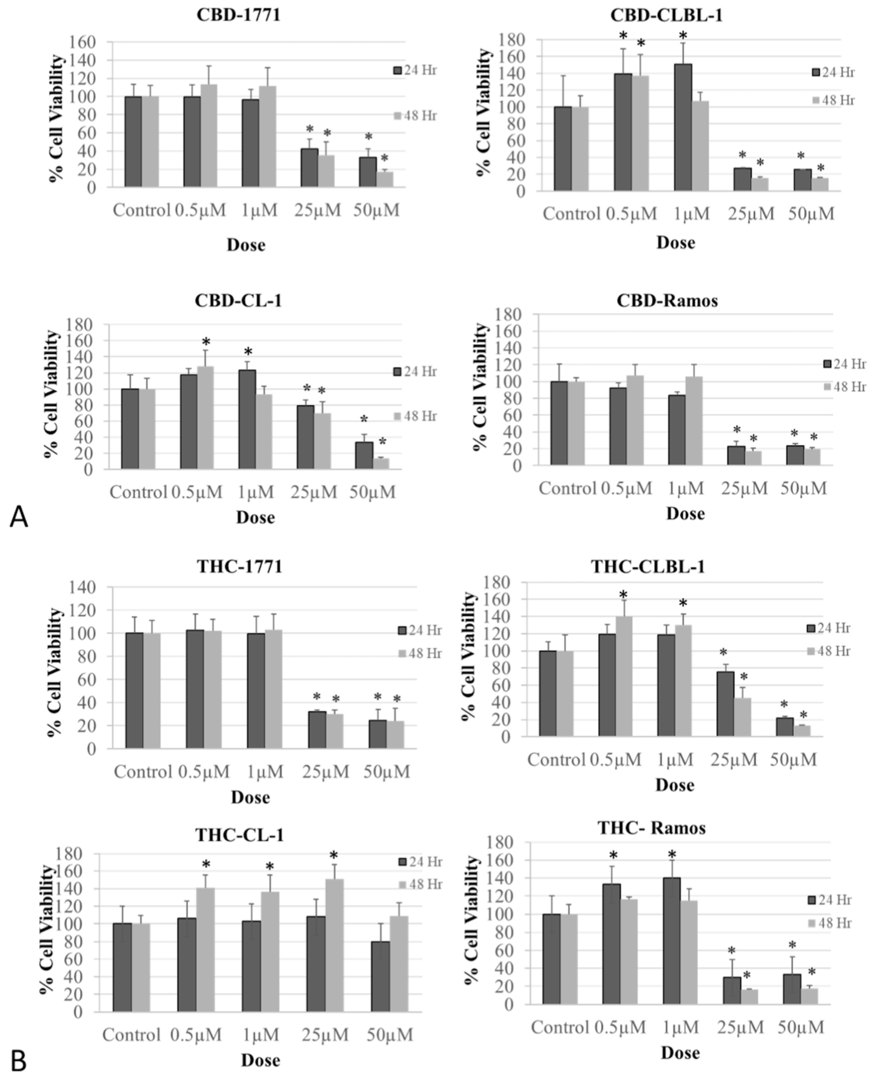
3.1.1. Treatment with CBD and THC Phytocannabinoids Altered Cell Viability in Human and Canine Lymphoma Cells

Treatment of canine B-cell lymphoma (1771 and CLBL-1) and human Ramos BCL cell lines with the phytocannabinoids CBD (Figure 1A) and THC (Figure 1B) demonstrated a biphasic effect with a stimulatory upshot at lower doses, followed by a significant reduction in cell viability at 25–50 μM concentrations (number of replicas for each treatment = 12, p < 0.0001). No such effect was detected in the canine CL-1 T-cell lymphoma cell line with THC treatment.

3.1.2. Treatment with WIN Altered NHL Cell Viability in Human and Canine Lymphoma Cells

Like the effect of phytocannabinoids, the WIN treatment at 25–50 µM final concentrations significantly decreased cell viability (p < 0.0001) in both canine and human non-Hodgkin BCL when compared to the vehicle treatment (Figure 2). We could not demonstrate a significant inhibitory effect of WIN on CL-1 canine TCL cell viability.

3.1.3. Treatment with CBD Does Not Affect Healthy Lymphocytes’ Viability

Unlike lymphoma cells, treatment of healthy lymphocytes with CBD at concentrations from 0.1 to 50 µM for 24 h demonstrated no significant effect on these lymphocytes’ (PBMCs) viability (Figure 3).

3.2. IC50 Calculation of Phytocannabinoids and a Synthetic Cannabinoid Based on Canine and Human Non-Hodgkin Lymphoma (NHL) Cell Viability

To further study the inhibitory effects of phytocannabinoids (CBD and THC) and a synthetic cannabinoid (WIN) on NHL cell viability, we calculated the IC50 of each cannabinoid in each NHL cell line used in this study. IC50 is a parameter that estimates the concentration of a drug that results in 50% inhibition of cell concentration when treated.
The analysis of IC50 shows that the phytocannabinoid CBD is the most potent cannabinoid among the cannabinoids used in 1771, Ramos, and CL-1, and WIN was found to be the most potent cannabinoid in CLBL-1 cell line (Table 1 and Supplementary Materials). All the cannabinoids studied are more effective against the canine and human B-cell lymphoma cell lines than against the T-cell lymphoma CL-1 cell line. Among the B-cell lymphoma cell lines, all the cannabinoids studied are more effective against the CLBL-1 and Ramos B-cell lines than the 1771 B-cell line. CBD was found to be the only effective cannabinoid against CL-1 canine T-cell lymphoma cells in terms of IC50 when compared to the other cannabinoids used in the study (Table 1 and Supplementary Materials).
The maximum concentration used to generate the dose–response curve was 50 µM; for cell lines where IC50 could not be calculated within the treated concentrations, it suggests that the concentrations of the drug (0.1–50 µM) did not effectively inhibit cell viability.

3.3. CBD, THC, and WIN Treatments Induced Oxidative Stress in Canine 1771 B-Cell Lymphoma Cell Line

Treatment with exogenous phytocannabinoids (CBD and THC) and a synthetic cannabinoid (WIN) increased oxidative stress, as seen by the significant increase in the generation of reactive oxygen species (ROS), NADH, and H2O2 and a concomitant decrease in the glutathione content (Figure 4A,B, n = 5, p < 0.05). However, regarding the variable effect of CBD and THC observed on the markers of oxidative stress analyzed, THC significantly increased lipid peroxidation; however, we could not demonstrate a significant effect of CBD on lipid peroxidation. A significant increase in the nitrite content was observed with CBD, but the opposite effect was observed with THC (Figure 4A, n = 5, p < 0.05).

3.4. CBD, THC, and WIN Treatments Induced Inflammation in Canine 1771 B-Cell Lymphoma Cell Line

Treatment of lymphoma cells with CBD, THC, and WIN induced inflammatory form of programmed cell death, as seen by the significant change in the activity of the inflammatory markers ICE-1 and COX (n = 5, p < 0.05; Figure 4C,D).

3.5. CBD, THC, and WIN Treatments Induced Apoptosis in Canine 1771 B-Cell Lymphoma Cell Line

Changes in the activity of various apoptotic markers indicated the apoptotic effect of the phytocannabinoids (CBD and THC) and the synthetic cannabinoid (WIN) against all lymphoma cell lines (Figure 5A,B). Treatment with THC caused a significant increase in all three of the caspases analyzed. Similarly, CBD and WIN increased the activities of caspase-3, caspase-8, and caspase-9 in a dose-dependent manner. The impact of CBD on caspase-3 and WIN on caspase-8 induction was appreciable, but it was not statistically significant.

3.6. Effects of CBD, THC, and WIN Cannabinoid Treatments on Mitochondrial Function

In canine B cells treated with CBD or WIN, complex-1 activity was significantly decreased when compared to the vehicle-treated control (n = 5, p < 0.001). In cells treated with THC, no significant effect on complex-1 activity could be demonstrated (Figure 5C,D and Table 2).

4. Discussion

It is well accepted that uncontrolled cellular growth, which may result from a defect in the cell cycle and apoptotic pathway, is one of the leading mechanisms for developing cancers, including lymphoma. Therefore, compounds that specifically induce apoptosis of cancer cells are likely to decrease tumor load and, consequently, be effective in cancer treatment. One of the most important and promising areas of current cannabinoid research is the ability of these natural products and endocannabinoids to modulate cell survival/death decisions, as described in several publications [6,20,41,42].
We recently studied endocannabinoid receptors and the effects of endocannabinoids on cell viability using canine and human NHL cell lines [21]. Our results demonstrated positive expression of cannabinoid receptors, CB1 and CB2, in both canine and human NHL cell lines, with a significantly higher expression in canine and human B-cell lymphoma compared to canine T-cell lymphoma. Also, we demonstrated decreased cell viability and apoptotic effect of endocannabinoids in canine and human NHL cell lines. The current study expanded our research from endogenous to exogenous cannabinoids. Among the exogenous cannabinoids studied, we focused on two phytocannabinoids, CBD and THC, and one synthetic cannabinoid, WIN. These selections were based on their well-documented anti-cancer effects across various human cancer models. Previous studies have highlighted the inhibitory effects of CBD and THC on cancer cell viability, along with their ability to trigger apoptotic mechanisms, leading to cell death in glioma, prostate, blood, breast, and other cancer cell lines [13,43,44,45,46]. Similarly, the synthetic cannabinoid WIN has been extensively studied for its potential anti-cancer activity against multiple cancers, including glioma, breast, and prostate cancers and mantle cell lymphoma [47,48,49,50].
Our objectives in this study were threefold: (1) an investigation of the inhibitory effects of CBD, THC, and WIN on canine and human NHL cell viability; (2) a study of the IC50 of phytocannabinoids (CBD and THC) and a synthetic cannabinoid(WIN) in canine and human NHL cell lines; and (3) an analysis of the effects of phytocannabinoids and a synthetic cannabinoid on oxidative stress, inflammation, apoptosis, and markers of mitochondrial function.
Our cell viability assay results demonstrated a growth stimulatory effect at lower cannabinoid concentrations. However, and in a clear contrast, a significant reduction in cell viability was consistently seen at higher non-molar final concentrations (25–50 µM) of the phytocannabinoids CBD and THC. Specifically, CBD significantly reduced cell viability in both B-cell (canine 1771 and CLBL1, and human Ramos) and canine CL-1 T-cell lymphoma cell lines. On the other hand, THC and the synthetic cannabinoid agonist WIN decreased cell viability significantly in only B-cell lymphoma cell lines. The lower expression of CB1 and CB2 receptors in CL-1 canine T-cell lymphoma cells, as compared to B-cell lymphoma reported in our previous study [21], could be the reason for the non-responsive behavior of CL-1 when treated with THC and WIN, both of which are selective cannabinoid receptor agonists. The hormesis-like effect of the lower phytocannabinoid concentrations on NHL cell viability seen in our study could be an adaptive response phenomenon previously documented in young adult C57BL/6 mice treated with a synthetic CP55940 cannabinoid and in other phytochemicals studies [51,52,53,54]. If future in vivo studies confirm our biphasic effect, the implications are that for some phytocannabinoids, too low a dose may be harmful, complicating individual responses. This phenomenon has been reported for resveratrol, a plant phenolic compound that induces a hormetic response in multiple well-characterized human cancer cell lines, including breast, prostate, colon, lung, uterine, and leukemia cell lines [55].
A further implication is that the hormesis-like response to CBD and THC could result in conflicting results among different studies if different concentrations are used. It is, thus, important to note that our understanding of the hormetic response to CBD and THC is very limited and further studies are needed to confirm this pharmacological phenomenon, especially in vivo.
The CBD-induced cytotoxicity in CL-1 T-cell lymphoma can potentially be mediated through receptors other than CB1 and CB2, as CBD has been shown in other studies to activate the transient receptor potential vanilloid subtype 1 (TRPV1), G protein-coupled receptor GPR55, the 5-HT1a receptor, and the α3 and α1 glycine receptors [56]. Several lymphoma studies have also demonstrated similar inhibitory effects of exogenous cannabinoids on cancer cell viability/proliferation [50,57,58,59].
To study the mechanism underlying the cytotoxic effects of CBD, THC, and WIN, markers of oxidative stress, inflammation, apoptosis, and mitochondrial function were analyzed in the 1771 B-cell lymphoma cell line. Our biochemical analysis results revealed a significant increase in ROS, H2O2, NADH, nitrite, and lipid peroxidation, and a concomitant reduction in the GSH content in cells treated with CBD, THC, and WIN at a 50 µM final concentration. Various studies from other laboratories have also demonstrated an increase in oxidative stress with exogenous and endogenous cannabinoid treatments in multiple cancer types, including human leukemia cells, human glioblastoma, and human glioma [46,60,61].
In our study, CBD, THC, and WIN treatments also significantly increased cyclooxygenase (COX) and Interleukin-1β-converting enzyme (ICE-1) activities. It is known that endocannabinoids are metabolized by COX-2 into pro-apoptotic prostaglandin derivatives, and both COX and ICE-1 enzymes are known to initiate pro-inflammatory mechanisms, leading to apoptosis or cell death [62,63,64]. The overproduction of COX and ICE-1 in cancer cells treated with CBD, THC, and WIN implies the possibility of the induction of an inflammatory pathway involved in cannabinoid-induced decrease in lymphoma cell viability. A similar effect of CBD has been previously demonstrated in glioblastoma [65,66].
Several cancer studies have demonstrated cannabinoid-induced apoptosis and associated this effect with increased caspase activity [46,61,67,68]. Likewise, we also demonstrated an increase in the activities of the apoptosis initiators Caspase-8 and -9 and the executioner Caspase-3 in lymphoma cells treated with the phytocannabinoids THC and CBD or with the synthetic cannabinoid WIN.
The inhibitory effect of cannabinoids on the mitochondrial respiratory chain has been demonstrated in past studies [44]. Similarly, compared to the vehicle-treated control cells, we found a significant reduction in complex-1 activity in 1771 B-cell lymphoma treated with WIN and THC when using concentrations as low as one micromole. However, we could not demonstrate a significant inhibitory effect of CBD on complex-1 activity. This differential effect of CBD could be attributed to the difference between these compounds triggering different anti-cancer signaling pathways. Further studies are needed to confirm this hypothesis.
In this study, we also analyzed the cytotoxic effects of phytocannabinoids (CBD and THC) and a synthetic cannabinoid (WIN) in lymphomas by calculating the IC50 value for each cell line. We found CBD to be the more potent phytocannabinoid (with a lower IC50) than THC. Overall, lymphomas with a higher expression of cannabinoid receptors responded more to the cannabinoid treatment compared to the ones with a lower expression (i.e., Ramos > CLBL-1 > 1771 > CL-1). However, the abovementioned anti-cancer effects can also be induced via mechanisms independent of CB1 and CB2 receptors.

5. Conclusions

Our study demonstrated a significant moderate inhibitory effect of CBD, THC, and WIN on canine and human NHL cell viability. Among the exogenous cannabinoids, the phytocannabinoid CBD was the most potent cannabinoid in 1771, Ramos, and CL-1, and the synthetic cannabinoid WIN was the most potent in the CLBL-1 cell line. Contrasting the inhibitory effect of CBD in B-cell versus T-cell lymphomas, we could not show a significant cytotoxic inhibitory effect of THC and WIN in the canine CL-1 T-cell lymphoma cell line. We surmised that the lack of a significant inhibitory effect may be due to the lower level of cannabinoid receptor expression in CL-1 T-cell cancer cells compared to B-cell lymphoma cell lines, as observed in our previous study [21].
Our results also revealed that CBD, THC, and WIN decreased lymphoma cell viability because they increased oxidative stress, leading to downstream apoptosis. Finally, our IC50 results could be lower than our findings due to serum binding. Furthermore, the results of our in vitro studies may not generalize to in vivo situations as many factors, including protein binding, could preclude direct extrapolation. In humans, THC may reach concentrations of approximately 1.4 µM in heavy users [69], and CBD may reach 2.5 µM [70] when administered orally therapeutically. Our study failed to demonstrate an inhibitory effect at these lower concentrations; the proliferative effects demonstrated in several cell lines with both CBD and THC may be problematic if these effects translate to in vivo responses. However, extrapolation of our in vitro results to in vivo situations would need to consider many other factors, including protein binding. This could preclude direct extrapolation.

Supplementary Materials

The following supporting information can be downloaded at https://www.mdpi.com/article/10.3390/biom14040495/s1, Figure S1: Calculation of IC50 for AEA, 2AG, CBD, THC, and WIN using MTT—cell viability percent inhibition dose–response curves expressed as log 10 of concentration (in nM) vs. percent inhibition of 1771 cell viability; Figure S2: Calculation of IC50 for AEA, 2AG, CBD, THC, and WIN using MTT—cell viability percent inhibition dose–response curves expressed as log 10 of concentration (in nM) vs. percent inhibition of CLBL1 cell viability; Figure S3: Calculation of IC50 for AEA, 2AG, CBD, THC, and WIN using MTT—cell viability percent inhibition dose– response curves expressed as log 10 of concentration (in nM) vs. percent inhibition of Ramos cell viability; Figure S4: Calculation of IC50 for AEA, 2AG, CBD, THC, and WIN using MTT—cell viability percent inhibition dose–response curves expressed as log 10 of concentration (in nM) vs. percent inhibition of CL1 cell viability; Figure S5: Graphic representation of IC50 values of endocannabinoids (AEA and 2AG), phytocannabinoids (CBD and THC), and a synthetic cannabinoid (WIN) in NHL cell lines.

Author Contributions

D.B. (Dawn Boothe), M.D. and S.O. conceived and designed the study, and outlined the research questions and objectives. S.O., S.P., R.N. and D.B. (Dylan Bowen) collected the data, conducted the experiments, performed the statistical analyses, and interpreted the data to draw meaningful conclusions. S.O. wrote the initial draft of the manuscript. M.M., D.B. (Dawn Boothe), S.R.P. and M.D. critically reviewed and revised the manuscript in terms of intellectual content, ensuring accuracy and coherence. They also supervised the overall research process, providing guidance and oversight. All authors have read and agreed to the published version of the manuscript.

Funding

This research did not receive any specific grant from funding agencies in the public, commercial, or not-for-profit sectors.

Institutional Review Board Statement

The animal study protocol was approved by the Institutional Review Board of College of Veterinary Medicine, Auburn University. (Approval number: 851, Date of approval: 10 June 2020).

Informed Consent Statement

Not applicable.

Data Availability Statement

The data that support the findings of this study are available from the corresponding author upon request. The data are not publicly available due to privacy or ethical restrictions.

Acknowledgments

We would like to thank Steven Suter from the Department of Medical Oncology, North Carolina State University, and Bruce F. Smith from Auburn University, Department of Pathobiology, for sharing the cell lines.

Conflicts of Interest

The authors declare no potential conflicts of interest.

References

  1. Ito, D.; Frantz, A.M.; Modiano, J.F. Canine lymphoma as a comparative model for human non-Hodgkin lymphoma: Recent progress and applications. Vet. Immunol. Immunopathol. 2014, 159, 192–201. [Google Scholar] [CrossRef] [PubMed]
  2. Zandvliet, M. Canine lymphoma: A review. Vet. Q. 2016, 36, 76–104. [Google Scholar] [CrossRef] [PubMed]
  3. Breen, M.; Modiano, J.F. Evolutionarily conserved cytogenetic changes in hematological malignancies of dogs and humans--man and his best friend share more than companionship. Chromosome Res. 2008, 16, 145–154. [Google Scholar] [CrossRef] [PubMed]
  4. Castor, A.; Nilsson, L.; Astrand-Grundstrom, I.; Buitenhuis, M.; Ramirez, C.; Anderson, K.; Strombeck, B.; Garwicz, S.; Bekassy, A.N.; Schmiegelow, K.; et al. Distinct patterns of hematopoietic stem cell involvement in acute lymphoblastic leukemia. Nat. Med. 2005, 11, 630–637. [Google Scholar] [CrossRef] [PubMed]
  5. Challa, A.; Eliopoulos, A.G.; Holder, M.J.; Burguete, A.S.; Pound, J.D.; Chamba, A.; Grafton, G.; Armitage, R.J.; Gregory, C.D.; Martinez-Valdez, H.; et al. Population depletion activates autonomous CD154-dependent survival in biopsylike Burkitt lymphoma cells. Blood 2002, 99, 3411–3418. [Google Scholar] [CrossRef] [PubMed]
  6. Guzman, M. Cannabinoids: Potential anticancer agents. Nat. Rev. Cancer 2003, 3, 745–755. [Google Scholar] [CrossRef] [PubMed]
  7. Sanchez, C.; de Ceballos, M.L.; Gomez del Pulgar, T.; Rueda, D.; Corbacho, C.; Velasco, G.; Galve-Roperh, I.; Huffman, J.W.; Ramon y Cajal, S.; Guzman, M. Inhibition of glioma growth in vivo by selective activation of the CB(2) cannabinoid receptor. Cancer Res. 2001, 61, 5784–5789. [Google Scholar] [PubMed]
  8. Bifulco, M.; Laezza, C.; Portella, G.; Vitale, M.; Orlando, P.; De Petrocellis, L.; Di Marzo, V. Control by the endogenous cannabinoid system of ras oncogene-dependent tumor growth. FASEB J. 2001, 15, 2745–2747. [Google Scholar] [CrossRef]
  9. Velasco, G.; Galve-Roperh, I.; Sanchez, C.; Blazquez, C.; Guzman, M. Hypothesis: Cannabinoid therapy for the treatment of gliomas? Neuropharmacology 2004, 47, 315–323. [Google Scholar] [CrossRef]
  10. Sarker, K.P.; Obara, S.; Nakata, M.; Kitajima, I.; Maruyama, I. Anandamide induces apoptosis of PC-12 cells: Involvement of superoxide and caspase-3. FEBS Lett. 2000, 472, 39–44. [Google Scholar] [CrossRef]
  11. Maccarrone, M.; Lorenzon, T.; Bari, M.; Melino, G.; Finazzi-Agro, A. Anandamide induces apoptosis in human cells via vanilloid receptors. Evidence for a protective role of cannabinoid receptors. J. Biol. Chem. 2000, 275, 31938–31945. [Google Scholar] [CrossRef]
  12. Blasco-Benito, S.; Seijo-Vila, M.; Caro-Villalobos, M.; Tundidor, I.; Andradas, C.; Garcia-Taboada, E.; Wade, J.; Smith, S.; Guzman, M.; Perez-Gomez, E.; et al. Appraising the “entourage effect”: Antitumor action of a pure cannabinoid versus a botanical drug preparation in preclinical models of breast cancer. Biochem. Pharmacol. 2018, 157, 285–293. [Google Scholar] [CrossRef]
  13. Milian, L.; Mata, M.; Alcacer, J.; Oliver, M.; Sancho-Tello, M.; Martin de Llano, J.J.; Camps, C.; Galbis, J.; Carretero, J.; Carda, C. Cannabinoid receptor expression in non-small cell lung cancer. Effectiveness of tetrahydrocannabinol and cannabidiol inhibiting cell proliferation and epithelial-mesenchymal transition in vitro. PLoS ONE 2020, 15, e0228909. [Google Scholar] [CrossRef]
  14. Sarfaraz, S.; Afaq, F.; Adhami, V.M.; Mukhtar, H. Cannabinoid receptor as a novel target for the treatment of prostate cancer. Cancer Res. 2005, 65, 1635–1641. [Google Scholar] [CrossRef]
  15. Patsos, H.A.; Greenhough, A.; Hicks, D.J.; Al Kharusi, M.; Collard, T.J.; Lane, J.D.; Paraskeva, C.; Williams, A.C. The endogenous cannabinoid, anandamide, induces COX-2-dependent cell death in apoptosis-resistant colon cancer cells. Int. J. Oncol. 2010, 37, 187–193. [Google Scholar] [CrossRef]
  16. Caffarel, M.M.; Andradas, C.; Mira, E.; Perez-Gomez, E.; Cerutti, C.; Moreno-Bueno, G.; Flores, J.M.; Garcia-Real, I.; Palacios, J.; Manes, S.; et al. Cannabinoids reduce ErbB2-driven breast cancer progression through Akt inhibition. Mol. Cancer 2010, 9, 196. [Google Scholar] [CrossRef]
  17. Guindon, J.; Hohmann, A.G. The endocannabinoid system and cancer: Therapeutic implication. Br. J. Pharmacol. 2011, 163, 1447–1463. [Google Scholar] [CrossRef]
  18. Zygmunt, P.M.; Petersson, J.; Andersson, D.A.; Chuang, H.; Sorgard, M.; Di Marzo, V.; Julius, D.; Hogestatt, E.D. Vanilloid receptors on sensory nerves mediate the vasodilator action of anandamide. Nature 1999, 400, 452–457. [Google Scholar] [CrossRef]
  19. Ramer, R.; Schwarz, R.; Hinz, B. Modulation of the Endocannabinoid System as a Potential Anticancer Strategy. Front. Pharmacol. 2019, 10, 430. [Google Scholar] [CrossRef]
  20. McAllister, S.D.; Christian, R.T.; Horowitz, M.P.; Garcia, A.; Desprez, P.Y. Cannabidiol as a novel inhibitor of ld-1 gene expression in aggressive breast cancer cells. Mol. Cancer Ther. 2007, 6, 2921–2927. [Google Scholar] [CrossRef]
  21. Omer, S.; Pathak, S.; Nadar, R.; Bowen, D.; Sandey, M.; Dhanasekaran, M.; Pondugula, S.; Mansour, M.; Boothe, D. Validating the anti-lymphoma pharmacodynamic actions of the endocannabinoids on canine non-Hodgkin lymphoma. Life Sci. 2023, 327, 121862. [Google Scholar] [CrossRef]
  22. Pondugula, S.R.; Ferniany, G.; Ashraf, F.; Abbott, K.L.; Smith, B.F.; Coleman, E.S.; Mansour, M.; Bird, R.C.; Smith, A.N.; Karthikeyan, C.; et al. Stearidonic acid, a plant-based dietary fatty acid, enhances the chemosensitivity of canine lymphoid tumor cells. Biochem. Biophys. Res. Commun. 2015, 460, 1002–1007. [Google Scholar] [CrossRef]
  23. McKallip, R.J.; Lombard, C.; Fisher, M.; Martin, B.R.; Ryu, S.; Grant, S.; Nagarkatti, P.S.; Nagarkatti, M. Targeting CB2 cannabinoid receptors as a novel therapy to treat malignant lymphoblastic disease. Blood 2002, 100, 627–634. [Google Scholar] [CrossRef]
  24. Massi, P.; Vaccani, A.; Ceruti, S.; Colombo, A.; Abbracchio, M.P.; Parolaro, D. Antitumor effects of cannabidiol, a nonpsychoactive cannabinoid, on human glioma cell lines. J. Pharmacol. Exp. Ther. 2004, 308, 838–845. [Google Scholar] [CrossRef]
  25. Guide for Biologists. Available online: https://www.sciencegateway.org/protocols/ (accessed on 2 March 2021).
  26. Sobotta, M.C.; Barata, A.G.; Schmidt, U.; Mueller, S.; Millonig, G.; Dick, T.P. Exposing cells to H2O2: A quantitative comparison between continuous low-dose and one-time high-dose treatments. Free Radic. Biol. Med. 2013, 60, 325–335. [Google Scholar] [CrossRef]
  27. Zheng, M.; Ahuja, M.; Bhattacharya, D.; Clement, T.P.; Hayworth, J.S.; Dhanasekaran, M. Evaluation of differential cytotoxic effects of the oil spill dispersant Corexit 9500. Life Sci 2014, 95, 108–117. [Google Scholar] [CrossRef]
  28. Ohkawa, H.; Ohishi, N.; Yagi, K. Assay for lipid peroxides in animal tissues by thiobarbituric acid reaction. Anal. Biochem. 1979, 95, 351–358. [Google Scholar] [CrossRef]
  29. Majrashi, M.; Almaghrabi, M.; Fadan, M.; Fujihashi, A.; Lee, W.; Deruiter, J.; Randall Clark, C.; Dhanasekaran, M. Dopaminergic neurotoxic effects of 3-TFMPP derivatives. Life Sci. 2018, 209, 357–369. [Google Scholar] [CrossRef]
  30. Hong, S.; Kim, C.Y.; Lee, J.E.; Seong, G.J. Agmatine protects cultured retinal ganglion cells from tumor necrosis factor-alpha-induced apoptosis. Life Sci. 2009, 84, 28–32. [Google Scholar] [CrossRef]
  31. Dhanasekaran, M.; Tharakan, B.; Manyam, B.V. Antiparkinson drug--Mucuna pruriens shows antioxidant and metal chelating activity. Phytother. Res. 2008, 22, 6–11. [Google Scholar] [CrossRef]
  32. Giustarini, D.; Rossi, R.; Milzani, A.; Dalle-Donne, I. Nitrite and nitrate measurement by Griess reagent in human plasma: Evaluation of interferences and standardization. Methods Enzymol. 2008, 440, 361–380. [Google Scholar] [CrossRef]
  33. Almaghrabi, M. Investigate the Dopaminergic Neurotoxicity Profile of Designer Drugs (Piperazine Derivatives). Ph.D. Thesis, Auburn University, Auburn, AL, USA, 2018. [Google Scholar]
  34. Ramsay, R.R.; Dadgar, J.; Trevor, A.; Singer, T.P. Energy-driven uptake of N-methyl-4-phenylpyridine by brain mitochondria mediates the neurotoxicity of MPTP. Life Sci. 1986, 39, 581–588. [Google Scholar] [CrossRef]
  35. Ahuja, M.; Buabeid, M.; Abdel-Rahman, E.; Majrashi, M.; Parameshwaran, K.; Amin, R.; Ramesh, S.; Thiruchelvan, K.; Pondugula, S.; Suppiramaniam, V.; et al. Immunological alteration and toxic molecular inductions leading to cognitive impairment and neurotoxicity in transgenic mouse model of Alzheimer’s disease. Life Sci. 2017, 177, 49–59. [Google Scholar] [CrossRef]
  36. Bhattacharya, D.; Majrashi, M.; Ramesh, S.; Govindarajulu, M.; Bloemer, J.; Fujihashi, A.; Crump, B.R.; Hightower, H.; Bhattacharya, S.; Moore, T.; et al. Assessment of the cerebellar neurotoxic effects of nicotine in prenatal alcohol exposure in rats. Life Sci. 2018, 194, 177–184. [Google Scholar] [CrossRef]
  37. Al Saqr, A.; Majrashi, M.; Alrbyawi, H.; Govindarajulu, M.; Fujihashi, A.; Gottumukkala, S.; Poudel, I.; Arnold, R.D.; Babu, R.J.; Dhanasekaran, M. Elucidating the anti-melanoma effect and mechanisms of Hispolon. Life Sci. 2020, 256, 117702. [Google Scholar] [CrossRef]
  38. Liu, K.; Kadannagari, S.; Deruiter, J.; Pathak, S.; Abbott, K.L.; Salamat, J.M.; Pondugula, S.R.; Akingbemi, B.T.; Dhanasekaran, M. Effects of developmental exposures to Bisphenol-A and Bisphenol-S on hepatocellular function in male Long-Evans rats. Life Sci. 2023, 326, 121752. [Google Scholar] [CrossRef]
  39. Copeland, R.A.; Williams, J.M.; Giannaras, J.; Nurnberg, S.; Covington, M.; Pinto, D.; Pick, S.; Trzaskos, J.M. Mechanism of selective inhibition of the inducible isoform of prostaglandin G/H synthase. Proc. Natl. Acad. Sci. USA 1994, 91, 11202–11206. [Google Scholar] [CrossRef]
  40. Usha, R.; Muralikrishnan, D.; Thomas, B.; Ghosh, S.; Mandal, C.; Mohanakumar, K.P. Region-specific attenuation of a trypsin-like protease in substantia nigra following dopaminergic neurotoxicity by 1-methyl-4-phenyl-1,2,3,6-tetrahydropyridine. Brain Res. 2000, 882, 191–195. [Google Scholar] [CrossRef]
  41. Ellert-Miklaszewska, A.; Ciechomska, I.A.; Kaminska, B. Cannabinoid Signaling in Glioma Cells. Adv. Exp. Med. Biol. 2020, 1202, 223–241. [Google Scholar] [CrossRef]
  42. Kovalchuk, O.; Kovalchuk, I. Cannabinoids as anticancer therapeutic agents. Cell Cycle 2020, 19, 961–989. [Google Scholar] [CrossRef] [PubMed]
  43. Sharma, M. In Vitro Anticancer Activity of Plant-Derived Cannabidiol on Prostate Cancer Cell Lines. Pharmacol. Pharm. 2014, 5, 806–820. [Google Scholar] [CrossRef]
  44. McAllister, S.D.; Soroceanu, L.; Desprez, P.Y. The Antitumor Activity of Plant-Derived Non-Psychoactive Cannabinoids. J. Neuroimmune Pharmacol. 2015, 10, 255–267. [Google Scholar] [CrossRef] [PubMed]
  45. Jia, W.; Hegde, V.L.; Singh, N.P.; Sisco, D.; Grant, S.; Nagarkatti, M.; Nagarkatti, P.S. Δ9-Tetrahydrocannabinol-Induced Apoptosis in Jurkat Leukemia T Cells Is Regulated by Translocation of Bad to Mitochondria. Mol. Cancer Res. 2006, 4, 549–562. [Google Scholar] [CrossRef] [PubMed]
  46. McKallip, R.J.; Jia, W.; Schlomer, J.; Warren, J.W.; Nagarkatti, P.S.; Nagarkatti, M. Cannabidiol-induced apoptosis in human leukemia cells: A novel role of cannabidiol in the regulation of p22phox and Nox4 expression. Mol. Pharmacol. 2006, 70, 897–908. [Google Scholar] [CrossRef] [PubMed]
  47. Emery, S.M.; Alotaibi, M.R.; Tao, Q.; Selley, D.E.; Lichtman, A.H.; Gewirtz, D.A. Combined antiproliferative effects of the aminoalkylindole WIN55,212-2 and radiation in breast cancer cells. J. Pharmacol. Exp. Ther. 2014, 348, 293–302. [Google Scholar] [CrossRef] [PubMed]
  48. Shah, S.A.; Gupta, A.S.; Kumar, P. Emerging role of cannabinoids and synthetic cannabinoid receptor 1/cannabinoid receptor 2 receptor agonists in cancer treatment and chemotherapy-associated cancer management. J. Cancer Res. Ther. 2021, 17, 1–9. [Google Scholar] [CrossRef] [PubMed]
  49. Ellert-Miklaszewska, A.; Ciechomska, I.A.; Kaminska, B. Synthetic Cannabinoids Induce Autophagy and Mitochondrial Apoptotic Pathways in Human Glioblastoma Cells Independently of Deficiency in TP53 or PTEN Tumor Suppressors. Cancers 2021, 13, 419. [Google Scholar] [CrossRef] [PubMed]
  50. Flygare, J.; Gustafsson, K.; Kimby, E.; Christensson, B.; Sander, B. Cannabinoid receptor ligands mediate growth inhibition and cell death in mantle cell lymphoma. FEBS Lett. 2005, 579, 6885–6889. [Google Scholar] [CrossRef] [PubMed]
  51. Hodges, E.L.; Marshall, J.P.; Ashpole, N.M. Age-dependent hormesis-like effects of the synthetic cannabinoid CP55940 in C57BL/6 mice. npj Aging Mech. Dis. 2020, 6, 7. [Google Scholar] [CrossRef]
  52. Murakami, A. Hormesis-Mediated Mechanisms Underlying Bioactivities of Phytochemicals. Curr. Pharmacol. Rep. 2020, 6, 325–334. [Google Scholar] [CrossRef]
  53. Jodynis-Liebert, J.; Kujawska, M. Biphasic Dose-Response Induced by Phytochemicals: Experimental Evidence. J. Clin. Med. 2020, 9, 718. [Google Scholar] [CrossRef] [PubMed]
  54. Son, T.G.; Camandola, S.; Mattson, M.P. Hormetic dietary phytochemicals. Neuromol. Med. 2008, 10, 236–246. [Google Scholar] [CrossRef] [PubMed]
  55. Calabrese, E.J.; Mattson, M.P.; Calabrese, V. Resveratrol commonly displays hormesis: Occurrence and biomedical significance. Hum. Exp. Toxicol. 2010, 29, 980–1015. [Google Scholar] [CrossRef] [PubMed]
  56. Anand, U.; Jones, B.; Korchev, Y.; Bloom, S.R.; Pacchetti, B.; Anand, P.; Sodergren, M.H. CBD Effects on TRPV1 Signaling Pathways in Cultured DRG Neurons. J. Pain. Res. 2020, 13, 2269–2278. [Google Scholar] [CrossRef] [PubMed]
  57. Gustafsson, K.; Wang, X.; Severa, D.; Eriksson, M.; Kimby, E.; Merup, M.; Christensson, B.; Flygare, J.; Sander, B. Expression of cannabinoid receptors type 1 and type 2 in non-Hodgkin lymphoma: Growth inhibition by receptor activation. Int. J. Cancer 2008, 123, 1025–1033. [Google Scholar] [CrossRef] [PubMed]
  58. Derocq, J.M.; Segui, M.; Marchand, J.; Le Fur, G.; Casellas, P. Cannabinoids enhance human B-cell growth at low nanomolar concentrations. FEBS Lett. 1995, 369, 177–182. [Google Scholar] [CrossRef] [PubMed]
  59. Carayon, P.; Marchand, J.; Dussossoy, D.; Derocq, J.M.; Jbilo, O.; Bord, A.; Bouaboula, M.; Galiegue, S.; Mondiere, P.; Penarier, G.; et al. Modulation and functional involvement of CB2 peripheral cannabinoid receptors during B-cell differentiation. Blood 1998, 92, 3605–3615. [Google Scholar] [CrossRef] [PubMed]
  60. Marcu, J.P.; Christian, R.T.; Lau, D.; Zielinski, A.J.; Horowitz, M.P.; Lee, J.; Pakdel, A.; Allison, J.; Limbad, C.; Moore, D.H.; et al. Cannabidiol enhances the inhibitory effects of delta9-tetrahydrocannabinol on human glioblastoma cell proliferation and survival. Mol. Cancer Ther. 2010, 9, 180–189. [Google Scholar] [CrossRef] [PubMed]
  61. Massi, P.; Vaccani, A.; Bianchessi, S.; Costa, B.; Macchi, P.; Parolaro, D. The non-psychoactive cannabidiol triggers caspase activation and oxidative stress in human glioma cells. Cell. Mol. Life Sci. 2006, 63, 2057–2066. [Google Scholar] [CrossRef]
  62. Wójcik, P.; Biernacki, M.; Domian, N.; Žarković, N.; Skrzydlewska, E. Influence of Inhibition of COX-2-Dependent Lipid Metabolism on Regulation of UVB-Induced Keratinocytes Apoptosis by Cannabinoids. Biomolecules 2022, 12, 842. [Google Scholar] [CrossRef]
  63. Meijerink, J.; Poland, M.; Balvers, M.G.J.; Plastina, P.; Lute, C.; Dwarkasing, J.; van Norren, K.; Witkamp, R.F. Inhibition of COX-2-mediated eicosanoid production plays a major role in the anti-inflammatory effects of the endocannabinoid N-docosahexaenoylethanolamine (DHEA) in macrophages. Br. J. Pharmacol. 2015, 172, 24–37. [Google Scholar] [CrossRef] [PubMed]
  64. Ruhaak, L.R.; Felth, J.; Karlsson, P.C.; Rafter, J.J.; Verpoorte, R.; Bohlin, L. Evaluation of the cyclooxygenase inhibiting effects of six major cannabinoids isolated from Cannabis sativa. Biol. Pharm. Bull. 2011, 34, 774–778. [Google Scholar] [CrossRef] [PubMed]
  65. Rimmerman, N.; Ben-Hail, D.; Porat, Z.; Juknat, A.; Kozela, E.; Daniels, M.P.; Connelly, P.S.; Leishman, E.; Bradshaw, H.B.; Shoshan-Barmatz, V.; et al. Direct modulation of the outer mitochondrial membrane channel, voltage-dependent anion channel 1 (VDAC1) by cannabidiol: A novel mechanism for cannabinoid-induced cell death. Cell Death Dis. 2013, 4, e949. [Google Scholar] [CrossRef] [PubMed]
  66. Singer, E.; Judkins, J.; Salomonis, N.; Matlaf, L.; Soteropoulos, P.; McAllister, S.; Soroceanu, L. Reactive oxygen species-mediated therapeutic response and resistance in glioblastoma. Cell Death Dis. 2015, 6, e1601. [Google Scholar] [CrossRef] [PubMed]
  67. Velasco, G.; Sanchez, C.; Guzman, M. Towards the use of cannabinoids as antitumour agents. Nat. Rev. Cancer 2012, 12, 436–444. [Google Scholar] [CrossRef] [PubMed]
  68. Gustafsson, K.; Christensson, B.; Sander, B.; Flygare, J. Cannabinoid receptor-mediated apoptosis induced by R(+)-methanandamide and Win55,212-2 is associated with ceramide accumulation and p38 activation in mantle cell lymphoma. Mol. Pharmacol. 2006, 70, 1612–1620. [Google Scholar] [CrossRef]
  69. Grotenhermen, F. Pharmacokinetics and pharmacodynamics of cannabinoids. Clin. Pharmacokinet. 2003, 42, 327–360. [Google Scholar] [CrossRef]
  70. Britch, S.C.; Babalonis, S.; Walsh, S.L. Cannabidiol: Pharmacology and therapeutic targets. Psychopharmacology 2021, 238, 9–28. [Google Scholar] [CrossRef]
Figure 1. Biphasic effect of the phytocannabinoids cannabidiol (CBD) and ∆9-tetrahydrocannabinol (THC) on cell viability of canine and human non-Hodgkin lymphomas. (A). CBD treatment of B-cell lymphomas (canine 1771 and CLBL1, and human Ramos) and canine T-cell lymphoma (CL-1). (B). THC treatment of B-cell lymphomas (canine 1771 and CLBL1 and human Ramos). The experiment was repeated three times at 24 and 48 h. Replica for each treatment = 12. The results are expressed as (%) change compared to the 100% cell viability in vehicle-treated control. Values over 100% are due to increased cell growth. Data are depicted as mean ± SD. Data were evaluated with analysis of variance (ANOVA), followed by Dunnett’s multiple comparison test. Of note, no biphasic effect was detected in CL-1 canine TCL cells when treated with THC (B). Overall, CBD demonstrated a more potent inhibitory effect on cell viability against BCL than against TCL (A). (* statistically significant increase/decrease in cell viability compared to the control).
Biomolecules 14 00495 g001
Figure 2. Effect of WIN on canine and human non-Hodgkin lymphoma cell viability. The WIN treatment decreased cell viability in canine and human Ramos B-cell lymphoma cell lines (1771, CLBL-1, and Ramos). No significant effects were detected in canine T-cell lymphoma cell line CL-1. The WIN treatment was at 0.5–50 µM concentrations for 24 h and 48 h. The experiment was repeated three times. Replica for each treatment = 12. The results are expressed as (%) change compared to the control, with mean ± SD. Data were evaluated using analysis of variance (ANOVA), followed by Dunnett’s multiple comparison test. (* statistically significant increase/decrease in cell viability compared to the control).
Biomolecules 14 00495 g002
Figure 3. Effect of CBD on canine healthy lymphocytes’ viability. CBD treatment caused no significant effect in canine healthy lymphocytes. CBD treatment was at concentrations of 0.1–50 µM for 24 h. Results are expressed as (%) change compared to control, with mean ± SD.
Biomolecules 14 00495 g003
Figure 4. (A,B) Effects of CBD, THC, and WIN on markers of oxidative stress in 1771 canine lymphoma cells. All markers of oxidative stress were measured spectrofluorometrically. (A) CBD treatment is associated with a dose-dependent increase in nitrite content, reactive oxygen species (ROS) generation, and NADH and lipid peroxidation, and a simultaneous dose-dependent decrease in the GSH content in lymphoma-treated cells when compared to the control. THC demonstrates a dose-dependent increase in lipid peroxidation and ROS content and a decrease in GSH and nitrite contents. A significant effect could not be demonstrated in the cellular content of NADH in cells treated with THC. (B) The synthetic cannabinoid (WIN) shows a significant dose-dependent increase in nitrite, ROS, and H2O2 contents and a decrease in GSH compared to the vehicle-treated control cells. (C,D) Effects of exogenous cannabinoids on Interleukin-1β-converting enzyme (ICE-1) and cyclooxygenase (COX) activities, showing a significant increase in lymphoma cells treated with THC and WIN at a 50 µM final concentration. CBD treatment increases cox activity significantly by does not alter the ICE-1 level. The vehicle treatment (50 μM) does not affect the activity of ICE-1 or COX. The results are expressed as % change compared to the vehicle-treated control, with mean ± SD. * p < 0.05, ** p < 0.01, *** p < 0.001, **** p ≤ 0.0001. n = 5. (asterisk significant compared to the control).
Biomolecules 14 00495 g004
Figure 5. Effects of CBD, THC, and WIN on canine 1771 B-cell lymphoma cells. Caspase-3, caspase-8, and caspase-9 activities were measured spectrofluorometrically using AC-DEVD-AMC, Ac-VETD-AMC, Ac-LEHD-pNa, respectively, as the substrate. (A) CBD and THC increased all caspase activities in a dose-dependent manner. Significant dose-dependent effects on caspase-3 activity could not be demonstrated with CBD when compared to the control. (B) A significant increase in caspase-3 and caspase-9 activities was demonstrated in cells treated with WIN. The significant effect of WIN could not be demonstrated on caspase-8 activity. (C,D) Effects of CBD, THC, and WIN on canine 1771 B-cell lymphoma cells’ complex-I activity. The complex-I activity was measured spectrophotometrically using NADH as the substrate. CBD and WIN showed a significant decrease in complex-I activity compared to the control. The significant effect on complex-1 activity could not be demonstrated with THC. The results are expressed as % change compared to the control, with mean ± SD. * p < 0.05, ** p < 0.01, *** p < 0.001, **** p ≤ 0.0001. n = 5. (asterisk significant compared to the control).
Biomolecules 14 00495 g005
Table 1. IC50 of phytocannabinoids and a synthetic cannabinoid in NHL cell lines.
Cell LineCannabinoidIC50SE *R2 †
Canine B-cell lymphoma 1771CBD14 µM20.9
THC41 µM9.60.9
WINNANANA
Canine B-cell lymphoma CLBL-1CBD18 µM6.10.6
THC34 µM2.20.9
WIN5.6 µM0.60.9
Canine T-cell lymphoma
CL-1
CBD39 µM3.20.8
THCNANANA
WINNANANA
Human B-cell lymphoma
Ramos
CBD8.6 µM1.30.9
THC21 µM2.60.8
WIN14 µM1.70.9
* SE: standard error, R2: goodness of fit, NA: not applicable.
Table 2. Summary of biochemical analysis results.
Cellular Response to CytotoxicityLymphoma Cells’ Response to Cannabinoid-Induced Cytotoxicity
Overall EffectCBDTHCWIN
Markers of Oxidative Stress
ROSBiomolecules 14 00495 i001Biomolecules 14 00495 i001Biomolecules 14 00495 i001Biomolecules 14 00495 i001
GSHBiomolecules 14 00495 i002Biomolecules 14 00495 i002Biomolecules 14 00495 i002Biomolecules 14 00495 i002
NADHBiomolecules 14 00495 i001Biomolecules 14 00495 i001Biomolecules 14 00495 i003Biomolecules 14 00495 i003
H2O2Biomolecules 14 00495 i001NDNDBiomolecules 14 00495 i001
NitriteBiomolecules 14 00495 i001Biomolecules 14 00495 i001Biomolecules 14 00495 i002Biomolecules 14 00495 i001
Lipid peroxidation (LP)Biomolecules 14 00495 i001Biomolecules 14 00495 i003Biomolecules 14 00495 i001ND
Markers of Inflammation
ICE-1Biomolecules 14 00495 i001Biomolecules 14 00495 i003Biomolecules 14 00495 i001Biomolecules 14 00495 i001
COXBiomolecules 14 00495 i001Biomolecules 14 00495 i001Biomolecules 14 00495 i001Biomolecules 14 00495 i001
Markers of Apoptosis
Caspase-3Biomolecules 14 00495 i001Biomolecules 14 00495 i003Biomolecules 14 00495 i001Biomolecules 14 00495 i001
Caspase-8Biomolecules 14 00495 i001Biomolecules 14 00495 i001Biomolecules 14 00495 i001Biomolecules 14 00495 i003
Caspase-9Biomolecules 14 00495 i001Biomolecules 14 00495 i001Biomolecules 14 00495 i001Biomolecules 14 00495 i001
Markers of Mitochondrial function
Complex-1Biomolecules 14 00495 i002Biomolecules 14 00495 i002Biomolecules 14 00495 i003Biomolecules 14 00495 i002
[Biomolecules 14 00495 i001 = Increased, Biomolecules 14 00495 i002 = Decreased, Biomolecules 14 00495 i003 = No significant effect, ND = Not determined].
Disclaimer/Publisher’s Note: The statements, opinions and data contained in all publications are solely those of the individual author(s) and contributor(s) and not of MDPI and/or the editor(s). MDPI and/or the editor(s) disclaim responsibility for any injury to people or property resulting from any ideas, methods, instructions or products referred to in the content.

