Development of a Simple Pretreatment Immunoassay Based on an Organic Solvent-Tolerant Nanobody for the Detection of Carbofuran in Vegetable and Fruit Samples

 ,
,
Abstract
:
1. Introduction
2. Materials and Methods
2.1. Materials and Reagents
2.2. Construction of Phage Display Nanobody Library
2.3. Selection and Identification of Anti-Carbofuran Phage Clones
2.4. Expression and Purification of Nanobody Protein
2.5. Stability Analysis of Anti-Carbofuran Nanobody
2.6. Nanobody-Based Indirect Competitive ELISA
2.7. Immunoassay Validation
3. Results and Discussion
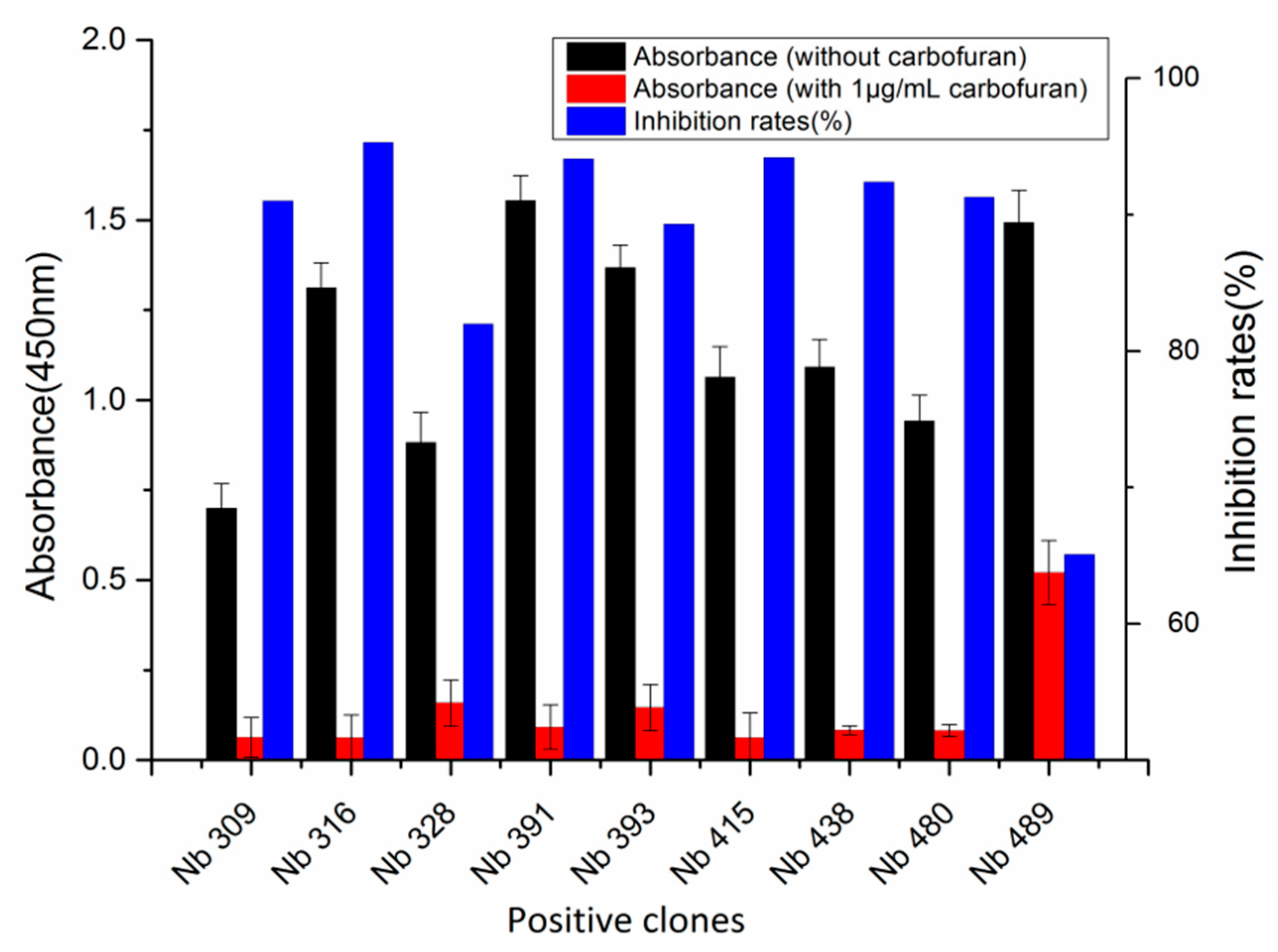
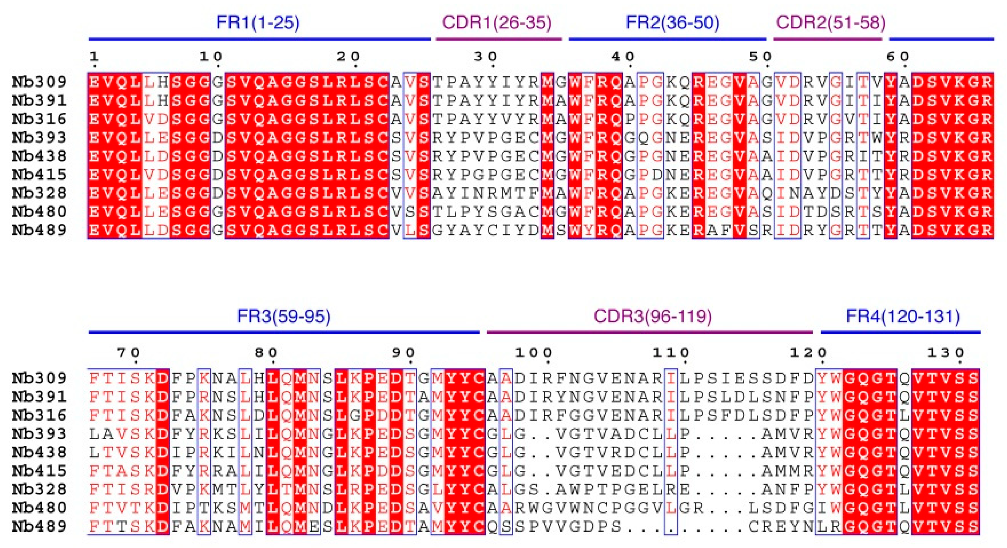
3.1. Library Construction and Selection of Anti-Carbofuran Phage Clones
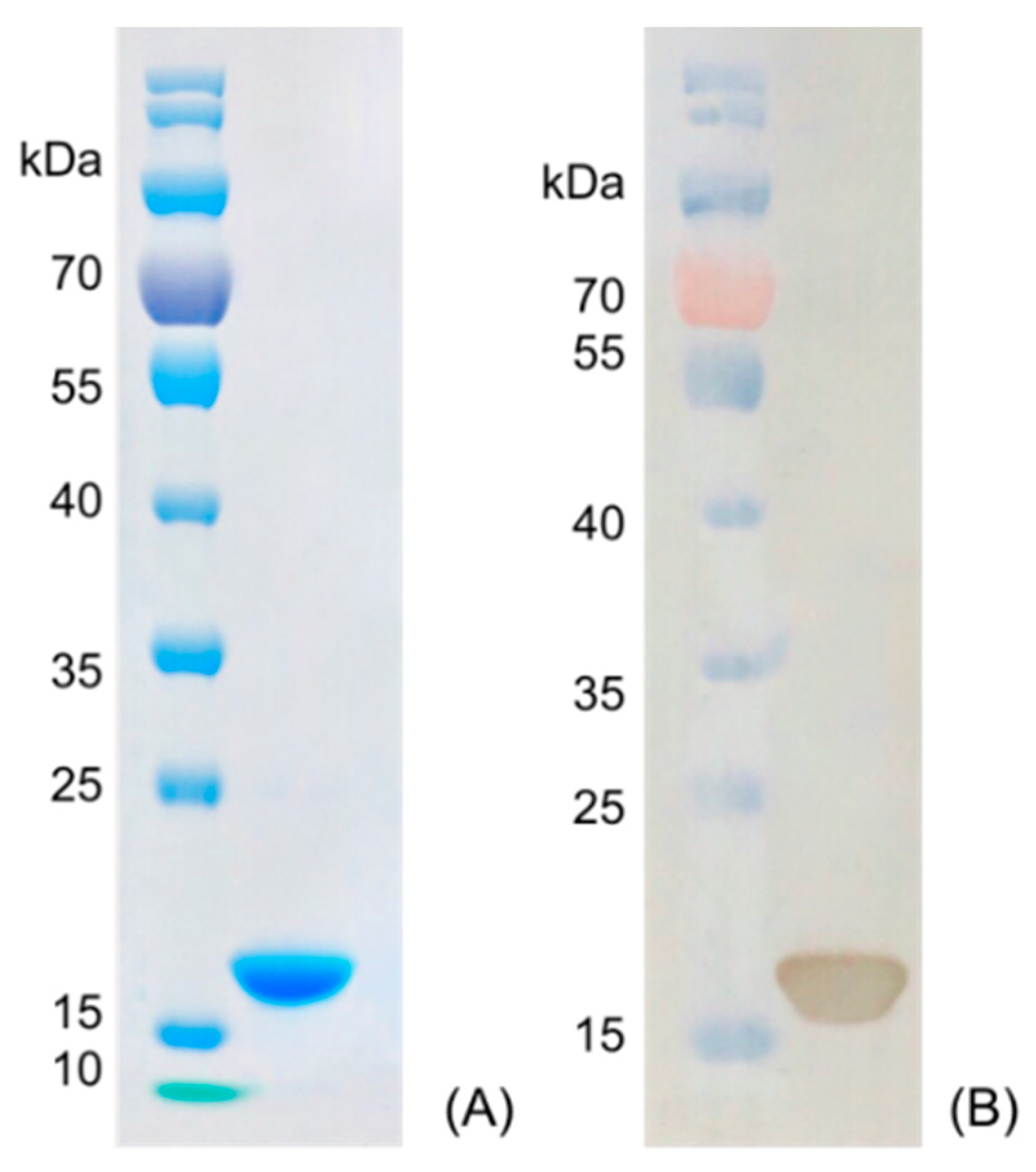
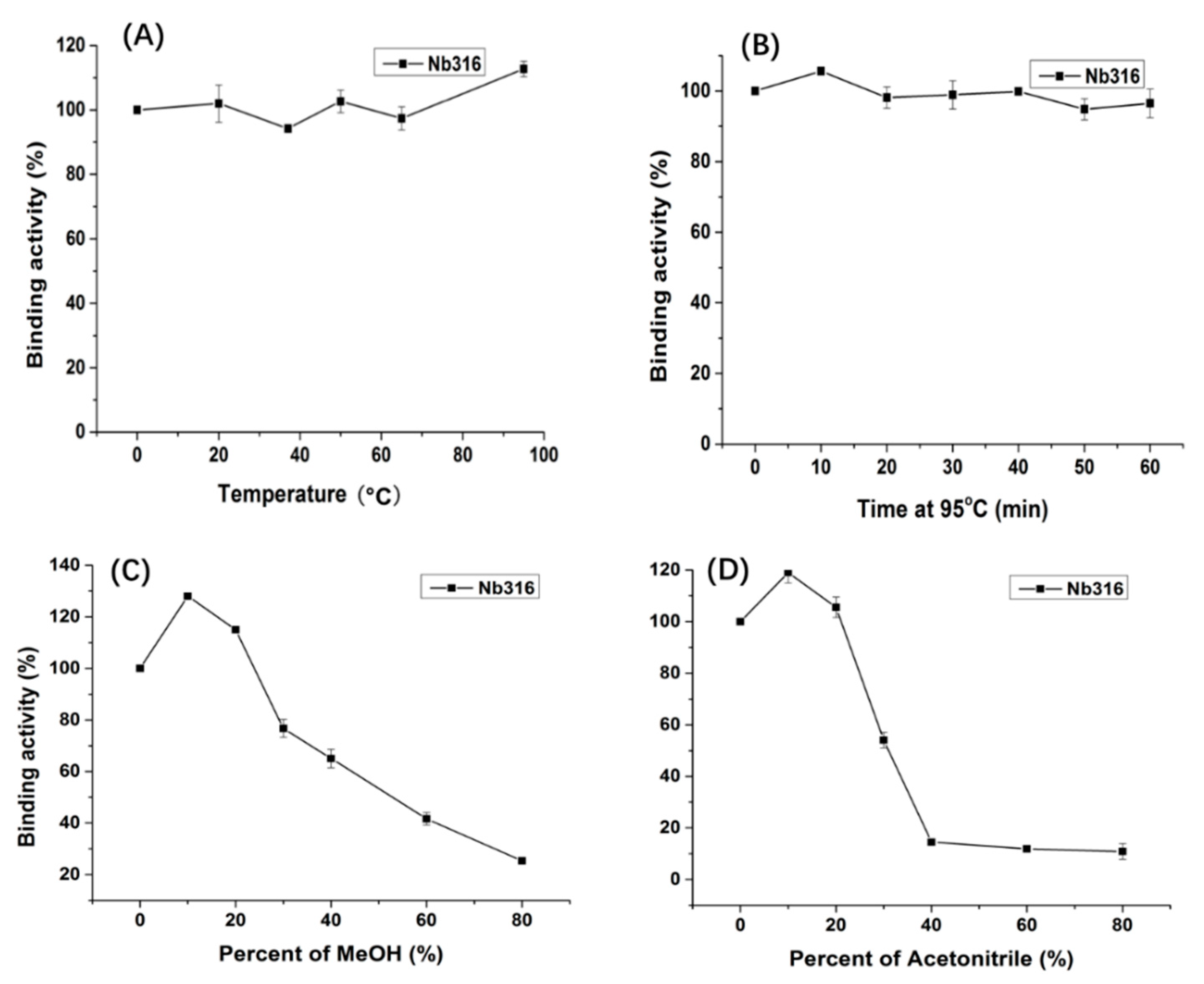
3.2. Preparation and Stability Analysis of Anti-Carbofuran Nanobody
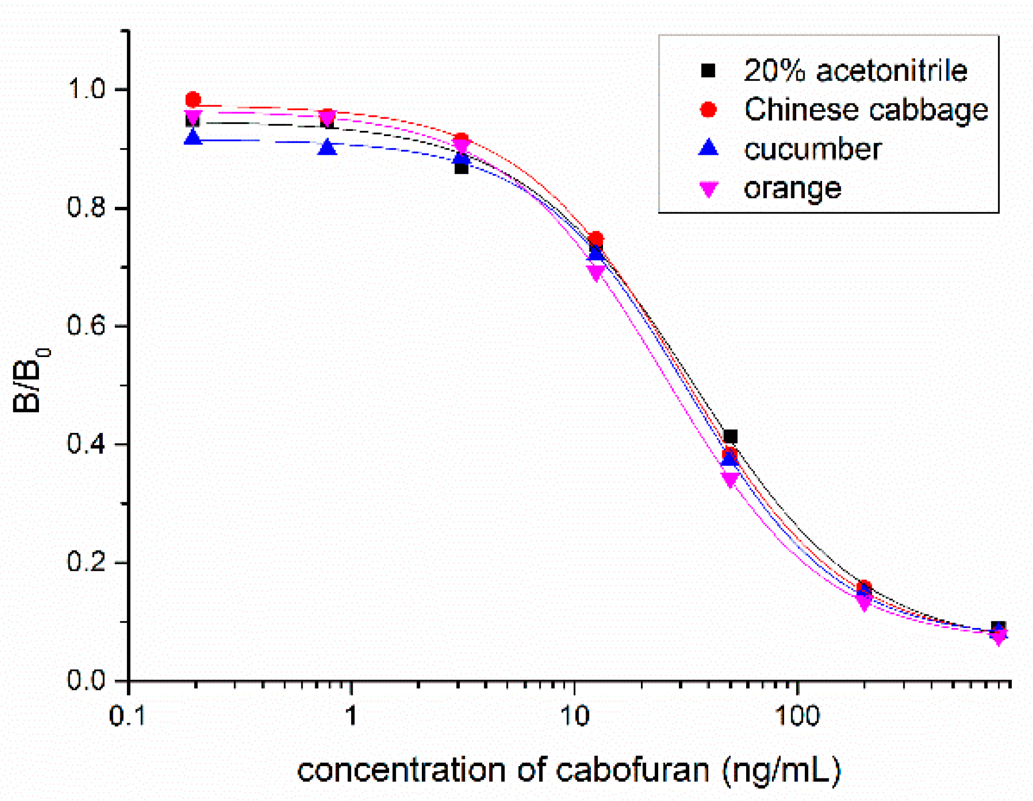
3.3. Nanobody-Based Ic-ELISA
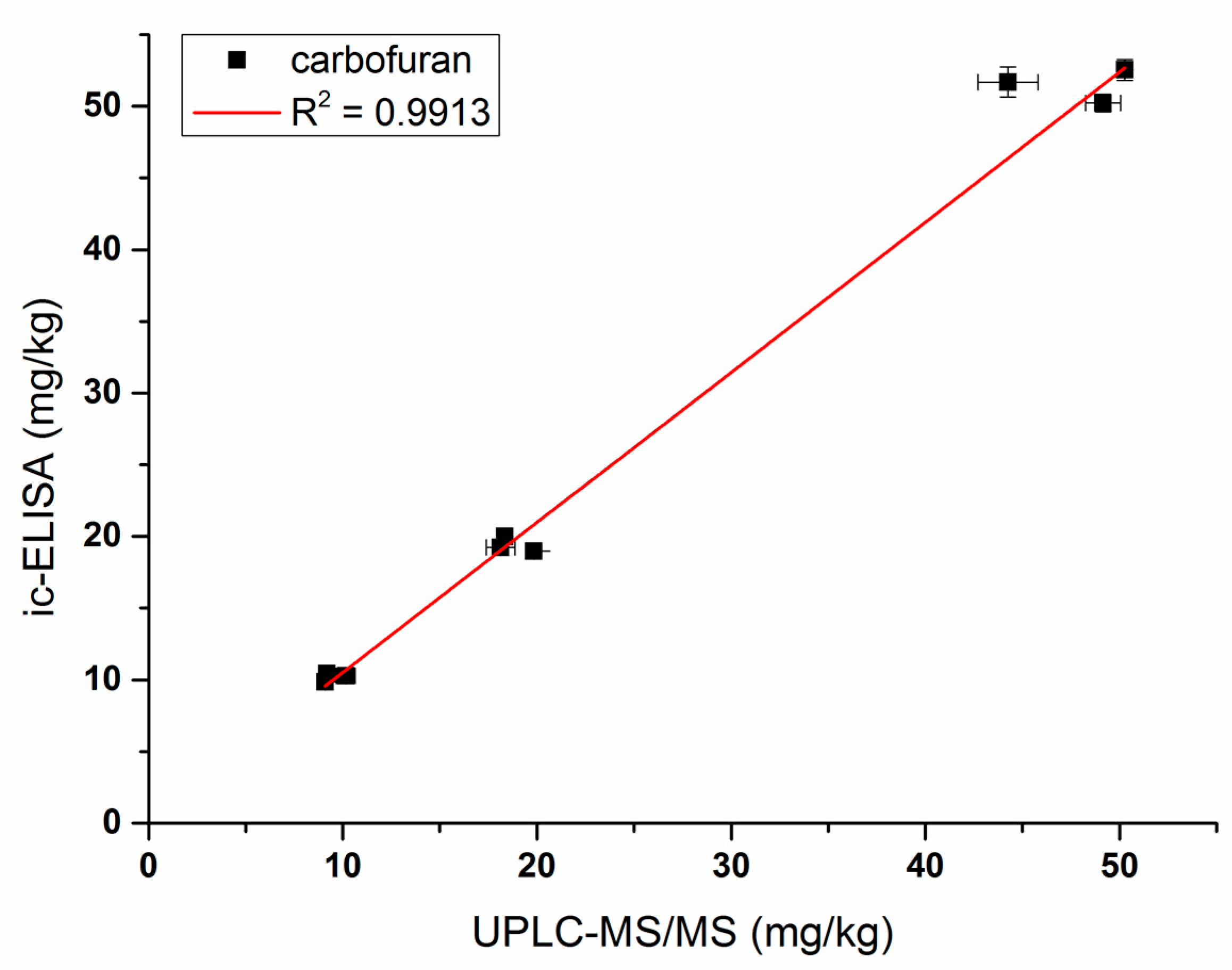
3.4. Sample Analysis by ic-ELISA and UPLC–MS/MS
4. Conclusions
Author Contributions
Funding
Conflicts of Interest
References
- Dai, Y.; Wang, T.; Hu, X.; Liu, S.; Zhang, M.; Wang, C. Highly sensitive microcantilever-based immunosensor for the detection of carbofuran in soil and vegetable samples. Food Chem. 2017, 229, 432–438. [Google Scholar] [CrossRef] [PubMed]
- Liu, L.; Xu, D.; Hu, Y.; Liu, S.; Wei, H.; Zheng, J.; Wang, G.; Hu, X.; Wang, C. Construction of an impedimetric immunosensor for label-free detecting carbofuran residual in agricultural and environmental samples. Food Control 2015, 53, 72–80. [Google Scholar] [CrossRef]
- Tien, C.; Huang, H.; Chen, C.S. Accessing the Carbofuran Degradation Ability of Cultures from Natural River Biofilms in Different Environments. CLEAN Soil Air Water 2017, 45, 1600380. [Google Scholar] [CrossRef]
- Gupta, R.C. Carbofuran toxicity. J. Toxicol. Environ. Health 1994, 43, 383–418. [Google Scholar] [CrossRef] [PubMed]
- Saraji, M.; Esteki, N. Analysis of carbamate pesticides in water samples using single-drop microextraction and gas chromatography-mass spectrometry. Anal. Bioanal. Chem. 2008, 391, 1091–1100. [Google Scholar] [CrossRef]
- Carla, S.; Brett, H.; Ambrose, F.; James, K.J.; Jordi, M.E.; Yolanda, P. Liquid chromatography quadrupole time-of-flight mass spectrometry analysis of carbosulfan, carbofuran, 3-hydroxycarbofuran, and other metabolites in food. Anal. Chem. 2007, 79, 1492–1501. [Google Scholar]
- Gui, W.; Jin, M.; Sun, L.; Guo, Y.; Zhu, G. Residues determination of carbofuran in vegetables based on sensitive time-resolved fluorescence immunoassay. Food Agric. Immunol. 2009, 20, 49–56. [Google Scholar] [CrossRef] [Green Version]
- Bellemjid, N.; Iddar, A.; Moussaif, A.; Abbadi, N.E.; Mesfioui, A. Analysis of Carbamates Pesticides: Immunogical Technique by Local Development of Enzyme-linked Immuno-Sorbent Assay. J. Pharm. Pharmacol. 2018, 6, 395–402. [Google Scholar] [CrossRef]
- Yang, J.Y.; Zhang, Y.; Wang, H.; Xu, Z.L.; Eremin, S.A.; Shen, Y.D.; Wu, Q.; Lei, H.T.; Sun, Y.M. Development of fluorescence polarisation immunoassay for carbofuran in food and environmental water samples. Food Agric. Immunol. 2015, 26, 340–355. [Google Scholar] [CrossRef]
- Liu, A.; Anfossi, L.; Shen, L.; Li, C.; Wang, X. Non-competitive immunoassay for low-molecular-weight contaminant detection in food, feed and agricultural products: A mini-review. Trends Food Sci. Tech. 2018, 71, 181–187. [Google Scholar] [CrossRef]
- Kim, H.; McCoy, M.R.; Majkova, Z.; Dechant, J.E.; Gee, S.J.; Tabares-da Rosa, S.; González-Sapienza, G.G.; Hammock, B.D. Isolation of Alpaca Anti-Hapten Heavy Chain Single Domain Antibodies for Development of Sensitive Immunoassay. Anal. Chem. 2012, 84, 1165–1171. [Google Scholar] [CrossRef] [PubMed]
- Hamers-Casterman, C.; Atarhouch, T.; Muyldermans, S.; Robinson, G.; Hammers, C.; Songa, E.B.; Bendahman, N.; Hammers, R. Naturally occurring antibodies devoid of light chains. Nature 1993, 363, 446–448. [Google Scholar] [CrossRef] [PubMed]
- Rossotti, M.A.; Pirez, M.; Gonzalez-Techera, A.; Cui, Y.; Bever, C.S.; Lee, K.S.S.; Morisseau, C.; Leizagoyen, C.; Gee, S.; Hammock, B.D.; et al. Method for Sorting and Pairwise Selection of Nanobodies for the Development of Highly Sensitive Sandwich Immunoassays. Anal. Chem. 2015, 87, 11907–11914. [Google Scholar] [CrossRef] [PubMed] [Green Version]
- Bever, C.S.; Dong, J.; Vasylieva, N.; Barnych, B.; Cui, Y.; Xu, Z.; Hammock, B.D.; Gee, S.J. VHH antibodies: Emerging reagents for the analysis of environmental chemicals. Anal. Bioanal. Chem. 2016, 408, 5985–6002. [Google Scholar] [CrossRef] [PubMed]
- Sun, Z.; Duan, Z.; Liu, X.; Deng, X.; Tang, Z. Development of a Nanobody-Based Competitive Dot ELISA for Visual Screening of Ochratoxin A in Cereals. Food Anal. Methods 2017, 10, 3558–3564. [Google Scholar] [CrossRef]
- Ren, W.; Li, Z.; Xu, Y.; Wan, D.; Barnych, B.; Li, Y.; Tu, Z.; He, Q.; Fu, J.; Hammock, B.D. One-Step Ultrasensitive Bioluminescent Enzyme Immunoassay Based on Nanobody/Nanoluciferase Fusion for Detection of Aflatoxin B1 in Cereal. J. Agric. Food Chem. 2019, 67, 5221–5229. [Google Scholar] [CrossRef]
- Xu, C.; Yang, Y.; Liu, L.; Li, J.; Liu, X.; Zhang, X.; Liu, Y.; Zhang, C.; Liu, X. Microcystin-LR nanobody screening from an alpaca phage display nanobody library and its expression and application. Ecotoxicol. Environ. Saf. 2018, 151, 220–227. [Google Scholar] [CrossRef]
- Wang, K.; Liu, Z.; Ding, G.; Li, J.; Vasylieva, N.; Li, Q.X.; Li, D.; Gee, S.J.; Hammock, B.D.; Xu, T. Development of a one-step immunoassay for triazophos using camel single-domain antibody-alkaline phosphatase fusion protein. Anal. Bioanal. Chem. 2019, 411, 1287–1295. [Google Scholar] [CrossRef]
- Zhang, Y.; Xu, Z.; Wang, F.; Cai, J.; Dong, J.; Zhang, J.; Si, R.; Wang, C.; Wang, Y.; Shen, Y.; et al. Isolation of Bactrian Camel Single Domain Antibody for Parathion and Development of One-Step dc-FEIA Method Using VHH-Alkaline Phosphatase Fusion Protein. Anal. Chem. 2018, 90, 12886–12892. [Google Scholar] [CrossRef]
- Wang, K.; Vasylieva, N.; Wan, D.; Eads, D.A.; Yang, J.; Tretten, T.; Barnych, B.; Li, J.; Li, Q.X.; Gee, S.J.; et al. Quantitative Detection of Fipronil and Fipronil-Sulfone in Sera of Black-Tailed Prairie Dogs and Rats after Oral Exposure to Fipronil by Camel Single-Domain Antibody-Based Immunoassays. Anal. Chem. 2019, 91, 1532–1540. [Google Scholar] [CrossRef]
- Liu, Z.; Wang, K.; Wu, S.; Wang, Z.; Ding, G.; Hao, X.; Li, Q.X.; Li, J.; Gee, S.J.; Hammock, B.D. Development of a camelid variable domain of heavy chain antibody-based immunoassay for the detection of carbaryl in cereals. J. Sci. Food Agric. 2019, 99, 4383–4390. [Google Scholar] [CrossRef] [PubMed]
- Yang, J.Y.; Wang, H.; Jiang, J.M.; Sun, Y.M.; Pan, K.; Lei, H.T.; Wu, Q.; Shen, Y.D.; Xiao, Z.L.; Xu, Z.L. Development of an enzyme-linked immuno-sorbent assay (ELISA) method for carbofuran residues. Molecules 2008, 13, 871–881. [Google Scholar] [CrossRef] [PubMed]
- Ebrahimizadeh, W.; Mousavi Gargari, S.; Rajabibazl, M.; Safaee Ardekani, L.; Zare, H.; Bakherad, H. Isolation and characterization of protective anti-LPS nanobody against V. cholerae O1 recognizing Inaba and Ogawa serotypes. Appl. Microbiol. Biot. 2013, 97, 4457–4466. [Google Scholar] [CrossRef] [PubMed]
- Olichon, A.; Schweizer, D.; Muyldermans, S.; Marco, A.D. Heating as a rapid purification method for recovering correctly-folded thermotolerant VH and VHH domains. BMC Biotechnol. 2007, 7, 7–14. [Google Scholar] [CrossRef] [PubMed]
- Cieślik, E.; Sadowska-Rociek, A.; Ruiz, J.M.M.; Surma-Zadora, M. Evaluation of QuEChERS method for the determination of organochlorine pesticide residues in selected groups of fruits. Food Chem. 2011, 125, 773–778. [Google Scholar] [CrossRef]
- Cécile, V.; Remy, L.; Dirk, S.; Sergio, M.R.; Serge, M.; Katja, C. General strategy to humanize a camelid single-domain antibody and identification of a universal humanized nanobody scaffold. J. Biol. Chem. 2009, 284, 3273–3284. [Google Scholar]
- Muyldermans, S.; Atarhouch, T.; Saldanha, J.; Barbosa, J.A.R.G.; Hamers, R. Sequence and structure of VH domain from naturally occurring camel heavy chain immunoglobulins lacking light chains. Protein Eng. 1994, 7, 1129–1135. [Google Scholar] [CrossRef]
- Pérez, J.M.J.; Renisio, J.G.; Prompers, J.J.; van Platerink, C.J.; Cambillau, C.; Darbon, H.; Frenken, L.G.J. Thermal Unfolding of a Llama Antibody Fragment: A Two-State Reversible Process. Biochemistry 2001, 40, 74–83. [Google Scholar] [CrossRef]
- Akazawaogawa, Y.; Uegaki, K.; Hagihara, Y. The role of intra-domain disulfide bonds in heat-induced irreversible denaturation of camelid single domain VHH antibodies. J. Biochem. 2016, 159, 111–121. [Google Scholar] [CrossRef]
- Turner, K.B.; Zabetakis, D.; Goldman, E.R.; Anderson, G.P. Enhanced stabilization of a stable single domain antibody for SEB toxin by random mutagenesis and stringent selection. Protein Eng. Des. Sel. 2014, 27, 89–95. [Google Scholar] [CrossRef] [Green Version]
- Zabetakis, D.; Olson, M.A.; Anderson, G.P.; Legler, P.M.; Goldman, E.R. Evaluation of Disulfide Bond Position to Enhance the Thermal Stability of a Highly Stable Single Domain Antibody. PLoS ONE 2014, 9, e115405. [Google Scholar] [CrossRef] [PubMed]
- Zhu, G.; Jin, M.; Gui, W.; Guo, Y.; Jin, R.; Wang, C.; Liang, C.; Liu, Y.; Wang, T. Development of a direct competitive enzyme-linked immunoassay for carbofuran in vegetables. Food Chem. 2008, 107, 1737–1742. [Google Scholar] [CrossRef]
- Moreno, M.; Abad, A.; Pelegrí, P.; Martínez, M.; Sáez, A.; Gamón, M.; Montoya, A. Validation of a Monoclonal Enzyme Immunoassay for the Determination of Carbofuran in Fruits and Vegetables. J. Agric. Food Chem. 2001, 49, 1713–1719. [Google Scholar] [CrossRef] [PubMed]


| Analogues | Molecular Structural | IC50 (ng/mL) | Cross-Reactivity (%) |
|---|---|---|---|
| Carbofuran | 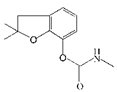 | 7.27 | 100 |
| Benfuracarb |  | 142.51 | 5.1 |
| Fenobucarb |  | 204.6 | 3.5 |
| Carbosulfan | 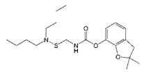 | 280.93 | 2.6 |
| 3-Hydroxycarbofuran | 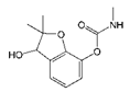 | 366.05 | 2.0 |
| Isoprocarb |  | 1351.82 | 0.5 |
| Carbaryl |  | >2000 | <0.1 |
| Aldicarb | 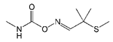 | >2000 | <0.1 |
| Methomyl | 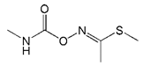 | >2000 | <0.1 |
| Pirimicarb |  | >2000 | <0.1 |
| Mercaptodimethur | 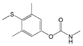 | >2000 | <0.1 |
| Tsumacide | 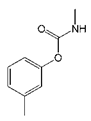 | >2000 | <0.1 |
| Added (mg/kg) | Chinese Cabbage | Cucumber | Orange | ||||||
|---|---|---|---|---|---|---|---|---|---|
| Found ± SD (mg/kg) | Recovery (%) | CV (%) | Found ± SD (mg/kg) | Recovery (%) | CV (%) | Found ± SD (mg/kg) | Recovery (%) | CV (%) | |
| 10 | 10.27 ± 0.34 | 102.7 | 3.33 | 9.73 ± 0.24 | 97.35 | 2.52 | 8.47 ± 0.2 | 84.74 | 2.35 |
| 20 | 18.44 ± 0.54 | 92.25 | 2.61 | 20.45 ± 0.31 | 102.26 | 1.50 | 16.46 ± 0.22 | 82.31 | 1.21 |
| 50 | 51.96 ± 0.75 | 103.92 | 1.44 | 46.24 ± 0.51 | 92.48 | 1.11 | 43.77 ± 0.91 | 87.54 | 20.76 |
© 2019 by the authors. Licensee MDPI, Basel, Switzerland. This article is an open access article distributed under the terms and conditions of the Creative Commons Attribution (CC BY) license (http://creativecommons.org/licenses/by/4.0/).
Share and Cite
Zhang, J.-r.; Wang, Y.; Dong, J.-x.; Yang, J.-y.; Zhang, Y.-q.; Wang, F.; Si, R.; Xu, Z.-l.; Wang, H.; Xiao, Z.-l.; et al. Development of a Simple Pretreatment Immunoassay Based on an Organic Solvent-Tolerant Nanobody for the Detection of Carbofuran in Vegetable and Fruit Samples. Biomolecules 2019, 9, 576. https://doi.org/10.3390/biom9100576
Zhang J-r, Wang Y, Dong J-x, Yang J-y, Zhang Y-q, Wang F, Si R, Xu Z-l, Wang H, Xiao Z-l, et al. Development of a Simple Pretreatment Immunoassay Based on an Organic Solvent-Tolerant Nanobody for the Detection of Carbofuran in Vegetable and Fruit Samples. Biomolecules. 2019; 9(10):576. https://doi.org/10.3390/biom9100576
Chicago/Turabian StyleZhang, Jin-ru, Yu Wang, Jie-xian Dong, Jin-yi Yang, Yu-qi Zhang, Feng Wang, Rui Si, Zhen-lin Xu, Hong Wang, Zhi-li Xiao, and et al. 2019. "Development of a Simple Pretreatment Immunoassay Based on an Organic Solvent-Tolerant Nanobody for the Detection of Carbofuran in Vegetable and Fruit Samples" Biomolecules 9, no. 10: 576. https://doi.org/10.3390/biom9100576