Article

Phytochemicals Profiling, Antimicrobial Activity and Mechanism of Action of Essential Oil Extracted from Ginger (Zingiber officinale Roscoe cv. Bentong) against Burkholderia glumae Causative Agent of Bacterial Panicle Blight Disease of Rice

by
Mahesh Tiran Gunasena
1,2,
Amara Rafi
1,
Syazwan Afif Mohd Zobir
1,*,
Mohd Zobir Hussein
3,
Asgar Ali
4,
Abdulaziz Bashir Kutawa
1,5,
Mohd Aswad Abdul Wahab
1,
Mohd Roslan Sulaiman
6,
Fariz Adzmi
7 and
Khairulmazmi Ahmad
1,8,*
1
Department of Plant Protection, Faculty of Agriculture, Universiti Putra Malaysia, Serdang 43400, Malaysia
2
Grain Legume and Oil Crop Research and Development Centre, Angunakolapelessa 82220, Sri Lanka
3
Institute of Nanoscience and Nanotechnology, Universiti Putra Malaysia, Serdang 43400, Malaysia
4
Centre of Excellence for Postharvest Biotechnology (CEPB), School of Biosciences, University of Nottingham Malaysia, Semenyih 43500, Malaysia
5
Department of Plant Science and Biotechnology, Faculty of Life Science, Federal University Dutsin-Ma, Dutsin-Ma 821101, Nigeria
6
Department of Biomedical Science, Faculty of Medicine and Health Sciences, Universiti Putra Malaysia, Serdang 43400, Malaysia
7
Institute of Plantation Studies (IKP), Universiti Putra Malaysia, Serdang 43400, Malaysia
8
Institute of Tropical Agriculture and Food Security (ITAFoS), Universiti Putra Malaysia, Serdang 43400, Malaysia
*
Authors to whom correspondence should be addressed.
Plants 2022, 11(11), 1466; https://doi.org/10.3390/plants11111466
Submission received: 16 March 2022 / Revised: 11 April 2022 / Accepted: 13 April 2022 / Published: 30 May 2022

Abstract

:
Essential oils protect plants, and due to their natural origin, there is much interest in using them as antimicrobial agents. The purpose of this study was to determine the phytochemical constituents of ginger essential oil (GEO), antimicrobial activity, and mode of action against Burkholderia glumae (Bg). In addition, the volatile active compounds (AIs) were studied using GC-MS, FTIR, and Raman spectroscopy. A total of 45 phytochemical components were detected and the most prevalent bioactive compounds were Geranial, 1,8-Cineole, Neral, Camphene, α-Zingiberene, and α-Farnesene. Furthermore, it was found that the most dominant terpenes in GEO were monoterpenes. The diameter zone of inhibition values varied from 7.1 to 15 mm depending on the concentration tested. In addition, the MIC and MBC values were 112.5 µL/mL. Faster killing time and lower membrane potential were observed in 1xMIC treatment compared to 0.5xMIC treatment, whereas the control had the maximum values. From observations of various images, it was concluded that the mode of action of GEO affected the cytoplasmic membrane, causing it to lose its integrity and increase its permeability. Therefore, the antibacterial study and mechanism of action revealed that GEO is very effective in suppressing the growth of B. glumae.

1. Introduction

Burkholderia glumae is a seed-borne rice pathogen and the most serious emerging pathogen in paddy-growing countries. The bacterial panicle blight (BPB) of rice caused by Bg can induce up to 75% yield loss in a severely infested field [1,2]. The Bg is Gram-negative, non-spore-forming, nonfluorescent, and rod-shaped with a polar flagellum bacterium [2,3]. The disease causes symptoms such as blight in rice panicles, discoloration of growing grains, seed rot, and spikelet sterility [3,4]. B. glumae’s incidence in plumules results in the production of toxic materials such as toxoflavin. The toxoflavin [1,6-dimethylpyrimido [5,4-e]-1, 2,4-triazine-5,7(1H,6H)-dione] is a yellow pigment required for pathogenicity and causes rice seedling rot [2]. Weather conditions, including excessive daily minimum temperatures and regular rainfall throughout rice panicle appearance and flowering stages, which increase the relative humidity conditions, have a considerable impact on BPB disease outbreaks [5]. B. glumae is a good example of minor plant pathogens progressing to a serious disease as environmental conditions change. Similarly to how it affects rice, the Bg also affects pepper, eggplant, sesame, and tomato plants [2]. Plant extracts, resistant varieties, seed treatments, and heating technologies that have been proven to inhibit Bg infections are among the most common methods used to overcome the disease [6].
Recently, significant control efforts have been made to limit pathogen dissemination using numerous control methods, such as the use of substitute antimicrobial compounds. Essential oils (EOs) as natural antimicrobial agents have a role in plant defense mechanisms, and they have a high probability of being used for controlling plant pathogens [7]. Aromatic volatile compounds derived from various plant parts were extracted by numerous techniques and are known as EOs [8,9]. For the commercialization process, a conventional hydrodistillation method that utilizes water that leaves nontoxic byproducts is the most effective method for EO extraction [10]. The EOs and their constituents have a long history of use as antimicrobial agents. This biological activity is frequently attributed to bioactive components developed by EOs during the secondary metabolism of the plant. The most EOs are classified into two types based on the number of isoprene units: monoterpenes and sesquiterpenes [11]. Monoterpenes are terpenes made up of two isoprene units that are either linear (acyclic) or comprise of rings (monocyclic and bicyclic). Sesquiterpenes are types of terpenes composed of three isoprene units [12].
Ginger (Zingiber officinale Roscoe) is one of the most widely known perennial herbs. It is derived from the underground stems or rhizomes of the plant [13]. In Malaysia, ginger is cultivated commercially in Pahang, Johor, Selangor, Sabah, and Sarawak. Zingiber officinale cv. ‘Tambunan’ is a popular cultivar of ginger in Sabah, and is mainly cultivated in the Tambunan and Keningau areas. Z. officinale cv. Bentong is the most expensive and popular ginger cultivar in Malaysia, and is mainly cultivated in Bentong, Pahang. The Zingiberaceae family is amongst the most commonly grown plant families in the tropics, specifically in Southeast Asia. This valuable natural resource produces a variety of useful items, such as food, spices, medications, colors, perfumes, and cosmetics. Ginger is widely used as a spice and flavoring agent around the world and is also used medicinally [13,14]. Additionally, Zingiberaceae species have been identified to have broad biological actions including antimicrobial, antioxidant, and anti-inflammatory properties. Ginger essential oil is a volatile oil obtained from the root of ginger and is known for its distinct aroma and biological activity [10]. Ginger has been extensively studied and it contains 1-2% EO, which gives the spice its distinct flavor [15]. As a result of the increasing attention to the usage of EOs in the control microbes, GEO has been studied for its role against different pathogens. Conventionally, chemical components of EOs can be detected and measured using chromatographic methods, which generate high-quality and consistent results. Raman spectroscopy procedures can offer important analytical information in significantly less time than conventional methods. There are numerous studies on the chemical content of fresh GEO and other natural flavoring compounds [15]. Terpenes, phenolics, and N- and S-containing derivatives are the three most common types of secondary metabolites produced by plants [16]. Ginger contains many active ingredients, like phenolic and terpene compounds. In vitro antimicrobial activity of EOs has been widely studied. Furthermore, it is a rich source of several active substances, such as bioactive phenols (gingerols, shogaols, and zingerones) [9]. It also contains several terpene components, including β-bisabolene, α-curcumin, zingiberene, α-farnesene, and β-sesquiphellandrene, which are thought to be the major components of GEOs [17]. More importantly, recent work emphasizes the contribution of the phenolic compounds (Eugenol, Shogaols, Zingerone, Gingerdiols, Gingerols, and so on) and their synergistic interactions with other substances such as β-Sesquiphellandrene, α-Zingiberene, α-Farnesene, α- and β-Bisabolene found in GEO, extracts, and Oleoresins, which played a crucial role as the antimicrobial agent [18]. Gram-negative bacteria are commonly more opposed to EO than Gram-positive ones. Gram-negative bacteria have an outer membrane made of hydrophilic lipopolysaccharides (LPS), which operate as a barrier against hydrophobic chemicals like those contained in EOs [19]. Moreover, GEO has also demonstrated the ability to suppress bacterial growth due to the reduction of bacterial biofilm formation [17]. GEO has been widely studied, with particular attention to its antifungal, antibacterial, and antioxidant properties, in addition to its potential use in preserving food [20]. The advantages of using GEO were highlighted in numerous studies, which demonstrated its favorable impacts against disease symptoms, acting as an anti-inflammatory, antitumor, anodyne, neuronal cell-protective agent [21]. Therefore, the primary objective of this study was to determine the phytochemical profiling, antibacterial activity, and mechanism of action of GEO against B. glumae.

2. Results

2.1. Analysis of Chemical Constituents of Ginger Essential Oils by Gas Chromatography-Mass Spectrometry

The results of the gas chromatography–mass spectrometry (GC–MS) study revealed various bioactive compounds identified and quantified in GEO. The chemical components of GEO through hydrodistillation was listed in Table 1 with their respective percentages. EO constituents were identified by comparing retention times and retention index with library data from FFNSC1.3.lib, NIST11.lib, and WILEY229. The retention index was calculated using the Kovats equation. In addition, the comparison of retention values obtained by Sharma et al. (2016) [15] and Babushok et al. (2009) [22] is also provided in Table 1.
The extraction yields were determined by dividing the amount of extracted oil by the amount of fresh material fed into the extractor. The extraction yields were estimated based on the results of several cycles. The results were presented as a percentage of extraction. The yield of GEO was between 1.2–1.5% (v/w). The EO was extracted from the ginger rhizome in which the color was changed from pale yellow to light amber. Gas chromatography analysis identified 45 peaks of active compounds available in GEO as displayed in Figure 1 It was also observed that the major compounds were Geranial (17.88%), 1,8-Cineole (14.96%), Neral (13.99%), Camphene (7.53%), and α-Farnesene (3.51%) and their combination was more than 50%. The GEO contained a high percentage of monoterpenes (74.6 %) and sesquiterpenes (20.73%). Geranial was the most prevalent monoterpene hydrocarbon compound in the total extracted essential oil. Among the 20 monoterpenes, the major ones were Geranial, 1,8-Cineole, Neral, Camphene, Citronellol, α-Pinene, and β-Linalool. The remaining monoterpenes were found in less than 2% of the samples. The predominant sesquiterpenes were α-Zingiberene (5.19%) α-Farnesene (3.51%), β-Sesquiphellandrene (2.40%), Curcumene (1.58%), and β-Bisabolene (1.16%). Less than 1% of the other sesquiterpenes were detected in the sample. Our findings are in line with the findings of Abdullahi et al., (2020) [23].

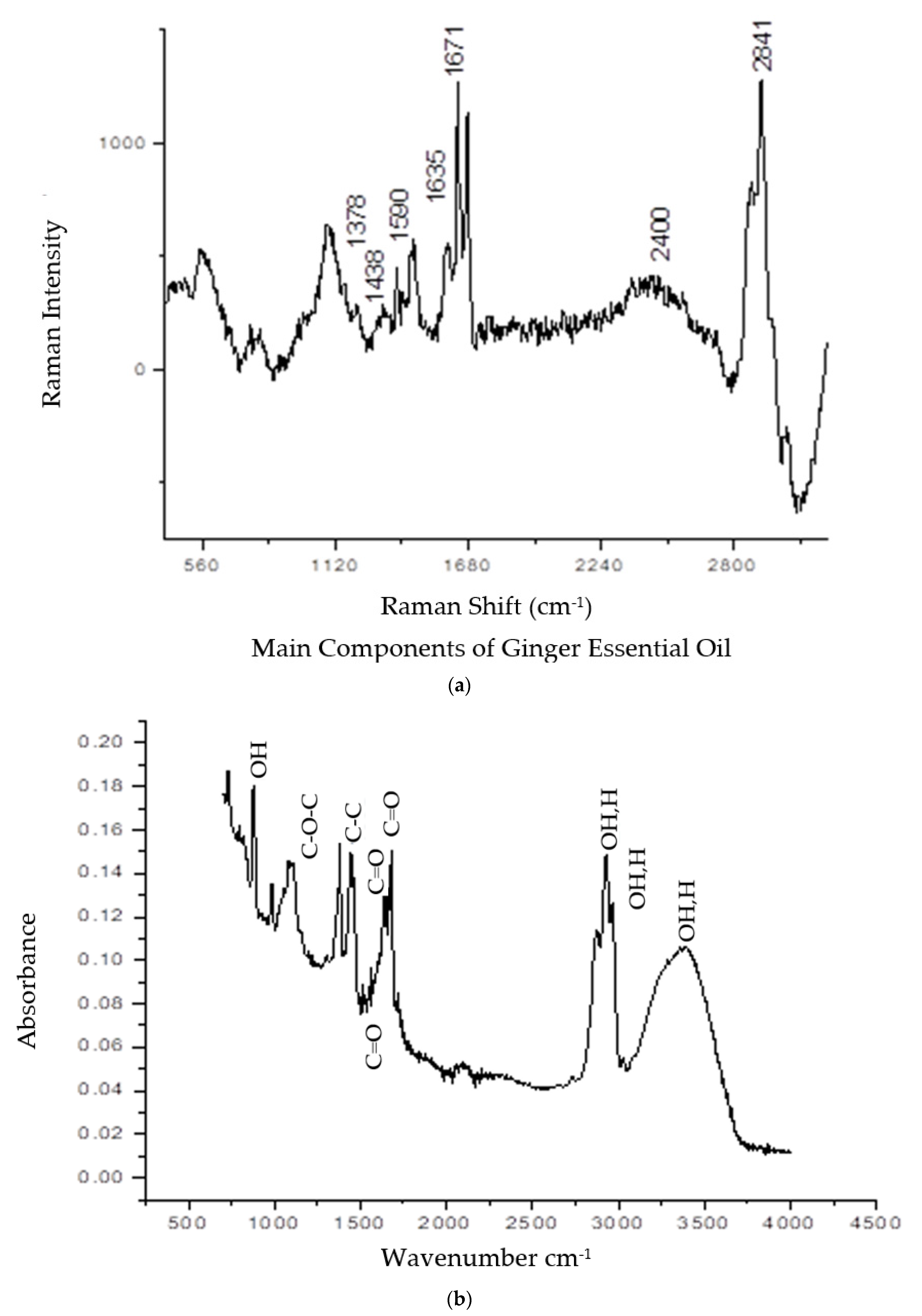
2.2. Vibrational Spectroscopy Analyses by Raman and FTIR

Both vibrational spectroscopy analyses of Raman and Fourier transform infrared (FTIR) spectra are displayed in Figure 2. The appearance of several peaks ranging from 400 cm−1 to 3000 cm−1 was attributed to the existence of multiple chemical bonds in the GEO (Figure 2a). The most important components in ginger essential oil are zingiberene, geranial, neral, and camphene [24]. At the lower frequency of the Raman spectrum (700–900 cm−1), the COC and CCO bands were detected. The CH-stretching band was at 1438 cm−1. Other appearance peaks for benzene rings were recorded at the middle range frequency: 1590 and 1602 cm−1 of the Raman spectrum. The C=C band was at 1642 and 1684 cm−1, which attributed to the CH3 group bending mode. In the chemical structure of geranial and neral, the double bond is conjugated with the aldehyde group. CH-bending mode was also detected at 2800–2841 cm−1. The Fourier Transform Infra-Red spectrum was recorded from 550–4000 cm−1, as displayed in Figure 2b. Plenty of functional groups were detected, such as OH bonds, COC, C-C stretch, C=O stretch, and OH stretch [25]. Detailed information about the peaks, wavelength, and functional groups was tabulated in Table 2.

2.3. Antibacterial Activity, Minimum Inhibitory Concentration (MIC), and Minimum Bactericidal Concentration (MBC) of Ginger Essential Oil

Ginger essential oil antibacterial activity was assessed qualitatively by measuring the diameter of the zone of inhibition (ZOI). Evaluation of the antibacterial activity of GEO was recorded in Table 3. The negative control was dimethyl sulfoxide (DMSO) buffer, and the positive control was streptomycin. The results revealed that all concentrations were potentially effective in suppressing the growth of Bg with variable potency. The widths of the inhibition zones increased as the concentrations of GEO increased. The bacterial sensitivity to GEO was ranked using zone-of-inhibition values expressed in millimeters (mm) [26]. The antibacterial activity of 100, 200, 300, 400, and 500 µL/mL of GEO against Bg indicated inhibition zone diameters of 7.1, 8.0, 10.6, 14.3, and 15.1 mm, respectively. It was found that the most effective retarding potent antibacterial action against Bg was at 500 µL/mL. These treatments incited sensitive reactions and a high percent of inhibition of growth. Compared to a standard positive control used in the experiment, the ZOI of streptomycin was 24.1 mm. The negative control DMSO demonstrated no inhibition of the growth of Bg.
The antibacterial effect of GEO was quantified using MIC and MBC values against Bg. The color appearance of the well contents was determined by the inhibition of Bg at various concentrations. The wells containing nonviable bacterial cells demonstrated no color change, whereas the wells containing viable cells presented red color formation. The MIC and MBC were determined to see if GEO could inhibit or kill the pathogenic bacteria Bg. The results for the MIC determination in this study revealed that GEO has potent antibacterial activity against Bg. It was critical to determine the efficient concentration in vitro. GEO had an MIC of 12.5 L/mL against B. glumae and the MBC value for B. glumae was 12.5 L/mL. The MIC and MBC for the GEO were 12.5 µL/mL, it served as the concentration whereby approximately all the Bg cells were killed. The MBC value for Bg was the same as the MIC value.

2.4. Time Killing Analysis

A bacterial growth-curve assay was carried out to determine the efficacy of GEO in killing or inhibiting the growth of Bg. The assessment of GEO inhibition or deactivation kinetics on Bg gives supportive evidence that supplements the data acquired from the MIC and MBC investigations, permitting for a more accurate estimation of the component efficiency. The analysis of Bg under various concentrations of GEO was illustrated in Figure 3. The time courses of Bg growth in the presence of different concentrations of GEO were plotted to confirm the antibacterial action of GEO against Bg.
The optical density values of the control treatment increased rapidly during 2–18 h of incubation periods. When compared to the 0.5xMIC of GEO, the optical density of Bg slightly increased from 2–8 h, which was mostly attributable to the bacteria’s growth cycle inhibition. Interestingly, pathogen growth was slowed down with an extended lag time when the concentration of GEO approached 1 × MIC. The optical density dropped from 0–4 h of incubation periods and the optical density value remained constant onward. Compared to the control, susceptible Bg treated with GEO at 1 × MIC level demonstrated a significant decrease in the number of the viable cells during the first 6-8 h of the assessment periods and needed up to 14 h of incubation to promote the entire elimination of Bg cells in this assay.

2.5. Morphological Changes of B. glumae by Scanning Electron Microscope (SEM)

The scanning electron microscope is commonly applied to monitor the external morphological deviations of the cells. The physical and morphological variations of Bg at different treatments were examined by SEM images, as demonstrated in Figure 4. SEM images revealed that the treated Bg surfaces underwent obvious morphological changes compared to the DMSO-treated ones (negative control). Figure 4a illustrates the morphology and microstructure of Bg exposed to DMSO, with distinguishing features such as normal rod-shaped, undamaged surface, striated cell walls, and no visible cellular debris. However, after 24 h of exposure to GEO at MIC (Figure 4b), morphological deviations of Bg were observed as distorted, irregular, and shriveled to different degrees. The bacterial colonies exposed to GEO were severely damaged, with contraction illustrated by the yellow arrows in Figure 4b. The cells incubated with streptomycin presented collapsed and ruptured cells in Figure 4c. Streptomycin treatment resulted in more severe morphological destruction than GEO treatment. This was most likely owing to the severely damaged bacterial peptidoglycan layer and cell membrane, which resulted in cell death and disconnection from the filter holder.

2.6. Morphological Changes of B. glumae by Transmission Electron Microscope (TEM)

The effects of GEO as an antimicrobial against Bg cells were further investigated using TEM, which determines the cell membrane integrity and internal morphology of the cells, and the results are displayed in Figure 5. The images of the Bg cells treated with GEO presented considerably differently from those of the untreated cells. TEM observations revealed that the cells treated with DMSO remained unchanged, with distinct, uniformly shaped, and intact cell walls, evenly dispersed cytochylema, and electron-dense substance within the cell (Figure 5a). However, there were lesser-intact Bg cells in the GEO treated samples, and it was difficult to identify the cells that had been exposed to the GEO (MIC). After being exposed to GEO for 24 h, the Bg cell was distorted and crumbled, demonstrating that the intracellular contents were leaked out of the cells (Figure 5b). Furthermore, the cell wall and cytoplasmic membrane had become irregular and thick; much lysis was observed. This has the potential to cause nutrients and genetic substances to be discharged from the cell.

2.7. Biofilm Breakdown Observed by Confocal Laser Scanning Microscope (CLSM)

Confocal laser scanning microscopy studies were used to investigate the bactericidal mode of action of the active compounds found in EOs. The fluorescence intensity of the stained (red or green) pathogen cells at a specific wavelength indicated the findings of the LIVE/DEAD analyses. The results demonstrated that after staining with the LIVE/DEAD stain, there were no detectible dead cells in the untreated control, having viable Bg cells. In the untreated control, the biofilm aggregation size appeared in a green color, indicating the presence of live and viable pathogen cells (Figure 6(a1)). Conversely, the images for the treated Bg cells with the MIC of GEO have exhibited membrane damage (Figure 6(b1)), with red staining confirming the existence of many dead cells. These findings revealed that GEO at MIC inhibited and broke down the produced biofilm, limiting its development and growth. The effect of GEO on Bg biofilm was further analyzed using 3D confocal microscopy software (Figure 6(a2,a3,b2,b3)). Comparatively, the untreated Bg biofilm with GEO images was displayed in Figure 6(a2,a3). Volumetric images revealed a significant reduction in Bg biofilm and a higher proportion of damaged/compromised cells (Figure 6(b2)). As depicted in Figure 6(b3), GEO manages to kill Bg, starting from the top and working down to the lower layers of the biofilm cells. Furthermore, fewer viable cells can be observed by the green color of the treated biofilm cells.

3. Discussion

The composition, structure, and functional groups of essential oils play an important role in determining their antimicrobial activity. GEO is a complex combination of volatile compounds formed as secondary metabolites in ginger that have been extensively utilized as possible substitutes for chemically produced antimicrobials and antioxidants [27]. Many analyses have stated the chemical composition of ginger essential oil [13,23,28,29]. However, the specific content of the extraction and the number of compounds in plant essential oils are varied and depend on the environmental factors; the growing season of plant, cultivation type, vegetative stage [30], harvesting period, plant part utilized, and method of extraction [21]. The 45 chemical compounds of ginger essential oil were identified in this study. Guimaraes et al. (2019) [31] reported that essential oils are composed of a complex mixture of compounds ranging from 20 to 60 at different concentrations. Based on the GC–MS analysis, the highest percentages of active compounds in GEO were Geranial, 1,8-Cineole, and Neral compounds (Table 1) which agrees with the findings of Mesomo et al., (2013) [21]. The work highlighted the GC–MS analysis of GEO AIs and concluded that the main volatile compounds existing in fresh ginger rhizome were Geranial, ar-Curcumene, and β-Bisabolene. In addition, according to Abdullahi et al. (2020) [23], sesquiterpenes and monoterpenes were found in EOs, which contributed to significant antimicrobial activity against pathogenic microorganisms. In concurrence with previous work conducted by Wang et al. (2020) [9], the essential oil contained a high concentration of monoterpenes, and was then followed by sesquiterpenes.
Those molecular structures are Geranial, 1,8 Cineole, Neral, Camphene, α-Zingiberene, and α-Farnesene, in addition to the combination of those AIs have 60% of GEO compositions. Next, the physicochemical characterizations were done using vibrational spectroscopy analyses, such as Raman and FTIR spectra, as demonstrated in Figure 2. In general, all AIs Have C=C, C-C, and C-H bonding, which was recorded on both spectra. However, the existence of benzene rings is exclusive only for 1,8 Cineole and α-Zingiberene which were recorded on the Raman spectrum at 1590 and 1602 cm−1.
The analysis of ZOI, MIC, MBC, and the time-killing analysis of Bg were studied to assess GEO antibacterial action. The EOs and their main constituents have demonstrated a high level of antimicrobial activity against pathogens [32]. Similarly, because of the high content of lipophilic compounds, EOs attached and interrupted the integrity of microbial cell wall surfaces and membrane structures, leading to cell lysis [33]. The antibacterial activity against Bg demonstrated varying degrees of growth inhibition, which were highly dependent on the concentration of GEO. A wider ring (higher ZOI) of no bacterial growth occurred because of a stronger inhibitory effect of antimicrobial EOs, whereas a lesser or no inhibition action results in few or no changes in the bacterial cell membrane. The EO concentration (100–500 µL/mL) affected the bacterial growth of Bg. The MIC study strongly indicates that GEO has potent antibacterial activities against Bg. In this circumstance, the MBC value was the same as the MIC value for Bg which was consistent with the time-killing study. The information clearly indicated that the GEOs at 1 × MIC could kill large amounts of Bg at different times compared to GEO at 0.5 MIC. Sharma et al. (2016) [15] found that GEO had an inhibitory action on a broad range of pathogenic bacteria and fungi, which was most likely due to the oil’s major components. Furthermore, the surface of Gram-negative bacteria cells acts as a penetration barrier, preventing macromolecules and hydrophobic compounds from penetrating the bacteria cell membrane [27]. As a result, Gram-negative bacteria are more tolerant to hydrophobic antibiotics than Gram-positive bacteria. However, Wang et al. (2020) [9] discovered that GEO had antibacterial properties against both Escherichia coli and Staphylococcus aureus. The findings of the MIC study and the growth curve from our investigation both pointed to the same conclusion. GEO and extracts were found to have great antimicrobial action and inhibited some food-spoilage microorganisms [34]. Our findings confirmed that GEO can significantly inhibit Bg growth by providing a theoretical basis for effective antibacterial activity. According to Dannenberg et al.’s (2018) [35] time-killing analysis, after 8 h of making contact with rosemary EOs (MBC = 0.1%), the total mortality of the bacteria (S. aureus) was observed while the main component (1,8-Cineole) was treated separately and needed 12 h. The differences were more accessible, which was similar to the findings of the SEM and the kill-time study.
Based on the different mechanisms of action, the antimicrobial substances on the membrane could cause depolarization or increased permeability. For example, several antimicrobial peptides form pores, whereas other compounds, such as certain EO constituents, have a fluidifying effect on the membrane [36]. SEM, TEM, and CLSM assays were utilized to discover the antibacterial mode of action of GEO against Bg, with the MIC concentration of GEO causing changes in cell viability, morphology, membrane leakage, and cell integrity. The integrity of the cell is critical for an organism to continue to exist because it is the main component for critical biological activities that take place [37]. Despite this, certain mechanisms have been suggested. The cell membrane breaks down and changes in ion channels (Na+, K+, Ca2+, or Cl) in the cell membrane. This could enhance permeability and cause the leakage of important intracellular substances [38] in addition to the inhibition of target enzymes [39]. The morphological deviations from the images clearly demonstrated that the cell membranes of the Bg treated with GEO were significantly disrupted, with noticeable abnormal structure and shape. Most importantly, GEO demonstrated the ability to enhance the permeability of cell membranes by causing the leakage of intracellular substances, which was thought to be a mechanism of the Bg downstream physiological phenomena. Most of the treated GEO bacteria formed irregular, sunken surfaces and shriveled to varying degrees. This study discovered similar research findings in bacterial morphological changes as other studies [20,40,41]. Next, CLSM analysis revealed that the GEO inhibited the treated cell, resulting in a notable breakdown and reduction in biofilm development. As a result, GEO decreased the quantity of viable Bg cells, and the Bg growing medium made the conditions unfavorable for biofilm development. The assay demonstrated a reduction of entire structures and cell density when compared to the control, where the living cells seemed to be embedded in the polysaccharide matrix. As previously reported by Kerekes et al., (2013) [42], the nonviable cells were surrounded by a layer of living cells.
We may make a preliminary conclusion, based on the results of transcriptional profiling tests, that GEO could be useful in treatment strategies, based on the considerable bacterial reduction/inhibition of Bg pathogen (in vitro). However, our knowledge of biocontrol for the individual bioactive compounds in the EO has some limitations. As a result, future studies should focus on the mechanisms of action of individual EO active compounds, and a systematic investigation into the mechanisms of synergistic interaction between compounds should be encouraged.

4. Materials and Methods

4.1. Ginger Essential Oil Extraction

The essential oil from fresh ginger rhizomes was extracted by utilizing a Clevenger-type apparatus and the hydrodistillation method [15]. The fresh rhizome of ginger (Z. officinalae cv. Bentong) (Voucher no: KM0006/22) was obtained from a commercial ginger farm in Bentong, Pahang, Malaysia, and the botanical name was confirmed by Dr. Khairil Mahmud of the Institute of Biological Sciences, Universiti Putra Malaysia (UPM). First, the rhizomes were cleaned with water to remove inert materials, then cut into slices and crushed into small particles using a blender. The ground ginger rhizomes were mixed in a 3:5 ratio with distilled water and boiled directly in the 10 L flask for 5 h. The aromatic compounds in the rhizome were let off by the hot steam. Then, the molecules of these volatile compounds were discharged from the plant parts and evaporated into the vapor inside the system, while the temperature of the steam was properly controlled. The sample was heated for 20 min at 100 °C, then decreased to 45 °C for 4 h for an effective extraction process. Finally, the extracted essential oil was placed in a 15 mL Falcon tube and kept in a refrigerator at 4 °C. According to Lucero et al., (2009) [43], the following equation was used to calculate retention indices of GEO.
I i = 100 [ n + log ( t i t 0 ) log ( t n t 0 ) log ( t n + 1 t 0 ) log ( t n t 0 ) ]
where the variables used are:
Ii, the retention index of peak i
n, the carbon number of n- alkane peak heading peak i
ti, the retention time of compound i, minutes
t0, the air peak, void time in average velocity u = L/t0, minutes.

4.2. Analysis of the Chemical Compound in Ginger Essential Oil

This study was performed to assess the volatile composition and its abundance in GEO. The GC–MS Shimadzu QP-2010 Ultra was used to analyze the volatile compounds according to the method of Wang et al. (2020). [9] The GC–MS system consists of a gas chromatograph interfaced with a mass spectrometer and an SLB-5ms capillary column (30 m × 0.25 mm I.D. × 0.25 µm film thickness). The temperature of the oven was initially fixed at 50 °C (isothermal for 3 min) with a 10 °C/min increase, then increased to 250 °C for 10 min, and finally to 300 °C for 10 min isothermal. At 70 EV, a scan interval of 0.1 s was used, and fragments ranging from 40 to 700 Da were recorded. The total time spent running the GC was 45 min. In this study, helium (99.999 %) was used as the carrier gas, with a flow rate of 0.8 mL/min, an injection volume of 1 µL, a split ratio of 10:1, and an injector temperature of 250 °C. The mass spectrum of ginger constituents was determined by matching retention times, retention index, and mass spectral data from FFNSC1.3.lib, NIST11.lib, and WILEY229.LIB as well as with available literature [9,15,29]. The proportion of the respective compound was indicated as a percentage of the peak area in comparison to the total peak area. The chemical compound, proportions, retention times, retention index, and molecular formulae of chemical compounds were determined.

4.3. Antibacterial Activity of Ginger Essential Oil

The antibacterial activity of GEO was evaluated against Bg, which is causing bacterial panicle blight disease in rice. In this study, the bacteria stock culture was obtained from the Bacteriology Laboratory at the Department of Plant Protection, Faculty of Agriculture, Universiti Putra Malaysia. The methodology for analyzing GEO antibacterial action was carried out by applying the standard agar disc diffusion technique, also known as the Kirby–Bauer antimicrobial susceptibility test, as described by Tu et al., (2018) [44] with a few modifications. The KB agar was used for the antibacterial activity test. Initially, 20 mL of KB agar was poured aseptically into a sterilized Petri plate and left for 30 min to allow the agar to solidify. Bacteria were subcultured overnight in KB agar at 37 °C, and bacterial growth was harvested using sterile distilled water. Using a Multiskan GO microplate spectrophotometer (Thermo Fisher Scientific, Vantaa, Finland), bacteria culture was diluted with distilled water to achieve an optical density of 0.15 OD 600 nm (1 × 10⁸ CFU/mL) for the Bg bacteria. Then, using a sterile L-shaped glass road, 100 µL of the bacterial suspension was evenly spread on agar plates and allowed to dry for 5 min. The GEO were dissolved in DMSO (at different concentrations of 100, 200, 300, 400, and 500 µL/mL). A Whatman No.1 sterile filter paper disc (6 mm diameter) was loaded with 10 µL/disc of different concentrations of GEO. Streptomycin sulfate (0.15 mg/mL) was applied as a positive control, and DMSO was used as a negative control. The plate was left at room temperature for 30 min to allow the oil to diffuse into the agar. After that, the discs were aseptically placed on the inoculated culture plates with sterile forceps, and finally, the plates were incubated at 37 °C for 24 h. The diameter of the ZOI against Bg was measured in millimeters to assess the antibacterial activity. The test was carried out in triplicate. According to Khalid et al., (2017) [45], the following equation was used to calculate the inhibition percentage of GEO at each concentration.
P e r c e n t   o f   I n h i b i t i o n = Z o n e   o f   i n h i b i t i o n   o f   t e s t   s a m p l e   ( m m ) Z o n e   o f   i n h i b i t i o n   o f   s t a n d a r d   p o s i t i v e   c o n t r o l   ( m m ) × 100

4.4. Assessment of MIC and MBC

The GEO MIC was determined using the method described by Al-Shuneigat et al. (2020) [46] with modifications. The MIC of GEO was determined by applying a twofold dilution assay in 96-well flat-bottom microtiter plates with liquid cultures. The EO were diluted to the subsequent serial dilutions: 20%, 10%, 5%, 2.5%, 1.25%, 0.62%, 0.31%, 0.15%, and 0.07%. Sterile MHB (150 µL) was pipetted into each well of the microtiter plate except for well number 10. The first column was then filled with 150 µL of diluted GEO (400 µL/mL), and serial dilution was carried out until well number 9 was reached. A 150 µL of well number 9 content was thoroughly mixed, and half of the volume was dispensed. In each well, an equal volume of 50 µL Bg suspension with an OD 600 nm spectrophotometer value of 0.15 was added. Only MHB and Bg suspension was used as a negative control in well number 11. Positive control (well 12) contained MHB, Bg suspension, and streptomycin. The plates were incubated for 24 h at 37 °C. A quantitative tetrazolium-based colorimetric method was used to determine the MIC. In each well except well number 10, 50 µL of 1.5% 2,3,5-tetrazolium chloride (TTC) was added. Then, the plate was incubated for 24 h at 37 °C inside the incubator, and the color change from pale yellow to pinkish red was considered positive. The test was performed in triplicate. The MIC was decided by choosing the lowest concentration (highest dilution) of GEO that fully inhibited the growth of the bacteria after 24 h.
MBC was defined as having the greatest dilution (the lowest concentration) and no bacterial growth on the plates. 100 µL of clear wells were cultured on KB medium plates using the drop plate procedure, and the plates were incubated at 37 °C for 24 h. The MBC value was interpreted as no bacterial growth on the KB medium plate. For each concentration, the tests were conducted in triplicate.

4.5. Time Killing Analysis

The logarithm of the relative population size as a function of time was used to create the growth curve, which typically illustrated the bacteria’s vigor. The antibacterial activity was determined by calculating the bacterial growth curve using the method presented by Cai et al., (2018) [47] with minor modifications. Bg suspension (1 × 10⁷ CFU/mL, 0.1 mL) was inoculated into a 100 mL sterile MHB medium in a culture flask, and ½ × MIC and 1 × MIC concentrations of GEOs were added to the bacterial suspension, respectively. For the control treatment, the same volume of DMSO was used in the MHB medium. The cultured broths were then incubated for 24 h at 37 °C and shaken at 180 rpm in a rotary shaker. The turbidity of the culture was measured every 2 h by measuring the optical density OD 600 nm for 24 h with a spectrophotometer. To determine an overall average, each set of tests was replicated three times.

4.6. Morphological Changes Observed by Scanning Electron Microscope

Scanning electron microscope studies were carried out to observe the morphological deviations of Bg treated with GEO, as described by de Oliveira et al., (2011) [48,49] with few adjustments. An SEM study was performed at the microscopy unit, Institute of Bioscience (IBS), UPM. The Bg was cultured in KB Petri plates at 37 °C for 24 h and used for SEM. The area of the ZOI around the disc treated with the GEO MIC concentration, Streptomycin, and DMSO (without inhibition) was cut into a small piece 10 × 10 mm in size with a sterile scalpel. All the test samples were subject to primary fixation with 4% glutaraldehyde at 4 °C for 2 h. Then the treated samples were rinsed three times with 0.1 M sodium cacodylate for every 10 min interval. For the post-fixation, the samples were treated with 1% osmium tetroxide at 4 °C for 2 h. The samples were washed three times at an interval of 10 min with 0.1 M sodium cacodylate. Subsequently, the samples were dehydrated with the sequence of acetone in ascending order (35, 50, 75, and 95%) for 10 min and 100% for 15 min 3 times. After completion of the dehydration, the specimens were transferred into a specimen basket and placed into the critical dryer for 90 min. The samples were adhered onto the stub using clean double side tape. Finally, the samples were coated with gold particles and ready for viewing.

4.7. Morphological Damage Observed by Transmission Electron Microscope (TEM)

A transmission electron microscope was used to determine the structural deviations of Bg bacterial cells, as reported by Sahu et al. (2018). [41] The Bg was incubated in MHB and then treaded with GEO MIC concentration, and DMSO, respectively. Following a 24 h incubation period at 37 °C, Bg cells were obtained by centrifuging them at 5000 rpm for 5 min and then washing them three times in PBS to eliminate any media or other materials. The bacteria pellet was then prefixed for 6 h at 4 °C with 4 % (v/v) glutaraldehyde. Following that, samples were rinsed three times in 0.1 M sodium cacodylate buffer for 10 min each. The specimens were post-fixed for 2 h at 4 °C in 1% osmium tetroxide. The samples were rinsed three times in 0.1 M sodium cacodylate buffer for 10 min each time and then dehydrated with a graded acetone sequence (35, 50, 75, 95 % for 10 min, and 100 % for 15 min each). The samples were treated with a 1:1 acetone: resin mixture for 1 h, 1:3 mixture for 2 h, 100% resin overnight, and 100% resin for 2 h. After that, the samples were embedded in beam capsules with resin. The polymerization was then performed in an oven at 60 °C for 48 h. The specimen were divided into ultrathin sections with an ultramicrotome and a diamond knife. The sections were mounted on copper grids and treated for 15 min with 2% uranyl acetate and washed with double distilled water. The sections were then washed with double distilled water after being stained with lead citrate solutions for 10 min. Finally, a transmission electron microscope was used to examine the sections.

4.8. Biofilm Breakdown Observations under Confocal Laser Scanning Microscope

The membrane damage of the bacterial cells was determined and verified using the LIVE/DEAD BacLight (L7012) bacterial viability assays. CLSM was used in this study to investigate the effect of GEO on Bg biofilm formation. Each falcon tube contained 25 mL of MHB and was inoculated with 100 µL of the Bg suspension (1 × 10⁶ CFU/mL). The falcon tubes were then treated with the MIC concentration of GEO (12.5 µL/mL), while the control tube contained only MHB and Bg suspension. The control and treated falcon tubes were incubated for 24 h at 37 °C before being centrifuged at 10,000× g for 10 min. The pellet was then suspended in 2 mL of wash buffer (PBS solution). 1 mL of this suspension was added to 20 mL of PBS solution, and the tubes were incubated at room temperature for 1 h, with 15 min mixing intervals. After being centrifuged at 10,000× g for 10 min, the samples were resuspended in 20 mL of PBS solution. The pellets were then centrifuged again at 10,000× g for 10 min and resuspended in 10 mL of PBS solution.
Then, according to the manufacturer’s specifications, SYSTO 9 dye-3.34 mM and Propidium iodide-20 mM were mixed in a microcentrifuge tube in a 1:1 ratio. 3 µL of mixed stains were pipetted into milliliters of each sample and incubated for 15 min at room temperature. To ensure stain viability, stained samples were kept away from light throughout the staining process. The stained samples (10 µL) were pipetted onto glass slides and covered with coverslips. On the same day, CLSM was used to view the stained-glass slides.

4.9. Statistical Analysis

All experiments were performed in triplicate and data was expressed as mean value ± standard deviation. The data presented in this work were analyzed using a SAS 9.4 version of PROC ANOVA and mean significant differences have been detected at a probability level of 0.05 with the least significant difference (LSD).

5. Conclusions

Natural antimicrobial agents are an important step toward reducing the negative effects of pesticides made from synthetic chemicals, such as slow biodegradation, toxic residues, resistance development in phytopathogens, and harmful effects on the environment, humans, and animals. Our findings demonstrated that ginger essential oil was primarily composed of a high percentage of monoterpenes and a low percentage of sesquiterpenes. The most abundant components in the hydrodistillation oil were Geranial, 1,8-Cineole, Neral, and Camphene. The in vitro analysis found that the ginger essential oil had significant antibacterial action against B. glumae.
Physicochemical characterization using vibrational spectroscopy analyses has revealed several functional groups and the chemical bonding for the AIs in GEO. It was also observed that the existence of benzene rings contributed by 1,8 Cineole and α-Zingiberene was determined by the Raman spectrum. Ginger essential oil antibacterial activity and inhibition of bacterial growth were dose-dependent. The treatment with ginger essential oil caused physical and morphological changes in the cell wall and membrane of B. glumae’s cells. Furthermore, the ginger essential oil was adsorbed, which resulted in cell wall damage or disintegration, and then penetrated the bacterial cells, causing intracellular leakage and cell death. The current findings have provided strong evidence that ginger essential oil is a strong antibacterial agent against B. glumae. However, further research should be performed to fully understand the control mechanisms to justify the practical applications of ginger essential oil in controlling bacterial diseases as a natural antibacterial agent.

Author Contributions

This study was carried out in collaboration with the authors. M.T.G. and A.R. performed the experiments and statistical analyses, and wrote the first draft of the manuscript. K.A. and S.A.M.Z. were the principal investigators of the study. M.R.S., M.Z.H., A.A., A.B.K., F.A. and M.A.A.W. performed supervision and correction of the manuscript. The final manuscript was read and approved by all authors. All authors have read and agreed to the published version of the manuscript.

Funding

Ministry of Higher Education Malaysia for providing research funding through the Long-term Research Grant Scheme (LRGS/1/2019/UPM/01/2/2).

Institutional Review Board Statement

Not applicable.

Informed Consent Statement

Not applicable.

Acknowledgments

The authors would like to thank Universiti Putra Malaysia (UPM) for the facilities and the Ministry of Higher Education Malaysia for providing research funding through the Long-term Research Grant Scheme (LRGS/1/2019/UPM/01/2/2): Sustainable short- and medium- to long-term strategies for managing bacterial panicle blight (BPB) under climate-resilience rice production. We give our sincere appreciation to the technical staff of the Department of Plant Protection and the Institute of Bioscience for their technical assistance in this study.

Conflicts of Interest

The authors have declared that they have no conflict of interest.

References

  1. Shew, A.M.; Durand-Morat, A.; Nalley, L.L.; Zhou, X.G.; Rojas, C.; Thoma, G. Warming increases Bacterial Panicle Blight (Burkholderia glumae) occurrences and impacts USA rice production. PLoS ONE 2019, 14, e021919. [Google Scholar] [CrossRef] [Green Version]
  2. Zhou-qi, C.; Bo, Z.; Guan-lin, X.; Bin, L.; Shi-wen, H. Research Status and Prospect of Burkholderia glumae, the Pathogen Causing Bacterial Panicle Blight. Rice Sci. 2016, 23, 111–118. [Google Scholar] [CrossRef] [Green Version]
  3. Kumar, S.; Meshram, S.; Sinha, A. Bacterial Diseases in Rice and Their Eco-Friendly Management. Int. J. Agric. Sci. Res. 2017, 7, 31–42. Available online: http://www.tjprc.org/publishpapers/2-50-1486807962-5.IJASRAPR20175.pdf (accessed on 5 June 2021).
  4. CABI. Burkholderia glumae (Bacterial Grain Rot). Modified 20 November 2019. 2019. Available online: http://www.cabi.org/isc/datasheet/44964 (accessed on 2 February 2021).
  5. Lee, H.H.; Park, J.; Kim, J.; Park, I.; Seo, Y.S. Understanding the direction of evolution in Burkholderia glumae through comparative genomics. Curr. Genet. 2016, 62, 115–123. [Google Scholar] [CrossRef] [PubMed]
  6. Syahri, S.; Somantri, R.U.; Sasmita, P. Detection and Control Bacteria Cause Grain Rot Burkholderia Glumae on Rice. J. Perlindungan Tanam. Indones. 2019, 23, 163–170. [Google Scholar] [CrossRef]
  7. Rao, J.; Chen, B.; McClements, D.J. Improving the Efficacy of Essential Oils as Antimicrobials in Foods: Mechanisms of Action. Annu. Rev. Food Sci. Technol. 2019, 10, 365–387. [Google Scholar] [CrossRef]
  8. Ashraf, S.A.; Al-Shammari, E.; Hussain, T.; Tajuddin, S.; Panda, B.P. In-vitro antimicrobial activity and identification of bioactive components using GC–MS of commercially available essential oils in Saudi Arabia. J. Food Sci. Technol. 2017, 54, 3948–3958. [Google Scholar] [CrossRef]
  9. Wang, X.; Shen, Y.; Thakur, K.; Han, J.; Zhang, J.; Hu, F. Antibacterial Activity and Mechanism of Ginger Essential Oil against Escherichia coli and Staphylococcus aureus. Molecules 2020, 25, 3955. [Google Scholar] [CrossRef]
  10. Souza, J.E.T.; Siqueira, L.M.; Lucas, A.M.; Silva, C.G.F.D.; Cassel, E.; Vargas, R.M.F. Comparison of Different Extraction Techniques of Zingiber officinale Essential Oil. Braz. Arch. Biol. Technol. 2020, 63, e20190213. [Google Scholar] [CrossRef]
  11. Zhang, J.; Ye, K.P.; Zhang, X.; Pan, D.D.; Sun, Y.Y.; Cao, J.X. Antibacterial activity and mechanism of action of black pepper essential oil on meat-borne Escherichia coli. Front. Microbiol. 2017, 7, 2094. [Google Scholar] [CrossRef] [Green Version]
  12. Chadwick, M.; Trewin, H.; Gawthrop, F.; Wagstaff, C. Sesquiterpenoids lactones: Benefits to plants and people. Int. J. Mol. Sci. 2013, 14, 12780–12805. [Google Scholar] [CrossRef] [PubMed] [Green Version]
  13. Sharifi-Rad, M.; Varoni, E.M.; Salehi, B.; Sharifi-Rad, J.; Matthews, K.R.; Ayatollahi, S.A.; Kobarfard, F.; Ibrahim, S.A.; Mnayer, D.; Zakaria, Z.A.; et al. Plants of the Genus Zingiber as a Source of Bioactive Phytochemicals: From Tradition to Pharmacy. Molecules 2017, 22, 2145. [Google Scholar] [CrossRef] [PubMed] [Green Version]
  14. Ogodo, A.C.; Abia, U. In-vitro antibacterial activity of garlic cloves and ginger rhizomes on food-borne pathogens. Int. J. Basic Appl. Sci. 2013, 2, 387–392. [Google Scholar] [CrossRef] [Green Version]
  15. Sharma, P.K.; Singh, V.; Ali, M. Chemical Composition and Antimicrobial Activity of Fresh Rhizome Essential Oil of Zingiber Officinale Roscoe. Pharmacogn. J. 2016, 8, 185–190. [Google Scholar] [CrossRef] [Green Version]
  16. Rahamah, B.M.; Farhana, M.D.S.N.; Khairulmazmi, A.; Idris, A.S.; Susilawati, K.; Sariah, M. Assessment of plant secondary metabolites in oil palm seedlings after being treated with calcium, copper ions and salicylic acid. Arch. Phytopathol. Plant Prot. 2014, 47, 1120–1135. [Google Scholar] [CrossRef]
  17. Mao, Q.; Xu, X.; Shi-Yu Cao, S.; Gan, R.; Corke, H.; Trust, T.; Li, H. Bioactive Compounds and Bioactivities of Ginger (Zingiber officinale Roscoe). Foods 2019, 8, 185. [Google Scholar] [CrossRef] [Green Version]
  18. Beristain-Bauza, S.D.C.; Hernandez-Carranza, P.; Cid-Perez, T.S.; Avila-Sosa, R.; Ruiz-Lopez, I.I.; Ochoa-Velasco, C.E. Antimicrobial Activity of Ginger (Zingiber Officinale) and Its Application in Food Products. Food Rev. Int. 2019, 35, 407–426. [Google Scholar] [CrossRef]
  19. O’Bryan, C.A.; Pendleton, S.J.; Crandall, P.G.; Ricke, S.C. Potential of plant essential oils and their components in animal agriculture—In vitro studies on the antibacterial mode of action. Front. Vet. Sci. 2015, 2, 35. [Google Scholar] [CrossRef] [Green Version]
  20. Ju, J.; Xie, Y.F.; Guo, Y.H.; Cheng, Y.L.; Qian, H.; Yao, W.R. Application of edible coating with essential oil in food preservation. Crit. Rev. Food Sci. Nutr. 2018, 59, 2467–2480. [Google Scholar] [CrossRef]
  21. Mesomo, M.C.; Corazza, M.L.; Ndiaye, P.M.; Dalla Santa, O.R.; Cardozo, L.; Scheer, A.D.P. Supercritical CO2 extracts and essential oil of ginger (Zingiber officinale R.): Chemical composition and antibacterial activity. J. Supercrit. Fluids 2013, 80, 44–49. [Google Scholar] [CrossRef]
  22. Babushok, V.I.; Zenkevich, I.G. Retention indices for most frequently reported essential oil compounds in GC. Chromatographia 2009, 69, 257–269. [Google Scholar] [CrossRef]
  23. Abdullahi, A.; Khairulmazmi, A.; Yasmeen, S.; Ismail, I.S.; Norhayu, A. Phytochemical profiling and antimicrobial activity of ginger (Zingiber officinale) essential oils against important phytopathogens. Arab. J. Chem. 2020, 13, 8012–8025. [Google Scholar] [CrossRef]
  24. Jentzsch, P.V.; Ramos, L.A.; Ciobota, V. Handheld Raman spectroscopy for the distinction of essential oils used in the cosmetics industry. Cosmetics 2015, 2, 162–176. [Google Scholar] [CrossRef] [Green Version]
  25. Farshbaf-Sadigh, A.; Jafarizadeh-Malmiri, H.; Anarjan, N.; Najian, Y. Preparation of ginger oil in water nanoemulsion using phase inversion composition technique: Effects of stirring and water addition rates on their Physico-chemical properties and stability. Z. Phys. Chem. 2021, 235, 295–314. [Google Scholar] [CrossRef]
  26. Li, Z.H.; Cai, M.; Liu, Y.S.; Sun, P.L.; Luo, S.L. Antibacterial Activity and Mechanisms of Essential Oil from Citrus medica L. var. sarcodactylis. Molecules 2019, 24, 1577. [Google Scholar] [CrossRef] [PubMed] [Green Version]
  27. Teerarak, M.; Laosinwattana, C. Essential oil from ginger as a novel agent in delaying senescence of cut fronds of the fern (Davallia solida (G. Forst.) Sw.). Postharvest Biol. Technol. 2019, 156, 110927. [Google Scholar] [CrossRef]
  28. Mahboubi, M. Zingiber officinale Rosc. essential oil, a review on its composition and bioactivity. Clin. Phytoscience 2019, 5, 6. [Google Scholar] [CrossRef] [Green Version]
  29. Nampoothiri, S.V.; Venugopalan, V.V.; Joy, B.; Sreekumar, M.M.; Menon, A.N. Comparison of Essential oil Composition of Three Ginger Cultivars from Sub Himalayan Region. Asian Pac. J. Trop. Biomed. 2012, 2 (Suppl. S3), S1347–S1350. [Google Scholar] [CrossRef]
  30. Ghasemzadeh, A.; Jaafar, H.Z.E.; Rahmat, A. Changes in antioxidant and antibacterial activities as well as phytochemical constituents associated with ginger storage and polyphenol oxidase activity. BMC Complement. Altern. Med. 2016, 16, 382. [Google Scholar] [CrossRef] [Green Version]
  31. Guimaraes, A.C.; Meireles, L.M.; Lemos, M.F.; Guimaraes, M.C.C.; Endringer, D.C.; Fronza, M.; Scherer, R. Antibacterial activity of terpenes and terpenoids present in essential oils. Molecules 2019, 24, 2471. [Google Scholar] [CrossRef] [Green Version]
  32. Hossain, S.; De Silva, B.C.J.; Wimalasena, S.H.M.P.; Pathirana, H.N.K.S.; Heo, G.J. In vitro antibacterial effect of ginger (Zingiber officinale) essential oil against fish pathogenic bacteria isolated from farmed olive flounder (Paralichthys olivaceus) in Korea. Iran. J. Fish. Sci. 2019, 18, 386–394. [Google Scholar]
  33. Brozyna, M.; Paleczny, J.; Kozłowska, W.; Chodaczek, G.; Dudek-Wicher, R.; Felinczak, A.; Gołębiewska, J.; Gorniak, A.; Junka, A. The antimicrobial and antibiofilm in vitro activity of liquid and vapour phases of selected essential oils against Staphylococcus aureus. Pathogens 2021, 10, 1207. [Google Scholar] [CrossRef] [PubMed]
  34. Nikolic, M.; Vasic, S.; Djurdjevic, J.; Stefanovic, O.; Comic, L. Antibacterial and anti-biofilm activity of ginger (Zingiber officinale (Roscoe)) ethanolic extract. Kragujev. J. Sci. 2014, 36, 129–136. [Google Scholar] [CrossRef] [Green Version]
  35. Dannenberg, G.d.S.; Funck, G.D.; da Silva, W.P.; Fiorentini, A.M. Essential oil from pink pepper (Schinus terebinthifolius Raddi): Chemical composition, antibacterial activity, and mechanism of action. Food Control 2018, 95, 115–120. [Google Scholar] [CrossRef]
  36. Hyldgaard, M.; Mygind, T.; Meyer, R.L. Essential oils in food preservation: Mode of action, synergies, and interactions with food matrix components. Front. Microbiol. 2012, 3, 12. [Google Scholar] [CrossRef] [PubMed] [Green Version]
  37. Chouhan, S.; Sharma, K.; Guleria, S. Antimicrobial Activity of Some Essential Oils—Present Status and Future Perspectives. Medicines 2017, 4, 58. [Google Scholar] [CrossRef] [Green Version]
  38. Oz, M.; Lozon, Y.; Sultan, A.; Yang, K.H.S.; Galadari, S. Effects of monoterpenes on ion channels of excitablecells. Pharmacol. Ther. 2015, 152, 83–97. [Google Scholar] [CrossRef]
  39. Ouattara, B.; Simard, R.E.; Holley, R.A.; Piette, G.J.P.; Begin, A. Antibacterial activity of selected fatty acids and essential oils against six meat spoilage organisms. Int. J. Food Microbiol. 1997, 37, 155–162. [Google Scholar] [CrossRef]
  40. Li, B.; Liu, B.; Shan, C.; Ibrahim, M.; Lou, Y.; Wang, Y.; Xie, G.; Li, H.Y.; Sun, G. Antibacterial activity of two chitosan solutions and their effect on rice bacterial leaf blight and leaf streak. Pest Manag. Sci. 2013, 69, 312–320. [Google Scholar] [CrossRef]
  41. Sahu, S.K.; Zheng, P.; Yao, N. Niclosamide Blocks Rice Leaf Blight by Inhibiting Biofilm Formation of Xanthomonas oryzae. Front. Plant Sci. 2018, 9, 408. [Google Scholar] [CrossRef] [Green Version]
  42. Kerekes, E.B.; Deak, E.; Tako, M.; Tserennadmid, R.; Petkovits, T.; Vagvolgyi, C.; Krisch, J. Anti-biofilm forming and an-ti-quorum sensing activity of selected essential oils and their main components on food-related micro-organisms. J. Appl. Microbiol. 2013, 115, 933–942. [Google Scholar] [CrossRef] [PubMed]
  43. Lucero, M.; Estell, R.; Tellez, M.; Fredrickson, E. A retention index calculator simplifies the identification of plant volatile organic compounds. Phytochem. Anal. 2009, 20, 378–384. [Google Scholar] [CrossRef] [PubMed]
  44. Tu, X.F.; Hu, F.; Thakura, K.; Li, X.L.; Zhang, Y.S.; Wei, Z.J. Comparison of antibacterial effects and fumigant toxicity of essential oils extracted from different plants. Ind. Crops Prod. 2018, 124, 192–200. [Google Scholar] [CrossRef]
  45. Khalid, A.; Ullah, H.; Ul-Islam, M.; Khan, R.; Khan, S.; Ahmad, F.; Khan, T.; Wahid, F. Bacterial cellulose-TiO2 nanocomposites promote healing and tissue regeneration in burn mice model. RSC Adv. 2017, 7, 47662–47668. [Google Scholar] [CrossRef] [Green Version]
  46. Al-Shuneigat, J.; Al-Sarayreh, S.; Al-Saraireh, Y.; Al-Qudah, M. Effect of Achillea santolina essential oil on bacterial biofilm and its mode of action. Curr. Issues Pharm. Med. Sci. 2020, 33, 83–89. [Google Scholar] [CrossRef]
  47. Cai, L.; Chen, J.; Liu, Z.; Wang, H.; Yang, H.; Ding, W. Magnesium oxide nanoparticles: Effective agricultural antibacterial agent against Ralstonia solanacearum. Front. Microbiol. 2018, 9, 790. [Google Scholar] [CrossRef] [PubMed]
  48. De Oliveira, A.G.; Murate, L.S.; Spago, F.R.; Lopes, L.d.P.; Beranger, J.P.d.O.; Martin, J.A.B.S.; Nogueira, M.A.; de Mello, J.C.P.; Andrade, C.G.T.d.J.; Andrade, G. Evaluation of the antibiotic activity of extracellular compounds produced by the Pseudomonas strain against the Xanthomonas citri pv. citri 306 strain. Biol. Control 2011, 56, 125–131. [Google Scholar] [CrossRef]
  49. Adamu, A.; Ahmad, K.; Siddiqui, Y.; Ismail, I.S.; Asib, N.; Bashir Kutawa, A.; Adzmi, F.; Ismail, M.R.; Berahim, Z. Ginger Essential Oils-Loaded Nanoemulsions: Potential Strategy to Manage Bacterial Leaf Blight Disease and Enhanced Rice Yield. Molecules 2021, 26, 3902. [Google Scholar] [CrossRef]
Figure 1. GC–MS chromatogram phytochemical analysis of ginger essential oil.
Plants 11 01466 g001
Figure 2. (a) The Raman spectrum (b) FTIR spectrum of ginger essential oil.
Plants 11 01466 g002
Figure 3. Kinetics of action of the GEO against B. glumae. The growth was recorded by quantifying the absorbance (O.D) at 600 nm hourly for 24 h. The error bars represent the standard deviations of three measurements.
Plants 11 01466 g003
Figure 4. Scanning electron microscope images for the mechanism of action of GEO against Bg. (a) Regular rod shape for the DMSO-treated negative control of Bg culture after 24 h of incubation, Arrows show the smooth and regular cell shape (b) Irregular shape of Bg cells treated with GEO at MIC concentration, Arrows show the shriveled appearance the cell surface (c) Irregular growth of Bg cells treated with streptomycin (positive control) (10,000×).
Plants 11 01466 g004
Figure 5. The mechanism of action of GEO against Bg was observed using a transmission electron microscope after being treated for 24 h. (a) Bg cells were treated with DMSO in normal rod shape (b) Bg cells were treated with GEO (MIC), causing the membrane cell to rupture.
Plants 11 01466 g005
Figure 6. Confocal laser scanning microscope analyses were performed after 24 h treatment period. (a1) The green color of untreated Bg cells indicated that the cells were alive and undamaged. (b1) The image displays orthogonal 3D biofilm untreated Bg cells. (a2,a3) GEO treatment of Bg cells MIC resulted in red-stained cells, indicating that more dead cells occurred. (b2,b3) The image displays orthogonal 3D biofilm GEO treatment of Bg cells.
Plants 11 01466 g006
Table 1. Chemical compounds in GEO using GC–MS by matching their retention time (Rt), retention index (RI), molecular formula, and mass spectral (MS). Data from the mass spectral databases FFNSC1.3.lib, NIST11.lib, and WILEY229. Lib.
Chemical
Compound
Rt
(min)
RI
(This Work)
RI
[15]
RI
[22]
Area
(%)
Molecular FormulaMS
1Heptan-2-ol 8.074900--1.23C7H16O 331
2α-Pinene 9.386933937922–9552.76C10H16 412
3Camphene 10.087951943933–9627.53C10H16 451
4β-Pinene 11.173978-959–9860.52C10H16349
56-Methyl-5-hepten-2-one 11.344983--0.95C8 H14 O347
6Myrcene 11.588989975973–9932.31C10H16359
7Sulcatol 11.706992--0.25C8H16O332
8α- Phellandrene 12.3701007987989–10130.30C10 H16322
91,8-Cineole 13.6461034-1005–103914.96C10H18O363
10Terpinolene 16.0741086--0.47C10H16408
11Methyl lavender ketone 16.2201089--0.44C10H20O2290
12β-Linalool 16.7051100--2.19C10H18O396
13Camphor 19.0811149-10450.26C10 H16 O299
14Citronellal 19.2241152--0.95C10H18O290
15Isoneral 19.6671161--0.22C10H16O407
16Endo-Borneol 20.3491175--1.70C10H18O355
17Isogeranial 20.5811179--0.34C10H18O404
184-Terpineol 20.7191182--0.39C10H18O361
19Cryptone 21.0211188--0.19C9H14O361
20α -Terpineol 21.4421197-1159–11931.77C10H18O404
21Citronellol 23.0551231-1203–12293.27C10H20O358
22Neral 23.6661244--13.99C10H16O364
23Geraniol 24.1701254-1228–12582.35C10H18O364
24Geranial 25.1461274-1232–126717.88C10H16O342
252-Undecanone 26.0141293--0.90C11H22O369
26Methyl nonyl carbinol26.4661302--0.20C11H24O334
27Citronellyl acetate 28.5951348--0.31C12H22O2339
28Geranyl acetate 29.911137713831344–13850.63C12H20O2357
29β-Elemene 30.5851392-1370–14040.51C15H24385
30Caryophyllene 32.0071424--0.37C15H24353
31Curcumene 34.5881483--1.58C15H22423
32Germacrene34.6981485--0.67C15 H24371
33α-Zingiberene 35.2471498--5.19C15H24312
34α-Farnesene 35.551150514331479–15183.51C15H24429
35β-Bisabolene 35.7661510-1485–15131.16C15H24396
36β-Sesquiphellandrene 36.46015271525-2.40C15H24418
37Muurola-4,10(14)-dien-1.beta.-ol37.3481548--0.23C15H24O386
38α-Elemol 37.4941552--0.89C15H26O401
39Nerolidiol 37.882156115581535–15650.65C15H26O309
40Sesquisabinene hydrate 39.1741592--0.34C15H26O315
41Zingiberenol 40.15216171601-0.68C15H26O370
42trans-Sesquisabinene hydrate 40.8301634--0.49C15H26O394
43Globulol 40.9561637--0.51C15H26O402
44Rosifoliol 41.9111661--0.89C15H26O372
45Shyobunol 43.0071689--0.66C15H26O358
Table 2. The detailed information on the peaks, wavelength, bonds, and functional groups for the FTIR spectrum of GEO.
Peaks NumberWavelength cm−1Chemical BondsFunctional Compounds
13271OH stretch;H-bondedCarboxylic acid
22920OH stretch;H-bondedCarboxylic acid
32871OH stretch;H-bondedCarboxylic acid
41739C=O stretchEsters RCOOR
51678C=O stretchCarbonyl Compound
61640C=O stretchCarbonyl Compound
71590C-C stretchAromatic stretch ring
81260C-O-C(R-O-R) Ether
9870OH bondPhenol
Table 3. Antibacterial activity of GEO at different concentrations and streptomycin against B. glumae using disc diffusion method.
GEO Concentration (µL/mL)DMSOAntibiotic
Streptomycin
100200300400500
Dimeter of zone of inhibition (mm)7.1 ± 0.16 f8.0 ± 0.28 e10.6 ± 0.44 d14.3 ± 0.16 c15.1 ± 0.16 b0.0 ± 0 g24.1 ± 0.44 a
Inhibition (%) 29.46%33.19%43.98%59.33%62.65%0.0%
Efficacy *Not sensitiveModerate sensitiveModerate sensitiveModerate SensitiveSensitiveNot sensitiveExtremely sensitive
a–g Means with different letters differed significantly (p < 0.05). Values are mean ± standard deviation (n = 3). * Efficacy grouping = Not sensitive (total zone diameters of ≤8.0 mm), moderately sensitive (diameters between 8 and 14.0 mm), sensitive (zone diameters between 15 and 19 mm;), and extremely sensitive (zone diameters of ≥20 mm).
Publisher’s Note: MDPI stays neutral with regard to jurisdictional claims in published maps and institutional affiliations.

