Review

The Hepatoprotective Effects of Moringa oleifera against Antiretroviral-Induced Cytotoxicity in HepG2 Cells: A Review

Department of Basic Medical Sciences, School of Biomedical Sciences, Faculty of Health Sciences, University of the Free State, Bloemfontein 9300, South Africa
*
Author to whom correspondence should be addressed.
Plants 2023, 12(18), 3235; https://doi.org/10.3390/plants12183235
Submission received: 4 July 2023 / Revised: 20 August 2023 / Accepted: 7 September 2023 / Published: 12 September 2023
(This article belongs to the Special Issue Advanced Research on African Medicinal Plants)

Abstract

:
The untreated human immunodeficiency virus (HIV), a lentivirus species that attacks immune cells (CD4+ T cells), causes acquired immunodeficiency syndrome (AIDS). HIV-positive people manage HIV/AIDS by using antiretroviral therapy (ART). The ART treatment regimen contains two nucleoside reverse transcriptase inhibitors (NRTIs) and one non-nucleoside reverse transcriptase inhibitor/integrase strand transfer inhibitor. Tenofovir, an NRTI approved for managing HIV infection, is associated with hepatic steatosis and lactic acidosis, which are linked to mitochondrial toxicity and oxidative stress. Due to side-effects associated with ART, people living with HIV often use medicinal plants or a combination of medicinal plants with ART to promote adherence and diminish the side-effects and cytotoxicity. The Moringa oleifera (MO) tree from the family of Moringaceae is among the medicinal trees studied in managing HIV/AIDS in sub-Saharan Africa. The MO tree extracts have been reported to have inhibitory activity primarily against HIV due to their bioactive compounds. However, there is a scarcity of knowledge about the use of the MO tree amongst HIV/AIDS patients receiving ART in South Africa and its effect on patient compliance and outcomes. Thus, this review aims to outline the impact of MO aqueous leaf extract on oxidative stress and antioxidant responses in human HepG2 liver cells after exposure to antiretrovirals such as tenofovir. The review will contribute to a comprehensive understanding of the potential protective effect of MO aqueous leaf extract on tenofovir-induced cytotoxicity.

1. Introduction

Acquired Immune Deficiency Syndrome (AIDS), caused by the Human Immunodeficiency Virus (HIV), is a highly communicable disease that continues to impose a significant burden on national healthcare systems across the world but specifically in sub-Saharan Africa [1]. Globally, more than 30 million HIV-positive people have lost their lives following the identification of the first HIV-positive patient [2]. However, since the availability of antiretroviral therapy (ART) in the mid-1990s, the number of HIV/AIDS-related deaths has steadily declined [3].
In South Africa (SA), the accepted standard of care for HIV treatment includes using a combination of three active drugs, namely Tenofovir–Lamivudine–Dolutegravir (TLD) [4]. The TLD treatment provides rapid viral suppression and a high genetic barrier to drug resistance [5]. Consequently, the use of TLD treatment has shifted HIV infection from a terminal illness to a long-term, manageable chronic disease. People living with HIV (PLHIV) can have a life expectancy similar to that of HIV-negative individuals [6]. Unfortunately, some patients receiving ART may develop severe side-effects such as drug-induced liver injury [1]. Due to the side-effects associated with ART, PLHIV, especially in the rural areas of SA, tend to use traditional remedies such as medicinal trees/plants to ameliorate the side-effects associated with the use of ART [7].
The Moringa oleifera (MO) tree from the family of Moringaceae is one such example of a medicinal plant; it has been used for centuries in traditional medicine [8]. MO’s traditional uses include healing skin infections, wounds, fever, diarrhea, and sore throats [9]. The MO tree is widely used due to its high concentration of phytochemicals that work synergistically to induce their medicinal effects [8]. Scientifically, MO is documented to possess anti-inflammatory, antihypertensive, antimicrobial, antioxidant, anti-diabetic, and antiviral effects [10]. In addition, MO has been shown to improve renal and hepatic functions [11]. Various parts of the MO tree (i.e., flowers, seeds, roots, and leaves) contain a wide range of bioactive compounds, including flavonoids and phenolic acids [12]. However, the leaves contain the most significant amount of bioactive compounds and therefore have a wide range of medicinal properties such as anti-inflammatory, anticancer, and antioxidant [12,13]. Even though there is evidence to support the health benefits associated with MO treatment, little is known about the use of the MO leaf extracts amongst HIV/AIDS patients receiving ART in SA and its effect on patient compliance and outcomes. Hence, this review focuses on exploring the potential hepatoprotective effect of MO leaf extract after exposure to ART, such as tenofovir.
Research methodology: A literature review on in vitro-based research studies investigating the hepatoprotective effects of MO aqueous leaf extracts against drug-induced oxidative stress was conducted.
Inclusion criteria for the literature review:
-
Literature published in English.
-
Literature published at least 5 years ago.
-
African-based research study articles.
-
Published literature on HIV, antiretroviral therapy, oxidative stress, antioxidants, MO, and its bioactive compounds.
-
Published literature intended to investigate the underlying mechanism of action.
-
Published literature with rationale and scientific evidence.
-
Research studies identified through scientific databases such as Google Scholar, PubMed, and Science Direct.
-
Biorender.com for creating figures.
Exclusion criteria for the literature review:
-
Conference papers.
-
Bioactive compounds not related to the antioxidant effect of MO.
-
Research studies that are not focusing on the HepG2 cell line.

2. Prevalence of HIV/AIDS

AIDS was first discovered in patients in the United States of America in 1981 [14]. It is caused by the human immunodeficiency virus 1 (HIV-1) and 2 (HIV-2) [15]. AIDS originated from two species of lentivirus [16], which entered the human population through cross-species transmission in the early twentieth century [17]. These viruses are spread by one of three modes of transmission: sexual, parenteral, and mother-to-child [18].
Globally, the vast majority (~98%) of HIV infections are caused by HIV-1 [14], a variant of HIV that primarily attacks the immune system’s CD4+ T cells [19]. The CD4+ T cells are thymus lymphocytes that recognize antigenic peptides in the form of MHC class II molecules [20]. These cells help B cells produce antibodies and are required to generate cytotoxic and memory CD8+ T cells that destroy infected cells [21]. However, when infected by HIV, CD4+ T cells replicate the virus [22]. HIV thus hijacks and manipulates the transcriptional and translational machinery of CD4+ T cells to replicate itself [23].
Worldwide, the number of HIV-1-infected people continues to increase. There are approximately 40 million HIV-positive people worldwide, with developing countries accounting for 95% of those infected [2]. Globally, SA has the largest number of HIV-1 infections [24]. Since the estimated 5.3 million PLHIV reported in 2004 [25], there has been an increase in HIV-1 infections in SA. Approximately 7.5 million PLHIV, an estimated 200,000 new HIV-1 infections, and 74,000 HIV/AIDS-related mortalities were reported in 2019 [24]. The prevalence of HIV is approximately 19.5% in the South African adult population [26]. Poverty, a lack of education about the virus and its modes of transmission, a high incidence of rape, the non-disclosure of HIV-positive status to partners, and mother-to-child HIV transmission are overarching factors that can be associated with the increasing number of HIV-1 infections in SA [27,28].
In 2004, SA started the national rollout of ART to treat HIV-1 infections [25]. Since 2005, the percentage of deaths related to HIV/AIDS decreased from 50.8% to 31.1% in 2016 [29]. This decrease is primarily attributed to the government’s rapid scale-up of public-sector HIV resources to make ART extensively accessible [29]. As of 2016 (see Figure 1), SA reportedly has the most extensive ART program, with approximately 3.9 million individuals receiving antiretrovirals (ARVs) [19]. This value is four times the number of all other ART programs globally, equating to 24% of all ART programs worldwide [19].
The program’s success is expected to increase as plans and policies initiated in 2004 have improved HIV patient outcomes and survival rates throughout the country [29]. The plans and policies include the provision of ART free of charge and the use of community-based programs expanding on HIV care and treatment services [29]. At first, only those with CD4+ T cell counts of 200 cells μL−1 or less were approved for treatment [30]. A low CD4+ T cell count indicates that the immune system has been compromised by HIV and/or the disease is progressing [31]. However, in 2016, SA implemented the Universal Test and Treat (UTT) program. All individuals who tested positive were allowed to receive treatment regardless of their CD4+ T cell count [30].
The initiation of the UTT program led to the Joint United Nations Programme on HIV/AIDS (UNAIDS) 90–90–90 mantra, which was adopted in 2016. In simple terms, it means that 90% of the populace should be aware of their HIV status, 90% of those who are aware of their HIV status should be on therapy, and 90% of those receiving treatment should achieve suppressed viral loads [32]. According to the UNAIDS 2020 report, 92% of people in SA are aware of their HIV status, 75% receive therapy, and 92% are virally suppressed (see Figure 2) [33].
To manage the HIV epidemic, it is critical to achieve optimum virologic suppression across key population groups [34]. Using ART is the only proven management strategy for HIV-1 infection that has improved the quality of life in PLHIV [35].

2.1. Antiretroviral Therapy

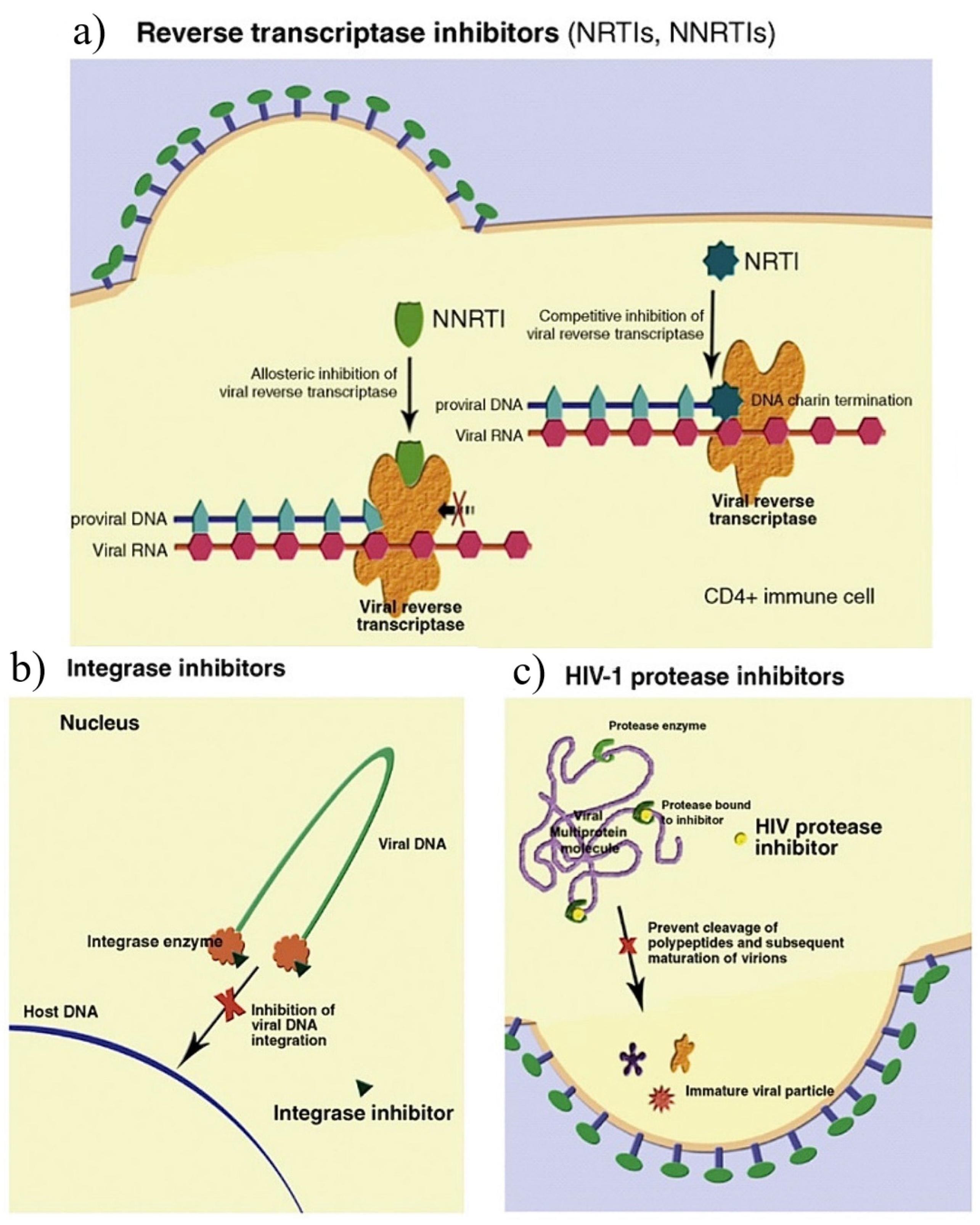
There are five ART drug categories approved by the USA Food and Drug Administration (FDA), which include NRTIs, non-nucleoside reverse transcriptase inhibitors (NNTRIs), integrase strand transfer inhibitors (InSTIs), protease inhibitors (PIs), and entry inhibitors [3]. Each class targets a specific phase of the HIV-1 replication cycle (see Figure 3) [19].
The NRTIs halt the elongation of the proviral deoxyribonucleic acid (DNA) strand by blocking the HIV-1 reverse transcriptase (RT) enzyme responsible for converting viral ribonucleic acid (RNA) into viral DNA [37,38]. The NNRTIs inhibit DNA polymerase activity by inducing a conformational change, disrupting the enzyme’s catalytic site [39]. The HIV integrase enzyme, responsible for integrating viral DNA into the host cell’s DNA, is inhibited by InSTIs [36]. The PIs are responsible for inhibiting HIV-1 protease, which cleaves newly synthesized polyproteins (Gag and Gag-Pol) into a mature infectious virus [40,41].

2.2. The Approved Standard Treatment for HIV

The approved standard treatment for HIV-1 consists of a combination of three drugs from at least two different categories [4]. One of the first NRTIs accepted to treat HIV-1 infection was zidovudine (AZT). Following its acceptance, several other NRTIs, such as tenofovir, was produced and used in combination with NNRTIs or PIs [42]. Until 2018, the first-line regimen for HIV-1 treatment in many countries was the combination of Tenofovir–Lamivudine–Efavirenz (TLE); that is, two NRTIs (Tenofovir–Lamivudine) and one NNRTI (Efavirenz) [43]. However, TLE has a low genetic barrier to drug resistance and causes neuropsychiatric side-effects [44]. Subsequently, in 2019, the South African National Department of Health changed the prescribed standard initial treatment from TLE to Tenofovir–Lamivudine–Dolutegravir (InSTI) (TLD), a fixed-dose combination [45].

2.3. The Types of Antiretroviral Drugs

The backbone for HIV-1 treatment, TLD, targets a specific phase of the HIV-1 replication cycle, aggressively suppresses viral replication, and halts the progression of HIV-1 infection [46]. The TLD treatment is more tolerable and has a high genetic barrier to drug resistance [6].

2.3.1. Tenofovir

Tenofovir is a nucleotide analog of adenosine 5′-monophosphate [47]. It is a highly hydrophilic compound with two negative charges resulting in low intestinal membrane permeability [48]. To improve oral bioavailability and membrane permeability, tenofovir is commercially available as a pro-drug, tenofovir disoproxil fumarate (TDF) [49]. Following oral administration, TDF is rapidly converted to tenofovir in the intestinal walls through esterase hydrolysis [50]. Tenofovir then enters cells through organic anion transporters 1 and 3 [51].
Intracellularly, tenofovir is phosphorylated by adenylate kinases and subsequently phosphorylated by nucleoside diphosphate kinases into its active form, tenofovir diphosphate [52]. Tenofovir diphosphate is an analog of deoxyadenosine-5′-triphosphate (dATP), a common substrate for DNA polymerase. Tenofovir diphosphate terminates the viral DNA chain elongation by competing with dATP to be incorporated into viral DNA [53]. The kidneys excrete Tenofovir through glomerular filtration and tubular secretion [51]. Organic anion transporters in the basolateral membrane actively transport about 20–30% of tenofovir into renal proximal tubule cells. Subsequently, tenofovir is secreted into the tubular lumen by the apical membrane transporters and multidrug-resistance proteins, MRP-4 and MRP-2 (encoded by ABCC4 and ABCC2 genes, respectively) [53].

2.3.2. Lamivudine

Lamivudine (3TC) forms part of the NRTIs; it inhibits viral DNA synthesis via RT DNA chain termination post phosphorylation [37]. 3TC is highly soluble and rapidly absorbed, with a bioavailability ranging from 82 to 86% for oral administration [5]. Intracellularly, 3TC is metabolized to its active triphosphate form, lamivudine triphosphate (3TC-TP), through kinase phosphorylation [54].
The 3TC-TP competes with the corresponding endogenous nucleoside triphosphate, deoxycytidine triphosphate (dCTP), for binding to the viral RT. Once incorporated into the viral DNA, chain termination results due to the absence of a 3′-hydroxy (3′-OH) group to enable the 3′-5′-phosphodiester linkages essential for DNA elongation [55]. The majority of 3TC is eliminated through filtration and active renal tubular secretion. Metabolism is a minor route of elimination, with only 10% of the parent drug metabolized to an inactive trans-sulfoxide metabolite that is excreted in the urine [5].

2.3.3. Dolutegravir

Dolutegravir (DTG), an orally bio-available integrase strand transfer inhibitor, is an efficacious, well-tolerated drug with a high barrier to drug resistance [56]. Upon oral administration, DTG binds and inhibits the active site of integrase, an HIV enzyme that catalyzes the integration of viral DNA into chromosomal DNA, leading to viral replication [57]. DTG is metabolized in the liver by uridine 5′-diphosphate-glucuronosyltransferase (UGT) 1A1 and cytochrome P450 (CYP) 3A4 [58].

2.4. Antiretroviral Drugs’ Side-Effects

The most common side-effects of ART-TLD-based regimens include nausea, diarrhea, hypoglycemia, insomnia, and headaches [59]. Appetite loss, vomiting, diarrhea, and abdominal pain caused by ART use can result in malnutrition [60]. HIV and malnutrition can be detrimental to the immune system, decreasing the number of CD4+ and CD8+ T cells. A compromised immune system will subsequently increase the body’s susceptibility to opportunistic infections, including pneumocystis pneumonia, cryptococcal meningitis, and mycobacterium tuberculosis [60,61]. Opportunistic infections and malnutrition can worsen disease progression and increase HIV-related mortality [60].
Reports from preclinical- and clinical-based studies have also linked ART with hepatotoxicity associated with oxidative stress [62]. Hepatotoxicity is a liver injury or impairment of liver function caused by exposure to xenobiotics such as drugs, alcohol, peroxidized fatty acids, environmental toxicants, and even some medicinal plants [63]. Hepatotoxicity may include hepatitis, granuloma, lactic acidosis, cholestasis, and hepatic steatosis [64]. The TDF regimen has been associated with severe lactic acidosis and hepatic steatosis [49]. The mechanism proposed behind TDF causing the latter complications is the inhibition of mitochondrial DNA (mtDNA) polymerase gamma (γ) [65]. Mitochondrial toxicity can manifest as nephrotoxicity, myopathy, pancreatitis, peripheral neuropathy, lactic acidosis, and hepatic steatosis [65]. Figure 4 illustrates the mechanism of ART-induced cytotoxicity in liver cells.
Figure 4 demonstrates that ARVs such as NRTIs inhibit DNA polymerase γ and lead to decreased mtDNA, loss of mitochondrial membrane potential, and oxidative phosphorylation, consequently precipitating oxidative stress [67]. NRTIs lack a 3′-OH group on their pentose rings, having nucleoside as their base. Due to the missing 3′-OH group, the NRTIs prevent the formation of the 3′-5′-phosphodiester bonds in growing DNA chains and terminate mtDNA chain elongation [68]. As a result, mtDNA copy numbers decrease, as well as mitochondrial-encoded genes, which are essential components of the mitochondrial respiratory chain (MRC) function. This leads to a disrupted electron transport chain and a concomitant reduction in membrane potential and ATP production by the mitochondrion. This destruction in mitochondrial function can result in increased ROS production and changes in mitochondrial morphology [66,69].

2.5. Oxidative Stress

Oxidative stress is an imbalance between the excessive generation of free radicals or reactive oxygen species (ROS) and their eradication by the antioxidant defense system [70]. Free radicals are atoms with an unpaired electron in their outer orbital [71]. Free radicals are unstable and highly reactive; however, they gain stability by attracting electrons from other compounds. The compound loses an electron and becomes a free radical, triggering a chain reaction cascade, ultimately damaging the living cell [72]. The term “reactive oxygen species” refers to any oxygen-containing molecule (radical or non-radical) capable of causing harmful reactions. These include the superoxide anion (O2ˉ), hydrogen peroxide (H2O2), hydroxyl radical (HO•), alkoxyl radical (RO), peroxyl radical (RO2), hydroperoxyl radical (HO2), hypochlorous acid (HOCl), and singlet oxygen (O2) [73].
During the oxidative stress reaction (illustrated below), the formation of superoxide results from the one-electron reduction of O2, the disproportionation of two superoxide molecules yields H2O2 and O2, and the oxidation of ferric iron by H2O2 yields HO• and the hydroxide anion [73]:
O2 + e → O2 (Superoxide)
2O2 + 2H+ → O2 + H2O2 (Hydrogen peroxide)
Fe2 + H2O2 → Fe3 + HO• + OH (Hydroxyl radical)
As illustrated in Figure 5, free radicals/ROS are formed due to adenosine triphosphate (ATP) production by the mitochondria when cells utilize oxygen to produce energy [74].
In addition to mitochondria, ROS are produced by various enzymes, such as NADPH oxidases and xanthine oxidase (XO) [75]. The sum of ROS produced by metabolic processes can be induced by environmental stimuli in the form of various stresses, including pollution, tobacco smoke, alcohol, transition metals, heavy metals, pesticides, industrial solvents, drugs such as ARVs, paracetamol, halothane, and radiation among others [76].
ROS include not only O2ˉ, H2O2, and HO•, but also a group of nitrogen-containing molecules called reactive nitrogen species (RNS) [77]. The nitroxyl anion, nitrosonium cation, higher oxides of nitrogen, S-nitrosothiols, and dinitrosyl iron complexes are all examples of RNS [78]. Another prominent effect of ROS is lipid peroxidation, which occurs when membrane phospholipids are brought into contact with an ROS oxidizing agent [75]. Lipid peroxidation is a process in which free radical species remove electrons from lipids. Subsequently, the lipids become reactive free radicals that can propagate lipid peroxidation chain reactions [79]. Lipid peroxidation forms several oxidation products, including lipid hydroperoxides (LOOH) and aldehydes such as malondialdehyde (MDA) and 4-hydroxynonenal (4-HNE) [80].
Among the aldehydes produced through lipid peroxidation, MDA has gained the most attention, given that MDA is made at high levels during lipid peroxidation and is commonly used as a measure of oxidative stress [81]. MDA is widely used as a biomarker for lipid peroxidation because of its ready reaction with thiobarbituric acid [82]. Uncontrolled free radicals/ROS production occurs when antioxidants (e.g., glutathione, superoxide dismutase, catalase, and vitamins) are saturated due to aging, stress, physical damage, or pathological disease [83].

2.6. Antioxidants

Antioxidants are compounds that scavenge free radicals/ROS and intracellularly retain a more reduced redox state [84]. The antioxidant defense system comprises endogenous and exogenous antioxidants [85]. Endogenous antioxidants produced by the body are divided into enzymatic and non-enzymatic antioxidants [86]. Enzymatic antioxidants include superoxide dismutases (SODs), catalases (CATs), and glutathione peroxidases (GPxs). Non-enzymatic antioxidants include polyamides, linolenic acid, bilirubin, albumin, uric acid, glutathione, transferrin, ceruloplasmin, and coenzyme Q10 [85]. Exogenous antioxidants are vitamins A, C, and E, selenium (Se), carotenoids, and polyphenols [87].
Antioxidants can directly decrease oxidative damage by accepting or donating an electron to eliminate the unpaired condition of the radical [88]. Antioxidants can also indirectly reduce free radicals by inhibiting the activity or expression of free-radical-generating enzymes. Examples of free-radical-generating enzymes are NADPH oxidase and XO [88]. Another function associated with antioxidants is increasing the activity or expression of intracellular enzymatic antioxidants such as SOD, CAT, and GPx [88]. SOD, CAT, and GPx are the first-line antioxidant enzymes that suppress or prevent the formation of free radicals/ROS. These enzymes are known to neutralize any molecule with the potential of developing into a free radical or any free radical with the potential to induce the production of other radicals [89]. The expression of SOD, CAT, and GPx is regulated by the non-coding DNA sequence antioxidant response element (ARE). ARE is activated by nuclear-factor-erythroid-2-related factor 2 (Nrf2) [90].

2.6.1. Superoxide Dismutase

Superoxide dismutases (SODs) are a group of metalloenzymes that decrease O2 levels by catalyzing the dismutation of the O2 free radical into molecular oxygen and H2O2 (see Figure 6) [91]. They require a metal cofactor for their antioxidant activity. In this regard, SODs are classified into three forms, including iron SOD (Fe-SOD), manganese-dependent SOD (Mn-SOD), and copper-zinc-SOD (Cu/Zn-SOD) [89]. Superoxide dismutase 2 (SOD2) is an Mn-SOD. The SOD2 gene mapping encodes it to chromosome 6, which is present in prokaryotes and mitochondria of eukaryotes [89]. The SOD2 is a vital component of the antioxidant defense system against mitochondrial superoxide radicals [92].

2.6.2. Catalase

Catalase (CAT) is a homotetramer in which each monomer (62.5 kDa) contains a haem B (also known as protoheme IX) group responsible for the enzymatic activity [93]. The CAT participates in the dismutation of H2O2 to oxygen and water in a two-step reaction [94]. The first step of the reaction involves the formation of compound I, the product of the reaction of H2O2 with catalase haem (Reaction (1)). Subsequently, compound I is decomposed upon reaction with a second H2O2 molecule in the catalytic reaction, releasing oxygen and water (Reaction (2)) [95]:
Compound I: catalase−Fe3+ + H2O2 → catalase−FeO3+ + H2O
Catalytic reaction: catalase−FeO3+ + H2O2 → catalase−Fe3+ + O2 + H2O
In sum: 2H2O2 → O2 + 2H2O

2.6.3. Glutathione

Glutathione (GSH) is a thiol tripeptide (γ-glutamyl cysteinyl glycine) comprising three amino acids (glutamic acid, cysteine, and glycine) [96]. The synthesis of GSH from cysteine, glutamate, and glycine is catalyzed sequentially by two cytosolic enzymes, glutamate cysteine ligase (GCL) and GSH synthetase [97]. GCL catalyzes the first of two ATP-dependent steps in GSH synthesis, the formation of γ-glutamylcysteine (γ-GC) from glutamate and cysteine. The second step is catalyzed by GSH synthetase, which joins glycine to γ-GC, thus forming GSH [98].
GCL consists of two separately coded proteins, a catalytic subunit (GCLC) and a modifier subunit (GCLM) [99]. It has been shown that GSH production is paralleled with GCLC gene expression, which is primarily regulated at the transcription level. The GCLC gene is shown to have oxidative-stress-responsive elements in the promoter/enhancer region. Several cis-acting DNA elements contribute to the transcriptional up-regulation of the GCLC gene in response to oxidative stress, providing a protective mechanism against oxidative stress [100].
In all mammalian tissues, GSH is the most abundant non-protein thiol that protects against oxidative stress [101]. The antioxidant function of GSH is primarily accomplished through GSH peroxidase-1 (GPx-1)-catalyzed reactions, reducing H2O2 to water and lipid peroxides to their corresponding alcohols, mainly in the mitochondria and cytosol [89]. The primary reaction that GPx-1 (selenocysteine (Sec)-containing enzyme) catalyzes is 2GSH + H2O2 → GSSG + 2H2O (see Figure 7) [102].
Reducing equivalents come from two equivalents of GSH, which are oxidized to glutathione disulfide (GSSG). For the maintenance of free radical detoxification in a cell, GSSG needs to be converted to GSH by the glutathione reductase (GR) enzyme using nicotinamide adenine dinucleotide phosphate (NADPH) (see Figure 8) [103].

2.6.4. Nuclear-Factor-Erythroid-2-Related Factor 2

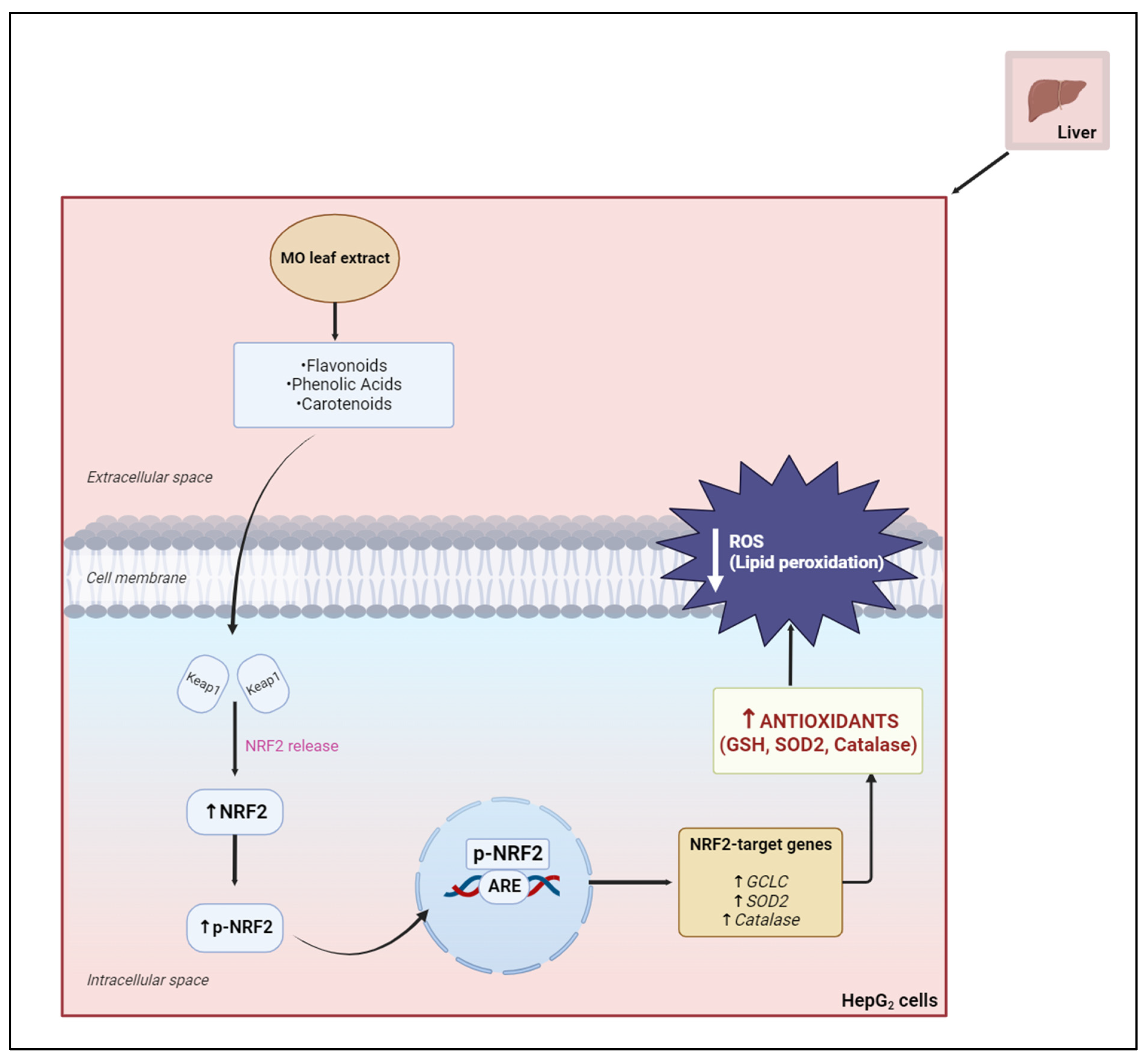
The expression of most antioxidant enzymes is tightly regulated by the antioxidant response element (ARE), a non-coding DNA sequence activated by nuclear-factor-erythroid-2-related factor 2 (Nrf2) [90]. The Nrf2, a nucleus transcription factor bound to its inhibitor, Kelch-like ECH-associated protein (Keap1), is a vital transcription factor regulating cellular redox homeostasis [104]. Following exposure to oxidants (see Figure 9), Nrf2 is dissociated from Keap1 through oxidation of the cysteine residues of the Nrf2–Keap1 complex [105]. Nrf2 translocates to the nucleus and binds to ARE in the upstream regulatory regions of genes, encoding for detoxification and antioxidant enzymes, thereby leading to enhanced transcription [106].

2.7. The Use of Traditional African Medicinal Plants

An estimated 72% of the Black South African population uses traditional medicines for primary healthcare needs [108]. This can be ascribed to several factors, including easy access to medicinal plants, low costs, and extensive knowledge and expertise within the local communities [109]. There are about ten prominently used South African medicinal plants, which include Aspalathus linearis (Fabaceae), Agathosma betulina (Rutaceae), Aloe ferox (Asphodelaceae), and Hypoxis hemerocallidea (African potato) [110]. Each medicinal plant contains a wide range of diverse bioactive compounds and high levels of phytochemicals, which act as natural antimicrobial, anticancer, antispasmodic, antipyretic, antioxidant, and antiviral agents in the human body [7,111]. South African medicinal plants have also shown efficacy in treating hypertension, heartburn, arthritis, rheumatism, type 2 diabetes mellitus, gastrointestinal disturbances, menstrual difficulties, headache, heartburn, and gout [110]. PLHIV frequently uses traditional African medicines (ATMs) with Western medications, including ARVs [35]. The ATMs may diminish the side-effects of ARVs and their cytotoxicity and promote treatment adherence [35]. However, research studies evaluating the potential herb–drug interactions in a clinical setting are still warranted.
Medicinal plants that traditional medicine practitioners most extensively use in Sub-Saharan Africa for adjuvant HIV/AIDS treatment and related disorders are Hypoxis hemerocallidea (African potato), Sutherlandia frutescens (Cancer bush) [112], and MO [7]. In SA, the African potato, together with the cancer bush, is considered one of the two most popular medicinal plants used to boost the immune system of HIV patients [113,114]. Despite the popularity of their use and the support of the Ministries of Health and NGOs in certain African nations, no clinical trials are investigating the efficacy and limited evidence of harm for the potential of drug interactions with antiretroviral drugs [113]. MO is considered a highly nutritive tree in many parts of Africa and Asian countries [115]. The various health benefits of MO, particularly its leaves, are well researched, documented, and confirmed in several studies. MO’s most well-studied and exploited uses are medicinal and nutritional [115].
A Nigerian study by Gambo and colleagues in 2021 showed that MO leaf powder supplementation increased the CD4+ T cell count of PLHIV on ART (TLE) [60]. This can be attributed to MO’s nutraceutical benefits [60]. A study by Monera-Penduka and colleagues in 2017 showed that MO was well tolerated when taken with nevirapine by HIV patients. MO inhibits the CYP3A4 enzyme, which is responsible for metabolizing nevirapine. However, the safety profile of nevirapine was not altered when co-administered with MO [116]. The MO tree extracts have also shown inhibitory activity, specifically against HIV-1, Herpes Simplex Virus (HSV), and Hepatitis B Virus (HBV), which damages the liver by causing inflammation, cirrhosis, and liver cancer [7]. However, there is limited information about the use of MO amongst HIV/AIDS patients receiving TLD in SA and its effects on patient compliance and outcomes.
Moringa oleifera is a medicinal tree from the family of Moringaceae, commonly found in Asia and Africa, including Nigeria, Namibia, Ghana, Senegal, and SA [12,60,117]. In SA, MO is farmed in several provinces, including Gauteng, Limpopo, Mpumalanga, and KwaZulu-Natal [115]. The MO tree is known for its anthelmintic, antiseptic, detergent, anti-ulcerogenic, anti-inflammatory, anti-microbial, antioxidant, anti-hyperglycaemic, anti-clastogenic, anticancer, and anti-fibrotic effects [8]. For centuries, many cultures worldwide have used MO to treat skin infections, blackheads, anxiety, anemia, asthma, bronchitis, catarrh, chest congestion, cholera, and many other illnesses [118].
Ayurvedic, the traditional Indian system of medicine, is an ancient yet living tradition that is equal to conventional Western medicine and traditional Chinese medicine [119]. It is based on drug discovery, whereby therapeutically active ingredients are first identified based on ethnic uses and then verified through clinical trials. It is a holistic healing system based on over 7000 plants and about 8000 remedies, all of which have been documented [120]. The traditional Ayurvedic system of medicine shows that MO can prevent approximately 300 diseases, and its leaves have been exploited for preventive and curative purposes [121].

2.8. Moringa oleifera

Moringa oleifera, commonly known as the ‘Drumstick’ or horseradish tree, is a small, soft-wooded deciduous tree with sparse foliage cover (see Figure 10) [122]. MO is a fast-growing, highly drought-tolerant, and multi-purpose tree. It is usually 5–10 m tall but can grow up to 15 m [123]. MO belongs to the monogeneric family of shrubs and trees, Moringaceae, considered to have its origin in Agra and Oudh, in the northwest region of India and south of the Himalayan Mountains. It is now cultivated throughout the Middle East, in almost the whole tropical belt, and it was introduced in Eastern Africa from India at the beginning of the 20th century [124]. The tree has been scientifically classified accordingly into the Plantae kingdom, Magnoliophyta division, Magnoliopsida class, Brassicales order, Moringaceae family, Moringa genus, and oleifera species [125]. This plant is widely used as a nutritional herb and contains valuable pharmacological actions like antiasthmatic, antidiabetic, hepatoprotective, anti-inflammatory, anticancer, and antioxidant [126].
MO trees excel mainly in tropical and sub-tropical regions and are known to thrive in a wide range of soil types, mostly heavy clay and waterlogged, with a pH between 4.5 and 8, at an altitude of up to 2000 m [121,124]. It thrives in dry to moist tropical or subtropical climes, with an annual precipitation of 760 to 2500 mm [121]. Furthermore, among all climatic factors that affect plant growth, temperature is one of the most important factors governing natural geographical plant distribution, tree performance, physiology, and productivity [124]. The tree requires between 25 and 35 °C to have optimal growth and a high production of pods and leaves, resulting in its most cost-effective cultivation [127].
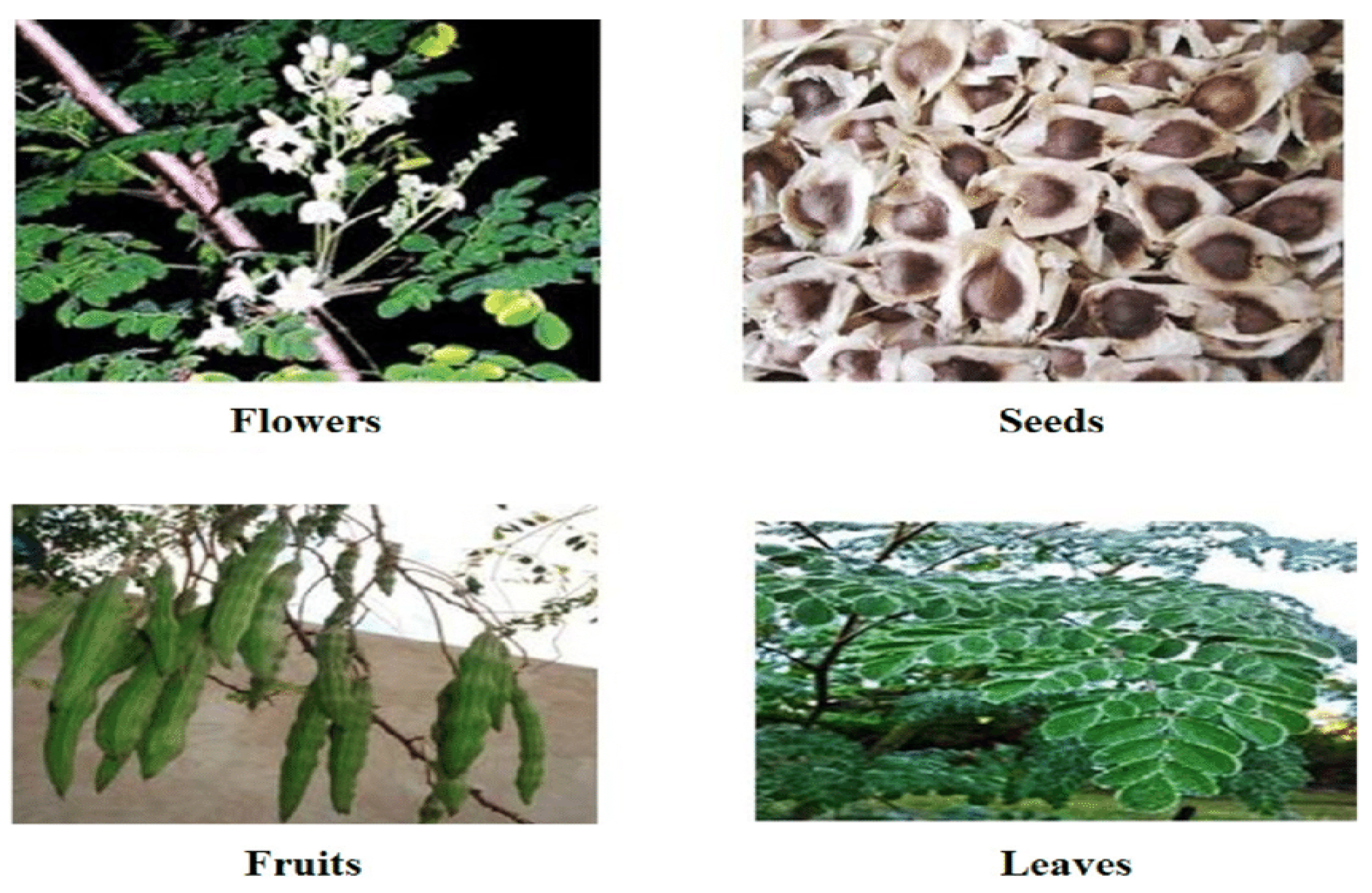
Various parts of the tree (Figure 11) consist of numerous bioactive components, including vitamins, polyphenols, isothiocyanates, tannins, and saponins [12]. The roots, bark, gum, leaf, flowers, fruit (pods), seed, and seed oil of the MO tree have various biological activities that protect against gastric ulceration and hypertension, in addition to anti-diabetic and anti-inflammatory effects [10].
The leaves are the most used part of the tree for nutritious and medicinal purposes. The leaves contain the most extensive amounts of vitamins C and A; flavonoids, including myricetin, quercetin, rutin, and phenolic acids; and carotenoids, such as lutein, β-carotene, and zeaxanthin [13]. The high content of bioactive compounds provides several health advantages, including antidiabetic, anticancer, anti-inflammatory, and antioxidant properties. The antioxidant effect of MO leaves is mostly attributed to flavonoids, phenolic acids, and carotenoids [128]. MO leaves predominantly contain quercetin (43.75%) and equal percentages (18.75%) of other flavonoids. The concentration of flavonoids varies with the environmental conditions. MO leaves harvested in South Africa and Namibia had 17 distinct flavonoids, with quercetin (35%), kaempferol (35%), isorhamnetin (24%), and apigenin (6%) derivatives, but leaves harvested in sub-Saharan Africa contained just 12 different flavonoids [129]. MO leaves contain gallic acid as their major phenolic acid. Ferulic acid, ellagic acid, caffeic acid, o-coumaric acid, and chlorogenic acid are also detected in the leaves [9]. Gallic acid is the most abundant, with a concentration of 1.034 mg/g of dry weight [12]. The leaves have abundant carotenoids with a total amount varying from 44.30 to 80.48 mg/100 g on a fresh weight basis among eight different cultivars [129].

2.8.1. Flavonoids

Flavonoids are polyphenolic phytochemicals found in fruits, vegetables, and grains [130]. Intake of flavonoids has been shown to protect against chronic diseases associated with oxidative stress. The main flavonoids (see Figure 12) found in MO leaves are myricetin, quercetin, and kaempferol, in concentrations of 5.8, 0.207, and 7.57 mg/g, respectively [12]. Flavonoids are a group of diphenyl propane compounds (C6-C3-C6). They have the general structure of a 15-carbon skeleton composed of two phenyl rings (A and B), linked through a heterocyclic pyran or pyrone ring in the middle [131].
The potent antioxidant activity exhibited by flavonoids in vitro is primarily due to their ability to trap free radicals via the metal chelation and donation of electrons or hydrogen atoms (see Figure 13) [131]. Flavonoids are oxidized by free radicals, resulting in a more stable, less-reactive radical [132].

2.8.2. Phenolic Acids

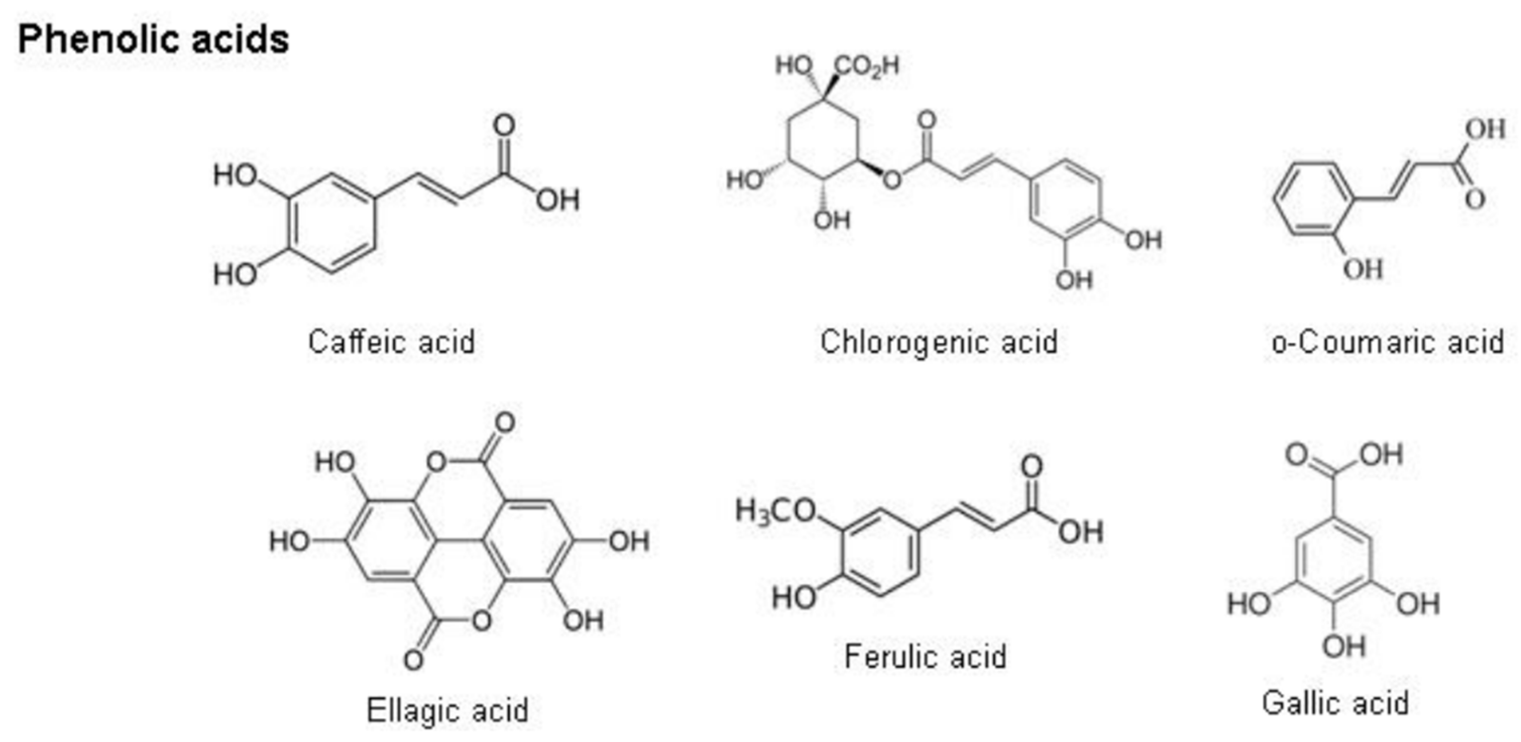
Phenolic acids are a sub-group of phenolic compounds derived from hydroxybenzoic acid and hydroxycinnamic acid, naturally present in plants [121]. Caffeic acid, ellagic acid, ferulic acid, and chlorogenic acid are found in the phenolic acid group (Figure 14). Phenolic acids are distributed ubiquitously in plants and play a significant protective role in oxidative stress conditions [133].
Phenolic acids possess antioxidant activity due to their chemical nature: hydroxyl groups attached to the pentyl ring. They stabilize free radicals by donating a hydroxyl group, forming a delocalized and stabilized unpaired electron, a phenoxy radical, across the phenolic ring (see Figure 15). The degree of antioxidant activity is determined by the position and number of the phenolic hydroxyl groups [134].

2.8.3. Carotenoids

Carotenoids are tetraterpene pigments, which exhibit yellow, orange, red, and purple colors [136]. Plants and some microorganisms such as bacteria, fungi, and yeasts produce these pigments. Among the carotenoids, the β-carotene is the most abundant in foods that have the highest activity of provitamin A [137]. Most carotenoids (Figure 16) consist of eight isoprene units with a 40-carbon skeleton. Their general structures commonly consist of a polyene chain with nine conjugated double bonds and an end group at both ends of the polyene chain [136].
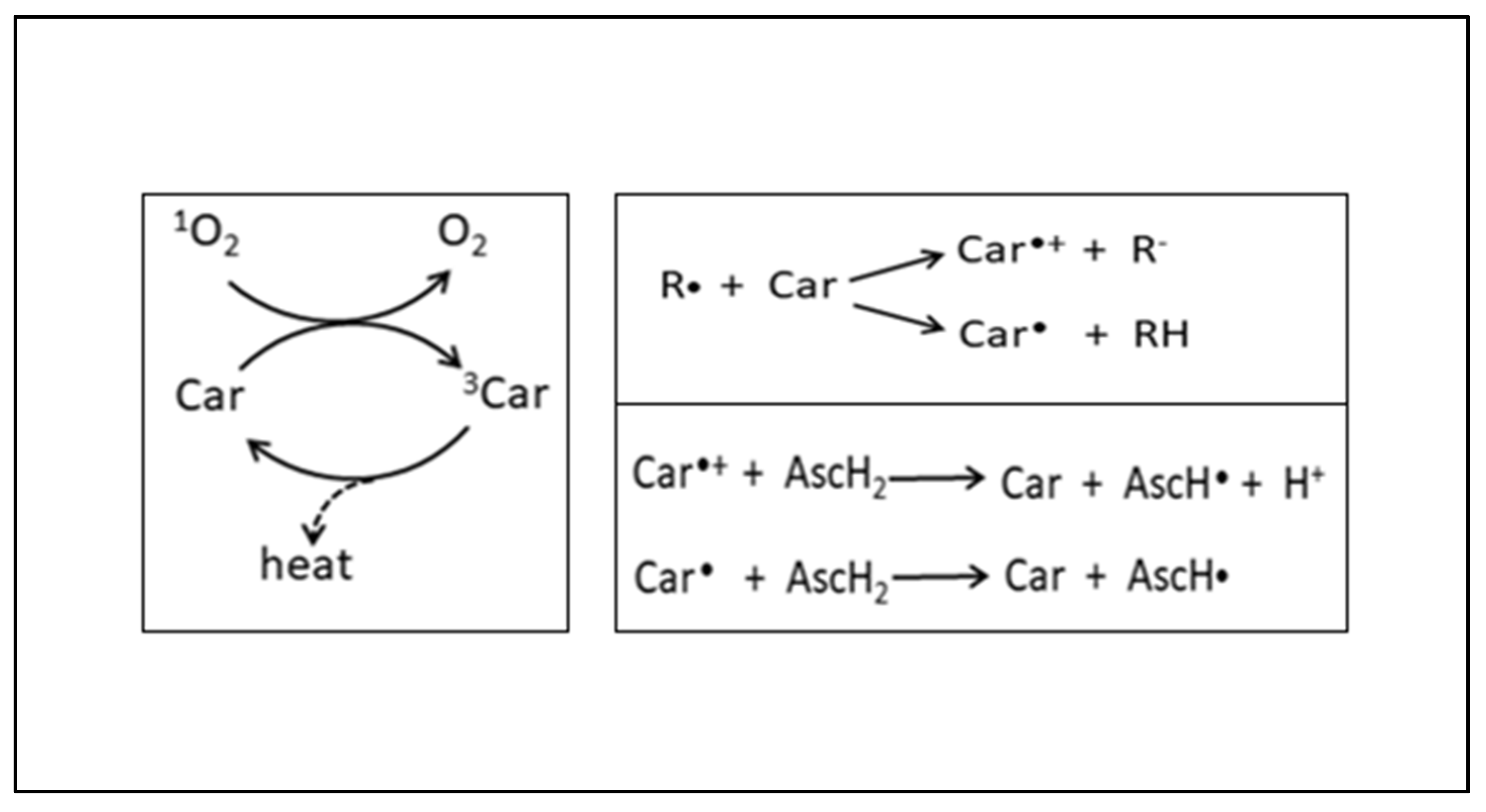
Carotenoids are well known for their ability to physically and chemically quench oxygen. They scavenge ROS and play a protective role in various ROS-mediated disorders, such as multiple forms of cancer, cardiovascular diseases, and neurological and eye-related disorders [138]. Carotenoids quench singlet oxygen, remove peroxy radicals, modulate carcinogen metabolism, inhibit cell proliferation, stimulate communication between cells, and increase the immune response (Figure 17) [137].

2.9. The Hepatoprotective Effects of Moringa oleifera against Oxidative Stress

The MO aqueous leaf extracts contain large amounts of bioactive compounds or high levels of phytochemicals such as flavonoids, phenolic acids, and carotenoids, which act synergistically to induce their medicinal effects [140]. Daily consumption of MO is recommended. However, an overdose of MO may cause a high accumulation of iron. High iron can cause gastrointestinal distress and hemochromatosis. Hence, a daily dose of 70 g of MO is suggested to be suitable and prevents the over-accumulation of nutrients [141]. In vivo, the aqueous leaf extract, a rich source of antioxidant compounds, is responsible for protecting against oxidative-stress-induced diseases [126]. In vitro, studies have reported MO’s renal and hepatoprotective properties against several drugs, such as gentamicin, pyrazinamide, rifampicin, and acetaminophen, mainly attributable to its leaves [142]. To determine the potency of MO as a protective agent in vitro, a half-maximal inhibition concentration (IC50) will have to be determined. Scientists often use the HepG2 cell line as an in vitro model system that mimics the natural in vivo environment to assess hepatoxicity and the protective effects of aqueous leaf extracts (Figure 18) [143,144].

2.10. The Use of Human HepG₂ Liver Cells In Vitro

Currently, drugs used to treat several viral infections, such as HBV, HSV, or HIV, display consistent side-effects, including mitochondrial toxicity [145]. Several in vitro models and techniques have been developed to analyze the impact of such drugs. The HepG2 cells (derived from the human hepatoma) are an excellent model to investigate mitochondrial toxicity due to their high organelle content and mtDNA. They are extensively used by several investigators [145]. The HepG2 cell line was established from a liver tumor biopsy obtained from a 15-year-old Caucasian male in the 1970s [11]. It is the most frequently used hepatoma cell line in the testing and research on drug-induced liver injury [11].
Several scientists have researched ART using the HepG2 cell line. In 2017, Paemanne and colleagues used the HepG2 cell line to investigate the effect of the nevirapine (NVP) regimen on mitochondrial dysfunction. The study results showed that NVP induces mitochondrial dysregulation in HepG2 cells [146]. Shamsabadi, 2014 investigated the hepatotoxic effects of the components of Atripla and Eviplera on HepG2 cells. The results showed that NRTIs, tenofovir, and emtricitabine had no hepatotoxic effects in vitro compared to the NNRTIs, efavirenz, and rilpivirine [147]. A research study by Nagiah and colleagues in 2015 has also established the appropriate application of this cell line as an in vitro model to evaluate drug metabolism or the toxicity of antiretroviral drugs, including tenofovir. Further research studies that take on a similar approach to assess tenofovir-induced cytotoxicity and incorporate MO treatment to evaluate the hepatoprotective effects will be beneficial.

3. Conclusions

To date, various biological activities of different parts of the MO plant have been reported. However, no in vitro studies have assessed the hepatoprotective effects of MO aqueous leaf extract against ART-induced cytotoxicity in liver cells. Therefore, further research is warranted to investigate the effect of MO aqueous leaf extract on oxidative stress and antioxidants.

4. Value of This Study

This study is of scientific, clinical, and public value. It contributes to the scientific knowledge by determining the extent to which the MO tree can commonly be used for medicinal purposes and its potential to minimize tenofovir-induced oxidative stress in human HepG2 liver cells. To achieve this, the use of various experimental techniques such as spectrophotometry, luminometry, ELISA, Western blotting, and qPCR is employed. Societal disparities that contribute to inadequate access to basic healthcare and the occurrence of malnutrition for a majority of low-income households result in the use of the MO tree. The use of the MO tree for the provision of medicine, food, as well as skin and hair supplements may serve as a promising alternative, more so for people residing in the rural areas of SA. HIV-infected people often use MO with ART in an attempt to mitigate side-effects, boost their immune system, and improve their health. However, using medicinal plants with drugs, including ART, mainly in Africa and other developing countries, is not well researched and thus poorly regulated. Therefore, more studies evaluating the efficacy of medicinal plants in relation to reducing the cytotoxicity associated with ART use are needed. Thus, results from this study will provide insight and broaden our understanding of how the bioactive compounds present in MO aqueous leaf extract may have a hepatoprotective effect against tenofovir-induced oxidative stress in human HepG2 liver cells.

Funding

This research study is funded by the University of the Free State Faculty Research Funding, the University of the Free State Master’s bursary, and the National Research Foundation (NRF). The application reference number for NRF is MND210524603414.

Acknowledgments

I would like to express my gratitude to my supervisor and co-supervisors, Charlette Tiloke, Helena De Villiers, and Claudia Ntsapi, for their guidance and support throughout this review. I would also like to acknowledge the University of the Free State, the Faculty of Health Sciences, and the Department of Basic Medical Sciences for evaluating this research study and providing valuable and constructive suggestions during the planning and development of this research work.

Conflicts of Interest

The authors declare that there are no conflict of interest.

Abbreviations

AIDS: Acquired immunodeficiency syndrome; ARE, Antioxdiant response element; ART, Antiretroviral therapy; ATP, Adenosine triphosphate; ATMs, Traditional African medicines, AZT, Zidovudine; CAT, Catalase; Cu/Zn-SOD, Copper-zinc-SOD; CYP, cytochrome P450; dATP, deoxyadenosine-5′-triphosphate; dCTP, deoxycytidine triphosphate; DNA, deoxyribonucleic acid; DTG, Dolutegravir; FDA, USA Food and Drug Administration; Fe-SOD, Iron SOD; GCL, Glutamate cysteine ligase; GCLC, Glutamate cysteine ligase catalytic subunit; GCLM, Glutamate cysteine ligase modifier subunit; GPx, Glutathione peroxidase; GSH, Glutathione; GSSG, Glutathione disulfide; HIV, Human immunodeficiency virus; H2O2, Hydrogen peroxide; HO2, hydroperoxyl radical; HO•, hydroxyl radical; HOCl, hypochlorus acid; InSTI, integrase strand transfer inhibitors; LOOH, lipid hydroperoxides; MDA, Malondialdehyde; mDNA, mitochondrial DNA; MO, Moringa oleifera; Mn-SOD, Manganese-dependent SOD; NADPH, Nicotinamide adenine dinucleotide phosphate; Nrf2, Nuclear-factor-erythroid-2-related factor 2; NNRTIs, Non-nucleoside reverse transcriptase inhibitors; NRTIs, Nucleoside reverse transcriptase inhibitors; O2, singlet oxygen; O2ˉ, anion; PLHIV, People living with HIV/AIDS; PIs, protease inhibitors; RNA, Ribonucleic acid; RNS, Reactive nitrogen species; RO, alkoxyl radical; RO2, peroxyl radical; ROS, Reactive oxygen species; Se, Selenium; SOD, Superoxide dismutase; TDF, Tenofovir disoproxil fumarate; TLD, Tenofovir–Lamivudine–dolutegravir; TLE, Tenofovir–lamivudine–efavirenz; UGT, 5′-diphosphate-glucuronosyltransferase; UNAIDS, Joint United Nations Programme on HIV/AIDS; UTT, Universal test and treat; XO, Xanthine oxidase; 3′-OH, 3′-hydroxy; 3TC, Lamivudine; 3TC-TP, lamivudine triphosphate; 4-HNE, 4-hydroxynonenal γ, gamma; γ-GC, γ-glutamylcysteine.

References

  1. Pillaye, J.N.; Marakalala, M.J.; Khumalo, N.; Spearman, W.; Ndlovu, H. Mechanistic insights into antiretroviral drug-induced liver injury. Pharmacol. Res. Perspect. 2020, 8, e00598. [Google Scholar] [CrossRef] [PubMed]
  2. Eilami, O.; Nazari, A.; Dousti, M.; Sayehmiri, F.; Ghasemi, M. Investigation of HIV/AIDS prevalence and associated risk factors among female sex workers from 2010 to 2017: A meta-analysis study. HIV/AIDS Res. Palliat. Care 2019, 11, 105–117. [Google Scholar] [CrossRef] [PubMed]
  3. Spach, D.H. Antiretroviral Medications and Initial Therapy HIV Life Cycle and Antiretroviral Drug Targets; National HIV Curriculum, University of Washington: Seattle, WA, USA, 2020. [Google Scholar]
  4. Waters, L.; Mehta, V.; Gogtay, J.; Boffito, M. The Evidence for Using Tenofovir Disoproxil Fumarate Plus Lamivudine as a Nucleoside Analogue Backbone for the Treatment of HIV. J. Virus Erad. 2021, 7, 100028. [Google Scholar] [CrossRef] [PubMed]
  5. Dumitrescu, T.P.; Peddiraju, K.; Fu, C.; Bakshi, K.; Yu, S.; Zhang, Z.; Tenorio, A.R.; Spancake, C.; Joshi, S.; Wolstenholme, A.; et al. Bioequivalence and Food Effect Assessment of 2 Fixed-Dose Combination Formulations of Dolutegravir and Lamivudine. Clin. Pharmacol. Drug Dev. 2020, 9, 189–202. [Google Scholar] [CrossRef]
  6. Umar, D.; Waziri, B.; Ndagi, U.; Mohammed, S.; Usman, N.; Abubakar-Muhammad, H. Impact of Tenofovir/Lamivudine/Dolutegravir (Tld) on the Health-Related Quality of Life and Clinical Outcomes of HIV/AIDS Patients at a Tertiary Health Facility in Niger State. 2020, pp. 1–18. Available online: https://www.researchsquare.com/article/rs-127277/v1 (accessed on 21 February 2021).
  7. Biswas, D.; Nandy, S.; Mukherjee, A.; Pandey, D.K.; Dey, A. Moringa oleifera Lam. and derived phytochemicals as promising antiviral agents: A review. S. Afr. J. Bot. 2020, 129, 272–282. [Google Scholar] [CrossRef]
  8. Salami, A.T.; Okonkwo, C.E.; Attah, F.A.; Olagoke, O.C. Bioactive Moringa olifera seed extracts attenuates cholesterol gall stones in hyperglycaemic Swiss mice. Comp. Clin. Pathol. 2021, 30, 207–216. [Google Scholar] [CrossRef]
  9. Rani, N.Z.A.; Husain, K.; Kumolosasi, E. Moringa genus: A review of phytochemistry and pharmacology. Front. Pharmacol. 2018, 9, 108. [Google Scholar] [CrossRef]
  10. Stohs, S.J.; Hartman, M.J. Review of the safety and efficacy of Moringa oleifera. Phytother. Res. 2015, 29, 796–804. [Google Scholar] [CrossRef] [PubMed]
  11. Ren, Z.; Chen, S.; Guo, L. Chapter 8 Use of Liver-Derived Cell Lines for the Study. In Drug-Induced Liver Toxicity; Humana: New York, NY, USA, 2018. [Google Scholar]
  12. Vergara-Jimenez, M.; Almatrafi, M.M.; Fernandez, M.L. Bioactive components in Moringa oleifera leaves protect against chronic disease. Antioxidants 2017, 6, 91. [Google Scholar] [CrossRef]
  13. Nizioł-ŁUkaszewska, Z.; Furman-Toczek, D.; Bujak, T.; Wasilewski, T.; Hordyjewicz-Baran, Z. Moringa oleifera L. Extracts as Bioactive Ingredients That Increase Safety of Body Wash Cosmetics. Dermatol. Res. Pract. 2020, 2020, 8197902. [Google Scholar] [CrossRef]
  14. Sharp, P.M.; Hahn, B.H. The evolution of HIV-1 and the origin of AIDS. Philos. Trans. R. Soc. B Biol. Sci. 2010, 365, 2487–2494. [Google Scholar] [CrossRef]
  15. Heeney, J.L.; Dalgleish, A.G.; Weiss, R.A. Origins of HIV and the evolution of resistance to AIDS. Science 2006, 313, 462–466. [Google Scholar] [CrossRef] [PubMed]
  16. Van Heuverswyn, F.; Peeters, M. The origins of HIV and implications for the global epidemic. Curr. Infect. Dis. Rep. 2007, 9, 338–346. [Google Scholar] [CrossRef] [PubMed]
  17. De Cock, K.M.; Jaffe, H.W.; Curran, J.W. The evolving epidemiology of HIV/AIDS. Aids 2012, 26, 1205–1213. [Google Scholar] [CrossRef] [PubMed]
  18. Delpech, V.; Gahagan, J. The global epidemiology of HIV. Medicine 2009, 37, 317–320. [Google Scholar] [CrossRef]
  19. Ncube, S.; Madikizela, L.M.; Chimuka, L.; Nindi, M.M. Environmental fate and ecotoxicological effects of antiretrovirals: A current global status and future perspectives. Water Res. 2018, 145, 231–247. [Google Scholar] [CrossRef] [PubMed]
  20. Nair, S.; Bayer, W.; Ploquin, M.J.Y.; Kassiotis, G.; Hasenkrug, K.J.; Dittmer, U. Distinct roles of CD4+ T cell subpopulations in retroviral immunity: Lessons from the Friend virus mouse model. Retrovirology 2011, 8, 76. [Google Scholar] [CrossRef] [PubMed]
  21. Swain, S.L.; McKinstry, K.K.; Strutt, T.M. Expanding roles for CD4+ T cells in immunity to viruses. Nat. Rev. Immunol. 2012, 12, 136–148. [Google Scholar] [CrossRef]
  22. Aavani, P.; Allen, L.J.S. The role of CD4 T cells in immune system activation and viral reproduction in a simple model for HIV infection. Appl. Math. Model. 2019, 75, 210–222. [Google Scholar] [CrossRef]
  23. Kirchhoff, F. HIV life cycle: Overview. Encycl. AIDS 2013, 1–9. [Google Scholar] [CrossRef]
  24. Pillay, Y.; Johnson, L. World AIDS day 2020: Reflections on global and South African progress and continuing challenges. S. Afr. J. HIV Med. 2021, 22, 1205. [Google Scholar] [CrossRef] [PubMed]
  25. Heyer, A.; Ogunbanjo, G.A. Adherence to HIV antiretroviral therapy. Part I: A review of factors that influence adherence. S. Afr. Fam. Pract. 2006, 48, 5–9. [Google Scholar] [CrossRef]
  26. Orton, P.M.; Sokhela, D.G.; Nokes, K.M.; Perazzo, J.D.; Webel, A.R. Factors related to functional exercise capacity amongst people with HIV in Durban, South Africa. Health SA Gesondheid 2021, 26, a1532. [Google Scholar] [CrossRef] [PubMed]
  27. Ramlagan, S.; Matseke, G.; Rodriguez, V.J.; Jones, D.L.; Peltzer, K.; Ruiter, R.A.C.; Sifunda, S. Determinants of disclosure and non-disclosure of HIV-positive status, by pregnant women in rural South Africa. Sahara J. 2018, 15, 155–163. [Google Scholar] [CrossRef] [PubMed]
  28. Mabaso, M.; Makola, L.; Naidoo, I.; Mlangeni, L.L.; Jooste, S.; Simbayi, L. HIV prevalence in South Africa through gender and racial lenses: Results from the 2012 population-based national household survey. Int. J. Equity Health 2019, 18, 167. [Google Scholar] [CrossRef] [PubMed]
  29. Loeliger, K.B.; Niccolai, L.M.; Mtungwa, L.N.; Moll, A.; Shenoi, S.V. “I have to push him with a wheelbarrow to the clinic”: Community health workers’ roles, needs, and strategies to improve HIV care in rural South Africa. AIDS Patient Care STDS 2016, 30, 385–394. [Google Scholar] [CrossRef] [PubMed]
  30. Bessong, P.O.; Matume, N.D.; Tebit, D.M. Potential challenges to sustained viral load suppression in the HIV treatment programme in South Africa: A narrative overview. AIDS Res. Ther. 2021, 18, 1. [Google Scholar] [CrossRef]
  31. Torti, C.; Prosperi, M.; Motta, D.; Digiambenedetto, S.; Maggiolo, F.; Paraninfo, G.; Ripamonti, D.; Cologni, G.; Fabbiani, M.; Caputo, S.L.; et al. Factors influencing the normalization of CD4+ T-cell count, percentage and CD4+/CD8+ T-cell ratio in HIV-infected patients on long-term suppressive antiretroviral therapy. Clin. Microbiol. Infect. 2012, 18, 449–458. [Google Scholar] [CrossRef]
  32. Levi, J.; Raymond, A.; Pozniak, A.; Vernazza, P.; Kohler, P.; Hill, A. Can the UNAIDS 90-90-90 target be achieved? A systematic analysis of national HIV treatment cascades. BMJ Glob. Health 2016, 1, e000010. [Google Scholar] [CrossRef]
  33. AVERT. South Africa 90-90-90 Progress. 2020. Available online: https://www.avert.org/infographics/south-africa-90-90-90-progress (accessed on 14 October 2021).
  34. Moses, K.; Adefisayo, O.A.; Maryam, B.; Adeoye, A.; Oluwatosin, A.; Abiye, K.; Kent, K.; Iyiola, F.; Tolu, A.; Homsuk, S.; et al. Virologic Response among Key Populations Living with HIV following a Switch to Dolutegravir-Based Regimen in Southern Nigeria. Int. J. Virol. AIDS 2020, 7, 69. [Google Scholar] [CrossRef]
  35. Sibanda, M.; Manimbulu, M.N.; Naidoo, P. Concurrent use of Antiretroviral and African traditional medicines amongst people living with HIV/AIDS (PLWA) in the eThekwini Metropolitan area of KwaZulu Natal. Afr. Health Sci. 2016, 16, 1118–1130. [Google Scholar] [CrossRef] [PubMed]
  36. Kis, O.; Robillard, K.; Chan, G.N.Y.; Bendayan, R. The complexities of antiretroviral drug-drug interactions: Role of ABC and SLC transporters. Trends Pharmacol. Sci. 2010, 31, 22–35. [Google Scholar] [CrossRef] [PubMed]
  37. Max, B.; Sherer, R. Management of the adverse effects of antiretroviral therapy and medication adherence. Clin. Infect. Dis. 2000, 30, S96–S116. [Google Scholar] [CrossRef] [PubMed]
  38. Ha, B.; Larsen, K.P.; Zhang, J.; Fu, Z.; Montabana, E.; Jackson, L.N.; Chen, D.-H.; Puglisi, E.V. High-resolution view of HIV-1 reverse transcriptase initiation complexes and inhibition by NNRTI drugs. Nat. Commun. 2021, 12, 2500. [Google Scholar] [CrossRef] [PubMed]
  39. Temesgen, Z.; Warnke, D.; Kasten, M.J. Current status of antiretroviral therapy. Expert Opin. Pharmacother. 2006, 7, 1541–1554. [Google Scholar] [CrossRef] [PubMed]
  40. Eggleton, J.; Nagalli, S. Highly Active Antiretroviral Therapy (HAART); StatPearls Publishing: Treasure Island, FL, USA, 2022. [Google Scholar]
  41. Weber, I.T.; Wang, Y.F.; Harrison, R.W. HIV protease: Historical perspective and current research. Viruses 2021, 13, 839. [Google Scholar] [CrossRef] [PubMed]
  42. Gabazana, Z.; Sitole, L. Raman-based metabonomics unravels metabolic changes related to a first-line tenofovir-based treatment in a small cohort of South African HIV-infected patients. Spectrochim. Acta A Mol. Biomol. Spectrosc. 2021, 248, 119256. [Google Scholar] [CrossRef]
  43. Kouanfack, C.; Mpoudi-Etame, M.; Bassega, O.; Eymard Duvernay, S.; Leroy, S.; Boyer, S.; Peeters, M.; Calmy, A.; Delaporte, E. Dolutegravir-Based or Low-Dose Efavirenz–Based Regimen for the Treatment of HIV-1. N. Engl. J. Med. 2019, 381, 816–826. [Google Scholar]
  44. Raffi, F.; Pozniak, A.L.; Wainberg, M.A. Has the time come to abandon efavirenz for first-line antiretroviral therapy? J. Antimicrob. Chemother. 2014, 69, 1742–1747. [Google Scholar] [CrossRef]
  45. Mendelsohn, A.S.; Ritchwood, T. COVID-19 and Antiretroviral Therapies: South Africa’s Charge Towards 90–90–90 in the Midst of a Second Pandemic. AIDS Behav. 2020, 24, 2754–2756. [Google Scholar] [CrossRef]
  46. Desta, A.; Biru, T.T.; Kefale, A.T. Health related quality of life of people receiving highly active antiretroviral therapy in Southwest Ethiopia. PLoS ONE 2020, 15, e0237013. [Google Scholar] [CrossRef] [PubMed]
  47. Anderson, P.L.; Kiser, J.J.; Gardner, E.M.; Rower, J.E.; Meditz, A.; Grant, R.M. Pharmacological considerations for tenofovir and emtricitabine to prevent HIV infection. J. Antimicrob. Chemother. 2011, 66, 240–250. [Google Scholar] [CrossRef] [PubMed]
  48. Geboers, S.; Haenen, S.; Mols, R.; Brouwers, J.; Tack, J.; Annaert, P.; Augustijns, P. Intestinal behavior of the ester prodrug tenofovir DF in humans. Int. J. Pharm. 2015, 485, 131–137. [Google Scholar] [CrossRef] [PubMed]
  49. Wassner, C.; Bradley, N.; Lee, Y. A Review and Clinical Understanding of Tenofovir: Tenofovir Disoproxil Fumarate versus Tenofovir Alafenamide. J. Int. Assoc. Provid. AIDS Care 2020, 19, 1–10. [Google Scholar] [CrossRef] [PubMed]
  50. Cressey, T.R.; Siriprakaisil, O.; Kubiak, R.W.; Klinbuayaem, V.; Sukrakanchana, P.; Quame-Amaglo, J.; Okochi, H.; Tawon, Y.; Cressey, R.; Baeten, J.M.; et al. Plasma pharmacokinetics and urinary excretion of tenofovir following cessation in adults with controlled levels of adherence to tenofovir disoproxil fumarate. Int. J. Infect. Dis. 2020, 97, 365–370. [Google Scholar] [CrossRef] [PubMed]
  51. James, A.M.; Ofotokun, I.; Sheth, A.; Acosta, E.P.; King, J.R. Tenofovir: Once-daily dosage in the management of HIV infection. Clin. Med. Insights Ther. 2012, 4, 201–216. [Google Scholar] [CrossRef]
  52. Kearney, B.P.; Flaherty, J.F.; Shah, J. Tenofovir disoproxil fumarate: Clinical pharmacology and pharmacokinetics. Clin. Pharmacokinet. 2004, 43, 595–612. [Google Scholar] [CrossRef] [PubMed]
  53. Fernandez-Fernandez, B.; Montoya-Ferrer, A.; Sanz, A.B.; Sanchez-Niño, M.D.; Izquierdo, M.C.; Poveda, J.; Sainz-Prestel, V.; Ortiz-Martin, N.; Parra-Rodriguez, A.; Selgas, R.; et al. Tenofovir nephrotoxicity: 2011 update. AIDS Res. Treat. 2011, 2011, 354908. [Google Scholar] [CrossRef]
  54. Taylor, K.; Fritz, K.; Parmar, M. Lamivudine. In StatPearls; StatPearls Publishing: Treasure Island, FL, USA, 2023. [Google Scholar]
  55. Else, L.J.; Jackson, A.; Puls, R.; Hill, A.; Fahey, P.; Lin, E.; Amara, A.; Siccardi, M.; Watson, V.; Tjia, J.; et al. Pharmacokinetics of Lamivudine and Lamivudine-Triphosphate after Administration of 300 Milligrams and 150 Milligrams Once Daily to Healthy Volunteers: Results of the ENCORE 2 Study. Antimicrob. Agents Chemother. 2012, 56, 1427–1433. [Google Scholar] [CrossRef]
  56. Mohan, H.; Lenis, M.G.; Laurette, E.Y.; Tejada, O.; Sanghvi, T.; Leung, K.Y.; Cahill, L.S.; Sled, J.G.; Delgado-Olguín, P.; Greene, N.D.E.; et al. Dolutegravir in pregnant mice is associated with increased rates of fetal defects at therapeutic but not at supratherapeutic levels. eBioMedicine 2021, 63, 103167. [Google Scholar] [CrossRef]
  57. Kandel, C.E.; Walmsley, S.L. Dolutegravir—A review of the pharmacology, efficacy, and safety in the treatment of HIV. Drug Des. Dev. Ther. 2015, 9, 3547–3555. [Google Scholar] [CrossRef] [PubMed]
  58. Castellino, S.; Moss, L.; Wagner, D.; Borland, J.; Song, I.; Chen, S.; Lou, Y.; Min, S.S.; Goljer, I.; Culp, A.; et al. Metabolism, excretion, and mass balance of the HIV-1 integrase inhibitor dolutegravir in humans. Antimicrob. Agents Chemother. 2013, 57, 3536–3546. [Google Scholar] [CrossRef] [PubMed]
  59. Kaeni, M.C. Safety, Tolerability and Adherence of Dtg-Based Regimen among Adult HIV Patients Attending Kenyatta National Hospital; University of Nairobi: Nairobi, Kenya, 2020. [Google Scholar]
  60. Gambo, A.; Moodley, I.; Babashani, M.; Babalola, T.K.; Gqaleni, N. A double-blind, randomized controlled trial to examine the effect of Moringa oleifera leaf powder supplementation on the immune status and anthropometric parameters of adult HIV patients on antiretroviral therapy in a resource-limited setting. PLoS ONE 2021, 16, e0261935. [Google Scholar] [CrossRef] [PubMed]
  61. Damtie, D.; Yismaw, G.; Woldeyohannes, D.; Anagaw, B. Common opportunistic infections and their CD4 cell correlates among HIV-infected patients attending at antiretroviral therapy clinic of Gondar University Hospital, Northwest Ethiopia. BMC Res. Notes 2013, 6, 534. [Google Scholar] [CrossRef] [PubMed]
  62. Elias, A.; Nelson, B.; Oputiri, D.; Geoffrey, O.B.P. Antiretroviral toxicity and oxidative stress. Am. J. Pharmacol. Toxicol. 2013, 8, 187–196. [Google Scholar] [CrossRef]
  63. Paniagua, A.C.; Amariles, P. Hepatotoxicity by Drugs, Pharmacokinetics and Adverse Effects of Drugs—Mechanisms and Risks Factors; IntechOpen: London, UK, 2018. [Google Scholar]
  64. Singh, A.; Bhat, T.K.; Sharma, O.P. Clinical biochemistry of hepatotoxicity. J. Clinic. Toxicol. S. 2011, 4, 2161-0495. [Google Scholar]
  65. Chhatwani, C.; Purohit, J.; Vakil, A.; Khunt, S. Tenofovir Induced Severe Lactic Acidosis and Hepatitis. Natl. J. Med. Res. 2016, 6, 288–289. [Google Scholar]
  66. Smith, R.L.; de Boer, R.; Brul, S.; Budovskaya, Y.; van der Spek, H. Premature and accelerated aging: HIV or HAART? Front. Genet. 2013, 3, 328. [Google Scholar] [CrossRef]
  67. Akay, C.; Cooper, M.; Odeleye, A.; Jensen, B.K.; White, M.G.; Vassoler, F.; Gannon, P.J.; Mankowski, J.; Dorsey, J.L.; Buch, A.M.; et al. Antiretroviral drugs induce oxidative stress and neuronal damage in the central nervous system. J. Neurovirol. 2014, 20, 39–53. [Google Scholar] [CrossRef]
  68. Warnke, D.; Barreto, J.; Temesgen, Z. Therapeutic review: Antiretroviral drugs. J. Clin. Pharmacol. 2007, 47, 1570–1579. [Google Scholar] [CrossRef]
  69. Kline, E.R.; Bassit, L.; Hernandez-Santiago, B.I.; Detorio, M.A.; Liang, B.; Kleinhenz, D.J.; Walp, E.R.; Dikalov, S.; Jones, D.P.; Schinazi, R.F.; et al. Long-term exposure to AZT, but not d4T, increases endothelial cell oxidative stress and mitochondrial dysfunction. Cardiovasc. Toxicol. 2009, 9, 1–12. [Google Scholar] [CrossRef]
  70. Ďuračková, Z. Some current insights into oxidative stress. Physiol. Res. 2010, 8408, 459–469. Available online: http://www.biomed.cas.cz/physiolres/pdf/59/59_459.pdf (accessed on 15 February 2021). [CrossRef] [PubMed]
  71. de Andrade Júnior, D.R.; Souza, R.B.; Santos, S.A. Oxygen free radicals and pulmonary disease. J. Bras. Pneumol. J. Bras. Pneumol. 2005, 31, 60–68. [Google Scholar]
  72. Phaniendra, A.; Jestadi, D.B.; Periyasamy, L. Free Radicals: Properties, Sources, Targets, and Their Implication in Various Diseases. Indian J. Clin. Biochem. 2015, 30, 11–26. [Google Scholar] [CrossRef] [PubMed]
  73. Lambert, A.J.; Brand, M.D. Reactive oxygen species production by mitochondria. Methods Mol. Biol. 2009, 554, 165–181. [Google Scholar] [PubMed]
  74. Pham-Huy, L.A.; He, H.; Pham-Huy, C. Free radicals, antioxidants in disease and health. Int. J. Biomed. Sci. 2008, 4, 89–96. [Google Scholar] [PubMed]
  75. Andrés Juan, C.; Manuel Pérez de la Lastra, J.; Plou, F.J.; Pérez-Lebeña, E.; Reinbothe, S. Molecular Sciences The Chemistry of Reactive Oxygen Species (ROS) Revisited: Outlining Their Role in Biological Macromolecules (DNA, Lipids and Proteins) and Induced Pathologies. Int. J. Mol. Sci. 2021, 22, 4642. [Google Scholar] [CrossRef]
  76. Gupta, S.D. Chapter 11 Role of Free Radicals and Antioxidants in In Vitro. 2010. Available online: https://www.researchgate.net/profile/Snehasish-Dutta-Gupta/publication/260227296_Role_of_free_radicals_and_antioxidants_in_in_vitro_morphogenesis/links/0deec53832658c2751000000/Role-of-free-radicals-and-antioxidants-in-in-vitro-morphogenesis.pdf (accessed on 16 February 2021).
  77. Doshi, S.B.; Khullar, K.; Sharma, R.K.; Agarwal, A. Role of reactive nitrogen species in male infertility. Reprod. Biol. Endocrinol. 2012, 10, 109. [Google Scholar] [CrossRef]
  78. Martínez, C.M.; Andriantsitohaina, R. Reactive Nitrogen Species: Molecular Mechanisms and Potential Significance in Health and Disease. Antioxid. Redox Signal. 2009, 11, 669–702. [Google Scholar] [CrossRef]
  79. Su, L.J.; Zhang, J.H.; Gomez, H.; Murugan, R.; Hong, X.; Xu, D.; Jiang, F.; Peng, Z.Y. Review Article Reactive Oxygen Species-Induced Lipid Peroxidation in Apoptosis, Autophagy, and Ferroptosis. Oxidative Med. Cell. Longev 2019, 2019, 5080843. [Google Scholar]
  80. Mas-Bargues, C.; Escrivá, C.; Dromant, M.; Borrás, C.; Viña, J. Lipid peroxidation as measured by chromatographic determination of malondialdehyde. Human plasma reference values in health and disease. Arch. Biochem. Biophys. 2021, 709, 108941. [Google Scholar] [CrossRef] [PubMed]
  81. Barrera, G.; Pizzimenti, S.; Daga, M.; Dianzani, C.; Arcaro, A.; Cetrangolo, G.P.; Giordano, G.; Cucci, M.A.; Graf, M.; Gentile, F. Lipid peroxidation-derived aldehydes, 4-hydroxynonenal and malondialdehyde in aging-related disorders. Antioxidants 2018, 7, 102. [Google Scholar] [CrossRef]
  82. Ayala, A.; Muñoz, M.F.; Argüelles, S. Lipid peroxidation: Production, metabolism, and signaling mechanisms of malondialdehyde and 4-hydroxy-2-nonenal. Oxid. Med. Cell. Longev. 2014, 2014, 360438. [Google Scholar] [CrossRef] [PubMed]
  83. Alkadi, H. A Review on Free Radicals and Antioxidants. Infect. Disord. Drug Targets 2018, 20, 16–26. [Google Scholar]
  84. Henriksen, E.J. Role of Oxidative Stress in the Pathogenesis of Insulin Resistance and Type 2 Diabetes. In Bioactive Food as Dietary Interventions for Diabetes; Elsevier: Amsterdam, The Netherlands, 2019; pp. 3–17. [Google Scholar]
  85. Kuciel-Lewandowska, J.M.; Pawlik-Sobecka, L.; Płaczkowska, S.; Kokot, I.; Paprocka-Borowicz, M. The assessment of the integrated antioxidant system of the body and the phenomenon of spa reaction in the course of radon therapy: A pilot study. Adv. Clin. Exp. Med. 2018, 27, 1341–1346. [Google Scholar] [PubMed]
  86. Matough, F.A.; Budin, S.B.; Hamid, Z.A.; Alwahaibi, N.; Mohamed, J. The role of oxidative stress and antioxidants in diabetic complications. Sultan. Qaboos Univ. Med. J. 2012, 12, 556–569. [Google Scholar] [CrossRef] [PubMed]
  87. Bouayed, J.; Bohn, T. Exogenous antioxidants—Double-edged swords in cellular redox state: Health beneficial effects at physiologic doses versus deleterious effects at high doses. Oxid. Med. Cell. Longev. 2010, 3, 228–237. [Google Scholar] [CrossRef] [PubMed]
  88. Lü, J.M.; Lin, P.H.; Yao, Q.; Chen, C. Chemical and molecular mechanisms of antioxidants: Experimental approaches and model systems. J. Cell Mol. Med. 2010, 14, 840–860. [Google Scholar] [CrossRef]
  89. Ighodaro, O.M.; Akinloye, O.A. First line defence antioxidants-superoxide dismutase (SOD), catalase (CAT) and glutathione peroxidase (GPX): Their fundamental role in the entire antioxidant defence grid. Alex. J. Med. 2018, 54, 287–293. [Google Scholar] [CrossRef]
  90. de Vries, H.E.; Witte, M.; Hondius, D.; Rozemuller, A.J.M.; Drukarch, B.; Hoozemans, J.; van Horssen, J. Nrf2-induced antioxidant protection: A promising target to counteract ROS-mediated damage in neurodegenerative disease? Free. Radic. Biol. Med. 2008, 4, 1375–1383. [Google Scholar] [CrossRef]
  91. Younus, H. Therapeutic potentials of superoxide dismutase. Int. J. Health Sci. 2018, 12, 88–93. Available online: http://www.ncbi.nlm.nih.gov/pubmed/29896077%0Ahttp://www.pubmedcentral.nih.gov/articlerender.fcgi?artid=PMC5969776 (accessed on 2 March 2021).
  92. Pourvali, K.; Abbasi, M.; Mottaghi, A. Role of superoxide dismutase 2 gene Ala16Val polymorphism and total antioxidant capacity in diabetes and its complications. Avicenna J. Med. Biotechnol. 2016, 8, 48–56. [Google Scholar] [PubMed]
  93. Glorieux, C.; Zamocky, M.; Sandoval, J.M.; Verrax, J.; Calderon, P.B. Regulation of catalase expression in healthy and cancerous cells. Free. Radic. Biol. Med. 2015, 87, 84–97. [Google Scholar] [CrossRef] [PubMed]
  94. Grigoras, A.G. Catalase immobilization—A review. Biochem. Eng. J. 2017, 117, 1–20. [Google Scholar] [CrossRef]
  95. Sies, H. Hydrogen peroxide as a central redox signaling molecule in physiological oxidative stress: Oxidative eustress. Redox Biol. 2017, 11, 613–619. [Google Scholar] [CrossRef] [PubMed]
  96. Noctor, G.; Queval, G.; Mhamdi, A.; Chaouch, S.; Foyer, C.H. Glutathione. Arab. Book 2011, 9, e0142. [Google Scholar] [CrossRef] [PubMed]
  97. Wu, G.; Fang, Y.Z.; Yang, S.; Lupton, J.R.; Turner, N.D. Glutathione Metabolism and Its Implications for Health. J. Nutr. 2004, 134, 489–492. [Google Scholar] [CrossRef]
  98. Franklin, C.C.; Backos, D.S.; Mohar, I.; White, C.C.; Forman, H.J.; Kavanagh, T.J. Structure, function, and post-translational regulation of the catalytic and modifier subunits of glutamate cysteine ligase. Mol. Asp. Med. 2009, 30, 86–98. [Google Scholar] [CrossRef]
  99. Krejsa, C.M.; Franklin, C.C.; White, C.C.; Ledbetter, J.A.; Schieven, G.L.; Kavanagh, T.J. Rapid activation of glutamate cysteine ligase following oxidative stress. J. Biol. Chem. 2010, 285, 16116–16124. [Google Scholar] [CrossRef]
  100. Koide, S.I.; Kugiyama, K.; Sugiyama, S.; Nakamura, S.I.; Fukushima, H.; Honda, O.; Yoshimura, M.; Ogawa, H. Association of polymorphism in glutamate-cysteine ligase catalytic subunit gene with coronary vasomotor dysfunction and myocardial infarction. J. Am. Coll. Cardiol. 2003, 41, 539–545. [Google Scholar] [CrossRef]
  101. Lu, S.C. Glutathione synthesis. Biochim. Biophys. Acta Gen. Subj. 2013, 1830, 3143–3153. [Google Scholar] [CrossRef]
  102. Behnisch-Cornwell, S.; Wolff, L.; Bednarski, P.J. The effect of glutathione peroxidase-1 knockout on anticancer drug sensitivities and reactive oxygen species in haploid HAP-1 cells. Antioxidants 2020, 9, 1300. [Google Scholar] [CrossRef] [PubMed]
  103. Sarıkaya, E.; Doğan, S. Glutathione Peroxidase in Health and Diseases; IntechOpen: London, UK, 2020; Volume 49, Available online: https://www.researchgate.net/publication/339093247_Glutathione_Peroxidase_in_Health_and_Diseases (accessed on 3 March 2021).
  104. Guo, K.; Ge, J.; Zhang, C.; Lv, M.W.; Zhang, Q.; Talukder, M.; Li, J.-L. Cadmium induced cardiac inflammation in chicken (Gallus gallus) via modulating cytochrome P450 systems and Nrf2 mediated antioxidant defense. Chemosphere 2020, 249, 125858. [Google Scholar] [CrossRef] [PubMed]
  105. Nagiah, S.; Phulukdaree, A.; Chuturgoon, A. Mitochondrial and Oxidative Stress Response in HepG2 Cells Following Acute and Prolonged Exposure to Antiretroviral Drugs. J. Cell Biochem. 2015, 116, 1939–1946. [Google Scholar] [CrossRef] [PubMed]
  106. Holmström, K.M.; Kostov, R.V.; Dinkova-Kostova, A.T. The multifaceted role of Nrf2 in mitochondrial function. Curr. Opin. Toxicol. 2016, 2, 80–91. [Google Scholar] [CrossRef] [PubMed]
  107. Lee, D.H.; Gold, R.; Linker, R.A. Mechanisms of oxidative damage in multiple sclerosis and neurodegenerative diseases: Therapeutic modulation via fumaric acid esters. Int. J. Mol. Sci. 2012, 13, 11783–11803. [Google Scholar] [CrossRef]
  108. Sobiecki, J.F. The Intersection of Culture and Science in South African Traditional Medicine. Indo-Pac. J. Phenomenol. 2014, 14, 1–10. [Google Scholar] [CrossRef]
  109. Street, R.A.; Stirk, W.A.; Van Staden, J. South African traditional medicinal plant trade-Challenges in regulating quality, safety and efficacy. J. Ethnopharmacol. 2008, 119, 705–710. [Google Scholar] [CrossRef]
  110. Street, R.A.; Prinsloo, G. Commercially important medicinal plants of South Africa: A review. J. Chem. 2013, 2013, 205048. [Google Scholar] [CrossRef]
  111. Altemimi, A.; Lakhssassi, N.; Baharlouei, A.; Watson, D.G.; Lightfoot, D.A. Phytochemicals: Extraction, isolation, and identification of bioactive compounds from plant extracts. Plants 2017, 6, 42. [Google Scholar] [CrossRef]
  112. Peltzer, K.; Preez, N.F.d.; Ramlagan, S.; Fomundam, H.; Anderson, J.; Chanetsa, L. Antiretrovirals and the Use of Traditional, Complementary and Alternative Medicine by HIV Patients in Kwazulu-Natal, South Africa: A longitudinal study. Afr. J. Tradit. Complement. Altern. Med. 2011, 8, 337–345. [Google Scholar] [CrossRef]
  113. Mills, E.; Cooper, C.; Seely, D.; Kanfer, I. African herbal medicines in the treatment of HIV: Hypoxis and Sutherlandia. An overview of evidence and pharmacology. Nutr. J. 2005, 4, 19. [Google Scholar] [CrossRef] [PubMed]
  114. Dukhi, N.; Taylor, M. A focus on four popular “functional foods” as part of a strategy to combat metabolic disease through the increased consumption of fruits and vegetables. Curr. Res. Nutr. Food Sci. 2018, 6, 294–306. [Google Scholar] [CrossRef]
  115. Luhlaza-ISS. Growing and Agro-Processing of Moringa Oleifera with Commercial Potential in South Africa; Industrial Development Corporation of South Africa (IDC); 2006; Volume 43, pp. 1–108. Available online: https://drive.google.com/file/d/1iZfoUMXG_2FZIucWnWxos0FdPnak_b1S/view (accessed on 23 October 2021).
  116. Monera-Penduka, T.G.; Maponga, C.C.; Wolfe, A.R.; Wiesner, L.; Morse, G.D.; Nhachi, C.F.B. Effect of Moringa oleifera Lam. leaf powder on the pharmacokinetics of nevirapine in HIV-infected adults: A one sequence cross-over study. AIDS Res. Ther. 2017, 14, 12. [Google Scholar] [CrossRef] [PubMed]
  117. Coppin, J. A Study of the Nutritional and Medicinal Values of Moringa oleifera Leaves from Sub-Saharan Africa: Ghana, Rwanda, Senegal and Zambia. Master’s Thesis, The State University of New Jersey, New Brunswick, NJ, USA, 2008. [Google Scholar]
  118. Razis, A.F.A.; Ibrahim, M.D.; Kntayya, S.B. Health benefits of Moringa oleifera. Asian Pac. J. Cancer Prev. 2014, 15, 8571–8576. [Google Scholar] [CrossRef] [PubMed]
  119. Patwardhan, B.; Warude, D.; Pushpangadan, P.; Bhatt, N. Ayurveda and traditional Chinese medicine: A comparative overview. Evid. Based Complement. Altern. Med. 2005, 2, 465–473. [Google Scholar] [CrossRef] [PubMed]
  120. Mukherjee, A.; Banerjee, M.; Mandal, V.; Shukla, A.C.; Mandal, S.C. Modernization of Ayurveda: A brief overview of Indian initiatives. Nat. Prod. Commun. 2014, 9, 287–290. [Google Scholar] [CrossRef] [PubMed]
  121. Leone, A.; Spada, A.; Battezzati, A.; Schiraldi, A.; Aristil, J.; Bertoli, S. Cultivation, Genetic, Ethnopharmacology, Phytochemistry and Pharmacology of Moringa oleifera Leaves: An Overview. Int. J. Mol. Sci. 2015, 16, 12791–12835. [Google Scholar] [CrossRef]
  122. Tomar, M.; Subha, K.; Reetu, D.; Bhargavi, H.A. Moringa oleifera: A health food for animal and human consumption. Food Sci. Rep. 2020, 1, 11–14. [Google Scholar]
  123. Devkota, S.; Bhusal, K.K. Moringa oleifera: A miracle multipurpose tree for agroforestry and climate change mitigation from the Himalayas—A review. Cogent Food Agric. 2020, 6, 1805951. [Google Scholar] [CrossRef]
  124. Mashamaite, C.V.; Pieterse, P.J.; Mothapo, P.N.; Phiri, E.E. Moringa oleifera in South Africa: A review on its production, growing conditions and consumption as a food source. S. Afr. J. Sci. 2021, 117, 1–7. [Google Scholar] [CrossRef] [PubMed]
  125. Tiloke, C. The Antiproliferative and Apoptosis Inducing Effects of Moringa oleifera Aqueous Leaf Extract and Its Synthesised Gold Nanoparticles-Modulation of Oncogenes and Tumour Supppressor Genes in Human Cancer Cell Lines. Ph.D. Thesis, University of KwaZulu-Natal, Durban, South Africa, 2015. [Google Scholar]
  126. Paikra, B.K.; Dhongade, H.K.J.; Gidwani, B. Phytochemistry and pharmacology of Moringa oleifera Lam. J. Pharmacopunct. 2017, 20, 194–200. [Google Scholar]
  127. Trigo, C.; Castelló, M.L.; Ortolá, M.D.; García-Mares, F.J.; Soriano, M.D. Moringa oleifera: An Unknown Crop in Developed Countries with Great Potential for Industry and Adapted to Climate Change. Foods 2021, 10, 31. [Google Scholar] [CrossRef] [PubMed]
  128. Peñalver, R.; Martínez-Zamora, L.; Lorenzo, J.M.; Ros, G.; Nieto, G. Nutritional and Antioxidant Properties of Moringa oleifera Leaves in Functional Foods. Foods 2022, 11, 1107. [Google Scholar] [CrossRef] [PubMed]
  129. Kashyap, P.; Kumar, S.; Riar, C.S.; Jindal, N.; Baniwal, P.; Guiné, R.P.F.; Correia, P.M.R.; Mehra, R.; Kumar, H. Recent Advances in Drumstick (Moringa oleifera) Leaves Bioactive Compounds: Composition, Health Benefits, Bioaccessibility, and Dietary Applications. Antioxidants 2022, 11, 402. [Google Scholar] [CrossRef] [PubMed]
  130. Safe, S.; Jayaraman, A.; Chapkin, R.S.; Howard, M.; Mohankumar, K.; Shrestha, R. Flavonoids: Structure–function and mechanisms of action and opportunities for drug development. Toxicol. Res. 2021, 37, 147–162. [Google Scholar] [CrossRef] [PubMed]
  131. Lin, M.; Zhang, J.; Chen, X. Bioactive flavonoids in Moringa oleifera and their health-promoting properties. J. Funct. Foods 2018, 47, 469–479. [Google Scholar] [CrossRef]
  132. Panche, A.N.; Diwan, A.D.; Chandra, S.R. Flavonoids: An overview. J. Nutr. Sci. 2016, 5, E47. [Google Scholar] [CrossRef]
  133. de la Rosa, L.A.; Moreno-Escamilla, J.O.; Rodrigo-García, J.; Alvarez-Parrilla, E. Phenolic compounds. In Postharvest Physiology and Biochemistry of Fruits and Vegetables; Elsevier: Amsterdam, The Netherlands, 2018; pp. 253–271. [Google Scholar]
  134. Chen, J.; Yang, J.; Ma, L.; Li, J.; Shahzad, N.; Kim, C.K. Structure-antioxidant activity relationship of methoxy, phenolic hydroxyl, and carboxylic acid groups of phenolic acids. Sci. Rep. 2020, 10, 2611. [Google Scholar] [CrossRef]
  135. Rungratanawanich, W.; Memo, M.; Uberti, D. Redox homeostasis and natural dietary compounds: Focusing on antioxidants of rice (Oryza sativa L.). Nutrients 2018, 10, 1605. [Google Scholar] [CrossRef]
  136. Maoka, T. Carotenoids as natural functional pigments. J. Nat. Med. 2020, 74, 1–16. [Google Scholar] [CrossRef] [PubMed]
  137. Mezzomo, N.; Ferreira, S.R.S. Carotenoids functionality, sources, and processing by supercritical technology: A review. J. Chem. 2016, 2016, 3164312. [Google Scholar] [CrossRef]
  138. Fiedor, J.; Burda, K. Potential role of carotenoids as antioxidants in human health and disease. Nutrients 2014, 6, 466–488. [Google Scholar] [CrossRef] [PubMed]
  139. Sandmann, G. Antioxidant protection from UV-and light-stress related to carotenoid structures. Antioxidants 2019, 8, 219. [Google Scholar] [CrossRef] [PubMed]
  140. Zhang, Y.J.; Gan, R.Y.; Li, S.; Zhou, Y.; Li, A.N.; Xu, D.P.; Li, H.B. Antioxidant phytochemicals for the prevention and treatment of chronic diseases. Molecules 2015, 20, 21138–21156. [Google Scholar] [CrossRef] [PubMed]
  141. Gopalakrishnan, L.; Doriya, K.; Kumar, D.S. Moringa oleifera: A review on nutritive importance and its medicinal application. Food Sci. Hum. Wellness 2016, 5, 49–56. [Google Scholar] [CrossRef]
  142. Brilhante, R.S.N.; Sales, J.A.; Pereira, V.S.; Castelo-Branco, D.d.S.C.M.; Cordeiro, R.d.A.; de Souza Sampaio, C.M.; Paiva, M.d.A.N.; dos Santos, J.B.F.; Sidrim, J.J.C.; Rocha, M.F.G. Research advances on the multiple uses of Moringa oleifera: A sustainable alternative for socially neglected population. Asian Pac. J. Trop Med. 2017, 10, 621–630. [Google Scholar] [CrossRef]
  143. Harjumäki, R.; Nugroho, R.W.N.; Zhang, X.; Lou, Y.R.; Yliperttula, M.; Valle-Delgado, J.J.; Österberg, M. Author Correction: Quantified forces between HepG2 hepatocarcinoma and WA07 pluripotent stem cells with natural biomaterials correlate with in vitro cell behavior. Sci. Rep. 2019, 9, 7354, Erratum in Sci. Rep. 2020, 10, 8803. [Google Scholar] [CrossRef]
  144. Donato, M.T.; Tolosa, L.; Gómez-Lechón, M.J. Culture and Functional Characterization of Human Hepatoma HepG2 Cells. In Protocols in In Vitro Hepatocyte Research; Humana Press: New York, NY, USA, 2015; pp. 77–93. [Google Scholar]
  145. Pinti, M.; Troiano, L.; Ferraresi, R.; Dobrucki, J.; Cossarizza, A. Hepatoma HepG2 cells as a model for in vitro studies on mitochondrial toxicity of antiviral drugs: Which correlation with the patient? J. Biol. Regul. Homeost. Agents 2003, 17, 166–171. [Google Scholar]
  146. Paemanee, A.; Sornjai, W.; Kittisenachai, S. Nevirapine induced mitochondrial dysfunction in HepG2 cells. Sci. Rep. 2017, 7, 9194. [Google Scholar] [CrossRef]
  147. Shamsabadi, A.A. Investigation into the Hepatotoxic Effects of Highly Active Anti-Retroviral Therapy (HAART) Medications. Master’s Thesis, University of Brighton, Brighton, UK, 2014. [Google Scholar]
Figure 1. Countries with the most extensive antiretroviral treatment programs (created by researcher, M. Saki, in 2023 [19]).
Plants 12 03235 g001
Figure 2. The progress toward the 90–90–90 targets [33].
Plants 12 03235 g002
Figure 3. Mechanism of action of antiretroviral therapy drugs: (a) reverse transcriptase inhibitors (NRTIs, NNRTIs); (b) integrase inhibitors; and (c) HIV-1 protease inhibitors [36].
Plants 12 03235 g003
Figure 4. Mechanism of ARV-drug-induced hepatotoxicity. ATP: adenosine triphosphate; MRC: mitochondrial respiratory chain; ROS: reactive oxygen species; ↑: increase; ↓: decrease (created by the researcher, M. Saki, in 2023 [66]).
Plants 12 03235 g004
Figure 5. Oxidative stress. Multiple factors induce oxidative stress by upregulating (↑) ROS (created by the researcher, M. Saki, in 2023 using Biorender.com).
Plants 12 03235 g005
Figure 6. Superoxide dismutase enzyme reaction (created by the researcher, M. Saki, in 2023).
Plants 12 03235 g006
Figure 7. Glutathione peroxidase enzymatic reaction 1 (created by the researcher, M. Saki, in 2023).
Plants 12 03235 g007
Figure 8. Glutathione peroxidase enzymatic reaction 2 (created by the researcher, M. Saki, in 2023).
Plants 12 03235 g008
Figure 9. Activation of the antioxidant transcription factor, nuclear-factor-erythroid-2-related factor 2 [107].
Plants 12 03235 g009
Figure 10. Moringa oleifera tree [121].
Plants 12 03235 g010
Figure 11. Constituents of the Moringa oleifera tree [126].
Plants 12 03235 g011
Figure 12. Chemical structures of flavonoids [121].
Plants 12 03235 g012
Figure 13. Antioxidant effect of flavonoids. Flavonoids decrease (↓) ROS by donating electrons to free radicals (created by the researcher, M. Saki, in 2023 using Biorender.com).
Plants 12 03235 g013
Figure 14. Chemical structures of phenolic acids [121].
Plants 12 03235 g014
Figure 15. Antioxidant effect of phenolic acids (created by the researcher, M. Saki, in 2023 [135]).
Plants 12 03235 g015
Figure 16. Basic structures of carotenoids and end groups [136].
Plants 12 03235 g016
Figure 17. Antioxidant effect of carotenoids [139]. Reactions of carotenoids with singlet oxygen or radicals and regeneration by ascorbate. 1O2: singlet oxygen; O2: oxygen; Car: carotenoids; R•: radicals.
Plants 12 03235 g017
Figure 18. The hepatoprotective effects of MO against oxidative stress (created by the researcher, M. Saki, in 2023 using Biorender.com). The antioxidant effect of MO responds to increased ROS. During oxidative stress, MO activates the Nrf2-Keap1 complex and releases Nrf2. p-Nrf2, the active form of Nrf2, translates to the nucleus and binds to ARE, and promotes the transcription of antioxidant genes. This results in the upregulation of antioxidants and the downregulation of ROS, thus decreasing oxidative stress.
Plants 12 03235 g018
Disclaimer/Publisher’s Note: The statements, opinions and data contained in all publications are solely those of the individual author(s) and contributor(s) and not of MDPI and/or the editor(s). MDPI and/or the editor(s) disclaim responsibility for any injury to people or property resulting from any ideas, methods, instructions or products referred to in the content.