Article

3‴-O-Foliamenthoyl-Rutin, a New Flavonoid Glycoside from the Roots of Nymphoides peltata

1
Department of Pharmacy, College of Pharmacy and Research Institute for Drug Development, Pusan National University, Busan 46241, Republic of Korea
2
School of Pharmacy, Sungkyunkwan University, Suwon 16419, Republic of Korea
3
Natural Products Research Institute, Korea Institute of Science and Technology, Gangneung 25451, Republic of Korea
4
Division of Bio-Medical Science and Technology, KIST School, University of Science and Technology, Seoul 02792, Republic of Korea
*
Authors to whom correspondence should be addressed.
These authors contributed equally to this study.
Plants 2023, 12(24), 4083; https://doi.org/10.3390/plants12244083
Submission received: 1 November 2023 / Revised: 28 November 2023 / Accepted: 1 December 2023 / Published: 6 December 2023

Abstract

:
Nymphoides peltata (Menyanthaceae) has been used as a medicinal herb in traditional medicines to treat conditions such as strangury, polyuria, swelling, and as a diuretic and antipyretic. In our ongoing research to discover novel structural and/or biological natural products in natural resources, five flavonoids, quercetin (1), quercitrin (2), isoquercetin (3), quercetin-3-O-vicianoside (4), and rutin (5), as well as a new flavonoid glycoside, 3‴-O-foliamenthoyl-rutin (6), were isolated from the MeOH extract of N. peltata roots. The chemical structure of the new compound (6) was determined by analyzing 1D and 2D NMR spectra and high-resolution (HR) electrospray ionization mass spectroscopy (ESIMS), along with a chemical reaction. The wound-healing activities of the isolated compounds (16) were evaluated using a HaCaT cell scratch test. Among the isolates, isoquercetin (3), quercetin-3-O-vicianoside (4), and 3‴-O-foliamenthoyl-rutin (6) promoted HaCaT cell migration over scratch wounds, with compound 4 being the most effective. Our findings provide experimental data supporting the potential of quercetin-3-O-vicianoside (4) as a wound-healing agent.

1. Introduction

Menyanthaceae is a family of perennial aquatic plants that consists of approximately 60 species in 6 genera [1]. The genus Nymphoides is the largest within Menyanthaceae, comprising around 55 species [2]. These plants are native to temperate and subtropical regions, with endemism in Korea, China, and Japan [3,4]. Phytochemical studies on the Nymphoides genus have revealed the presence of polyphenols, flavonoids, triterpenes, and ferulic acid as major compounds. Furthermore, these plants exhibit various pharmacological activities, including anticonvulsant, antioxidant, and antidiabetic effects [5,6,7,8]. Nymphoides peltata, also known as the yellow floating heart, is a medicinal herb with historical use in traditional Chinese medicines like Ben Cao Gang Mu, as well as in traditional Indian medicines like Ayurveda. It is employed to treat conditions such as heat strangury and polyuria, as a diuretic or antipyretic, and for addressing swelling [5]. In a recent pharmacological study, an MTT assay revealed that a 95% ethanol extract of N. peltata exhibited significant antitumor activity against prostate cancer (PC3) and osteosarcoma cells (U2OS) [9]. Despite the numerous studies reporting the pharmacological activity of N. peltata extract, limited research has been conducted on its phytochemical composition at the species level.
The skin, being the largest organ of humans, serves as an essential physical shield against harmful pathogens and radiation, playing a pivotal role in maintaining life on an ongoing basis. However, this robust defense can be compromised by various forms of trauma, such as battles, bites, thermal burns, car accidents, and more, leading to breaks in the continuity of the skin [10,11]. Healing these skin injuries is a complex and dynamic process, remaining a significant clinical challenge. Over the years, numerous techniques have been proposed to facilitate the healing of skin injuries, including the application of ointments, the use of specialized wound dressings, and even graft transplantation to ensure proper tissue repair [12,13]. However, synthetic drugs, despite their efficacy, come with a high risk of side effects, including allergic reactions and the development of drug resistance. In light of these concerns, natural compounds have emerged as a powerful alternative in the field of wound-healing medicine [14]. Alternative herbal medicine, in particular, has gained prominence in the context of skin injury healing. This approach emphasizes moist wound healing, eliminating the need for prolonged dressing, and introducing a more efficient and patient-friendly healing process [15]. Additionally, certain herbs used in this alternative medicine have been recognized for their remarkable antimicrobial properties, which can effectively combat bacteria and prevent infection in wounds [16]. Thus, the use of natural compounds and herbal remedies not only promises effective wound management but also offers a safer and more sustainable approach.
Wound healing is a sophisticated process that commences following an injury and unfolds through the well-orchestrated interplay of various pathways, growth factors, and a diverse array of cell and tissue types. The pattern of wound healing is characterized by the gradual contraction of the wound area, facilitated by the inward movement of the wound border toward the wound core, ultimately leading to wound closure. The pursuit of medications that can accelerate wound healing and potentially contribute to all phases of the healing process remains imperative for effective treatment. These medications should ideally be cost-effective and entail fewer side effects, underscoring the importance of exploring natural compounds in this context.
Polyphenolic compounds, with a particular emphasis on flavonoids, are ubiquitous constituents found throughout the plant kingdom. These compounds have earned recognition for their remarkable anti-inflammatory properties and their role in promoting wound healing [17]. Due to their potent anti-inflammatory effects, certain flavonoids have emerged as significant players in the realm of wound healing, demonstrating their efficacy in both in vitro and in vivo models. Among the notable flavonoids with documented roles in wound healing are apigenin, rutin, and catechin [17,18]. These compounds have showcased their potential to facilitate the wound-healing process. Additionally, sativanone-7-O-glucoside and ononin have been identified as contributors to wound healing, further expanding the roster of flavonoids known for their healing properties [19]. Prunetin and genistein are also recognized for their wound-healing capabilities, reflecting the diversity of flavonoids with therapeutic potential [20]. The list of wound-healing flavonoids extends to include luteolin, linarin, and 6-hydroxyluteolin, which have exhibited promising effects in facilitating the healing of various types of wounds [21]. Kaempferol and vicenin-2 are additional flavonoids that have demonstrated their potential as valuable agents in the wound-healing process [22]. The wealth of evidence surrounding these polyphenolic compounds, particularly flavonoids, underscores their multifaceted role as potent anti-inflammatory and wound-healing agents. Their presence in the plant kingdom offers a rich source of natural remedies with the potential to revolutionize wound care and bring about more effective and sustainable approaches to healing. As research in this field continues to advance, the healing properties of these compounds may pave the way for innovative and accessible wound-healing therapies.
In this study, we directed our focus toward investigating the presence of new bioactive metabolites in the methanol (MeOH) extract of N. peltate root. In our recent study of N. peltate root, it was demonstrated that a 95% EtOH extract of N. peltata root inhibited IL-4 expression in PMA/ionomycin-induced RBL-2H3 cells and exhibited anti-atopic effects in oxazolone- and 2,4-dinitrochlorobenzene (DNCB)-induced mouse models [23]. In the present study of N. peltate root, which is aimed at identifying new secondary metabolites, the MeOH extract of N. peltate root was investigated through extensive chromatographic purifications and LC/MS analysis in conjunction with the references of an in-house UV spectra library. As a result, we successfully identified and isolated six flavonoids (16), including a new flavonoid glycoside (6). The chemical structure of the new compound (6) was unequivocally elucidated using a combination of 1D and 2D NMR spectroscopic techniques, high-resolution (HR) electrospray ionization mass spectroscopy (ESIMS), and a chemical reaction. Further, we evaluated the isolated compounds (16) for their wound-healing activities using a HaCaT cell scratch test. Here, we report the isolation and structural characterizations of the compounds isolated, as well as their wound-healing activities.

2. Results and Discussion

2.1. Isolation and Structural Elucidation of Compounds 16

The roots of N. peltata were extracted with methanol (MeOH), and the resulting MeOH extract was fractionated using solvent partitioning with n-hexane (Hx), ethyl acetate (EtOAc), and n-butanol (BuOH) to obtain three major fractions. Column chromatography procedures, including open-column chromatography, Sephadex LH-20, and reverse-phase HPLC purification, together with LC/MS analysis in conjunction with reference to an in-house UV spectra library, were applied to the EtOAc fractions. This process led to the isolation of six flavonoid derivatives (16) (Figure 1). The chemical structures of the isolated compounds were definitively determined to be quercetin (1) [24], quercitrin (2) [25], isoquercetin (3) [24], quercetin-3-O-vicianoside (4) [26], and rutin (5) [24] through comparison and validation of their NMR spectroscopic data with those previously reported and high-resolution electrospray ionization mass spectrometry (HR-ESI-MS) analyses (Table S1). Among the isolated compounds, compound 6 was identified as a new flavonoid glycoside derivative.
Compound 6 was isolated as a yellowish amorphous powder. The molecular formula was confirmed to be C37H44O18 based on the proton-adducted molecular ion peak [M + H]+ m/z 777.2561 (calculated for C37H45O18, 777.2606) from the positive-ion high-resolution electrospray ionization mass spectrometry (Figure S1). The IR spectrum exhibited absorption bands corresponding to hydroxyl (3400 cm−1), carbonyl (1655 cm−1), and aromatic (1515 cm−1) functionalities. The 1H NMR data (Table 1, Figure S2) of 6 showed the presence of proton signals for H-6 [δH 6.16 (1H, d, J = 2.0 Hz)] and H-8 [δH 6.36 (1H, d, J = 2.0 Hz)] of the A-ring and H-5′ [6.84 (1H, d, J = 8.0 Hz)], H-2′ [δH 7.52 (1H, d, J = 8.0, 2.0 Hz)], and H-6′ [7.53 (1H, dd, J = 8.0, 2.0 Hz)] of the B-ring in the flavonoid backbone. The characteristic proton signals suggested that the flavonoid skeleton is a quercetin, which was also supported by the 13C NMR (Table 1, Figure S3) data of 6, showing 15 carbon signals corresponding to quercetin aglycone, including one carboxyl carbon (δC 177.8) [24]. In addition, two anomeric NMR signals for the sugar units were observed at δH 5.34 (1H, d, J = 7.0 Hz)/δC 101.8 and at δH 4.41 (1H, br s)/δC 101.3, and the sugar units were assigned to rutinose (6-O-α-L-rhamnosyl-D-glucose) composed of glucose (δC 101.8, 76.9, 76.3, 74.5, 71.8, 68.0) and rhamnose (δC 101.3, 74.7, 69.4, 68.9, 68.3, 18.1) using the 13C NMR (Table 1) data of the sugar units [24] that were obtained with the assistance of the HSQC experiment (Figure S5). Based on this information, compound 6 was determined to possess a rutin moiety [24].
Additionally, interesting signals assignable to a monoterpene unit [δH 6.70 (1H, t, J = 6.5 Hz, H-3⁗), 5.31 (1H, t, J = 6.5 Hz, H-7⁗), 3.94 (2H, d, J = 6.5 Hz, H-8⁗), 2.25 (2H, dt, J = 6.5, 7.0 Hz, H-4⁗), 2.07 (2H, t, J = 7.0 Hz, H-5⁗), 1.77 (3H, s, H-10⁗), and 1.61 (3H, s, H-9⁗), and δC 167.3 (C-1⁗), 142.0 (C-3⁗), 135.6 (C-6⁗), 128.1 (C-2⁗), 126.1 (C-7⁗), 58.0 (C-8⁗), 38.0 (C-5⁗), 27.1 (C-4⁗), 16.5 (C-9⁗), and 12.8 (C-10⁗)] were observed. Collectively, the NMR spectra (Table 1) of 6 were almost identical to those of rutin (5) [24], with the only difference being the presence of a monoterpene group. The monoterpene group was determined to be a foliamenthoyl moiety [27] based on two methyl groups at δH 1.61 (3H, s, H-9⁗) and δH 1.77 (3H, s, H-10⁗), two olefinic protons at δH 6.70 (1H, t, J = 6.5, H-3⁗) and δH 5.31 (1H, t, J = 6.5, H-7⁗), and one oxygenated methylene proton at δH 3.94 (2H, d, J = 6.5 Hz, H-8⁗), as well as one carboxyl carbon signal (δC 167.3). The foliamenthoyl moiety was also confirmed with the analysis of the 1H-1H COSY correlations of H-3⁗/H-4⁗/H-5⁗ and H-7⁗/H-8⁗ and the HMBC correlations of H-9⁗/C-5⁗, H-9⁗/C-7⁗, H-8⁗/C-6⁗, H-10⁗/C-1⁗, H-10⁗/C-3⁗, and H-4⁗/C-2⁗ (Figure 2). The location of the foliamenthoyl moiety was established at C-3 of rhamnose through a relatively deshielded proton signal at δH 4.63 for H-3 of rhamnose and the HMBC correlation of H-3 of rhamnose (δH 4.63)/C-1⁗ (δC 167.3) (Figure 2). Further detailed analysis of the HMBC (Figure S6) and 1H-1H COSY spectra (Figure S4 and Figure 2) revealed the complete planar structure of 6, as shown in Figure 1. Finally, the coupling constants of the anomeric protons at δH 5.34 (1H, d, J = 7.0 Hz) and δH 4.41 (1H, br s) were confirmed as β-glucopyranose and α-rhamnopyranose, and acid hydrolysis of compound 6 gave D-glucopyranose and L-rhamnopyranose, which was confirmed using LC/MS analysis [28]. Therefore, based on the obtained data, compound 6 was elucidated as the new flavonoid glycoside shown in Figure 1 and named 3‴-O-foliamenthoyl-rutin.

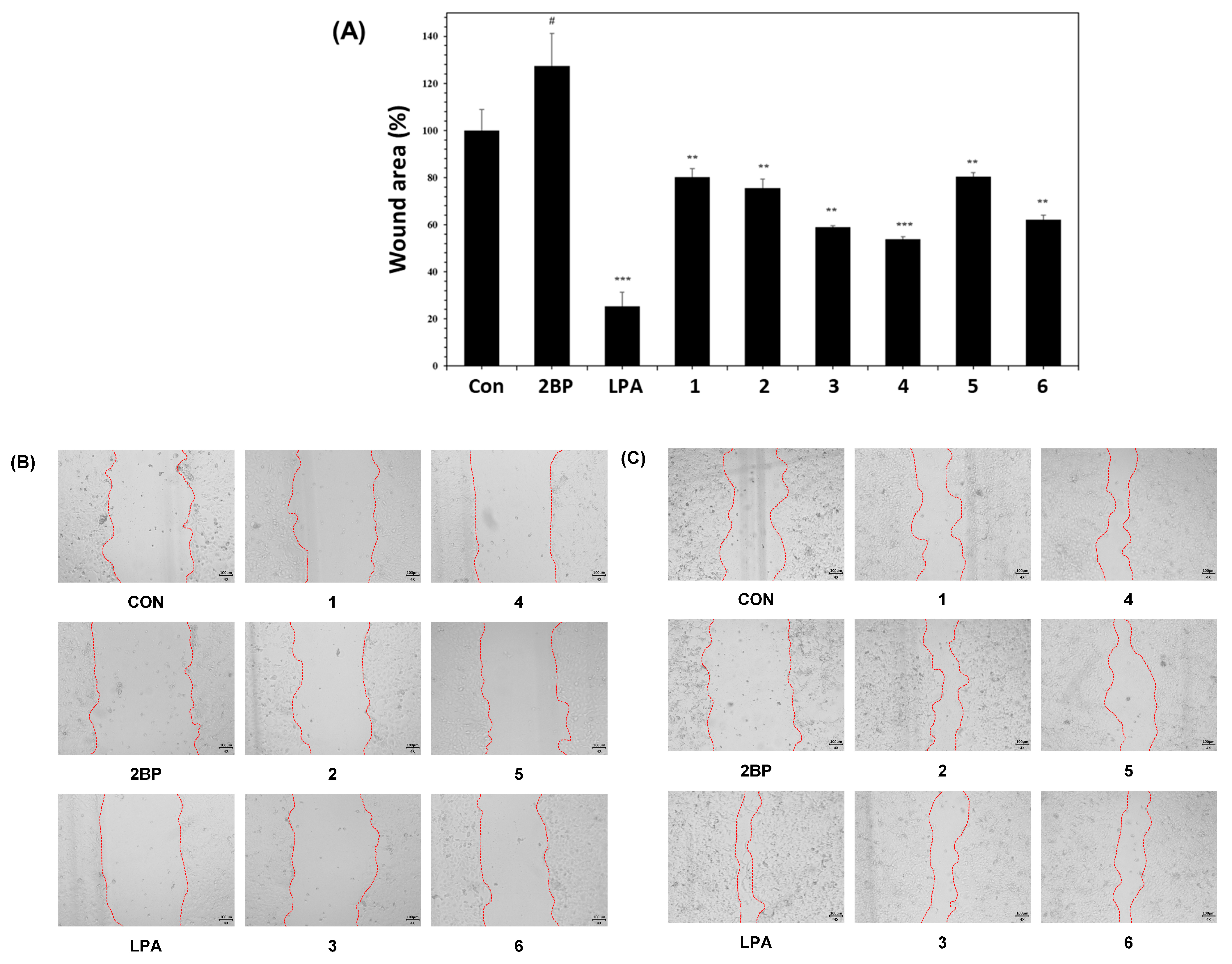
2.2. Wound-Healing Activities of Compounds 16

Keratinocytes are essential constituents of the epidermis and play a pivotal role in the regeneration of the epidermis during wound healing, encompassing processes like hemostasis, inflammation, proliferation, and remodeling [29,30]. The wound-healing activities of the compounds 16 isolated from N. peltata were evaluated using a HaCaT cell scratch test, an in vitro method that proves valuable for assessing cell migration from wound edges and over scratches [31]. The negative control, 2-bromo-palmitate (2BP), resulted in reduced cell migration, increasing the wound area to 124.04%. As a positive control, lysophosphatidic acid (LPA), known for its role in epidermal regeneration by modulating cellular responses [32], decreased the wound area to 23.97%. Furthermore, our results revealed that HaCaT cells exhibited enhanced wound healing in the presence of isoquercetin (3) (reducing the wound area to 66.48%), quercetin-3-O-vicianoside (4) (reducing the wound area to 61.08%), and 3‴-O-foliamenthoyl-rutin (6) (reducing the wound area to 71.42%) (Figure 3). Among these active compounds, quercetin-3-O-vicianoside (4) proved to be the most effective in promoting wound repair.
Quercetin (1), a bioactive flavonoid often found in the form of quercetin glycoside, is notable for its safety within reasonable dosage ranges [33]. It boasts a diverse range of healing properties, including anti-inflammatory, anti-infectious, angiogenic, immune-regulating, and antioxidant attributes [33]. When applied topically, quercetin demonstrates the ability to elevate interleukin-10 levels (an anti-inflammatory cytokine) while reducing tumor necrosis factor-α (TNF-α), an inflammatory factor [33]. Furthermore, quercetin exhibits promise in wound healing by increasing levels of vascular endothelial growth factor (VEGF), transforming growth factor-β (TGF-β), and antioxidants, while simultaneously diminishing the expression of inflammatory elements [33]. Despite these positive effects, a therapeutic effect of quercetin in healing wounds was not found in the current study. Quercitrin (2) has been reported to possess in vitro wound-healing capabilities. Previous studies have highlighted the involvement of the Wnt/β-catenin signaling pathway for quercitrin in enhancing wound healing by regulating cell proliferation [34]. Quercitrin was found to enhance motility in HaCaT keratinocytes by increasing β-catenin levels and activating the Wnt/β-catenin signaling pathway [34]. This discovery indicates its potential in promoting wound healing by facilitating cellular processes. Furthermore, quercitrin demonstrated non-cytotoxic effects under therapeutic concentrations (10 μM), suggesting its suitability as a potential therapeutic agent for wound healing. However, the observed therapeutic concentration of quercitrin (10 μM) did not demonstrate the expected effects in wound healing in the current study. In the case of isoquercetin (3), wound-healing activity was confirmed in isoquercetin that was isolated sequentially through sequential fractionation, beginning with the Sambucus ebulus extract from a previous study [35]. However, it was noted that the independent activity of isoquercetin, which was identified as the primary single compound in the active extract with confirmed wound-healing activity, was significantly lower than that of the subfractions [35]. Those results were partially consistent with our findings, wherein compound 3 exhibited weak activity. Contrastingly, quercetin-3-O-vicianoside (4), which is characterized by different sugar units, demonstrated activity in wound healing in our bioassay system, marking the first report of such activity in the current study. In the case of rutin (5), previous experiments were conducted to evaluate the relationship between wound healing in streptozotocin-induced hyperglycemic rats and the antioxidant and anti-inflammatory effects of rutin [36]. Eighteen male Wistar rats were randomly divided into three groups: normal, hyperglycemic, and rutin-containing hyperglycemic groups. After inducing hyperglycemia for 2 days, a wound was induced on the back of each rat. It was confirmed that intraperitoneal injection of rutin significantly improved diabetes-induced weight loss and alleviated metabolic dysfunction in hyperglycemic rats [36]. Finally, the new compound identified in this study, 3‴-O-foliamenthoyl-rutin (6), demonstrated the ability to promote HaCaT cell migration over scratch wounds, indicating its potential as a wound-healing agent.
Normal wound healing is a dynamic and complex process that involves a series of coordinated events, including bleeding, coagulation, initiation of an acute inflammatory response to the initial injury, and subsequent regeneration, migration, and proliferation of connective tissue, along with collagen deposition. Among these events, cell migration and proliferation play crucial roles in wound healing. The wound site progresses through various stages, relying on cellular processes such as collagen synthesis, angiogenesis, granulation tissue formation, and epithelialization to undergo recovery [37].
In this study, we investigated the effects of compounds 16 on cell migration and proliferation in HaCaT cells, which are keratinocytes. Our findings revealed that compounds 3, 4, and 6 positively influence wound healing. Consequently, additional experiments are deemed necessary to validate whether changes in concentration contribute to wound healing. If wound healing is concentration-dependent, further research on the various mechanisms mentioned above would be warranted.

3. Materials and Methods

3.1. General Experimental Procedures

Nuclear magnetic resonance (NMR) spectra analysis (1H, 13C, 1H-1H COSY, HMBC, and HMQC) spectra were observed on a JEOL JNM-ECZ400S 400 MHz NMR spectrometer (JEOL, Ltd., Tokyo, Japan) and Bruker AVANCE NEO 500 MHz NMR spectrometer (Bruker Corp., Billerica, MA, USA). HR-ESI-MS was measured using Agilent 6530 Accurate-Mass Q/TOF-LC/MS system (Agilent Technologies, Santa Clara, CA, USA). The high-performance liquid chromatography–photodiode array (HPLC-PDA) analysis was performed on the Waters HPLC system with an e2695 separation module and a 2998 PDA detector (Waters Corporation, Milford, MA, USA) and Empower®3 Chromatography Software version 3 (Build 3471, Waters Corp., MA, USA) and a reversed-phase column (Aegispak C18-L, 5 μm, 4.6 × 250 mm, Young Jin Biochrom, Sungnam, Republic of Korea). Semipreparative HPLC was performed on a Gilson HPLC system with two pumps (305 master pump, 307 slave pump) and a mixer (811C dynamic mixer) and a Shimadzu HPLC system with a pump (LC-20AT), UV/vis detector (SPD-20A), and system controller (CBM-20A) using the reversed-phase column (Watchers 120 ODS-BP, S-10 μm, 150 × 10 mm, Isu Industry Corp., Seoul, Republic of Korea). The resin for open-column chromatography used Silica gel 60 with pore size of 6 nm and particle size of 63–200 µm (Product No. 1.07734, Merck, Darmstadt, Germany) and Sephadex™ LH-20 with bead size of 25–100 µm (GE Healthcare Bio-Sciences AB, Uppsala, Sweden). The gel-coated plate used in the thin-layer chromatography (TLC) detection assay was silica gel 60 F254 Art (GF254, No. 1.05554, Merck, Darmstadt, Germany), and spots were observed using UV spectrum and anisaldehyde–sulfuric acid reagent.

3.2. Plant Material

The root parts of Nymphoides peltate (Gmel.) O. Kuntze were collected in the Hantaek Botanical Garden Foundation, Yongin-si, Gyeonggi-do, Republic of Korea, in June 2021. The plants were authenticated by Dr. Jung Hwa Kang. A voucher specimen (PNU-0040) was deposited in the Medicinal Herb Garden, Pusan National University.

3.3. Extraction and Isolation

The dried and powdered root parts of N. peltata (2.8 kg) were ultrasonically extracted twice with MeOH (28 L, 90 min each) at room temperature. The solvent was concentrated under reduced pressure at 45 °C and immediately freeze dried to obtain the crude N. peltata extract (427 g, yield: 15.2%). The crude extract obtained was suspended in H2O (2 L) and then partitioned with hexane (Hx, 8 L), ethyl acetate (EtOAc, 8 L), and n-butanol (BuOH, 8 L) to obtain three fractions, including soluble fractions of Hx (80.3 g), EtOAc (24 g), and BuOH (108 g). The soluble fraction of EtOAc was subjected to normal-phase silica column chromatography using a gradient mobile phase (EtOAc:MeOH from 10:1 to 100% MeOH) to obtain 7 subfractions (E1~E7). E1 (463.2 mg) was separated into 9 subfractions (E1-1~E1-9) with normal-phase silica column chromatography using a gradient mobile phase (EtOAc:MeOH from 20:1 to 100% MeOH). E1-6 (30.5 mg) was subjected to Sephadex LH-20 using 100% MeOH to obtain compound 1 (3 mg). E2 (1.025 g) was separated into 9 subfractions (E2-1~E2-9) with normal-phase silica column chromatography using a gradient mobile phase [chloroform (CHCl3):MeOH from 20:1 to 100% MeOH]. E2-9 (206 mg) was separated into 3 subfractions (E2-9-1~E2-9-3) with Sephadex LH-20 using 100% MeOH. E2-9-2 (75 mg) was separated into 3 subfractions (E2-9-2-1~E2-9-2-3) with Sephadex LH-20 using 100% MeOH. E2-9-2-3 (18.7 mg) was subjected to a Shimadzu preparative-HPLC system (UV wavelengths at 250 and 330 nm; flow rate of 2 mL/min) using an isocratic mobile phase (0.1% formic acid + MeCN:0.1% formic acid + H2O = 25:75) to obtain compound 2 (8.8 mg, tR = 13.2 min). E3 (1.1036 g) was separated into 6 subfractions (E3-1~E3-6) with Sephadex LH-20 using a 100% MeOH. E3-2 (357 mg) was separated into 5 subfractions (E3-2-1~E3-2-5) with normal-phase silica column chromatography using a gradient mobile phase (EtOAc:MeOH from 20:1 to 100% MeOH). E3-2-3 (137.9 mg) was separated into 3 subfractions (E3-2-3-1~E3-2-3-3) with Sephadex LH-20 using a 100% MeOH. E3-2-3-2 (67.8 mg) was subjected to a Shimadzu preparative-HPLC system (UV wavelengths at 250 and 330 nm; flow rate of 2 mL/min) using an isocratic mobile phase (0.1% formic acid + MeCN:0.1% formic acid + H2O = 25:75) to obtain compound 6 (31 mg, tR = 35 min). E7 (1.893 g) was subjected to a Shimadzu preparative-HPLC system (UV wavelengths at 250 and 330 nm; flow rate of 2 mL/min) using an isocratic mobile phase (0.1% formic acid + MeCN:0.1% formic acid + H2O = 20:80) to obtain compounds 4 (20.9 mg, tR = 18 min), 5 (22.4 mg, tR = 22 min), and 3 (21.7 mg, tR = 27.5 min).

3‴-O-Foliamenthoyl-Rutin (6)

Yellowish amorphous powder; [ α ] D 25 +15.8(c 0.02, MeOH); UV (MeOH) λmax 254, 354 nm (Figure S7); IR (KBr) νmax 3400, 2925, 2715, 1655, 1605, 1515, 1380, 1015 cm−1; 1H (500 MHz, DMSO-d6) and 13C (100 MHz, DMSO-d6) NMR data, see Table 1; HR-ESI-MS (positive ion mode) m/z 777.2561 [M + H]+ (calcd. for C37H45O18, 777.2606).

3.4. Acid Hydrolysis and Absolute Configuration Determination of Sugar Moieties

The absolute configuration of the sugar moieties was determined using an HPLC-UV-based method [28,38,39,40]. For compound 6, 1.5 mg was hydrolyzed in the presence of 1 N HCl at 80 °C for 2 h and EtOAc was used for the extraction. The aqueous layer was neutralized with repeated evaporation under a vacuum evaporator and dissolved in anhydrous pyridine (0.5 mL) with the addition of L-cysteine methyl ester hydrochloride (1.0 mg). After the reaction mixture was heated at 60 °C for 1 h, o-tolylisothiocyanate (50 μL) was added and the mixture was kept at 60 °C for 1 h. The reaction product was evaporated under a vacuum evaporator and dissolved in MeOH. Next, the dissolved reaction product was directly analyzed with LC/MS [MeOH/H2O, 1:9 → 7:3 gradient system (0–20 min), 100% MeOH (21–31 min), 0% MeOH (32–42 min); flow rate of 0.3 mL/min] using an analytical Kinetex C18 100 Å column (100 mm × 2.1 mm i.d., 5 μm). The sugar moieties from 6 were identified as β-glucopyranose and α-rhamnopyranose based on comparison with the standard using LC/MS analysis.

3.5. Cell Culture

Human keratinocytes (HaCaT) were purchased from CLS Cell Lines Service GmbH (Eppelheim, Baden-Württemberg, Germany) and cultured as single layers at 37 °C in a 5% CO2 incubator (Forma Direct Heat CO2 Incubator, Thermo Fisher Scientific, Madison, WI, USA) in DMEM (HyClone; Cytiva, Washington, DC, USA) supplemented with 10% fetal bovine serum (FBS; Gibco, Grand Island, NY, USA), 100 U/mL penicillin, and 100 μg/mL streptomycin (Gibco, Grand Island, NY, USA) (growth medium).

3.6. Wound-Healing Assay and MTT Assay

The wound-healing assay was performed as previously described [31] with some modifications. HaCaT cells were seeded in 24-well plates at a density of 2 × 10⁵ cells per well and allowed to attach for one day. Scratches were then created in each well using a 200 μL pipette tip. After washing with PBS, cells were treated with 2-bromo-palmitate (2BP) at 10 μM (3.3532 μg/mL) or lysophosphatidic acid (LPA) at 10 μM (4.3652 μg/mL) as negative and positive controls, respectively, and compounds 16 at 10 μM (1: 3.0224 μg/mL, 2: 4.4838 μg/mL, 3: 4.641 μg/mL, 4: 5.965 μg/mL, 5: 6.1052 μg/mL, and 6: 7.76 μg/mL). In addition, samples were cultured in a growth medium containing 1% FBS and 1% penicillin-streptomycin for 48 h to minimize the effects of growth factors. Wound images were captured immediately after wounding and after 24 or 48 h of culture using a microscope camera, HK5.1 UCMOS (KOPTIC, Republic of Korea). Captured images were quantified using Image J 1.52a software (National Institutes of Health, Bethesda, MD, USA). The wound area was recorded through phase-contrast imaging using an optical microscope. For quantification using Image J, the wound area was manually measured at both the 0 h and 48 h time points for each dataset. The results were expressed as the efficiency of wound closure, calculated as [(sample wound area/control wound area) × 100%].

3.7. Statistics Analysis

Data were expressed as the means ± standard deviation. GraphPad Prism, version 5.04 (GraphPad Software Inc., San Diego, CA, USA) was used for statistical processing in this experiment. The differences between the experimental groups were analyzed through the ANOVA test, and the significant level difference was set to p < 0.05.

4. Conclusions

A new flavonoid glycoside, 3‴-O-foliamenthoyl-rutin (6) and five known flavonoids, namely quercetin (1), quercitrin (2), isoquercetin (3), quercetin-3-O-vicianoside (4), and rutin (5), were isolated from the MeOH extract of N. peltata roots. The chemical structure of the newly isolated compound (6) was determined via 1D and 2D NMR spectra, HR-ESIMS analysis, and a chemical reaction. In our evaluation of the wound-healing activities of the isolated compounds 1–6 using a HaCaT cell scratch test, isoquercetin (3), quercetin-3-O-vicianoside (4), and 3‴-O-foliamenthoyl-rutin (6) promoted HaCaT cell migration over scratch wounds. Quercetin-3-O-vicianoside (4) proved to be the most effective in promoting wound repair. Our findings provide supportive data for the potential of quercetin-3-O-vicianoside (4) as a wound-healing agent. It is essential to acknowledge the limitations of our study. The biological evaluation, which was conducted with a minimum of three repetitions and a single concentration, may constrain the validity of our conclusions. Therefore, future studies are warranted to further investigate and substantiate the implications of our findings.

Supplementary Materials

The following are available online at https://www.mdpi.com/article/10.3390/plants12244083/s1: Table S1: 1H and 13C NMR Spectrum Data of Compounds 15; Figure S1: HR-ESI-MS data of 6; Figure S2: 1H NMR spectrum of 6 in DMSO-d6 (500 MHz); Figure S3: 13C NMR spectrum of 6 in DMSO-d6 (100 MHz); Figure S4: 1H-1H COSY spectrum of 6 in DMSO-d6; Figure S5: HSQC spectrum of 6 in DMSO-d6; Figure S6: HMBC spectrum of 6 in DMSO-d6; Figure S7: UV/PDA spectrum of 6.

Author Contributions

Conceptualization, S.-N.K., K.H.K. and M.H.Y.; methodology, T.-Y.K., B.S.L., B.-G.J., S.P.H., M.-J.K. and T.-H.K.; formal analysis, T.-Y.K., B.S.L., B.-G.J., S.P.H., M.-J.K. and T.-H.K.; writing—original draft preparation, T.-Y.K., B.S.L. and K.H.K.; writing—review and editing, S.-N.K., K.H.K. and M.H.Y.; supervision, S.-N.K., K.H.K. and M.H.Y.; funding acquisition, K.H.K. All authors have read and agreed to the published version of the manuscript.

Funding

This research was supported by National Research Foundation of Korea (NRF) grants funded by the Ministry of Science, ICT, and Future Planning (grant Nos. NRF-2019M3A9I3080265, NRF-2019M3A9I3080266, and NRF-2022R1A2C1003088). This work was supported by the Korea Environment Industry & Technology Institute (KEITI) through a project to improve the development of multi-ministerial national biological research resources, which is funded by the Republic of Korea Ministry of Environment (MOE) (grant number 2021003420003).

Data Availability Statement

Data are contained within the article and supplementary materials.

Conflicts of Interest

The authors declare no conflict of interest.

References

  1. Chuang, T.; Ornduff, R. Seed morphology and systematics of Menyanthaceae. AM. J. Bot. 1992, 79, 1396–1406. [Google Scholar] [CrossRef]
  2. Njuguna, A.W.; Li, Z.-Z.; Saina, J.K.; Munywoki, J.M.; Gichira, A.W.; Gituru, R.W.; Wang, Q.-F.; Chen, J.-M. Comparative analyses of the complete chloroplast genomes of nymphoides and menyanthes species (menyanthaceae). Aquat. Bot. 2019, 156, 73–81. [Google Scholar] [CrossRef]
  3. Uesugi, R.; Tani, N.; Goka, K.; Nishihiro, J.; Tsumura, Y.; Washitani, I. Isolation and characterization of highly polymorphic microsatellites in the aquatic plant, Nymphoides peltata (Menyanthaceae). Mole. Ecol. Notes. 2005, 5, 343–345. [Google Scholar] [CrossRef]
  4. National List of Species of Korea 2022. National Institute of Biological Resources. Available online: http://kbr.go.kr/ (accessed on 10 September 2023).
  5. Khan, Z.R.; Chowdhury, N.S.; Sharmin, S.; Sohrab, M.H. Medicinal values of aquatic plant genus Nymphoides grown in Asia: A review. Asian Pac. J. Trop. Biomed. 2018, 8, 113–119. [Google Scholar] [CrossRef]
  6. Nocchi, N.; Duarte, H.M.; Pereira, R.C.; Konno, T.U.P.; Soares, A.R. Effects of UV-B radiation on secondary metabolite production, antioxidant activity, photosynthesis and herbivory interactions in Nymphoides humboldtiana (Menyanthaceae). J. Photochem. Photobiol. B Biol. 2020, 212, 112021. [Google Scholar] [CrossRef] [PubMed]
  7. Amin, A.; Tuenter, E.; Exarchou, V.; Upadhyay, A.; Cos, P.; Maes, L.; Apers, S.; Pieters, L. Phytochemical and pharmacological investigations on Nymphoides indica leaf extracts. Phytother. Res. 2016, 30, 1624–1633. [Google Scholar] [CrossRef] [PubMed]
  8. Murali, A.; Sudha, C.; Madhavan, V.; Yoganarasimhan, S. Anticonvulsant and Sedative Activity of Tagara (Nymphoides macrospermum). Pharm. Biol. 2007, 45, 407–410. [Google Scholar] [CrossRef]
  9. Du, Y.; Wang, R.; Zhang, H.; Liu, J. Antitumor constituents of the wetland plant Nymphoides peltata: A case study for the potential utilization of constructed wetland plant resources. Nat. Prod. Commun. 2015, 10, 1934578X1501000203. [Google Scholar] [CrossRef]
  10. Zhou, X.; Ning, K.; Ling, B.; Chen, X.; Cheng, H.; Lu, B.; Gao, Z.; Xu, J. Multiple injections of autologous adipose-derived stem cells accelerate the burn wound healing process and promote blood vessel regeneration in a rat model. Stem Cells Dev. 2019, 28, 1463–1472. [Google Scholar] [CrossRef]
  11. Mathias, E.; Srinivas, M.M. Pediatric thermal burns and treatment: A review of progress and future prospects. Medicines 2017, 4, 91. [Google Scholar] [CrossRef]
  12. Gautam, S.; Chou, C.-F.; Dinda, A.K.; Potdar, P.D.; Mishra, N.C. Surface modification of nanofibrous polycaprolactone/gelatin composite scaffold by collagen type I grafting for skin tissue engineering. Mater. Sci. Eng. C 2014, 34, 402–409. [Google Scholar] [CrossRef]
  13. Childs, D.R.; Murthy, A.S. Overview of wound healing and management. Surg. Clin. 2017, 97, 189–207. [Google Scholar] [CrossRef]
  14. Sun, M.-L.; Zhao, F.; Chen, X.-L.; Zhang, X.-Y.; Zhang, Y.-Z.; Song, X.-Y.; Sun, C.-Y.; Yang, J. Promotion of wound healing and prevention of frostbite injury in rat skin by exopolysaccharide from the arctic marine bacterium Polaribacter sp. SM1127. Mar. Drugs 2020, 18, 48. [Google Scholar] [CrossRef] [PubMed]
  15. Dorai, A.A. Wound care with traditional, complementary and alternative medicine. Indian J. Plast. Surg. 2012, 45, 418–424. [Google Scholar] [CrossRef]
  16. Yeng, N.K.; Shaari, R.; Nordin, M.L.; Sabri, J. Investigation of wound healing effect of Acalypha indica extract in sprague dawley rats. Biomed. Pharmacol. J. 2019, 12, 1857–1865. [Google Scholar] [CrossRef]
  17. Schmidt, C.A.; Murillo, R.; Bruhn, T.; Bringmann, G.; Goettert, M.; Heinzmann, B.; Brecht, V.; Laufer, S.A.; Merfort, I. Catechin derivatives from Parapiptadenia rigida with in vitro wound-healing properties. J. Nat. Prod. 2010, 73, 2035–2041. [Google Scholar] [CrossRef] [PubMed]
  18. Tran, N.Q.; Joung, Y.K.; Lih, E.; Park, K.D. In situ forming and rutin-releasing chitosan hydrogels as injectable dressings for dermal wound healing. Biomacromolecules 2011, 12, 2872–2880. [Google Scholar] [CrossRef]
  19. Öz, B.E.; İşcan, G.S.; Akkol, E.K.; Süntar, İ.; Acıkara, Ö.B. Isoflavonoids as wound healing agents from Ononidis Radix. J. Ethnopharmacol. 2018, 211, 384–393. [Google Scholar] [CrossRef]
  20. Muralidhar, A.; Babu, K.S.; Sankar, T.R.; Reddanna, P.; Latha, J. Wound healing activity of flavonoid fraction isolated from the stem bark of Butea monosperma (Lam) in albino wistar rats. Eur. J. Exp. Biol. 2013, 3, 1–6. [Google Scholar]
  21. Mensah, A.; Sampson, J.; Houghton, P.; Hylands, P.; Westbrook, J.; Dunn, M.; Hughes, M.; Cherry, G. Effects of Buddleja globosa leaf and its constituents relevant to wound healing. J. Ethnopharmacol. 2001, 77, 219–226. [Google Scholar] [CrossRef]
  22. Muhammad, A.A.; Pauzi, N.A.S.; Arulselvan, P.; Abas, F.; Fakurazi, S. In vitro wound healing potential and identification of bioactive compounds from Moringa oleifera Lam. BioMed Res. Int. 2013, 2013, 974580. [Google Scholar] [CrossRef] [PubMed]
  23. Kim, T.-Y.; Park, N.-J.; Jegal, H.; Paik, J.-H.; Choi, S.; Kim, S.-N.; Yang, M.H. Nymphoides peltata root extracts improve atopic dermatitis by regulating skin inflammatory and anti-oxidative enzymes in 2, 4-dinitrochlorobenzene (DNCB)-induced SKH-1 hairless mice. Antioxidants 2023, 12, 873. [Google Scholar] [CrossRef] [PubMed]
  24. Blunder, M.; Orthaber, A.; Bauer, R.; Bucar, F.; Kunert, O. Efficient identification of flavones, flavanones and their glycosides in routine analysis via off-line combination of sensitive NMR and HPLC experiments. Food Chem. 2017, 218, 600–609. [Google Scholar] [CrossRef] [PubMed]
  25. Hardiyanti, R.; Marpaung, L.; Adnyana, I.K.; Simanjuntak, P. Isolation of quercitrin from Dendrophthoe pentandra (L.) Miq leaves and it’s antioxidant and antibacterial activities. Rasayan. J. Chem. 2019, 12, 1822–1827. [Google Scholar] [CrossRef]
  26. Slimestad, R.; Torskangerpoll, K.; Nateland, H.S.; Johannessen, T.; Giske, N.H. Flavonoids from black chokeberries, Aronia melanocarpa. J. Food Compos. Anal. 2005, 18, 61–68. [Google Scholar] [CrossRef]
  27. Iwagawa, T.; Asai, H.; Hase, T.; Sako, S.; Su, R.; Hagiwara, N.; Kim, M. Monoterpenoids from Radermachia sinica. Phytochem 1990, 29, 1913–1916. [Google Scholar] [CrossRef]
  28. Itoh, A.; Tanahashi, T.; Nagakura, N.; Takenaka, Y.; Chen, C.-C.; Pelletier, J. Flavonoid glycosides from Adina racemosa and their inhibitory activities on eukaryotic protein synthesis. J. Nat. Prod. 2004, 67, 427–431. [Google Scholar] [CrossRef]
  29. Piipponen, M.; Li, D.; Landén, N.X. The immune functions of keratinocytes in skin wound healing. Int. J. Mol. Sci. 2020, 21, 8790. [Google Scholar] [CrossRef]
  30. Li, J.; Chen, J.; Kirsner, R. Pathophysiology of acute wound healing. Clin. Dermatol. 2007, 25, 9–18. [Google Scholar] [CrossRef]
  31. Grada, A.; Otero-Vinas, M.; Prieto-Castrillo, F.; Obagi, Z.; Falanga, V. Research techniques made simple: Analysis of collective cell migration using the wound healing assay. J. Investig. Dermatol. 2017, 137, e11–e16. [Google Scholar] [CrossRef]
  32. Mazereeuw-Hautier, J.; Gres, S.; Fanguin, M.; Cariven, C.; Fauvel, J.; Perret, B.; Chap, H.; Salles, J.-P.; Saulnier-Blache, J.-S. Production of lysophosphatidic acid in blister fluid: Involvement of a lysophospholipase D activity. J. Investig. Dermatol. 2005, 125, 421–427. [Google Scholar] [CrossRef]
  33. Mi, Y.; Zhong, L.; Lu, S.; Hu, P.; Pan, Y.; Ma, X.; Yan, B.; Wei, Z.; Yang, G. Quercetin promotes cutaneous wound healing in mice through Wnt/β-catenin signaling pathway. J. Ethnopharmacol. 2022, 290, 115066. [Google Scholar] [CrossRef]
  34. Chen, J.; Li, G.; Sun, C.; Peng, F.; Yu, L.; Chen, Y.; Tan, Y.; Cao, X.; Tang, Y.; Xie, X.; et al. Chemistry, pharmacokinetics, pharmacological activities, and toxicity of Quercitrin. Phytother. Res. 2022, 36, 1545–1575. [Google Scholar] [CrossRef] [PubMed]
  35. Süntar, I.P.; Akkol, E.K.; Yalçin, F.N.; Koca, U.; Keleş, H.; Yesilada, E. Wound healing potential of Sambucus ebulus L. leaves and isolation of an active component, quercetin 3-O-glucoside. J. Ethnopharmacol. 2010, 129, 106–114. [Google Scholar] [CrossRef] [PubMed]
  36. Chen, L.-Y.; Huang, C.-N.; Liao, C.-K.; Chang, H.-M.; Kuan, Y.-H.; Tseng, T.-J.; Yen, K.-J.; Yang, K.-L.; Lin, H.-C. Effects of Rutin on Wound Healing in Hyperglycemic Rats. Antioxidants 2020, 9, 1122. [Google Scholar] [CrossRef] [PubMed]
  37. Velnar, T.; Bailey, T.; Smrkolj, V. The wound healing process: An overview of the cellular and molecular mechanisms. J. Int. Med. Res. 2009, 37, 1528–1542. [Google Scholar] [CrossRef]
  38. Sun, Y.; Gao, M.; Chen, F.; Han, R.; Du, K.; Zhang, Y.; Li, M.; Si, Y.; Feng, W. Six New Coumarin Glycosides from the Aerial Parts of Gendarussa vulgaris. Molecules 2019, 24, 1456. [Google Scholar] [CrossRef]
  39. Lee, K.H.; Kim, J.K.; Yu, J.S.; Jeong, S.Y.; Choi, J.H.; Kim, J.-C.; Ko, Y.-J.; Kim, S.-H.; Kim, K.H. Ginkwanghols A and B, osteogenic coumaric acid-aliphatic alcohol hybrids from the leaves of Ginkgo biloba. Arch. Pharm. Res. 2021, 44, 514–524. [Google Scholar] [CrossRef]
  40. Yu, J.S.; Jeong, S.Y.; Li, C.; Oh, T.; Kwon, M.; Ahn, J.S.; Ko, S.K.; Ko, Y.J.; Cao, S.; Kim, K.H. New phenalenone derivatives from the Hawaiian volcanic soil-associated fungus Penicillium herquei FT729 and their inhibitory effects on indoleamine 2,3-dioxygenase 1 (IDO1). Arch. Pharm. Res. 2022, 45, 105–113. [Google Scholar] [CrossRef]
Figure 1. Chemical structures of compounds 16.
Plants 12 04083 g001
Figure 2. Key 1H-1H COSY (Plants 12 04083 i001) and HMBC (Plants 12 04083 i002) correlations for compound 6.
Plants 12 04083 g002
Figure 3. Effects of compounds 16 on HaCaT cell wound-healing assay results. Scratched HaCaT monolayers were treated with 10 μM of 2BP, LPA, or compounds 16, and cell migration was observed under a microscope after 48 h of culture (A). Wound closure was monitored (B) and wound spacing was measured (C). Each bar is presented as mean ± SD (n = 3). Wound area photographs and wound area over time (# p < 0.05 vs. CON; *** p < 0.001, ** p < 0.005 vs. 2BP). CON: untreated group, 2BP: 2-bromo-palmitate-treated group, LPA: lysophosphatidic acid-treated group.
Plants 12 04083 g003
Table 1. 1H and 13C NMR data for compound 6 in DMSO-d6 (δ ppm) a.
Position6
δCcδH (J in Hz) b
2157.0 C
3133.8 C
4177.8 C
5161.7 C
699.1 CH6.16 (d, 2.0)
7164.5 C
894.0 CH6.36 (d, 2.0)
9156.9 C
10104.5 C
1′121.7 C
2′116.7 CH7.52 (d, 2.0)
3′145.2 C
4′148.9 C
5′115.7 CH6.84 (d, 8.0)
6′122.0 CH7.53 (dd, 8.0, 2.0)
1″101.8 CH5.34 (d, 7.0)
2″74.5 CH3.23 (m)
3″76.9 CH3.23 (m)
4″71.8 CH3.24 (m)
5″76.3 CH3.29 (m)
6″68.0 CH23.76 (br d, 10.5); 3.33 (overlap)
1‴101.3 CH4.41 (br s)
2‴68.3 CH3.60 (br s)
3‴74.7 CH4.63 (dd, 9.5, 3.5)
4‴69.4 CH3.37 (m)
5‴68.9 CH3.43 (m)
6‴18.1 CH31.03 (d, 6.0)
1⁗167.3 C
2⁗128.1 C
3⁗142.0 CH6.70 (t, 6.5)
4⁗27.1 CH22.25 (dt, 6.5, 7.0)
5⁗38.0 CH22.07 (t, 7.0)
6⁗135.6 C
7⁗126.1 CH5.31 (t, 6.5)
8⁗58.0 CH23.94 (d, 6.5)
9⁗16.5 CH31.61 (s)
10⁗12.8 CH31.77 (s)
a Coupling constants (Hz) are given in parentheses. b Measured at 500 MHz in DMSO-d6. c Measured at 100 MHz in DMSO-d6.
Disclaimer/Publisher’s Note: The statements, opinions and data contained in all publications are solely those of the individual author(s) and contributor(s) and not of MDPI and/or the editor(s). MDPI and/or the editor(s) disclaim responsibility for any injury to people or property resulting from any ideas, methods, instructions or products referred to in the content.

