Review

Bioactive Compounds in Moringa oleifera: Mechanisms of Action, Focus on Their Anti-Inflammatory Properties

by
Adina Chiș
1,†,
Paul Aimé Noubissi
2,†,
Oana-Lelia Pop
3,4,
Carmen Ioana Mureșan
3,4,
Michel Archange Fokam Tagne
5,
René Kamgang
2,
Adriana Fodor
6,
Adela-Viviana Sitar-Tăut
7,
Angela Cozma
7,
Olga Hilda Orășan
7,
Simona Codruța Hegheș
8,*,
Romana Vulturar
1,‡ and
Ramona Suharoschi
3,4,‡
1
Department of Molecular Sciences, “Iuliu Hațieganu” University of Medicine and Pharmacy, 6 Louis Pasteur St, 400349 Cluj-Napoca, Romania
2
Department of Animal Biology and Conservation, Faculty of Science, University of Buea, Buea P.O. Box 63, Cameroon
3
Department of Food Science, University of Agricultural Science and Veterinary Medicine, 3-5 Calea Mănăștur, 400372 Cluj-Napoca, Romania
4
Molecular Nutrition and Proteomics Lab, CDS3, Life Science Institute, University of Agricultural Science and Veterinary Medicine, 3-5 Calea Mănăștur, 400372 Cluj-Napoca, Romania
5
Department of Biological Sciences, Faculty of Science, University of Ngaoundéré, Ngaoundéré P.O. Box 454, Cameroon
6
Clinical Center of Diabetes, Nutrition and Metabolic Diseases, “Iuliu Hațieganu” University of Medicine and Pharmacy, 2-4 Clinicilor St., 400012 Cluj-Napoca, Romania
7
Department of Internal Medicine, Faculty of Medicine, “Iuliu Hațieganu” University of Medicine and Pharmacy, 400012 Cluj-Napoca, Romania
8
Department of Drug Analysis, Faculty of Pharmacy, “Iuliu Hațieganu” University of Medicine and Pharmacy, Louis Pasteur Street 6, 400349 Cluj-Napoca, Romania
*
Author to whom correspondence should be addressed.
These authors contributed equally to this work.
Plants 2024, 13(1), 20; https://doi.org/10.3390/plants13010020 (registering DOI)
Submission received: 10 November 2023 / Revised: 6 December 2023 / Accepted: 15 December 2023 / Published: 20 December 2023

Abstract

:
Moringa oleifera (M. oleifera) is a tropical tree native to Pakistan, India, Bangladesh, and Afghanistan; it is cultivated for its nutritious leaves, pods, and seeds. This scientific study was conducted to outline the anti-inflammatory properties and mechanisms of action of bioactive compounds from M. oleifera. The existing research has found that the plant is used in traditional medicine due to its bioactive compounds, including phytochemicals: flavonoids and polyphenols. The compounds are thought to exert their anti-inflammatory effects due to: (1) inhibition of pro-inflammatory enzymes: quercetin and kaempferol inhibit the pro-inflammatory enzymes (cyclooxygenase and lipoxygenase); (2) regulation of cytokine production: isothiocyanates modulate signaling pathways involved in inflammation, such as the nuclear factor-kappa B (NF-kappa B) pathway; isothiocyanates inhibit the production of pro-inflammatory cytokines such as TNF-α (tumor necrosis factor α) and IL-1β (interleukin-1β); and (3) antioxidant activity: M. oleifera contains flavonoids, polyphenols, known to reduce oxidative stress and inflammation. The review includes M. oleifera’s effects on cardiovascular protection, anti-hypertensive activities, type 2 diabetes, inflammatory bowel disease, and non-alcoholic fatty liver disease (NAFLD). This research could prove valuable for exploring the pharmacological potential of M. oleifera and contributing to the prospects of developing effective medicines for the benefit of human health.

1. Introduction

The wide range of therapeutic effects exhibited by plant-based bioactive substances has positioned them as promising candidates in contemporary drug development; many phytochemicals have shown antibacterial, antioxidant, anticancer, anti-hepatitis C virus, and anti-inflammatory potentials such as quercetin, curcumin, capsaicin, resveratrol, taxol, and others; in addition, plant-based natural products have shown their therapeutic potential against various cardiovascular diseases or brain and neurodegenerative diseases (such as Parkinson’s disease, Alzheimer’s disease, and Huntington’s disease) [1].
Moringa stands as the sole genus in the Moringaceae family of flowering plants [2,3]. Among its various species, M. oleifera Lam (Moringa pterygosperma G.) is the most popular and widespread and is native to Pakistan, India, Bangladesh, and Afghanistan. It was found to withstand even the driest and harshest of soils [4]. It is largely cultivated in subtropical and tropical areas, with its young leaves, seed pods, and mature seeds serving as vegetables for populations in many countries [5], especially in the northern part of Cameroon. Also known as “horseradish tree”, or “drumstick tree”, all parts of M. oleifera have long been consumed by humans for nutritional and medicinal purposes [5]. Due to its medicinal properties, the plant was sometimes named “mother’s best friend” or “miracle tree” [6]. A recent article showed that the supplementation of polar extracts of M. oleifera was highly effective in controlling oxidative stress, inducing the retrieval of sensory and motor functions, and therefore facilitating accelerated nerve generation [7]. In a recent study, the phenolic composition and antioxidant capacity of various dietary supplements derived from M. oleifera were examined. The study found an important correlation, revealing that greater phenolic content corresponded to increased antioxidant activity within these supplements, regardless of their different forms of presentation [8].
Recently, it has been demonstrated that M. oleifera leaf extract alleviates hepatotoxicity caused by antiretroviral drugs through its ability to neutralize harmful oxidants and activate the NRF2 antioxidant pathway. This highlights M. oleifera’s substantial therapeutic promise and suggests its potential role as a valuable supplement for mitigating the toxicity associated with antiretroviral drugs [9].
Regarding the fortified foods, the yogurts fortified with M. oleifera exhibited superior antioxidant properties compared to the negative control; these results underscore the potential utilization of M. oleifera powder and extract as natural supplements for creating fortified foods that may help address malnutrition [10]. In addition, a recent publication has shown the development of a palatable and standardized pharmacologically active formulation using M. oleifera leaves, designed as a functional food. This formulation aims to activate NRF2 signaling and can be consumed either as a beverage (such as hot soup) or in the form of a freeze-dried powder. Its goal is to reduce the risk of environmental respiratory diseases by harnessing the power of isothiocyanate moringin and polyphenols, both strong stimulators of NRF2 signaling [11].
Additionally, recent studies are outlining the interest in enhancing the viable bioactive compounds in the culture of M. oleifera, i.e., the in vitro-based elicitation approach (a biotechnological tool for enhancing the production of secondary metabolites) and demonstrating that utilizing varying spectral lights represents an effective method for increasing the production of nutraceuticals and novel pharmacologically significant metabolites in the in vitro callus culture of M. oleifera [12]. The high nutritional, nutraceutical, and therapeutic profile (Table 1) is mainly attributed to its rich repertoire of biologically active molecules: proteins (peptides and protein hydrolysates), flavonoids, saponins, phenolic acids, tannins, isothiocyanates, lipids, minerals, and vitamins, amongst others [13].
The protein quality of M. oleifera leaves was shown to be similar to that of milk and eggs [36], and the plant has been described as performing numerous pharmacological properties and is long known in Ayurvedic medicine. Many recent studies have proven that leaves, pods, seeds, flowers, roots, bark, and stem has anti-inflammatory effects (Figure 1) [2,37,38,39,40,41,42,43,44].
M. oleifera was reported to be richer in potassium than bananas, with a higher content of calcium compared to milk, more iron than spinach, more vitamin C than oranges, and a higher vitamin A content compared to carrots [45].
Figure 1. Summary of pharmacological (direct and indirect) anti-inflammatory actions of different parts of M. oleifera (leaves, pods, seeds, flowers, roots, bark, and stem) demonstrated on an animal model (based on details cited in references [2,13,20,32,34,37,38,39,40,41,42,43,44,46]).
Plants 13 00020 g001
This review aims to provide detailed information on M. oleifera anti-inflammatory compounds and their possible specific mechanisms of action.

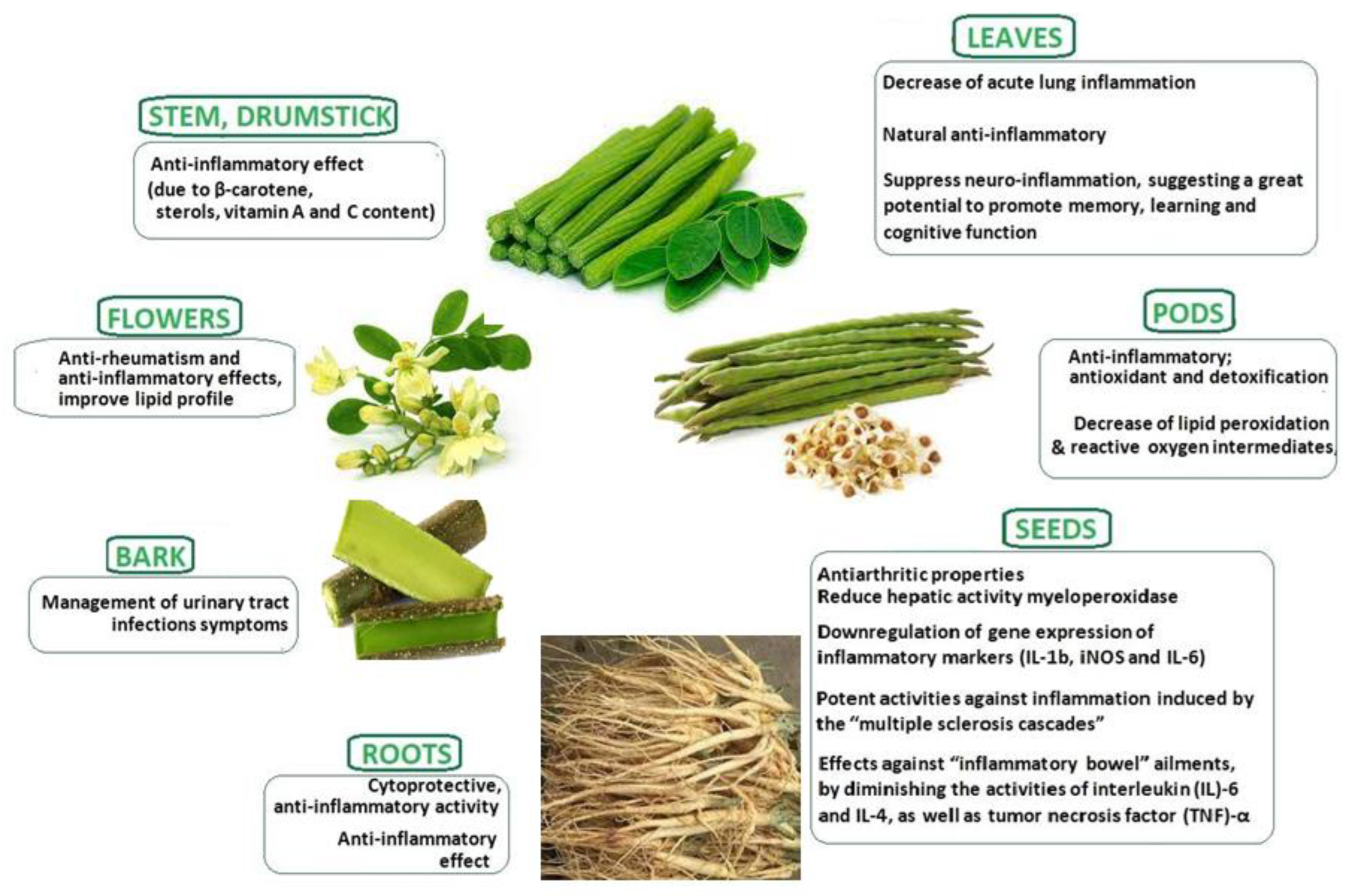
2. Description of Moringa oleifera

The genus is composed of about 13 species, namely M. arborea Verdcourt (Kenya, Somalia), M. borziana Mattei (Kenya and Somalia), M. concanensis Nimmo (India), M. drouhardii Jumelle (Southern Madagascar), M. hildebrandtii Engler (Southwest Madagascar), M. longituba Engler (Kenya, Southeast Ethiopia, and Somalia), M. oleifera Lam. (Cameroun and India), M. ovalifolia Dinter ex Berger (Namibia and Southwest Angola), M. peregrina Forssk. Ex Fiori (Red Sea, Arabia, and Northeast Africa), M. pygmaea Verdcourt (North Somalia), M. rivae Chiovenda (Kenya and Ethiopia), M. ruspoliana Engler (Kenya, Ethiopia, and Somalia), and M. stenopetala (Baker f.) Cufodontis (Kenya, Southwest Ehiopia, and Somalia), is distributed in Southwest Asia, Africa, and Madagascar [47].
Moringa tree has a diameter of about 45 cm (1.5 ft) and is about 10–12 m (32–40 ft) in height. Its fragrant bisexual flowers are surrounded by five thinly and unequally veined yellow to white petals of about 1.0 to 1.5 cm long and broad of about 2.0 cm. Fruits referred to as pods are tri-lobed pendulous capsules that are brown and triangular and split lengthwise into three parts when dried. Fruits contain about 26 seeds and are mostly produced between March and April. Immature pods are green in color; they turn brown at maturity. Seeds are around 1 cm in diameter and range in color from brown to black but can also be white when the viability of the kernels is low. Seeds germinate in 2 weeks, while each tree can produce around 15,000 to 25,000 seeds/year. The average weight is 0.3 g/seed. Propagation is usually performed by wind and water [36].
Taxonomically, M. oleifera is a member of the Kingdom of Plantae; sub kingdom of Tracheobionta; super-division of Spermatophyta; the division of Magnoliophyta; class of Magnoliopsida; subclass of Dilleniidae; order of Capparales; family of Moringceae; genus Moringa; and specie, oleifera [36,48].

3. Bioactive Components of M. oleifera and Their Mechanisms of Action in Inflammatory Diseases

In recent years, M. oleifera, a versatile and nutritionally rich plant, has garnered significant attention for its potential therapeutic benefits in combating inflammatory diseases. The description of bioactive compounds found in M. oleifera and the approach to intricate mechanisms of action are important in addressing inflammatory conditions, providing promising insights for future health interventions.

3.1. M. oleifera Essential Oil and Fatty Acids

The phytochemical composition of the volatile constituents of the ethanolic extract from the leaves of M. oleifera was reported by Kute in 2017; a total of forty-four volatile compounds have been identified with pentacosane (1) (17.4%), hexacosane (2) (11.2%), (E)-phytol (3) (7.7%), and 1-[2,3,6-trimethylphenyl]-2-butanone (4) (3.4%) as major constituents [2]. M. oleifera seed oil content ranges between 35 and 40% [49]. Oleic acid (5) (65%), linoleic acid (6) (16%), palmitic acid (7) (12.31%), stearic acid (8) (5.1%), and palmitoleic acid (9) (2.1%) are found to be its most important fatty acids [50] (Figure 2).

3.2. Bioactive Phytochemical Components

In the strictest sense, phytochemicals are the chemicals generated by plants. They encompass a broad group of compounds, known as secondary metabolites, that naturally occur and accumulate in plants at high concentrations [51]. Phytochemicals are found in roots, seeds, leaves, stems, flowers, and pods [52]. According to their chemical structure and characteristics, plant phytochemicals are classified into five main groups: polyphenols, sulfur-containing compounds, carotenoids, alkaloids, and terpenoids [52,53].
Polyphenols are classified as phenolic acids (with only one phenol ring present) and flavonoids (with more than one phenol ring present) [53] (Figure 3). Phenolic acids and flavonoids (especially tannins) are among the most important polyphenols found in the Moringa tree [53]. Leaves were reported to have the highest total phenolic contents, with about 2000 to 12200 mg GAE/100 g [53]. Except for the roots and seeds, the most common flavonoids found in various parts of the Moringa tree are kaempferol glycosides [glucosides (11), malonyl glucosides (12), and rutinosides (13)] and quercetin (14) [53]; other flavonols found in lower amounts include myricetin (15), epicatechin (16), and rutin (17). Among the abundant phenolic acids in the Moringa tree are caffeic acid (18), chlorogenic acid (19), coumaric acid (20), gallic acid (21), and ellagic acid (22) [53]. Leaves of Moringa were also reported to contain an important amount of tannins whose concentration can vary, with the highest being reported in dried leaves. A low quantity of tannins was also reported in seeds [53].
M. oleifera contains various sulfur compounds, including glucosinolates and isothiocyanates (Figure 4). When the plant is damaged, processed, harvested, or chewed, the enzyme called myrosinase is activated. This enzyme catalyzes the hydrolysis of β-D-glucose at neutral pH, resulting in the formation of isothiocyanates, thiocyanates, sulfates, and nitriles [53].
Glucosinolates represent a diverse category of glycosidic compounds containing sulfur and nitrogen, found in abundance in numerous plant species. Various types of glucosinolates have been identified in different parts of the Moringa plant, including the leaves, pods, stem, and roots. The most common glucosinolate in this plant is the 4-(α-l-rhamnopyranosiloxy) benzyl glucosinolate called glucomoringin (23). Glucomoringin (23) is commonly present in stems, flowers, pods, leaves, and seeds, while in the roots the predominant glucosinolate is benzyl glucosinolate (24). These compounds contribute to the pungent taste and odor of the plant and exhibit biological properties such as antifungal and antibacterial effects [54].
Isothiocyanates with thiocyanates and thiocarbamates are secondary metabolites derived from glucosinolates and have garnered significant attention because of their pharmacological properties. The principal isothiocyanates isolated from M. oleifera include niazidin (25), niazicin (26), or niazinin (27) [54].
Carotenoids are highly unsaturated fat-soluble pigments that provide fruits, vegetable fungi, bacteria, and algae their characteristic red, orange, or yellow color [55,56] (Figure 5). Carotenoids are subdivided into two groups: the carotenes (precursor of vitamin A) with a single long carbon chain and the xanthophylls, with oxygen atoms attached to their structure [56]. Moringa fresh leaves were found to contain an important amount of β-carotene (6.6–17.4 mg/100 g), higher than in carrots, pumpkins, and apricots [55,57,58]. The β-carotene content in Moringa dried leaves was even higher, about 23.31 to 39.6 mg per 100 g of dry matter. Other different carotenoids were identified in the flowers, fruits, and fresh leaves of eight Moringa oleifera commercially grown cultivars in India and included all-E-β-carotene (28), all-E-zeaxanthin (29), all-E-lutein (30), all-E-luteoxanthin (31), 15-Z-β-carotene (32), and 13-Z-lutein (33) [55].
Alkaloids are an assembly of naturally occurring chemical composites, typically comprising basic nitrogen atoms; they are miscellaneous elements and biomolecules, secondary compounds derived from amino acids or transamination [59] (Figure 6). There are three major alkaloid groups, namely pseudoalkaloids, true alkaloids, and protoalkaloids. Compared to pseudoalkaloids, the true alkaloids and protoalkaloids are derived from amino acids. Almost all true alkaloids have a bitter taste [59]. The presence of many alkaloids has been reported in the Moringa tree. N,α-l-rhamnopyranosyl vincosamide (34) was the most commonly reported Moringa plant indol alkaloid, which was isolated from the leaves. These leaves were also reported to contain glycosides of a pyrol alkaloid such as marumosides A (pyrrolemarumine 4″-O-α-l-rhamnopyranoside) (35) and marumosides B (4′-hydroxyphenylethanamide) (36) [53]. M. oleifera was also reported to contain two, as trigonelline (37) [60] or moringinine (benzylamine) (38) [61].
Terpenoids are compounds synthesized from the condensation of the five-carbon precursor isopentenyl pyrophosphate (IPP) with dimethylallyl pyrophosphate (DMAPP), the functional isomer [62,63] (Figure 7). Under the enzyme isoprenyl diphosphate synthase, IPP and DMAPP are condensed into acyclic and achiral isoprenyl diphosphate/pyrophosphate (ID, C5n) intermediates considered the universal precursors of terpenoid. Terpene synthases (TPSs) action on one or more of these precursors produces a diversity of terpenes [63]. Lupeol acetate (39), α-amyrin (40), and β-amyrin (41) are terpenes isolated from a n-hexane fraction of the ethanol extract of Moringa peregrina aerial parts [47].
Furthermore, proteins and peptide fractions with a high nutritional profile have been studied as promising components in Moringa [22].
Indeed, these phytochemicals in the Moringa tree are largely believed to be responsible for its diverse biological activities and disease-preventive potential. The presence and amount of these metabolites depend on geographical location, soil type, and climate [64].

3.3. Anti-Inflammatory Activities of M. oleifera Compounds and Their Mechanisms of Action

The compounds of M. oleifera are thought to exert their anti-inflammatory effects through several mechanisms [39,41]: (a) inhibition of pro-inflammatory enzymes: quercetin (14) and kaempferol (10) are M. oleifera’s compounds that inhibit the activity of pro-inflammatory enzymes [cyclooxygenase (COX) and lipoxygenase (LOX)], which are key enzymes involved in the production of inflammatory mediators such as prostaglandins and leukotrienes; (b) regulation of cytokine production: isothiocyanates (a class of M. oleifera’s compounds) have been shown to modulate signaling pathways involved in inflammation, as is the nuclear factor-kappa B (NF-kappa B) pathway; this ensures modulation of signaling pathways. Isothiocyanates have also been shown to inhibit the production of pro-inflammatory cytokines [tumor necrosis factor-α (TNF-α) and interleukin-1β (IL-1β)] and to increase the production of anti-inflammatory cytokines such as interleukin-10 (IL-10); and (c) antioxidant activity: flavonoids and polyphenols help to reduce oxidative stress and inflammation. These compounds may also inhibit the activity of pro-inflammatory enzymes and modulate cytokine production.
Inflammation constitutes a vital and intricate aspect of an organism’s reaction to biological, chemical, and/or physical stimuli [65]. Inflammation is commonly characterized by distinct acute and chronic phases, although there is some overlap between these stages. During the acute phase, primarily granulocytes, guided by a chemotactic gradient, migrate to the site of injury. This orchestrated response, facilitated by acute phase proteins and cytokines, aims to eliminate the inflammatory stimulus (e.g., infectious agents or foreign material) and remove damaged cells, initiating the healing process [66]. Depending on the severity of the injury, this acute cellular phase could be enough to resolve any damage. As a result of either prolonged exposure to inflammatory stimuli or an inappropriate reaction to self-molecules, persistent inflammation can probably lead to the chronic phase. The active immune cell populations shift to include a mononuclear phenotype, resulting in tissue damage and fibrosis. During inflammation, activated macrophages secrete a certain number of different pro-inflammatory cytokines, including TNF-α, IL-1β, interferon-γ (IFN-γ), interleukin-6 (IL-6), and oxidative stress mediators, such as nitric oxide (NO), produced by iNOS, the inducible nitric oxide synthase [67]. Chronic inflammation is implicated in the pathophysiology of numerous disorders, including cardiovascular diseases (atherosclerosis and hypertension), type 2 diabetes, enterocolitis, and non-alcoholic fatty liver disease (NAFLD). M. oleifera extract has shown potential activities against these diseases (see Figure 1) [68].

3.3.1. Cardiovascular Protection and Anti-Hypertensive Activities of M. oleifera

Chronic inflammatory diseases such as systemic lupus erythematosus, rheumatoid arthritis, psoriasis, and HIV infection affect up to 18% of the global population [69]. They have a higher risk of developing inflammation-related cardiovascular diseases than the general population [69,70,71,72,73]. Inflammation leads to atherosclerosis, ischemic heart disease, and heart failure, partly through atherosclerotic plaque formation. Notwithstanding the considerable enhancements in our insight into the etiology of cardiovascular diseases (CVDs), stroke, coronary artery disease, and other vasculopathies still account for over 31% of all fatalities globally [74]. Although much of these cardiovascular (CV) risks are due to traditionally known cardiovascular disease risk factors such as diabetes, hyperlipidemia, hypertension, and smoking, inflammation has been considered and identified as the key factor in the development, evolution, and aggravation of atherosclerosis [75]. Increased levels of pro-inflammatory factors in the serum, such as IL-6, IL-12, and TNF-α, and inflammatory biomarkers, such as C-reactive protein (CRP), high-sensitivity C-reactive protein (hs-CRP), fibrinogen, and homocysteine YKL-40, have been observed in atherosclerotic patients. YKL-40 has been confirmed to be a key factor in the pathogenesis of cardiovascular diseases such as insulin resistance and obesity. Furthermore, it is highly expressed in atherosclerotic plaques [76].
Hypertension is usually characterized by high systolic and diastolic blood pressure resulting from increased arginase, acetylcholinesterase, phosphodiesterase-5, and angiotensin-1-converting enzymes [77]. Hypertension has also been associated with insufficient production of the principal vasodilator, nitric oxide (NO), and vascular remodeling. Endogenous production of nitric oxide by NO synthases (NOS) requires L-arginine as the substrate. L-arginine also serves as a substrate for arginases, metabolizing L-arginine into urea and L-ornithine. It was recently found that arginase activity can result from many hypertensive stimuli, such as persistent chronic inflammation and salt loading. Stimulation of arginase enzymatic activity reduces the bioavailability of L-arginine (to NOS), thereby decreasing endogenous NO production in the vasculature. L-ornithine can be converted into polyamines and proline, metabolites central to vascular remodeling and proliferation of vascular smooth muscle cells. Thus, arginase implication in hypertension pathogenesis promotes vascular remodeling and inhibition of endogenous NO production [78]. The extracts from M. oleifera leaves and seeds demonstrated a reduction in activity of the enzyme arginase. These inhibitory effects may be attributed to the actions of phenolic compounds, which have been reported to inhibit the activity of the arginase enzyme [77]. Ethanolic extracts from the leaves of M. oleifera showed notable anti-hypertensive or hypotensive activity [47]. Thiocarbamates such as 4-[(4′-O-acetyl-α-l-rhamnosyloxy)benzyl]isothiocyanate, 4-[(α-l-rhamnosyloxy)benzyl] isothiocyanate, niazinin A (27), niazicin A (26), and niazirin [79] have been identified and isolated from the M. oleifera ethyl acetate fraction. Intravenous administration of any of these compounds at doses of 1 to 10 mg/kg resulted in hypotensive and bradycardiac actions in anesthetized rats, possibly mediated through calcium antagonist effects [47]. In vivo activity showed that isothiocyanate glycosides and thiocarbamate were responsible for this robust hypotensive activity [80].
Acetylcholine Esterase (AchE) is an enzyme widely distributed in neuromuscular junctions and the brain cholinergic synapses. Its principal biological activity in cholinergic synapses is to prevent the transmission of impulses through the rapid decomposition (hydrolysis) of acetylcholine (Ach) to acetate and choline [81]. Extracts from M. oleifera leaves and seeds were shown to reduce the activity of AchE, probably because of their phenolic constituents [77]. Phenolics are an important class of phytochemicals. Because of their (poly) hydroxyl groups, especially the 3′OH and 4′OH of their three-carbon chain, phenolics can donate electrons, therefore terminating the chain reaction process [77]. Structural similarity exists between naturally occurring polyphenols and the inhibitors of cholinesterase in terms of the hydrophobic component, molecular weight, and phenolic rings [82].
No biological effects were proven to be mediated through guanylyl cyclase activation. This fact leads to increased cyclic guanosine monophosphate (cGMP) synthesis, which, in turn, activates specific proteins, resulting in different actions, including smooth muscle relaxation, cardiac protection, neuronal plasticity, and endothelial permeability [83]. The cGMP activities were found to be terminated by the enzyme phosphodiesterase 5 (PDE-5) [83]. M. oleifera extracts decreased PDE-5 activity [77]. Previous reports revealed the capacity of medicinal plant extracts, especially those with high flavonoid contents, to inhibit the activity of PDE-5 [84]. The potential of M. oleifera extract to inhibit PDE-5 action could therefore be associated with its essential flavonoid content [14,84].
One of the primary critical regulators of hypertension is the renin–angiotensin system (RAS). It exhibits its anti-hypertensive actions mainly through the vasoactive peptide angiotensin II, released under angiotensin-converting enzyme (ACE) action following a blood pressure increase [85]. Inhibition of ACE has hypotensive effects. Diets supplemented with extracts from Moringa oleifera seed and leaves in rats reduced ACE activity [77]. There is evidence that phenolic compounds exhibit ACE inhibitory actions through the establishment of hydrogen bonds and hydrophobic interactions with the hydrophobic enzyme active site [86].
In addition, the anti-atherosclerotic and hypolipidemic effects of M. oleifera leaves were also shown in a different study [87,88]. Atherosclerosis, a highly chronic inflammatory disease, is closely associated with an increase in serum malondialdehyde (MDA). An increase in serum MDA suggests an increase in oxygen radical levels. Thus, the endothelial cell injury represents a critical initial event in atherosclerosis pathogenesis. Atherosclerosis pathogenesis begins with the ‘fatty streak’ lesions (accumulation of excess cholesterol and cholesteryl esters) in macrophage ‘foam’ cells within the intima of arteries [89,90]. In the pathogenesis of atherosclerosis, lipid accumulation is followed by chronic inflammation of the major arteries at some susceptible sites in their walls [90]. This chronic inflammation could result in ‘fatty streaks’ and then evolve into fibrous plaques [89]. The rupture of the plaque is performed through the action of enzymes released by activated macrophages. Once the plaque ruptures, the content is exposed to blood and could finally result in thrombosis. This thrombosis, therefore, may modify the shape of plaque and occlude the blood vessel lumen. The final results of stenosis provoked by the plaques are acute coronary syndrome, fatal arrhythmias, myocardial infarction, and sudden cardiac death [89,90]. The extract from the leaves of M. oleifera significantly prevented atherosclerotic plaque formation and development in the internal carotid of rabbits nourished with a diet highly supplemented with cholesterol. Interestingly, the capacity of the extract to prevent the formation of atherosclerotic plaque was highly comparable to that of simvastatin, the oral antilipemic agent that belongs to the statin class of medications and is largely used to manage abnormal lipid levels by inhibiting the endogenous liver production of cholesterol [88,89].

3.3.2. Type 2 Diabetes: Chronic Inflammatory Disease and M. oleifera

Diabetes is a metabolic disease mainly manifesting through chronic hyperglycemia, resulting from impairment in insulin secretion and/or insulin action, with severe consequences [90]. Symptoms of elevated blood sugar are, among others: frequent micturition, increased thirst, and hunger. Hyperglycemia in diabetes is followed by impairment in lipids, carbohydrates, and protein metabolism [91]. Without any treatment, diabetes may lead to many complications: cardiovascular disease, diabetic ketoacidosis, hyperosmolar hyperglycemic state, foot ulcers, stroke, chronic kidney disease, eye damage, or death [92]. Type 2 diabetes is traditionally characterized by insulin resistance/reduced systemic insulin sensitivity, and islet β-cell dysfunction [93]. Chronic tissue inflammation is the key contributing factor to type 2 diabetes [93]. Elevated glucose and lipid levels, particularly saturated fatty acids, are hallmarks of insulin resistance and synergistically increase FAS expression within the cell [94]. This fact contributes to diabetes type 2 pathogenesis via endoplasmic reticulum stress and the subsequent generation of reactive oxygen species. Both events culminate and induce pro-inflammatory cytokine production [94]. In particular, IL1b secretion has been known as the mediator of β-cell dysfunction and death, and its effects are potentiated by interferon c (IFNc) and TNF-α. Inflammatory cytokines act on an inhibitor of the kappa light polypeptide gene enhancer in b-cells (IKKB) and on mitogen-activated protein kinase 8/JNK1 to inhibit insulin action directly via serine phosphorylation of substrates one and two of the insulin receptor [94].
In addition, in streptozotocin-induced diabetic rats, after 21 treatment days with M. oleifera aqueous leaf extract, the blood glucose level decreased. When diabetic and non-treated animals were compared to the control group, levels of organ damage markers differed significantly (p values: 0.0001). Treatment with the M. oleifera extract significantly reduced oxidative stress markers (hydrogen peroxide, MDA, and protein carbonyl) in the kidney, heart, and liver. Antioxidants in the diabetic non-treated group were reduced, while an increase in the group treated with Moringa extract was observed. From the pancreas and liver histologies, varied levels of inflammatory cell infiltration were observed, along with congestion and necrotic lesions. These tissue lesions were mild in Moringa-treated groups [95]. Furthermore, M. oleifera extract caused upregulation of glucose transporter 4 (GLUT 4), which is relevant in reversing insulin resistance in a similar way to pioglitazone, a standard antidiabetic agent [95]. GLUT 4 was downregulated in the untreated diabetic group compared to Moringa-treated groups, in which it was well expressed. Niazirin is a phenolic glycoside isolated from M. oleifera seed. Reports indicated that it could improve insulin resistance, hyperglycemia, hyperlipidemia, and non-alcohol fatty liver disease [96]. The significant biological effects of niazirin were shown to be mediated by its capacity to reduce lipid accumulation and gluconeogenesis and its capacity to improve lipid oxidation and glycolysis. Niazirin maintains energy homeostasis via the activation of the adenosine monophosphate-activated protein kinase (AMPK) signal pathway [96]. According to pharmacological and genetic investigations, AMPK is essential in maintaining glucose homeostasis [96]. The phosphorylation of its α-subunit induces AMPK activation. Activated AMPK then regulates its downstream targets, SirT1 and PFKFB3, and finally ameliorates glucose metabolism. Niazirin-induced phosphorylation of AMPKα and PFKFB3 in the db/db mice liver [96]. Other studies have shown that phosphorylation of AMPKα can activate SirT1, and SirT1 can then interact with PGC-1α to deacetylate it [97]. However, after niazirin treatment, SirT1 and PGC-1α expressions were almost brought to normal levels [96]. Insulin resistance in metabolic syndrome may result from the high secretion of TNF-α and the low secretion of IL-10 [96,98]. Inadequate secretion of cytokines such as TNF-α and IL-10 strengthens insulin resistance in db/db mice, resulting in adipocytes’ increased insensitivity to insulin [98]. After 4 treatment weeks with niazirin, in db/db mice, pro-inflammatory cytokine levels decreased, hyperglycemia and insulin resistance were alleviated, lipid metabolism was brought back to normal, and lipotoxicity was reduced [96].
Many antidiabetic plants are rich in phenolic compounds [99,100]. The antidiabetic properties of phenolic compounds may include, among others: inhibition of glucose metabolism enzymes, like α-glucosidase, α-amylase, and aldolase reductase; inhibition of insulin sensitization; induction of insulin-like glucose transport into adipocytes; inhibition of gluconeogenesis; increased GLP-1 receptor binding; insulin secretagogue activity; PPAR-γ-agonist; insulin-like activity; and an insulinotropic effect [100,101]. Some phenols of plant origin promote insulin secretagogue activity in β-cells via ATP-dependent K-channels or insulin–mimetic mechanisms. In contrast, others are essentially insulinotropic [101].
Moreover, phytofabrication of selenium nanoparticles with M. oleifera (MO-SeNPs) exhibited encouraging antidiabetic characteristics, displaying inhibition of alpha-amylase (ranging from 26.7% to 44.53%) and inhibition of the alpha-glucosidase enzyme (ranging from 4.73% to 19.26%), with the degree of inhibition being dependent on the dosage [102].
Also, in a clinical study, M. oleifera ameliorated the plasma lipid and glucose levels of type 2 diabetic subjects [103].

3.3.3. Inflammatory Bowel Disease and M. oleifera

The intestine is the body’s largest digestive organ and is critical for digestion and nutrient absorption [104]. In normal physiological conditions, intestinal epithelial cells undergo selective permeation, allowing the passage of nutrients while preventing harmful substances from invading the intestinal epithelial cells [104,105]. The intestine is home to a group of microorganisms known as the “microbiota” [105]. The microbiota is a metabolically and immunologically complex active ecosystem composed of hundreds of thousands of microorganisms (viruses, bacteria, and some eukaryotes) that invade and colonize the digestive tract [105,106]. There is a dynamic relationship of mutual profits (symbiosis) established between the microbiota and human organism, which contributes to regular metabolic, immunological, and motor function maintenance, as well as to correct and adequate digestion and nutrient absorption [106,107]. The imbalance between the microbiota and gut defense system may result in aberrant inflammatory responses leading to neutrophil infiltration and chronic intestinal inflammation, as it is in the case of inflammatory bowel disease (IBD) [3,108]. IBD constitutes an immunological, histopathologically, and sometimes genetically heterogeneous group of bowel inflammation disorders, including Crohn’s disease (CD) and ulcerative colitis (UC) [3,108]. UC is a chronic illness at the origin of inflammation and/or ulceration of the large intestine’s (colon and rectum) epithelial lining, whereas Crohn’s disease can affect the layers of the alimentary tract and may even skip segments [109]. Their clinical manifestations include frequent diarrhea episodes, bloody feces, abdominal cramps, and over medium- to long-term weight loss. CD closely resembles UC [110]. Pathophysiological events associated with IBD are, among others, increases in specific pro-inflammatory mediators such as TNF-α, IL-1β, and IL-6 [109]; increased oxidative stress; impaired mucosa glycosaminoglycan (GAG) content; reduced short-chain fatty acid oxidation; increased permeability in the intestine; high sulfide synthesis; and reduced methylation [111].
Moreover, Kim et al. (2017) showed that, on dextran sulfate sodium (DSS)-induced acute and chronic UC, M. oleifera seed extract reduced colitis severity by attenuating the disease activity index (DAI) scores, increasing the colon lengths, and decreasing the colon weight/length ratios [111]. Furthermore, the extract also reduced the histopathological scores and colonic damage in acute UC. It decreased pro-inflammatory cytokines (myeloperoxidase (MPO), nitric oxide (NO), and TNF-α secretion) in the colon during acute and chronic colitis [112]. In acute UC, M. oleifera seed extract treatment was shown to reduce fecal lipocalin-2, downregulated gene expression of pro-inflammatory interleukin (IL)-1, IL-6, TNF-α, and inducible iNOS; upregulated claudin-1 and ZO-1 expression in acute and chronic colitis; as well as in chronic UC, upregulated GSTP1, which is an Nrf2 key mediator of phase II detoxifying enzyme [112]. Investigations by Noubissi et al. 2022 using M. oleifera leaf-aqueous extract on acetic acid-induced acute UC corroborate these findings [3].
Kaempferol (3,5,7-trihydroxy-2-(4-hydroxyphenyl)-4H-1-benzopyran-4-one is a flavonoid compound isolated from M. oleifera. Numerous preclinical investigations presented kaempferol (10) and some of its numerous glycosides as exhibiting a wide range of biological activities, including antioxidant and anti-inflammatory [113]. Feeding kaempferol (10) (0.1% to 0.3%) showed an effective decrease in the severity of colitis in DSS-induced colitis in mice. At 0.3%, kaempferol (10) decreased the plasma leukotriene B4 [LTB(4)] level in all treated animals, while NO and PGE2 contents decreased significantly [110]. Kaempferol (10) also suppressed MPO activity in the colon mucosa [114]. Additionally, in kaempferol (10) pre-treated animals, the quantity of TFF3 (a marker of goblet cell function) mRNA was upregulated, indicating its usefulness [110,115].
Astragalin, another flavonoid compound from M. oleifera, as well as KETTTIVR, an active peptide isolated from M. oleifera seeds, prevented weight loss, reduced the disease activity index, prevented colon shortening, and improved colon-damaging tissue in colitis mice [116,117]. Peng et al., 2020, also found that Astragalin reduced pro-inflammatory cytokines (such as TNF-α, IL-6, and IL-1β), and their related mRNA expression prevented macrophages and neutrophils colonic infiltration and ameliorated mucosal barrier function in the intestine [116]. They also revealed through Western blot analysis that Astragalin downregulated the NF-κB signaling pathway. Moreover, these authors showed that Astragalin or KETTTIVR partially reversed the gut microbiota alterations in colitis mice, mainly by increasing the potentially beneficial bacteria load (such as Ruminococcaceae) and decreasing the potentially harmful bacteria load (such as Escherichia-Shigella) [116], or by remodeling the intestinal mucosal barrier through inhibiting the JAK–STAT activation in colitis [117].

3.3.4. M. oleifera Potential Effects on Non-Alcoholic Fatty Liver Disease (NAFLD)

Non-alcoholic fatty liver disease (NAFLD) is a global public health concern. The general term encompasses two subsets of patients [118]: individuals with non-alcoholic fatty liver (NAFL), with at least 5% hepatic steatosis without evidence of hepatocellular injury, and individuals with non-alcoholic steatohepatitis (NASH), defined by the presence of at least 5% hepatic steatosis and inflammation with hepatocellular injury, with or without fibrosis [118,119]. Non-alcoholic fatty liver disease (NAFLD) is associated with several metabolic risk factors, such as dyslipidemia, obesity, and type 2 diabetes mellitus, in many cases involving genetic predisposition [118,120]. Thus, NAFLD exists in two forms: simple steatosis, or NAFL, and non-alcoholic steatohepatitis (NASH) [118,121]. In NAFL (which is usually considered benign and reversible, with minimal risk of progression to cirrhosis or liver failure), there is a degree of hepatic steatosis without significant inflammation leading to hepatocellular injury or fibrosis [122,123]. NASH, in contrast, refers to hepatic inflammation and injury (with steatosis), which results in cellular necrosis [123]. The risk of NASH progression to cirrhosis and/or liver failure and hepatocellular carcinoma is increased [122,123]. Non-alcoholic fatty liver disease is usually caused by an imbalance in lipid acquisition (fatty acid uptake and de novo lipogenesis) and lipid removal (mitochondrial fatty acid oxidation) [124,125]. This leads to an increase in adipose tissue mass and, thus, to overweight and obesity [125]. The primary storage site for energy in the form of triglycerides (TG) is adipose tissue. It represents an important endocrine organ secreting hormones, cytokines, and chemokines called adipokines [122]. In obesity, enlarged adipose tissue experiences a dysregulation of adipokine production. The level of pro-inflammatory chemokines and cytokines such as monocyte chemotactic protein (MCP)-1, TNF-α, interleukin (IL)-6, and IL-8 is increased. These increased cytokines have been associated with insulin resistance [124,126].
Furthermore, free fatty acid infiltration in obesity leads to adipose tissue dysfunction. Increased levels of hepatic free fatty acids lead to increased lipid synthesis and gluconeogenesis [127,128]. Increased hepatic free fatty acids lead to peripheral insulin resistance, contributing to inflammation by serving as ligands for Toll-like receptor (TLR) 4 and inducing cytokine production, thereby contributing to NAFLD [122,129]. Hepatic fat accumulation can also result in oxidative stress, which ultimately leads to free oxygen radicals (ROS), which cause damage by oxidation of the cell components [130]. At high concentrations, ROS causes oxidative modifications of cellular macromolecules (DNA, lipids, proteins, and others), while the accumulation of these damaged macromolecules will induce liver injury [131,132].
High fat and sugar accumulation has been associated with opioid and dopamine receptor activation in the nucleus accumbens, the brain area controlling the development of cravings [121,133]. Furthermore, fructose increases blood flow to the brain region responsible for motivation and reward, failing to reduce satiety and contributing to obesity in NAFLD [121]. This activation of reward centers in response to certain macronutrients is coupled to the systemic reduction of glucagon-like peptide 1 (GLP-1) and the increase in ghrelin, both gut-derived hormones that promote satiety and stimulate hunger [133]. As a result of these changes, circulating triglyceride levels increase, which has implications for pathogenesis [134]. Leptin and adiponectin, adipose-derived hormones, are also implicated in NAFLD pathogenesis [135,136]. Leptin acts primarily and centrally to inhibit food intake and stimulate energy expenditure [135]. Adiponectin increases hepatic insulin sensitivity and reduces body fat [136]. It has been demonstrated that NAFLD patients have low adiponectin levels and are resistant to leptin, no matter their high leptin level [121]. In addition, leptin can promote stellate cell fibrogenesis by stimulating the expression of fibrogenic genes and inflammation in T cells [124].
Almatrafi et al. investigated the activities of M. oleifera leaf extract on the guinea pig hepatic steatosis model. In the control animals, they observed an accumulation of lipids while the M. oleifera treatment dose-dependently reduced cholesterol and TG levels and prevented the development of a steatotic phenotype. This fact could potentially be under the action of bioactive compounds present in M. oleifera, such as quercetin (14) and chlorogenic acid (19) (CGA), known to alter gene expression of major regulators of hepatic cholesterol and triglyceride synthesis and uptake [137]. In another investigation, CGA significantly inhibited fatty acid synthase activities, 3-hydroxy-3-methylglutaryl CoA reductase, and acyl-CoA cholesterol acyltransferase; it increased fatty acid β-oxidation and PPARα expression in mouse livers compared to a control group [134]. Lipid biosynthesis is regulated at the level of transcription by SREBP-1 and SREBP-2. SREBP-1c coordinates and controls fatty acids and TG synthesis, while SREBP-2 controls cholesterol. M. oleifera decreased SREBP-1c expression in guinea pigs [137]. In other studies, CGA decreased TG via reductions in SREBP-1c [138]. M. oleifera leaf biological activities were assessed in high fat diet-induced obese mice for 12 weeks [139]. Waterman et al. found a reduction of pro-inflammatory cytokines, IL-6, and IL-1β gene expression in the ileum and liver tissues of M. oleifera treated mice. M. oleifera CGA may have contributed to such effects since it was shown to suppress inflammatory cytokine transcription and inhibit NF-κB signaling pathway activation [139]. Histological evaluation of livers also demonstrated less lipid droplet accumulation in M. oleifera treated groups [139].
The ability of M. oleifera to decrease hepatic TG could be associated with its DGAT2 expression-lowering capacity. DGAT2 is one of the key enzymes from the endoplasmic reticulum involved in TG biosynthesis. It acylates at the sn-3 position the diacylglycerol, using fatty acyl CoAs [137]. Quercetin (14), a flavonoid abundantly found in M. oleifera, has been shown to prevent TG synthesis in Caco-2 cells, partly through inhibition of DGAT2 action [137]. Quercetin (14) reduced liver fat storage and serum lipid profiles via an alteration of the expression of genes related to fat metabolism. This effect is mediated by reduced expression of the peroxisome proliferator-activated receptor-alpha (PPARα) gene, increased expression of a liver gene associated with lipid metabolism, and a reduction in non-esterified fatty acid levels [130]. In addition, Joung et al. [140] found that fermented M. oleifera leaf extract upregulated the expression of genes related to fatty acid uptake (CD36), fatty acid β-oxidation (ACOX1), and lipolysis (ATGL and HSL). AMPK25 acts like an energy sensor, and it has been considered the therapeutic target in NAFLD and associated metabolic diseases. Fermented M. oleifera leaf extract increased AMPK phosphorylation. Greater AMPK activation, decreased lipogenesis, and increased lipolysis by fermented M. oleifera may decrease hepatic lipid accumulation. Thus, fermented M. oleifera supplementation may slow NAFLD progression [140].
In addition, Asgari-Kafrani et al. (2020) [130] identified three phenolic compounds: caffeic acid (18), quercetin (14), and gallic acid (21) in the leaves and stem of M. oleifera extracts. They investigated the antioxidant status of M. oleifera in NAFLD rats. They concluded that this plant exhibits its antioxidant activities in NAFLD rats by reducing total cholesterol, triglyceride, low-density lipoprotein, very low-density lipoprotein, alanine aminotransferase, and aspartate aminotransferase. The gallic acid (21) and caffeic acid (18) reported in M. oleifera may have directly contributed to the observed antioxidative effect. Hydroxyl groups in ring B and the 3-OH group are related to the superoxide-scavenging activity of flavonoids [130]. Various investigations have demonstrated that quercetin (14) decreases lipogenesis-associated genes [130]. Quercetin (14) ameliorates steatosis through an increase in fatty acid oxidation [130].
In another study, Bao et al. investigated niazirin’s (a bioactive compound isolated from M. oleifera seed) effects as well as its mechanisms of action on metabolic syndrome in db/db diabetic mice. They found that niazirin treatment reduced in the liver of db/db mice the two gluconeogenic enzymes’ abnormally intense activity, namely, PEPCK and G6Pase [96]. They realized that, after a four week niazirin treatment, they observed a reduction in liver cell swelling, inhibition of lipid accumulation in the liver, a decrease in inflammatory cells, and restoration of hepatocyte tissue architecture. Additionally, the observation of histology scores for NAFLD activity indicated a significant restructuring of the liver histology in db/db mice due to niazirin. They also found that niazirin significantly reduced lipid profile area percentage in the liver, indicating that niazirin treatment significantly ameliorated NAFLD in db/db mice. Thus, niazirin could improve liver steatosis and NAFLD [96].

4. Conclusions

Chronic inflammation contributes to the pathophysiology of numerous diseases, including cardiovascular diseases (atherosclerosis and hypertension), type 2 diabetes, enterocolitis, and NAFLD. Nowadays, the most commonly used drugs in modern medicine, including anti-inflammatory drugs, are from plant sources and are regarded as safe. The plant kingdom, therefore, represents an alternative to conventional drugs. In this overview, we present the pathophysiology of a certain number of chronic inflammation-induced diseases. We also reported the biological activities of M. oleifera and the chemical compounds underlying these observed activities, not forgetting their potential mechanisms of action. Taken together, the knowledge on M. oleifera phytochemical compounds we gathered in this review will provide a guide concerning inflammatory disease management. The regulatory effects of M. oleifera phytochemical compounds on the various steps of inflammation and different metabolic pathways were highlighted, and they may serve as therapeutic targets. Although there has been recent progress towards understanding the mechanisms underlying the diverse bioactivities of the M. oleifera plant, further studies are required to establish and confirm these activities firmly. This research could prove valuable for exploring the pharmacological potential of M. oleifera and contributing to the prospects of developing effective medicines for the benefit of human health.

Author Contributions

Conceptualization, P.A.N., A.C. (Adina Chiș), and R.S.; methodology, A.C. (Adina Chiș) and P.A.N.; writing—original draft preparation, P.A.N., A.C. (Adina Chiș), R.S., R.V., O.-L.P., C.I.M., and S.C.H.; visualization, R.S., M.A.F.T., R.K., R.V., A.F., O.H.O., and O.-L.P.; writing—review and editing, P.A.N., A.C. (Adina Chiș); R.S., R.V., C.I.M., A.-V.S.-T.; A.F., and A.C. (Angela Cozma); supervision, R.S., A.C. (Adina Chiș), S.C.H., and R.V. All authors have read and agreed to the published version of the manuscript.

Funding

This work was granted by project PDI-PFE-CDI 2021, entitled Increasing the Performance of Scientific Research, Supporting Excellence in Medical Research and Innovation, PROGRES, no. 40PFE/30.12.2021.

Data Availability Statement

Not applicable.

Conflicts of Interest

The authors declare no conflict of interest.

References

  1. Ezzat, S.M.; Jeevanandam, J.; Egbuna, C.; Kumar, S.; Ifemeje, J.C. Phytochemicals as Sources of Drugs. In Phytochemistry: An In-Silico and In-Vitro Update; Kumar, S., Egbuna, C., Eds.; Springer: Singapore, 2019; pp. 3–22. [Google Scholar]
  2. Kuete, V. Moringa oleifera. In Medicinal Spices and Vegetables from Africa; Kuete, V., Ed.; Elsevier: Amsterdam, The Netherlands, 2017; pp. 485–496. [Google Scholar]
  3. Noubissi, P.A.; Njilifac, Q.; Tagne, M.A.F.; Nguepi, M.S.D.; Fondjo, A.F.; Emégam, N.K.; Mukam, J.N.; Zintchem, R.; Wambe, H.; Fankem, G.O.; et al. Anxiolytic and anti-colitis effects of Moringa oleifera leaf-aqueous extract on acetic acid-induced colon inflammation in rat. Biomed. Pharmacother. 2022, 154, 113652. [Google Scholar] [CrossRef] [PubMed]
  4. Foidl, N.; Makkar, H.; Becker, K. The Potential of Moringa oleifera for agricultural and industrial uses. In The Miracle Tree: The Multiple Attributes of Moringa; Fuglie, L.J., Ed.; CTA: Wageningen, The Netherlands; CWS: Dakar, Senegal, 2001; pp. 45–76. [Google Scholar]
  5. Hodas, F.; Zorzenon, M.R.T.; Milani, P.G. Moringa oleifera potential as a functional food and a natural food additive: A biochemical approach. An. Acad. Bras. Cienc. 2021, 93 (Suppl. 4), e20210571. [Google Scholar] [CrossRef] [PubMed]
  6. Martín Ortega, A.M.; Segura Campos, M.R. Medicinal Plants and Their Bioactive Metabolites in Cancer Prevention and Treatment. In Bioactive Compounds; Segura Campos, M.R., Ed.; Elsevier: Amsterdam, The Netherlands, 2019; pp. 85–109. [Google Scholar] [CrossRef]
  7. Imran, M.; Hussain, G.; Hameed, A.; Iftikhar, I.; Ibrahim, M.; Asghar, R.; Nisar, I.; Farooq, T.; Khalid, T.; Rehman, K.; et al. Metabolites of Moringa oleifera Activate Physio-Biochemical Pathways for an Accelerated Functional Recovery after Sciatic Nerve Crush Injury in Mice. Metabolites 2022, 12, 1242. [Google Scholar] [CrossRef] [PubMed]
  8. Llorent-Martínez, E.J.; Gordo-Moreno, A.I.; Córdova, M.L.F.-D.; Ruiz-Medina, A. Preliminary Phytochemical Screening and Antioxidant Activity of Commercial Moringa oleifera Food Supplements. Antioxidants 2023, 12, 110. [Google Scholar] [CrossRef] [PubMed]
  9. Ndlovu, S.S.; Chuturgoon, A.A.; Ghazi, T. Moringa oleifera Lam Leaf Extract Stimulates NRF2 and Attenuates ARV-Induced Toxicity in Human Liver Cells (HepG2). Plants 2023, 12, 1541. [Google Scholar] [CrossRef] [PubMed]
  10. Gomes, S.M.; Leitão, A.; Alves, A.; Santos, L. Incorporation of Moringa oleifera Leaf Extract in Yoghurts to Mitigate Children’s Malnutrition in Developing Countries. Molecules 2023, 28, 2526. [Google Scholar] [CrossRef] [PubMed]
  11. Manjunath, S.H.; Natarajan, P.; Swamy, V.H.; Sugur, K.; Dey, S.K.; Ranganathan, V.; Daniel, S.; Leihang, Z.; Sharon, V.; Chandrashekharappa, S.; et al. Development of Moringa oleifera as functional food targeting NRF2 signaling: Antioxidant and anti-inflammatory activity in experimental model systems. Food Funct. 2023, 14, 4734–4751. [Google Scholar] [CrossRef]
  12. Bajwa, M.N.; Khanum, M.; Zaman, G.; Ullah, M.A.; Farooq, U.; Waqas, M.; Ahmad, N.; Hano, C.; Abbasi, B.H. Effect of Wide-Spectrum Monochromatic Lights on Growth, Phytochemistry, Nutraceuticals, and Antioxidant Potential of In Vitro Callus Cultures of Moringa oleifera. Molecules 2023, 28, 1497. [Google Scholar] [CrossRef]
  13. Dzuvor, C.K.O.; Pan, S.; Amanze, C.; Amuzu, P.; Asakiya, C.; Kubi, F. Bioactive components from Moringa oleifera seeds: Production, functionalities and applicationsA critical review. Crit. Rev. Biotechnol. 2021, 42, 271–293. [Google Scholar] [CrossRef]
  14. Singh, B.N.; Singh, B.R.; Singh, R.L.; Prakash, D.; Dhakarey, R.; Upadhyay, G.; Singh, H.B. Oxidative DNA damage protective activity, antioxidant and anti-quorum sensing potentials of Moringa oleifera. Food Chem. Toxicol. 2009, 47, 1109–1116. [Google Scholar] [CrossRef]
  15. Singh, R.G.; Negi, P.S.; Radha, C. Phenolic composition, antioxidant and antimicrobial activities of free and bound phenolic extracts of Moringa oleifera seed flour. J. Funct. Foods 2013, 5, 1883–1891. [Google Scholar] [CrossRef]
  16. Wang, S.; Liu, S.; Hao, G.; Zhao, L.; Lü, X.; Wang, H.; Wang, L.; Zhang, J.; Ge, W. Antimicrobial activity and mechanism of isothiocyanate from Moringa oleifera seeds against Bacillus cereus and Cronobacter sakazakii and its application in goat milk. Food Control 2022, 139, 109067. [Google Scholar] [CrossRef]
  17. Sodvadiya, M.; Patel, H.; Mishra, A.; Nair, S. Emerging Insights into Anticancer Chemopreventive Activities of Nutraceutical Moringa oleifera: Molecular Mechanisms, Signal Transduction and In Vivo Efficacy. Curr. Pharmacol. Rep. 2020, 6, 38–51. [Google Scholar] [CrossRef]
  18. Amina, M.; Al Musayeib, N.M.; Alarfaj, N.A.; El-Tohamy, M.F.; Orabi, H.E.; Bukhari, S.I.; Mahmoud, A.Z. Exploiting the Potential of Moringa oleifera Oil/Polyvinyl Chloride Polymeric Bionanocomposite Film Enriched with Silver Nanoparticles for Antimicrobial Activity. Int. J. Polym. Sci. 2019, 2019, 5678149. [Google Scholar] [CrossRef]
  19. Sultan, R.; Ahmed, A.; Wei, L.; Saeed, H.; Islam, M.; Ishaq, M. The anticancer potential of chemical constituents of Moringa oleifera targeting CDK-2 inhibition in estrogen receptor positive breast cancer using in-silico and in vitro approches. BMC Complement. Med. Ther. 2023, 23, 396. [Google Scholar] [CrossRef] [PubMed]
  20. Meireles, D.; Gomes, J.; Lopes, L.; Hinzmann, M.; Machado, J. A review of properties, nutritional and pharmaceutical applications of Moringa oleifera: Integrative approach on conventional and traditional Asian medicine. Adv. Trad. Med. (ADTM) 2020, 20, 495–515. [Google Scholar] [CrossRef]
  21. Kumar, S.; Verma, P.K.; Shukla, A.; Singh, R.K.; Patel, A.K.; Yadav, L.; Kumar, S.; Kumar, N.; Kaushalendra; Acharya, A. Moringa oleifera L. leaf extract induces cell cycle arrest and mitochondrial apoptosis in Dalton’s Lymphoma: An in vitro and in vivo study. J. Ethnopharmacol. 2023, 302 Pt A, 115849. [Google Scholar] [CrossRef]
  22. Aderinola, T.A.; Alashi, A.M.; Fagbemi, A.N.; Enujiugha, V.N.; Aluko, R.E.; Fagbemi, T.N.; Desk, S. Moringa oleifera flour protein fractions as food ingredients with antioxidant properties. SDRP J. Food Sci. Technol. 2019, 4, 720–728. [Google Scholar] [CrossRef]
  23. Aderinola, T.A.; Alashi, A.M.; Nwachukwu, I.D.; Fagbemi, T.N.; Enujiugha, V.N.; Aluko, R.E.; Nwachukwu, I.D. Antihypertensive and Antioxidant Properties of Moringa Oleifera Seed Enzymatic Protein Hydrolysate and Ultrafiltration Fractions. Curr. Top. Nutraceutical. Res. 2019, 17, 437–444. [Google Scholar]
  24. Aderinola, T.A.; Alashi, A.M.; Nwachukwu, I.D.; Fagbemi, T.N.; Enujiugha, V.N.; Aluko, R.E. In vitro digestibility, structural and functional properties of Moringa oleifera seed proteins. Food Hydrocoll. 2020, 101, 105574. [Google Scholar] [CrossRef]
  25. Aderinola, T.A.; Fagbemi, T.N.; Enujiugha, V.N.; Alashi, A.M.; Aluko, R.E. Amino acid composition and antioxidant properties of Moringa oleifera seed protein isolate and enzymatic hydrolysates. Heliyon 2018, 4, e00877. [Google Scholar] [CrossRef]
  26. Garza, N.G.G.; Koyoc, J.A.C.; Castillo, J.A.T.; Zambrano, E.A.G.; Ancona, D.B.; Guerrero, L.C.; García, S.R.S. Biofunctional properties of bioactive peptide fractions from protein isolates of moringa seed (Moringa oleifera). J. Food Sci. Technol. 2017, 54, 4268–4276. [Google Scholar] [CrossRef] [PubMed]
  27. Liang, L.; Wang, C.; Li, S.; Chu, X.; Sun, K. Nutritional compositions of Indian Moringa oleifera seed and antioxidant activity of its polypeptides. Food Sci. Nutr. 2019, 7, 1754–1760. [Google Scholar] [CrossRef] [PubMed]
  28. Liang, L.-L.; Cai, S.-Y.; Gao, M.; Chu, X.-M.; Pan, X.-Y.; Gong, K.-K.; Xiao, C.-W.; Chen, Y.; Zhao, Y.-Q.; Wang, B.; et al. Purification of antioxidant peptides of Moringa oleifera seeds and their protective effects on H2O2 oxidative damaged Chang liver cells. J. Funct. Foods 2020, 64, 103698. [Google Scholar] [CrossRef]
  29. Oluduro, O.A.; Aderiye, B.I.; Connolly, J.D.; Akintayo, E.T.; Famurewa, O. Characterization and antimicrobial activity of 4-(β-d-glucopyranosyl-1→4-α-l-rhamnopyranosyloxy)-benzyl thiocarboxamide: A novel bioactive compound from Moringa oleifera seed extract. Folia Microbiol. 2010, 55, 422–426. [Google Scholar] [CrossRef] [PubMed]
  30. Jeon, S.R.; Lee, K.H.; Shin, D.H.; Kwon, S.S.; Hwang, J.S. Synergistic antimicrobial efficacy of mesoporous ZnO loaded with 4-(α-l-rhamnosyloxy)-benzyl isothiocyanate isolated from the Moringa oleifera seed. J. Gen. Appl. Microbiol. 2014, 60, 251–255. [Google Scholar] [CrossRef] [PubMed]
  31. Galuppo, M.; De Nicola, G.R.; Iori, R.; Dell’Utri, P.; Bramanti, P.; Mazzon, E. Antibacterial activity of glucomoringin bioactivated with myrosinase against two important pathogens affecting the health of long-term patients in hospitals. Molecules 2013, 18, 14340–14348. [Google Scholar] [CrossRef] [PubMed]
  32. Galuppo, M.; Giacoppo, S.; De Nicola, G.R.; Iori, R.; Navarra, M.; Lombardo, G.E.; Bramanti, P.; Mazzon, E. Antiinflammatory activity of glucomoringin isothiocyanate in a mouse model of experimental autoimmune encephalomyelitis. Fitoterapia 2014, 95, 160–174. [Google Scholar] [CrossRef]
  33. Giacoppo, S.; Galuppo, M.; De Nicola, G.R.; Iori, R.; Bramanti, P.; Mazzon, E. 4(α-l-rhamnosyloxy)-benzyl isothiocyanate, a bioactive phytochemical that attenuates secondary damage in an experimental model of spinal cord injury. Bioorganic Med. Chem. 2015, 23, 80–88. [Google Scholar] [CrossRef]
  34. Jaja-Chimedza, A.; Graf, B.L.; Simmler, C.; Kim, Y.; Kuhn, P.; Pauli, G.F.; Raskin, I. Biochemical characterization and anti-inflammatory properties of an isothiocyanate-enriched moringa (Moringa oleifera) seed extract. PLoS ONE 2017, 12, e0182658. [Google Scholar] [CrossRef]
  35. Jaja-Chimedza, A.; Zhang, L.; Wolff, K.; Graf, B.L.; Kuhn, P.; Moskal, K.; Carmouche, R.; Newman, S.; Salbaum, J.M.; Raskin, I. A dietary isothiocyanate-enriched moringa (Moringa oleifera) seed extract improves glucose tolerance in a high-fat-diet mouse model and modulates the gut microbiome. J. Funct. Foods 2018, 47, 376–385. [Google Scholar] [CrossRef]
  36. Dhongade, H.K.J.; Paikra, B.K.; Gidwani, B. Phytochemistry and Pharmacology of Moringa oleifera Lam. J. Pharmacopunct. 2017, 20, 194–200. [Google Scholar] [CrossRef] [PubMed]
  37. Tan, W.S.; Arulselvan, P.; Karthivashan, G.; Fakurazi, S. Moringa oleifera flower extract suppresses the activation of inflammatory mediators in lipopolysaccharide-stimulated RAW 264.7 macrophages via NF-κB pathway. Mediat. Inflamm. 2015, 2015, 720171. [Google Scholar] [CrossRef] [PubMed]
  38. Vasanth, K.; Minakshi, G.; Ilango, K.; Kumar, R.; Agrawal, A.; Dubey, G. Moringa oleifera attenuates the release of pro-inflammatory cytokines in lipopolysaccharide stimulated human monocytic cell line. Ind. Crop. Prod. 2015, 77, 44–50. [Google Scholar] [CrossRef]
  39. Adebayo, S.A.; Amoo, S.O. South African botanical resources: A gold mine of natural pro-inflammatory enzyme inhibitors? South Afr. J. Bot. 2019, 123, 214–227. [Google Scholar] [CrossRef]
  40. Albaayit, S.F.A.; Al-Khafaji, A.S.K.; Alnaimy, H.S. In vitro macrophage nitric oxide and interleukin-1 beta suppression by moringa peregrina seed. Turk. J. Pharm. Sci. 2019, 16, 362–365. [Google Scholar] [CrossRef] [PubMed]
  41. Cui, C.; Chen, S.; Wang, X.; Yuan, G.; Jiang, F.; Chen, X.; Wang, L. Characterization of Moringa oleifera roots polysaccharide MRP-1 with anti-inflammatory effect. Int. J. Biol. Macromol. 2019, 132, 844–851. [Google Scholar] [CrossRef] [PubMed]
  42. Luetragoon, T.; Sranujit, R.P.; Noysang, C.; Thongsri, Y.; Potup, P.; Suphrom, N.; Nuengchamnong, N.; Usuwanthim, K. Bioactive compounds in Moringa oleiferaLam. leaves inhibit the pro-inflammatory mediators in lipopolysaccharide-induced human monocyte-derived macrophages. Molecules 2020, 25, 191–207. [Google Scholar] [CrossRef]
  43. Afonso, A.F.; Pereira, O.R.; Cardoso, S.M. Health-Promoting Effects of Thymus Phenolic-Rich Extracts: Antioxidant, Anti-inflammatory and Antitumoral Properties. Antioxidants 2020, 9, 814. [Google Scholar] [CrossRef]
  44. Lopez-Rodriguez, N.A.; Gaytán-Martínez, M.; Reyes-Vega, M.d.l.L.; Loarca-Piña, G. Glucosinolates and Isothiocyanates from Moringa oleifera: Chemical and Biological Approaches. Plant Foods Hum. Nutr. 2020, 75, 447–457. [Google Scholar] [CrossRef]
  45. Fahey, J. Moringa oleifera: A Review of the Medical Evidence for Its Nutritional, Therapeutic, and Prophylactic Properties. Part 1. Trees Life J. 2005, 1, 1–15. [Google Scholar]
  46. Maurya, S.K.; Singh, A.K. Clinical Efficacy of Moringa oleifera Lam. Stems Bark in Urinary Tract Infections. Int. Sch. Res. Not. 2014, 2014, 906843. [Google Scholar] [CrossRef] [PubMed]
  47. Rani, N.Z.A.; Husain, K.; Kumolosasi, E. Moringa genus: A review of phytochemistry and pharmacology. Front. Pharmacol. 2018, 9, 108. [Google Scholar] [CrossRef] [PubMed]
  48. Olson, M.E.; Carlquist, S. Stem and root anatomical correlations with life form diversity, ecology, and systematics in Moringa (Moringaceae). Bot. J. Linnean Soc. 2001, 13i5, 315–348. [Google Scholar] [CrossRef]
  49. Nadeem, M.; Imran, M. Promising features of Moringa oleifera oil: Recent updates and perspectives. Lipids Heal. Dis. 2016, 15, 212. [Google Scholar] [CrossRef] [PubMed]
  50. Basuny, A.M.; Al-Marzouq, M.A. Biochemical Studies on Moringa Oleifera Seed Oil. MOJ Food Process. Technol. 2016, 2, 40–46. [Google Scholar] [CrossRef]
  51. Huang, Y.; Xiao, D.; Burton-Freeman, B.M.; Edirisinghe, I. Chemical Changes of Bioactive Phytochemicals during Thermal Processing. In Reference Module in Food Science; Elsevier: Amsterdam, The Netherlands, 2016. [Google Scholar]
  52. Prabu, S.L.; Umamaheswari, A.; Puratchikody, A. Phytopharmacological potential of the natural gift Moringa oleifera Lam and its therapeutic application: An overview. Asian Pac. J. Trop. Med. 2019, 12, 485–498. [Google Scholar] [CrossRef]
  53. Ma, Z.; Ahmad, J.; Zhang, H.; Khan, I.; Muhammad, S. Evaluation of phytochemical and medicinal properties of Moringa (Moringa oleifera) as a potential functional food. South. Afr. J. Bot. 2020, 129, 40–46. [Google Scholar] [CrossRef]
  54. Kołodziejski, D.; Koss-Mikołajczyk, I.; Abdin, A.Y.; Jacob, C.; Bartoszek, A. Chemical Aspects of Biological Activity of Isothiocyanates and Indoles, the Products of Glucosinolate Decomposition. Curr. Pharm. Des. 2019, 25, 1717–1728. [Google Scholar] [CrossRef]
  55. Saini, R.K.; Shetty, N.P.; Giridhar, P. Carotenoid content in vegetative and reproductive parts of commercially grown Moringa oleifera Lam. cultivars from India by LC–APCI–MS. Eur. Food Res. Technol. 2014, 238, 971–978. [Google Scholar] [CrossRef]
  56. Bhatt, T.; Patel, K. Carotenoids: Potent to Prevent Diseases Review. Nat. Prod. Bioprospecting 2020, 10, 109–117. [Google Scholar] [CrossRef]
  57. Haroen, U.; Syafwan, S.; Kurniawan, K.; Budiansyah, A. Determination of nutrient content, β-carotene, and antioxidant activity of Moringa oleifera extraction using organic solution. J. Adv. Veter-Anim. Res. 2022, 9, 246–254. [Google Scholar] [CrossRef] [PubMed]
  58. Muteeb, G.; Aatif, M.; Farhan, M.; Alsultan, A.; Alshoaibi, A.; Alam, M.W. Leaves of Moringa oleifera Are Potential Source of Bioactive Compound β-Carotene: Evidence from In Silico and Quantitative Gene Expression Analysis. Molecules 2023, 28, 1578. [Google Scholar] [CrossRef] [PubMed]
  59. Dey, P.; Kundu, A.; Kumar, A.; Gupta, M.; Lee, B.M.; Bhakta, T.; Dash, S.; Kim, H.S. Analysis of alkaloids (indole alkaloids, isoquinoline alkaloids, tropane alkaloids). In Recent Advances in Natural Products Analysis; Sanches Silva, A., Nabavi, S.F., Saeedi, M., Nabavi, S.M., Eds.; Elsevier: Amsterdam, The Netherlands, 2020; pp. 1–9. [Google Scholar] [CrossRef]
  60. Xie, J.; Luo, F.-X.; Shi, C.-Y.; Jiang, W.-W.; Qian, Y.-Y.; Yang, M.-R.; Song, S.; Dai, T.-Y.; Peng, L.; Gao, X.-Y.; et al. Moringa oleifera Alkaloids Inhibited PC3 Cells Growth and Migration Through the COX-2 Mediated Wnt/β-Catenin Signaling Pathway. Front. Pharmacol. 2020, 11, 523962. [Google Scholar] [CrossRef] [PubMed]
  61. Adedapo, A.A.; Etim, U.; Falayi, O.O.; Ogunpolu, B.S.; Omobowale, T.O.; Oyagbemi, A.A.; Oguntibeju, O.O. Methanol stem extract of Moringa oleifera mitigates glycerol-induced acute kidney damage in rats through modulation of KIM-1 and NF-kB signaling pathways. Sci. Afr. 2020, 9, e00493. [Google Scholar] [CrossRef]
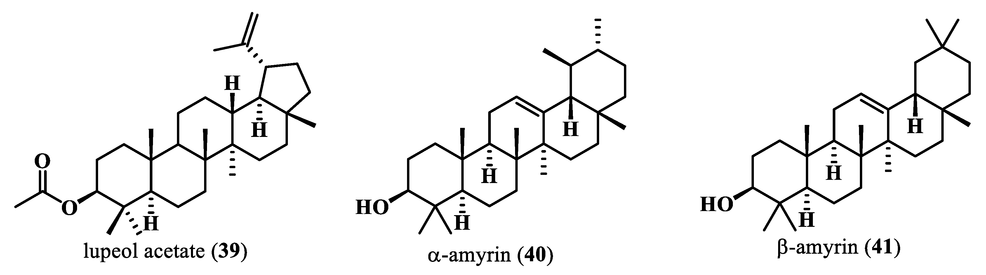
  62. Fongang Fotsing, Y.S.; Bankeu Kezetas, J.J. Terpenoids as Important Bioactive Constituents of Essential Oils. In Essential Oils—Bioactive Compounds, New Perspectives and Applications; Santana de Oliveira, M., Almeida da Costa, W., Gomes Silva, S., Eds.; IntechOpen: London, UK, 2020. [Google Scholar] [CrossRef]
  63. Boncan, D.A.T.; Tsang, S.S.; Li, C.; Lee, I.H.; Lam, H.-M.; Chan, T.-F.; Hui, J.H. Terpenes and terpenoids in plants: Interactions with environment and insects. Int. J. Mol. Sci. 2020, 21, 7382. [Google Scholar] [CrossRef] [PubMed]
  64. Singh, A.K.; Rana, H.K.; Tshabalala, T.; Kumar, R.; Gupta, A.; Ndhlala, A.R.; Pandey, A.K. Phytochemical, nutraceutical and pharmacological attributes of a functional crop Moringa oleifera Lam: An overview. South. Afr. J. Bot. 2020, 129, 209–220. [Google Scholar] [CrossRef]
  65. Korniluk, A.; Koper, O.; Kemona, H.; Dymicka-Piekarska, V. From inflammation to cancer. Ir. J. Med. Sci. 2017, 186, 57–62. [Google Scholar] [CrossRef]
  66. Germolec, D.R.; Shipkowski, K.A.; Frawley, R.P.; Evans, E. Markers of Inflammation. In Immunotoxicity Testing: Methods and Protocols, Methods in Molecular Biology; Humana Press: New York, NY, USA, 2018; pp. 57–79. [Google Scholar] [CrossRef]
  67. Rajan, T.S.; Giacoppo, S.; Iori, R.; De Nicola, G.R.; Grassi, G.; Pollastro, F.; Bramanti, P.; Mazzon, E. Anti-inflammatory and antioxidant effects of a combination of cannabidiol and moringin in LPS-stimulated macrophages. Fitoterapia 2016, 112, 104–115. [Google Scholar] [CrossRef]
  68. Ribaudo, G.; Povolo, C.; Zagotto, G. Moringa oleifera Lam.: A Rich Source of Phytoactives for the Health of Human Being. In Studies in Natural Products Chemistry, 1st ed.; Rahman, A., Ed.; Elsevier: Amsterdam, The Netherlands, 2019. [Google Scholar] [CrossRef]
  69. Lazou, A.; Ikonomidis, I.; Bartekova, M.; Benedek, T.; Makavos, G.; Palioura, D.; Fuentes, H.C.; Andreadou, I. Chronic inflammatory diseases, myocardial function and cardioprotection. Br. J. Pharmacol. 2020, 177, 5357–5374. [Google Scholar] [CrossRef]
  70. Mason, J.C.; Libby, P. Cardiovascular disease in patients with chronic inflammation: Mechanisms underlying premature cardiovascular events in rheumatologic conditions. Eur. Hear. J. 2015, 36, 482–489. [Google Scholar] [CrossRef]
  71. Raaby, L.; Ahlehoff, O.; de Thurah, A. Psoriasis and cardiovascular events: Updating the evidence. Arch. Dermatol. Res. 2017, 309, 225–228. [Google Scholar] [CrossRef] [PubMed]
  72. Sun, H.-H.; Tian, F. Inflammatory bowel disease and cardiovascular disease incidence and mortality: A meta-analysis. Eur. J. Prev. Cardiol. 2018, 25, 1623–1631. [Google Scholar] [CrossRef] [PubMed]
  73. Grundy, S.M.; Stone, N.J.; Bailey, A.L.; Beam, C.; Birtcher, K.K.; Blumenthal, R.S.; Braun, L.T.; de Ferranti, S.; Faiella-Tommasino, J.; Forman, D.E.; et al. 2018 AHA/ACC/AACVPR/AAPA/ABC/ACPM/ADA/AGS/APhA/ASPC/NLA/PCNA Guideline on the Management of Blood Cholesterol: A Report of the American College of Cardiology/American Heart Association Task Force on Clinical Practice Guidelines. Circulation 2019, 139, E1082–E1143. [Google Scholar] [CrossRef]
  74. Aksentijevich, M.; Lateef, S.S.; Anzenberg, P.; Dey, A.K.; Mehta, N.N. Chronic inflammation, cardiometabolic diseases and effects of treatment: Psoriasis as a human model. Trends Cardiovasc. Med. 2020, 30, 472–478. [Google Scholar] [CrossRef] [PubMed]
  75. Mehta, N.N.; Dey, A.K.; Maddineni, R.; Kraus, W.E.; Huffman, K.M. GlycA measured by NMR spectroscopy is associated with disease activity and cardiovascular disease risk in chronic inflammatory diseases. Am. J. Prev. Cardiol. 2020, 4, 100120. [Google Scholar] [CrossRef] [PubMed]
  76. Deng, Y.; Li, G.; Chang, D.; Su, X. YKL-40 as a novel biomarker in cardio-metabolic disorders and inflammatory diseases. Clin. Chim. Acta 2020, 511, 40–46. [Google Scholar] [CrossRef] [PubMed]
  77. Adefegha, S.A.; Oboh, G.; Iyoha, A.E.; Oyagbemi, A.A. Comparative effects of horseradish (Moringa oleifera) leaves and seeds on blood pressure and crucial enzymes relevant to hypertension in rat. PharmaNutrition 2019, 9, 100152. [Google Scholar] [CrossRef]
  78. Nelin, L.D.; Stenger, M.R.; Malleske, D.T.; Chicoine, L.G. Vascular Arginase and Hypertension. Curr. Hypertens. Rev. 2007, 3, 242–249. [Google Scholar] [CrossRef]
  79. Cheenpracha, S.; Park, E.-J.; Yoshida, W.Y.; Barit, C.; Wall, M.; Pezzuto, J.M.; Chang, L.C. Potential anti-inflammatory phenolic glycosides from the medicinal plant Moringa oleifera fruits. Bioorganic Med. Chem. 2010, 18, 6598–6602. [Google Scholar] [CrossRef]
  80. Gilani, A.H.; Aftab, K.; Suria, A.; Siddiqui, S.; Salem, R.; Siddiqui, B.S.; Faizi, S. Pharmacological Studies on Hypotensive and Spasmolytic Activities of Pure Compounds from Moringa oleifera. Phytother. Res. 1994, 8, 87–91. [Google Scholar] [CrossRef]
  81. Zibrila, A.I.; Li, Y.; Wang, Z.; Zhao, G.; Liu, H.; Leng, J.; Ali, A.; Osei, J.A.; Kang, Y.-M.; Liu, J. Acetylcholinesterase inhibition with pyridostigmine attenuates hypertension and neuroinflammation in the paraventricular nucleus in rat model for Preeclampsia. Int. Immunopharmacol. 2021, 101, 108365. [Google Scholar] [CrossRef] [PubMed]
  82. Nebbioso, M.; Pascarella, A.; Cavallotti, C.; Pescosolido, N. Monoamine oxidase enzymes and oxidative stress in the rat optic nerve: Age-related changes. Int. J. Exp. Pathol. 2012, 93, 401–405. [Google Scholar] [CrossRef] [PubMed]
  83. Elhwuegi, A. The Wonders of Phosphodiesterase-5 Inhibitors: A Majestic History. Ann. Med. Heal. Sci. Res. 2016, 6, 139–145. [Google Scholar] [CrossRef] [PubMed]
  84. Oboh, G.; Oyeleye, S.I.; Akintemi, O.A.; Olasehinde, T.A. Moringa oleifera supplemented diet modulates nootropic-related biomolecules in the brain of STZ-induced diabetic rats treated with acarbose. Metab. Brain Dis. 2018, 33, 457–466. [Google Scholar] [CrossRef] [PubMed]
  85. Clarke, N.E.; Turner, A.J. Angiotensin-Converting Enzyme 2: The First Decade. Int. J. Hypertens. 2012, 2012, 307315. [Google Scholar] [CrossRef] [PubMed]
  86. Oboh, G.; Ademiluyi, A.O.; Ademosun, A.O.; Olasehinde, T.A.; Oyeleye, S.I.; Boligon, A.A.; Athayde, M.L. Phenolic Extract fromMoringa oleifera Leaves Inhibits Key Enzymes Linked to Erectile Dysfunction and Oxidative Stress in Rats’ Penile Tissues. Biochem. Res. Int. 2015, 2015, 175950. [Google Scholar] [CrossRef] [PubMed]
  87. Ramamurthy, S.; Varghese, S.; Sudarsan, S.; Muruganandhan, J.; Mushtaq, S.; Patil, P.B.; Raj, A.T.; Zanza, A.; Testarelli, L.; Patil, S. Moringa oleifera: Antioxidant, Anticancer, Anti-inflammatory, and Related Properties of Extracts in Cell Lines: A Review of Medicinal Effects, Phytochemistry, and Applications. J. Contemp. Dent. Pract. 2021, 22, 1483–1492. [Google Scholar] [CrossRef]
  88. Alia, F.; Putri, M.; Anggraeni, N.; A Syamsunarno, M.R.A. The Potency of Moringa oleifera Lam. as Protective Agent in Cardiac Damage and Vascular Dysfunction. Front. Pharmacol. 2022, 12, 724439. [Google Scholar] [CrossRef]
  89. Aziz, M.; Yadav, K. Pathogenesis of Atherosclerosis A Review. Med. Clin. Rev. 2016, 2, 22. [Google Scholar] [CrossRef]
  90. Frąk, W.; Wojtasińska, A.; Lisińska, W.; Młynarska, E.; Franczyk, B.; Rysz, J. Pathophysiology of Cardiovascular Diseases: New Insights into Molecular Mechanisms of Atherosclerosis, Arterial Hypertension, and Coronary Artery Disease. Biomedicines 2022, 10, 1938. [Google Scholar] [CrossRef]
  91. Eller-Vainicher, C.; Cairoli, E.; Grassi, G.; Grassi, F.; Catalano, A.; Merlotti, D.; Falchetti, A.; Gaudio, A.; Chiodini, I.; Gennari, L. Pathophysiology and Management of Type 2 Diabetes Mellitus Bone Fragility. J. Diabetes Res. 2020, 2020, 7608964. [Google Scholar] [CrossRef] [PubMed]
  92. Fagninou Nonsito, A.; Ulbad, T.P.; Magloire, N.; Ruffine, F.; Koutinhouin, G.B.; Akadiri, Y. Diabetes mellitus: Classification, epidemiology, physiopathology, immunology, risk factors, prevention and nutrition. Int. J. Adv. Res. 2019, 7, 855–863. [Google Scholar]
  93. Liu, H.; Cao, M.-M.; Wang, Y.; Li, L.-C.; Zhu, L.-B.; Xie, G.-Y.; Li, Y.-B. Endoplasmic reticulum stress is involved in the connection between inflammation and autophagy in type 2 diabetes. Gen. Comp. Endocrinol. 2015, 210, 124–129. [Google Scholar] [CrossRef] [PubMed]
  94. Trivedi, P.M.; Fynch, S.; Kennedy, L.M.; Chee, J.; Krishnamurthy, B.; O’reilly, L.A.; Strasser, A.; Kay, T.W.H.; Thomas, H.E. Soluble FAS ligand is not required for pancreatic islet inflammation or beta-cell destruction in non-obese diabetic mice. Cell Death Discov. 2019, 5, 136. [Google Scholar] [CrossRef] [PubMed]
  95. Adedapo, A.A.; Ogunmiluyi, I.O.; Falayi, O.O.; Ogunpolu, B.S.; Oyagbemi, A.A.; Orishadipe, A.; Omobowale, T.O.; Yakubu, M.A.; Oguntibeju, O.O. The lyophilized aqueous leaf extract of Moringa oleifera blunts streptozocin-induced diabetes in rats through upregulation of GLUT 4 signaling pathway and anti-oxidant effect. Sci. Afr. 2020, 10, e00619. [Google Scholar] [CrossRef]
  96. Bao, Y.; Xiao, J.; Weng, Z.; Lu, X.; Shen, X.; Wang, F. A phenolic glycoside from Moringa oleifera Lam. improves the carbohydrate and lipid metabolisms through AMPK in db/db mice. Food Chem. 2020, 311, 125948. [Google Scholar] [CrossRef]
  97. Wu, H.; Deng, X.; Shi, Y.; Su, Y.; Wei, J.; Duan, H. PGC-1α, glucose metabolism and type 2 diabetes mellitus. J. Endocrinol. 2016, 229, R99–R115. [Google Scholar] [CrossRef]
  98. Gao, T.; Chen, S.; Han, Y.; Zhang, D.; Tan, Y.; He, Y.; Liu, M. Ameliorating Inflammation in Insulin-resistant Rat Adipose Tissue with Abdominal Massage Regulates SIRT1/NF-κB Signaling. Cell Biochem. Biophys. 2022, 80, 579–589. [Google Scholar] [CrossRef]
  99. Njume, C.; Donkor, O.; McAinch, A.J. Predisposing factors of type 2 diabetes mellitus and the potential protective role of native plants with functional properties. J. Funct. Foods 2019, 53, 115–124. [Google Scholar] [CrossRef]
  100. Praparatana, R.; Maliyam, P.; Barrows, L.R.; Puttarak, P. Flavonoids and Phenols, the Potential Anti-Diabetic Compounds from Bauhinia strychnifolia Craib. Stem. Molecules 2022, 27, 2393. [Google Scholar] [CrossRef]
  101. Bharti, S.K.; Krishnan, S.; Kumar, A.; Kumar, A. Antidiabetic phytoconstituents and their mode of action on metabolic pathways. Ther. Adv. Endocrinol. Metab. 2018, 9, 81–100. [Google Scholar] [CrossRef] [PubMed]
  102. Tarmizi, A.A.A.; Ramli, N.N.N.; Adam, S.H.; Mutalib, M.A.; Mokhtar, M.H.; Tang, S.G.H. Phytofabrication of Selenium Nanoparticles with Moringa oleifera (MO-SeNPs) and Exploring Its Antioxidant and Antidiabetic Potential. Molecules 2023, 28, 5322. [Google Scholar] [CrossRef] [PubMed]
  103. Shabab, S.; Gholamnezhad, Z.; Mahmoudabady, M. Protective effects of medicinal plant against diabetes induced cardiac disorder: A review. J. Ethnopharmacol. 2021, 265, 113328. [Google Scholar] [CrossRef] [PubMed]
  104. Xiong, W.; Ma, H.; Zhang, Z.; Jin, M.; Wang, J.; Xu, Y.; Wang, Z. The protective effect of icariin and phosphorylated icariin against LPS-induced intestinal epithelial cells injury. Biomed. Pharmacother. 2019, 118, 109246. [Google Scholar] [CrossRef] [PubMed]
  105. Luo, H.; Li, M.; Wang, F.; Yang, Y.; Wang, Q.; Zhao, Y.; Du, F.; Chen, Y.; Shen, J.; Zhao, Q.; et al. The role of intestinal stem cell within gut homeostasis: Focusing on its interplay with gut microbiota and the regulating pathways. Int. J. Biol. Sci. 2022, 18, 5185–5206. [Google Scholar] [CrossRef] [PubMed]
  106. Passos, M.D.C.F.; Moraes-Filho, J.P. Intestinal microbiota in digestive diseases. Arq. De Gastroenterol. 2017, 54, 255–262. [Google Scholar] [CrossRef] [PubMed]
  107. Salguero, M.V.; Al-Obaide, M.A.I.; Singh, R.; Siepmann, T.; Vasylyeva, T.L. Dysbiosis of Gram-negative gut microbiota and the associated serum lipopolysaccharide exacerbates inflammation in type 2 diabetic patients with chronic kidney disease. Exp. Ther. Med. 2019, 18, 3461–3469. [Google Scholar] [CrossRef] [PubMed]
  108. Guazelli, C.F.; Fattori, V.; Ferraz, C.R.; Borghi, S.M.; Casagrande, R.; Baracat, M.M.; Verri, W.A. Antioxidant and anti-inflammatory effects of hesperidin methyl chalcone in experimental ulcerative colitis. Chem. Interactions 2020, 333, 109315. [Google Scholar] [CrossRef]
  109. Liu, K.; Li, G.; Guo, W.; Zhang, J. The protective effect and mechanism of pedunculoside on DSS (dextran sulfate sodium) induced ulcerative colitis in mice. Int. Immunopharmacol. 2020, 88, 107017. [Google Scholar] [CrossRef]
  110. Saldanha, E.; Saxena, A.; Kaur, K.; Kalekhan, F.; Venkatesh, P.; Fayad, R.; Rao, S.; George, T.; Baliga, M.S. Polyphenols in the Prevention of Ulcerative Colitis: A Revisit. In Dietary Interventions in Gastrointestinal Diseases; Watson, R.R., Preedy, V.R., Eds.; Academic Press: Cambridge, MA, USA; Elsevier Inc.: London, UK, 2019; Volume 2, pp. 277–287. [Google Scholar] [CrossRef]
  111. Gholap, P.A.; Nirmal, S.A.; Pattan, S.R.; Pal, S.C.; Mandal, S.C. Potential of Moringa oleifera root and Citrus sinensis fruit rind extracts in the treatment of ulcerative colitis in mice. Pharm. Biol. 2012, 50, 1297–1302. [Google Scholar] [CrossRef]
  112. Kim, Y.; Wu, A.G.; Jaja-Chimedza, A.; Graf, B.L.; Waterman, C.; Verzi, M.P.; Raskin, I. Isothiocyanate-enriched moringa seed extract alleviates ulcerative colitis symptoms in mice. PLoS ONE 2017, 12, e0184709. [Google Scholar] [CrossRef] [PubMed]
  113. Nejabati, H.R.; Roshangar, L. Kaempferol: A potential agent in the prevention of colorectal cancer. Physiol. Rep. 2022, 10, e15488. [Google Scholar] [CrossRef] [PubMed]
  114. Tao, J.; Huang, Z.; Wang, Y.; Liu, Y.; Zhao, T.; Wang, Y.; Tian, L.; Cheng, G. Ethanolic Extract from Pteris wallichiana Alleviates DSS-Induced Intestinal Inflammation and Intestinal Barrier Dysfunction by Inhibiting the TLR4/NF-κB Pathway and Regulating Tight Junction Proteins. Molecules 2022, 27, 3093. [Google Scholar] [CrossRef] [PubMed]
  115. Park, M.-Y.; Ji, G.E.; Sung, M.-K. Dietary kaempferol suppresses inflammation of dextran sulfate sodium-induced colitis in mice. Dig. Dis. Sci. 2011, 57, 355–363. [Google Scholar] [CrossRef] [PubMed]
  116. Peng, L.; Gao, X.; Nie, L.; Xie, J.; Dai, T.; Shi, C.; Tao, L.; Wang, Y.; Tian, Y.; Sheng, J. Astragalin Attenuates Dextran Sulfate Sodium (DSS)-Induced Acute Experimental Colitis by Alleviating Gut Microbiota Dysbiosis and Inhibiting NF-κB Activation in Mice. Front. Immunol. 2020, 11, 2058. [Google Scholar] [CrossRef] [PubMed]
  117. Hong, Z.-S.; Xie, J.; Wang, X.-F.; Dai, J.-J.; Mao, J.-Y.; Bai, Y.-Y.; Sheng, J.; Tian, Y. Moringa oleifera Lam. Peptide Remodels Intestinal Mucosal Barrier by Inhibiting JAK-STAT Activation and Modulating Gut Microbiota in Colitis. Front. Immunol. 2022, 13, 924178. [Google Scholar] [CrossRef] [PubMed]
  118. Gluvic, Z.; Tomasevic, R.; Bojovic, K.; Obradovic, M.; Isenovic, E.R. Non-alcoholic fatty liver disease: A multidisciplinary clinical practice approach—The institutional adaptation to existing Clinical Practice Guidelines. Emerg. Crit. Care Med. 2021, 2, 12–22. [Google Scholar] [CrossRef]
  119. Rinella, M.E.; Tacke, F.; Sanyal, A.J.; Anstee, Q.M. Report on the AASLD/EASL joint workshop on clinical trial endpoints in NAFLD. J. Hepatol. 2019, 71, 823–833. [Google Scholar] [CrossRef]
  120. Wang, X.J.; Malhi, H. Nonalcoholic Fatty Liver Disease. Ann. Intern. Med. 2018, 169, ITC65–ITC80. [Google Scholar] [CrossRef]
  121. Carr, R.M.; Oranu, A.; Khungar, V. Nonalcoholic Fatty Liver Disease: Pathophysiology and management. Gastroenterol. Clin. North. Am. 2016, 45, 639–652. [Google Scholar] [CrossRef]
  122. Sweet, P.H.; Khoo, T.; Nguyen, S. Nonalcoholic Fatty Liver Disease. Prim. Care Clin. Off. Primers 2017, 44, 599–607. [Google Scholar] [CrossRef] [PubMed]
  123. Nguyen, X.-V.K.; Zhang, J.; Chin, K.L.; Bloom, S.; Nicoll, A.J. Is Hepatocellular Carcinoma in Fatty Liver Different to Non-Fatty Liver? Nutrients 2022, 14, 3875. [Google Scholar] [CrossRef] [PubMed]
  124. Manne, V.; Handa, P.; Kowdley, K.V. Pathophysiology of Nonalcoholic Fatty Liver Disease/Nonalcoholic Steatohepatitis. Clin. Liver Dis. 2018, 22, 23–37. [Google Scholar] [CrossRef]
  125. Petrelli, F.; Manara, M.; Colombo, S.; De Santi, G.; Ghidini, M.; Mariani, M.; Iaculli, A.; Rausa, E.; Rampulla, V.; Arru, M.; et al. Hepatocellular carcinoma in patients with nonalcoholic fatty liver disease: A systematic review and meta-analysis: HCC and Steatosis or Steatohepatitis. Neoplasia 2022, 30, 100809. [Google Scholar] [CrossRef] [PubMed]
  126. Püschel, G.P.; Klauder, J.; Henkel, J. Macrophages, Low-Grade Inflammation, Insulin Resistance and Hyperinsulinemia: A Mutual Ambiguous Relationship in the Development of Metabolic Diseases. J. Clin. Med. 2022, 11, 4358. [Google Scholar] [CrossRef] [PubMed]
  127. Zhang, D.; Wei, Y.; Huang, Q.; Chen, Y.; Zeng, K.; Yang, W.; Chen, J.; Chen, J. Important Hormones Regulating Lipid Metabolism. Molecules 2022, 27, 7052. [Google Scholar] [CrossRef]
  128. Berndt, N.; Hudert, C.A.; Eckstein, J.; Loddenkemper, C.; Henning, S.; Bufler, P.; Meierhofer, D.; Sack, I.; Wiegand, S.; Wallach, I.; et al. Alterations of Central Liver Metabolism of Pediatric Patients with Non-Alcoholic Fatty Liver Disease. Int. J. Mol. Sci. 2022, 23, 11072. [Google Scholar] [CrossRef]
  129. Palma, R.; Pronio, A.; Romeo, M.; Scognamiglio, F.; Ventriglia, L.; Ormando, V.M.; Lamazza, A.; Pontone, S.; Federico, A.; Dallio, M. The Role of Insulin Resistance in Fueling NAFLD Pathogenesis: From Molecular Mechanisms to Clinical Implications. J. Clin. Med. 2022, 11, 3649. [Google Scholar] [CrossRef]
  130. Asgari-Kafrani, A.; Fazilati, M.; Nazem, H. Hepatoprotective and antioxidant activity of aerial parts of Moringa oleifera in prevention of non-alcoholic fatty liver disease in Wistar rats. S. Afr. J. Bot. 2019, 129, 82–90. [Google Scholar] [CrossRef]
  131. Chen, Z.; Tian, R.; She, Z.; Cai, J.; Li, H. Role of oxidative stress in the pathogenesis of nonalcoholic fatty liver disease. Free Radic. Biol. Med. 2020, 152, 116–141. [Google Scholar] [CrossRef]
  132. Ma, Y.; Lee, G.; Heo, S.-Y.; Roh, Y.-S. Oxidative stress is a key modulator in the development of nonalcoholic fatty liver disease. Antioxidants 2021, 11, 91. [Google Scholar] [CrossRef] [PubMed]
  133. Morgan, C.; Sáez-Briones, P.; Barra, R.; Reyes, A.; Zepeda-Morales, K.; Constandil, L.; Ríos, M.; Ramírez, P.; Burgos, H.; Hernández, A. Prefrontal Cortical Control of Activity in Nucleus Accumbens Core Is Weakened by High-Fat Diet and Prevented by Co-Treatment with N-Acetylcysteine: Implications for the Development of Obesity. Int. J. Mol. Sci. 2022, 23, 10089. [Google Scholar] [CrossRef] [PubMed]
  134. Cho, A.-S.; Jeon, S.-M.; Kim, M.-J.; Yeo, J.; Seo, K.-I.; Choi, M.-S.; Lee, M.-K. Chlorogenic acid exhibits anti-obesity property and improves lipid metabolism in high-fat diet-induced-obese mice. Food Chem. Toxicol. 2010, 48, 937–943. [Google Scholar] [CrossRef] [PubMed]
  135. Jiménez-Cortegana, C.; García-Galey, A.; Tami, M.; del Pino, P.; Carmona, I.; López, S.; Alba, G.; Sánchez-Margalet, V. Role of leptin in non-alcoholic fatty liver disease. Biomedicines 2021, 9, 762. [Google Scholar] [CrossRef] [PubMed]
  136. Francisco, V.; Sanz, M.J.; Real, J.T.; Marques, P.; Capuozzo, M.; Eldjoudi, D.A.; Gualillo, O. Adipokines in Non-Alcoholic Fatty Liver Disease: Are We on the Road toward New Biomarkers and Therapeutic Targets? Biology 2022, 11, 1237. [Google Scholar] [CrossRef] [PubMed]
  137. Almatrafi, M.M.; Vergara-Jimenez, M.; Murillo, A.G.; Norris, G.H.; Blesso, C.N.; Fernandez, M.L. Moringa leaves prevent hepatic lipid accumulation and inflammation in guinea pigs by reducing the expression of genes involved in lipid metabolism. Int. J. Mol. Sci. 2017, 18, 1330. [Google Scholar] [CrossRef]
  138. Li, X.-J.; Mu, Y.-M.; Li, T.-T.; Yang, Y.-L.; Zhang, M.-T.; Li, Y.-S.; Zhang, W.K.; Tang, H.-B.; Shang, H.-C. Gynura procumbens Reverses Acute and Chronic Ethanol-Induced Liver Steatosis through MAPK/SREBP-1c-Dependent and -Independent Pathways. J. Agric. Food Chem. 2015, 63, 8460–8471. [Google Scholar] [CrossRef]
  139. Waterman, C.; Cheng, D.M.; Rojas-Silva, P.; Poulev, A.; Dreifus, J.; Lila, M.A.; Raskin, I. Stable, water extractable isothiocyanates from Moringa oleifera leaves attenuate inflammation in vitro. Phytochemistry 2014, 103, 114–122. [Google Scholar] [CrossRef]
  140. Joung, H.; Kim, B.; Park, H.; Lee, K.; Kim, H.-H.; Sim, H.-C.; Do, H.-J.; Hyun, C.-K.; Do, M.-S. Fermented Moringa oleifera Decreases Hepatic Adiposity and Ameliorates Glucose Intolerance in High-Fat Diet-Induced Obese Mice. J. Med. Food 2017, 20, 439–447. [Google Scholar] [CrossRef]
Figure 2. Volatile compounds and fatty acids from M. oleifera.
Plants 13 00020 g002
Figure 3. Polyphenolic compounds from M. oleifera.
Plants 13 00020 g003
Figure 4. Sulfur compounds from M. oleifera.
Plants 13 00020 g004
Figure 5. Carotenoidic compounds from M. oleifera.
Plants 13 00020 g005
Figure 6. Alkaloids isolated from M. oleifera.
Plants 13 00020 g006
Figure 7. Terpenoids isolated from M. oleifera.
Plants 13 00020 g007
Table 1. Bioactive compounds from M. oleifera, their bioactivity and therapeutic characteristics (in vivo and in vitro activities).
Part Used/Type of ExtractExperimental ModelModel of StudyDuration of the TreatmentDosageControlExtracts/Secondary
Metabolites
Bioactivity/Therapeutic CharacteristicsRef.
Leaves, fruits, and seeds/aqueous extractsThe experiment investigated the capacity to restrain oxidative DNA damage, antioxidant, and anti-quorum sensing (QS) capabilities -5–10 µg/mL
50 mg/mL
Positive control: furanone and garlic extractPolyphenolic compounds (aqueous extracts)Inhibition of proteins and lipid oxidation
Antioxidant activities: nitric oxide and superoxide anion scavenging activities
[14]
LeavesThe study screened the functional metabolites of M. oleifera to evaluate their possible role in nerve regeneration after injuryin vivo
Male albino mice
12 days2 g/kg bwControl: standard dietLeaf extracts in n-Hexane, dichloromethane, ethyl acetate, ethanol, and methanolNerve regeneration (restoration of sensory and motor function)
Oxidative stress management
[7]
Defatted seed flourThe study assessed the antioxidant and antibacterial activities of free and bound phenolic extractsin vitro
B. cereus
S. aureus
E. coli
Y. enterocolitica
-0.5–15.0 mg in 150 µL
0.5–1.5 mg/mL
Positive control: ascorbic acidPhenolic compounds (extracts in ethanol, methanol, acetone, hexane, and chloroform)Antioxidant, antimicrobial activities[15]
SeedsThe experiments evaluated the antibacterial and antifungal activities of secondary metabolitesin vitro
Bacteria:
S. aureus
S. epidermidis
E. coli
E. aerogenes
K. pneumonia
P. aeruginosa
B. subtilis
Fungal strains:
C. albicans
T. rubrum
E. floccosum
-10 mg/mL
1 mg/mL
100 µg/mL
10 µg/mL
Positive control: Ofloxacin and Clotrimazole
Negative control: sterile distilled water
Glucosinolates (secondary metabolites extracts in acetone in CH2Cl2)Antimicrobial activities[16]
SeedsThe study evaluated the antitumoral activity of eight isolate compounds from M. oleiferain vitro
EBV genome carrying lymphoblastoid cell

in vivo
Specific pathogen-free female ICR mice
-


20 weeks
100, 10, 1, and 0.1 µg/mL


85 nmol in 0.1 mL acetone
Positive control: n-butyric acid


Negative control: 12-O-tetradecanoyl-phorbol-13-acetate (TPA)


Glucosinolates, isothiocyanates, and sterols
Antitumor promoting activities[17]
SeedsThe experiments assessed the antimicrobial activity of a polymeric, naturally extracted M. oleifera oil bionanocomposite film enriched with silver nanoparticlesin vitro
S. aureus,
E. coli
K. pneumoniae
S. typhi
P. aeruginosa
S. flexneri
C. albicans
-5–10 wt%Ciprofloxacin and FluconazoleFatty acids, sterols, alkanes, and alcohol compoundsAntibacterial activity[18]
SeedsThe study assessed the cytotoxic activity of seed essential oil obtained from M. oleiferain vitro
HeLa, HepG2, MCF-7, CACO-2, and L929 cell lines
24 h0.15 to 1.0 mg/mLDMSOEssential oils (extracted from seeds through cold pressing)Antiproliferative activity[19]
Flowers PC3 cell lines 0.01–100 µg/mLDMSOMethanol extractsAnticancer activity[20]
LeavesThe study has investigated the anticancer activity of the M. oleifera leaf extractin vitro
murine Non-Hodgkin Lymphoma (NHL)

in vivo
Balb/c mice
24 h


16 days
100 to 450 µg/mL


100 and 200 mg/kg


Healthy mice without Dalton’s lymphoma cell transplantation


Methanol-based leaf extract


Triggers apoptosis and inhibits the growth of Dalton’s lymphoma


[21]
SeedsExperiments assessed the antioxidant, antihypertensive, and potential cardioprotective properties of bioactive peptides

in vivo
Wistar rats
-


-
-

200 mg/kg
-


-
Peptides obtained through enzymatic hydrolysis of M. oleifera seed (shorter peptides (1–3 kDa) and longer peptides (>10 kDa))Oxidative stress management;

antihypertensive, and cardioprotective properties
[22,23]
SeedsThe study compared the structural and functional properties of albumin and globulin in M. oleifera seeds with those of the isoelectric pH-precipitated protein isolate--6.25 mg/mL-Globulins, Albumins, Iso–electric precipitated isolatesReduce free radicals
Globulin-enhanced metal ion chelation activity
Antioxidant properties
[24]
SeedsThe experiments compared the antioxidant and angiotensin-converting enzyme (ACE) inhibitory properties of M. oleifera seed protein isolate (ISO)----Protein hydrolysate fractions (<1 kDa, 3–5 kDa, and 5–10 kDa)Antioxidative properties
ACE inhibition;
[25]
SeedsThe study evaluated the biofunctional properties of total hydrolysates and peptide fractions from protein isolates of moringa seedsin vitro
ACE
extracted from rabbit lungs
5 h--Protein hydrolysate fractions >10 kDa)Antioxidant, antihypertensive, and antidiabetic properties;[26]
SeedsThe study evaluated the nutritional composition of Indian M. oleifera seed, the antioxidant activity of its polypeptides, and the protective effects on H2O2 oxidative-damaged Chang liver cellsin vitro
Chang liver cell line
3 h100, 300, and 500 µMPBSPeptide isolates and hydrolysate
Fractions (>3.5 kDa) PFE, GY, YTR, QY, FG, SF, IN, SP, YFE, IY, LY
Oxidative stress management
Antioxidant activity
Hepatoprotector
[27,28]
SeedsSeed powder was extracted in hexane, petroleum ether, ethyl acetate, or methanol, and the study evaluated the extracts antimicrobial activityin vitro
E. coli
P. aeruginosa
S. aureus
C. cladosporioides
P. sclerotigenum
---Glucosinolates and isothiocyanatesFlocculating and antimicrobial activities[29]
SeedsThe experiments assessed the antimicrobial activities of isolated compounds from seed extracts of M. oleifera and their synergistic effect through a hybridized complex of organic–inorganic composite materialsin vitro
S. aureus
E. coli
P. aeruginosa
C. albicans
A. niger


24 h (37 °C)

48 h (25 °C)
5 days (25 °C)
5 mg-Glucosinolates and isothiocyanates
(ethanol, methanol, hexane, acetate ethanol extracts)
Antimicrobial activities[30]
SeedsThe study evaluated the antimicrobial activities of 4-(α-l-rhamnosyloxy) benzyl glucosinolate isolated from M. oleifera seed and its protective effect on an experimental model of spinal cord injuryin vitro
S. aureus
E. casseliflavus
C. albicans


in vivo
Male adult C57Bl/6 mice
24 h


8 days
-


10 mg/kg
PBS

Gentamicin Chloramphenicol


Negative control: naive group (no treatment)
Positive control: GMG-ITC (control group)
Glucosinolates and isothiocyanatesAntibiotic activity


Nerve regeneration
[31,32,33]
LeavesThe study evaluated the anxiolytic and anti-colitis effects of M. oleifera leaf-aqueous extract on acetic acid-induced colon inflammation in ratin vivo
Adult albino Wistar rats
20 days25, 50, and 100 mg/kgLoperamide-Anxiolytic, anti-inflammatory
Antioxidant and anti-colitis properties
[3]
SeedsThe study evaluated the dietary isothiocyanate-enriched moringa seed extract on glucose tolerance in a high fat diet mouse model and its modulatory activity on the gut microbiomein vivo
Male Sprague–Dawley rats
12 weeks0.54 and 0.73% of moringa seed extract containing moringa isothiocyanate-1Vehicle control: 15% sodium carboxymethyl celluloseGlucosinolates, phenolic
glycosides, flavonoids, and carbohydrates
Anti-inflammatory
Antioxidant properties
Improves glucose tolerance and modulates the gut microbiome
[34,35]
Legend: DPPH: 2,2-diphenyl-1-picrylhydrazyl; HeLa: human cervical cancer; HepG2: human hepatocellular carcinoma; ACE: angiotensin-I converting enzyme; Amino acid nomenclature: I: isoleucine; S: serine; Y: tyrosine; F: phenylalanine; E: glutamic acid; N: asparagine; Q: glutamine; G: glycine; P: proline; L: leucine; T: threonine; R: arginine.
Disclaimer/Publisher’s Note: The statements, opinions and data contained in all publications are solely those of the individual author(s) and contributor(s) and not of MDPI and/or the editor(s). MDPI and/or the editor(s) disclaim responsibility for any injury to people or property resulting from any ideas, methods, instructions or products referred to in the content.

