

Mass Spectrometry-Based Proteomics of Human Milk to Identify Differentially Expressed Proteins in Women with Breast Cancer versus Controls


Abstract
:1. Introduction
2. Materials and Methods
2.1. Human Subjects and Milk Samples
2.2. Reagents
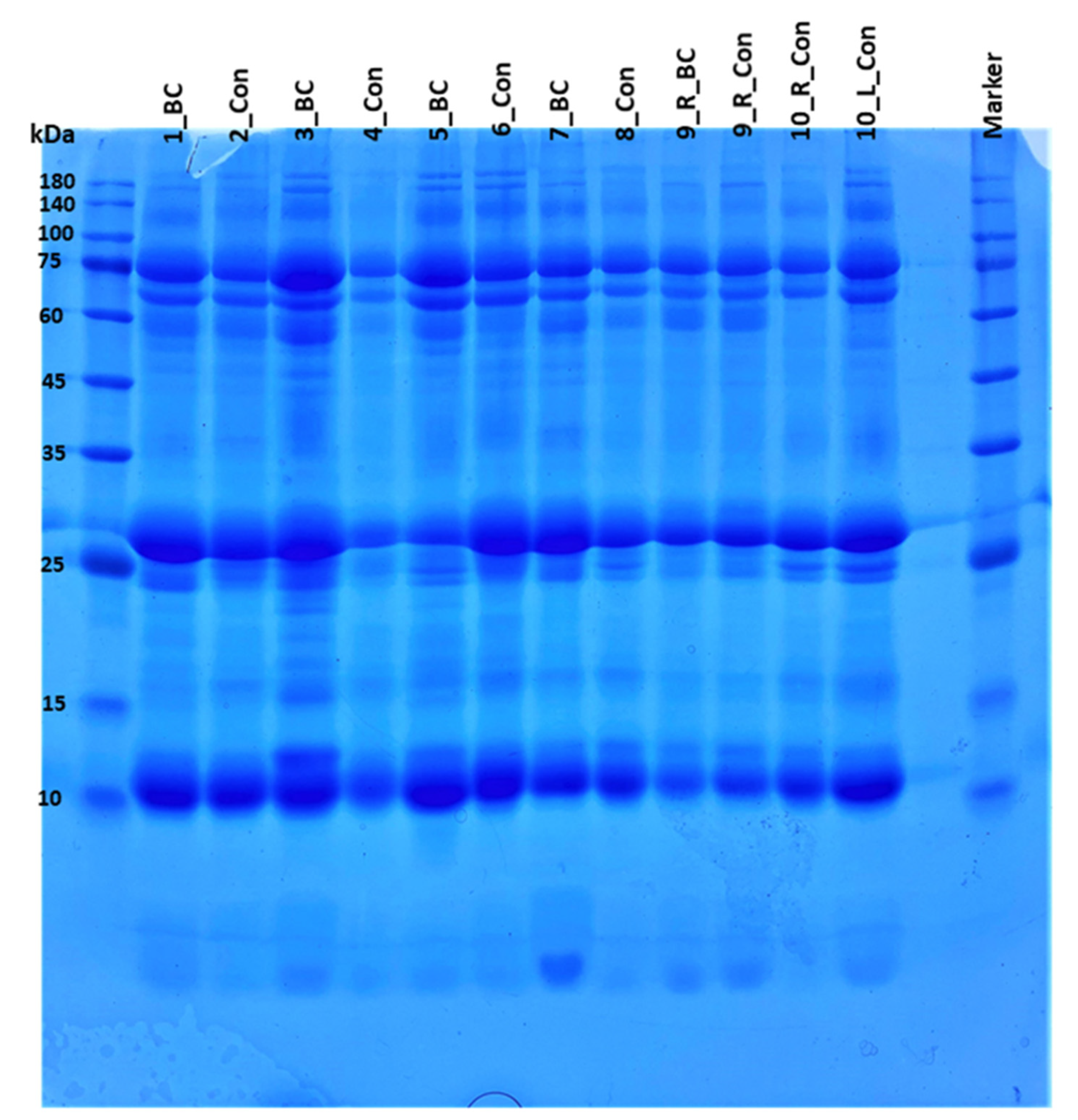
2.3. MS-Based Proteomics Analysis
2.4. Data Availability
3. Results and Discussion
3.1. Differentially Expressed Proteins in BC vs. Control That Were Identified in the Current Study (and Also Identified Erentially Expressed in Our Previous Studies on Human Milk)
3.2. Dysregulated Proteins Specific to the Current Study
4. Conclusions
5. Limitations
Supplementary Materials
Author Contributions
Funding
Institutional Review Board Statement
Informed Consent Statement
Data Availability Statement
Acknowledgments
Conflicts of Interest
Abbreviations
References
- Arcaro, K.F.; Browne, E.P.; Qin, W.; Zhang, K.; Anderton, D.L.; Sauter, E.R. Differential expression of cancer–related proteins in paired breast milk samples from women with breast cancer. J. Hum. Lact. 2012, 28, 543–546. [Google Scholar] [CrossRef] [PubMed]
- Qin, W.; Zhang, K.; Kliethermes, B.; Ruhlen, R.L.; Browne, E.P.; Arcaro, K.F.; Sauter, E.R. Differential expression of cancer associated proteins breast milk based on age at first full term pregnancy. BMC Cancer 2012, 12, 100. [Google Scholar] [CrossRef] [PubMed] [Green Version]
- Yang, H.P.; Schneider, S.S.; Chisholm, C.M.; Browne, E.P.; Mahmood, S.; Gierach, G.L.; Lenington, S.; Anderton, D.L.; Sherman, M.E.; Arcaro, K.F. Association of TGF–β2 levels in breast milk with severity of breast biopsy diagnosis. Cancer Causes Control 2015, 26, 345–354. [Google Scholar] [CrossRef] [Green Version]
- Schneider, S.S.; Aslebagh, R.; Wetie, A.G.N.; Sturgeon, S.R.; Darie, C.C.; Arcaro, K.F. Using breast milk to assess breast cancer risk: The role of mass spectrometry–based proteomics. Adv. Exp. Med. Biol. 2014, 806, 399–408. [Google Scholar] [PubMed]
- Aslebagh, R.; Channaveerappa, D.; Arcaro, K.F.; Darie, C.C. Proteomics analysis of human breast milk to assess breast cancer risk. Electrophoresis 2018, 39, 653–665. [Google Scholar] [CrossRef]
- Faupel-Badger, J.M.; Arcaro, K.F.; Balkam, J.J.; Eliassen, A.H.; Hassiotou, F.; Lebrilla, C.B.; Michels, K.B.; Palmer, J.R.; Schedin, P.; Stuebe, A.M. Postpartum remodeling, lactation, and breast cancer risk: Summary of a National Cancer Institute—Sponsored workshop. J. Natl. Cancer Inst. 2013, 105, 166–174. [Google Scholar] [CrossRef] [Green Version]
- Woods, A.G.; Sokolowska, I.; Wetie, A.G.N.; Wormwood, K.; Aslebagh, R.; Patel, S.; Darie, C.C. Mass spectrometry for proteomics–based investigation. In Advancements of Mass Spectrometry in Biomedical Research; Springer: Cham, Switzerland, 2014; pp. 1–32. [Google Scholar]
- Afzal, S.; Hassan, M.; Ullah, S.; Abbas, H.; Tawakkal, F.; Khan, M.A. Breast Cancer; Discovery of Novel Diagnostic Biomarkers, Drug Resistance, and Therapeutic Implications. Front. Mol. Biosci. 2022, 9, 783450. [Google Scholar] [CrossRef] [PubMed]
- Li, J.; Guan, X.; Fan, Z.; Ching, L.-M.; Li, Y.; Wang, X.; Cao, W.-M.; Liu, D.-X. Non–invasive biomarkers for early detection of breast cancer. Cancers 2020, 12, 2767. [Google Scholar] [CrossRef] [PubMed]
- Browne, E.P.; Punska, E.C.; Lenington, S.; Otis, C.N.; Anderton, D.L.; Arcaro, K.F. Increased promoter methylation in exfoliated breast epithelial cells in women with a previous breast biopsy. Epigenetics 2011, 6, 1425–1435. [Google Scholar] [CrossRef]
- Gu, Y.-Q.; Gong, G.; Xu, Z.-L.; Wang, L.-Y.; Fang, M.-L.; Zhou, H.; Xing, H.; Wang, K.-R. miRNA profiling reveals a potential role of milk stasis in breast carcinogenesis. Int. J. Mol. Med. 2014, 33, 1243–1249. [Google Scholar] [CrossRef]
- Thompson, P.; Kadlubar, F.; Vena, S.; Hill, H.; McClure, G.; McDaniel, L.; Ambrosone, C. Exfoliated ductal epithelial cells in human breast milk: A source of target tissue DNA for molecular epidemiologic studies of breast cancer. Cancer Epidemiol. Biomark. Prev. 1998, 7, 37–42. [Google Scholar]
- Wong, C.M.; Anderton, D.L.; Smith-Schneider, S.; Wing, M.A.; Greven, M.C.; Arcaro, K.F. Quantitative analysis of promoter methylation in exfoliated epithelial cells isolated from breast milk of healthy women. Epigenetics 2010, 5, 645–655. [Google Scholar] [CrossRef] [PubMed] [Green Version]
- Murphy, J.; Sherman, M.E.; Browne, E.P.; Caballero, A.I.; Punska, E.C.; Pfeiffer, R.M.; Yang, H.P.; Lee, M.; Yang, H.; Gierach, G.L. Potential of breastmilk analysis to inform early events in breast carcinogenesis: Rationale and considerations. Breast Cancer Res. Treat. 2016, 157, 13–22. [Google Scholar] [CrossRef] [PubMed] [Green Version]
- Aslebagh, R.; Channaveerappa, D.; Arcaro, K.F.; Darie, C.C. Comparative two-dimensional polyacrylamide gel electrophoresis (2D-PAGE) of human milk to identify dysregulated proteins in breast cancer. Electrophoresis 2018, 39, 1723–1734. [Google Scholar] [CrossRef]
- Ngounou Wetie, A.G.; Wormwood, K.L.; Russell, S.; Ryan, J.P.; Darie, C.C.; Woods, A.G. A pilot proteomic analysis of salivary biomarkers in autism spectrum disorder. Autism Res. 2015, 8, 338–350. [Google Scholar] [CrossRef]
- Sokolowska, I.; Dorobantu, C.; Woods, A.G.; Macovei, A.; Branza-Nichita, N.; Darie, C.C. Proteomic analysis of plasma membranes isolated from undifferentiated and differentiated HepaRG cells. Proteome Sci. 2012, 10, 47. [Google Scholar] [CrossRef] [Green Version]
- Nesvizhskii, A.I.; Keller, A.; Kolker, E.; Aebersold, R. A statistical model for identifying proteins by tandem mass spectrometry. Anal. Chem. 2003, 75, 4646–4658. [Google Scholar] [CrossRef]
- Burke, H.B. Predicting clinical outcomes using molecular biomarkers. Biomark. Cancer 2016, 6, 89–99. [Google Scholar] [CrossRef]
- Bártková, J.; Burchell, J.; Bártek, J.; Vojtěšek, B.; Taylor-Papadimitriou, J.; Rejthar, A.; Stašková, Z.; Kovařík, J. Lack of β–casein production by human breast tumours revealed by monoclonal antibodies. Eur. J. Cancer Clin. Oncol. 1987, 23, 1557–1563. [Google Scholar] [CrossRef]
- Bar, I.; Merhi, A.; Larbanoix, L.; Constant, M.; Haussy, S.; Laurent, S.; Canon, J.-L.; Delrée, P. Silencing of casein kinase 1 delta reduces migration and metastasis of triple negative breast cancer cells. Oncotarget 2018, 9, 30821. [Google Scholar] [CrossRef] [Green Version]
- Xu, K.; Ling, M.; Wang, X.; Wong, Y.C. Evidence of a novel biomarker, αs1–Casein, a milk protein, in benign prostate hyperplasia. Prostate Cancer Prostatic Dis. 2006, 9, 293–297. [Google Scholar] [CrossRef] [PubMed]
- Seve, P.; Ray-Coquard, I.; Trillet-Lenoir, V.; Sawyer, M.; Hanson, J.; Broussolle, C.; Negrier, S.; Dumontet, C.; Mackey, J.R. Low serum albumin levels and liver metastasis are powerful prognostic markers for survival in patients with carcinomas of unknown primary site. Cancer 2006, 107, 2698–2705. [Google Scholar] [CrossRef] [PubMed]
- Fu, X.; Yang, Y.; Zhang, D. Molecular mechanism of albumin in suppressing invasion and metastasis of hepatocellular carcinoma. Liver Int. 2022, 42, 696. [Google Scholar] [CrossRef] [PubMed]
- Gopal, S.H.; Das, S.K. Role of lactoferrin in the carcinogenesis of triple–negative breast cancer. J. Cancer Clin. Trials 2016, 1, e105. [Google Scholar]
- Zhang, Z.; Lu, M.; Chen, C.; Tong, X.; Li, Y.; Yang, K.; Lv, H.; Xu, J.; Qin, L. Holo–lactoferrin: The link between ferroptosis and radiotherapy in triple–negative breast cancer. Theranostics 2021, 11, 3167. [Google Scholar] [CrossRef]
- Benaïssa, M.; Peyrat, J.P.; Hornez, L.; Mariller, C.; Mazurier, J.; Pierce, A. Expression and prognostic value of lactoferrin mRNA isoforms in human breast cancer. Int. J. Cancer 2005, 114, 299–306. [Google Scholar] [CrossRef] [PubMed]
- Naleskina, L.; Lukianova, N.Y.; Sobchenko, S.; Storchai, D.; Chekhun, V. Lactoferrin expression in breast cancer in relation to biologic properties of tumors and clinical features of disease. Exp. Oncol. 2016, 38, 181–186. [Google Scholar] [CrossRef]
- Schramm, G.; Surmann, E.-M.; Wiesberg, S.; Oswald, M.; Reinelt, G.; Eils, R.; König, R. Analyzing the regulation of metabolic pathways in human breast cancer. BMC Med. Genom. 2010, 3, 39. [Google Scholar] [CrossRef] [Green Version]
- Fini, M.A.; Monks, J.; Farabaugh, S.M.; Wright, R.M. Contribution of Xanthine Oxidoreductase to Mammary Epithelial and Breast Cancer Cell Differentiation In Part Modulates Inhibitor of Differentiation–1XOR Promotes HC11 Differentiation and Breast Cancer Suppression. Mol. Cancer Res. 2011, 9, 1242–1254. [Google Scholar] [CrossRef] [Green Version]
- Harrison, R. Structure and function of xanthine oxidoreductase: Where are we now? Free. Radic. Biol. Med. 2002, 33, 774–797. [Google Scholar] [CrossRef]
- Sturge, J.; Todd, S.K.; Kogianni, G.; McCarthy, A.; Isacke, C.M. Mannose receptor regulation of macrophage cell migration. J. Leukoc. Biol. 2007, 82, 585–593. [Google Scholar] [CrossRef] [PubMed]
- Fiani, M.L.; Barreca, V.; Sargiacomo, M.; Ferrantelli, F.; Manfredi, F.; Federico, M. Exploiting manipulated small extracellular vesicles to subvert immunosuppression at the tumor microenvironment through mannose receptor/CD206 targeting. Int. J. Mol. Sci. 2020, 21, 6318. [Google Scholar]
- Yamamura, J.; Miyoshi, Y.; Tamaki, Y.; Taguchi, T.; Iwao, K.; Monden, M.; Kato, K.; Noguchi, S. mRNA expression level of estrogen-inducible gene, α1-antichymotrypsin, is a predictor of early tumor recurrence in patients with invasive breast cancers. Cancer Sci. 2004, 95, 887–892. [Google Scholar] [CrossRef] [PubMed]
- Higashiyama, M.; Doi, O.; Yokouchi, H.; Kodama, K.; Nakamori, S.; Tateishi, R. Alpha-1-antichymotrypsin expression in lung adenocarcinoma and its possible association with tumor progression. Cancer 1995, 76, 1368–1376. [Google Scholar] [CrossRef]
- Cho, N.H.; Park, C.; Park, D.S. Expression of alpha–1–antichymotrypsin in prostate carcinoma. J. Korean Med. Sci. 1997, 12, 228–233. [Google Scholar] [CrossRef] [Green Version]
- Hassan, M.I.; Waheed, A.; Yadav, S.; Singh, T.P.; Ahmad, F. Zinc α2–glycoprotein: A multidisciplinary protein. Mol. Cancer Res. 2008, 6, 892–906. [Google Scholar] [CrossRef] [Green Version]
- Ubois, V.; Delort, L.; Mishellany, F.; Jarde, T.; Billard, H.; Lequeux, C.; Damour, O.; Penault-Llorca, F.; Vasson, M.-P.; Caldefie-Chezet, F. Zinc–α2–glycoprotein: A new biomarker of breast cancer? Anticancer. Res. 2010, 30, 2919–2925. [Google Scholar]
- Díez-Itza, I.; Sánchez, L.M.; Allende, M.T.; Vizoso, F.; Ruibal, A.; López-Otín, C. Zn–α2–glycoprotein levels in breast cancer cytosols and correlation with clinical, histological and biochemical parameters. Eur. J. Cancer 1993, 29, 1256–1260. [Google Scholar] [CrossRef]
- Freije, J.P.; Fueyo, A.; Uría, J.; López-Otin, C. Human Zn-α2-glycoprotein cDNA cloning and expression analysis in benign and malignant breast tissues. FEBS Lett. 1991, 290, 247–249. [Google Scholar] [CrossRef] [Green Version]
- Flavin, R.; Peluso, S.; Nguyen, P.L.; Loda, M. Fatty acid synthase as a potential therapeutic target in cancer. Future Oncol. 2010, 6, 551–562. [Google Scholar] [CrossRef] [Green Version]
- Wang, Y.Y.; Kuhajda, F.P.; Li, J.; Finch, T.T.; Cheng, P.; Koh, C.; Li, T.; Sokoll, L.J.; Chan, D.W. Fatty acid synthase as a tumor marker: Its extracellular expression in human breast cancer. J. Exp. Ther. Oncol. 2004, 4, 101–110. [Google Scholar] [PubMed]
- Xu, S.; Chen, T.; Dong, L.; Li, T.; Xue, H.; Gao, B.; Ding, X.; Wang, H.; Li, H. Fatty acid synthase promotes breast cancer metastasis by mediating changes in fatty acid metabolism. Oncol. Lett. 2021, 21, 27. [Google Scholar] [CrossRef]
- Wang, Y.Y.; Kuhajda, F.P.; Li, J.N.; Pizer, E.S.; Han, W.F.; Sokoll, L.J.; Chan, D.W. Fatty acid synthase (FAS) expression in human breast cancer cell culture supernatants and in breast cancer patients. Cancer Lett. 2001, 167, 99–104. [Google Scholar] [CrossRef]
- Ammamieh, R.; Chakraborty, N.; Barmada, M.; Das, R.; Jett, M. Expression patterns of fatty acid binding proteins in breast cancer cells. J. Exp. Oncol 2005, 5, 133–143. [Google Scholar]
- Erukainure, O.L.; Zaruwa, M.Z.; Choudhary, M.I.; Naqvi, S.A.; Ashraf, N.; Hafizur, R.M.; Muhammad, A.; Ebuehi, O.A.; Elemo, G.N. Dietary fatty acids from leaves of clerodendrum volubile induce cell cycle arrest, downregulate matrix metalloproteinase–9 expression, and modulate redox status in human breast cancer. Nutr. Cancer 2016, 68, 634–645. [Google Scholar] [CrossRef]
- Das, R.; Hammamieh, R.; Neill, R.; Melhem, M.; Jett, M. Expression pattern of fatty acid–binding proteins in human normal and cancer prostate cells and tissues. Clin. Cancer Res. 2001, 7, 1706–1715. [Google Scholar]
- Xiao, S.; Liu, L.; Lu, X.; Long, J.; Zhou, X.; Fang, M. The prognostic significance of bromodomain PHD–finger transcription factor in colorectal carcinoma and association with vimentin and E–cadherin. J. Cancer Res. Clin. Oncol. 2015, 141, 1465–1474. [Google Scholar] [CrossRef]
- Li, P.; Sun, J.; Ruan, Y.; Song, L. High PHD Finger Protein 19 (PHF19) expression predicts poor prognosis in colorectal cancer: A retrospective study. Peer J. 2021, 9, e11551. [Google Scholar] [CrossRef]
- Ostler, D.A.; Prieto, V.G.; Reed, J.A.; Deavers, M.T.; Lazar, A.J.; Ivan, D. Adipophilin expression in sebaceous tumors and other cutaneous lesions with clear cell histology: An immunohistochemical study of 117 cases. Mod. Pathol. 2010, 23, 567–573. [Google Scholar] [CrossRef] [Green Version]
- Straub, B.K.; Herpel, E.; Singer, S.; Zimbelmann, R.; Breuhahn, K.; Macher-Goeppinger, S.; Warth, A.; Lehmann-Koch, J.; Longerich, T.; Heid, H. Lipid droplet–associated PAT–proteins show frequent and differential expression in neoplastic steatogenesis. Mod. Pathol. 2010, 23, 480–492. [Google Scholar] [CrossRef] [Green Version]
- Kubota, N.; Ojima, H.; Hatano, M.; Yamazaki, K.; Masugi, Y.; Tsujikawa, H.; Fujii-Nishimura, Y.; Ueno, A.; Kurebayashi, Y.; Shinoda, M. Clinicopathological features of hepatocellular carcinoma with fatty change: Tumors with macrovesicular steatosis have better prognosis and aberrant expression patterns of perilipin and adipophilin. Pathol. Int. 2020, 70, 199–209. [Google Scholar] [CrossRef] [PubMed]
- Nakashima, D.; Uzawa, K.; Kasamatsu, A.; Koike, H.; Endo, Y.; Saito, K.; Hashitani, S.; Numata, T.; Urade, M.; Tanzawa, H. Protein expression profiling identifies maspin and stathmin as potential biomarkers of adenoid cystic carcinoma of the salivary glands. Int. J. Cancer 2006, 118, 704–713. [Google Scholar] [CrossRef] [PubMed]
- Spencer, V.A. Actin—Towards a deeper understanding of the relationship between tissue context, cellular function and tumorigenesis. Cancers 2011, 3, 4269–4280. [Google Scholar] [CrossRef] [PubMed] [Green Version]
- Zhang, S.; Nguyen, L.H.; Zhou, K.; Tu, H.-C.; Sehgal, A.; Nassour, I.; Li, L.; Gopal, P.; Goodman, J.; Singal, A.G. Knockdown of anillin actin binding protein blocks cytokinesis in hepatocytes and reduces liver tumor development in mice without affecting regeneration. Gastroenterology 2018, 154, 1421–1434. [Google Scholar] [CrossRef] [Green Version]
- Available online: https://www.ncbi.nlm.nih.gov/gene/7273 (accessed on 24 October 2022).
- Chang, Y.-W.; Weng, H.-Y.; Tsai, S.-F.; Fan, F.S. Disclosing an in–frame deletion of the titin gene as the possible predisposing factor of anthracycline–induced cardiomyopathy: A case report. Int. J. Mol. Sci. 2022, 23, 9261. [Google Scholar] [CrossRef]
- Bresnick, A.R.; Weber, D.J.; Zimmer, D.B. S100 proteins in cancer. Nat. Rev. Cancer 2015, 15, 96–109. [Google Scholar] [CrossRef]
- Ghavami, S.; Rashedi, I.; Dattilo, B.M.; Eshraghi, M.; Chazin, W.J.; Hashemi, M.; Wesselborg, S.; Kerkhoff, C.; Los, M. S100A8/A9 at low concentration promotes tumor cell growth via RAGE ligation and MAP kinase-dependent pathway. J. Leukoc. Biol. 2008, 83, 1484–1492. [Google Scholar] [CrossRef] [Green Version]
- Sun, F.; Ding, W.; He, J.-H.; Wang, X.-J.; Ma, Z.-B.; Li, Y.-F. Stomatin–like protein 2 is overexpressed in epithelial ovarian cancer and predicts poor patient survival. BMC Cancer 2015, 15, 746. [Google Scholar] [CrossRef] [Green Version]
- Skryabin, G.O.; Komelkov, A.V.; Galetsky, S.A.; Bagrov, D.V.; Evtushenko, E.G.; Nikishin, I.I.; Zhordaniia, K.I.; Savelyeva, E.E.; Akselrod, M.E.; Paianidi, I.G. Stomatin is highly expressed in exosomes of different origin and is a promising candidate as an exosomal marker. J. Cell. Biochem. 2021, 122, 100–115. [Google Scholar] [CrossRef]
- Yang, C.; Hayashida, T.; Forster, N.; Li, C.; Shen, D.; Maheswaran, S.; Chen, L.; Anderson, K.S.; Ellisen, L.W.; Sgroi, D. The Integrin αvβ3–5 Ligand MFG–E8 Is a p63/p73 Target Gene in Triple–Negative Breast Cancers but Exhibits Suppressive Functions in ER+ and erbB2+ Breast CancersMFG–E8 in Breast Cancer. Cancer Res. 2011, 71, 937–945. [Google Scholar] [CrossRef] [Green Version]
- Carrascosa, C.; Obula, R.G.; Missiaglia, E.; Lehr, H.A.; Delorenzi, M.; Frattini, M.; Rüegg, C.; Mariotti, A. MFG–E8/lactadherin regulates cyclins D1/D3 expression and enhances the tumorigenic potential of mammary epithelial cells. Oncogene 2012, 31, 1521–1532. [Google Scholar] [CrossRef] [PubMed] [Green Version]
- Ma, Z.; Vosseller, K. Cancer metabolism and elevated O–GlcNAc in oncogenic signaling. J. Biol. Chem. 2014, 289, 34457–34465. [Google Scholar] [CrossRef] [PubMed] [Green Version]
- Ferrer, C.M.; Lynch, T.P.; Sodi, V.L.; Falcone, J.N.; Schwab, L.P.; Peacock, D.L.; Vocadlo, D.J.; Seagroves, T.N.; Reginato, M.J. O–GlcNAcylation regulates cancer metabolism and survival stress signaling via regulation of the HIF–1 pathway. Mol. Cell 2014, 54, 820–831. [Google Scholar] [CrossRef] [Green Version]
- Sodi, V.L.; Khaku, S.; Krutilina, R.; Schwab, L.P.; Vocadlo, D.J.; Seagroves, T.N.; Reginato, M.J. mTOR/MYC Axis Regulates O–GlcNAc Transferase Expression and O–GlcNAcylation in Breast Cancerc–MYC Regulates OGT Expression in Cancer Cells. Mol. Cancer Res. 2015, 13, 923–933. [Google Scholar] [CrossRef] [Green Version]
- Lynch, T.P.; Ferrer, C.M.; Jackson, S.R.; Shahriari, K.S.; Vosseller, K.; Reginato, M.J. Critical role of O–Linked β–N–acetylglucosamine transferase in prostate cancer invasion, angiogenesis, and metastasis. J. Biol. Chem. 2012, 287, 11070–11081. [Google Scholar] [CrossRef] [PubMed] [Green Version]
- Mi, W.; Gu, Y.; Han, C.; Liu, H.; Fan, Q.; Zhang, X.; Cong, Q.; Yu, W. O–GlcNAcylation is a novel regulator of lung and colon cancer malignancy. Biochim. Biophys. Acta 2011, 1812, 514–519. [Google Scholar] [CrossRef]
- Vizin, T.; Kos, B. Gamma–enolase: A well–known tumour marker, with a less–known role in cancer. Radiol. Oncol. 2015, 49, 217–226. [Google Scholar] [CrossRef] [Green Version]
- Ji, H.; Wang, J.; Guo, J.; Li, Y.; Lian, S.; Guo, W.; Yang, H.; Kong, F.; Zhen, L.; Guo, L. Progress in the biological function of alpha–enolase. Anim. Nutr. 2016, 2, 12–17. [Google Scholar] [CrossRef]
- Soh, M.A.; Garrett, S.H.; Somji, S.; Dunlevy, J.R.; Zhou, X.D.; Sens, M.A.; Bathula, C.S.; Allen, C.; Sens, D.A. Arsenic, cadmium and neuron specific enolase (ENO2, γ–enolase) expression in breast cancer. Cancer Cell Int. 2011, 11, 41. [Google Scholar] [CrossRef] [Green Version]
- Tu, S.-H.; Chang, C.-C.; Chen, C.-S.; Tam, K.-W.; Wang, Y.-J.; Lee, C.-H.; Lin, H.-W.; Cheng, T.-C.; Huang, C.-S.; Chu, J.-S. Increased expression of enolase α in human breast cancer confers tamoxifen resistance in human breast cancer cells. Breast Cancer Res. Treat. 2010, 121, 539–553. [Google Scholar] [CrossRef]
- Choi, H.-J.; Chung, T.-W.; Kim, C.-H.; Jeong, H.-S.; Joo, M.; Youn, B.; Ha, K.-T. Estrogen induced β–1,4–galactosyltransferase 1 expression regulates proliferation of human breast cancer MCF–7 cells. Biochem. Biophys. Res. Commun. 2012, 426, 620–625. [Google Scholar] [CrossRef]
- Villegas-Comonfort, S.; Serna-Marquez, N.; Galindo-Hernandez, O.; Navarro-Tito, N.; Salazar, E.P. Arachidonic acid induces an increase of β-1, 4-galactosyltransferase I expression in MDA-MB-231 breast cancer cells. J. Cell. Biochem. 2012, 113, 3330–3341. [Google Scholar] [CrossRef] [PubMed]
- Dziȩgiel, P.; Owczarek, T.; Plazuk, E.; Gomułkiewicz, A.; Majchrzak, M.; Podhorska-Okołów, M.; Driouch, K.; Lidereau, R.; Ugorski, M. Ceramide galactosyltransferase (UGT8) is a molecular marker of breast cancer malignancy and lung metastases. Br. J. Cancer 2010, 103, 524–531. [Google Scholar] [CrossRef] [Green Version]
- Zhu, X.; Jiang, J.; Shen, H.; Wang, H.; Zong, H.; Li, Z.; Yang, Y.; Niu, Z.; Liu, W.; Chen, X. Elevated β1,4–galactosyltransferase I in highly metastatic human lung cancer cells: Identification of E1AF as important transcription activator. J. Biol. Chem. 2005, 280, 12503–12516. [Google Scholar] [CrossRef] [PubMed] [Green Version]
- Rzechonek, A.; Cygan, M.; Blasiak, P.; Muszczynska-Bernhard, B.; Bobek, V.; Lubicz, M.; Adamiak, J. Expression of Ceramide Galactosyltransferase (UGT8) in primary and metastatic lung tissues of non–small–cell lung Cancer. In Advancements in Clinical Research 2016; Springer: Berlin/Heidelberg, Germany, 2016; pp. 51–58. [Google Scholar]
- Bazhin, A.V.; Schadendorf, D.; Philippov, P.P.; Eichmüller, S.B. Recoverin as a cancer–retina antigen. Cancer Immunology. Immunotherapy 2007, 56, 110–116. [Google Scholar]
- Available online: https://www.ncbi.nlm.nih.gov/gene/5957#gene–expression (accessed on 24 October 2022).
- Grzybowska–Szatkowska, L.; Ślaska, B. Mitochondrial NADH dehydrogenase polymorphisms are associated with breast cancer in Poland. J. Appl. Genet. 2014, 55, 173–181. [Google Scholar] [CrossRef] [Green Version]
- Czarnecka, A.M.; Klemba, A.; Krawczyk, T.; Zdrozny, M.; Arnold, R.S.; Bartnik, E.; Petros, J.A. Mitochondrial NADH–dehydrogenase polymorphisms as sporadic breast cancer risk factor. Oncol. Rep. 2010, 23, 531–535. [Google Scholar]
- Gazi, N.N.S.; Atiqur, R.; Rokeya, B.; Rowsan, A.B. Breast cancer risk associated mitochondrial NADH–dehydrogenase subunit–3 (ND3) polymorphisms (G10398A and T10400C) in Bangladeshi women. J. Med. Genet. Genom. 2011, 3, 131–135. [Google Scholar]
- Tirinato, L.; Pagliari, F.; Limongi, T.; Marini, M.; Falqui, A.; Seco, J.; Candeloro, P.; Liberale, C.; Di Fabrizio, E. An overview of lipid droplets in cancer and cancer stem cells. Stem Cells Int. 2017, 2017, 1656053. [Google Scholar] [CrossRef]
- Available online: https://www.proteinatlas.org/ENSG00000105355–PLIN3/pathology (accessed on 24 October 2022).
- Campone, M.; Valo, I.; Jézéquel, P.; Moreau, M.; Boissard, A.; Campion, L.; Loussouarn, D.; Verriele, V.; Coqueret, O.; Guette, C. Prediction of recurrence and survival for triple–negative breast cancer (TNBC) by a protein signature in tissue samples. Mol. Cell. Proteom. 2015, 14, 2936–2946. [Google Scholar] [CrossRef] [Green Version]
- Available online: https://www.proteinatlas.org/ENSG00000116874–WARS2/pathology (accessed on 24 October 2022).
- Michalak, E.M.; Visvader, J.E. Dysregulation of histone methyltransferases in breast cancer–Opportunities for new targeted therapies? Mol. Oncol. 2016, 10, 1497–1515. [Google Scholar] [CrossRef] [PubMed] [Green Version]
- Nigam, V.N.; MacDonald, H.L.; Cantero, A. Limiting Factors for Glycogen Storage in Tumors: I. Limiting Enzymes. Cancer Res. 1962, 22, 131–138. [Google Scholar] [PubMed]
- Zeng, Z.; Zeng, X.; Guo, Y.; Wu, Z.; Cai, Z.; Pan, D. Determining the Role of UTP–Glucose–1–Phosphate Uridylyltransferase (GalU) in Improving the Resistance of Lactobacillus acidophilus NCFM to Freeze–Drying. Foods 2022, 11, 1719. [Google Scholar] [CrossRef] [PubMed]
- Available online: https://www.proteinatlas.org/ENSG00000169764–UGP2/pathology (accessed on 24 October 2022).
- Xu, X.; Xiong, X.; Sun, Y. The role of ribosomal proteins in the regulation of cell proliferation, tumorigenesis, and genomic integrity. Sci. China Life Sci. 2016, 59, 656–672. [Google Scholar] [CrossRef]
- Park, S.; Lee, K.M.; Ju, J.H.; Kim, J.; Noh, D.Y.; Lee, T.; Shin, I. Protein expression profiling of primary mammary epithelial cells derived from MMTV-neu mice revealed that HER2/NEU-driven changes in protein expression are functionally clustered. IUBMB Life 2010, 62, 41–50. [Google Scholar]
- Kreunin, P.; Yoo, C.; Urquidi, V.; Lubman, D.M.; Goodison, S. Differential expression of ribosomal proteins in a human metastasis model identified by coupling 2–D liquid chromatography and mass spectrometry. Cancer Genom. Proteom. 2007, 4, 329–339. [Google Scholar]
- Lee, E. Emerging roles of protein disulfide isomerase in cancer. BMB Rep. 2017, 50, 401. [Google Scholar] [CrossRef] [Green Version]
- Parakh, S.; Atkin, J.D. Novel roles for protein disulphide isomerase in disease states: A double edged sword? Front. Cell Dev. Biol. 2015, 3, 30. [Google Scholar] [CrossRef] [Green Version]
- Hassan, M.K.; Kumar, D.; Naik, M.; Dixit, M. The expression profile and prognostic significance of eukaryotic translation elongation factors in different cancers. PLoS ONE 2018, 13, e0191377. [Google Scholar] [CrossRef] [Green Version]
- Oji, Y.; Tatsumi, N.; Fukuda, M.; Nakatsuka, S.-I.; Aoyagi, S.; Hirata, E.; Nanchi, I.; Fujiki, F.; Nakajima, H.; Yamamoto, Y. The translation elongation factor eEF2 is a novel tumor-associated antigen overexpressed in various types of cancers. Int. J. Oncol. 2014, 44, 1461–1469. [Google Scholar] [CrossRef] [Green Version]
- Kulkarni, G.; Turbin, D.A.; Amiri, A.; Jeganathan, S.; Andrade-Navarro, M.A.; Wu, T.D.; Huntsman, D.G.; Lee, J.M. Expression of protein elongation factor eEF1A2 predicts favorable outcome in breast cancer. Breast Cancer Res. Treat. 2007, 102, 31–41. [Google Scholar] [CrossRef] [PubMed]
- Available online: https://www.proteinatlas.org/ENSG00000141367–CLTC/pathology (accessed on 24 October 2022).
- Li, J.; Bai, T.-R.; Gao, S.; Zhou, Z.; Peng, X.-M.; Zhang, L.-S.; Dou, D.-L.; Zhang, Z.-S.; Li, L.-Y. Human rhomboid family–1 modulates clathrin coated vesicle–dependent pro–transforming growth factor α membrane trafficking to promote breast cancer progression. eBioMedicine 2018, 36, 229–240. [Google Scholar] [CrossRef] [PubMed]


| Participant | Cancer Diagnosis ER/PR/Her2 | Age (Years) | Age at First Birth | Number of Live Births | Baby’s Age (Days) | Family History of BC | Milk Sample Code * | Time of Cancer Diagnosis |
|---|---|---|---|---|---|---|---|---|
| 1 (2008) | IDC, DCIS Not available | 37 | 34 | 2 | 210 | yes | 1_BC | 40 days after milk donation |
| 2 (2013) | NA | 37 | 34 | 2 | 30 | yes | 2_Con | NA |
| 3 (2010) | Carcinoma Not available | 43 | 29 | 3 | 570 | no | 3_BC | 6.2 years before milk donation |
| 4 (2012) | NA | 38 | 32 | 3 | 60 | no | 4_Con | NA |
| 5 (2009) | IDC +/+/2+ | 39 | 38 | 1 | 164 | no | 5_BC | 1 week before milk donation |
| 6 (2012) | NA | 40 | 40 | 1 | 60 | yes | 6_Con | NA |
| 7 (2013) | IDC +/+/− | 34 | 30 | 2 | 270 | yes | 7_BC | 5 months after milk donation |
| 8 (2013) | NA | 36 | 32 | 2 | 240 | no | 8_Con | NA |
| 9 (2015) | IDC Not available | 38 | 32 | 3 | 600 | no | 9_R_BC | 2 weeks before milk donation |
| 9_L_Con | ||||||||
| 10 (2015) | NA | 33 | 30 | 2 | 180 | yes | 10_R_Con | NA |
| 10_L_Con |
| Protein Family | Identified Protein | Accession Number | Sample Code | Total Spectrum Count | Fold Change | Fisher’s Exact Test (p–Value): (p ≤ 0.05) | |
|---|---|---|---|---|---|---|---|
| BC * | Con * | ||||||
| casein | PREDICTED: alpha–S1–casein isoform X2 | gi|578808784 (+1) | 1_BC vs. 2_Con | 40 | 8 | 5 | 0.00032 |
| 5_BC vs. 6_Con | 0 | 11 | –INF | 0.034 | |||
| 7_BC vs. 8_Con | 0 | 8 | –INF | 0.034 | |||
| beta–casein | gi|29674 | 1_BC vs. 2_Con | 0 | 42 | –INF | <0.00010 | |
| Casein alpha s1 | gi|118764211 | 1_BC vs. 2_Con | 0 | 5 | –INF | 0.011 | |
| 5_BC vs. 6_Con | 0 | 11 | –INF | 0.034 | |||
| 7_BC vs. 8_Con | 0 | 8 | –INF | 0.034 | |||
| beta–casein isoform 1 precursor | gi|4503087 (+1) | 3_BC vs. 4_Con | 0 | 94 | –INF | <0.00010 | |
| 5_BC vs. 6_Con | 6 | 68 | –11.3 | <0.00010 | |||
| 7_BC vs. 8_Con | 0 | 46 | –INF | <0.00010 | |||
| kappa–casein precursor | gi|148491103 (+2) | 5_BC vs. 6_Con | 0 | 37 | –INF | <0.00010 | |
| 7_BC vs. 8_Con | 0 | 8 | –INF | 0.034 | |||
| albumin | alpha–lactalbumin precursor | gi|4504947 (+7) | 1_BC vs. 2_Con | 7 | 0 | INF | 0.026 |
| 5_BC vs. 6_Con | 0 | 11 | –INF | 0.034 | |||
| PRO2675 | gi|7770217 | 3_BC vs. 4_Con | 0 | 52 | –INF | <0.00010 | |
| 5_BC vs. 6_Con | 0 | 107 | –INF | <0.00010 | |||
| 7_BC vs. 8_Con | 0 | 45 | –INF | <0.00010 | |||
| albumin | gi|332356380 | 3_BC vs. 4_Con | 0 | 71 | –INF | <0.00010 | |
| 5_BC vs. 6_Con | 0 | 228 | –INF | < 0.00010 | |||
| 7_BC vs. 8_Con | 16 | 84 | −5.2 | < 0.00010 | |||
| serum albumin | gi|62113341 | 3_BC vs. 4_Con | 0 | 68 | –INF | < 0.00010 | |
| 5_BC vs. 6_Con | 0 | 217 | –INF | <0.00010 | |||
| 7_BC vs. 8_Con | 0 | 82 | –INF | <0.00010 | |||
| serum albumin | gi|28592 | 5_BC vs. 6_Con | 0 | 217 | –INF | <0.00010 | |
| 9_R_BC vs. 9_L_Con | 0 | 111 | –INF | <0.00010 | |||
| Chain A, Human Serum Albumin Complexed with Myristate and Aspirin | gi|122920512 | 5_BC vs. 6_Con | 0 | 229 | –INF | <0.00010 | |
| serum vitamin D–binding protein precursor (a member of albumin family) | gi|181482 (+2) | 9_R_BC vs. 9_L_Con | 5 | 0 | INF | 0.036 | |
| antichymotrypsin | alpha–1–antichymotrypsin | gi|177809 (+1) | 7_BC vs. 8_Con | 8 | 3 | 2.7 | 0.01 |
| Chain A, Crystal Structure of Cleaved Human Alpha1–antichymotrypsin at 2.7 Angstroms Resolution and Its Comparison with Other Serpins | gi|443345 | 7_BC vs. 8_Con | 9 | 0 | INF | <0.00010 | |
| 10_R_Con vs. 10_L_Con | 10 | 0 | INF | 0.00023 | |||
| Zn–alpha2–glycoprotein | Zn–alpha2–glycoprotein | gi|38026 | 3_BC vs. 4_Con | 5 | 0 | INF | 0.00026 |
| 10_R_Con vs. 10_L_Con | 6 | 0 | INF | 0.0066 | |||
| lactoferrin | lactoferrin | gi|193527456 | 3_BC vs. 4_Con | 0 | 459 | –INF | <0.00010 |
| 5_BC vs. 6_Con | 0 | 592 | –INF | <0.00010 | |||
| 7_BC vs. 8_Con | 0 | 217 | –INF | <0.00010 | |||
| lactoferrin | gi|58372399 | 3_BC vs. 4_Con | 0 | 442 | –INF | <0.00010 | |
| 5_BC vs. 6_Con | 109 | 583 | −5.3 | <0.00010 | |||
| Chain A, R210k N–Terminal Lobe Human Lactoferrin | gi|7245541 | 3_BC vs. 4_Con | 0 | 261 | –INF | <0.00010 | |
| 5_BC vs. 6_Con | 0 | 335 | –INF | <0.00010 | |||
| Chain A, Structure of Human Diferric Lactoferrin At 2.5a Resolution Using Crystals Grown at Ph 6.5 | gi|48425709 | 3_BC vs. 4_Con | 0 | 382 | –INF | <0.00010 | |
| 5_BC vs. 6_Con | 0 | 494 | –INF | <0.00010 | |||
| 7_BC vs. 8_Con | 0 | 173 | –INF | <0.00010 | |||
| Lactotransferrin | gi|18490850 | 3_BC vs. 4_Con | 0 | 455 | –INF | <0.00010 | |
| 5_BC vs. 6_Con | 0 | 590 | –INF | <0.00010 | |||
| Chain A, Molecular Replacement Solution of The Structure of Apolactoferrin, A Protein Displaying Large–Scale Conformational Change | gi|157831799 | 5_BC vs. 6_Con | 113 | 575 | −5.1 | <0.00010 | |
| lactoferrin precursor | gi|12083188 (+1) | 5_BC vs. 6_Con | 111 | 583 | −5.2 | <0.00010 | |
| 7_BC vs. 8_Con | 0 | 217 | –INF | <0.00010 | |||
| lactoferrin | gi|38154680 | 5_BC vs. 6_Con | 103 | 553 | −5.4 | <0.00010 | |
| 7_BC vs. 8_Con | 0 | 209 | –INF | <0.00010 | |||
| bile salt stimulated lipase | carboxyl ester lipase (bile salt–stimulated lipase), isoform CRA_b, partial | gi|119608437 | 3_BC vs. 4_Con | 0 | 156 | –INF | <0.00010 |
| 5_BC vs. 6_Con | 22 | 191 | −8.7 | <0.00010 | |||
| 7_BC vs. 8_Con | 0 | 105 | –INF | <0.00010 | |||
| carboxyl ester lipase (bile salt–stimulated lipase), isoform CRA_c | gi|119608438 | 3_BC vs. 4_Con | 0 | 111 | –INF | <0.00010 | |
| Chain A, Structure of The Catalytic Domain of Human Bile Salt Activated Lipase | gi|11514505 | 3_BC vs. 4_Con | 23 | 160 | −7 | 0.0085 | |
| 5_BC vs. 6_Con | 21 | 192 | −9.1 | <0.00010 | |||
| 7_BC vs. 8_Con | 26 | 105 | −4 | <0.00010 | |||
| xanthine dehydrogenase | xanthine dehydrogenase | gi|984267 | 3_BC vs. 4_Con | 0 | 78 | –INF | <0.00010 |
| 5_BC vs. 6_Con | 0 | 93 | –INF | <0.00010 | |||
| 7_BC vs. 8_Con | 0 | 22 | –INF | <0.00010 | |||
| 10_R_Con vs. 10_L_Con | 121 | 44 | 2.75 | <0.00010 | |||
| Chain A, Crystal Structure of Human Xanthine Oxidoreductase Mutant, Glu803val | gi|158428225 (+1) | 5_BC vs. 6_Con | 19 | 97 | −5.1 | 0.0062 | |
| 10_R_Con vs. 10_L_Con | 124 | 45 | 2.76 | <0.00010 | |||
| fatty acid synthase | FASN variant protein | gi|68533031 | 3_BC vs. 4_Con | 0 | 41 | –INF | 0.00014 |
| 5_BC vs. 6_Con | 18 | 84 | −4.7 | 0.023 | |||
| 7_BC vs. 8_Con | 37 | 18 | 2.1 | <0.00010 | |||
| 10_R_Con vs. 10_L_Con | 65 | 16 | 4.1 | <0.00010 | |||
| fatty acid synthase | gi|41584442 | 3_BC vs. 4_Con | 0 | 40 | –INF | 0.00018 | |
| 5_BC vs. 6_Con | 0 | 80 | –INF | <0.00010 | |||
| 10_R_Con vs. 10_L_Con | 65 | 0 | INF | <0.00010 | |||
| encodes region of fatty acid synthase activity; FAS; multifunctional protein | gi|1049053 | 5_BC vs. 6_Con | 0 | 63 | –INF | <0.00010 | |
| 10_R_Con vs. 10_L_Con | 46 | 13 | 3.5 | <0.00010 | |||
| Chain A, Enoyl–acyl Carrier Protein–reductase Domain from Human Fatty Acid Synthase | gi|697351654 | 5_BC vs. 6_Con | 0 | 13 | –INF | 0.018 | |
| 7_BC vs. 8_Con | 9 | 0 | INF | <0.00010 | |||
| 10_R_Con vs. 10_L_Con | 12 | 0 | INF | <0.00010 | |||
| Chain A, Crystal Structure of The Human Fatty Acid Synthase Thioesterase Domain with an Activate Site–Specific Polyunsaturated Fatty Acyl Adduct | gi|347948699 | 9_R_BC vs. 9_L_Con | 0 | 33 | –INF | <0.00010 | |
| mannose receptor | mannose receptor | gi|109895388 | 3_BC vs. 4_Con | 0 | 28 | –INF | 0.0024 |
| 5_BC vs. 6_Con | 0 | 40 | –INF | <0.00010 | |||
| 7_BC vs. 8_Con | 0 | 31 | –INF | <0.00010 | |||
| 9_R_BC vs. 9_L_Con | 51 | 20 | 2.5 | 0.00032 | |||
| 10_R_Con vs. 10_L_Con | 69 | 32 | 2.1 | <0.00010 | |||
| fatty acid–binding protein | fatty acid–binding protein, heart isoform 2 | gi|4758328 (+6) | 3_BC vs. 4_Con | 0 | 21 | –INF | 0.011 |
| 5_BC vs. 6_Con | 0 | 12 | –INF | 0.025 | |||
| zinc finger protein | zinc finger protein 292 | gi|150170718 | 5_BC vs. 6_Con | 3 | 0 | INF | 0.018 |
| bassoon (Zinc finger protein 231) (presynaptic cytomatrix protein), isoform CRA_a | gi|119585396 (+1) | 9_R_BC vs. 9_L_Con | 0 | 7 | –INF | 0.0065 | |
| CXXC–type zinc finger protein 5 [Homo sapiens] | gi|158261990 | 10_R_Con vs. 10_L_Con | 7 | 0 | INF | 0.019 | |
| adipophilin | adipophilin | gi|1806040 (+2) | 5_BC vs. 6_Con | 0 | 30 | –INF | <0.00010 |
| 7_BC vs. 8_Con | 5 | 0 | INF | 0.0046 | |||
| 10_R_Con vs. 10_L_Con | 43 | 10 | 4.3 | <0.00010 | |||
| apolipoprotein | apolipoprotein J precursor | gi|178855 (+4) | 5_BC vs. 6_Con | 0 | 10 | –INF | 0.046 |
| 10_R_Con vs. 10_L_Con | 23 | 7 | 3.3 | 0.00021 | |||
| actin | gamma–actin, partial | gi|178045 | 9_R_BC vs. 9_L_Con | 0 | 13 | –INF | <0.00010 |
| titin | titin isoform IC | gi|642945631 | 9_R_BC vs. 9_L_Con | 0 | 11 | –INF | 0.00036 |
| S100 family | Hornerin | gi|57546919 | 9_R_BC vs. 9_L_Con | 0 | 5 | –INF | 0.027 |
| 10_R_Con vs. 10_L_Con | 6 | 0 | INF | 0.033 | |||
| stomatin | band 7.2b stomatin | gi|1103842 | 9_R_BC vs. 9_L_Con | 5 | 0 | INF | 0.036 |
| lactadherin | PREDICTED: lactadherin isoform X1 | gi|530407155 | 1_BC vs. 2_Con | 8 | 0 | INF | 0.016 |
| 3_BC vs. 4_Con | 0 | 23 | –INF | 0.0071 | |||
| 5_BC vs. 6_Con | 0 | 28 | –INF | 0.00018 | |||
| lactadherin isoform a preproprotein | gi|167830475 | 1_BC vs. 2_Con | 8 | 0 | INF | 0.016 | |
| 3_BC vs. 4_Con | 0 | 35 | –INF | 0.00053 | |||
| 5_BC vs. 6_Con | 0 | 33 | –INF | <0.00010 | |||
| O–linked N–acetylglucosamine (GlcNAc) transferase | Chain E, Human O–Glcnac Transferase (Ogt) In Complex with Udp–5sglcnac Additionally, Substrate Peptide | gi|409973764 | 3_BC vs. 4_Con | 3 | 0 | INF | 0.0071 |
| enolase | gamma–enolase | gi|5803011 (+6) | 3_BC vs. 4_Con | 2 | 0 | INF | 0.037 |
| galactosyltransferase | beta–1,4–galactosyltransferase 1 | gi|13929462 | 5_BC vs. 6_Con | 4 | 0 | INF | 0.0048 |
| recoverin | Chain A, Crystal Structure of Human Recoverin At 2.2 A Resolution | gi|134104098 | 5_BC vs. 6_Con | 3 | 0 | INF | 0.018 |
| NADH dehydrogenase | NADH dehydrogenase subunit 5 (mitochondrion) | gi|416949295 | 5_BC vs. 6_Con | 3 | 0 | INF | 0.018 |
| NADH dehydrogenase subunit 5, partial (mitochondrion) | gi|416949335 | 9_R_BC vs. 9_L_Con | 6 | 0 | INF | 0.018 | |
| NADH dehydrogenase subunit 5 (mitochondrion) | gi|381243849 | 10_R_Con vs. 10_L_Con | 6 | 0 | INF | 0.033 | |
| perilipin | perilipin–3 isoform 1 | gi|255958282 (+1) | 5_BC vs. 6_Con | 3 | 0 | INF | 0.018 |
| 7_BC vs. 8_Con | 4 | 0 | INF | 0.014 | |||
| tRNA synthetase–tRNA complex | Chain A, Charged and Uncharged Trnas Adopt Distinct Conformations When Complexed with Human Tryptophanyl–Trna Synthetase | gi|112490030 | 5_BC vs. 6_Con | 3 | 0 | INF | 0.018 |
| histone–lysine methyltransferase | histone–lysine N–methyltransferase SETD2 | gi|197313748 (+3) | 5_BC vs. 6_Con | 3 | 0 | INF | 0.018 |
| UTP––glucose–1–phosphate uridylyltransferase | UTP––glucose–1–phosphate uridylyltransferase isoform a | gi|48255966 (+3) | 5_BC vs. 6_Con | 0 | 16 | –INF | 0.0072 |
| ribosomal protein | 40S ribosomal protein S5 | gi|13904870 (+3) | 7_BC vs. 8_Con | 3 | 0 | INF | 0.04 |
| human protein disulfide isomerase (Hpdi) | Chain A, Crystal Structure of Reduced Hpdi (abb’xa’) | gi|478247271 | 9_R_BC vs. 9_L_Con | 11 | 0 | INF | 0.00064 |
| 10_R_Con vs. 10_L_Con | 5 | 0 | INF | 0.015 | |||
| elongation factor | elongation factor 2 | gi|4503483 | 9_R_BC vs. 9_L_Con | 7 | 0 | INF | 0.0093 |
| clathrin | clathrin heavy chain1 isoform1 | gi|4758012 (+8) | 9_R_BC vs. 9_L_Con | 9 | 2 | 4.5 | 0.039 |
| Protein Family | Dysregulation in the Current Study | Dysregulation in Our Previous Studies on Human Milk | Selected Functions | Cancer Related Investigations |
|---|---|---|---|---|
| casein |
|
|
|
|
| albumin |
|
|
|
|
| lactoferrin |
|
|
| |
| bile salt–stimulated lipase |
|
|
|
|
| xanthine dehydrogenase |
|
|
| |
| mannose receptor |
|
|
|
|
| antichymotrypsin |
|
|
| |
| Zn–alpha2–glycoprotein |
|
|
| |
| fatty acid synthase |
|
|
| |
| fatty acid–binding protein |
|
|
|
|
| zinc finger protein |
|
|
|
|
| adipophilin |
|
|
|
|
| apolipoprotein |
|
|
|
|
| actin |
|
|
|
|
| titin |
|
|
|
|
| S100 family |
|
|
| |
| Stomatin |
|
|
|
|
| Protein Family | Dysregulation in the Current Study | Selected Functions | Cancer Related Investigations |
|---|---|---|---|
| lactadherin | Four downregulations in 2 out of 5 pairs Two upregulations in 1 out of 5 pairs |
| Downregulated in ER positive BC progression, although upregulated in triple negative BC [62]. High expression of MFG–E8 (gene that encodes lactadherin) observed in breast carcinomas [63]. |
| O–linkedN–acetyl Glucosamine transferase (GlcNAc) | One upregulation in 1 out of 5 pairs |
| Upregulated in cancers (including BC) and is involved in cancer progression [64]. Upregulated in BC and plays a role in cancer cells glycolysis [65]. Upregulated in BC cell lines [66]. Upregulated in prostate cancer cell lines [67]. Upregulated in lung and colon cancer tissues [68]. |
| Enolase | One upregulation in 1 out of 5 pairs |
| Upregulated in different types of cancers including BC [69,70]. Elevated levels in BC, resulted from environmental pollutants [71]. Upregulated in BC tissues [72]. |
| galactosyltransferase | One upregulation in 1 out of 5 pairs |
| Plays a role in BC cell line proliferation [73]. Plays a role in cell adhesion in BC cell line [74]. Plays a role in cell transformation to malignancy [75]. Upregulated in malignant BC tissues and cell lines [75]. Upregulated in lung cancer cells [75,76,77]. |
| recoverin | One upregulation in 1 out of 5 pairs |
| Altered levels have been reported in different cancers including BC [78]. Based on NCBI, Plays a role in retia damage, caused by cancer [79]. |
| NADH dehydrogenase | Two upregulations in 2 out of 5 pairs One dysregulation in control samples from participant 10 |
| Gene polymorphisms happen in BC patients [80,81,82]. |
| perilipin | Two upregulations in 2 out of 5 pairs |
| Plays a role in cancer development [83]. Highly expressed in BC based on the Human Protein Atlas [84]. |
| tRNA synthetase–tRNA complex | One upregulation in 1 out of 5 pairs |
| Tryptophanyl–tRNA synthetase has been reported to be upregulated in BC tumors [85]. Tryptophanyl–tRNA synthetase is highly expressed in BC based on the Human Protein Atlas [86] |
| histone–lysine methyltransferase | One upregulation in 1 out of 5 pairs |
| Plays a role in BC development and is dysregulated in BC [87]. |
| UTP––glucose–1–phosphate uridylyltransferase | One downregulation in 1 out of 5 pairs |
| Downregulated in different types of tumors [88,89]. Lower expression in BC based on the Human Protein Atlas [90]. |
| ribosomal protein | One upregulation in 1 out of 5 pairs |
| Play a role in tumor development and has shown altered levels in different cancers [91]. Upregulated in mice mammary gland tumors [92]. Upregulated in M4A4 BC cell line [93] |
| human protein disulfide isomerase (Hpdi) | One upregulation in 1 out of 5 pairs One dysregulation in control samples from participant 10 |
| Involved in cancer development and progression [94]. Upregulated in different types of cancers [95]. |
| elongation factor | One upregulation in 1 out of 5 pairs |
| Upregulation has been reported in different cancers [96,97] Overexpression is reported in BC tumors [98] |
| clathrin | One upregulation in 1 out of 5 pairs |
| High expression has been reported in BC based on the Human Protein Atlas [99,100]. |
Publisher’s Note: MDPI stays neutral with regard to jurisdictional claims in published maps and institutional affiliations. |
© 2022 by the authors. Licensee MDPI, Basel, Switzerland. This article is an open access article distributed under the terms and conditions of the Creative Commons Attribution (CC BY) license (https://creativecommons.org/licenses/by/4.0/).
Share and Cite
Aslebagh, R.; Whitham, D.; Channaveerappa, D.; Mutsengi, P.; Pentecost, B.T.; Arcaro, K.F.; Darie, C.C. Mass Spectrometry-Based Proteomics of Human Milk to Identify Differentially Expressed Proteins in Women with Breast Cancer versus Controls. Proteomes 2022, 10, 36. https://doi.org/10.3390/proteomes10040036
Aslebagh R, Whitham D, Channaveerappa D, Mutsengi P, Pentecost BT, Arcaro KF, Darie CC. Mass Spectrometry-Based Proteomics of Human Milk to Identify Differentially Expressed Proteins in Women with Breast Cancer versus Controls. Proteomes. 2022; 10(4):36. https://doi.org/10.3390/proteomes10040036
Chicago/Turabian StyleAslebagh, Roshanak, Danielle Whitham, Devika Channaveerappa, Panashe Mutsengi, Brian T. Pentecost, Kathleen F. Arcaro, and Costel C. Darie. 2022. "Mass Spectrometry-Based Proteomics of Human Milk to Identify Differentially Expressed Proteins in Women with Breast Cancer versus Controls" Proteomes 10, no. 4: 36. https://doi.org/10.3390/proteomes10040036