
Immunomodulation of Oxidative Stress during Organ Donation Process: Preliminary Results

 , and
, and
Abstract
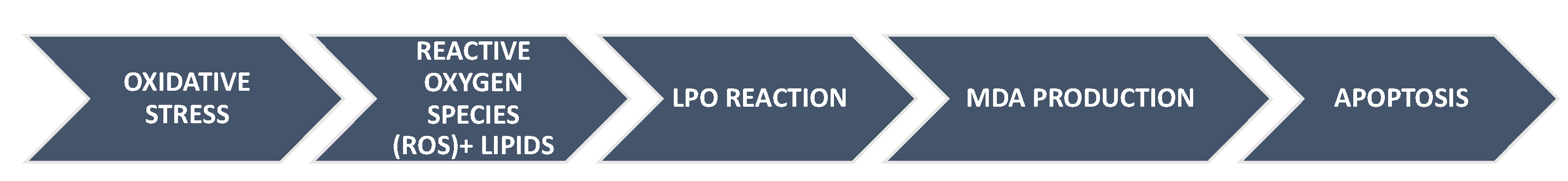
:1. Introduction
2. Materials and Methods
2.1. Quantification of Malondialdehyde
2.2. Statistical Analysis
3. Results
4. Discussion
5. Conclusions
Author Contributions
Funding
Institutional Review Board Statement
Informed Consent Statement
Data Availability Statement
Acknowledgments
Conflicts of Interest
References
- Palomo-López, N.; Martín-Villén, L.; Roldán-Reina, Á.J.; de Azúa-López, Z.R.; Cuenca-Apolo, D.X.; Adriaensens-Pérez, M.; Corcia-Palomo, Y.; Egea-Guerrero, J.J. Results of Controlled Donation after Circulatory Death in a Third-Level Hospital. Transpl. Proc. 2018, 50, 536–553. [Google Scholar] [CrossRef] [PubMed]
- Lazzeri, C.; Bonizzoli, M.; Valente, S.; Cianchi, G.; Migliaccio, M.L.; Gensini, G.F.; Peris, A. The role of extracorporeal membrane oxygenation in donation after circulatory death. Minerva Anestesiol. 2014, 80, 1217–1227. [Google Scholar] [PubMed]
- Donación en Asistolia en España: Situación Actual y Recomendcdiones. Documento de Consenso Nacional 2012. Available online: http://www.ont.es/infesp/DocumentosDeConsenso/DONACIÓN%20EN%20ASISTOLIA%20EN%20ESPAÑA.%20SITUACIÓN%20ACTUAL%20Y%20RECOMENDCDIONES.pdf (accessed on 8 December 2020).
- Wadei, H.M.; Heckman, M.G.; Rawal, B.; Taner, C.B.; Farahat, W.; Nur, L.; Mai, M.L.; Prendergast, M.; Gonwa, T.A. Comparison of kidney function between donation after cardiac death and donation after brain death kidney transplantation. Transplantation 2013, 96, 274–281. [Google Scholar] [CrossRef] [PubMed]
- Bellingham, J.M.; Santhanakrishnan, C.; Neidlinger, N.; Wai, P.; Kim, J.; Niederhaus, S.; Leverson, G.E.; Fernandez, L.A.; Foley, D.P.; Mezrich, J.D.; et al. Donation after cardiac death: A 29-year experience. Surgery 2011, 150, 692–702. [Google Scholar] [CrossRef] [PubMed] [Green Version]
- Aslaner, A.; Gunal, O.; Turgut, H.T.; Celik, E.; Yildirim, U.; Demirci, R.K.; Gunduz, U.R.; Calis, H.; Dogan, S. Effect of melatonin on kidney cold ischemic preservation injury. Int. J. Clin. Exp. Med. 2013, 25, 794–798. [Google Scholar]
- Tsikas, D. Assessment of lipid peroxidation by measuring malondialdehyde (MDA) and relatives in biological samples: Analytical and biological challenges. Anal. Biochem. 2017, 524, 13–30. [Google Scholar] [CrossRef]
- Negre-Salvayre, A.; Augé, N.; Ayala, V.; Basaga, H.; Boada, J.; Brenke, R.; Chapple, S.; Cohen, G.; Feher, J.; Grune, T.; et al. Pathological aspects of lipid peroxidation. Free Radic. Res. 2010, 44, 1125–1171. [Google Scholar] [CrossRef]
- Miñambres, E.; Rubio, J.J.; Coll, E.; Domínguez-Gil, B. Donation after circulatory death and its expansion in Spain. Curr. Opin. Organ Transpl. 2018, 23, 120–129. [Google Scholar] [CrossRef] [PubMed]
- Palomo-López, N.; Martín-Sastre, S.; Martín-Villén, L.; de Azúa-López, Z.R.; Solis-Clavijo, D.; Caballero-Gálvez, S.; Carballo-Caro, J.M.; Egea-Guerrero, J.J. Normothermic Regional Perfusion and Donation After Circulatory Death (Controlled and Uncontrolled): Metabolic Differences and Kidney Transplantation Evolution. Transpl. Proc. 2019, 51, 3044–3046. [Google Scholar] [CrossRef] [PubMed]
- Singhanat, K.; Apaijai, N.; Chattipakorn, S.C.; Chattipakorn, N. Roles of melatonin and its receptors in cardiac ischemia-reperfusion injury. Cell Mol. Life Sci. 2018, 75, 4125–4149. [Google Scholar] [CrossRef] [PubMed]
- Hardeland, R. Melatonin and inflammation-Story of a double-edged blade. J. Pineal. Res. 2018, 65, e12525. [Google Scholar] [CrossRef] [PubMed] [Green Version]
- Dianatkhah, M.; Najafi, A.; Sharifzadeh, M.; Ahmadi, A.; Sharifnia, H.; Mojtahedzadeh, M.; Najmeddin, F.; Moghaddas, A. Melatonin Supplementation May Improve the Outcome of Patients with Hemorrhagic Stroke in the Intensive Care Unit. J. Res. Pharm. Pract. 2017, 6, 173–177. [Google Scholar] [PubMed]
- Mistraletti, G.; Umbrello, M.; Sabbatini, G.; Miori, S.; Taverna, M.; Cerri, B.; Mantovani, E.S.; Formenti, P.; Spanu, P.; D’Agostino, A.; et al. Melatonin reduces the need for sedation in ICU patients: A randomized controlled trial. Minerva Anestesiol. 2015, 81, 1298–1310. [Google Scholar] [PubMed]
- Protocolo Nacional de Donación y Trasplante Hepático en Donación en Asistolia Controlada. Documento de Consenso 2015. Página web Organización Nacional de Trasplantes. Available online: http://www.ont.es/infesp/DocumentosDeConsenso/PROTOCOLO%20NACIONAL%20DE%20DONACIÓN%20Y%20TRASPLANTE%20HEPÁTICO%20EN%20DONACIÓN%20EN%20ASISTOLIA%20CONTROLADA_Agosto%202015_FINAL.pdf?Mobile=1&Source=%2Finfesp%2F%5Flayouts%2Fmobile%2Fdispform%2Easpx%3FList%3Dc6aba339%252D9d35%252D4da7%252D9c6e%252D774daa2bef18%26View%3D593da1d1%252Dbba9%252D4578%252D93f0%252D39353d2fc8de%26ID%3D26%26CurrentPage%3D1 (accessed on 1 December 2020).
- Cardinali, D.P.; Vigo, D.E. Melatonin, mitochondria, and the metabolic syndrome. Cell Mol. Life Sci. 2017, 74, 3941–3954. [Google Scholar] [CrossRef] [PubMed]
- Lee, J.H.; Yoon, Y.M.; Han, Y.S.; Jung, S.K.; Lee, S.H. Melatonin protects mesenchymal stem cells from autophagy-mediated death under ischaemic ER-stress conditions by increasing prion protein expression. Cell Prolif. 2019, 52, e12545. [Google Scholar] [CrossRef] [PubMed] [Green Version]
- Li, Y.; Yang, Y.; Feng, Y.; Yan, J.; Fan, C.; Jiang, S.; Qu, Y. A review of melatonin in hepatic ischemia/reperfusion injury and clinical liver disease. Ann. Med. 2014, 46, 503–511. [Google Scholar] [CrossRef] [PubMed]
- Baykara, B.; Tekmen, I.; Pekcetin, C.; Ulukus, C.; Tuncel, P.; Sagol, O.; Ormen, M.; Ozogul, C. The protective effects of carnosine and melatonin in ischemia-reperfusion injury in the rat liver. Acta Histochem. 2009, 111, 42–51. [Google Scholar] [CrossRef] [PubMed]
- Lan, H.; Su, Y.; Liu, Y.; Deng, C.; Wang, J.; Chen, T.; Jules, K.E.; Masau, J.F.; Li, H.; Wei, X. Melatonin protects circulatory death heart from ischemia/reperfusion injury via the JAK2/STAT3 signalling pathway. Life Sci. 2019, 228, 35–46. [Google Scholar] [CrossRef] [PubMed]
- Liang, Y.; Harris, F.L.; Brown, L.A.S. Alcohol induced mitochondrial oxidative stress and alveolar macrophage dysfunction. BioMed Res. Int. 2014, 2014, 371953. [Google Scholar] [CrossRef] [PubMed] [Green Version]


| Variables | Melatonin Group | Placebo Group | p | |
|---|---|---|---|---|
| Age | 56 (42.3–64.7) | 63 (46.6–69.9) | 0.75 | |
| Gender, male | 10 (58.8) | 9 (60) | 0.62 | |
| Type of brain injury | Hemorrhagic CVA | 7 (41.1) | 7 (40.8) | 0.41 |
| TBI | 6 (35.3) | 4 (26.7) | ||
| Ischemic CVA | 71 (5.9) | 4 (26.7) | ||
| Anoxic encephalopathy | 3 (17.6) | 1 (6.7) | ||
| Smoker | 7 (41.2) | 4 (28.6) | 0.36 | |
| Alcoholism | 7 (41.2) | 1 (7.1) | 0.04 | |
| AH | 7 (41.2) | 8 (57.1) | 0.30 | |
| DM | 3 (17.6) | 2 (14.3) | 0.59 | |
| ASA | 2 (11.8) | 2 (11.4) | 0.62 | |
| OAC | 3 (17.6) | 1 (7.1) | 0.38 | |
| GCS at admission | 6 (3.9–7.9) | 3 (2.9–7.9) | 1 | |
| Diabetes Insipidus | 12 (70.6) | 7 (46.7) | 0.16 | |
| Length of stay in ICU | 2 (1.9–9.9) | 1 (0.84–3.3) | 0.47 | |
| Length of stay in Hospital | 2 (1.9–9.9) | 2 (1.39–4) | 0.50 | |
| Melatonin Group | Placebo Group | p | |
|---|---|---|---|
| Age, median (RI) | 61.5 (53.5–67.8) | 61 (53–8.5) | 0.75 |
| Gender, male n (%) | 6 (75) | 7 (53.8) | 0.40 |
| Smoker n (%) | 4 (32.8) | 5 (62.5) | 0.16 |
| Alcoholism n (%) | 3 (23.1) | 3 (37.5) | 0.13 |
| AH n (%) | 11 (84.6) | 7 (87.5) | 0.92 |
| DM n (%) | 4 (30.8) | 2 (25) | 0.78 |
| Liver disease n (%) | 2 (15.4) | 2 (25) | 0.59 |
| Kidney disease n (%) | 0 (0) | 1 (12.5) | 0.20 |
| Heart disease n (%) | 4 (30.8) | 3 (37.5) | 0.71 |
| Neurologic disease n (%) | 2 (15.4) | 0 (0) | 0.23 |
| Cause for LTE n (%) | 0.86 | ||
| * Anoxic encephalopathy | 6 (38.5) | 3 (50) | |
| * Hemorrhagic CVA | 5 (46.2) | 4 (37.5) | |
| * Other causes | 2 (15.4) | 1 (12.5) | |
| Length of stay in ICU | 10 (4.3–20) | 7 (3–16) | 0.09 |
| Length of stay in Hospital | 12 (4.3–20) | 7 (3–17) | 0.06 |
| f-WIT (minutes) | 19 (10.8–21.5) | 14 (13–17) | 0.09 |
| TIT (minutes) | 24 (17.5–27.5) | 26 (19–33) | 1 |
| NRP (minutes) | 132 (99.8–150.5) | 103 (90–125) | 0.15 |
| Malondialdehyde (MDA), Median (IR) | Melatonin Group | Placebo Group | p |
|---|---|---|---|
| Difference at 0–60’ | 0.05 (−1.2; −0.6) | 0.06 (−1.6; −0.9) | 0.78 |
| Difference at 0–90’ | −0.17 (−1.9; −0.6) | −0.0083 (−1.9; −1.6) | 0.55 |
| Malondialdehyde, Median (IR) | Melatonin Group | Placebo Group | p |
|---|---|---|---|
| Difference at 0–60’ | −3.1 (−6.1; −0.8) | −1.2 (−3.1; −0.5) | 0.21 |
| Difference at 0–90’ | −4.3 (−6.7; −2.6) | −1.6 (−2.9; −0.75) | 0.004 |
Publisher’s Note: MDPI stays neutral with regard to jurisdictional claims in published maps and institutional affiliations. |
© 2022 by the authors. Licensee MDPI, Basel, Switzerland. This article is an open access article distributed under the terms and conditions of the Creative Commons Attribution (CC BY) license (https://creativecommons.org/licenses/by/4.0/).
Share and Cite
Palomo-López, N.; Rodríguez-Rodríguez, A.; Martín-Villén, L.; Mendoza-Prieto, M.; Ruiz de Azúa-López, Z.; Sempere-Bordes, L.; Boyero-Corral, L.; Daga-Ruiz, D.; Gordillo-Brenes, A.; Pacheco-Sánchez, M.; et al. Immunomodulation of Oxidative Stress during Organ Donation Process: Preliminary Results. Healthcare 2022, 10, 762. https://doi.org/10.3390/healthcare10050762
Palomo-López N, Rodríguez-Rodríguez A, Martín-Villén L, Mendoza-Prieto M, Ruiz de Azúa-López Z, Sempere-Bordes L, Boyero-Corral L, Daga-Ruiz D, Gordillo-Brenes A, Pacheco-Sánchez M, et al. Immunomodulation of Oxidative Stress during Organ Donation Process: Preliminary Results. Healthcare. 2022; 10(5):762. https://doi.org/10.3390/healthcare10050762
Chicago/Turabian StylePalomo-López, Nora, Ana Rodríguez-Rodríguez, Luis Martín-Villén, María Mendoza-Prieto, Zaida Ruiz de Azúa-López, Lluis Sempere-Bordes, Laura Boyero-Corral, Domingo Daga-Ruiz, Antonio Gordillo-Brenes, María Pacheco-Sánchez, and et al. 2022. "Immunomodulation of Oxidative Stress during Organ Donation Process: Preliminary Results" Healthcare 10, no. 5: 762. https://doi.org/10.3390/healthcare10050762