
Graphene-Based Surface-Enhanced Raman Scattering (SERS) Sensing: Bibliometrics Based Analysis and Review

 ,
,
Abstract
:1. Introduction
- (1)
- Can graphene effectively improve the sensitivity of conventional SERS substrates?
- (2)
- Are the SERS properties of graphene itself promising for applications?
- (3)
- What nanomaterials and morphologies are often used to compound with graphene to prepare SERS substrates?
- (4)
- Do graphene-based SERS substrates already have a specific application at this stage?
- (5)
- Has the enthusiasm for research on this topic waned, as attention has gradually shifted from graphene to other novel materials?
2. Developments in the Research Field
2.1. Literature Development Trends
2.2. Journals, Cited Journals, and Research Subjects
2.3. Geographic Distribution
3. Keyword Analysis and Evolution of the Field
- #0
- (Graphene-based composite) This one cluster contains the largest number of papers among all clusters. Most of them are concerned with preparing graphene and noble metal (Ag and Au) composites and investigating their SERS properties. The metal nanomaterials used for the composite have different morphologies such as nanocube [85,346], flower-like particle [57], nano-disc [61], nanorod [104], nanostar [136] and 3D butterfly wing structure [109]. In addition to Au and Ag, Cu [83], MoS2 [347], Fe3O4 [58,282] and ZnO [98] have been used for the preparation of SERS substrates as well.
- #1
- (Graphene property) This cluster has a relatively low silhouette value, so its clustering effect is not particularly obvious. The graphene and Au/Ag composite continue to be a key content in this cluster. Unlike #0, this cluster contains a series of investigations on the SERS properties of graphene itself. For example, Ramanauskaite et al. [163] investigated the reduction process GO undergoes when used in SERS substrates and the changes in its properties. Li et al. [157] investigated that wrapping silicon nanowires with graphene allows silicon nanowires, which otherwise have no SERS properties, to become a novel SERS substrate. Han et al. [156] investigated the relationship between the chemistry and structure of graphene and its SERS properties.
- #2
- (Sandwich structure) The sandwich structure of the SERS substrate is the main content of interest in this cluster. Different investigations have found sandwich structures to be effective in enhancing the performance of SERS. For example, Wu et al. [172] synthesized an AgNP-graphene-AgNP sandwiched structure using a wet chemical method and an autonomous loading technique. Plasma coupling between AgNPs from both sides of the graphene can greatly enhance the performance of SERS. Zhao et al. [173] prepared AuNP-graphene-AgNP sandwiched substrates, which have a detection sensitivity of 10−13 M. Other sandwich structures include AgNPs-silica-GO [174], AuNPs-graphene-Au array [175], silicon nanowire-graphene-AuNPs [169,182], Ag-graphene-Au [183], AgNPs-TiO2-graphene [144], Ag nanohole array-graphene-AuNPs [184] and AgNPs-silica-graphene [188].
- #3
- (Doping) The silhouette value of this cluster is low, so the direction of the papers contained in it is divergent. Two directions are worth noting. The first one is about the doping and modification of graphene. Some papers report that doping or modification of graphene can lead to more excellent SERS properties. For example, Kasztelan et al. [214] found that a simple treatment of GO with ammonia solution improved the SERS detection. This may be due to the partial reduction of GO by NH3 and the introduction of nitrogen functionalization. Nair et al. [185] found that nitrogen sulfur co-doped RGO could be used to adsorb different forms of AgNPs and therefore exhibited more sensitive SERS performance. Another direction is the preparation of free-standing SERS substrates. Zhao et al. [220] synthesized a flexible film combining graphene and AgNPs for SERS applications. Fan et al. [346] also prepared a free-standing substrate containing GO and AgNPs for SERS. Lee and Kim [73] loaded AuNPs and GO on a hydrophobic paper, which can be used as a SERS substrate for analytical detection.
- #4
- (Modeling) This cluster focuses on the modeling of SERS. Al-Otaibi et al. [221] calculated the structural, nonlinear optical, electronic and biological properties of three anastrozole-based triazole analogues on graphene surfaces. The results demonstrated the enhancement of SERS for all three molecules. They also calculated three aminobenzoate derivatives and their SERS active graphene complexes [348]. Ullah et al. [349] performed theoretical calculations for adsorbed antimalarial-graphene dimers and predicted the SERS signal.
- #5
- (Magnetic composite) This cluster shares many papers with #0 and contains two directions. The first direction is the synthesis of graphene-Ag nanostructure-based composite for SERS. It is worth mentioning that this cluster does not contain any paper related to graphene-Au nanostructure-based composite. Another direction is the synthesis of graphene-based nanocomposites with magnetic properties. The fast magnetic response enables rapid separation of the composite material from the solution, and the practical application of SERS can be achieved by first using the material for adsorption on the analyte, followed by detection after rapid separation [217,231].
- #6
- (Detection) The papers in this cluster begin to focus further on the sensing applications of the prepared SERS substrates. Their titles will not only describe the preparation of a particular structure of the substrate but will also emphasize the detection of a particular analyte. For example, the work of Xu et al. [237] and Qiu et al. [242] both emphasized the detection of adenosine. Jinbin et al. [239] highlighted that their substrate could be used to detect circulating breast cancer cells. Naqvi et al. [240] highlighted that their SERS sensor is used for explosive detection. The SERS platform proposed by Dutta et al. [91] was used for uranyl ion sensing.
- #7
- (Fabrication method) This cluster mainly highlights the preparation techniques of different graphene-based SERS substrates and the way of optimization in the preparation process. Saha et al. [245] used stabilization of hot spots in GO liquid crystals to improve the reproducibility of SERS. Kovaricek et al. [246] investigated the covalent reaction during CVD to optimize the growth of graphene. Hu et al. [249] prepared SERS substrates by electrostatic self-assembly. Ouyang et al. [254] used a filtration-assisted fabrication technique to synthesize large-size SERS substrates.
- #8
- #9
- (SERS) The silhouette value of this cluster is only 0.664. According to the CiteSpace manual, clusters with a silhouette value below 8.5 do not have a significant similarity. After analyzing the papers in this cluster one by one, we did not find any strong correlation between them.
- #10
- (Biosensing) This cluster mainly highlights the references of graphene-based SERS in biosensing. For example, the SERS substrate proposed by Fu et al. [302] to detect of cardiac troponin I. Chen et al. [303] focused on the detection of clenbuterol residues in animal-origin food samples by SERS. Lv et al. [306] tried the detection of adenine by SERS. Li et al. [308] attempted the detection of trace amounts of ferritin by SERS.
- #11
- (Graphene film) The content of this cluster is entirely covered by #1, #2, #6 and #8 as seen in Figure 8. The papers in this cluster mainly compare the SERS performance of noble metal nanomaterials enhanced with the assistance of graphene.
- #12
- (Morphology) The content of this cluster mainly emphasizes the effect of graphene morphology (number of layers) and location (center or edge) on SERS. For example, Xu et al. [322] investigated the SERS performance of highly ordered graphene-isolated silver nanodot arrays. Matz et al. [323] investigated the SERS fingerprint of monolayer graphene grown by CVD. D’Urso et al. [255] investigated the SERS properties of 1D-2D graphene-based structures.
- #13
- (Fluorescence) This cluster appears to utilize graphene quantum dots as a material for the SERS substrate. As a quantum dot, its fluorescence properties impact the Raman signal. Therefore, this series of work involves the investigation of the fluorescence properties. On the other hand, graphene has been observed to have a fluorescence quenching effect, which is one of the important reasons why it is widely used in SERS.
- #14
- (Nanoparticle) This cluster is also entirely covered by surrounding clusters, and its papers overlap with parts #0, #4, #5 and #10. It includes not only the composite of AuNPs or AgNPs with graphene but also the ternary composite of all three of them.
- #15
- (SERS property) This cluster includes only two papers. Guo et al. [344] used a photocatalytic method to grow Ag nanocrystals on the surface of TiO2/RGO and examined their SERS properties. Liu and Luo [61] synthesized two gold nanostructures with different morphologies for compounding with graphene and evaluated their SERS properties.
- #16
- (Nanodendrites) This cluster contains only one paper. This paper describes the SERS properties after covering silver nanodendrites with graphene films [345].
- (1)
- The content of this topic does not show a considerable divergence. Most of the works have focused on investigating the performance of conventional SERS materials after graphene compounding.
- (2)
- These SERS substrates prepared using graphene-based composites have much to investigate. For example, whether there is a difference in their SERS effect when different nanostructures and graphene are compounded. Whether the different oxidation states of graphene affect the SERS effect. Whether the number of layers of graphene affects the SERS effect of the composites.
- (3)
- Investigation of the effect of graphene’s own SERS. Mechanistic analysis of this phenomenon and whether it has practical value.
- (4)
- The advantages of SERS in analytical assays. Which analytes are easier and more sensitive to detect using graphene-based SERS than other traditional detection methods.
4. Conclusions
- (1)
- Graphene-based SERS has been widely discussed since it was proposed, and the publication of related papers gradually rose and peaked in 2017. This trend has not continued until today. Starting in 2018, the annual number of publications on this topic began to decline, with only 42 in 2021. The annual number of publications shows that researchers are gradually shifting their attention from this topic to other areas.
- (2)
- Although SERS is an optical-based analytical sensing technique, the investigations on this topic were initially focused on materials science and chemistry. This is because the SERS properties generated by graphene or by the composite of graphene and other conventional SERS nanomaterials need to be explained mechanistically. Therefore, most of the published papers on this topic simply choose a commonly used probe to evaluate the performance of the prepared SERS substrates rather than a custom development for specific detection needs. Starting in 2013, the topic gradually shifted from the investigation of materials science/optics/chemistry to different application areas, including food science, environmental science, pharmacology, molecular biology, etc.
- (3)
- Chinese scientists contribute the most significant number of papers in this field, with the Chinese Academy of Sciences being the most influential institution. USA, India, and South Korea also play an important role in this topic. Nanyang Technological University in Singapore and Massachusetts Institute of Technology in USA have not published many papers on this topic. However, their work has had a significant impact. Based on the geographical analysis, this topic attracts the attention of scientists from global regions. Although the annual publication of this topic is decreasing yearly, countries continue to participate in this topic for the first time every year.
- (4)
- The analysis of the keywords proves that the investigation of this topic focuses on the preparation of SERS substrates. Among them, GO in many cases replaces graphene for composite synthesis. The most commonly used SERS materials, AuNPs and AgNPs, continue to be the most widely used choices for composite with graphene. In addition to nanoparticles, other nanostructures have also been widely investigated. On the other hand, the nature of the graphene also affects the SERS performance, where a range of factors are included, such as the degree of oxidation, number of layers, size, folds, etc.
- (5)
- Although graphene-based SERS has been studied for more than a decade, it has not yet presented a particular application dedicated to it in the sensing field. This may be due to the fact that while the assistance of graphene can provide enhancement of the SERS signal, it does not have the property of specific identification of the analyte. Therefore, it is indeed a very sensitive analytical tool when optimized, but it is more difficult to overcome the challenges posed by interferents in sensing.
5. Perspectives
- (1)
- Graphene-based SERS substrates are an analytical platform that can be produced on a large scale with easily controlled stability. The development of practical applications based on this platform is a direction that needs to be focused on in the future. It is believed that with the participation of scientists from different fields, such as contaminant detection, food safety detection, drug detection, etc., it is possible to find suitable assay needs for this platform.
- (2)
- Since graphene is a two-dimensional lamellar material, it changes its morphology when compounded with other nanomaterials. For example, it has the ability to combine into three-dimensional structures. These structural changes have been shown to affect the performance of SERS substrates. Some of these particular structures have also been shown to possess extraordinary properties. However, whether such structures can be controlled with high quality still needs to be verified. Therefore, how to tune graphene in SERS substrates is an important direction. Finding a balance between the reproducibility of the prepared substrates and SERS performance is challenging.
Author Contributions
Funding
Institutional Review Board Statement
Informed Consent Statement
Data Availability Statement
Conflicts of Interest
References
- Fleischmann, M.; Hendra, P.J.; McQuillan, A.J. Raman Spectra of Pyridine Adsorbed at a Silver Electrode. Chem. Phys. Lett. 1974, 26, 163–166. [Google Scholar] [CrossRef]
- Jeanmaire, D.L.; Van Duyne, R.P. Surface Raman Spectroelectrochemistry: Part I. Heterocyclic, Aromatic, and Aliphatic Amines Adsorbed on the Anodized Silver Electrode. J. Electroanal. Chem. Interfacial Electrochem. 1977, 84, 1–20. [Google Scholar] [CrossRef]
- Li, Z.-Y.; Xia, Y. Metal Nanoparticles with Gain toward Single-Molecule Detection by Surface-Enhanced Raman Scattering. Nano Lett. 2010, 10, 243–249. [Google Scholar] [CrossRef] [PubMed]
- Han, Y.; Liu, S.; Liu, B.; Jiang, C.; Zhang, Z. In Situ Loading of Ag Nanocontacts onto Silica Nanospheres: A SERS Platform for Ultrasensitive Detection. RSC Adv. 2014, 4, 2776–2782. [Google Scholar] [CrossRef]
- Jiang, S.-M.; Wu, D.-J.; Wu, X.-W.; Liu, X.-J. Enormous Enhancement of Electric Field in Active Gold Nanoshells. Chin. Phys. B 2014, 23, 047807. [Google Scholar] [CrossRef]
- Zhou, Q.; Zhu, J.; Yuan, J.; Fang, X. Numerical Simulation of Surface-Enhanced Coherent Anti-Stokes Raman Scattering on Gold Nanoparticle Substrate. J. Nanosci. Nanotechnol. 2017, 17, 2152–2156. [Google Scholar] [CrossRef]
- Kneipp, K.; Wang, Y.; Kneipp, H.; Perelman, L.T.; Itzkan, I.; Dasari, R.R.; Feld, M.S. Single Molecule Detection Using Surface-Enhanced Raman Scattering (SERS). Phys. Rev. Lett. 1997, 78, 1667. [Google Scholar] [CrossRef] [Green Version]
- Tian, Z.-Q.; Ren, B.; Wu, D.-Y. Surface-Enhanced Raman Scattering: From Noble to Transition Metals and from Rough Surfaces to Ordered Nanostructures. J. Phys. Chem. B 2002, 106, 9463–9483. [Google Scholar] [CrossRef]
- Ren, B.; Huang, Q.; Cai, W.; Mao, B.; Liu, F.; Tian, Z. Surface Raman Spectra of Pyridine and Hydrogen on Bare Platinum and Nickel Electrodes. J. Electroanal. Chem. 1996, 415, 175–178. [Google Scholar] [CrossRef]
- Lombardi, J.R.; Birke, R.L. A Unified View of Surface-Enhanced Raman Scattering. Acc. Chem. Res. 2009, 42, 734–742. [Google Scholar] [CrossRef] [PubMed] [Green Version]
- Sun, P.; Yu, H.; Liu, T.; Li, Y.; Wang, Z.; Xiao, Y.; Dong, X. Efficiently Photothermal Conversion in a MnOx-Based Monolithic Photothermocatalyst for Gaseous Formaldehyde Elimination. Chin. Chem. Lett. 2022, 33, 2564–2568. [Google Scholar] [CrossRef]
- Wallace, P.R. The Band Theory of Graphite. Phys. Rev. 1947, 71, 622. [Google Scholar] [CrossRef]
- Novoselov, K.S.; Geim, A.K.; Morozov, S.V.; Jiang, D.; Zhang, Y.; Dubonos, S.V.; Grigorieva, I.V.; Firsov, A.A. Electric Field Effect in Atomically Thin Carbon Films. Science 2004, 306, 666–669. [Google Scholar] [CrossRef] [PubMed] [Green Version]
- Zhang, Y.; Tan, Y.-W.; Stormer, H.L.; Kim, P. Experimental Observation of the Quantum Hall Effect and Berry’s Phase in Graphene. Nature 2005, 438, 201–204. [Google Scholar] [CrossRef] [PubMed] [Green Version]
- Ling, X.; Xie, L.; Fang, Y.; Xu, H.; Zhang, H.; Kong, J.; Dresselhaus, M.S.; Zhang, J.; Liu, Z. Can Graphene Be Used as a Substrate for Raman Enhancement? Nano Lett. 2010, 10, 553–561. [Google Scholar] [CrossRef]
- Huh, S.; Park, J.; Kim, Y.S.; Kim, K.S.; Hong, B.H.; Nam, J.-M. UV/Ozone-Oxidized Large-Scale Graphene Platform with Large Chemical Enhancement in Surface-Enhanced Raman Scattering. ACS Nano 2011, 5, 9799–9806. [Google Scholar] [CrossRef]
- Yu, X.; Cai, H.; Zhang, W.; Li, X.; Pan, N.; Luo, Y.; Wang, X.; Hou, J. Tuning Chemical Enhancement of SERS by Controlling the Chemical Reduction of Graphene Oxide Nanosheets. ACS Nano 2011, 5, 952–958. [Google Scholar] [CrossRef] [PubMed]
- Colomban, P.; March, G.; Mazerolles, L.; Karmous, T.; Ayed, N.; Ennabli, A.; Slim, H. Raman Identification of Materials Used for Jewellery and Mosaics in Ifriqiya. J. Raman Spectrosc. 2003, 34, 205–213. [Google Scholar] [CrossRef]
- Huang, C.; Kim, M.; Wong, B.M.; Safron, N.S.; Arnold, M.S.; Gopalan, P. Raman Enhancement of a Dipolar Molecule on Graphene. J. Phys. Chem. C 2014, 118, 2077–2084. [Google Scholar] [CrossRef]
- Butmee, P.; Samphao, A.; Tumcharern, G. Reduced Graphene Oxide on Silver Nanoparticle Layers-Decorated Titanium Dioxide Nanotube Arrays as SERS-Based Sensor for Glyphosate Direct Detection in Environmental Water and Soil. J. Hazard. Mater. 2022, 437, 129344. [Google Scholar] [CrossRef]
- Xie, Y.; Li, Y.; Niu, L.; Wang, H.; Qian, H.; Yao, W. A Novel Surface-Enhanced Raman Scattering Sensor to Detect Prohibited Colorants in Food by Graphene/Silver Nanocomposite. Talanta 2012, 100, 32–37. [Google Scholar] [CrossRef] [PubMed]
- Ponlamuangdee, K.; Hornyak, G.L.; Bora, T.; Bamrungsap, S. Graphene Oxide/Gold Nanorod Plasmonic Paper—A Simple and Cost-Effective SERS Substrate for Anticancer Drug Analysis. New J. Chem. 2020, 44, 14087–14094. [Google Scholar] [CrossRef]
- Cao, Y.; Cheng, Y.; Sun, M. Graphene-Based SERS for Sensor and Catalysis. Appl. Spectrosc. Rev. 2021; in press. [Google Scholar] [CrossRef]
- Kang, L.; Chu, J.; Zhao, H.; Xu, P.; Sun, M. Recent Progress in the Applications of Graphene in Surface-Enhanced Raman Scattering and Plasmon-Induced Catalytic Reactions. J. Mater. Chem. C 2015, 3, 9024–9037. [Google Scholar] [CrossRef]
- Zhang, N.; Tong, L.; Zhang, J. Graphene-Based Enhanced Raman Scattering toward Analytical Applications. Chem. Mater. 2016, 28, 6426–6435. [Google Scholar] [CrossRef]
- Sharma, S.; Prakash, V.; Mehta, S. Graphene/Silver Nanocomposites-Potential Electron Mediators for Proliferation in Electrochemical Sensing and SERS Activity. TrAC Trends Anal. Chem. 2017, 86, 155–171. [Google Scholar] [CrossRef]
- Chen, C. Science Mapping: A Systematic Review of the Literature. J. Data Inf. Sci. 2017, 2, 1–40. [Google Scholar] [CrossRef] [Green Version]
- Xueshu, D. Wenxian COOC Is a Software for Bibliometrics and Knowledge Mapping [CP/OL]. Available online: https://mp.weixin.qq.com/s/3Iy7h203BDwWrFHxRqcG8w (accessed on 3 August 2022).
- Schedin, F.; Lidorikis, E.; Lombardo, A.; Kravets, V.G.; Geim, A.K.; Grigorenko, A.N.; Novoselov, K.S.; Ferrari, A.C. Surface-Enhanced Raman Spectroscopy of Graphene. ACS Nano 2010, 4, 5617–5626. [Google Scholar] [CrossRef]
- Wang, Y.; Ni, Z.; Hu, H.; Hao, Y.; Wong, C.P.; Yu, T.; Thong, J.T.; Shen, Z.X. Gold on Graphene as a Substrate for Surface Enhanced Raman Scattering Study. Appl. Phys. Lett. 2010, 97, 163111. [Google Scholar] [CrossRef]
- Fu, X.; Bei, F.; Wang, X.; O’Brien, S.; Lombardi, J.R. Excitation Profile of Surface-Enhanced Raman Scattering in Graphene–Metal Nanoparticle Based Derivatives. Nanoscale 2010, 2, 1461–1466. [Google Scholar] [CrossRef]
- Huang, J.; Zhang, L.; Chen, B.; Ji, N.; Chen, F.; Zhang, Y.; Zhang, Z. Nanocomposites of Size-Controlled Gold Nanoparticles and Graphene Oxide: Formation and Applications in SERS and Catalysis. Nanoscale 2010, 2, 2733–2738. [Google Scholar] [CrossRef] [PubMed]
- Zheng, Y.; Karimi-Maleh, H.; Fu, L. Advances in Electrochemical Techniques for the Detection and Analysis of Genetically Modified Organisms: An Analysis Based on Bibliometrics. Chemosensors 2022, 10, 194. [Google Scholar] [CrossRef]
- Zheng, Y.; Mao, S.; Zhu, J.; Fu, L.; Zare, N.; Karimi, F. Current Status of Electrochemical Detection of Sunset Yellow Based on Bibliometrics. Food Chem. Toxicol. 2022, 164, 113019. [Google Scholar] [CrossRef] [PubMed]
- Shen, Y.; Mao, S.; Chen, F.; Zhao, S.; Su, W.; Fu, L.; Zare, N.; Karimi, F. Electrochemical Detection of Sudan Red Series Azo Dyes: Bibliometrics Based Analysis. Food Chem. Toxicol. 2022, 163, 112960. [Google Scholar] [CrossRef]
- Zheng, Y.; Karimi-Maleh, H.; Fu, L. Evaluation of Antioxidants Using Electrochemical Sensors: A Bibliometric Analysis. Sensors 2022, 22, 3238. [Google Scholar] [CrossRef] [PubMed]
- Fu, L.; Mao, S.; Chen, F.; Zhao, S.; Su, W.; Lai, G.; Yu, A.; Lin, C.-T. Graphene-Based Electrochemical Sensors for Antibiotic Detection in Water, Food and Soil: A Scientometric Analysis in CiteSpace (2011–2021). Chemosphere 2022, 297, 134127. [Google Scholar] [CrossRef]
- Jin, M.; Liu, J.; Wu, W.; Zhou, Q.; Fu, L.; Zare, N.; Karimi, F.; Yu, J.; Lin, C.-T. Relationship between Graphene and Pedosphere: A Scientometric Analysis. Chemosphere 2022, 300, 134599. [Google Scholar] [CrossRef]
- Li, Y.-T.; Qu, L.-L.; Li, D.-W.; Song, Q.-X.; Fathi, F.; Long, Y.-T. Rapid and Sensitive In-Situ Detection of Polar Antibiotics in Water Using a Disposable Ag–Graphene Sensor Based on Electrophoretic Preconcentration and Surface-Enhanced Raman Spectroscopy. Biosens. Bioelectron. 2013, 43, 94–100. [Google Scholar] [CrossRef]
- Nguyen, T.H.D.; Zhang, Z.; Mustapha, A.; Li, H.; Lin, M. Use of Graphene and Gold Nanorods as Substrates for the Detection of Pesticides by Surface Enhanced Raman Spectroscopy. J. Agric. Food Chem. 2014, 62, 10445–10451. [Google Scholar] [CrossRef]
- Qiu, X.; You, X.; Chen, X.; Chen, H.; Dhinakar, A.; Liu, S.; Guo, Z.; Wu, J.; Liu, Z. Development of Graphene Oxide-Wrapped Gold Nanorods as Robust Nanoplatform for Ultrafast near-Infrared SERS Bioimaging. Int. J. Nanomed. 2017, 12, 4349. [Google Scholar] [CrossRef] [Green Version]
- Muntean, C.M.; Dina, N.E.; Biter, T.-L.; Bratu, I.; Coroş, M.; Socaci, C.; Coste, A. Surface Dynamics of Genomic DNAs upon Lowering the PH, in the Presence of Graphene/AgNPs-Based SERS Detection Platform. J. Mol. Model. 2020, 26, 211. [Google Scholar] [CrossRef] [PubMed]
- Konkena, B.; Vasudevan, S. Understanding Aqueous Dispersibility of Graphene Oxide and Reduced Graphene Oxide through p K a Measurements. J. Phys. Chem. Lett. 2012, 3, 867–872. [Google Scholar] [CrossRef] [PubMed]
- Kurapati, R.; Russier, J.; Squillaci, M.A.; Treossi, E.; Ménard-Moyon, C.; Del Rio-Castillo, A.E.; Vazquez, E.; Samorì, P.; Palermo, V.; Bianco, A. Dispersibility-dependent Biodegradation of Graphene Oxide by Myeloperoxidase. Small 2015, 11, 3985–3994. [Google Scholar] [CrossRef]
- Jabłońska, A.; Jaworska, A.; Kasztelan, M.; Berbeć, S.; Pałys, B. Graphene and Graphene Oxide Applications for SERS Sensing and Imaging. Curr. Med. Chem. 2019, 26, 6878–6895. [Google Scholar] [CrossRef]
- Olea-Mejía, O.; Fernández-Mondragón, M.; Rodríguez-de la Concha, G.; Camacho-López, M. SERS-Active Ag, Au and Ag–Au Alloy Nanoparticles Obtained by Laser Ablation in Liquids for Sensing Methylene Blue. Appl. Surf. Sci. 2015, 348, 66–70. [Google Scholar] [CrossRef]
- Wang, Y.; Polavarapu, L.; Liz-Marzán, L.M. Reduced Graphene Oxide-Supported Gold Nanostars for Improved SERS Sensing and Drug Delivery. ACS Appl. Mater. Interfaces 2014, 6, 21798–21805. [Google Scholar] [CrossRef]
- Ren, W.; Fang, Y.; Wang, E. A Binary Functional Substrate for Enrichment and Ultrasensitive SERS Spectroscopic Detection of Folic Acid Using Graphene Oxide/Ag Nanoparticle Hybrids. ACS Nano 2011, 5, 6425–6433. [Google Scholar] [CrossRef]
- Ma, X.; Qu, Q.; Zhao, Y.; Luo, Z.; Zhao, Y.; Ng, K.W.; Zhao, Y. Graphene Oxide Wrapped Gold Nanoparticles for Intracellular Raman Imaging and Drug Delivery. J. Mater. Chem. B 2013, 1, 6495–6500. [Google Scholar] [CrossRef]
- Lin, D.; Qin, T.; Wang, Y.; Sun, X.; Chen, L. Graphene Oxide Wrapped SERS Tags: Multifunctional Platforms toward Optical Labeling, Photothermal Ablation of Bacteria, and the Monitoring of Killing Effect. ACS Appl. Mater. Interfaces 2014, 6, 1320–1329. [Google Scholar] [CrossRef]
- Huang, J.; Zong, C.; Shen, H.; Liu, M.; Chen, B.; Ren, B.; Zhang, Z. Mechanism of Cellular Uptake of Graphene Oxide Studied by Surface-enhanced Raman Spectroscopy. Small 2012, 8, 2577–2584. [Google Scholar] [CrossRef] [Green Version]
- Kong, X.; Chen, Q.; Sun, Z. Enhanced SERS of the Complex Substrate Using Au Supported on Graphene with Pyridine and R6G as the Probe Molecules. Chem. Phys. Lett. 2013, 564, 54–59. [Google Scholar] [CrossRef]
- Kong, X.; Chen, Q.; Sun, Z. The Positive Influence of Boron-Doped Graphyne on Surface Enhanced Raman Scattering with Pyridine as the Probe Molecule and Oxygen Reduction Reaction in Fuel Cells. RSC Adv. 2013, 3, 4074–4080. [Google Scholar] [CrossRef]
- Zhao, X.; Chen, M. Charge Transfer Mechanism of SERS for Metal–Molecule–Metal Junction Supported by Graphene and Boron-Doped Graphene. RSC Adv. 2014, 4, 63596–63602. [Google Scholar] [CrossRef]
- Zhao, Y.; Li, X.; Wang, M.; Zhang, L.; Chu, B.; Yang, C.; Liu, Y.; Zhou, D.; Lu, Y. Constructing Sub-10-Nm Gaps in Graphene-Metal Hybrid System for Advanced Surface-Enhanced Raman Scattering Detection. J. Alloys Compd. 2017, 720, 139–146. [Google Scholar] [CrossRef]
- Fan, W.; Yue-E, M.; Ling, X.; Liu, T. Free-Standing Silver Nanocube/Graphene Oxide Hybrid Paper for Surface-Enhanced Raman Scattering. Chin. J. Chem. 2016, 34, 73–81. [Google Scholar] [CrossRef]
- Zhang, C.; Hao, R.; Zhao, B.; Fu, Y.; Zhang, H.; Moeendarbari, S.; Pickering, C.S.; Hao, Y.; Liu, Y. Graphene Oxide-Wrapped Flower-like Sliver Particles for Surface-Enhanced Raman Spectroscopy and Their Applications in Polychlorinated Biphenyls Detection. Appl. Surf. Sci. 2017, 400, 49–56. [Google Scholar] [CrossRef]
- Duan, N.; Shen, M.; Wu, S.; Zhao, C.; Ma, X.; Wang, Z. Graphene Oxide Wrapped Fe3O4@ Au Nanostructures as Substrates for Aptamer-Based Detection of Vibrio Parahaemolyticus by Surface-Enhanced Raman Spectroscopy. Microchim. Acta 2017, 184, 2653–2660. [Google Scholar] [CrossRef]
- Jiang, Y.; Carboni, D.; Malfatti, L.; Innocenzi, P. Graphene Oxide-Silver Nanoparticles in Molecularly-Imprinted Hybrid Films Enabling SERS Selective Sensing. Materials 2018, 11, 1674. [Google Scholar] [CrossRef] [Green Version]
- Zhou, Y.; Cheng, X.; Du, D.; Yang, J.; Zhao, N.; Ma, S.; Zhong, T.; Lin, Y. Graphene–Silver Nanohybrids for Ultrasensitive Surface Enhanced Raman Spectroscopy: Size Dependence of Silver Nanoparticles. J. Mater. Chem. C 2014, 2, 6850–6858. [Google Scholar] [CrossRef]
- Liu, Y.; Luo, F. Large-Scale Highly Ordered Periodic Au Nano-Discs/Graphene and Graphene/Au Nanoholes Plasmonic Substrates for Surface-Enhanced Raman Scattering. Nano Res. 2019, 12, 2788–2795. [Google Scholar] [CrossRef]
- Zhang, C.-Y.; Zhao, B.-C.; Hao, R.; Wang, Z.; Hao, Y.-W.; Zhao, B.; Liu, Y.-Q. Graphene Oxide-Highly Anisotropic Noble Metal Hybrid Systems for Intensified Surface Enhanced Raman Scattering and Direct Capture and Sensitive Discrimination in PCBs Monitoring. J. Hazard. Mater. 2020, 385, 121510. [Google Scholar] [CrossRef]
- Shanta, P.V.; Cheng, Q. Graphene Oxide Nanoprisms for Sensitive Detection of Environmentally Important Aromatic Compounds with SERS. ACS Sens. 2017, 2, 817–827. [Google Scholar] [CrossRef]
- Sun, H.; Liu, H.; Wu, Y. Three-Dimensional (3D) Crumpled Graphene-Silver Hybrid Nanostructures on Shape Memory Polymers for Surface-Enhanced Raman Scattering. Appl. Surf. Sci. 2019, 467, 554–560. [Google Scholar] [CrossRef]
- Liu, X.; Li, Y.; Xue, W.; Ge, J.; Wang, J.; Sun, J. 3D Nano-Arrays of Silver Nanoparticles and Graphene Quantum Dots with Excellent Surface-Enhanced Raman Scattering. Mater. Sci. Technol. 2018, 34, 679–687. [Google Scholar] [CrossRef]
- Liu, Q.; Wei, L.; Wang, J.; Peng, F.; Luo, D.; Cui, R.; Niu, Y.; Qin, X.; Liu, Y.; Sun, H. Cell Imaging by Graphene Oxide Based on Surface Enhanced Raman Scattering. Nanoscale 2012, 4, 7084–7089. [Google Scholar] [CrossRef]
- Wen, G.; Jing, Q.; Liang, A.; Jiang, Z. A New SERS Strategy for Quantitative Analysis of Trace Microalbuminuria Based on Immunorecognition and Graphene Oxide Nanoribbon Catalysis. Int. J. Nanomed. 2018, 13, 6099. [Google Scholar] [CrossRef] [Green Version]
- Jian, T.; Ai-Ping, L.; Pei-Gang, L.; Jing-Qin, S.; Wei-Hua, T. Surface-Enhanced Raman Scattering of Gold/Graphene Oxide Composite Materials Fabricated by Interface Self-Assembling. ACTA Phys. Sin. 2014, 63, 107801. [Google Scholar] [CrossRef]
- Wan, M.; Liu, Z.; Li, S.; Yang, B.; Zhang, W.; Qin, X.; Guo, Z. Silver Nanoaggregates on Chitosan Functionalized Graphene Oxide for High-Performance Surface-Enhanced Raman Scattering. Appl. Spectrosc. 2013, 67, 761–766. [Google Scholar] [CrossRef]
- Zhao, Y.; Li, X.; Du, Y.; Chen, G.; Qu, Y.; Jiang, J.; Zhu, Y. Strong Light–Matter Interactions in Sub-Nanometer Gaps Defined by Monolayer Graphene: Toward Highly Sensitive SERS Substrates. Nanoscale 2014, 6, 11112–11120. [Google Scholar] [CrossRef]
- Lu, G.; Li, H.; Liusman, C.; Yin, Z.; Wu, S.; Zhang, H. Surface Enhanced Raman Scattering of Ag or Au Nanoparticle-Decorated Reduced Graphene Oxide for Detection of Aromatic Molecules. Chem. Sci. 2011, 2, 1817–1821. [Google Scholar] [CrossRef] [Green Version]
- Barman, B.K.; Nanda, K.K. Hexamethylenetetramine Mediated Simultaneous Nitrogen Doping and Reduction of Graphene Oxide for a Metal-Free SERS Substrate. RSC Adv. 2014, 4, 44146–44150. [Google Scholar] [CrossRef]
- Lee, D.-J.; Kim, D.Y. Hydrophobic Paper-Based SERS Sensor Using Gold Nanoparticles Arranged on Graphene Oxide Flakes. Sensors 2019, 19, 5471. [Google Scholar] [CrossRef] [Green Version]
- Zhao, B.; Hao, R.; Wang, Z.; Zhang, H.; Hao, Y.; Zhang, C.; Liu, Y. Green Synthesis of Multi-Dimensional Plasmonic Coupling Structures: Graphene Oxide Gapped Gold Nanostars for Highly Intensified Surface Enhanced Raman Scattering. Chem. Eng. J. 2018, 349, 581–587. [Google Scholar] [CrossRef]
- Kuo, C.-C.; Chen, C.-H. Graphene Thickness-Controlled Photocatalysis and Surface Enhanced Raman Scattering. Nanoscale 2014, 6, 12805–12813. [Google Scholar] [CrossRef] [Green Version]
- Tang, X.-Z.; Srikanth, N.; Feng, X.-Q.; Chua, C.K.; Zhou, K. Reduced Graphene Oxide/Silver Hybrid with N,N-Dimethyl Formamide for Oxygen Reduction Reactions and Surface Enhanced Raman Scattering. RSC Adv. 2016, 6, 102519–102527. [Google Scholar] [CrossRef]
- Segal, E.; Gedanken, A. Can R-Graphene Oxide Replace the Noble Metals in SERS Studies: The Detection of Acrylamide. Environ. Chem. 2015, 13, 58–67. [Google Scholar] [CrossRef] [Green Version]
- Yang, Y.-K.; He, C.-E.; He, W.-J.; Yu, L.-J.; Peng, R.-G.; Xie, X.-L.; Wang, X.-B.; Mai, Y.-W. Reduction of Silver Nanoparticles onto Graphene Oxide Nanosheets with N, N-Dimethylformamide and SERS Activities of GO/Ag Composites. J. Nanoparticle Res. 2011, 13, 5571–5581. [Google Scholar] [CrossRef]
- Sun, D.; Tang, M.; Zhang, L.; Falzon, B.G.; Padmanaban, D.B.; Mariotti, D.; Maguire, P.; Xu, H.; Chen, M.; Sun, D. Microplasma Assisted Synthesis of Gold Nanoparticle/Graphene Oxide Nanocomposites and Their Potential Application in SERS Sensing. Nanotechnology 2019, 30, 455603. [Google Scholar] [CrossRef]
- Chettri, P.; Vendamani, V.; Tripathi, A.; Singh, M.K.; Pathak, A.P.; Tiwari, A. Green Synthesis of Silver Nanoparticle-Reduced Graphene Oxide Using Psidium Guajava and Its Application in SERS for the Detection of Methylene Blue. Appl. Surf. Sci. 2017, 406, 312–318. [Google Scholar] [CrossRef]
- Amicucci, C.; D’Andrea, C.; de Angelis, M.; Banchelli, M.; Pini, R.; Matteini, P. Cost Effective Silver Nanowire-Decorated Graphene Paper for Drop-on SERS Biodetection. Nanomaterials 2021, 11, 1495. [Google Scholar] [CrossRef]
- Pandey, A.; Qureshi, A. Surface Modified Graphene Oxide Nanosheets by Gold Ion Implantation as a Substrate for Surface Enhanced Raman Scattering. J. Alloys Compd. 2017, 703, 500–507. [Google Scholar] [CrossRef]
- Guo, M.; Zhao, Y.; Zhang, F.; Xu, L.; Yang, H.; Song, X.; Bu, Y. Reduced Graphene Oxide-Stabilized Copper Nanocrystals with Enhanced Catalytic Activity and SERS Properties. RSC Adv. 2016, 6, 50587–50594. [Google Scholar] [CrossRef]
- Wu, T.; Li, K.; Zhang, N.; Xia, J.; Zeng, Q.; Wen, X.; Dinish, U.S.; Olivo, M.; Shen, Z.; Liu, Z. Ultrawideband Surface Enhanced Raman Scattering in Hybrid Graphene Fragmented-Gold Substrates via Cold-Etching. Adv. Opt. Mater. 2019, 7, 1900905. [Google Scholar] [CrossRef]
- Banchelli, M.; Tiribilli, B.; Pini, R.; Dei, L.; Matteini, P.; Caminati, G. Controlled Graphene Oxide Assembly on Silver Nanocube Monolayers for SERS Detection: Dependence on Nanocube Packing Procedure. Beilstein J. Nanotechnol. 2016, 7, 9–21. [Google Scholar] [CrossRef] [Green Version]
- Zheng, Y.; Wang, A.; Wang, Z.; Fu, L.; Peng, F. Facial Synthesis of Carrageenan/Reduced Graphene Oxide/Ag Composite as Efficient SERS Platform. Mater. Res. 2016, 20, 15–20. [Google Scholar] [CrossRef] [Green Version]
- Pan, S.; Liu, X.; Wang, X. Preparation of Ag2S–Graphene Nanocomposite from a Single Source Precursor and Its Surface-Enhanced Raman Scattering and Photoluminescent Activity. Mater. Charact. 2011, 62, 1094–1101. [Google Scholar] [CrossRef]
- Phung, V.-D.; Jung, W.-S.; Kim, J.-H.; Lee, S.-W. Gold Nanostructures Electrodeposited on Graphene Oxide-Modified Indium Tin Oxide Glass as a Surface-Enhanced Raman Scattering-Active Substrate for Ultrasensitive Detection of Dopamine Neurotransmitter. Jpn. J. Appl. Phys. 2018, 57, 08PF02. [Google Scholar] [CrossRef]
- Jia, S.; Li, D.; Fodjo, E.K.; Xu, H.; Deng, W.; Wu, Y.; Wang, Y. Simultaneous Preconcentration and Ultrasensitive On-Site SERS Detection of Polycyclic Aromatic Hydrocarbons in Seawater Using Hexanethiol-Modified Silver Decorated Graphene Nanomaterials. Anal. Methods 2016, 8, 7587–7596. [Google Scholar] [CrossRef]
- Liu, X.; Cao, L.; Song, W.; Ai, K.; Lu, L. Functionalizing Metal Nanostructured Film with Graphene Oxide for Ultrasensitive Detection of Aromatic Molecules by Surface-Enhanced Raman Spectroscopy. ACS Appl. Mater. Interfaces 2011, 3, 2944–2952. [Google Scholar] [CrossRef]
- Dutta, S.; Ray, C.; Sarkar, S.; Pradhan, M.; Negishi, Y.; Pal, T. Silver Nanoparticle Decorated Reduced Graphene Oxide (RGO) Nanosheet: A Platform for SERS Based Low-Level Detection of Uranyl Ion. ACS Appl. Mater. Interfaces 2013, 5, 8724–8732. [Google Scholar] [CrossRef]
- Chen, H.; Wang, Z.; Zong, S.; Wu, L.; Chen, P.; Zhu, D.; Wang, C.; Xu, S.; Cui, Y. SERS-Fluorescence Monitored Drug Release of a Redox-Responsive Nanocarrier Based on Graphene Oxide in Tumor Cells. ACS Appl. Mater. Interfaces 2014, 6, 17526–17533. [Google Scholar] [CrossRef]
- Kostadinova, T.; Politakos, N.; Trajcheva, A.; Blazevska-Gilev, J.; Tomovska, R. Effect of Graphene Characteristics on Morphology and Performance of Composite Noble Metal-Reduced Graphene Oxide SERS Substrate. Molecules 2021, 26, 4775. [Google Scholar] [CrossRef]
- Siljanovska Petreska, G.; Salsamendi, M.; Arzac, A.; Leal, G.P.; Alegret, N.; Blazevska Gilev, J.; Tomovska, R. Covalent-Bonded Reduced Graphene Oxide–Fluorescein Complex as a Substrate for Extrinsic SERS Measurements. ACS Omega 2017, 2, 4123–4131. [Google Scholar] [CrossRef] [Green Version]
- Mehl, H.; Oliveira, M.M.; Zarbin, A.J.G. Thin and Transparent Films of Graphene/Silver Nanoparticles Obtained at Liquid–Liquid Interfaces: Preparation, Characterization and Application as SERS Substrates. J. Colloid Interface Sci. 2015, 438, 29–38. [Google Scholar] [CrossRef] [Green Version]
- Zhao, Y.; Chu, B.; Zhang, L.; Zhao, F.; Yan, J.; Li, X.; Liu, Q.; Lu, Y. Constructing Sensitive SERS Substrate with a Sandwich Structure Separated by Single Layer Graphene. Sens. Actuators B Chem. 2018, 263, 634–642. [Google Scholar] [CrossRef]
- Kim, Y.-K.; Ok, G.; Choi, S.-W.; Jang, H.; Min, D.-H. The Interfacing Structural Effect of Ag/Graphene Oxide Nanohybrid Films on Surface Enhanced Raman Scattering. Nanoscale 2017, 9, 5872–5878. [Google Scholar] [CrossRef]
- Zhu, Q.; Xu, C.; Wang, D.; Liu, B.; Qin, F.; Zhu, Z.; Liu, Y.; Zhao, X.; Shi, Z. Femtomolar Response of a Plasmon-Coupled ZnO/Graphene/Silver Hybrid Whispering-Gallery Mode Microcavity for SERS Sensing. J. Mater. Chem. C 2019, 7, 2710–2716. [Google Scholar] [CrossRef]
- Iliut, M.; Leordean, C.; Canpean, V.; Teodorescu, C.-M.; Astilean, S. A New Green, Ascorbic Acid-Assisted Method for Versatile Synthesis of Au–Graphene Hybrids as Efficient Surface-Enhanced Raman Scattering Platforms. J. Mater. Chem. C 2013, 1, 4094–4104. [Google Scholar] [CrossRef]
- Hsu, K.-C.; Chen, D.-H. Microwave-Assisted Green Synthesis of Ag/Reduced Graphene Oxide Nanocomposite as a Surface-Enhanced Raman Scattering Substrate with High Uniformity. Nanoscale Res. Lett. 2014, 9, 1–9. [Google Scholar] [CrossRef] [Green Version]
- Fu, W.L.; Zhen, S.J.; Huang, C.Z. One-Pot Green Synthesis of Graphene Oxide/Gold Nanocomposites as SERS Substrates for Malachite Green Detection. Analyst 2013, 138, 3075–3081. [Google Scholar] [CrossRef]
- Qian, Z.; Cheng, Y.; Zhou, X.; Wu, J.; Xu, G. Fabrication of Graphene Oxide/Ag Hybrids and Their Surface-Enhanced Raman Scattering Characteristics. J. Colloid Interface Sci. 2013, 397, 103–107. [Google Scholar] [CrossRef]
- Chen, S.; Li, X.; Zhao, Y.; Chang, L.; Qi, J. Graphene Oxide Shell-Isolated Ag Nanoparticles for Surface-Enhanced Raman Scattering. Carbon 2015, 81, 767–772. [Google Scholar] [CrossRef]
- Liu, M.; Zheng, C.; Cui, M.; Zhang, X.; Yang, D.-P.; Wang, X.; Cui, D. Graphene Oxide Wrapped with Gold Nanorods as a Tag in a SERS Based Immunoassay for the Hepatitis B Surface Antigen. Microchim. Acta 2018, 185, 458. [Google Scholar] [CrossRef]
- Xu, L.; Zhang, H.; Tian, Y.; Jiao, A.; Li, S.; Tan, Y.; Chen, M.; Chen, F. Modified Photochemical Strategy to Support Highly-Purity, Dense and Monodisperse Au Nanospheres on Graphene Oxide for Optimizing SERS Detection. Talanta 2020, 209, 120535. [Google Scholar] [CrossRef]
- Yan, T.; Zhang, L.; Jiang, T.; Bai, Z.; Yu, X.; Dai, P.; Wu, M. Controllable SERS Performance for the Flexible Paper-like Films of Reduced Graphene Oxide. Appl. Surf. Sci. 2017, 419, 373–381. [Google Scholar] [CrossRef]
- Liu, Z.; Wang, Y.; Deng, R.; Yang, L.; Yu, S.; Xu, S.; Xu, W. Fe3O4@Graphene Oxide@Ag Particles for Surface Magnet Solid-Phase Extraction Surface-Enhanced Raman Scattering (SMSPE-SERS): From Sample Pretreatment to Detection All-in-One. ACS Appl. Mater. Interfaces 2016, 8, 14160–14168. [Google Scholar] [CrossRef]
- Ma, X.; Guo, Y.; Jin, J.; Zhao, B.; Song, W. Bi-Functional Reduced Graphene Oxide/AgCo Composite Nanosheets: An Efficient Catalyst and SERS Substrate for Monitoring the Catalytic Reactions. RSC Adv. 2017, 7, 41962–41969. [Google Scholar] [CrossRef] [Green Version]
- Zhang, M.; Meng, J.; Wang, D.; Tang, Q.; Chen, T.; Rong, S.; Liu, J.; Wu, Y. Biomimetic Synthesis of Hierarchical 3D Ag Butterfly Wing Scale Arrays/Graphene Composites as Ultrasensitive SERS Substrates for Efficient Trace Chemical Detection. J. Mater. Chem. C 2018, 6, 1933–1943. [Google Scholar] [CrossRef]
- Shi, Z.; Hao, X.; Xu, C. In Situ Synthesis of Ag Nanoparticles-Graphene Oxide Nanocomposites with Strong SERS Activity. Mater. Res. Express 2018, 5, 015034. [Google Scholar] [CrossRef]
- Ghopry, S.A.; Sadeghi, S.M.; Farhat, Y.; Berrie, C.L.; Alamri, M.; Wu, J.Z. Intermixed WS2+MoS2 Nanodisks/Graphene van Der Waals Heterostructures for Surface-Enhanced Raman Spectroscopy Sensing. ACS Appl. Nano Mater. 2021, 4, 2941–2951. [Google Scholar] [CrossRef]
- Han, Q.; Lu, Z.; Gao, W.; Wu, M.; Wang, Y.; Wang, Z.; Qi, J.; Dong, J. Three-Dimensional AuAg Alloy NPs/Graphene/AuAg Alloy NP Sandwiched Hybrid Nanostructure for Surface Enhanced Raman Scattering Properties. J. Mater. Chem. C 2020, 8, 12599–12606. [Google Scholar] [CrossRef]
- Chen, F.; Liang, W.; Qin, X.; Jiang, L.; Zhang, Y.; Fang, S.; Luo, D. Ag@ AgCl Photocatalyst Loaded on the 3D Graphene/PANI Hydrogel for the Enhanced Adsorption-Photocatalytic Degradation and In Situ SERS Monitoring Properties. ChemistrySelect 2021, 6, 4166–4177. [Google Scholar] [CrossRef]
- Lei, Y.; Du, P.; Hu, J.; Ouyang, Z.; Jiang, Z.; Lin, Y.; Wu, Y. Graphene Quantum Dots Modified W18O49 as SERS Substrate for MB Detection. J. Mater. Sci. Mater. Electron. 2021, 32, 956–966. [Google Scholar] [CrossRef]
- Qiu, H.; Guo, J.; Wang, M.; Ji, S.; Cao, M.; Padhiar, M.A.; Bhatti, A.S. Reduced Graphene Oxide Supporting Ag Meso-Flowers and Phenyl-Modified Graphitic Carbon Nitride as Self-Cleaning Flexible SERS Membrane for Molecular Trace-Detection. Colloids Surf. Physicochem. Eng. Asp. 2019, 560, 9–19. [Google Scholar] [CrossRef]
- Xie, Y.; Meng, Y.; Wu, M. Visible-light-driven Self-cleaning SERS Substrate of Silver Nanoparticles and Graphene Oxide Decorated Nitrogen-doped Titania Nanotube Array. Surf. Interface Anal. 2016, 48, 334–340. [Google Scholar] [CrossRef]
- Bharadwaj, S.; Pandey, A.; Yagci, B.; Ozguz, V.; Qureshi, A. Graphene Nano-Mesh-Ag-ZnO Hybrid Paper for Sensitive SERS Sensing and Self-Cleaning of Organic Pollutants. Chem. Eng. J. 2018, 336, 445–455. [Google Scholar] [CrossRef]
- Lin, S.; Zhao, X.; Li, Y.; Liang, C.; Huang, K.; Sheng, Y.; Wang, H.; Ye, C.; Xu, X.; Zhou, Y. One-Step Synthesis of Ag–Reduced Graphene Oxide Nanocomposites and Their Surface-Enhanced Raman Scattering Activity. Powder Diffr. 2014, 29, 356–360. [Google Scholar] [CrossRef]
- Liang, X.; You, T.; Liu, D.; Lang, X.; Tan, E.; Shi, J.; Yin, P.; Guo, L. Direct Observation of Enhanced Plasmon-Driven Catalytic Reaction Activity of Au Nanoparticles Supported on Reduced Graphene Oxides by SERS. Phys. Chem. Chem. Phys. 2015, 17, 10176–10181. [Google Scholar] [CrossRef]
- Yu, X.; Tao, J.; Shen, Y.; Liang, G.; Liu, T.; Zhang, Y.; Wang, Q.J. A Metal–Dielectric–Graphene Sandwich for Surface Enhanced Raman Spectroscopy. Nanoscale 2014, 6, 9925–9929. [Google Scholar] [CrossRef] [Green Version]
- Wang, X.; Zhu, C.; Huang, Z.; Hu, X.; Zhu, X. In Situ Synthesis of Pristine-Graphene/Ag Nanocomposites as Highly Sensitive SERS Substrates. RSC Adv. 2016, 6, 91579–91583. [Google Scholar] [CrossRef]
- Yang, B.; Liu, Z.; Guo, Z.; Zhang, W.; Wan, M.; Qin, X.; Zhong, H. In Situ Green Synthesis of Silver–Graphene Oxide Nanocomposites by Using Tryptophan as a Reducing and Stabilizing Agent and Their Application in SERS. Appl. Surf. Sci. 2014, 316, 22–27. [Google Scholar] [CrossRef] [Green Version]
- Kwon, Y.-B.; Cho, S.Y.; Jang, H.; Kim, J.-H.; Kim, Y.-K. Lateral Size Effect of Graphene Oxide on Its Surface-Enhanced Raman Scattering Property. Langmuir 2021, 37, 14205–14213. [Google Scholar] [CrossRef]
- Liu, C.-Y.; Liang, K.-C.; Chen, W.; Tu, C.; Liu, C.-P.; Tzeng, Y. Plasmonic Coupling of Silver Nanoparticles Covered by Hydrogen-Terminated Graphene for Surface-Enhanced Raman Spectroscopy. Opt. Express 2011, 19, 17092–17098. [Google Scholar] [CrossRef]
- Xiu, X.; Hou, L.; Yu, J.; Jiang, S.; Li, C.; Zhao, X.; Peng, Q.; Qiu, S.; Zhang, C.; Man, B. Manipulating the Surface-Enhanced Raman Spectroscopy (SERS) Activity and Plasmon-Driven Catalytic Efficiency by the Control of Ag NP/Graphene Layers under Optical Excitation. Nanophotonics 2021, 10, 1529–1540. [Google Scholar] [CrossRef]
- Losurdo, M.; Bergmair, I.; Dastmalchi, B.; Kim, T.; Giangregroio, M.M.; Jiao, W.; Bianco, G.V.; Brown, A.S.; Hingerl, K.; Bruno, G. Graphene as an Electron Shuttle for Silver Deoxidation: Removing a Key Barrier to Plasmonics and Metamaterials for SERS in the Visible. Adv. Funct. Mater. 2014, 24, 1864–1878. [Google Scholar] [CrossRef]
- Cheng, Y.-W.; Wu, C.-H.; Chen, W.-T.; Liu, T.-Y.; Jeng, R.-J. Manipulated Interparticle Gaps of Silver Nanoparticles by Dendron-Exfoliated Reduced Graphene Oxide Nanohybrids for SERS Detection. Appl. Surf. Sci. 2019, 469, 887–895. [Google Scholar] [CrossRef]
- Quan, J.; Zhang, J.; Li, J.; Zhang, X.; Wang, M.; Wang, N.; Zhu, Y. Three-Dimensional AgNPs-Graphene-AgNPs Sandwiched Hybrid Nanostructures with Sub-Nanometer Gaps for Ultrasensitive Surface-Enhanced Raman Spectroscopy. Carbon 2019, 147, 105–111. [Google Scholar] [CrossRef]
- Zhou, Y.; Cheng, X.; Yang, J.; Zhao, N.; Ma, S.; Li, D.; Zhong, T. Fast and Green Synthesis of Flexible Free-Standing Silver Nanoparticles–Graphene Substrates and Their Surface-Enhanced Raman Scattering Activity. RSC Adv. 2013, 3, 23236–23241. [Google Scholar] [CrossRef]
- He, L.; Liu, C.; Hu, J.; Gu, W.; Zhang, Y.; Dong, L.; Fu, X.; Tang, J. Hydrophobic Ligand-Mediated Hierarchical Cu Nanoparticles on Reduced Graphene Oxides for SERS Platform. CrystEngComm 2016, 18, 7764–7771. [Google Scholar] [CrossRef]
- Wadhwa, H.; Kumar, D.; Mahendia, S.; Kumar, S. Microwave Assisted Facile Synthesis of Reduced Graphene Oxide-Silver (RGO-Ag) Nanocomposite and Their Application as Active SERS Substrate. Mater. Chem. Phys. 2017, 194, 274–282. [Google Scholar] [CrossRef]
- Gong, T.; Zhang, J.; Zhu, Y.; Wang, X.; Zhang, X.; Zhang, J. Optical Properties and Surface-Enhanced Raman Scattering of Hybrid Structures with Ag Nanoparticles and Graphene. Carbon 2016, 102, 245–254. [Google Scholar] [CrossRef]
- Wang, X.; Zhu, C.; Hu, X.; Xu, Q.; Zhao, H.; Meng, G.; Lei, Y. Highly Sensitive Surface-Enhanced Raman Scattering Detection of Organic Pesticides Based on Ag-Nanoplate Decorated Graphene-Sheets. Appl. Surf. Sci. 2019, 486, 405–410. [Google Scholar] [CrossRef]
- Zhang, M.; Zheng, Z.; Liu, H.; Wang, D.; Chen, T.; Liu, J.; Wu, Y. Rationally Designed Graphene/Bilayer Silver/Cu Hybrid Structure with Improved Sensitivity and Stability for Highly Efficient SERS Sensing. ACS Omega 2018, 3, 5761–5770. [Google Scholar] [CrossRef] [PubMed]
- Zhang, W.; Man, P.; Wang, M.; Shi, Y.; Xu, Y.; Li, Z.; Yang, C.; Man, B. Roles of Graphene Nanogap for the AgNFs Electrodeposition on the Woven Cu Net as Flexible Substrate and Its Application in SERS. Carbon 2018, 133, 300–305. [Google Scholar] [CrossRef]
- Jalani, G.; Cerruti, M. Nano Graphene Oxide-Wrapped Gold Nanostars as Ultrasensitive and Stable SERS Nanoprobes. Nanoscale 2015, 7, 9990–9997. [Google Scholar] [CrossRef]
- Volodina, M.O.; Polyakov, A.Y.; Sidorov, A.V.; Grigorieva, A.V.; Eremina, E.A.; Savilov, S.V.; Goodilin, E.A. One-Pot Preparation of SERS Nanocomposites of Silver and Graphene Oxide with Tunable Properties. Mendeleev Commun. 2016, 3, 231–234. [Google Scholar] [CrossRef]
- Zhu, W.; Feng, X.; Liu, Z.; Zhao, M.; He, P.; Yang, S.; Tang, S.; Chen, D.; Guo, Q.; Wang, G.; et al. Sensitive, Reusable, Surface-Enhanced Raman Scattering Sensors Constructed with a 3D Graphene/Si Hybrid. ACS Appl. Mater. Interfaces 2021, 13, 23081–23091. [Google Scholar] [CrossRef]
- Nguyen, T.-A.; Lee, S.-W. Hierarchical Au Nanostructure Electrodeposited on Graphene Oxide-Modified ITO Glass as an Ultrasensitive SERS Substrate. Mater. Res. Bull. 2016, 83, 550–555. [Google Scholar] [CrossRef]
- Kim, Y.-K.; Han, S.W.; Min, D.-H. Graphene Oxide Sheath on Ag Nanoparticle/Graphene Hybrid Films as an Antioxidative Coating and Enhancer of Surface-Enhanced Raman Scattering. ACS Appl. Mater. Interfaces 2012, 4, 6545–6551. [Google Scholar] [CrossRef]
- Benítez–Martínez, S.; López-Lorente, Á.I.; Valcárcel, M. Multilayer Graphene–Gold Nanoparticle Hybrid Substrate for the SERS Determination of Metronidazole. Microchem. J. 2015, 121, 6–13. [Google Scholar] [CrossRef]
- Liu, Z.; Guo, Z.; Zhong, H.; Qin, X.; Wan, M.; Yang, B. Graphene Oxide Based Surface-Enhanced Raman Scattering Probes for Cancer Cell Imaging. Phys. Chem. Chem. Phys. 2013, 15, 2961–2966. [Google Scholar] [CrossRef] [PubMed]
- Cao, X.; Yan, S.; Cheng, Y.; Wang, J.; Zhu, Y.; Sun, B.; Xiao, Z. Cysteine-Modified Graphene/Gold Nanorod Composites toward Rhodamine 6G Detection by Surface-Enhanced Raman Scattering. J. Nanosci. Nanotechnol. 2016, 16, 6697–6704. [Google Scholar] [CrossRef]
- Hsu, K.-C.; Chen, D.-H. Highly Sensitive, Uniform, and Reusable Surface-Enhanced Raman Scattering Substrate with TiO2 Interlayer between Ag Nanoparticles and Reduced Graphene Oxide. ACS Appl. Mater. Interfaces 2015, 7, 27571–27579. [Google Scholar] [CrossRef]
- Li, X.; Li, J.; Zhou, X.; Ma, Y.; Zheng, Z.; Duan, X.; Qu, Y. Silver Nanoparticles Protected by Monolayer Graphene as a Stabilized Substrate for Surface Enhanced Raman Spectroscopy. Carbon 2014, 66, 713–719. [Google Scholar] [CrossRef]
- Ghopry, S.A.; Alamri, M.A.; Goul, R.; Sakidja, R.; Wu, J.Z. Extraordinary Sensitivity of Surface-Enhanced Raman Spectroscopy of Molecules on MoS2 (WS2) Nanodomes/Graphene van Der Waals Heterostructure Substrates. Adv. Opt. Mater. 2019, 7, 1801249. [Google Scholar] [CrossRef]
- Chen, S.; Bu, M.; You, X.; Dai, Z.; Shi, J. High-Performance Detection of p-Nitroaniline on Defect-Graphene SERS Substrate Utilizing Molecular Imprinting Technique. Microchem. J. 2021, 168, 106536. [Google Scholar] [CrossRef]
- Oluwafemi, O.S.; Saha, A.; Thomas, S.; Kalarikkal, N. Ultrasensitive Detection of a 1-Pyrenecarboxylic Acid by Surface Enhanced Raman Scattering Hot Spot with Reduced Graphene Oxide/Silver Nanoparticles Composites. Mater. Lett. 2016, 171, 137–141. [Google Scholar]
- Xie, Y.; Meng, Y. SERS Performance of Graphene Oxide Decorated Silver Nanoparticle/Titania Nanotube Array. RSC Adv. 2014, 4, 41734–41743. [Google Scholar] [CrossRef]
- Dong, Y.; Xie, Y.; Hu, L.; Xu, C.; Guo, W.; Pan, G.; Wang, Q.; Qian, F.; Sun, J. Graphene-Assisted Preparation of Large-Scale Single-Crystal Ag (111) Nanoparticle Arrays for Surface-Enhanced Raman Scattering. Nanotechnology 2020, 32, 025301. [Google Scholar] [CrossRef]
- Mandal, P.; Mondal, S.; Behera, G.; Sharma, S.; Parmar, K. Plasmonic Ladder–like Structure and Graphene Assisted High Surface Enhanced Raman Scattering Detection. J. Appl. Phys. 2016, 120, 173101. [Google Scholar] [CrossRef]
- Wang, X.; Xu, Q.; Hu, X.; Han, F.; Zhu, C. Silver-Nanoparticles/Graphene Hybrids for Effective Enrichment and Sensitive SERS Detection of Polycyclic Aromatic Hydrocarbons. Spectrochim. Acta. A Mol. Biomol. Spectrosc. 2020, 228, 117783. [Google Scholar] [CrossRef] [PubMed]
- Ghosh, P.; Paria, D.; Balasubramanian, K.; Ghosh, A.; Narayanan, R.; Raghavan, S. Directed Microwave-assisted Self-assembly of Au–Graphene–Au Plasmonic Dimers for SERS Applications. Adv. Mater. Interfaces 2019, 6, 1900629. [Google Scholar] [CrossRef]
- Zhang, X.; Si, S.; Zhang, X.; Wu, W.; Xiao, X.; Jiang, C. Improved Thermal Stability of Graphene-Veiled Noble Metal Nanoarrays as Recyclable SERS Substrates. ACS Appl. Mater. Interfaces 2017, 9, 40726–40733. [Google Scholar] [CrossRef] [PubMed]
- Huang, C.-W.; Lin, B.-J.; Lin, H.-Y.; Huang, C.-H.; Shih, F.-Y.; Wang, W.-H.; Liu, C.-Y.; Chui, H.-C. Surface-Enhanced Raman Scattering of Suspended Monolayer Graphene. Nanoscale Res. Lett. 2013, 8, 480. [Google Scholar] [CrossRef] [PubMed] [Green Version]
- Han, D.J.; Choi, K.S.; Liu, F.; Seo, T.S. Effect of Chemical and Structural Feature of Graphene on Surface Enhanced Raman Scattering. J. Nanosci. Nanotechnol. 2013, 13, 8154–8161. [Google Scholar] [CrossRef]
- Li, H.; Yang, B.; Yu, B.; Huang, N.; Liu, L.; Lu, J.; Jiang, X. Graphene-Coated Si Nanowires as Substrates for Surface-Enhanced Raman Scattering. Appl. Surf. Sci. 2021, 541, 148486. [Google Scholar] [CrossRef]
- Liu, D.; Wu, T.; Zhang, Q.; Wang, X.; Guo, X.; Su, Y.; Zhu, Y.; Shao, M.; Chen, H.; Luo, Y.; et al. Probing the In-Plane Near-Field Enhancement Limit in a Plasmonic Particle-on-Film Nanocavity with Surface-Enhanced Raman Spectroscopy of Graphene. ACS Nano 2019, 13, 7644–7654. [Google Scholar] [CrossRef]
- Yao, A.; Fu, Q.; Xu, L.; Xu, Y.; Jiang, W.; Wang, D. Synthesis of PH-Responsive Nanocomposites of Gold Nanoparticles and Graphene Oxide and Their Applications in SERS and Catalysis. RSC Adv. 2017, 7, 56519–56527. [Google Scholar] [CrossRef] [Green Version]
- Wu, J.; Wang, P.; Wang, F.; Fang, Y. Investigation of the Microstructures of Graphene Quantum Dots (GQDs) by Surface-Enhanced Raman Spectroscopy. Nanomaterials 2018, 8, 864. [Google Scholar] [CrossRef] [Green Version]
- Muntean, C.M.; Dina, N.E.; Coroş, M.; Toşa, N.; Turza, A.I.; Dan, M. Graphene/Silver Nanoparticles-based Surface-enhanced Raman Spectroscopy Detection Platforms: Application in the Study of DNA Molecules at Low PH. J. Raman Spectrosc. 2019, 50, 1849–1860. [Google Scholar] [CrossRef]
- Li, H.; Huang, X.; Hassan, M.M.; Zuo, M.; Wu, X.; Chen, Y.; Chen, Q. Dual-Channel Biosensor for Hg2+ Sensing in Food Using Au@ Ag/Graphene-Upconversion Nanohybrids as Metal-Enhanced Fluorescence and SERS Indicators. Microchem. J. 2020, 154, 104563. [Google Scholar] [CrossRef]
- Ramanauskaite, L.; Xu, H.; Snitka, V. Localized Plasmon-Stimulated Nanochemistry of Graphene Oxide on a SERS Substrate. ChemPhysChem 2016, 17, 873–878. [Google Scholar] [CrossRef] [PubMed]
- Tian, H.; Zhang, N.; Tong, L.; Zhang, J. In Situ Quantitative Graphene-Based Surface-Enhanced Raman Spectroscopy. Small Methods 2017, 1, 1700126. [Google Scholar] [CrossRef]
- Zhang, Z.-C.; Jiang, H.-Y.; Yu, Z.-W. Surface-Enhanced Raman Scattering, Electron Paramagnetic Resonance, and Electrochemical Activity of Copper (II) l-Methionine Complex/Silver Nanoparticles/Graphene-Coupled Nanoaggregates. J. Coord. Chem. 2015, 68, 18–26. [Google Scholar] [CrossRef]
- Xu, X.; Ma, Y.; Du, Y.; Jiang, T.; Zhou, J.; Zhao, Z. Sensitive Surface-Enhanced Raman Scattering Activity of Triple Gold/Silver/Graphene Oxide Nanostructures Decorated on Gold Nanowire Arrays. Mater. Res. Express 2018, 5, 015013. [Google Scholar] [CrossRef]
- Alamri, M.; Sakidja, R.; Goul, R.; Ghopry, S.; Wu, J.Z. Plasmonic Au Nanoparticles on 2D MoS2/Graphene van Der Waals Heterostructures for High-Sensitivity Surface-Enhanced Raman Spectroscopy. ACS Appl. Nano Mater. 2019, 2, 1412–1420. [Google Scholar] [CrossRef]
- Wang, J.; Gao, X.; Sun, H.; Su, B.; Gao, C. Monodispersed Graphene Quantum Dots Encapsulated Ag Nanoparticles for Surface-Enhanced Raman Scattering. Mater. Lett. 2016, 162, 142–145. [Google Scholar] [CrossRef]
- Guo, J.; Xu, S.; Liu, X.; Li, Z.; Hu, L.; Li, Z.; Chen, P.; Ma, Y.; Jiang, S.; Ning, T. Graphene Oxide-Ag Nanoparticles-Pyramidal Silicon Hybrid System for Homogeneous, Long-Term Stable and Sensitive SERS Activity. Appl. Surf. Sci. 2017, 396, 1130–1137. [Google Scholar] [CrossRef]
- Zhao, Y.; Li, X.; Zhang, L.; Chu, B.; Liu, Q.; Lu, Y. Graphene Sandwiched Platform for Surface-Enhanced Raman Scattering. RSC Adv. 2017, 7, 49303–49308. [Google Scholar] [CrossRef] [Green Version]
- Luo, Y.; Ma, L.; Zhang, X.; Liang, A.; Jiang, Z. SERS Detection of Dopamine Using Label-Free Acridine Red as Molecular Probe in Reduced Graphene Oxide/Silver Nanotriangle Sol Substrate. Nanoscale Res. Lett. 2015, 10, 230. [Google Scholar] [CrossRef] [Green Version]
- Wu, J.; Xu, Y.; Xu, P.; Pan, Z.; Chen, S.; Shen, Q.; Zhan, L.; Zhang, Y.; Ni, W. Surface-Enhanced Raman Scattering from AgNP–Graphene–AgNP Sandwiched Nanostructures. Nanoscale 2015, 7, 17529–17537. [Google Scholar] [CrossRef] [PubMed]
- Zhao, Y.; Zeng, W.; Tao, Z.; Xiong, P.; Qu, Y.; Zhu, Y. Highly Sensitive Surface-Enhanced Raman Scattering Based on Multi-Dimensional Plasmonic Coupling in Au–Graphene–Ag Hybrids. Chem. Commun. 2015, 51, 866–869. [Google Scholar] [CrossRef]
- Pham, X.-H.; Shim, S.; Kim, T.-H.; Hahm, E.; Kim, H.-M.; Rho, W.-Y.; Jeong, D.H.; Lee, Y.-S.; Jun, B.-H. Glucose Detection Using 4-Mercaptophenyl Boronic Acid-Incorporated Silver Nanoparticles-Embedded Silica-Coated Graphene Oxide as a SERS Substrate. BioChip J. 2017, 11, 46–56. [Google Scholar] [CrossRef]
- Zhao, Y.; Zhao, S.; Zhang, L.; Liu, Y.; Li, X.; Lu, Y. A Three-Dimensional Au Nanoparticle–Monolayer Graphene–Ag Hexagon Nanoarray Structure for High-Performance Surface-Enhanced Raman Scattering. RSC Adv. 2017, 7, 11904–11912. [Google Scholar] [CrossRef] [Green Version]
- Jose, P.P.A.; Kala, M.; Joseph, A.V.; Kalarikkal, N.; Thomas, S. Reduced Graphene Oxide/Silver Nanohybrid as a Multifunctional Material for Antibacterial, Anticancer, and SERS Applications. Appl. Phys. A 2020, 126, 1–16. [Google Scholar] [CrossRef]
- Hou, H.; Wang, P.; Zhang, J.; Li, C.; Jin, Y. Graphene Oxide-Supported Ag Nanoplates as LSPR Tunable and Reproducible Substrates for SERS Applications with Optimized Sensitivity. ACS Appl. Mater. Interfaces 2015, 7, 18038–18045. [Google Scholar] [CrossRef]
- Wu, H.; Lai, Y.; Hsieh, M.; Lin, S.; Li, Y.; Lin, T. Highly Intensified Surface Enhanced Raman Scattering through the Formation of p, P′-Dimercaptoazobenzene on Ag Nanoparticles/Graphene Oxide Nanocomposites. Adv. Mater. Interfaces 2014, 1, 1400119. [Google Scholar] [CrossRef]
- Liu, A.; Xu, T.; Ren, Q.; Yuan, M.; Dong, W.; Tang, W. Graphene Modulated 2D Assembly of Plasmonic Gold Nanostructure on Diamond-like Carbon Substrate for Surface-Enhanced Raman Scattering. Electrochem. Commun. 2012, 25, 74–78. [Google Scholar] [CrossRef]
- Zhao, Y.; Li, X.; Liu, Y.; Zhang, L.; Wang, F.; Lu, Y. High Performance Surface-Enhanced Raman Scattering Sensing Based on Au Nanoparticle-Monolayer Graphene-Ag Nanostar Array Hybrid System. Sens. Actuators B Chem. 2017, 247, 850–857. [Google Scholar] [CrossRef]
- Jiang, Y.; Wang, J.; Malfatti, L.; Carboni, D.; Senes, N.; Innocenzi, P. Highly Durable Graphene-Mediated Surface Enhanced Raman Scattering (G-SERS) Nanocomposites for Molecular Detection. Appl. Surf. Sci. 2018, 450, 451–460. [Google Scholar] [CrossRef]
- Li, Y.; Dykes, J.; Gilliam, T.; Chopra, N. A New Heterostructured SERS Substrate: Free-Standing Silicon Nanowires Decorated with Graphene-Encapsulated Gold Nanoparticles. Nanoscale 2017, 9, 5263–5272. [Google Scholar] [CrossRef] [Green Version]
- Liu, A.; Xu, T.; Tang, J.; Wu, H.; Zhao, T.; Tang, W. Sandwich-Structured Ag/Graphene/Au Hybrid for Surface-Enhanced Raman Scattering. Electrochim. Acta 2014, 119, 43–48. [Google Scholar] [CrossRef]
- Zhao, Y.; Yang, D.; Li, X.; Liu, Y.; Hu, X.; Zhou, D.; Lu, Y. Toward Highly Sensitive Surface-Enhanced Raman Scattering: The Design of a 3D Hybrid System with Monolayer Graphene Sandwiched between Silver Nanohole Arrays and Gold Nanoparticles. Nanoscale 2017, 9, 1087–1096. [Google Scholar] [CrossRef]
- Nair, A.K.; Bhavitha, K.; Perumbilavil, S.; Sankar, P.; Rouxel, D.; Kala, M.; Thomas, S.; Kalarikkal, N. Multifunctional Nitrogen Sulfur Co-Doped Reduced Graphene Oxide–Ag Nano Hybrids (Sphere, Cube and Wire) for Nonlinear Optical and SERS Applications. Carbon 2018, 132, 380–393. [Google Scholar] [CrossRef]
- Li, C.; Wang, X.; Liang, A.; Luo, Y.; Wen, G.; Jiang, Z. A Simple Gold Nanoplasmonic SERS Method for Trace Hg2+ Based on Aptamer-regulating Graphene Oxide Catalysis. Luminescence 2018, 33, 1113–1121. [Google Scholar] [CrossRef]
- Chen, H.; Liu, Z.; Li, S.; Su, C.; Qiu, X.; Zhong, H.; Guo, Z. Fabrication of Graphene and AuNP Core Polyaniline Shell Nanocomposites as Multifunctional Theranostic Platforms for SERS Real-Time Monitoring and Chemo-Photothermal Therapy. Theranostics 2016, 6, 1096. [Google Scholar] [CrossRef]
- Juang, R.-S.; Cheng, Y.-W.; Chen, W.-T.; Wang, K.-S.; Fu, C.-C.; Liu, S.-H.; Jeng, R.-J.; Chen, C.-C.; Yang, M.-C.; Liu, T.-Y. Silver Nanoparticles Embedded on Mesoporous-Silica Modified Reduced Graphene-Oxide Nanosheets for SERS Detection of Uremic Toxins and Parathyroid Hormone. Appl. Surf. Sci. 2020, 521, 146372. [Google Scholar] [CrossRef]
- Zheng, X.; Peng, Y.; Yang, Y.; Chen, J.; Tian, H.; Cui, X.; Zheng, W. Hydrothermal Reduction of Graphene Oxide; Effect on Surface-enhanced Raman Scattering. J. Raman Spectrosc. 2017, 48, 97–103. [Google Scholar] [CrossRef]
- Li, C.; Fan, P.; Liang, A.; Liu, Q.; Jiang, Z. Aptamer Based Determination of Pb (II) by SERS and by Exploiting the Reduction of HAuCl4 by H2O2 as Catalyzed by Graphene Oxide Nanoribbons. Microchim. Acta 2018, 185, 1–8. [Google Scholar] [CrossRef]
- Xiong, R.; Hu, K.; Zhang, S.; Lu, C.; Tsukruk, V.V. Ultrastrong Freestanding Graphene Oxide Nanomembranes with Surface-Enhanced Raman Scattering Functionality by Solvent-Assisted Single-Component Layer-by-Layer Assembly. ACS Nano 2016, 10, 6702–6715. [Google Scholar] [CrossRef] [PubMed]
- Huang, Q.; Wang, J.; Wei, W.; Yan, Q.; Wu, C.; Zhu, X. A Facile and Green Method for Synthesis of Reduced Graphene Oxide/Ag Hybrids as Efficient Surface Enhanced Raman Scattering Platforms. J. Hazard. Mater. 2015, 283, 123–130. [Google Scholar] [CrossRef]
- Chang, C.-J.; Liu, C.-A.; Pu, Y.-H.; Yang, T.-Y.; Chiu, H.-T.; Chen, C.-H.; Huang, G.G. Gold Nanoparticles Grown by Galvanic Replacement on Graphene-Coated Aluminum Panels as Large-Area Substrates for Surface-Enhanced Raman Scattering. ACS Appl. Nano Mater. 2020, 3, 5783–5793. [Google Scholar] [CrossRef]
- Qiu, H.; Wang, M.; Zhang, L.; Cao, M.; Ji, Y.; Kou, S.; Dou, J.; Sun, X.; Yang, Z. Wrinkled 2H-Phase MoS2 Sheet Decorated with Graphene-Microflowers for Ultrasensitive Molecular Sensing by Plasmon-Free SERS Enhancement. Sens. Actuators B Chem. 2020, 320, 128445. [Google Scholar] [CrossRef]
- Xiao, G.; Li, Y.; Shi, W.; Shen, L.; Chen, Q.; Huang, L. Highly Sensitive, Reproducible and Stable SERS Substrate Based on Reduced Graphene Oxide/Silver Nanoparticles Coated Weighing Paper. Appl. Surf. Sci. 2017, 404, 334–341. [Google Scholar] [CrossRef]
- Liu, K.; Yu, Z.; Zhu, X.; Zhang, S.; Zou, F.; Zhu, Y. A Universal Surface Enhanced Raman Spectroscopy (SERS)-Active Graphene Cathode for Lithium–Air Batteries. RSC Adv. 2016, 6, 102272–102279. [Google Scholar] [CrossRef]
- He, S.; Liu, K.-K.; Su, S.; Yan, J.; Mao, X.; Wang, D.; He, Y.; Li, L.-J.; Song, S.; Fan, C. Graphene-Based High-Efficiency Surface-Enhanced Raman Scattering-Active Platform for Sensitive and Multiplex DNA Detection. Anal. Chem. 2012, 84, 4622–4627. [Google Scholar] [CrossRef] [PubMed]
- Liu, P.; Huang, Y.; Wang, L. Ordered Mesoporous Carbon-Reduced Graphene Oxide Composites Decorating with Ag Nanoparticles for Surface Enhanced Raman Scattering. Mater. Lett. 2013, 97, 173–176. [Google Scholar] [CrossRef]
- Meng, Y.; Yan, X.; Wang, Y. A Simple Preparation of Ag@graphene Nanocomposites for Surface-Enhanced Raman Spectroscopy of Fluorescent Anticancer Drug. Chem. Phys. Lett. 2016, 651, 84–87. [Google Scholar] [CrossRef]
- Krishnan, S.K.; Chipatecua Godoy, Y. Deep Eutectic Solvent-Assisted Synthesis of Au Nanostars Supported on Graphene Oxide as an Efficient Substrate for SERS-Based Molecular Sensing. ACS Omega 2020, 5, 1384–1393. [Google Scholar] [CrossRef] [PubMed]
- Liu, Y.; Hu, Y.; Zhang, J. Few-Layer Graphene-Encapsulated Metal Nanoparticles for Surface-Enhanced Raman Spectroscopy. J. Phys. Chem. C 2014, 118, 8993–8998. [Google Scholar] [CrossRef]
- Meng, X.; Wang, H.; Chen, N.; Ding, P.; Shi, H.; Zhai, X.; Su, Y.; He, Y. A Graphene–Silver Nanoparticle–Silicon Sandwich SERS Chip for Quantitative Detection of Molecules and Capture, Discrimination, and Inactivation of Bacteria. Anal. Chem. 2018, 90, 5646–5653. [Google Scholar] [CrossRef]
- Yang, C.; Zhang, C.; Huo, Y.; Jiang, S.; Qiu, H.; Xu, Y.; Li, X.; Man, B. Shell-Isolated Graphene@ Cu Nanoparticles on Graphene@ Cu Substrates for the Application in SERS. Carbon 2016, 98, 526–533. [Google Scholar] [CrossRef]
- Zhang, A.; Chang, J.; Chen, Y.; Huang, Z.; Alfranca, G.; Zhang, Q.; Cui, D. Spontaneous Implantation of Gold Nanoparticles on Graphene Oxide for Salivary SERS Sensing. Anal. Methods 2019, 11, 5089–5097. [Google Scholar] [CrossRef]
- Xing, G.; Wang, K.; Li, P.; Wang, W.; Chen, T. 3D Hierarchical Ag Nanostructures Formed on Poly (Acrylic Acid) Brushes Grafted Graphene Oxide as Promising SERS Substrates. Nanotechnology 2018, 29, 115503. [Google Scholar] [CrossRef]
- Mevold, A.H.; Hsu, W.-W.; Hardiansyah, A.; Huang, L.-Y.; Yang, M.-C.; Liu, T.-Y.; Chan, T.-Y.; Wang, K.-S.; Su, Y.-A.; Jeng, R.-J. Fabrication of Gold Nanoparticles/Graphene-PDDA Nanohybrids for Bio-Detection by SERS Nanotechnology. Nanoscale Res. Lett. 2015, 10, 1–7. [Google Scholar] [CrossRef] [Green Version]
- Du, Y.; Zhao, Y.; Qu, Y.; Chen, C.-H.; Chen, C.-M.; Chuang, C.-H.; Zhu, Y. Enhanced Light–Matter Interaction of Graphene–Gold Nanoparticle Hybrid Films for High-Performance SERS Detection. J. Mater. Chem. C 2014, 2, 4683–4691. [Google Scholar] [CrossRef]
- Das, R.; Parveen, S.; Bora, A.; Giri, P. Origin of High Photoluminescence Yield and High SERS Sensitivity of Nitrogen-Doped Graphene Quantum Dots. Carbon 2020, 160, 273–286. [Google Scholar] [CrossRef]
- Lu, Z.; Liu, Y.; Wang, M.; Zhang, C.; Li, Z.; Huo, Y.; Li, Z.; Xu, S.; Man, B.; Jiang, S. A Novel Natural Surface-Enhanced Raman Spectroscopy (SERS) Substrate Based on Graphene Oxide-Ag Nanoparticles-Mytilus Coruscus Hybrid System. Sens. Actuators B Chem. 2018, 261, 1–10. [Google Scholar] [CrossRef]
- Liu, X.; Wang, J.; Wu, Y.; Fan, T.; Xu, Y.; Tang, L.; Ying, Y. Compact Shielding of Graphene Monolayer Leads to Extraordinary SERS-Active Substrate with Large-Area Uniformity and Long-Term Stability. Sci. Rep. 2015, 5, 17167. [Google Scholar] [CrossRef] [Green Version]
- Fu, L.; Zhu, D.; Yu, A. Galvanic Replacement Synthesis of Silver Dendrites-Reduced Graphene Oxide Composites and Their Surface-Enhanced Raman Scattering Characteristics. Spectrochim. Acta. A Mol. Biomol. Spectrosc. 2015, 149, 396–401. [Google Scholar] [CrossRef]
- Kasztelan, M.; Studzinska, A.; Żukowska, G.Z.; Pałys, B. Silver–Graphene Oxide Nanohybrids for Highly Sensitive, Stable SERS Platforms. Front. Chem. 2021, 9, 391. [Google Scholar] [CrossRef] [PubMed]
- Fan, Z.; Kanchanapally, R.; Ray, P.C. Hybrid Graphene Oxide Based Ultrasensitive SERS Probe for Label-Free Biosensing. J. Phys. Chem. Lett. 2013, 4, 3813–3818. [Google Scholar] [CrossRef]
- Kasztelan, M.; Słoniewska, A.; Gorzkowski, M.; Lewera, A.; Pałys, B.; Zoladek, S. Ammonia Modified Graphene Oxide–Gold Nanoparticles Composite as a Substrate for Surface Enhanced Raman Spectroscopy. Appl. Surf. Sci. 2021, 554, 149060. [Google Scholar] [CrossRef]
- Murphy, S.; Huang, L.; Kamat, P.V. Reduced Graphene Oxide–Silver Nanoparticle Composite as an Active SERS Material. J. Phys. Chem. C 2013, 117, 4740–4747. [Google Scholar] [CrossRef]
- Mahigir, A.; Chang, T.-W.; Behnam, A.; Liu, G.L.; Gartia, M.R.; Veronis, G. Plasmonic Nanohole Array for Enhancing the SERS Signal of a Single Layer of Graphene in Water. Sci. Rep. 2017, 7, 14044. [Google Scholar] [CrossRef] [PubMed]
- Yang, L.; Hu, J.; He, L.; Tang, J.; Zhou, Y.; Li, J.; Ding, K. One-Pot Synthesis of Multifunctional Magnetic N-Doped Graphene Composite for SERS Detection, Adsorption Separation and Photocatalytic Degradation of Rhodamine 6G. Chem. Eng. J. 2017, 327, 694–704. [Google Scholar] [CrossRef]
- Gong, X.; Tang, J.; Ji, Y.; Wu, B.; Wu, H.; Liu, A. Adjustable Plasmonic Optical Properties of Hollow Gold Nanospheres Monolayers and LSPR-Dependent Surface-Enhanced Raman Scattering of Hollow Gold Nanosphere/Graphene Oxide Hybrids. RSC Adv. 2015, 5, 42653–42662. [Google Scholar] [CrossRef]
- Zhou, L.; Gu, H.; Wang, C.; Zhang, J.; Lv, M.; He, R. Study on the Synthesis and Surface Enhanced Raman Spectroscopy of Graphene-Based Nanocomposites Decorated with Noble Metal Nanoparticles. Colloids Surf. Physicochem. Eng. Asp. 2013, 430, 103–109. [Google Scholar] [CrossRef]
- Zhao, N.; Cheng, X.; Zhou, Y.; Yang, M.; Yang, J.; Zhong, T.; Zheng, S. Synthesis of Flexible Free-Standing Silver Nanoparticles-Graphene Films and Their Surface-Enhanced Raman Scattering Activity. J. Nanoparticle Res. 2014, 16, 1–11. [Google Scholar] [CrossRef]
- Al-Otaibi, J.S.; Almuqrin, A.H.; Mary, Y.S.; Mary, Y.S. Comprehensive Quantum Mechanical Studies on Three Bioactive Anastrozole Based Triazole Analogues and Their SERS Active Graphene Complex. J. Mol. Struct. 2020, 1217, 128388. [Google Scholar] [CrossRef]
- Mary, Y.S.; Mary, Y.S. Utilization of Doped/Undoped Graphene Quantum Dots for Ultrasensitive Detection of Duphaston, a SERS Platform. Spectrochim. Acta. A Mol. Biomol. Spectrosc. 2021, 244, 118865. [Google Scholar] [CrossRef] [PubMed]
- Zhou, Y.; Huang, J.; Shi, W.; Li, Y.; Wu, Y.; Liu, Q.; Zhu, J.; Zhao, N.; Zhang, L.; Yang, J. Ecofriendly and Environment-Friendly Synthesis of Size-Controlled Silver Nanoparticles/Graphene Composites for Antimicrobial and SERS Actions. Appl. Surf. Sci. 2018, 457, 1000–1008. [Google Scholar] [CrossRef]
- Wang, Y.; Chen, H.; Jiang, L. A Highly Reproducible SERS Sensor Based on an Au Nanoparticles/Graphene Oxide Hybrid Nanocomposite for Label-Free Quantitative Detection of Antibiotics. Analyst 2021, 146, 5740–5746. [Google Scholar] [CrossRef] [PubMed]
- Sun, L.; Jiang, C.; Chen, X.; Yu, F.; Zhao, X.; Xu, X.; Xu, S. Effect of Ag Nanoparticles on Wafer-Scale Quasi-Free-Standing Graphene Characterization by Surface Enhanced Raman Spectroscopy. Mater. Res. Express 2020, 7, 106412. [Google Scholar] [CrossRef]
- Gong, T.; Zhu, Y.; Zhang, J.; Ren, W.; Quan, J.; Wang, N. Study on Surface-Enhanced Raman Scattering Substrates Structured with Hybrid Ag Nanoparticles and Few-Layer Graphene. Carbon 2015, 87, 385–394. [Google Scholar] [CrossRef]
- Li, Y.; Yang, J.; Zhong, T.; Zhao, N.; Liu, Q.; Shi, H.; Xu, H. Fast and Green Synthesis of Silver Nanoparticles/Reduced Graphene Oxide Composite as Efficient Surface-Enhanced Raman Scattering Substrate for Bacteria Detection. Monatshefte Chem. Chem. Mon. 2017, 148, 1155–1163. [Google Scholar] [CrossRef]
- Ouyang, L.; Hu, Y.; Zhu, L.; Cheng, G.J.; Irudayaraj, J. A Reusable Laser Wrapped Graphene-Ag Array Based SERS Sensor for Trace Detection of Genomic DNA Methylation. Biosens. Bioelectron. 2017, 92, 755–762. [Google Scholar] [CrossRef] [Green Version]
- Marin, B.C.; Liu, J.; Aklile, E.; Urbina, A.D.; Chiang, A.S.-C.; Lawrence, N.; Chen, S.; Lipomi, D.J. SERS-Enhanced Piezoplasmonic Graphene Composite for Biological and Structural Strain Mapping. Nanoscale 2017, 9, 1292–1298. [Google Scholar] [CrossRef] [Green Version]
- Liu, Q.; Zhang, X.; Wen, G.; Luo, Y.; Liang, A.; Jiang, Z. A Sensitive Silver Nanorod/Reduced Graphene Oxide SERS Analytical Platform and Its Application to Quantitative Analysis of Iodide in Solution. Plasmonics 2015, 10, 285–295. [Google Scholar] [CrossRef]
- Xu, J.; Wang, C.; Rong, Z.; Xiao, R. A Graphene-Interlayered Magnetic Composite as a Multifunctional SERS Substrate. RSC Adv. 2015, 5, 62101–62109. [Google Scholar] [CrossRef]
- Yang, L.; Zhen, S.J.; Li, Y.F.; Huang, C.Z. Silver Nanoparticles Deposited on Graphene Oxide for Ultrasensitive Surface-Enhanced Raman Scattering Immunoassay of Cancer Biomarker. Nanoscale 2018, 10, 11942–11947. [Google Scholar] [CrossRef] [PubMed]
- Zhu, C.; Wang, X.; Shi, X.; Yang, F.; Meng, G.; Xiong, Q.; Ke, Y.; Wang, H.; Lu, Y.; Wu, N. Detection of Dithiocarbamate Pesticides with a Spongelike Surface-Enhanced Raman Scattering Substrate Made of Reduced Graphene Oxide-Wrapped Silver Nanocubes. ACS Appl. Mater. Interfaces 2017, 9, 39618–39625. [Google Scholar] [CrossRef] [PubMed]
- Liang, X.; Liang, B.; Pan, Z.; Lang, X.; Zhang, Y.; Wang, G.; Yin, P.; Guo, L. Tuning Plasmonic and Chemical Enhancement for SERS Detection on Graphene-Based Au Hybrids. Nanoscale 2015, 7, 20188–20196. [Google Scholar] [CrossRef] [PubMed]
- Li, J.-J.; An, H.-Q.; Zhu, J.; Zhao, J.-W. Improve the Surface Enhanced Raman Scattering of Gold Nanorods Decorated Graphene Oxide: The Effect of CTAB on the Electronic Transition. Appl. Surf. Sci. 2015, 347, 856–860. [Google Scholar] [CrossRef]
- Zou, F.; Zhou, H.; Tan, T.V.; Kim, J.; Koh, K.; Lee, J. Dual-Mode SERS-Fluorescence Immunoassay Using Graphene Quantum Dot Labeling on One-Dimensional Aligned Magnetoplasmonic Nanoparticles. ACS Appl. Mater. Interfaces 2015, 7, 12168–12175. [Google Scholar] [CrossRef]
- Xu, S.; Man, B.; Jiang, S.; Wang, J.; Wei, J.; Xu, S.; Liu, H.; Gao, S.; Liu, H.; Li, Z. Graphene/Cu Nanoparticle Hybrids Fabricated by Chemical Vapor Deposition as Surface-Enhanced Raman Scattering Substrate for Label-Free Detection of Adenosine. ACS Appl. Mater. Interfaces 2015, 7, 10977–10987. [Google Scholar] [CrossRef]
- Li, X.; Zhang, Y.; Wu, Y.; Duan, Y.; Luan, X.; Zhang, Q.; An, Q. Combined Photothermal and Surface-Enhanced Raman Spectroscopy Effect from Spiky Noble Metal Nanoparticles Wrapped within Graphene-Polymer Layers: Using Layer-by-Layer Modified Reduced Graphene Oxide as Reactive Precursors. ACS Appl. Mater. Interfaces 2015, 7, 19353–19361. [Google Scholar] [CrossRef]
- Jibin, K.; Babu, V.R.; Jayasree, R.S. Graphene–Gold Nanohybrid-Based Surface-Enhanced Raman Scattering Platform on a Portable Easy-to-Use Centrifugal Prototype for Liquid Biopsy Detection of Circulating Breast Cancer Cells. ACS Sustain. Chem. Eng. 2021, 9, 15496–15505. [Google Scholar] [CrossRef]
- Naqvi, T.K.; Sree Satya Bharati, M.; Srivastava, A.K.; Kulkarni, M.M.; Siddiqui, A.M.; Rao, S.V.; Dwivedi, P.K. Hierarchical Laser-Patterned Silver/Graphene Oxide Hybrid SERS Sensor for Explosive Detection. ACS Omega 2019, 4, 17691–17701. [Google Scholar] [CrossRef] [Green Version]
- Dai, Z.; Mei, F.; Xiao, X.; Liao, L.; Wu, W.; Zhang, Y.; Ying, J.; Wang, L.; Ren, F.; Jiang, C. Monolayer Graphene on Nanostructured Ag for Enhancement of Surface-Enhanced Raman Scattering Stable Platform. Nanotechnology 2015, 26, 125603. [Google Scholar] [CrossRef]
- Qiu, H.; Xu, S.; Chen, P.; Gao, S.; Li, Z.; Zhang, C.; Jiang, S.; Liu, M.; Li, H.; Feng, D. A Novel Surface-Enhanced Raman Spectroscopy Substrate Based on Hybrid Structure of Monolayer Graphene and Cu Nanoparticles for Adenosine Detection. Appl. Surf. Sci. 2015, 332, 614–619. [Google Scholar] [CrossRef]
- Liu, J.; Liu, L.; Wu, X.; Zhang, X.; Li, T. Environmentally Friendly Synthesis of Graphene–Silver Composites with Surface-Enhanced Raman Scattering and Antibacterial Activity via Reduction with l-Ascorbic Acid/Water Vapor. New J. Chem. 2015, 39, 5272–5281. [Google Scholar] [CrossRef]
- Li, Y.; Yang, J.; Zhou, Y.; Zhao, N.; Zeng, W.; Wang, W. Fabrication of Gold Nanoparticles/Graphene Oxide Films with Surface-Enhanced Raman Scattering Activity by a Simple Electrostatic Self-Assembly Method. Colloids Surf. Physicochem. Eng. Asp. 2017, 512, 93–100. [Google Scholar] [CrossRef] [Green Version]
- Saha, A.; Palmal, S.; Jana, N.R. Highly Reproducible and Sensitive Surface-Enhanced Raman Scattering from Colloidal Plasmonic Nanoparticle via Stabilization of Hot Spots in Graphene Oxide Liquid Crystal. Nanoscale 2012, 4, 6649–6657. [Google Scholar] [CrossRef]
- Kovaříček, P.; Bastl, Z.; Valeš, V.; Kalbac, M. Covalent Reactions on Chemical Vapor Deposition Grown Graphene Studied by Surface-Enhanced Raman Spectroscopy. Chem. Eur. J. 2016, 22, 5404–5408. [Google Scholar] [CrossRef]
- Qiu, H.; Huo, Y.; Li, Z.; Zhang, C.; Chen, P.; Jiang, S.; Xu, S.; Ma, Y.; Wang, S.; Li, H. Surface-Enhanced Raman Scattering Based on Controllable-Layer Graphene Shells Directly Synthesized on Cu Nanoparticles for Molecular Detection. ChemPhysChem 2015, 16, 2953–2960. [Google Scholar] [CrossRef]
- Wang, X.; Wang, N.; Gong, T.; Zhu, Y.; Zhang, J. Preparation of Graphene-Ag Nanoparticles Hybrids and Their SERS Activities. Appl. Surf. Sci. 2016, 387, 707–719. [Google Scholar] [CrossRef]
- Hu, C.; Rong, J.; Cui, J.; Yang, Y.; Yang, L.; Wang, Y.; Liu, Y. Fabrication of a Graphene Oxide–Gold Nanorod Hybrid Material by Electrostatic Self-Assembly for Surface-Enhanced Raman Scattering. Carbon 2013, 51, 255–264. [Google Scholar] [CrossRef]
- Sun, T.; Gu, F.; Pu, L.; Liu, X.; Zhang, W.; Yu, L.; Yang, J.; Yu, C.; Huang, D.; Xu, Z. In Situ Fabrication of Graphene Nanowalls as Active Surface-Enhanced Raman Scattering Substrate. Mater. Express 2017, 7, 398–404. [Google Scholar] [CrossRef]
- Sun, S.; Wu, P. Competitive Surface-Enhanced Raman Scattering Effects in Noble Metal Nanoparticle-Decorated Graphene Sheets. Phys. Chem. Chem. Phys. 2011, 13, 21116–21120. [Google Scholar] [CrossRef] [Green Version]
- Nwahara, N.; Achadu, O.J.; Nyokong, T. In-Situ Synthesis of Gold Nanoparticles on Graphene Quantum Dots-Phthalocyanine Nanoplatforms: First Description of the Photophysical and Surface Enhanced Raman Scattering Behaviour. J. Photochem. Photobiol. Chem. 2018, 359, 131–144. [Google Scholar] [CrossRef]
- Liu, Z.; Li, S.; Hu, C.; Zhang, W.; Zhong, H.; Guo, Z. PH-dependent Surface-enhanced Raman Scattering of Aromatic Molecules on Graphene Oxide. J. Raman Spectrosc. 2013, 44, 75–80. [Google Scholar] [CrossRef]
- Ouyang, L.; Wang, Y.; Zhu, L.; Irudayaraj, J.; Tang, H. Filtration-assisted Fabrication of Large-area Uniform and Long-term Stable Graphene Isolated Nano-Ag Array Membrane as Surface Enhanced Raman Scattering Substrate. Adv. Mater. Interfaces 2018, 5, 1701221. [Google Scholar] [CrossRef]
- D’Urso, L.; Forte, G.; Russo, P.; Caccamo, C.; Compagnini, G.; Puglisi, O. Surface-Enhanced Raman Scattering Study on 1D-2D Graphene-Based Structures. Carbon 2011, 49, 3149–3157. [Google Scholar] [CrossRef]
- You, T.; Yang, N.; Shu, Y.; Yin, P. A DFT Study on Graphene-based Surface-enhanced Raman Spectroscopy of Benzenedithiol Adsorbed on Gold/Graphene. J. Raman Spectrosc. 2019, 50, 1510–1518. [Google Scholar] [CrossRef]
- Zhang, Y.; Liu, S.; Wang, L.; Qin, X.; Tian, J.; Lu, W.; Chang, G.; Sun, X. One-Pot Green Synthesis of Ag Nanoparticles-Graphene Nanocomposites and Their Applications in SERS, H2O2, and Glucose Sensing. RSC Adv. 2012, 2, 538–545. [Google Scholar] [CrossRef]
- Ouyang, Y.; Chen, L. Surface-Enhanced Raman Scattering Studies of Few-Layer Graphene on Silver Substrate with 514 Nm Excitation. J. Mol. Struct. 2011, 992, 48–51. [Google Scholar] [CrossRef]
- Xu, W.; Xiao, J.; Chen, Y.; Chen, Y.; Ling, X.; Zhang, J. Graphene-veiled Gold Substrate for Surface-enhanced Raman Spectroscopy. Adv. Mater. 2013, 25, 928–933. [Google Scholar] [CrossRef]
- Yi, N.; Zhang, C.; Song, Q.; Xiao, S. A Hybrid System with Highly Enhanced Graphene SERS for Rapid and Tag-Free Tumor Cells Detection. Sci. Rep. 2016, 6, 1–8. [Google Scholar] [CrossRef] [Green Version]
- Liang, A.; Li, X.; Zhang, X.; Wen, G.; Jiang, Z. A Sensitive Sers Quantitative Analysis Method for Ni2+ by the Dimethylglyoxime Reaction Regulating a Graphene Oxide Nanoribbon Catalytic Gold Nanoreaction. Luminescence 2018, 33, 1033–1039. [Google Scholar] [CrossRef]
- Zhang, X.; Xu, S.; Jiang, S.; Wang, J.; Wei, J.; Xu, S.; Gao, S.; Liu, H.; Qiu, H.; Li, Z. Growth Graphene on Silver–Copper Nanoparticles by Chemical Vapor Deposition for High-Performance Surface-Enhanced Raman Scattering. Appl. Surf. Sci. 2015, 353, 63–70. [Google Scholar] [CrossRef]
- Nien, L.-W.; Chien, M.-H.; Chao, B.-K.; Chen, M.-J.; Li, J.-H.; Hsueh, C.-H. 3D Nanostructures of Silver Nanoparticle-Decorated Suspended Graphene for SERS Detection. J. Phys. Chem. C 2016, 120, 3448–3457. [Google Scholar] [CrossRef]
- Fan, W.; Lee, Y.H.; Pedireddy, S.; Zhang, Q.; Liu, T.; Ling, X.Y. Graphene Oxide and Shape-Controlled Silver Nanoparticle Hybrids for Ultrasensitive Single-Particle Surface-Enhanced Raman Scattering (SERS) Sensing. Nanoscale 2014, 6, 4843–4851. [Google Scholar] [CrossRef] [PubMed]
- Zheng, T.; Feng, E.; Wang, Z.; Gong, X.; Tian, Y. Mechanism of Surface-Enhanced Raman Scattering Based on 3D Graphene–TiO2 Nanocomposites and Application to Real-Time Monitoring of Telomerase Activity in Differentiation of Stem Cells. ACS Appl. Mater. Interfaces 2017, 9, 36596–36605. [Google Scholar] [CrossRef]
- Wang, P.; Zhang, D.; Zhang, L.; Fang, Y. The SERS Study of Graphene Deposited by Gold Nanoparticles with 785 Nm Excitation. Chem. Phys. Lett. 2013, 556, 146–150. [Google Scholar] [CrossRef]
- Hao, Q.; Wang, B.; Bossard, J.A.; Kiraly, B.; Zeng, Y.; Chiang, I.-K.; Jensen, L.; Werner, D.H.; Huang, T.J. Surface-Enhanced Raman Scattering Study on Graphene-Coated Metallic Nanostructure Substrates. J. Phys. Chem. C 2012, 116, 7249–7254. [Google Scholar] [CrossRef] [Green Version]
- Petreska, G.S.; Blazevska-Gilev, J.; Fajgar, R.; Tomovska, R. Surface-Enhanced Raman Scattering Activity of Ag/Graphene/Polymer Nanocomposite Films Synthesized by Laser Ablation. Thin Solid Films 2014, 564, 115–120. [Google Scholar] [CrossRef]
- Lin, T.-W.; Wu, H.-Y.; Tasi, T.-T.; Lai, Y.-H.; Shen, H.-H. Surface-Enhanced Raman Spectroscopy for DNA Detection by the Self-Assembly of Ag Nanoparticles onto Ag Nanoparticle–Graphene Oxide Nanocomposites. Phys. Chem. Chem. Phys. 2015, 17, 18443–18448. [Google Scholar] [CrossRef]
- Hinnemo, M.; Ahlberg, P.; Hägglund, C.; Ren, W.; Cheng, H.-M.; Zhang, S.-L.; Zhang, Z.-B. Scalable Residue-Free Graphene for Surface-Enhanced Raman Scattering. Carbon 2016, 98, 567–571. [Google Scholar] [CrossRef] [Green Version]
- Zhao, Y.; Xie, Y.; Bao, Z.; Tsang, Y.H.; Xie, L.; Chai, Y. Enhanced SERS Stability of R6G Molecules with Monolayer Graphene. J. Phys. Chem. C 2014, 118, 11827–11832. [Google Scholar] [CrossRef]
- Hu, Y.; Lu, L.; Liu, J.; Chen, W. Direct Growth of Size-Controlled Gold Nanoparticles on Reduced Graphene Oxide Film from Bulk Gold by Tuning Electric Field: Effective Methodology and Substrate for Surface Enhanced Raman Scattering Study. J. Mater. Chem. 2012, 22, 11994–12000. [Google Scholar] [CrossRef]
- Zhu, H.; Liu, A.; Li, D.; Zhang, Y.; Wang, X.; Yang, W.; Gooding, J.J.; Liu, J. Wafer-Scale Fabrication of a Cu/Graphene Double-Nanocap Array for Surface-Enhanced Raman Scattering Substrates. Chem. Commun. 2017, 53, 3273–3276. [Google Scholar] [CrossRef] [PubMed]
- Zhu, C.; Zhao, Q.; Wang, X.; Li, Z.; Hu, X. Ag-Nanocubes/Graphene-Oxide/Au-Nanoparticles Composite Film with Highly Dense Plasmonic Hotspots for Surface-Enhanced Raman Scattering Detection of Pesticide. Microchem. J. 2021, 165, 106090. [Google Scholar] [CrossRef]
- Bramhaiah, K.; Singh, V.N.; Kavitha, C.; John, N.S. Films of Reduced Graphene Oxide with Metal Oxide Nanoparticles Formed at a Liquid/Liquid Interface as Reusable Surface Enhanced Raman Scattering Substrates for Dyes. J. Nanosci. Nanotechnol. 2017, 17, 2711–2719. [Google Scholar] [CrossRef]
- Jie, Z.; Pengyue, Z.; Yimin, D.; Xiaolei, Z.; Jiamin, Q.; Yong, Z. Ag-Cu Nanoparticles Encaptured by Graphene with Magnetron Sputtering and CVD for Surface-Enhanced Raman Scattering. Plasmonics 2016, 11, 1495–1504. [Google Scholar] [CrossRef]
- Yang, H.; Hu, H.; Ni, Z.; Poh, C.K.; Cong, C.; Lin, J.; Yu, T. Comparison of Surface-Enhanced Raman Scattering on Graphene Oxide, Reduced Graphene Oxide and Graphene Surfaces. Carbon 2013, 62, 422–429. [Google Scholar] [CrossRef]
- Hwang, H.-J.; Jung, C.-H.; Choi, J.-H.; Lee, S.-Y.; Park, D.-J.; Ha, K.-S.; Oh, B.-K. Surface Enhanced Raman Scattering by Gold Nanoparticle-Decorated Reduced Graphene Oxide on ITO-Coated Glass. Sci. Adv. Mater. 2014, 6, 2566–2571. [Google Scholar] [CrossRef]
- Long, K.; Luo, X.; Nan, H.; Du, D.; Zhao, W.; Ni, Z.; Qiu, T. Surface-Enhanced Raman Scattering from Graphene Covered Gold Nanocap Arrays. J. Appl. Phys. 2013, 114, 183520. [Google Scholar] [CrossRef]
- Shi, G.; Wang, M.; Zhu, Y.; Shen, L.; Wang, Y.; Ma, W.; Chen, Y.; Li, R. A Flexible and Stable Surface-Enhanced Raman Scattering (SERS) Substrate Based on Au Nanoparticles/Graphene Oxide/Cicada Wing Array. Opt. Commun. 2018, 412, 28–36. [Google Scholar] [CrossRef]
- Panyathip, R.; Sucharitakul, S.; Phaduangdhitidhada, S.; Ngamjarurojana, A.; Kumnorkaew, P.; Choopun, S. Surface Enhanced Raman Scattering in Graphene Quantum Dots Grown via Electrochemical Process. Molecules 2021, 26, 5484. [Google Scholar] [CrossRef]
- Wang, P.; Chen, H.; Tian, J.; Dai, Z.; Zou, X. Electrochemical Evaluation of DNA Methylation Level Based on the Stoichiometric Relationship between Purine and Pyrimidine Bases. Biosens. Bioelectron. 2013, 45, 34–39. [Google Scholar] [CrossRef] [PubMed]
- Coca-López, N.; Hartmann, N.F.; Mancabelli, T.; Kraus, J.; Günther, S.; Comin, A.; Hartschuh, A. Remote Excitation and Detection of Surface-Enhanced Raman Scattering from Graphene. Nanoscale 2018, 10, 10498–10504. [Google Scholar] [CrossRef]
- Reza, K.K.; Dey, S.; Wuethrich, A.; Sina, A.A.I.; Korbie, D.; Wang, Y.; Trau, M. Parallel Profiling of Cancer Cells and Proteins Using a Graphene Oxide Functionalized Ac-EHD SERS Immunoassay. Nanoscale 2018, 10, 18482–18491. [Google Scholar] [CrossRef] [PubMed]
- Chen, Z.; Qiu, L.; Tian, Y.; Lee, Y.-I.; Hou, X.; Wu, L. Surface-Enhanced Raman Scattering Using Monolayer Graphene-Encapsulated Ag Nanoparticles as a Substrate for Sensitive Detection of 2,4,6-Trinitrotoluene. Anal. Methods 2017, 9, 3105–3113. [Google Scholar] [CrossRef]
- Li, C.; Liu, A.; Zhang, C.; Wang, M.; Li, Z.; Xu, S.; Jiang, S.; Yu, J.; Yang, C.; Man, B. Ag Gyrus-Nanostructure Supported on Graphene/Au Film with Nanometer Gap for Ideal Surface Enhanced Raman Scattering. Opt. Express 2017, 25, 20631–20641. [Google Scholar] [CrossRef]
- Jie, Z.; Xinyu, W.; Pengyue, Z.; Jiamin, Q.; Yong, Z. Surface-Enhanced Raman Scattering Activities of Graphene-Wrapped Cu Particles by Chemical Vapor Deposition Assisted with Thermal Annealing. Opt. Express 2016, 24, 24551–24566. [Google Scholar] [CrossRef]
- Su, K.; Zhang, Y.; Chen, S.; Zuo, S.; Ha, Y.; Dan, J.; Chen, W.; Sun, C.; Dai, Z.; Shi, X. Selectively Encapsulating Ag Nanoparticles on the Surface of Two-Dimensional Graphene for Surface-Enhanced Raman Scattering. Appl. Surf. Sci. 2019, 492, 108–115. [Google Scholar] [CrossRef]
- Tao, L.; Lou, Y.; Zhao, Y.; Hao, M.; Yang, Y.; Xiao, Y.; Tsang, Y.H.; Li, J. Silver Nanoparticle-Decorated Graphene Oxide for Surface-Enhanced Raman Scattering Detection and Optical Limiting Applications. J. Mater. Sci. 2018, 53, 573–580. [Google Scholar] [CrossRef]
- Wang, L.; Zhang, Y.; Yang, Y.; Zhang, J. Strong Dependence of Surface Enhanced Raman Scattering on Structure of Graphene Oxide Film. Materials 2018, 11, 1199. [Google Scholar] [CrossRef] [Green Version]
- Mahurin, S.M.; Surwade, S.P.; Crespo, M.; Dai, S. Probing the Interaction of Ionic Liquids with Graphene Using Surface-enhanced Raman Spectroscopy. J. Raman Spectrosc. 2016, 47, 585–590. [Google Scholar] [CrossRef]
- Ouyang, L.; Yao, L.; Zhou, T.; Zhu, L. Accurate SERS Detection of Malachite Green in Aquatic Products on Basis of Graphene Wrapped Flexible Sensor. Anal. Chim. Acta 2018, 1027, 83–91. [Google Scholar] [CrossRef] [PubMed]
- Zhan, Z.; Liu, L.; Wang, W.; Cao, Z.; Martinelli, A.; Wang, E.; Cao, Y.; Chen, J.; Yurgens, A.; Sun, J. Ultrahigh Surface-enhanced Raman Scattering of Graphene from Au/Graphene/Au Sandwiched Structures with Subnanometer Gap. Adv. Opt. Mater. 2016, 4, 2021–2027. [Google Scholar] [CrossRef]
- Ding, G.; Xie, S.; Liu, Y.; Wang, L.; Xu, F. Graphene Oxide-Silver Nanocomposite as SERS Substrate for Dye Detection: Effects of Silver Loading Amount and Composite Dosage. Appl. Surf. Sci. 2015, 345, 310–318. [Google Scholar] [CrossRef]
- Ge, J.; Li, Y.; Wang, J.; Pu, Y.; Xue, W.; Liu, X. Green Synthesis of Graphene Quantum Dots and Silver Nanoparticles Compounds with Excellent Surface Enhanced Raman Scattering Performance. J. Alloys Compd. 2016, 663, 166–171. [Google Scholar] [CrossRef]
- Vianna, P.G.; Grasseschi, D.; Costa, G.K.; Carvalho, I.C.; Domingues, S.H.; Fontana, J.; de Matos, C.J. Graphene Oxide/Gold Nanorod Nanocomposite for Stable Surface-Enhanced Raman Spectroscopy. ACS Photonics 2016, 3, 1027–1035. [Google Scholar] [CrossRef]
- Pham, X.-H.; Hahm, E.; Kim, H.-M.; Shim, S.; Kim, T.H.; Jeong, D.H.; Lee, Y.-S.; Jun, B.-H. Silver Nanoparticle-Embedded Thin Silica-Coated Graphene Oxide as an SERS Substrate. Nanomaterials 2016, 6, 176. [Google Scholar] [CrossRef] [Green Version]
- Cui, J.; Chen, S.; Ma, X.; Shao, H.; Zhan, J. Galvanic Displacement-Induced Codeposition of Reduced-Graphene-Oxide/Silver on Alloy Fibers for Non-Destructive SPME@ SERS Analysis of Antibiotics. Microchim. Acta 2019, 186, 1–8. [Google Scholar] [CrossRef]
- Lopes, J.L.; Fateixa, S.; Estrada, A.C.; Gouveia, J.D.; Gomes, J.R.; Trindade, T. Surface-Enhanced Raman Scattering Due to a Synergistic Effect on ZnS and Graphene Oxide. J. Phys. Chem. C 2020, 124, 12742–12751. [Google Scholar] [CrossRef]
- Achadu, O.J.; Abe, F.; Suzuki, T.; Park, E.Y. Molybdenum Trioxide Nanocubes Aligned on a Graphene Oxide Substrate for the Detection of Norovirus by Surface-Enhanced Raman Scattering. ACS Appl. Mater. Interfaces 2020, 12, 43522–43534. [Google Scholar] [CrossRef]
- Chen, J.; Zheng, X.; Wang, H.; Zheng, W. Graphene Oxide-Ag Nanocomposite: In Situ Photochemical Synthesis and Application as a Surface-Enhanced Raman Scattering Substrate. Thin Solid Films 2011, 520, 179–185. [Google Scholar] [CrossRef]
- Fu, X.; Wang, Y.; Liu, Y.; Liu, H.; Fu, L.; Wen, J.; Li, J.; Wei, P.; Chen, L. A Graphene Oxide/Gold Nanoparticle-Based Amplification Method for SERS Immunoassay of Cardiac Troponin I. Analyst 2019, 144, 1582–1589. [Google Scholar] [CrossRef] [PubMed]
- Cheng, J.; Fan, M.; Wang, P.; Su, X.-O. The Twice-Oxidized Graphene Oxide/Gold Nanoparticles Composite SERS Substrate for Sensitive Detection of Clenbuterol Residues in Animal-Origin Food Samples. Food Anal. Methods 2020, 13, 902–910. [Google Scholar] [CrossRef]
- Ali, A.; Hwang, E.Y.; Choo, J.; Lim, D.W. Nanoscale Graphene Oxide-Induced Metallic Nanoparticle Clustering for Surface-Enhanced Raman Scattering-Based IgG Detection. Sens. Actuators B Chem. 2018, 255, 183–192. [Google Scholar] [CrossRef]
- Guo, Y.; Wang, H.; Ma, X.; Jin, J.; Ji, W.; Wang, X.; Song, W.; Zhao, B.; He, C. Fabrication of Ag–Cu2O/Reduced Graphene Oxide Nanocomposites as Surface-Enhanced Raman Scattering Substrates for in Situ Monitoring of Peroxidase-like Catalytic Reaction and Biosensing. ACS Appl. Mater. Interfaces 2017, 9, 19074–19081. [Google Scholar] [CrossRef]
- Lv, W.; Liu, C.; Ma, Y.; Wang, X.; Luo, J.; Ye, W. Multi-Hydrogen Bond Assisted SERS Detection of Adenine Based on Multifunctional Graphene Oxide/Poly (Diallyldimethyl Ammonium Chloride)/Ag Nanocomposites. Talanta 2019, 204, 372–378. [Google Scholar] [CrossRef]
- Gao, N.; Yang, T.; Liu, T.; Zou, Y.; Jiang, J. Graphene Oxide Wrapped Individual Silver Nanocomposites with Improved Stability for Surface-Enhanced Raman Scattering. RSC Adv. 2015, 5, 55801–55807. [Google Scholar] [CrossRef]
- Li, C.; Liu, Y.; Liang, A.; Jiang, Z. SERS Quantitative Analysis of Trace Ferritin Based on Immunoreaction Regulation of Graphene Oxide Catalytic Nanogold Reaction. Sens. Actuators B Chem. 2018, 263, 183–189. [Google Scholar] [CrossRef]
- Khalil, I.; Yehye, W.A.; Julkapli, N.M.; Sina, A.A.I.; Chowdhury, F.I.; Khandaker, M.U.; Hsiao, V.K.; Basirun, W.J. Simultaneous Detection of Dual Food Adulterants Using Graphene Oxide and Gold Nanoparticle Based Surface Enhanced Raman Scattering Duplex DNA Biosensor. Vib. Spectrosc. 2021, 116, 103293. [Google Scholar] [CrossRef]
- Hussein, M.A.; El-Said, W.A.; Abu-Zied, B.M.; Choi, J.-W. Nanosheet Composed of Gold Nanoparticle/Graphene/Epoxy Resin Based on Ultrasonic Fabrication for Flexible Dopamine Biosensor Using Surface-Enhanced Raman Spectroscopy. Nano Converg. 2020, 7, 1–12. [Google Scholar] [CrossRef]
- Dai, Y.; Yan, T.; Shi, Y.; Hong, R.; Tao, C.; Lin, H.; Wang, Q.; Zhang, D. Tunable Surface Enhanced Raman Scattering of Silver Thin Films by the Graphene Oxide. Phys. E Low-Dimens. Syst. Nanostruct. 2021, 130, 114696. [Google Scholar] [CrossRef]
- Chen, J.; Zhou, Y.; Wang, W. Spontaneous Growth of Au Microflowers on Poly (N-Isopropylacrylamide) Brushes-Grafted-Graphene Oxide Films for Surface-Enhanced Raman Spectroscopy. Chem. Lett. 2020, 49, 1159–1162. [Google Scholar] [CrossRef]
- Zhu, C.; Hu, X.; Wang, X. Silver Nanocubes/Graphene Oxide Hybrid Film on a Hydrophobic Surface for Effective Molecule Concentration and Sensitive SERS Detection. Appl. Surf. Sci. 2019, 470, 423–429. [Google Scholar] [CrossRef]
- Zhang, K. Fabrication of Copper Nanoparticles/Graphene Oxide Composites for Surface-Enhanced Raman Scattering. Appl. Surf. Sci. 2012, 258, 7327–7329. [Google Scholar] [CrossRef]
- Zhao, Y.; Chen, G.; Du, Y.; Xu, J.; Wu, S.; Qu, Y.; Zhu, Y. Plasmonic-Enhanced Raman Scattering of Graphene on Growth Substrates and Its Application in SERS. Nanoscale 2014, 6, 13754–13760. [Google Scholar] [CrossRef] [PubMed]
- Song, W.; Nie, G.; Ji, W.; Jiang, Y.; Lu, X.; Zhao, B.; Ozaki, Y. Synthesis of Bifunctional Reduced Graphene Oxide/CuS/Au Composite Nanosheets for in Situ Monitoring of a Peroxidase-like Catalytic Reaction by Surface-Enhanced Raman Spectroscopy. RSC Adv. 2016, 6, 54456–54462. [Google Scholar] [CrossRef]
- Gupta, V.K.; Atar, N.; Yola, M.L.; Eryılmaz, M.; Torul, H.; Tamer, U.; Boyacı, İ.H.; Üstündağ, Z. A Novel Glucose Biosensor Platform Based on Ag@ AuNPs Modified Graphene Oxide Nanocomposite and SERS Application. J. Colloid Interface Sci. 2013, 406, 231–237. [Google Scholar] [CrossRef]
- Ding, X.; Kong, L.; Wang, J.; Fang, F.; Li, D.; Liu, J. Highly Sensitive SERS Detection of Hg2+ Ions in Aqueous Media Using Gold Nanoparticles/Graphene Heterojunctions. ACS Appl. Mater. Interfaces 2013, 5, 7072–7078. [Google Scholar] [CrossRef]
- Zhang, Y.; Qi, M.; Qi, Q.; Wu, Q.; Lin, L.; Li, S.; Zhang, H. A Preliminary Study of Surface Enhanced Raman Scattering Immunoassay Based on Graphene Oxide Substrate. Optik 2018, 170, 146–151. [Google Scholar] [CrossRef]
- Guo, S.; Jin, S.; Park, E.; Chen, L.; Mao, Z.; Jung, Y.M. Photo-Induced Charge Transfer Enhancement for SERS in a SiO2–Ag–Reduced Graphene Oxide System. ACS Appl. Mater. Interfaces 2021, 13, 5699–5705. [Google Scholar] [CrossRef]
- Ek Weis, J.; Costa, S.; Frank, O.; Fridrichova, M.; Vlčková, B.; Vejpravova, J.; Kalbac, M. SERS of Isotopically Labeled 12C/13C Graphene Bilayer–Gold Nanostructured Film Hybrids: Graphene Layer as Spacer and SERS Probe. J. Phys. Chem. C 2017, 121, 11680–11686. [Google Scholar] [CrossRef]
- Xu, S.; Jiang, S.; Hu, G.; Wei, J.; Wang, L.; Zhang, J.; Li, Q. Highly Ordered Graphene-Isolated Silver Nanodot Arrays as SERS Substrate for Detection of Urinary Nucleosides. Laser Phys. 2015, 25, 115601. [Google Scholar] [CrossRef]
- Matz, D.L.; Sojoudi, H.; Graham, S.; Pemberton, J.E. Signature Vibrational Bands for Defects in CVD Single-Layer Graphene by Surface-Enhanced Raman Spectroscopy. J. Phys. Chem. Lett. 2015, 6, 964–969. [Google Scholar] [CrossRef] [PubMed]
- Thilawala, K.G.N.; Kim, J.-K.; Lee, J.-M. Potential of Graphene for Shape-Directing Agent Free Growth of Highly Oriented Silver Particles and Their Application in Surface Enhanced Raman Scattering. J. Alloys Compd. 2019, 787, 893–902. [Google Scholar] [CrossRef]
- Tatarkin, D.E.; Yakubovsky, D.I.; Ermolaev, G.A.; Stebunov, Y.V.; Voronov, A.A.; Arsenin, A.V.; Volkov, V.S.; Novikov, S.M. Surface-Enhanced Raman Spectroscopy on Hybrid Graphene/Gold Substrates near the Percolation Threshold. Nanomaterials 2020, 10, 164. [Google Scholar] [CrossRef] [Green Version]
- Costa, Í.A.; Maciel, A.P.; Sales, M.J.; Moreira, S.G.; Paterno, L.G. The Role Played by Graphene Oxide in the Photodeposition and Surface-Enhanced Raman Scattering Activity of Plasmonic Ag Nanoparticle Substrates. Phys. Status Solidi A 2020, 217, 1900965. [Google Scholar] [CrossRef]
- Botti, S.; Rufoloni, A.; Laurenzi, S.; Gay, S.; Rindzevicius, T.; Schmidt, M.S.; Santonicola, M.G. DNA Self-Assembly on Graphene Surface Studied by SERS Mapping. Carbon 2016, 109, 363–372. [Google Scholar] [CrossRef]
- Han, Y.; Wang, H.; Qiang, L.; Gao, Y.; Li, Q.; Pang, J.; Liu, H.; Han, L.; Wu, Y.; Zhang, Y. Fabrication of a Uniform Au Nanodot Array/Monolayer Graphene Hybrid Structure for High-Performance Surface-Enhanced Raman Spectroscopy. J. Mater. Sci. 2020, 55, 591–602. [Google Scholar] [CrossRef]
- Ayhan, M.E. A Single-Step Fabrication of Ag Nanoparticles and CVD Graphene Hybrid Nanostructure as SERS Substrate. Microelectron. Eng. 2020, 233, 111421. [Google Scholar] [CrossRef]
- Prasad, A.; Chaichi, A.; Mahigir, A.; Sahu, S.P.; Ganta, D.; Veronis, G.; Gartia, M.R. Ripple Mediated Surface Enhanced Raman Spectroscopy on Graphene. Carbon 2020, 157, 525–536. [Google Scholar] [CrossRef]
- Wang, Y.; Chen, H.; Sun, M.; Yao, Z.; Quan, B.; Liu, Z.; Weng, Y.; Zhao, J.; Gu, C.; Li, J. Ultrafast Carrier Transfer Evidencing Graphene Electromagnetically Enhanced Ultrasensitive SERS in Graphene/Ag-Nanoparticles Hybrid. Carbon 2017, 122, 98–105. [Google Scholar] [CrossRef]
- Jiang, T.; Wang, X.; Tang, S.; Zhou, J.; Gu, C.; Tang, J. Seed-Mediated Synthesis and SERS Performance of Graphene Oxide-Wrapped Ag Nanomushroom. Sci. Rep. 2017, 7, 9795. [Google Scholar] [CrossRef] [PubMed]
- Zhang, D.; Wang, P.; Fang, Y. The Surface-Enhanced Raman Spectroscopy of Graphene Deposited by Silver Nanoparticle Islands. Integr. Ferroelectr. 2013, 147, 90–96. [Google Scholar] [CrossRef]
- Sidorov, A.N.; Sławiński, G.W.; Jayatissa, A.; Zamborini, F.P.; Sumanasekera, G.U. A Surface-Enhanced Raman Spectroscopy Study of Thin Graphene Sheets Functionalized with Gold and Silver Nanostructures by Seed-Mediated Growth. Carbon 2012, 50, 699–705. [Google Scholar] [CrossRef]
- Tao, G.; Wang, J. Gold Nanorod@ Nanoparticle Seed-SERSnanotags/Graphene Oxide Plasmonic Superstructured Nanocomposities as an “on-off” SERS Aptasensor. Carbon 2018, 133, 209–217. [Google Scholar] [CrossRef]
- Liu, J.; Qin, L.; Kang, S.-Z.; Li, G.; Li, X. Gold Nanoparticles/Glycine Derivatives/Graphene Quantum Dots Composite with Tunable Fluorescence and Surface Enhanced Raman Scattering Signals for Cellular Imaging. Mater. Des. 2017, 123, 32–38. [Google Scholar] [CrossRef]
- Zhang, Y.; Zhou, H.; Shen, Q.; Shao, Z.; Xu, L.; Luo, Z. Silver Nanostructures on Graphene Oxide as the Substrate for Surface-Enhanced Raman Scattering (SERS). Anal. Lett. 2019, 52, 1477–1486. [Google Scholar] [CrossRef]
- Lan, C.; Zhao, J.; Zhang, L.; Wen, C.; Huang, Y.; Zhao, S. Self-Assembled Nanoporous Graphene Quantum Dot-Mn3O4 Nanocomposites for Surface-Enhanced Raman Scattering Based Identification of Cancer Cells. RSC Adv. 2017, 7, 18658–18667. [Google Scholar] [CrossRef] [Green Version]
- Park, W.-H.; Jung, M. Out-of-Plane Directional Charge Transfer-Assisted Chemical Enhancement in the Surface-Enhanced Raman Spectroscopy of a Graphene Monolayer. J. Phys. Chem. C 2016, 120, 24354–24359. [Google Scholar] [CrossRef]
- Park, W.-H.; Cheong, H. Exploring the SERS Background Using a Sandwiched Graphene Monolayer with Gap-Plasmon Junctions. J. Phys. Appl. Phys. 2016, 49, 105302. [Google Scholar] [CrossRef]
- Xu, S.; Jiang, S.; Wang, J.; Wei, J.; Yue, W.; Ma, Y. Graphene Isolated Au Nanoparticle Arrays with High Reproducibility for High-Performance Surface-Enhanced Raman Scattering. Sens. Actuators B Chem. 2016, 222, 1175–1183. [Google Scholar] [CrossRef]
- Xu, S.; Wang, J.; Zou, Y.; Liu, H.; Wang, G.; Zhang, X.; Jiang, S.; Li, Z.; Cao, D.; Tang, R. High Performance SERS Active Substrates Fabricated by Directly Growing Graphene on Ag Nanoparticles. RSC Adv. 2015, 5, 90457–90465. [Google Scholar] [CrossRef]
- Zhao, L.; Gu, W.; Zhang, C.; Shi, X.; Xian, Y. In Situ Regulation Nanoarchitecture of Au Nanoparticles/Reduced Graphene Oxide Colloid for Sensitive and Selective SERS Detection of Lead Ions. J. Colloid Interface Sci. 2016, 465, 279–285. [Google Scholar] [CrossRef] [PubMed]
- Guo, T.-L.; Li, J.-G.; Sun, X.; Sakka, Y. Photocatalytic Growth of Ag Nanocrystals on Hydrothermally Synthesized Multiphasic TiO2/Reduced Graphene Oxide (RGO) Nanocomposites and Their SERS Performance. Appl. Surf. Sci. 2017, 423, 1–12. [Google Scholar] [CrossRef]
- Mohammadi, A.; Nicholls, D.L.; Docoslis, A. Improving the Surface-Enhanced Raman Scattering Performance of Silver Nanodendritic Substrates with Sprayed-on Graphene-Based Coatings. Sensors 2018, 18, 3404. [Google Scholar] [CrossRef] [Green Version]
- Karimi-Maleh, H.; Karimi, F.; Fu, L.; Sanati, A.L.; Alizadeh, M.; Karaman, C.; Orooji, Y. Cyanazine Herbicide Monitoring as a Hazardous Substance by a DNA Nanostructure Biosensor. J. Hazard. Mater. 2022, 423, 127058. [Google Scholar] [CrossRef] [PubMed]
- Lu, W.; Liu, L.; Zhu, T.; Li, Z.; Shao, M.; Zhang, C.; Yu, J.; Zhao, X.; Yang, C.; Li, Z. MoS2/Graphene van Der Waals Heterojunctions Combined with Two-Layered Au NP for SERS and Catalysis Analyse. Opt. Express 2021, 29, 38053–38067. [Google Scholar] [CrossRef]
- Al-Otaibi, J.S.; Almuqrin, A.H.; Sheena Mary, Y.; Mary, Y.S.; Thomas, R. Modeling the Conformational Preference, Spectral Analysis and Other Quantum Mechanical Studies on Three Bioactive Aminobenzoate Derivatives and Their SERS Active Graphene Complexes. Polycycl. Aromat. Compd. 2022, 42, 2076–2086. [Google Scholar] [CrossRef]
- Ullah, Z.; Sonawane, P.M.; Mary, Y.S.; Mary, Y.S.; Mane, P.; Chakraborty, B.; Churchill, D.G. Theoretical Model Study of Adsorbed Antimalarial-Graphene Dimers: Doping Effects, Photophysical Parameters, Intermolecular Interactions, Edge Adsorption, and SERS. J. Biomol. Struct. Dyn. 2021; in press. [Google Scholar] [CrossRef]
- Xu, W.; Ling, X.; Xiao, J.; Dresselhaus, M.S.; Kong, J.; Xu, H.; Liu, Z.; Zhang, J. Surface Enhanced Raman Spectroscopy on a Flat Graphene Surface. Proc. Natl. Acad. Sci. USA 2012, 109, 9281–9286. [Google Scholar] [CrossRef] [PubMed] [Green Version]
- Zhang, Z.; Xu, F.; Yang, W.; Guo, M.; Wang, X.; Zhang, B.; Tang, J. A Facile One-Pot Method to High-Quality Ag-Graphene Composite Nanosheets for Efficient Surface-Enhanced Raman Scattering. Chem. Commun. 2011, 47, 6440–6442. [Google Scholar] [CrossRef]
- Xu, W.; Mao, N.; Zhang, J. Graphene: A Platform for Surface-enhanced Raman Spectroscopy. Small 2013, 9, 1206–1224. [Google Scholar] [CrossRef] [PubMed]
- Khalil, I.; Julkapli, N.M.; Yehye, W.A.; Basirun, W.J.; Bhargava, S.K. Graphene–Gold Nanoparticles Hybrid—Synthesis, Functionalization, and Application in a Electrochemical and Surface-Enhanced Raman Scattering Biosensor. Materials 2016, 9, 406. [Google Scholar] [CrossRef] [PubMed] [Green Version]
- Lai, H.; Xu, F.; Zhang, Y.; Wang, L. Recent Progress on Graphene-Based Substrates for Surface-Enhanced Raman Scattering Applications. J. Mater. Chem. B 2018, 6, 4008–4028. [Google Scholar] [CrossRef] [PubMed]
- Zhao, X.; Yu, J.; Zhang, C.; Chen, C.; Xu, S.; Li, C.; Li, Z.; Zhang, S.; Liu, A.; Man, B. Flexible and Stretchable SERS Substrate Based on a Pyramidal PMMA Structure Hybridized with Graphene Oxide Assivated AgNPs. Appl. Surf. Sci. 2018, 455, 1171–1178. [Google Scholar] [CrossRef]

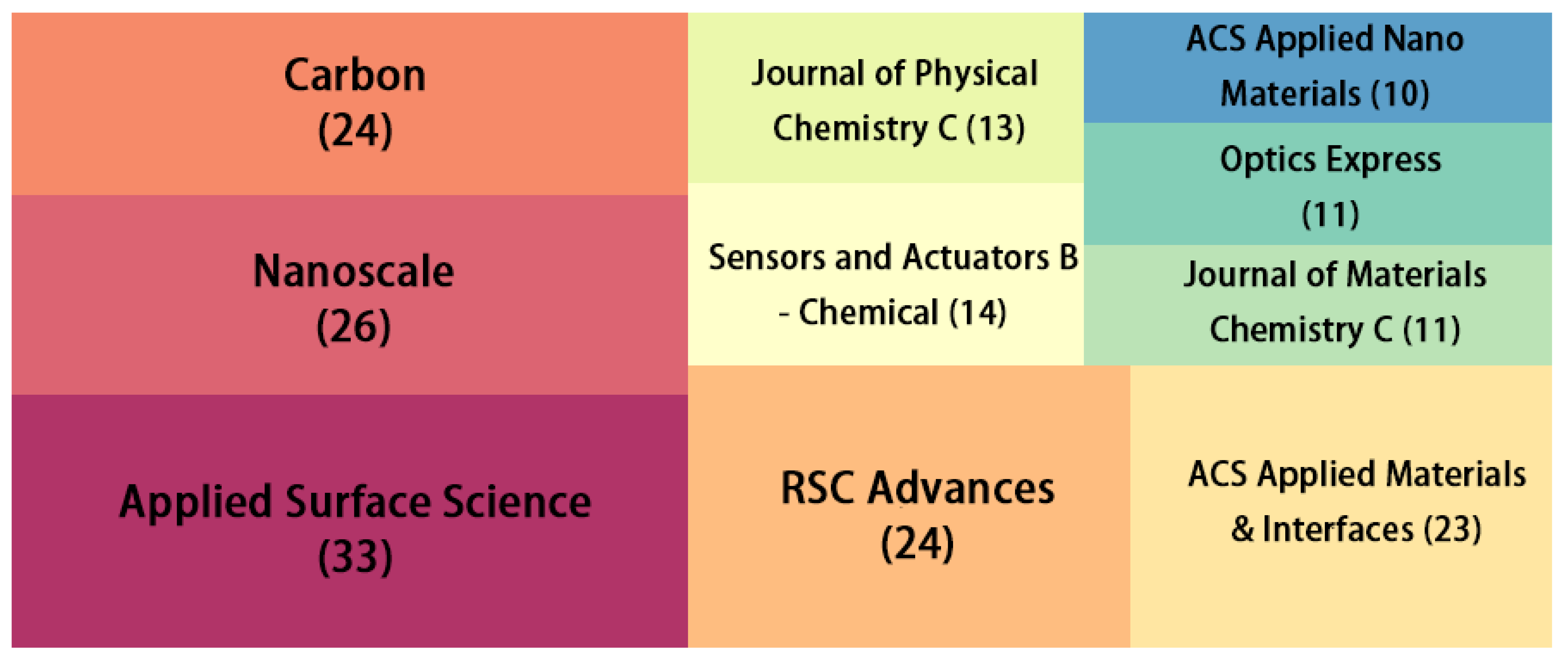

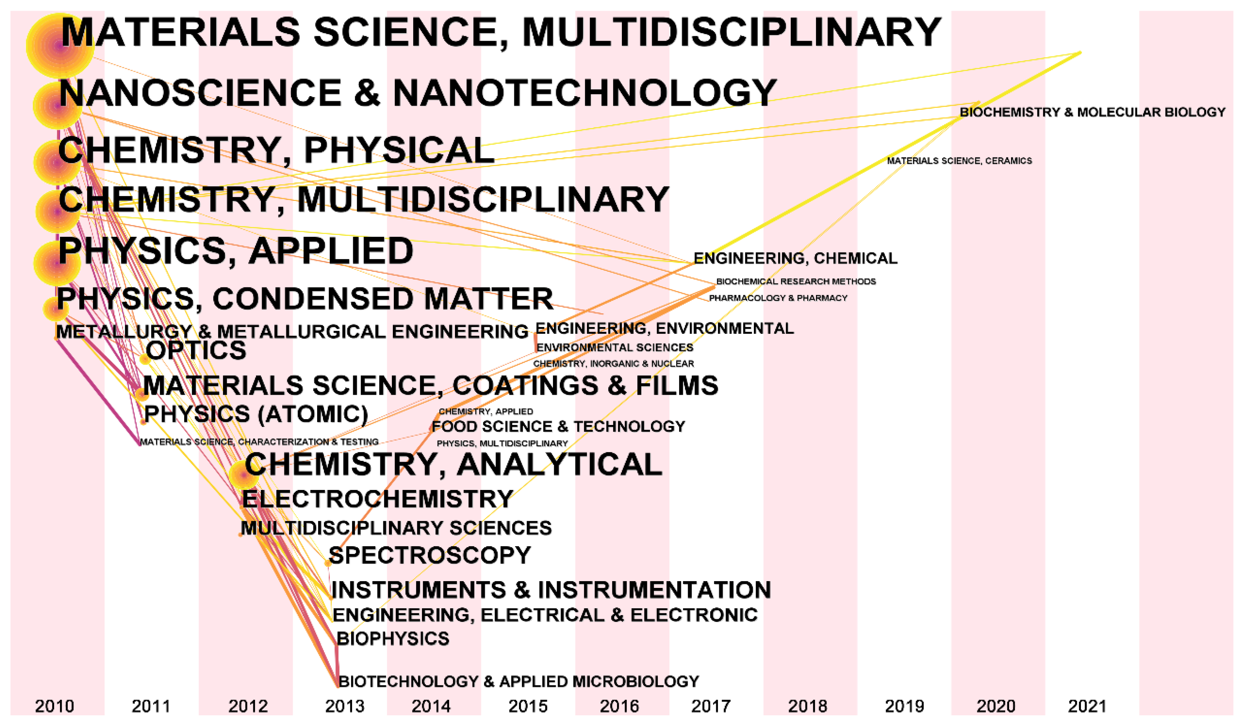
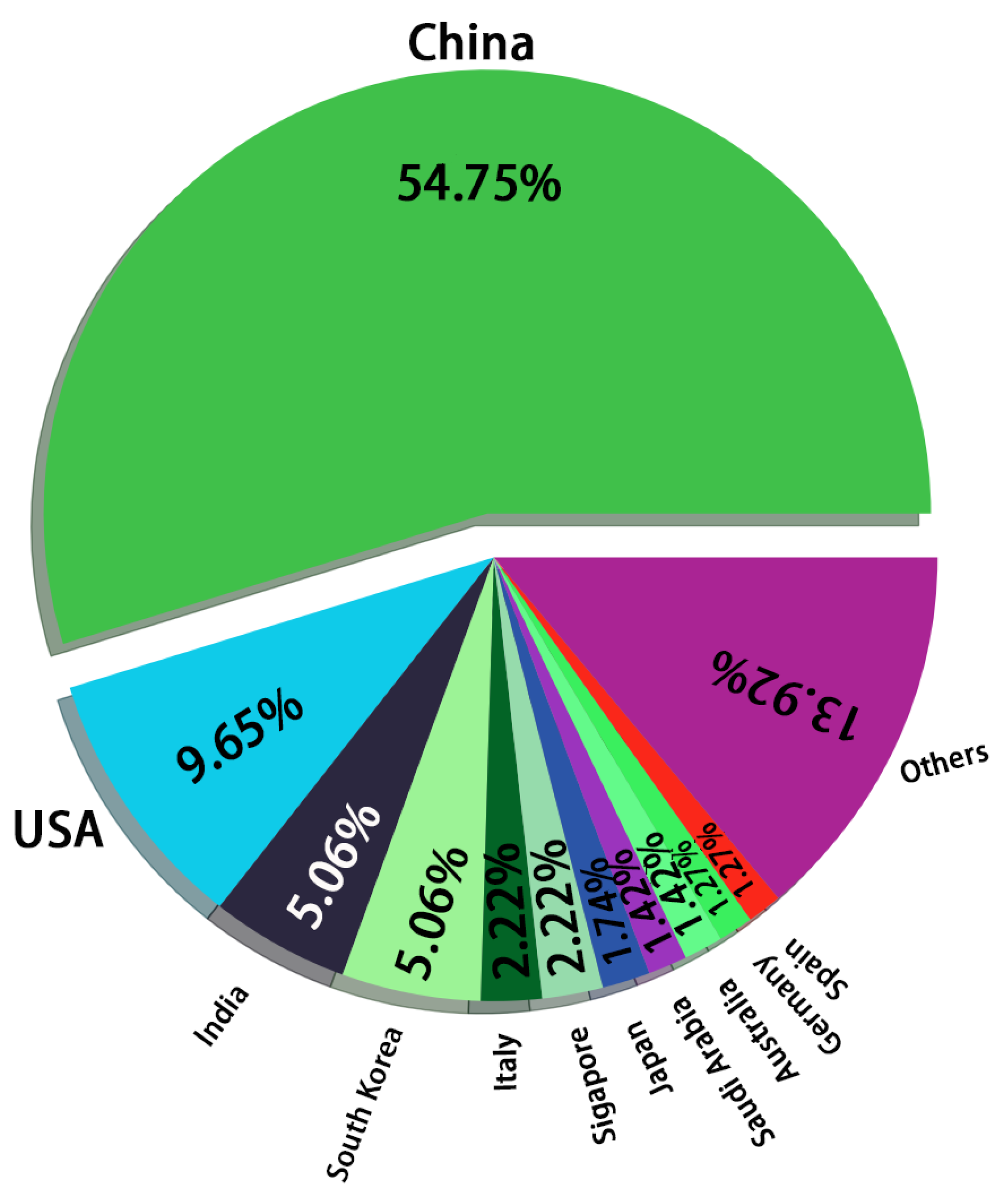


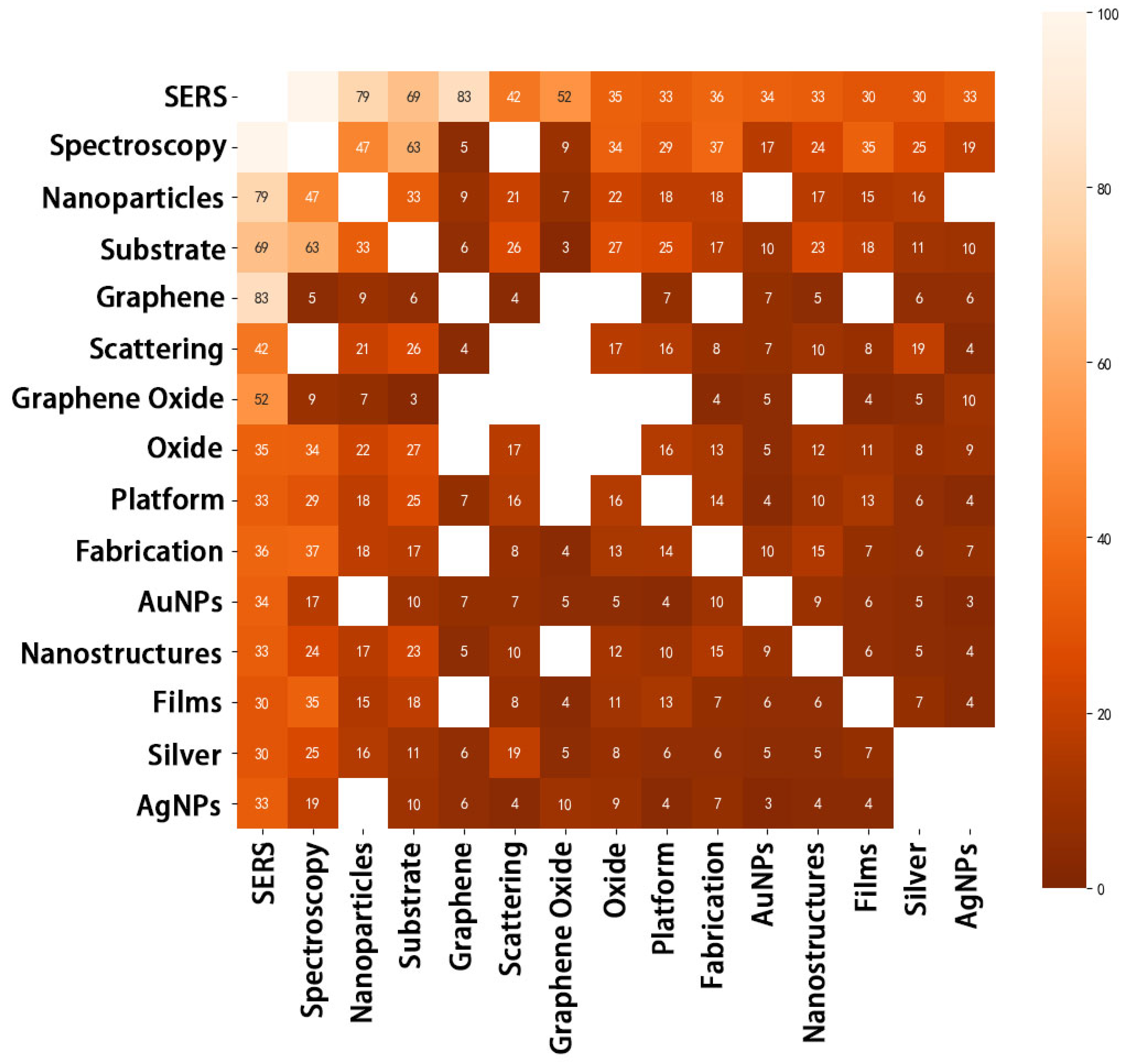
| No. | Citation | Cited Journal |
|---|---|---|
| 1 | 375 | ACS Nano |
| 2 | 369 | Journal of the American Chemical Society |
| 3 | 359 | Nano Letters |
| 4 | 345 | The Journal of Physical Chemistry C |
| 5 | 311 | Nanoscale |
| 6 | 295 | ACS Applied Materials & Interfaces |
| 7 | 289 | Small |
| 8 | 275 | Advanced Materials |
| 9 | 260 | Science |
| 10 | 259 | Carbon |
| 11 | 234 | Chemical Society Reviews |
| 12 | 233 | Analytical Chemistry |
| 13 | 202 | Chemical Communications |
| 14 | 188 | Langmuir |
| 15 | 186 | Nature |
| 16 | 184 | RSC Advances |
| 17 | 183 | Physical Chemistry Chemical Physics |
| 18 | 182 | Applied Physics Letters |
| 19 | 180 | Physical Review Letters |
| 20 | 178 | Applied Surface Science |
| Year | Journal Name |
|---|---|
| 2021 | ChemistrySelect, Journal of Materials Science & Technology, Science of The Total Environment, ACS Applied Bio Materials, The New England Journal of Medicine, Coatings, Frontiers in Chemistry, Nucleic Acids Research, Advanced Biology, Polycyclic Aromatic Compounds, Heliyon, Cellulose, Royal Society Open Science, Nature Reviews Chemistry, Fullerenes, Nanotubes and Carbon Nanostructures, Environmental Science: Nano, Dyes and Pigments, Journal of Biomolecular Structure and Dynamics |
| 2020 | Molecules, Biosensors, Journal of Pharmaceutical and Biomedical Analysis, NPG Asia Materials, Optical Materials Express, Nanoscale Horizons, Nanomaterials, JOSA B, Spectroscopy and Spectral Analysis, Bioinformatics, Composites Part B: Engineering, Nano Materials Science, Computational and Theoretical Chemistry, International Nano Letters, Synthetic Metals, Journal of Physics D, Trends in Food Science and Technology, American Journal of Psychiatry, American Mineralogist, Advanced Healthcare Materials, American Journal of Nuclear Medicine and Molecular Imaging, Advanced Synthesis & Catalysis, Advanced Device Materials, ACM Transactions on Sensor Networks |
| No. | Count | Institution | Centrality |
|---|---|---|---|
| 1 | 49 | Chinese Academy of Sciences * | 0.18 |
| 2 | 13 | Nanyang Technological University | 0.08 |
| 3 | 3 | Massachusetts Institute of Technology | 0.07 |
| 4 | 9 | Jiangsu University | 0.04 |
| 5 | 2 | Jiangnan University | 0.04 |
| 6 | 14 | University of Science and Technology of China | 0.03 |
| 7 | 3 | Academia Sinica * | 0.03 |
| No. | Freq | Centrality | Keywords |
|---|---|---|---|
| 1 | 179 | 0.26 | Spectroscopy |
| 2 | 129 | 0.21 | Nanoparticle |
| 3 | 125 | 0.15 | Substrate |
| 4 | 88 | 0.07 | SERS |
| 5 | 79 | 0.08 | Film |
| 6 | 76 | 0.07 | Oxide |
| 7 | 69 | 0.05 | Fabrication |
| 8 | 69 | 0.06 | Platform |
| 9 | 65 | 0.06 | Nanostructure |
| 10 | 62 | 0.07 | Graphene Oxide |
| 11 | 58 | 0.04 | Molecule |
| 12 | 50 | 0.05 | Silver |
| 13 | 49 | 0.04 | Silver Nanoparticle |
| 14 | 48 | 0.06 | Gold Nanoparticle |
| 15 | 45 | 0.04 | Reduction |
| Keywords | Strength | Begin | End | 2010–2021 |
|---|---|---|---|---|
| Sheet | 5.53 | 2010 | 2014 |  |
| Delivery | 3.39 | 2010 | 2013 |  |
| Graphene oxide | 3.21 | 2010 | 2014 |  |
| Silver | 3.07 | 2010 | 2013 |  |
| Spectra | 2.95 | 2010 | 2014 |  |
| Gold nanostructure | 2.78 | 2011 | 2013 |  |
| Pyridine | 3.32 | 2013 | 2014 |  |
| SERS detection | 4.07 | 2017 | 2018 |  |
| Graphene | 3.92 | 2019 | 2021 |  |
| Surface-enhanced Raman spectroscopy | 2.90 | 2019 | 2021 |  |
| Cluster ID | Size | Silhouette | Keywords | References |
|---|---|---|---|---|
| 0 | 38 | 0.937 | Nanoparticle, Substrate, Enhanced Raman scattering, Film, Fabrication, Platform, Reduction, Nanocomposite, Composite, Sheet | [32,54,55,56,57,58,59,60,61,62,63,64,65,66,67,68,69,70,71,72,73,74,75,76,77,78,79,80,81,82,83,84,85,86,87,88,89,90,91,92,93,94,95,96,97,98,99,100,101,102,103,104,105,106,107,108,109,110,111,112,113,114,115,116,117,118,119,120,121,122,123,124,125,126,127,128,129,130,131,132,133,134,135,136,137,138,139,140,141,142] |
| 1 | 32 | 0.840 | Nanostructure, Surface-enhanced Raman scattering, Array, Ag nanoparticle, Graphene, Water, Oxidation, Photoluminescence, Facile fabrication | [55,57,62,64,65,68,74,76,77,79,84,96,98,112,143,144,145,146,147,148,149,150,151,152,153,154,155,156,157,158,159,160,161,162,163,164,165,166,167,168,169] |
| 2 | 30 | 0.935 | Oxide, Silver nanoparticle, Reduced graphene oxide, Carbon, Light matter interaction, Green synthesis, Au nanoparticle, | [55,62,76,94,95,143,144,170,171,172,173,174,175,176,177,178,179,180,181,182,183,184,185,186,187,188,189,190,191,192,193,194,195,196,197,198,199,200,201] |
| 3 | 29 | 0.824 | Surface, Enhanced Raman spectroscopy, Rhodamine 6G, Graphite oxide, Electrode, Bacteria, Monolayer, Absorption | [56,60,70,73,77,104,146,147,171,177,185,202,203,204,205,206,207,208,209,210,211,212,213,214,215,216,217,218,219,220] |
| 4 | 27 | 0.960 | Growth, Raman, Plasmon resonance, Epitaxial graphene, Doped graphene, Facile synthesis | [72,97,221,222,223,224,225] |
| 5 | 26 | 0.943 | Sensitivity, Adsorption, Sensitive detection, SERS detection, Surface plasmon resonance | [21,57,75,89,171,217,226,227,228,229,230,231,232,233] |
| 6 | 24 | 0.941 | Hybrid, Au, Deposition, Size, Acid | [47,50,86,91,100,108,234,235,236,237,238,239,240,241,242,243,244] |
| 7 | 23 | 0.983 | Spectroscopy, Molecule, Silver, Carbon nanotube, Few layer graphene, Pyridine | [16,31,55,56,61,66,67,69,82,88,101,106,109,170,172,173,175,178,182,186,218,220,226,234,235,237,245,246,247,248,249,250,251,252,253,254,255,256,257,258,259,260,261,262,263,264,265,266,267,268,269,270,271,272,273,274,275,276,277,278,279,280,281,282,283,284,285,286,287,288,289,290] |
| 8 | 23 | 0.931 | Surface enhanced Raman scattering, Surface-enhanced Raman spectroscopy, Monolayer graphene, Malachite green, Temperature | [42,58,74,80,85,92,247,291,292,293,294,295] |
| 9 | 22 | 0.664 | Chemical enhancement, Aromatic molecule, Chemistry, Cell, Optical property, Quantum dot | [54,246,249,253,264,292,296,297,298,299,300] |
| 10 | 22 | 0.954 | Performance, Single molecule, Immunoassay, Surface enhanced Raman, Ultrasensitive detection | [80,88,89,103,176,182,214,248,261,301,302,303,304,305,306,307,308,309] |
| 11 | 21 | 0.926 | Graphene oxide, Ag, Gold, Charge transfer, Gold nanostructure | [52,53,56,58,63,68,73,75,78,80,90,93,102,143,145,148,174,183,251,263,301,310,311,312,313,314,315,316,317,318,319,320] |
| 12 | 19 | 0.908 | Scattering, Spectra, Raman spectroscopy, Graphite | [29,40,60,66,120,124,210,214,215,216,219,234,236,246,255,256,258,259,291,310,321,322,323,324,325,326,327,328,329,330,331,332,333,334,335] |
| 13 | 17 | 0.917 | Fluorescence, Sensor, Gold nanorod, Shape | [41,59,143,181,245,260,336,337,338,339,340] |
| 14 | 10 | 0.978 | Gold nanoparticle, Label free detection, Hybrid film, Folic acid, Surface enhanced Raman scattering | [48,58,96,173,234,252,254,262,278,304,310,317,341,342,343] |
| 15 | 6 | 0.988 | Surface-enhanced Raman scattering (SERS), Anatase | [61,344] |
| 16 | 6 | 0.968 | Trace detection, Silver dendrite, Agent, Dendrite | [345] |
| No. | Title | Citation | Year | Reference |
|---|---|---|---|---|
| Research Article | ||||
| 1 | Surface enhanced Raman spectroscopy on a flat graphene surface | 455 | 2012 | [350] |
| 2 | Surface-enhanced Raman spectroscopy of graphene | 397 | 2010 | [29] |
| 3 | Nanocomposites of size-controlled gold nanoparticles and graphene oxide: Formation and applications in SERS and catalysis | 379 | 2010 | [32] |
| 4 | A binary functional substrate for enrichment and ultrasensitive SERS spectroscopic detection of folic acid using graphene oxide/Ag nanoparticle hybrids | 322 | 2011 | [48] |
| 5 | UV/ozone-oxidized large-scale graphene platform with large chemical enhancement in surface-enhanced Raman scattering | 297 | 2011 | [16] |
| 6 | Tuning chemical enhancement of SERS by controlling the chemical reduction of graphene oxide nanosheets | 274 | 2011 | [17] |
| 7 | One-pot green synthesis of Ag nanoparticles-graphene nanocomposites and their applications in SERS, H2O2, and glucose sensing | 261 | 2012 | [257] |
| 8 | A facile one-pot method to high-quality Ag-graphene composite nanosheets for efficient surface-enhanced Raman scattering | 238 | 2011 | [351] |
| 9 | Silver nanoparticle decorated reduced graphene oxide (rGO) nanosheet: A platform for SERS based low-level detection of uranyl ion | 226 | 2013 | [91] |
| 10 | Surface enhanced Raman scattering of Ag or Au nanoparticle-decorated reduced graphene oxide for detection of aromatic molecules | 225 | 2011 | [71] |
| Review Article | ||||
| 1 | Graphene: A platform for surface-enhanced Raman spectroscopy | 393 | 2013 | [352] |
| 2 | Graphene-gold nanoparticles hybrid-synthesis, functionalization, and application in an electrochemical and surface-enhanced Raman scattering biosensor | 130 | 2016 | [353] |
| 3 | Recent progress in the applications of graphene in surface-enhanced Raman scattering and plasmon-induced catalytic reactions | 103 | 2015 | [24] |
| 4 | Recent progress on graphene-based substrates for surface-enhanced Raman scattering applications | 71 | 2018 | [354] |
| 5 | Flexible and stretchable SERS substrate based on a pyramidal PMMA structure hybridized with graphene oxide assivated AgNPs | 45 | 2018 | [355] |
Publisher’s Note: MDPI stays neutral with regard to jurisdictional claims in published maps and institutional affiliations. |
© 2022 by the authors. Licensee MDPI, Basel, Switzerland. This article is an open access article distributed under the terms and conditions of the Creative Commons Attribution (CC BY) license (https://creativecommons.org/licenses/by/4.0/).
Share and Cite
Zhou, Q.; Jin, M.; Wu, W.; Fu, L.; Yin, C.; Karimi-Maleh, H. Graphene-Based Surface-Enhanced Raman Scattering (SERS) Sensing: Bibliometrics Based Analysis and Review. Chemosensors 2022, 10, 317. https://doi.org/10.3390/chemosensors10080317
Zhou Q, Jin M, Wu W, Fu L, Yin C, Karimi-Maleh H. Graphene-Based Surface-Enhanced Raman Scattering (SERS) Sensing: Bibliometrics Based Analysis and Review. Chemosensors. 2022; 10(8):317. https://doi.org/10.3390/chemosensors10080317
Chicago/Turabian StyleZhou, Qingwei, Meiqing Jin, Weihong Wu, Li Fu, Chengliang Yin, and Hassan Karimi-Maleh. 2022. "Graphene-Based Surface-Enhanced Raman Scattering (SERS) Sensing: Bibliometrics Based Analysis and Review" Chemosensors 10, no. 8: 317. https://doi.org/10.3390/chemosensors10080317