
Electrochemical Sensors and Their Applications: A Review


1
DBT-ICGEB Centre for Advanced Bioenergy Research, International Centre for Genetic Engineering & Biotechnology, Aruna Asaf Ali Marg, New Delhi 110067, India
2
Department of Electrical and Electronic Engineering, University of Cagliari, Via Marengo 2, 09123 Cagliari, Italy
3
Department of Analytical Chemistry, University of Chemistry and Technology Prague, Technická 5, 166 28 Prague, Czech Republic
*
Authors to whom correspondence should be addressed.
Chemosensors 2022, 10(9), 363; https://doi.org/10.3390/chemosensors10090363
Submission received: 1 August 2022
/
Revised: 1 September 2022
/
Accepted: 5 September 2022
/
Published: 9 September 2022
(This article belongs to the Special Issue Electrochemical Detection: Analytical and Biological Challenges)
Abstract
:The world of sensors is diverse and is advancing at a rapid pace due to the fact of its high demand and constant technological improvements. Electrochemical sensors provide a low-cost and convenient solution for the detection of variable analytes and are widely utilized in agriculture, food, and oil industries as well as in environmental and biomedical applications. The popularity of electrochemical sensing stems from two main advantages: the variability of the reporting signals, such as the voltage, current, overall power output, or electrochemical impedance, and the low theoretical detection limits that originate from the differences in the Faradaic and nonFaradaic currents. This review article attempts to cover the latest advances and applications of electrochemical sensors in different industries. The role of nanomaterials in electrochemical sensor research and advancements is also examined. We believe the information presented here will encourage further efforts on the understanding and progress of electrochemical sensors.

1. Introduction
A chemical sensor is defined by the IUPAC [1] as “a device that converts chemical data, ranging from the concentration of a single sample component to complete composition analysis, into an analytically usable signal”. For the most part, a chemical sensor is constituted of two essential functional units: a receptor and a physicochemical transducer. The receptors are variable and can range from activated or doped surfaces to complex (macro)molecules that create highly specific interactions with the analyte (Figure 1).
If the receptor is of biological origin (e.g., DNA, antibodies, and enzymes), the device is referred to as a biosensor. The receptor interacts with the analyte, converting the recognition event into a predetermined output signal. One of the primary requirements of sensors is to maintain a high degree of specificity for the intended analyte in the presence of potentially interfering chemical species to avoid false-positive outcomes. Another critical component of sensors is the transducer, which is responsible for converting the signal created by the receptor–analyte interaction into a readable value. Thus, both chemical and biosensors can be classified into catalytic or affinity-based devices. Whereas catalytic sensors utilize catalytic activity to generate the signal, as in the case of enzymatic, DNAzyme, or functionalized surfaces that can perform redox reactions under certain conditions, affinity-based devices rely on highly specific interactions between the receptor and analyte, e.g., using the specific affinity of nucleic acids (i.e., ssDNA and aptamers), antibodies–antigens, or host–guest interactions. The monitoring of the recognition events can be performed using several methods (e.g., optical, gravimetric, or electrochemical) depending on the type of transducer utilized [2].
Being the market leaders, electrochemical sensors are, by far, the most frequently employed type of sensor due to the fact of their advantages associated with low detection limits, as low as picomoles, rapidness, and the low-cost equipment utilized for sensing. Electrochemical sensors come in a variety of form factors ranging from the top-bench to fully integrated wearable devices [3]. The utility of a chemical sensor is to deliver accurate real-time information regarding the chemical composition of its surroundings. In an ideal scenario, such a device would be able to respond constantly and reversibly without interfering with the sample. In such devices, a biological or chemical identification layer is coated on a transduction element. In electrochemical sensors, the analytical information is taken from the electrical signal produced by the interaction of the target analyte and the recognition layer. Various electrochemical devices can be employed for environmental monitoring depending on the nature of the analyte, the character of the sample matrix, and the sensitivity or selectivity requirements. The bulk of these devices fall into several categories such as amperometric and potentiometric electrochemical sensors (depending on the device’s nature) [4,5]. Electroactive species that are involved in chemical or biological identification are detected using amperometric sensors.
2. Types of Electrochemical Sensors
Electrochemical sensors can be classified into several categories including amperometric, potentiometric, impedimetric, photoelectrochemical, and electrogenerated chemiluminescence. For potentiometric sensors, as a result of specific sensor–analyte interactions, a local Nernstian equilibrium is formed at the sensor interface, when no current is allowed to flow in the system giving information about the analyte’s concentration. Amperometric sensors employ a voltage placed between a reference and working electrodes to initiate electrochemical oxidation or reduction, measuring the resulting current as a quantitative indicator of the analyte’s concentration, according to the Cottrell equation:
where:
i = Current (in ampere);
n = Number of electrons;
F = Faraday constant (96,485 C/mol);
A = Area of the (planar) electrode in cm2;
c0j = Initial concentration of the reducible analyte {\displaystyle j}in mol/cm3;
Dj = Diffusion coefficient for species {\displaystyle j}in cm2/s;
t = time in seconds.
Conductometric sensors, frequently referred to as impedimetric sensors, on the other hand, measure changes in the surface impedance to detect and quantify analyte-specific recognition events on the electrode. The extraordinary success of electrochemical sensor research, and its growing influence on analytical chemistry, make it difficult to address all of the achievements within the scope of this review and, therefore, we aimed to demonstrate the variability in the field, rather than deep immersion into a certain type of electrochemical sensor. Table 1 lists the different analytes, types of biosensors, and electrochemical measurement techniques [6].
2.1. Potentiometric Sensors
Due to the fact of their simplicity and low cost, since the early 1930s, potentiometric sensors have been the most extensively used practical sensors. Potentiometric devices can be classified into three categories:
- 1.
- Ion-selective electrodes (ISEs);
- 2.
- Coated-wire electrodes (CWEs);
- 3.
- Field-effect transistors (FETs).
The type and content of the membrane material play a significant role in constructing an electrode that is selective to a single species. The research in this sector has paved the way for a variety of applications involving an almost infinite number of analytes, with the only restriction being the membrane matrix’s dopant and ionophore composition. ISEs may be classified into three categories based on the type of the membrane: glass electrodes, liquid electrodes, and solid electrodes. Over two dozen ISEs are commercially available from companies, such as Corning, Orion, Radiometer, Beckman, and Hitachi, and they are widely used for the analysis of organic ions and anionic or cationic species in a variety of effluents, in the oil industry and in the manufacturing process and monitoring of drugs, using response membrane electrodes specifically designed for this purpose [11,12,13]. Wearable device technology paired with potentiometric ion sensors based on an all-solid-state concept offers significant potential in the tracking of physical status during athletic performance along with clinical medicine via sweat analysis [14].
pH electrodes have been the most extensively used potentiometric device for several decades and are the most widely used potentiometric device overall. A thin ion-sensitive glass membrane is used to create glass electrodes, which are the most common type and are available in a variety of forms and sizes. Nonetheless, additional types of potentiometric sensors that utilize organic polymers (e.g., polymethylene blue) or redox-active molecules (e.g., ferrocenes and quinones) can be used to detect pH in addition to those described above. Additionally, it has been reported that glass electrodes for monovalent cations, such as sodium, lithium [15,16], ammonium, and potassium sensors, can be employed [17]. These electrodes are composed of novel glass compositions. The use of glass membrane electrodes to determine pH solutions has proven highly effective; however, it is now confined to aqueous measurements. It is essential to make corrections when determining the concentration of hydrogen ions in nonaqueous liquids [18].
2.2. Amperometric Sensors
Amperometric measurements are frequently used as an analytical method of high accuracy and sensitivity in which the applied voltage serves as a driving force for electrocatalytic redox reactions that generate electrical currents proportional to the concentration of the analyte. A controlled-potential system is required for the fundamental instrumentation, and the electrochemical cell is composed of two electrodes submerged in an electrolyte of an appropriate composition. A more sophisticated and common design is the employment of a three-electrode cell, with one of the electrodes functioning as a reference electrode [19]. However, whereas a working electrode is defined as the electrode where the reaction of interest occurs, a reference electrode (such as Ag/AgCl or Hg/Hg2Cl2) is defined as the one that maintains a constant potential when compared to a working electrode [20]. As an auxiliary electrode, an inert conducting substance (e.g., platinum or graphite) is often employed. In controlled-potential studies, a supporting electrolyte is necessary to prevent electromigration effects, lowering the resistance of the solution and maintaining the ionic strength constant. Theoretical considerations, as well as practical approaches, have been well described [21,22].
2.3. Impedimetric Sensors
One strategy is to stimulate the cell with a small-magnitude alternating potential and then see how the system responds in a steady state. This strategy has many benefits. The most significant ones are the ability to perform sensitive measurements using an experiment because the response may be permanently steady and can thus be averaged over a long period of time, the capability to treat the response theoretically using generalized linear current-potential characteristics, and measurement over a broad time or frequency range [6]. Polymers, either by themselves or in combination with a conductor, are also often utilized. For example, polypyrrole is capable of detecting volatile amines and when doped with ClO4- and tosylate, it can be used as an NH3 sensor [23].
3. Electrochemical Sensor Applications
Electrochemical sensors have long been desirable for the investigation of biological, environmental, industrial, and pharmacological species, not only for their long-term dependability, high sensitivity, and accuracy but also for their low cost, speed, and ease of downsizing. For more than two decades, numerous nanomaterials with extraordinary characteristics, such as metals, conductive polymers, metal oxides, and metal–organic and carbon-based nanomaterial frameworks, have been included in electrochemical assays to promote analytical performance. This modification allows for increasing the loading capacity through the use of recognition molecules, such as enzymes, antibodies, and aptamers, as well as bioinspired receptors, which can capture targets specifically and effectively, thereby increasing the specificity of the electrochemical sensors. This is closely related to the aim of providing strong electrocatalytic activity for certain electrochemical processes. Additionally, by altering the surface shape and structure, it is possible to increase both the electrical conductivity and surface area, which should enhance the sensitivity of these tests. Electrochemical sensors have gained popularity recently owing to new applications such as single-molecule sensing, in vivo analysis, wearables, and point-of-care diagnostics [28].
Electrochemical sensors have a number of advantages, including their high sensitivity, which enables low LODs and LOQs; their rapid analytical response, which makes them ideal for flow analysis and alert systems; their simplicity, which allows for a virtually limitless variety of geometries, electrode materials, and configurations; their ease of use (simple and low-cost equipment, the ability to be integrated as a detection module in a variety of analytical systems).
Biosensors are interesting analytical instruments for environmental and biological investigations, because they have the following additional advantages:
- i.
- Quick data collection;
- ii.
- Detection of the important substrate is frequently accomplished without previous separation;
- iii.
- A sensitivity that can reach ng/mL;
- iv.
- Good selectivity and, occasionally, even specificity;
- v.
Biosensors are composed of three major components:
- Biocomponents or systems of biological detection: Biocomponents include enzymes, antibodies, other similar binding molecules, DNA probes, live cells, and organelles;
- Transducers: Converts the signal generated by the analyte’s interaction with the biocomponent into a quantifiable electrical signal;
- A signal processing system: It turns the measured signal into a form that is accessible and readable.
3.1. Biomolecule Electrochemical Detection
Biomolecules of a small size (e.g., hormones, nucleic acids, and enzymes) are detected based on their physiological and biological roles, which include transferring regulating biological activity, genetic information, and catalyzing cellular processes [32,33,34]. Nonetheless, creating biomolecule-sensing technology continues to be a difficult task [35]. Biomolecular methods, such as Western blot, polymerase chain reaction (PCR), and gel electrophoresis, have been developed for the analysis of biomolecules [36]. Despite their precision, they are limited by constraints such as large reagent needs, laboriousness, and long-time requirements [37].
Various studies on electrochemical approaches for identifying biomolecules as an early diagnostic tool have been published [38,39,40]. Mohan et al. [41] developed an integrated electrochemical biosensor that could detect biomarkers in urine. This may help improve the effectiveness of clinical disease management and indicates that pathogen identification in combination with quantitative detection of lactoferrin can provide important information for the diagnosis of urinary tract infections (Figure 2).
3.1.1. Electrochemical Biosensing for Viral Infections
Electrochemical biosensors are robust, easy to use, portable, and inexpensive analytical systems that can operate in turbid media and provide highly sensitive readouts [42]. DNA and RNA electrochemistry have been utilized to diagnose viral illnesses such as hepatitis E, coronavirus, HIV, influenza virus, bacterium, malaria, and Zika virus [43,44,45,46,47] (Figure 3).
Aptamers are short, single-stranded oligonucleotides (i.e., DNA or RNA) that range in size from 10 to 100 nucleotides. They are created using the SELEX method [48], which stands for the systematic evolution of ligands by exponential enrichment. By monitoring the change in the current response or electrical resistivities from the redox interaction between the targets and the aptamers attached on the electrode surface of the sensor, the electrochemical aptasensor determines the concentration of the interested targets [49].
Another example is the use of electrochemical techniques for detecting enzymes and hormones to monitor for pregnancy-related disease and cancer [50,51]. In comparison to traditional procedures, such as Western blot and PCR, in terms of the time and cost, an electrochemical approach is a preferable option [52]. Nevertheless, its effectiveness is dependent on the conductivity characteristics of the sensing surface [53,54]. Electrochemical performance with complex samples necessitates preventing signal overlapping due to the fact of interference.
3.1.2. Electrochemical Sensors: Recognition of the SARS-CoV-2 Virus
Coronavirus (COVID-19), a pandemic that has killed many people, is on the rise again, and has spread all over the world, causing many global health problems [65,66]. Though new ways of detecting COVID-19 are being worked on all the time, there is still a need for new ways to detect COVID-19 early and ensure its monitoring. RT-PCR tests have been widely used because they can be life-saving diagnostic tools. However, because of their multiple steps, time-consuming process, need for highly skilled people, and high costs, these tests may not be good for monitoring many different samples at the same time. Over the last few years, electrochemical-sensor-based techniques have been used to detect SARS-CoV-2. These methods are fast and cheap as well as sensitive and specific. Neither a serological or RT-PCR assay nor the electrochemical detection of SARS-CoV-2 by sensors/biosensors is the best way to identify COVID-19. However, these methods can be used together [67,68,69]. There is a lot of hope that electrochemical sensors and biosensors can help improve point-of-care tests for the deadly SARS-CoV-2 virus.
Yakoh et al. [70] developed an electrochemical paper-based analytical device (ePAD), which was used in the detection of SARS-CoV-2 immunoglobulins (i.e., IgG and IgM) and specifically targeted SARS-CoV-2 antibodies. Antibodies can interfere with the redox conversion of [Fe(CN)6] 3/4 or create immunocomplexes, hence, decreasing the current response [71]. The sensing mechanism of the ePAD is due to the interruption of the redox conversion caused by the development of a complex between the captured immunoglobulins produced in response to COVID-19 infection in people and immobilized SARS-CoV-2 spiking protein. This procedure was examined for cross-reactivity [72] with anti-Epstein–Barr virus (anti-EBV), anti-hepatitis B surface antigen (anti-HBsAg), anti-hepatitis C virus (anti-HCV), anti-Rubella, and anti-cytomegalovirus (anti-CMV), but no cross-reactivity was observed [73].
3.2. Enzyme-Based Electrosensor Applications
Enzymes are organic catalytic molecules created by living organisms. They accelerate biological processes by decreasing activation energy, and they can accelerate the conversion of substrates to products in cellular metabolism by a factor of at least 10 million [74]. Enzyme-mediated substrate conversion is very specific. Numerous enzymes are selective for a single substrate, whereas another type of enzyme can affect multiple structurally similar substrates. In order to begin an enzyme-catalyzed reaction, the enzyme must form a complex with its substrate. Enzymes are unaltered by the processes they catalyze and are recyclable and effective in minute quantities. Equally, the enzyme catalyzes either the forward or reverse process [75].
Enzymatic activity monitoring is in great demand. For measuring enzymatic activity, many analytical techniques have been reported, e.g., mass spectrometry [76], spectrophotometry, Raman spectroscopy, and electrochemical techniques. Because of their ease of use, cheap cost, and speed, electrochemical procedures are favored over other analytical techniques [77], which may need sophisticated pretreatment, filtering, and a knowledgeable operator. Enzymatic sensors are created by immobilizing an enzyme on an electrode and then used to determine the concentration of the matching substrate. The primary distinction between enzyme-based biosensors is the immobilization technique and the mediator used [78].
In a recent study, the authors constructed an amperometric Glc biosensor with Gox immobilized on MWCNTs, as the biorecognition element, and RuO2 acting as the mediator. To boost the sensor’s stability, the enzyme was coated with a Nafion® membrane. The designed sensor was used to determine the concentrations of hydrogen peroxide and glycol. The developed sensor was employed as an electroanalytical technique for studying the inhibition of the enzyme’s function, and the influence of the heavy metal cations (i.e., Cd2+, Hg2+, and Ag+) on the activity of the Gox enzyme was examined [78].
3.3. Ion-Selective Electrodes (ISEs): Application in Medicine
Clinical chemistry, namely, the determination of physiologically relevant electrolytes in physiological fluids, continues to be the predominant application sector for ISEs [79], with billions of regular ISE measurements conducted worldwide each year [80]. The International Federation of Clinical Chemistry (IFCC) has certified sensors for pH and ionized calcium, potassium, and sodium for use in commercially available clinical analyzers [81]. Additionally, magnesium, chloride, and lithium ions are commonly identified by matching ISEs in blood plasma, urine, and hemodialysis solutions [82], among other locations. Sensors for the characterization of physiologically significant polyions (i.e., heparin and protamine), phosphates [83], dissolved carbon dioxide, and other blood analytes have been extensively studied over the years and are on the verge of displacing less reliable and/or inconvenient analytical techniques for blood analysis. In comparison to conventional analytical techniques, ISEs respond to ion activity rather than the concentration, which makes them particularly interesting for clinical applications, because ion activity is typically connected with health issues. While the majority of ISEs are utilized in vitro, the ability to take measurements in vivo and continually use implanted sensors might prove a helpful diagnostic tool for physicians. Sensors must meet two strict requirements: first, they must cause the least amount of disruption to the in vivo environment, which can be problematic due to the injuries and inflammation frequently caused by implanted sensors, as well as the leaching of sensing materials; second, they must be immune to the environment, with the effects of cell adhesion, protein adsorption, and extraction of lipophilic species on a sensor. Nonetheless, microfabricated sensor arrays have been used to successfully detect electrolytes in situ in rabbit muscles [84].
Pharmaceutical analysis is another area in which ISEs excel [85]. ISEs have been used to identify a wide variety of pharmaceuticals in pharmaceutical formulations and manufacturing processes. Drugs and their metabolites can be quantified in actual bodily fluids. Though ISEs are not frequently employed in pharmaceutical chemistry at the moment, they offer significant promise, as seen by the development of a number of ISE applications in recent years [86,87,88,89]. The majority of drug-selective electrodes are ion exchange based and take advantage of the frequently high lipophilicity of drugs and metabolites [90].
3.4. Biosensors’ Distinct Characteristics in Health Services
Diabetes prevalence and diabetes patients’ use of biosensors are significant contributors to worldwide business profitability. Rapid and preventive diabetes detection is becoming increasingly popular. Biosensor developments have made it possible to detect blood glucose in the presence of various intervening substances throughout a wide temperature range. Using ZnO nanorods to detect glucose is a low-cost, safe, accurate, rapid, and safe method [91]. The sensitivity and accuracy of biosensors within a minute sample volume are improving, and they are now widely employed in the diabetes domain, with significant market demand projected in the coming years. Portable electronic gadgets are an important part of the overall healthcare system because of their high capacity for monitoring, therapy, diagnosis, fitness, and well-being. They will increase preventative measures and obtain a better perspective of their well-being by combining therapeutic technologies accessible in hospitals and emergency care centers.
3.5. Electrochemical Sensors: Environmental Applications
Electroanalytical chemistry has the potential to be a game-changer in terms of environmental protection. Electrochemical sensors and detectors, in particular, are tempting for on-site monitoring of priority pollutants and other environmental requirements. Such technology may meet a lot of the needs for on-site environmental analysis. Such capabilities have already tremendously aided decentralized clinical analysis (Figure 5). Electrochemical sensors for pollution management are still in the early phases of development, despite their tremendous potential for environmental monitoring [95].
The identification of inorganic ions, metal cations, organic chemicals, and biomolecules is possible using the trace electroanalytical method, known as electrochemical stripping analysis (ESA). It is based on a step where the target analyte(s) or a compound of the target is preconcentrated on an appropriate working electrode. The remaining accumulated analyte is then removed using an electroanalytical method [96]. Electrochemical stripping analysis has long been acknowledged as a highly effective method for determining trace metal concentrations [6,96,97,98,99,100]. Its extraordinary sensitivity is a result of the “built-in” accumulation process, which preconcentrates the target metals onto the working electrode. Electrochemical devices’ intrinsic miniaturization and low power needs meet a large number of criteria for on-site and in situ hazardous metals measurements. Due to the fact of its sensitivity to both low metal concentrations and the chemical form of metals in solution, stripping analysis is ideally suited for speciation research. Recent technological advancements have overcome past barriers to such field uses. As a result of these advancements, significant attention is currently being paid to decentralized electrochemical testing for trace metals. Additionally, nonelectrolytic (adsorptive) accumulation strategies have been devised to broaden the scope of stripping analysis to include trace metals that cannot be electrodeposited. Strip analysis is a two-step process. Typically, the first, or deposition phase, comprises the electrolytic deposition of a small number of metal ions in solution onto the mercury electrode to preconcentrate the metals. Following the preconcentration process, the stripping (measurement) step is performed, which entails the dissolving (stripping) of the deposit. Stripping analysis can be performed in a variety of ways, depending on the nature of the deposition and measuring stages [95]. In potentiometric sensors, the analytical data are obtained by converting the recognition process into a potential signal that is proportional (logarithmically) to the concentration (activity) of species created or consumed during the recognition event. Such devices rely on the use of ion-selective electrodes to obtain the potential signal [101]. In Table 4, examples of environmental analyses using electrochemical sensors and biosensors are listed.
3.6. Recent Uses of Carbon-Based Nanosensors in Pharmaceutical Analysis
Carbon-based nanosensors have seen a lot of use in pharmacological analysis in recent years as well as in real-world applications such as tablets and human serum. Cheemalapati et al. [116] used multiwalled carbon nanotubes on a glassy-carbon electrode to establish an electroanalytical measurement of anxiolytic buspirone hydrochloride. They employed MWCNTs were synthesized in dimethylformamide and had lengths of 0.1–10 μm. The linear range was determined using cyclic voltammetry, differential pulse voltammetry, and amperometry. Baytak and Aslanoglu [117] used a glassy-carbon electrode to make a nanosensor containing multiwalled carbon nanotubes and indium-tin oxide nanoparticles to determine the beta-adrenergic agonist metaproterenol. In pills and urine, the differential pulse voltammetric technique was used, which had a linear range of 1.2 × 10⁻⁸ M. Kutluay and Aslanoglu [118] employed MWCNTs functionalized with nickel nanoparticles to determine Bromhexine, a mucolytic medication.
3.7. Electrochemical Sensors: Design of Analytical Kits
Electrochemical techniques have been demonstrated to offer more benefits over other analytical methods due to the fact of their mobility and inexpensive cost. The majority of large firms have used this sort of analytical technology due to the fact of its rapid and selective analysis. Electroanalytical sensors are projected to be the next generation of analytical systems due to the fact of their ease of operation and great variety. As a result, several scientists and researchers have concentrated their efforts on developing and fabricating electroanalytical sensors with excellent selectivity and sensitivity for a variety of chemicals including pharmaceuticals, food, and environmental toxins. In a recent review paper, Karimi-Maleh et al. [119] discussed the mechanism and several applications of DNA, enzymatic, and electrocatalytic techniques for electroanalytical evaluation of medicinal, food, and environmental chemicals. In Table 5, a summary of the several types of electrochemical sensors and their applications in a variety of fields is presented. The interaction of DNA with analytes, particularly the intercalation reaction, is a highly helpful technique for determining anticancer medicines. Anticancer medicines deactivate the guanine and adenine bases and alter the oxidation base signals utilized for drug analysis. On the other hand, due to the selective interaction between the enzyme and the analyte, enzymatic biosensors may be utilized as selective analytical instruments.
4. Role of Nanomaterials in Electrochemical Sensors
Over the past years, nanotechnology has gained a lot of traction in the sensor industry. It is considered that the employment of such technologies, as well as the usage of nanosized materials, has a positive impact on sensor performance. Nanomaterials have been discovered to offer a variety of unique and intriguing physical and chemical characteristics [131,134]. In recent decades, low-dimensional nanometer-sized materials and systems have established a new field of study in condensed-matter physics. In addition to the aforementioned categories of materials, there are a variety of materials of various sorts that may be used to create nanosensors. Carbon is known as a one-of-a-kind element because of its numerous uses. Carbon is a fascinating element that exists in a variety of forms including graphite, diamond, fullerenes, and graphene. Some of the most significant and recent advances, made possible by the use of carbon-based nanostructures in nanotechnology for chemical and biological sensor creation as well as their use in the pharmaceutical and biomedical fields, have been reviewed in previous studies [135,136,137,138,139].
Recent advances in nanomaterials’ unique physicochemical features have been extremely successful in improving biosensors, and the potential for enhancing desirable molecular interaction has boosted the diagnostic sensitivity of these biosensors [140,141].
In the majority of reported biosensors, nanocomposites are used to increase the sensitivity, selectivity, and repeatability. Nanocomposites are solid materials composed of numerous phase domains with nanoscale features in at least one of them. The distinct and intriguing features of nanocomposites have attracted interest in recent years. Nanocomposites have several benefits in the manufacturing of sensors including a high surface-to-volume ratio, reactive capacity, biocompatibility, and high adsorption [142,143]. Nanocomposites are accountable for electrochemical reaction catalysis, biomolecule immobilization, biomolecule labeling, and electron transfer rate increase [144,145]. Conducting polymers, nanofibers, graphene, carbon nanotubes (CNTs), metal–organic frameworks (MOFs) [146], and nanoparticles (Nps) [147] are the most common nanocomposites [148] utilized to change the electrode surface [149,150,151]. Due to the fact of their evenly distributed metal centers, MOFs can be used as electrocatalysts for CO2 reduction reactions (CO2RR) [152].
Figure 6 shows the principle of an electrochemical sensor based on carbon nanomaterials for detecting biomarkers of metabolic diseases [153].
4.1. Carbon Nanotubes
Due to the fact of their tiny size and favorable electrochemical characteristics, carbon nanotubes have sparked great interest in electrochemistry. The great majority of research to date has employed carbon nanotube ensembles on nanostructure macroscopic electrodes, either randomly distributed nanotubes or aligned carbon nanotubes [154]. Carbon nanotubes (CNTs) are allotropes of elongated fullerenes with a modest average diameter of 100 nm. Due to the large surface area (l/d ratio), a superb platform for the efficient transport of active chemical, biological, or biochemical components is accessible. Carbon nanotubes are classified primarily according to their shape and size, as carbon nanotubes are classified primarily according to their shape and size as single-walled carbon nanotubes (SWCNTs) or multiwalled carbon nanotubes (MWCNTs). Carbon nanotube-based sensors for the detection of paracetamol and hesperidin have been developed [155,156,157]. The variable surface morphology of carbon materials permits a variety of surface functionalities for the development of highly efficient electrochemical sensors with long-term stability [158]. The conductivity of the tubes is critical for their involvement in electrochemistry as a result of their electrical characteristics. MWNTs are considered to be metallic conductors, which is an extremely desirable feature for an electrode.
4.2. Graphene in Sensors
Dresselhaus et al. [159] established graphene as the most commonly utilized nanomaterial for a range of applications [160,161,162,163,164,165]. The enormous specific area of graphene encourages high quantities of biomolecules to be loaded onto [166] the detecting base, resulting in high detection sensitivity. Electrons may easily pass between the graphene surface and biomolecules due to the fact of its tiny bandgap and good conductivity. Highly pure graphene, with no contaminants, and its derivative materials are harmless in character and cost effective due to the fact of their superb uniform surfaces.
Graphene’s large sensing surface area increases the loading of specific chemical species, such as proteins and enzymes, either by passive adsorption or chemical cross-linking to the analyte’s active groups [167]. The conductivity of graphene varies depending on the production or treatment method used. The electrical conductivity of graphene is 60 times that of SWCNTs, with particulate graphene having a reported electroconductivity of 64 mS/cm [168] compared to graphene’s 108 mS/cm [169]. The graphene family is being utilized to create and enhance electrocatalysis for increasing biomolecule loading and increasing the surface area.
4.3. Electrosensing Using Gold Nanoparticles
In electrosensing, gold nanoparticles are commonly utilized. The current tendency is to use “green” chemistry to photosynthesize gold nanoparticles (Phyto-AuNPs). Because Phyto-AuNPs are physiologically and catalytically stable, active, and biocompatible, they have a wide range of uses, including tactile and wearable (bio)sensors [170]. Because of their unique visual, physical, and electrochemical features, gold nanoparticles stand out [171]. To produce gold nanoparticles, a range of physical and chemical processes are utilized. Chemical synthesis, in comparison to physical synthesis, is extremely straightforward, labor saving, and economical. Chemicals and solvents, as well as reaction byproducts, can, nevertheless, be harmful to both people and the environment [172,173]. Recently, alternative techniques for nanoparticle production have evolved, which are based on a “green” chemical approach. The term “green” refers to reducing the use of hazardous chemicals and increasing the use of organic materials such as plants, for instance, the use of plant extracts (phytosynthesis) as reducing, stabilizing, and capping agents. The “green” approach is an efficient and environmentally beneficial way of creating gold nanoparticles [174]. The primary advantages of phytosynthesis include simplicity, environmental safety, a high synthesis rate, an absence of extra reagents, and the possibility of large-scale nanoparticle production [174,175,176].
The unique properties of phytosynthesized gold nanoparticles (Phyto-AuNPs) include their high catalytic activity in the degradation of organic dyes [172,177] and their anticancer [178], antioxidant [179], and antibacterial activity [180] as well as their biocompatibility and low cytotoxicity [175]. These properties make Phyto-AuNPs extremely attractive for biomedical applications such as diagnostic studies, theranostics, cell imaging, and protein as well as drug and photodynamic therapy and gene delivery [175,181].
Glassy-carbon electrodes are often used in sensors, and adding gold nanoparticles to glassy-carbon electrodes makes them more stable and sensitive electrochemically. Using a layer-by-layer method, gold nanoparticles and methylene blue were combined to created laminated films on a glassy-carbon electrode that could detect human chorionic gonadotropin (HCG). Due to the large area of the nanoparticles that can be used to load anti-HCG, this immunosensor could be used to measure the amount of HCG in blood or urine samples. Jena et al. [182] came up with the idea of using gold nanoparticles to make a sensor that can measure the amount of polyionic drugs such as protamine and heparin.
Nanosensors have advanced rapidly in recent decades, and they now play an increasingly important role in pharmaceutical and therapeutic applications where measurements in technology and science are critical. In recent years, the use of nanomaterials in the design of electrochemical nanosensors has piqued attention. Because of the improved chemical and physical characteristics resulting from discrete nanoelectrode devices or alterations of the surface of electrodes with nanomaterials, these devices offer an appealing choice for improving the present electroanalytical techniques in the pharmaceutical field [183,184,185,186,187,188].
5. Future Aspects
Research on electrochemical sensors is a promising area. It must be acknowledged that selection continues to be at the center of most of the problems in this area. However, electrochemical sensors’ quick analytical speed and capacity to detect exceedingly minute amounts without significantly damaging the sample remain highly desirable characteristics when direct detection in undamaged samples is attainable [175].
In the future, biosensor approaches may lead to the creation of a cell-friendly analyte for precision medical diagnostics and point-of-care testing. The development of electrochemical sensors has significantly advanced biological research. Sensitivity, selectivity, and processing speed are further benefits, all of which will help future industries. As a result, rapid, nondestructive, and adjustable electrochemical sensors may be used in sophisticated big systems for disease detection and quality standards of stem-cell-based products.
Recent advances in this sector include the use of arrays to monitor a broad variety of inorganic and organic contaminants as well as the development of various biological recognition materials, microelectronic industrial advancements, and micro- and disposable sensors. Additionally, flow-injection systems and online systems have been designed for monitoring a variety of contaminants. Recent advancements in the field of nanomaterials have also enhanced sensors’ features. Using sensor arrays to build multianalyte detection systems might be useful not only for pollution management but also for therapeutic and diagnostic monitoring.
6. Conclusions
This review summarizes the latest advancements in electrochemical sensors designed to identify minute biomolecules (DNA, enzymes, hormones, etc.) and to keep track of a variety of inorganic and organic pollutants updated electrochemically. The introduction of new sensors made from various chemical or biological sensing materials is ongoing. Furthermore, the development of incredibly small, reproducible, and affordable (disposable) sensor devices is made possible by mass production technology, which is ideal for the microelectronics industry. These devices are combined with lightweight, user-friendly microprocessor-based instrumentation. Other breakthroughs in selective and stable identification elements, such as “smart” sensors and remote electrodes, molecular devices, multiparameter sensor arrays or micromachining, and nanotechnology, will undoubtedly have a significant influence on pollution management. Electrochemical sensors are a further development in biological research. In addition, there are advantages in the future industry in terms of sensitivity, selectivity, and processing time. Electrochemical methods are fast, accurate, and nondestructive tools for analyzing a wide range of targeted targets. Functional peptides, aptamers, and nanomaterials (for example, carbon nanotubes, graphene, graphene derivatives, metal nanoparticles, and gold nanoparticles) have been used to increase sensitivity. The interaction of the target with a particular probe or composite produces a detectable read signal during the electrochemical measurement.
Author Contributions
Conceptualization, A.K.; methodology, J.B., B.B. and A.K.; software and formal analysis, J.B.; investigation, J.B., B.B., G.G., G.B. and A.K.; resources, G.G. and G.B.; data curation, J.B. and A.K.; writing—original draft preparation, J.B. and A.K.; writing—review and editing, J.B., B.B., G.G., G.B. and A.K.; supervision, A.K. and B.B.; project administration, A.K. and B.B.; funding acquisition, G.B. All authors have read and agreed to the published version of the manuscript.
Funding
This research received no external funding.
Institutional Review Board Statement
Not applicable.
Informed Consent Statement
Not applicable.
Data Availability Statement
Not applicable.
Conflicts of Interest
The authors declare no conflict of interest.
References
- Hulanicki, A.; Glab, S.; Ingman, F. Chemical sensors: Definitions and classification. Pure Appl. Chem. 1991, 63, 1247–1250. [Google Scholar] [CrossRef]
- Miri, P.S.; Khosroshahi, N.; Darabi Goudarzi, M.; Safarifard, V. MOF-biomolecule nanocomposites for electrosensing. Nanochem. Res. 2021, 6, 213–222. [Google Scholar] [CrossRef]
- Shetti, N.P.; Nayak, D.S.; Reddy, K.R.; Aminabhvi, T.M. Graphene–Clay-Based Hybrid Nanostructures for Electrochemical Sensors and Biosensors. In Graphene-Based Electrochemical Sensors for Biomolecules; Elsevier: Amsterdam, The Netherlands, 2019; pp. 235–274. [Google Scholar] [CrossRef]
- Meti, M.D.; Abbar, J.C.; Lin, J.; Han, Q.; Zheng, Y.; Wang, Y.; Huang, J.; Xu, X.; Hu, Z.; Xu, H. Nanostructured Au-graphene modified electrode for electrosensing of chlorzoxazone and its biomedical applications. Mater. Chem. Phys. 2021, 266. [Google Scholar] [CrossRef]
- Neiva, E.G.C.; Bergamini, M.F.; Oliveira, M.M.; Marcolino, L.H.; Zarbin, A.J.G. PVP-capped nickel nanoparticles: Synthesis, characterization and utilization as a glycerol electrosensor. Sens. Actuators B Chem. 2014, 196, 574–581. [Google Scholar] [CrossRef]
- Rahman, M.; Kumar, P.; Park, D.-S.; Shim, Y.-B. Electrochemical Sensors Based on Organic Conjugated Polymers. Sensors 2008, 8, 118–141. [Google Scholar] [CrossRef] [PubMed]
- Yunus, S.; Jonas, A.M.; Lakard, B. Potentiometric Biosensors. In Encyclopedia of Biophysics; Springer: Berlin/Heidelberg, Germany, 2013; pp. 1941–1946. [Google Scholar] [CrossRef]
- Thiruppathi, M.; Thiyagarajan, N.; Ho, J.-A.A. Applications of Metals, Metal Oxides, and Metal Sulfides in Electrochemical Sensing and Biosensing. In Metal, Metal-Oxides and Metal Sulfides for Batteries, Fuel Cells, Solar Cells, Photocatalysis and Health Sensors; Springer: Cham, Switzerland, 2021; pp. 209–244. [Google Scholar] [CrossRef]
- Pollap, A.; Kochana, J. Electrochemical Immunosensors for Antibiotic Detection. Biosensors 2019, 9, 61. [Google Scholar] [CrossRef]
- Cosnier, S. Electrochemical Biosensors; Jenny Stanford Publishing: Singapore, 2015. [Google Scholar] [CrossRef]
- Yoshinobu, T.; Schöning, M.J. Light-addressable potentiometric sensors for cell monitoring and biosensing. Curr. Opin. Electrochem. 2021, 28, 100727. [Google Scholar] [CrossRef]
- Isildak, Ö.; Özbek, O. Application of Potentiometric Sensors in Real Samples. Crit. Rev. Anal. Chem. 2020, 51, 218–231. [Google Scholar] [CrossRef]
- ElDin, N.B.; El-Rahman, M.K.A.; Zaazaa, H.E.; Moustafa, A.A.; Hassan, S.A. Microfabricated potentiometric sensor for personalized methacholine challenge tests during the COVID-19 pandemic. Biosens. Bioelectron. 2021, 190, 113439. [Google Scholar] [CrossRef] [PubMed]
- Cuartero, M.; Parrilla, M.; Crespo, G. Wearable Potentiometric Sensors for Medical Applications. Sensors 2019, 19, 363. [Google Scholar] [CrossRef] [PubMed]
- Bohnke, C.; Duroy, H.; Fourquet, J.L. pH sensors with lithium lanthanum titanate sensitive material: Applications in food industry. Sens. Actuators B Chem. 2003, 89, 240–247. [Google Scholar] [CrossRef]
- Yuskina, E.; Tugashov, K.; Shur, V.B.; Tikhonova, I.A.; Babain, V.; Kirsanov, D. Cross-Sensitive Potentiometric Sensors Based on Anti-Crown (C6HgF4)3. In Proceedings of the 1st International Electronic Conference on Chemical Sensors and Analytical Chemistry, Online, 1–15 July 2021. [Google Scholar]
- Trujillo, R.M.; Barraza, D.E.; Zamora, M.L.; Cattani-Scholz, A.; Madrid, R.E. Nanostructures in Hydrogen Peroxide Sensing. Sensors 2021, 21, 2204. [Google Scholar] [CrossRef] [PubMed]
- Cho, G.; Azzouzi, S.; Zucchi, G.; Lebental, B. Electrical and Electrochemical Sensors Based on Carbon Nanotubes for the Monitoring of Chemicals in Water—A Review. Sensors 2021, 22, 218. [Google Scholar] [CrossRef] [PubMed]
- Hu, J.; Sang, G.; Zeng, N.; Lv, C.; Xu, C. Amperometric sensor for the detection of hydrogen stable isotopes based on Pt nanoparticles confined within single-walled carbon nanotubes (SWNTs). Sens. Actuators B Chem. 2022, 356, 131344. [Google Scholar] [CrossRef]
- Stasyuk, N.Y.; Gayda, G.Z.; Zakalskiy, A.E.; Fayura, L.R.; Zakalska, O.M.; Sibirny, A.A.; Nisnevitch, M.; Gonchar, M.V. Amperometric biosensors for L-arginine and creatinine assay based on recombinant deiminases and ammonium-sensitive Cu/Zn(Hg)S nanoparticles. Talanta 2022, 238, 122996. [Google Scholar] [CrossRef] [PubMed]
- Matthews, C.J.; Andrews, E.S.V.; Patrick, W.M. Enzyme-based amperometric biosensors for malic acid—A review. Anal. Chim. Acta 2021, 1156, 338218. [Google Scholar] [CrossRef] [PubMed]
- Emir, G.; Dilgin, Y.; Ramanaviciene, A.; Ramanavicius, A. Amperometric nonenzymatic glucose biosensor based on graphite rod electrode modified by Ni-nanoparticle/polypyrrole composite. Microchem. J. 2021, 161, 105751. [Google Scholar] [CrossRef]
- Janata, J.; Josowicz, M.; Vanýsek, P.; DeVaney, D.M. Chemical Sensors. Anal. Chem. 1998, 70, 179–208. [Google Scholar] [CrossRef]
- Thévenot, D.R.; Toth, K.; Durst, R.A.; Wilson, G.S. Electrochemical biosensors: Recommended definitions and classification 1 International Union of Pure and Applied Chemistry: Physical Chemistry Division, Commission I.7 (Biophysical Chemistry); Analytical Chemistry Division, Commission V.5 (Electroanalytical Chemistry) 1. Biosens. Bioelectron. 2001, 16, 121–131. [Google Scholar] [CrossRef]
- Fatibello-Filho, O. Potentiometric Biosensors. In Tools and Trends in Bioanalytical Chemistry; Springer International Publishing: Cham, Switzerland, 2022; pp. 265–272. [Google Scholar] [CrossRef]
- Haritha, V.S.; Kumar, S.R.S.; Rakhi, R.B. Amperometric cholesterol biosensor based on cholesterol oxidase and Pt-Au/ MWNTs modified glassy carbon electrode. Mater. Today Proc. 2022, 50, 34–39. [Google Scholar] [CrossRef]
- Soldatkin, O.O.; Soldatkina, O.V.; Piliponskiy, I.I.; Rieznichenko, L.S.; Gruzina, T.G.; Dybkova, S.M.; Dzyadevych, S.V.; Soldatkin, A.P. Correction to: Application of gold nanoparticles for improvement of analytical characteristics of conductometric enzyme biosensors. Appl. Nanosci. 2021, 12, 1005. [Google Scholar] [CrossRef]
- Zhou, Y.; Kubota, L.T. Trends in Electrochemical Sensing. ChemElectroChem 2020, 7, 3684–3685. [Google Scholar] [CrossRef]
- Castillo, J.; Gáspár, S.; Leth, S.; Niculescu, M.; Mortari, A.; Bontidean, I.; Soukharev, V.; Dorneanu, S.A.; Ryabov, A.D.; Csöregi, E. Biosensors for life quality. Sens. Actuators B Chem. 2004, 102, 179–194. [Google Scholar] [CrossRef]
- Chaubey, A.; Malhotra, B.D. Mediated biosensors. Biosens. Bioelectron. 2002, 17, 441–456. [Google Scholar] [CrossRef]
- Llorent-Martínez, E.J.; Ortega-Barrales, P.; Fernández-de Córdova, M.L.; Ruiz-Medina, A. Trends in flow-based analytical methods applied to pesticide detection: A review. Anal. Chim. Acta 2011, 684, 30–39. [Google Scholar] [CrossRef]
- Knutson, S.D.; Sanford, A.A.; Swenson, C.S.; Korn, M.M.; Manuel, B.A.; Heemstra, J.M. Thermoreversible Control of Nucleic Acid Structure and Function with Glyoxal Caging. J. Am. Chem. Soc. 2020, 142, 17766–17781. [Google Scholar] [CrossRef]
- Hannocks, M.J.; Zhang, X.; Gerwien, H.; Chashchina, A.; Burmeister, M.; Korpos, E.; Song, J.; Sorokin, L. The gelatinases, MMP-2 and MMP-9, as fine tuners of neuroinflammatory processes. Matrix Biol. 2019, 75–76, 102–113. [Google Scholar] [CrossRef]
- Lu, M.; Flanagan, J.U.; Langley, R.J.; Hay, M.P.; Perry, J.K. Targeting growth hormone function: Strategies and therapeutic applications. Signal Transduct. Target. Ther. 2019, 4, 3. [Google Scholar] [CrossRef]
- Kurbanoglu, S.; Ozkan, S.A.; Merkoçi, A. Nanomaterials-based enzyme electrochemical biosensors operating through inhibition for biosensing applications. Biosens. Bioelectron. 2017, 89, 886–898. [Google Scholar] [CrossRef]
- Wang, C.-F.; Sun, X.-Y.; Su, M.; Wang, Y.-P.; Lv, Y.-K. Electrochemical biosensors based on antibody, nucleic acid and enzyme functionalized graphene for the detection of disease-related biomolecules. Analyst 2020, 145, 1550–1562. [Google Scholar] [CrossRef]
- Asal, M.; Özen, Ö.; Şahinler, M.; Polatoğlu, İ. Recent Developments in Enzyme, DNA and Immuno-Based Biosensors. Sensors 2018, 18, 1924. [Google Scholar] [CrossRef] [PubMed]
- Maduraiveeran, G.; Sasidharan, M.; Ganesan, V. Electrochemical sensor and biosensor platforms based on advanced nanomaterials for biological and biomedical applications. Biosens. Bioelectron. 2018, 103, 113–129. [Google Scholar] [CrossRef] [PubMed]
- Ezzati Nazhad Dolatabadi, J.; de la Guardia, M. Nanomaterial-based electrochemical immunosensors as advanced diagnostic tools. Anal. Methods 2014, 6, 3891–3900. [Google Scholar] [CrossRef]
- Cheng, M.; Cuda, G.; Bunimovich, Y.; Gaspari, M.; Heath, J.; Hill, H.; Mirkin, C.; Nijdam, A.; Terracciano, R.; Thundat, T. Nanotechnologies for biomolecular detection and medical diagnostics. Curr. Opin. Chem. Biol. 2006, 10, 11–19. [Google Scholar] [CrossRef] [PubMed]
- Mohan, R.; Mach, K.E.; Bercovici, M.; Pan, Y.; Dhulipala, L.; Wong, P.K.; Liao, J.C. Clinical Validation of Integrated Nucleic Acid and Protein Detection on an Electrochemical Biosensor Array for Urinary Tract Infection Diagnosis. PLoS ONE 2011, 6, e26846. [Google Scholar] [CrossRef]
- Campuzano, S.; Yáñez-Sedeño, P.; Pingarrón, J.M. Electrochemical Biosensing for the Diagnosis of Viral Infections and Tropical Diseases. ChemElectroChem 2017, 4, 753–777. [Google Scholar] [CrossRef]
- Faria, H.A.M.; Zucolotto, V. Label-free electrochemical DNA biosensor for zika virus identification. Biosens. Bioelectron. 2019, 131, 149–155. [Google Scholar] [CrossRef] [PubMed]
- Salimian, R.; Shahrokhian, S.; Panahi, S. Enhanced Electrochemical Activity of a Hollow Carbon Sphere/Polyaniline-Based Electrochemical Biosensor for HBV DNA Marker Detection. ACS Biomater. Sci. Eng. 2019, 5, 2587–2594. [Google Scholar] [CrossRef]
- Shabaninejad, Z.; Yousefi, F.; Movahedpour, A.; Ghasemi, Y.; Dokanehiifard, S.; Rezaei, S.; Aryan, R.; Savardashtaki, A.; Mirzaei, H. Electrochemical-based biosensors for microRNA detection: Nanotechnology comes into view. Anal. Biochem. 2019, 581, 113349. [Google Scholar] [CrossRef]
- Cui, F.; Zhou, Z.; Zhou, H.S. Molecularly Imprinted Polymers and Surface Imprinted Polymers Based Electrochemical Biosensor for Infectious Diseases. Sensors 2020, 20, 996. [Google Scholar] [CrossRef]
- Chowdhury, A.D.; Takemura, K.; Li, T.-C.; Suzuki, T.; Park, E.Y. Electrical pulse-induced electrochemical biosensor for hepatitis E virus detection. Nat. Commun. 2019, 10, 3737. [Google Scholar] [CrossRef] [PubMed]
- Ellington, A.D.; Szostak, J.W. In vitro selection of RNA molecules that bind specific ligands. Nature 1990, 346, 818–822. [Google Scholar] [CrossRef]
- Lou, B.; Liu, Y.; Shi, M.; Chen, J.; Li, K.; Tan, Y.; Chen, L.; Wu, Y.; Wang, T.; Liu, X.; et al. Aptamer-based biosensors for virus protein detection. TrAC Trends Anal. Chem. 2022, 157. [Google Scholar] [CrossRef]
- Khunseeraksa, V.; Kongkaew, S.; Thavarungkul, P.; Kanatharana, P.; Limbut, W. Electrochemical sensor for the quantification of iodide in urine of pregnant women. Microchim. Acta 2020, 187, 591. [Google Scholar] [CrossRef] [PubMed]
- Crulhas, B.P.; Basso, C.R.; Castro, G.R.; Pedrosa, V.A. Review—Recent Advances Based on a Sensor for Cancer Biomarker Detection. ECS J. Solid State Sci. Technol. 2021, 10, 047004. [Google Scholar] [CrossRef]
- Mishra, G.; Barfidokht, A.; Tehrani, F.; Mishra, R. Food Safety Analysis Using Electrochemical Biosensors. Foods 2018, 7, 141. [Google Scholar] [CrossRef] [PubMed]
- Luong, J.H.T.; Narayan, T.; Solanki, S.; Malhotra, B.D. Recent Advances of Conducting Polymers and Their Composites for Electrochemical Biosensing Applications. J. Funct. Biomater. 2020, 11, 71. [Google Scholar] [CrossRef] [PubMed]
- Sedlackova, E.; Bytesnikova, Z.; Birgusova, E.; Svec, P.; Ashrafi, A.M.; Estrela, P.; Richtera, L. Label-Free DNA Biosensor Using Modified Reduced Graphene Oxide Platform as a DNA Methylation Assay. Materials 2020, 13, 4936. [Google Scholar] [CrossRef]
- Suhito, I.R.; Koo, K.-M.; Kim, T.-H. Recent Advances in Electrochemical Sensors for the Detection of Biomolecules and Whole Cells. Biomedicines 2020, 9, 15. [Google Scholar] [CrossRef]
- Cinti, S.; Proietti, E.; Casotto, F.; Moscone, D.; Arduini, F. Paper-Based Strips for the Electrochemical Detection of Single and Double Stranded DNA. Anal. Chem. 2018, 90, 13680–13686. [Google Scholar] [CrossRef]
- Dutta, S.; Dutta Chowdhury, A.; Biswas, S.; Park, E.Y.; Agnihotri, N.; De, A.; De, S. Development of an effective electrochemical platform for highly sensitive DNA detection using MoS2—Polyaniline nanocomposites. Biochem. Eng. J. 2018, 140, 130–139. [Google Scholar] [CrossRef]
- Cheng, L.; Xu, C.; Cui, H.; Liao, F.; Hong, N.; Ma, G.; Xiong, J.; Fan, H. A sensitive homogenous aptasensor based on tetraferrocene labeling for thrombin detection. Anal. Chim. Acta 2020, 1111, 1–7. [Google Scholar] [CrossRef] [PubMed]
- Zhang, Y.; Xia, J.; Zhang, F.; Wang, Z.; Liu, Q. Ultrasensitive label-free homogeneous electrochemical aptasensor based on sandwich structure for thrombin detection. Sens. Actuators B Chem. 2018, 267, 412–418. [Google Scholar] [CrossRef]
- Chen, Y.; Song, X.; Li, L.; Tang, B. A High-Fidelity Electrochemical Platform Based on Au–Se Interface for Biological Detection. Anal. Chem. 2020, 92, 5855–5861. [Google Scholar] [CrossRef] [PubMed]
- Lee, J.; Yun, J.Y.; Lee, W.C.; Choi, S.; Lim, J.; Jeong, H.; Shin, D.-S.; Park, Y.J. A reference electrode-free electrochemical biosensor for detecting MMP-9 using a concentric electrode device. Sens. Actuators B Chem. 2017, 240, 735–741. [Google Scholar] [CrossRef]
- Ahirwar, R.; Dalal, A.; Sharma, J.G.; Yadav, B.K.; Nahar, P.; Kumar, A.; Kumar, S. An aptasensor for rapid and sensitive detection of estrogen receptor alpha in human breast cancer. Biotechnol. Bioeng. 2019, 116, 227–233. [Google Scholar] [CrossRef]
- Liu, M.; Ke, H.; Sun, C.; Wang, G.; Wang, Y.; Zhao, G. A simple and highly selective electrochemical label-free aptasensor of 17β-estradiol based on signal amplification of bi-functional graphene. Talanta 2019, 194, 266–272. [Google Scholar] [CrossRef] [PubMed]
- Rizwan, M.; Hazmi, M.; Lim, S.A.; Ahmed, M.U. A highly sensitive electrochemical detection of human chorionic gonadotropin on a carbon nano-onions/gold nanoparticles/polyethylene glycol nanocomposite modified glassy carbon electrode. J. Electroanal. Chem. 2019, 833, 462–470. [Google Scholar] [CrossRef]
- Khatri, R.; Parray, H.A.; Agrahari, A.K.; Rizvi, Z.A.; Kaul, R.; Raj, S.; Asthana, S.; Mani, S.; Samal, S.; Awasthi, A.; et al. Designing and characterization of a SARS-CoV-2 immunogen with receptor binding motif grafted on a protein scaffold: An epitope-focused vaccine approach. Int. J. Biol. Macromol. 2022, 209, 1359–1367. [Google Scholar] [CrossRef]
- Perra, C.; Kumar, A.; Losito, M.; Pirino, P.; Moradpour, M.; Gatto, G. Monitoring Indoor People Presence in Buildings Using Low-Cost Infrared Sensor Array in Doorways. Sensors 2021, 21, 4062. [Google Scholar] [CrossRef]
- Cui, J.; Li, F.; Shi, Z.-L. Origin and evolution of pathogenic coronaviruses. Nat. Rev. Microbiol. 2019, 17, 181–192. [Google Scholar] [CrossRef] [PubMed]
- Li, X.; Geng, M.; Peng, Y.; Meng, L.; Lu, S. Molecular immune pathogenesis and diagnosis of COVID-19. J. Pharm. Anal. 2020, 10, 102–108. [Google Scholar] [CrossRef] [PubMed]
- Udugama, B.; Kadhiresan, P.; Kozlowski, H.N.; Malekjahani, A.; Osborne, M.; Li, V.Y.C.; Chen, H.; Mubareka, S.; Gubbay, J.B.; Chan, W.C.W. Diagnosing COVID-19: The Disease and Tools for Detection. ACS Nano 2020, 14, 3822–3835. [Google Scholar] [CrossRef] [PubMed]
- Yakoh, A.; Chaiyo, S.; Siangproh, W.; Chailapakul, O. 3D Capillary-Driven Paper-Based Sequential Microfluidic Device for Electrochemical Sensing Applications. ACS Sens. 2019, 4, 1211–1221. [Google Scholar] [CrossRef] [PubMed]
- Faustini, S.E.; Jossi, S.E.; Perez-Toledo, M.; Shields, A.M.; Allen, J.D.; Watanabe, Y.; Newby, M.L.; Cook, A.; Willcox, C.R.; Salim, M.; et al. Detection of antibodies to the SARS-CoV-2 spike glycoprotein in both serum and saliva enhances detection of infection. medRxiv 2020. preprint. [Google Scholar] [CrossRef]
- Kumar, A.; Delogu, F. Dynamical footprint of cross-reactivity in a human autoimmune T-cell receptor. Sci. Rep. 2017, 7, 42496. [Google Scholar] [CrossRef] [PubMed]
- Kumar, N.; Shetti, N.P.; Jagannath, S.; Aminabhavi, T.M. Electrochemical sensors for the detection of SARS-CoV-2 virus. Chem. Eng. J. 2022, 430, 132966. [Google Scholar] [CrossRef]
- Mäntsälä, P.; Niemi, J. Enzymes: The Biological Catalysts of Life. In Physiology and Maintenance—Volume II; Hanninen, O.O.P., Atalay, M., Eds.; EOLSS Publications: Abu Dhabi, United Arab Emirates, 2009; pp. 1–22. [Google Scholar]
- Aledo, J.C.; Lobo, C.; del Valle, A.E. Energy diagrams for enzyme-catalyzed reactions: Concepts and misconcepts. Biochem. Mol. Biol. Educ. 2003, 31, 234–236. [Google Scholar] [CrossRef]
- Mohammed, A.M.; Rahim, R.A.; Ibraheem, I.J.; Loong, F.K.; Hisham, H.; Hashim, U.; Al-Douri, Y. Application of Gold Nanoparticles for Electrochemical DNA Biosensor. J. Nanomater. 2014, 2014, 1–7. [Google Scholar] [CrossRef]
- Kavita, V. DNA Biosensors—A Review. J. Bioeng. Biomed. Sci. 2017, 7, 2. [Google Scholar] [CrossRef]
- Ashrafi, A.M.; Sýs, M.; Sedláčková, E.; Farag, A.S.; Adam, V.; Přibyl, J.; Richtera, L. Application of the Enzymatic Electrochemical Biosensors for Monitoring Non-Competitive Inhibition of Enzyme Activity by Heavy Metals. Sensors 2019, 19, 2939. [Google Scholar] [CrossRef] [PubMed]
- Rutkowska, M.; Lindfors, T.; Boeva, Z.; Strawski, M. Low-cost flexible laminated graphene paper solid-contact ion-selective electrodes. Sens. Actuators B Chem. 2021, 337, 129808. [Google Scholar] [CrossRef]
- Burnett, R.W.; Covington, A.K.; Fogh-Andersen, N.; Külpmann, W.R.; Lewenstam, A.; Maas, A.H.J.; Müller-Plathe, O.; VanKessel, A.L.; Zijlstra, W.G. Use of Ion-Selective Electrodes for Blood-Electrolyte Analysis. Recommendations for Nomenclature, Definitions and Conventions. CCLM 2000, 38, 363–370. [Google Scholar] [CrossRef]
- Burnett, R.W.; Covington, A.K.; Fogh-Andersen, N.; Kulpmann, W.R.; Maas, A.H.; Muller-Plathe, O.; Siggaard-Andersen, O.; VanKessel, A.; Wimberley, P.D.; Zijlstra, W.G. International Federation of Clinical Chemistry (IFCC). Recommendation on mean molar activity coefficients and single ion activity coefficients of solutions for calibration of ion-selective electrodes for sodium, potassium and calcium determination. Eur. J. Clin. Chem. Clin. Biochem. 1997, 35, 345–349. [Google Scholar]
- Lewenstam, A. Chapter 1 Clinical analysis of blood gases and electrolytes by ion-selective sensors. In Electrochemical Sensor Analysis; Elsevier: Amsterdam, The Netherlands, 2007; pp. 5–24. [Google Scholar] [CrossRef]
- Umezewa, Y.; Aoki, H. Peer Reviewed: Ion Channel Sensors Based on Artificial Receptors. Anal. Chem. 2004, 76, 320.A–326.A. [Google Scholar] [CrossRef] [Green Version]
- Coşofre, V.V.; Buck, R.P. Recent Advances in Pharmaceutical Analysis with Potentiometric Membrane Sensors. Crit. Rev. Anal. Chem. 1993, 24, 1–58. [Google Scholar] [CrossRef]
- Mondal, N.K. Diagnosis of Fluorosis by Analysis of Fluoride Content in Body Fluids Using Ion Selective Electrode Method. In Translational Urinomics; Springer: Cham, Switzerland, 2021; pp. 121–127. [Google Scholar] [CrossRef]
- Lim, H.-R.; Lee, S.M.; Mahmood, M.; Kwon, S.; Kim, Y.-S.; Lee, Y.; Yeo, W.-H. Development of Flexible Ion-Selective Electrodes for Saliva Sodium Detection. Sensors 2021, 21, 1642. [Google Scholar] [CrossRef]
- Dimeski, G.; Badrick, T.; John, A.S. Ion Selective Electrodes (ISEs) and interferences—A review. Clin. Chim. Acta 2010, 411, 309–317. [Google Scholar] [CrossRef]
- Datta, S.K.; Chopra, P. Interference in Ion-Selective Electrodes Due to Proteins and Lipids. J. Appl. Lab. Med. 2022, 7, 589–595. [Google Scholar] [CrossRef]
- Monteyne, T.; Oyaert, M.; Van Dalem, A.; Godefroid, M.; Cuykx, M.; Callewaert, N. Impact of Bicarbonate Interference on Routine Ion-Selective Electrode Chloride Measurements. Ann. Lab. Med. 2022, 42, 566–574. [Google Scholar] [CrossRef]
- Gupta, V.K.; Singh, A.K.; Gupta, B. Development of membrane electrodes for selective determination of some antiepileptic drugs in pharmaceuticals, plasma and urine. Anal. Bioanal. Chem. 2007, 389, 2019–2028. [Google Scholar] [CrossRef] [PubMed]
- Marie, M.; Mandal, S.; Manasreh, O. An Electrochemical Glucose Sensor Based on Zinc Oxide Nanorods. Sensors 2015, 15, 18714–18723. [Google Scholar] [CrossRef] [PubMed]
- Haleem, A.; Javaid, M.; Singh, R.P.; Suman, R.; Rab, S. Biosensors applications in medical field: A brief review. Sensors 2021, 2, 100100. [Google Scholar] [CrossRef]
- Sciutto, G.; Zangheri, M.; Anfossi, L.; Guardigli, M.; Prati, S.; Mirasoli, M.; Di Nardo, F.; Baggiani, C.; Mazzeo, R.; Roda, A. Miniaturized Biosensors to Preserve and Monitor Cultural Heritage: From Medical to Conservation Diagnosis. Angew. Chem. 2018, 130, 7507–7511. [Google Scholar] [CrossRef]
- Caygill, R.L.; Blair, G.E.; Millner, P.A. A review on viral biosensors to detect human pathogens. Anal. Chim. Acta 2010, 681, 8–15. [Google Scholar] [CrossRef] [PubMed]
- Moro, G.; De Wael, K.; Moretto, L.M. Challenges in the electrochemical (bio)sensing of nonelectroactive food and environmental contaminants. Curr. Opin. Electrochem. 2019, 16, 57–65. [Google Scholar] [CrossRef]
- Ariño, C.; Banks, C.E.; Bobrowski, A.; Crapnell, R.D.; Economou, A.; Królicka, A.; Pérez-Ràfols, C.; Soulis, D.; Wang, J. Electrochemical stripping analysis. Nat. Rev. Methods Prim. 2022, 2, 63. [Google Scholar] [CrossRef]
- Lin, X.-Y.; Zhou, Q.-W.; Huo, X.-L.; Bao, N. Copper tape to improve analytical performance of disposable carbon electrodes in stripping analysis. Microchem. J. 2022, 179, 107428. [Google Scholar] [CrossRef]
- Liu, Q.; Liu, L.; Perdicakis, M.; Walcarius, A. Electrochemical stripping analysis from micro-counter electrode. Electrochim. Acta 2021, 393, 139095. [Google Scholar] [CrossRef]
- Mohamad Nor, N.; Arivalakan, S.; Zakaria, N.D.; Nilamani, N.; Lockman, Z.; Abdul Razak, K. Self-Assembled Iron Oxide Nanoparticle-Modified APTES-ITO Electrode for Simultaneous Stripping Analysis of Cd(II) and Pb(II) Ions. ACS Omega 2022, 7, 3823–3833. [Google Scholar] [CrossRef]
- Jovanovski, V.; Xhanari, K.; Finšgar, M. Editorial: Recent advances of metal-film electrodes for trace electrochemical analysis. Front. Chem. 2022, 10, 973672. [Google Scholar] [CrossRef]
- Hanrahan, G.; Patil, D.G.; Wang, J. Electrochemical sensors for environmental monitoring: Design, development and applications. J. Environ. Monit. 2004, 6, 657–664. [Google Scholar] [CrossRef] [PubMed]
- Ugo, P.; Ballarin, B.; Daniele, S.; Mazzocchin, G.A. Electrochemical behaviour and preconcentration of uranyl(VI) at Nafion-coated glassy carbon electrodes. J. Electroanal. Chem. 1992, 324, 145–159. [Google Scholar] [CrossRef]
- Wang, J.; Freiha, B.; Naser, N.; Gonzalez Romero, E.; Wollenberger, U.; Ozsoz, M.; Evans, O. Amperometric biosensing of organic peroxides with peroxidase-modified electrodes. Anal. Chim. Acta 1991, 254, 81–88. [Google Scholar] [CrossRef]
- Csoeregi, E.; Gorton, L.; Marko-Varga, G.; Tuedoes, A.J.; Kok, W.T. Peroxidase-Modified Carbon Fiber Microelectrodes in Flow-Through Detection of Hydrogen Peroxide and Organic Peroxides. Anal. Chem. 2002, 66, 3604–3610. [Google Scholar] [CrossRef]
- Wang, J.; Lu, Z. Electrocatalysis and determination of hydrazine compounds at glassy carbon electrodes coated with mixed-valent ruthenium(III, II) cyanide films. Electroanalysis 1989, 1, 517–521. [Google Scholar] [CrossRef]
- Simon, W.; Pretsch, E.; Morf, W.E.; Ammann, D.; Oesch, U.; Dinten, O. Design and application of neutral carrier-based ion-selective electrodes. Plenary lecture. Analyst 1984, 109, 207–209. [Google Scholar] [CrossRef]
- Wang, J. Preconcentration and voltammetric measurement of mercury with a crown-ether modified carbon-paste electrode. Talanta 1988, 35, 277–280. [Google Scholar] [CrossRef]
- Baldwin, R.P.; Christensen, J.K.; Kryger, L. Voltammetric determination of traces of nickel(II) at a chemically modified electrode based on dimethylglyoxime-containing carbon paste. Anal. Chem. 2002, 58, 1790–1798. [Google Scholar] [CrossRef]
- Smit, M.H.; Rechnitz, G.A. Toxin detection using a tyrosinase-coupled oxygen electrode. Anal. Chem. 1993, 65, 380–385. [Google Scholar] [CrossRef]
- Kalcher, K. A new method for the voltammetric determination of nitrite. Talanta 1986, 33, 489–494. [Google Scholar] [CrossRef]
- Gorski, W.; Cox, J.A. Amperometric Determination of N-Nitrosamines in Aqueous Solution at an Electrode Coated with a Ruthenium-Based Inorganic Polymer. Anal. Chem. 2002, 66, 2771–2774. [Google Scholar] [CrossRef]
- Wang, J.; Chen, Q. Microfabricated Phenol Biosensors Based on Screen Printing of Tyrosinase Containing Carbon Ink. Anal. Lett. 1995, 28, 1131–1142. [Google Scholar] [CrossRef]
- Wang, J.; Fang, L.; Lopez, D. Amperometric biosensor for phenols based on a tyrosinase–graphite–epoxy biocomposite. Analyst 1994, 119, 455–458. [Google Scholar] [CrossRef] [PubMed]
- Nader, P.A.; Vives, S.S.; Mottola, H.A. Studies with a sulfite oxidase-modified carbon paste electrode for detection/determination of sulfite ion and SO2(g) in continuous-flow systems. J. Electroanal. Chem. Interfacial Electrochem. 1990, 284, 323–333. [Google Scholar] [CrossRef]
- Tan, H.-M.; Cheong, S.-P.; Tan, T.-C. An amperometric benzene sensor using whole cell Pseudomonas putida ML2. Biosensors Bioelectron. 1994, 9, 1–8. [Google Scholar] [CrossRef]
- Cheemalapati, S.; Palanisamy, S.; Chen, S.-M. A simple and sensitive electroanalytical determination of anxiolytic buspirone hydrochloride drug based on multiwalled carbon nanotubes modified electrode. J. Appl. Electrochem. 2013, 44, 317–323. [Google Scholar] [CrossRef]
- Baytak, A.K.; Aslanoglu, M. Decorating carbon nanotubes with nanoparticles of indium tin oxide for the voltammetric determination of metaproterenol. J. Electroanal. Chem. 2015, 757, 210–215. [Google Scholar] [CrossRef]
- Kutluay, A.; Aslanoglu, M. Nickel nanoparticles functionalized multi-walled carbon nanotubes at platinum electrodes for the detection of bromhexine. Sens. Actuators B Chem. 2014, 192, 720–724. [Google Scholar] [CrossRef]
- Karimi-Maleh, H.; Karimi, F.; Alizadeh, M.; Sanati, A.L. Electrochemical Sensors, a Bright Future in the Fabrication of Portable Kits in Analytical Systems. Chem. Rec. 2019, 20, 682–692. [Google Scholar] [CrossRef] [PubMed]
- Wu, G.; Zheng, H.; Xing, Y.; Wang, C.; Yuan, X.; Zhu, X. A sensitive electrochemical sensor for environmental toxicity monitoring based on tungsten disulfide nanosheets/hydroxylated carbon nanotubes nanocomposite. Chemosphere 2022, 286, 131602. [Google Scholar] [CrossRef] [PubMed]
- Chen, J.; Tan, L.; Qu, K.; Cui, Z.; Wang, J. Novel electrochemical sensor modified with molecularly imprinted polymers for determination of enrofloxacin in marine environment. Microchim. Acta 2022, 189, 95. [Google Scholar] [CrossRef] [PubMed]
- Shi, H.; Fu, L.; Chen, F.; Zhao, S.; Lai, G. Preparation of highly sensitive electrochemical sensor for detection of nitrite in drinking water samples. Environ. Res. 2022, 209, 112747. [Google Scholar] [CrossRef]
- Park, J.; Kim, J.; Min, A.; Choi, M.Y. Fabrication of nonenzymatic electrochemical sensor based on Zn@ZnO core-shell structures obtained via pulsed laser ablation for selective determination of hydroquinone. Environ. Res. 2022, 204, 112340. [Google Scholar] [CrossRef] [PubMed]
- Zhao, H.; Xie, W.; Zhang, R.-L.; Wang, X.-D.; Liu, H.-F.; Li, J.; Sha, T.; Guo, X.-S.; Li, J.; Sun, Q.-M.; et al. Electrochemical sensor for human norovirus based on covalent organic framework/pillararene heterosupramolecular nanocomposites. Talanta 2022, 237, 122896. [Google Scholar] [CrossRef] [PubMed]
- Panda, A.K.; Keerthi, M.; Sakthivel, R.; Dhawan, U.; Liu, X.; Chung, R.-J. Biocompatible Electrochemical Sensor Based on Platinum-Nickel Alloy Nanoparticles for In Situ Monitoring of Hydrogen Sulfide in Breast Cancer Cells. Nanomaterials 2022, 12, 258. [Google Scholar] [CrossRef]
- Ramírez-Chavarría, R.G.; Castillo-Villanueva, E.; Alvarez-Serna, B.E.; Carrillo-Reyes, J.; Ramírez-Zamora, R.M.; Buitrón, G.; Alvarez-Icaza, L. Loop-mediated isothermal amplification-based electrochemical sensor for detecting SARS-CoV-2 in wastewater samples. J. Environ. Chem. Eng. 2022, 10, 107488. [Google Scholar] [CrossRef]
- Zhu, C.; Xue, H.; Zhao, H.; Fei, T.; Liu, S.; Chen, Q.; Gao, B.; Zhang, T. A dual-functional polyaniline film-based flexible electrochemical sensor for the detection of pH and lactate in sweat of the human body. Talanta 2022, 242, 123289. [Google Scholar] [CrossRef] [PubMed]
- Yeasmin, S.; Wu, B.; Liu, Y.; Ullah, A.; Cheng, L.-J. Nano gold-doped molecularly imprinted electrochemical sensor for rapid and ultrasensitive cortisol detection. Biosens. Bioelectron. 2022, 206, 114142. [Google Scholar] [CrossRef]
- González-Fernández, E.; Staderini, M.; Marland, J.R.K.; Gray, M.E.; Uçar, A.; Dunare, C.; Blair, E.O.; Sullivan, P.; Tsiamis, A.; Greenhalgh, S.N.; et al. In vivo application of an implantable tri-anchored methylene blue-based electrochemical pH sensor. Biosens. Bioelectron. 2022, 197, 113728. [Google Scholar] [CrossRef]
- Fang, X.; Duan, R. Highly Sensitive Capsaicin Electrochemical Sensor Based on Bimetallic Metal-Organic Framework Nanocage. Front. Chem. 2022, 10, 822619. [Google Scholar] [CrossRef] [PubMed]
- Şenocak, A.; Sanko, V.; Tümay, S.O.; Orooji, Y.; Demirbas, E.; Yoon, Y.; Khataee, A. Ultrasensitive electrochemical sensor for detection of rutin antioxidant by layered Ti3Al0.5Cu0.5C2 MAX phase. Food Chem. Toxicol. 2022, 164, 113016. [Google Scholar] [CrossRef] [PubMed]
- Meng, F.; Qin, Y.; Zhang, W.; Chen, F.; Zheng, L.; Xing, J.; Aihaiti, A.; Zhang, M. Amplified electrochemical sensor employing Ag NPs functionalized graphene paper electrode for high sensitive analysis of Sudan I. Food Chem. 2022, 371, 131204. [Google Scholar] [CrossRef] [PubMed]
- Li, J.; Bo, X. Laser-enabled flexible electrochemical sensor on finger for fast food security detection. J. Hazard. Mater. 2022, 423, 127014. [Google Scholar] [CrossRef]
- Kurbanoglu, S.; Ozkan, S.A. Electrochemical carbon based nanosensors: A promising tool in pharmaceutical and biomedical analysis. J. Pharm. Biomed. Anal. 2018, 147, 439–457. [Google Scholar] [CrossRef]
- Sanchez, F.; Sobolev, K. Nanotechnology in concrete—A review. Constr. Build. Mater. 2010, 24, 2060–2071. [Google Scholar] [CrossRef]
- Bhushan, B. Springer Handbook of Nanotechnology; Springer: Berlin/Heidelberg, Germany, 2007. [Google Scholar] [CrossRef]
- Pérez-López, B.; Merkoçi, A. Nanomaterials based biosensors for food analysis applications. Trends Food Sci. Technol. 2011, 22, 625–639. [Google Scholar] [CrossRef]
- Pandey, P.; Datta, M.; Malhotra, B.D. Prospects of Nanomaterials in Biosensors. Anal. Lett. 2008, 41, 159–209. [Google Scholar] [CrossRef]
- Petrie, A.A.; van der Ven, A.M.; Honek, J.F. Nanomaterial-based biosensors. In Biosensors and Their Applications in Healthcare; Future Science Group: London, UK, 2013; pp. 68–82. [Google Scholar] [CrossRef]
- Negahdary, M.; Angnes, L. Application of electrochemical biosensors for the detection of microRNAs (miRNAs) related to cancer. Coord. Chem. Rev. 2022, 464, 214565. [Google Scholar] [CrossRef]
- Pérez-Fernández, B.; de la Escosura-Muñiz, A. Electrochemical biosensors based on nanomaterials for aflatoxins detection: A review (2015–2021). Anal. Chim. Acta 2022, 1212, 339658. [Google Scholar] [CrossRef]
- Shrivastava, S.; Jadon, N.; Jain, R. Next-generation polymer nanocomposite-based electrochemical sensors and biosensors: A. review. TrAC Trends Anal. Chem. 2016, 82, 55–67. [Google Scholar] [CrossRef]
- John, B. Polymer Nanocomposite-Based Electrochemical Sensors and Biosensors. In Nanorods and Nanocomposites; InTech Open: London, UK, 2020. [Google Scholar] [CrossRef] [Green Version]
- Putzbach, W.; Ronkainen, N. Immobilization Techniques in the Fabrication of Nanomaterial-Based Electrochemical Biosensors: A Review. Sensors 2013, 13, 4811–4840. [Google Scholar] [CrossRef] [PubMed]
- Batool, R.; Rhouati, A.; Nawaz, M.H.; Hayat, A.; Marty, J.L. A Review of the Construction of Nano-Hybrids for Electrochemical Biosensing of Glucose. Biosensors 2019, 9, 46. [Google Scholar] [CrossRef]
- Zhang, H.-W.; Li, H.-K.; Han, Z.-Y.; Yuan, R.; He, H. Incorporating Fullerenes in Nanoscale Metal–Organic Matrixes: An Ultrasensitive Platform for Impedimetric Aptasensing of Tobramycin. ACS Appl. Mater. Interfaces 2022, 14, 7350–7357. [Google Scholar] [CrossRef]
- Li, L.; Liu, X.; Su, B.; Zhang, H.; Li, R.; Liu, Z.; Chen, Q.; Huang, T.; Cao, H. An innovative electrochemical immunosensor based on nanobody heptamer and AuNPs@ZIF-8 nanocomposites as support for the detection of alpha fetoprotein in serum. Microchem. J. 2022, 179, 107463. [Google Scholar] [CrossRef]
- Zhu, Q.; Li, R.; Sun, X.; Zaijun, L. Highly sensitive and selective electrochemical aptasensor with gold-aspartic acid, glycine acid-functionalized and boron-doped graphene quantum dot nanohybrid for detection of α-amanitin in blood. Anal. Chim. Acta 2022, 1219, 340033. [Google Scholar] [CrossRef]
- Filik, H.; Avan, A.A.; Özyürek, M. Electrochemical Immunosensors Based on Nanostructured Materials for Sensing of Prostate-Specific Antigen: A Review. Curr. Med. Chem. 2021, 28, 4023–4048. [Google Scholar] [CrossRef]
- Zhang, H.-W.; Zhu, Q.-Q.; Yuan, R.; He, H. Crystal engineering of MOF@COF core-shell composites for ultra-sensitively electrochemical detection. Sens. Actuators B Chem. 2021, 329, 129144. [Google Scholar] [CrossRef]
- Daniel, M.; Mathew, G.; Anpo, M.; Neppolian, B. MOF based electrochemical sensors for the detection of physiologically relevant biomolecules: An overview. Coord. Chem. Rev. 2022, 468, 214627. [Google Scholar] [CrossRef]
- Yan, T.; Wang, P.; Xu, Z.-H.; Sun, W.-Y. Copper(II) Frameworks with Varied Active Site Distribution for Modulating Selectivity of Carbon Dioxide Electroreduction. ACS Appl. Mater. Interfaces 2022, 14, 13645–13652. [Google Scholar] [CrossRef]
- Zhang, C.; Du, X. Electrochemical Sensors Based on Carbon Nanomaterial Used in Diagnosing Metabolic Disease. Front. Chem. 2020, 8, 651. [Google Scholar] [CrossRef]
- Gooding, J.J. Nanostructuring electrodes with carbon nanotubes: A review on electrochemistry and applications for sensing. Electrochim. Acta 2005, 50, 3049–3060. [Google Scholar] [CrossRef]
- Sims, M.J.; Li, Q.; Kachoosangi, R.T.; Wildgoose, G.G.; Compton, R.G. Using multiwalled carbon nanotube modified electrodes for the adsorptive striping voltammetric determination of hesperidin. Electrochim. Acta 2009, 54, 5030–5034. [Google Scholar] [CrossRef]
- Martinez, N.A.; Messina, G.A.; Bertolino, F.A.; Salinas, E.; Raba, J. Screen-printed enzymatic biosensor modified with carbon nanotube for the methimazole determination in pharmaceuticals formulations. Sens. Actuators B Chem. 2008, 133, 256–262. [Google Scholar] [CrossRef]
- Fanjul-Bolado, P.; Lamas-Ardisana, P.J.; Hernández-Santos, D.; Costa-García, A. Electrochemical study and flow injection analysis of paracetamol in pharmaceutical formulations based on screen-printed electrodes and carbon nanotubes. Anal. Chim. Acta 2009, 638, 133–138. [Google Scholar] [CrossRef] [PubMed]
- Savk, A.; Özdil, B.; Demirkan, B.; Nas, M.S.; Calimli, M.H.; Alma, M.H.; Inamuddin; Asiri, A. M.; Şen, F. Multiwalled carbon nanotube-based nanosensor for ultrasensitive detection of uric acid, dopamine, and ascorbic acid. Mater. Sci. Eng. C 2019, 99, 248–254. [Google Scholar] [CrossRef] [PubMed]
- Dresselhaus, M.S.; Araujo, P.T. Perspectives on the 2010 Nobel Prize in Physics for Graphene. ACS Nano 2010, 4, 6297–6302. [Google Scholar] [CrossRef]
- Liu, Y.; Dong, X.; Chen, P. Biological and chemical sensors based on graphene materials. Chem. Soc. Rev. 2012, 41, 2283–2307. [Google Scholar] [CrossRef]
- Geim, A.K. Graphene: Status and Prospects. Science 2009, 324, 1530–1534. [Google Scholar] [CrossRef]
- Pumera, M.; Ambrosi, A.; Bonanni, A.; Chng, E.L.K.; Poh, H.L. Graphene for electrochemical sensing and biosensing. TrAC Trends Anal. Chem. 2010, 29, 954–965. [Google Scholar] [CrossRef]
- Goenka, S.; Sant, V.; Sant, S. Graphene-based nanomaterials for drug delivery and tissue engineering. J. Control. Release 2014, 173, 75–88. [Google Scholar] [CrossRef] [PubMed]
- Artiles, M.S.; Rout, C.S.; Fisher, T.S. Graphene-based hybrid materials and devices for biosensing. Adv. Drug Del. Rev. 2011, 63, 1352–1360. [Google Scholar] [CrossRef] [PubMed]
- Nguyen, P.; Berry, V. Graphene Interfaced with Biological Cells: Opportunities and Challenges. J. Phys. Chem. Lett. 2012, 3, 1024–1029. [Google Scholar] [CrossRef] [PubMed]
- Rao, C.N.R.; Sood, A.K.; Voggu, R.; Subrahmanyam, K.S. Some Novel Attributes of Graphene. J. Phys. Chem. Lett. 2010, 1, 572–580. [Google Scholar] [CrossRef]
- Zheng, D.; Vashist, S.K.; Al-Rubeaan, K.; Luong, J.H.T.; Sheu, F.-S. Mediatorless amperometric glucose biosensing using 3-aminopropyltriethoxysilane-functionalized graphene. Talanta 2012, 99, 22–28. [Google Scholar] [CrossRef]
- Alwarappan, S.; Erdem, A.; Liu, C.; Li, C.-Z. Probing the Electrochemical Properties of Graphene Nanosheets for Biosensing Applications. J. Phys. Chem. C 2009, 113, 8853–8857. [Google Scholar] [CrossRef]
- Novoselov, K.S.; Geim, A.K.; Morozov, S.V.; Jiang, D.; Zhang, Y.; Dubonos, S.V.; Grigorieva, I.V.; Firsov, A.A. Electric Field Effect in Atomically Thin Carbon Films. Science 2004, 306, 666–669. [Google Scholar] [CrossRef]
- Stozhko, N.Y.; Bukharinova, M.A.; Khamzina, E.I.; Tarasov, A.V. Electrochemical Properties of Phytosynthesized Gold Nanoparticles for Electrosensing. Sensors 2021, 22, 311. [Google Scholar] [CrossRef]
- Brainina, K.; Stozhko, N.; Bukharinova, M.; Vikulova, E. Nanomaterials: Electrochemical Properties and Application in Sensors. Phys. Sci. Rev. 2018, 3, 20188050. [Google Scholar] [CrossRef]
- Herrero-Calvillo, R.; Santoveña-Uribe, A.; Esparza, R.; Rosas, G. A photocatalytic and electrochemical study of gold nanoparticles synthesized by a green approach. Mater. Res. Express 2020, 7, 015019. [Google Scholar] [CrossRef]
- Paiva-Santos, A.C.; Herdade, A.M.; Guerra, C.; Peixoto, D.; Pereira-Silva, M.; Zeinali, M.; Mascarenhas-Melo, F.; Paranhos, A.; Veiga, F. Plant-mediated green synthesis of metal-based nanoparticles for dermopharmaceutical and cosmetic applications. Int. J. Pharm. 2021, 597, 120311. [Google Scholar] [CrossRef]
- Jadoun, S.; Arif, R.; Jangid, N.K.; Meena, R.K. Green synthesis of nanoparticles using plant extracts: A review. Environ. Chem. Lett. 2020, 19, 355–374. [Google Scholar] [CrossRef]
- Lee, K.X.; Shameli, K.; Yew, Y.P.; Teow, S.-Y.; Jahangirian, H.; Rafiee-Moghaddam, R.; Webster, T. Recent Developments in the Facile Bio-Synthesis of Gold Nanoparticles (AuNPs) and Their Biomedical Applications. Int. J. Nanomed. 2020, 15, 275–300. [Google Scholar] [CrossRef]
- Can, M. Green gold nanoparticles from plant-derived materials: An overview of the reaction synthesis types, conditions, and applications. Rev. Chem. Eng. 2020, 36, 859–877. [Google Scholar] [CrossRef]
- Hashmi, S.S.; Shah, M.; Muhammad, W.; Ahmad, A.; Ullah, M.A.; Nadeem, M.; Abbasi, B.H. Potentials of phyto-fabricated nanoparticles as ecofriendly agents for photocatalytic degradation of toxic dyes and waste water treatment, risk assessment and probable mechanism. J. Indian Chem. Soc. 2021, 98, 100019. [Google Scholar] [CrossRef]
- El-Borady, O.M.; Fawzy, M.; Hosny, M. Antioxidant, anticancer and enhanced photocatalytic potentials of gold nanoparticles biosynthesized by common reed leaf extract. Appl. Nanosci. 2021. [Google Scholar] [CrossRef]
- Dauthal, P.; Mukhopadhyay, M. Antioxidant activity of phytosynthesized biomatrix-loaded noble metallic nanoparticles. Chin. J. Chem. Eng. 2018, 26, 1200–1208. [Google Scholar] [CrossRef]
- Boomi, P.; Ganesan, R.; Prabu Poorani, G.; Jegatheeswaran, S.; Balakumar, C.; Gurumallesh Prabu, H.; Anand, K.; Marimuthu Prabhu, N.; Jeyakanthan, J.; Saravanan, M. Phyto-Engineered Gold Nanoparticles (AuNPs) with Potential Antibacterial, Antioxidant, and Wound Healing Activities Under in vitro and in vivo Conditions. Int. J. Nanomed. 2020, 15, 7553–7568. [Google Scholar] [CrossRef] [PubMed]
- Kalimuthu, K.; Cha, B.S.; Kim, S.; Park, K.S. Eco-friendly synthesis and biomedical applications of gold nanoparticles: A review. Microchem. J. 2020, 152, 104296. [Google Scholar] [CrossRef]
- Jena, B.K.; Raj, C.R. Optical sensing of biomedically important polyionic drugs using nano-sized gold particles. Biosensors Bioelectron. 2008, 23, 1285–1290. [Google Scholar] [CrossRef]
- Ozkan, S.A.; Uslu, B. From mercury to nanosensors: Past, present and the future perspective of electrochemistry in pharmaceutical and biomedical analysis. J. Pharm. Biomed. Anal. 2016, 130, 126–140. [Google Scholar] [CrossRef]
- Clark, H.A.; Hoyer, M.; Philbert, M.A.; Kopelman, R. Optical Nanosensors for Chemical Analysis inside Single Living Cells. 1. Fabrication, Characterization, and Methods for Intracellular Delivery of PEBBLE Sensors. Anal. Chem. 1999, 71, 4831–4836. [Google Scholar] [CrossRef] [PubMed]
- Anker, J.N.; Hall, W.P.; Lyandres, O.; Shah, N.C.; Zhao, J.; Van Duyne, R.P. Biosensing with plasmonic nanosensors. In Nanoscience and Technology; World Scientific: Singapore, 2009; pp. 308–319. [Google Scholar] [CrossRef]
- Patolsky, F.; Lieber, C.M. Nanowire nanosensors. Mater. Today 2005, 8, 20–28. [Google Scholar] [CrossRef]
- Wang, W.U.; Chen, C.; Lin, K.-h.; Fang, Y.; Lieber, C.M. Label-free detection of small-molecule–protein interactions by using nanowire nanosensors. Proc. Natl. Acad. Sci. USA 2005, 102, 3208–3212. [Google Scholar] [CrossRef] [PubMed]
- Pison, U.; Welte, T.; Giersig, M.; Groneberg, D.A. Nanomedicine for respiratory diseases. Eur. J. Pharmacol. 2006, 533, 341–350. [Google Scholar] [CrossRef] [PubMed]
Figure 1.
A schematic diagram illustrating the major components of a standard sensor. Adapted with permission from Shetti, N.P.; Nayak, D.S.; Reddy, K.R.; Aminabhvi, T.M. Graphene–Clay-Based Hybrid Nanostructures for Electrochemical Sensors and Biosensors. In Graphene-Based Electrochemical Sensors for Biomolecules; Elsevier, 2019; pp. 235–274, ISBN 9780128153949, Reference [3].

Figure 2.
Urine-based diagnostics for the electrochemical biosensor detection of nucleic acids and proteins: (A) scheme for pathogen identification based on sandwich hybridization between bacterial 16S rRNA and capture and detector oligonucleotide probes; (B) sandwich detection using capture and detection antibodies. The schematic diagrams of the immunoassay based on host urinary proteins were adapted from Mohan, R.; Mach, K.E.; Bercovici, M.; Pan, Y.; Dhulipala, L.; Wong, P.K.; Liao, J.C. Clinical Validation of Integrated Nucleic Acid and Protein Detection on an Electrochemical Biosensor Array for Urinary Tract Infection Diagnosis. PLoS One 2011, Reference [41].

Figure 3.
Schematic representation of the electrochemical sensor for the diagnosis of viral and tropical diseases.
Figure 3.
Schematic representation of the electrochemical sensor for the diagnosis of viral and tropical diseases.

Figure 4.
Biosensors’ typical functions in healthcare services.

Figure 5.
Schematic depiction of the application of electrochemical sensors.

Figure 6.
Schematic diagram of an electrochemical biosensor based on nanomaterials.


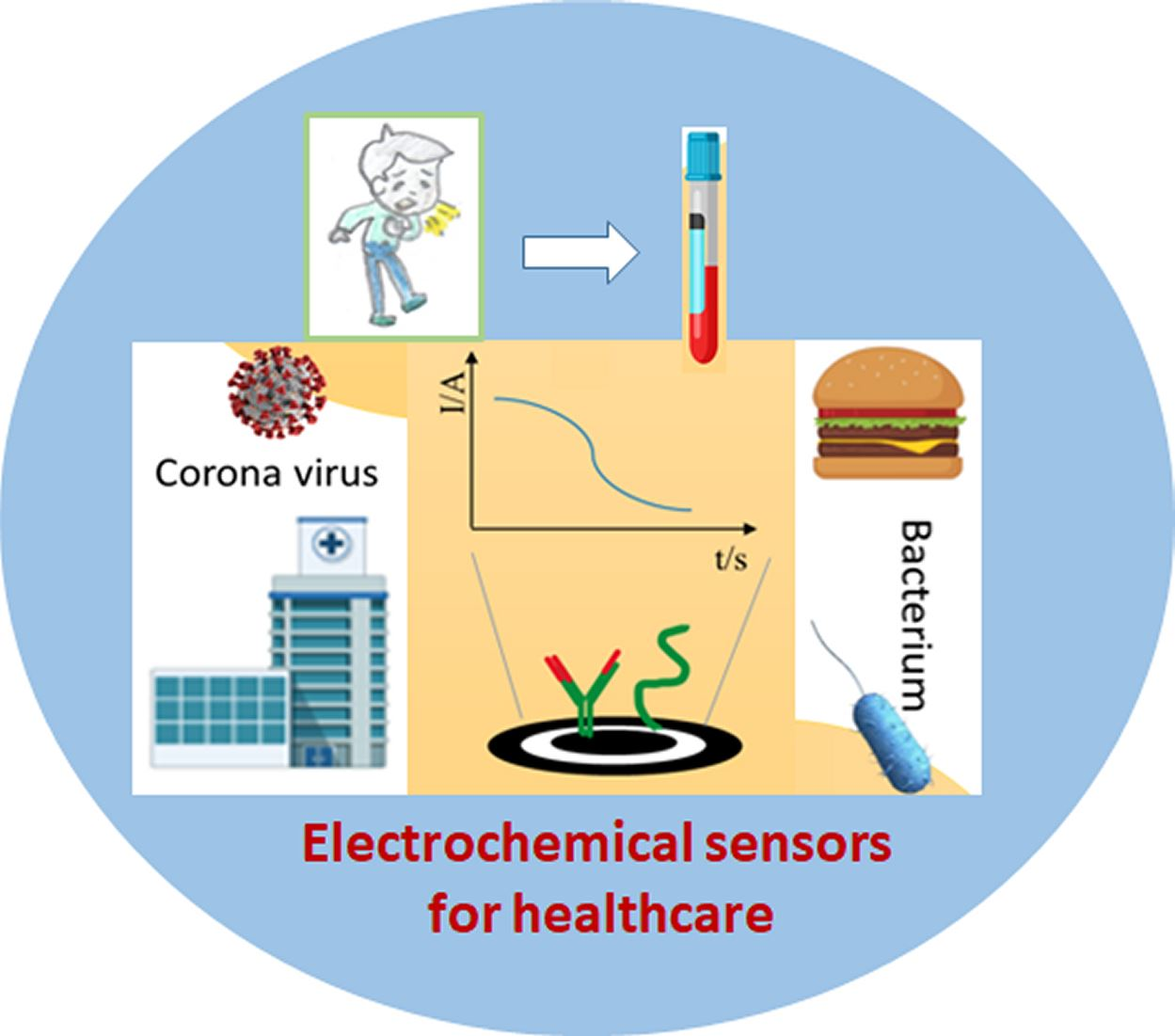
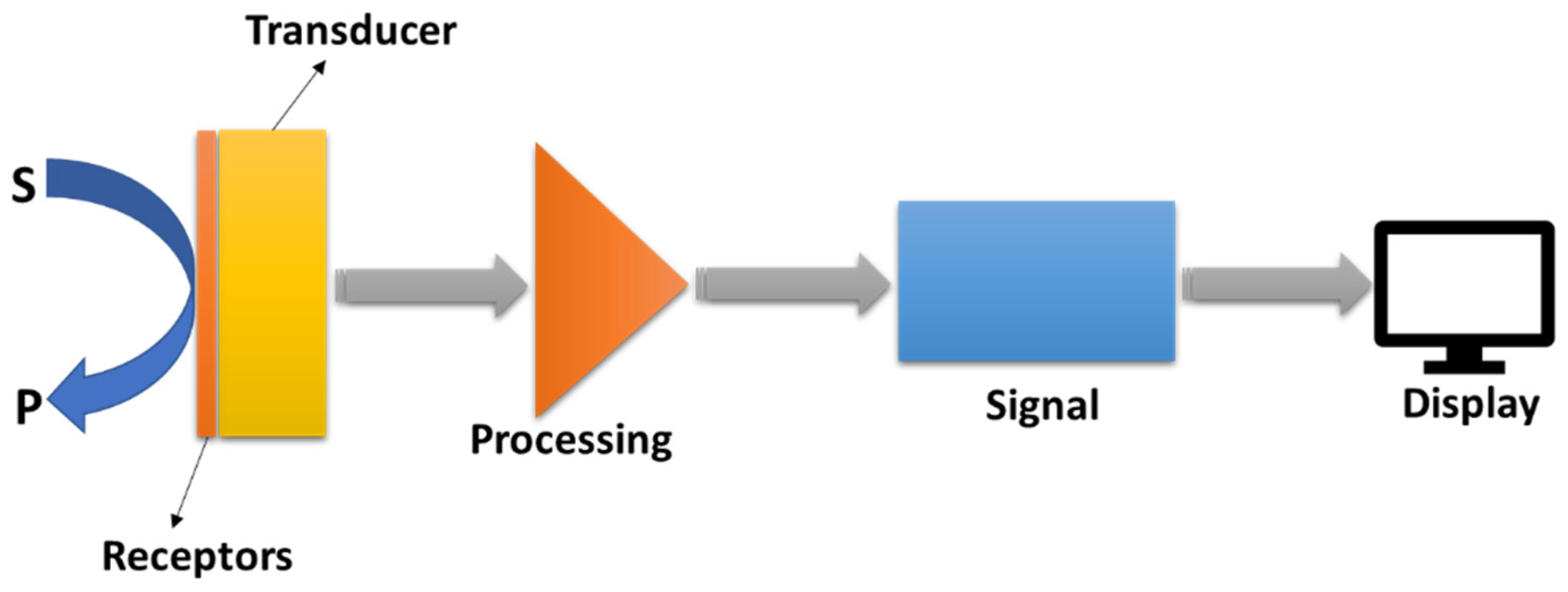
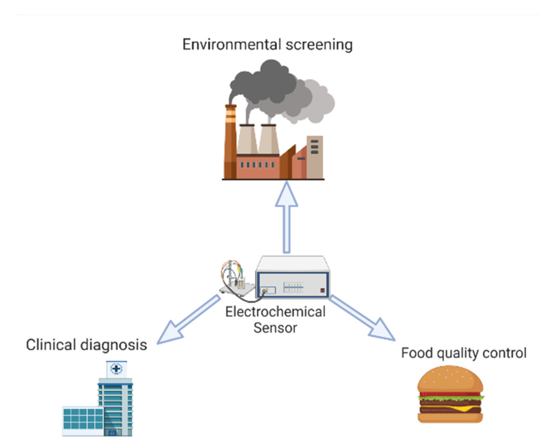
Table 1.
Biosensor receptors and electrochemical measurement methods.
| Analytes | Receptor/Chemical Recognition System | Measurement Approach | Reference |
|---|---|---|---|
| Ions | Permselective, ion-conductive inorganic crystals, or biological ionophores enzyme(s) | Potentiometric | [7] |
| Dissolved gases, vapors | Inert metal, enzyme(s), antibody, receptor | Amperometric or potentiometric | [8] |
| Antibody/antigen | Antigen/antibody oligonucleotide duplex, aptamer enzyme-labeled | Amperometric, potentiometric, or conductometric | [9] |
| Various proteins and low-molecular weight substrates | Specific ligands | Amperometric or potentiometric | [10] |
Table 2.
Types of electrochemical transducers for various kinds of sensors along with the analytes they measure.
Table 2.
Types of electrochemical transducers for various kinds of sensors along with the analytes they measure.
| Measurement Category | Transducer | Transducer Analyte | Reference |
|---|---|---|---|
| Potentiometric | Ion-selective electrode (ISE), glass electrode, gas electrode, metal electrode | K+, Cl−, Ca2+, F−, H+, Na+, CO2, NH3 redox species | [25] |
| Amperometric | Carbon electrode, chemically modified electrodes (CMEs) | O2, sugars, alcohols, phenols, oligonucleotides | [26] |
| Conductometric | Interdigitated electrodes, metal electrode | Urea, charged species, oligonucleotides | [27] |
Table 3.
Electronic biosensor systems for the detection of biomolecules.
| Target | Substrate | Immobilization Process | Detection Methods | Reference |
|---|---|---|---|---|
| DNA | Screen-printed electrode | Au nanoparticles/TFO probe/methylene blue/target DNA (ssDNA or dsDNA) | Cyclic voltammetry (CV)/square wave voltammetry (SWV) | [56] |
| DNA | Platinum electrode | MoS2-polyaniline/ssDNA/methylene blue (MB) | CV/differential pulse voltammetry (DPV) | [57] |
| Thrombin | Glassy-carbon electrode | Graphene oxide/MNP-TBA1 (magnetic nanoparticle thrombin-binding aptamer)/HAP-TBA2 (hydroxyapatite-TBA2) | CV/SWV | [58] |
| Thrombin | Au electrode | Thiol-group/aptamer/tetra-ferrocene | DPV/electrochemical impedance spectroscopy (EIS) | [59] |
| MMP-2 | Au electrode | Selenium/peptide/Na2MoO4/ssDNA | CV/EIS | [60] |
| MMP-9 | Au electrode | L-cysteine/EDC/NHS/peptide/MB | CV | [61] |
| Estrogen (ER alpha) | Screen-printed electrode | 5’-thiol-modified DNA aptamer/Tris-(2-carboxyethyl) phosphine hydrochloride | DPV | [62] |
| Estrogen (17-β Estradiol) | Au electrode | 6-mercapto-1-hexanol (MCH)/aptamer-graphene | DPV/EIS | [63] |
| Human chorionic gonadotropin (hCG) | Glassy-carbon electrode | Carbon nano-onions (CNOs)/gold nanoparticles (AuNPs)/polyethylene glycol (PEG) | CV/SWV | [64] |
Table 4.
Environmental analyses using electrochemical sensors and biosensors.
| Analyte | Recognition | Recognition Element | Detection Method | Reference |
|---|---|---|---|---|
| Uranium | Preconcentration | Nafion | Voltammetry | [102] |
| Peroxides | Biocatalysis | Peroxidase | Amperometry | [103,104] |
| Pesticides | Enzyme inhibition | Acetylcholinesterase choline oxidase | Amperometry | [103,104] |
| Hydrazines | Electrocatalysis | Ruthenium catalyst | Amperometry | [105] |
| Lead | Ion recognition | Macrocyclic ionophore | Potentiometry | [106] |
| Mercury | Preconcentration | Crown ether | Voltammetry | [107] |
| Nickel | Preconcentration | Dimethylglyoxine | Voltammetry | [108] |
| Cyanide | Enzyme inhibition | Tyrosinase | Amperometry | [109] |
| Nitrite | Preconcentration | Aliquat 336 ion exchanger | Voltammetry | [110] |
| Nitrosamines | Electrocatalysis | Ruthenium catalyst | Amperometry | [111] |
| Phenol | Biocatalysis | Tyrosinase | Amperometry | [112,113] |
| Sulfite | Biocatalysis | Sulfite oxidase | Amperometry | [114] |
| Benzene | Modulated-microbial activity | Whole-cell | Amperometry | [115] |
Table 5.
Electrochemical sensor types and its relevance in diverse disciplines.
| Class of Sensor | Electrode(s) Type | Application | Industry | Reference |
|---|---|---|---|---|
| Novel Cell-Based | Tungsten Disulfide Nanosheets/Hydroxylated Carbon Nanotube Nanocomposites | Environmental Toxicity Monitoring | Environment | [120] |
| Molecular Imprinted | Multiwalled Carbon Nanotubes | Determination of Trace Enrofloxacin in Marine Environment Samples | Environment | [121] |
| CeO2 Nanostructured | Graphite Sensors Modified by Cerium Oxide Nanoparticles (Cpe-Ceo2 Nps) | Detecting Diethylstilbestrol (DES) and 17Β-Estradiol (E2) in Environmental Samples | Environment | [122] |
| Nonenzymatic | Zinc/Zinc Oxide Core–Shell Nanostructures | Determination of Hydroquinone | Medical | [123] |
| Novel Sandwich-Type Biosensor | Magnetic Covalent Organic Framework/Pillararene Heterosupramolecular Nanocomposites | Human Norovirus (HuNOV) Detection | Medical | [124] |
| Disposable | PtNi Alloy Nanoparticles | Monitoring of H2S Released by Human Breast Cancer Cells | Medical | [125] |
| RT-LAMP | Screen-Printed Electrodes | Detect N and Orf1Ab Genes of the SARS-CoV-2 Genome | Medical | [126] |
| Dual Functional | Macroscopic Polyaniline (PANI) | Detection of pH and Lactate in Sweat of the Human Body | Medical | [127] |
| Molecularly Imprinted Polymer (Mip)-Based | Nano Gold-Doped Poly O-Phenylenediamine (Poly-O-Pd) Film | Rapid and Ultrasensitive Cortisol Detection | Medical | [128] |
| Implantable Tri-Anchored Methylene Blue-Based pH | Chip-Based Ag/AgCl Reference Electrode | Real-Time Intratumoral Tissue pH Detection | Medical | [129] |
| Capsaicin | Bimetallic Metal–Organic Framework Nanocage | Rapid Detection of Capsaicin | Food | [130] |
| Ultrasensitive | Glassy-Carbon Electrode (GCE) | Determination of Rutin Antioxidants in Mandarin and Kiwi Samples | Food | [131] |
| Amplified | Graphene Oxide (RGO) Paper Electrode Composed of Silver Nanoparticles | Detection of Sudan I in Chili Powder | Food | [132] |
| Laser-Enabled Flexible | Flexible Graphene Electrodes (FGEs) | Fast Food Security Detection (Real-Time On-Site Identification of Chloramphenicol, Clenbuterol, and Ractopamine in Meat) | Food | [133] |
Publisher’s Note: MDPI stays neutral with regard to jurisdictional claims in published maps and institutional affiliations. |
© 2022 by the authors. Licensee MDPI, Basel, Switzerland. This article is an open access article distributed under the terms and conditions of the Creative Commons Attribution (CC BY) license (https://creativecommons.org/licenses/by/4.0/).
Share and Cite
MDPI and ACS Style
Baranwal, J.; Barse, B.; Gatto, G.; Broncova, G.; Kumar, A. Electrochemical Sensors and Their Applications: A Review. Chemosensors 2022, 10, 363. https://doi.org/10.3390/chemosensors10090363
AMA Style
Baranwal J, Barse B, Gatto G, Broncova G, Kumar A. Electrochemical Sensors and Their Applications: A Review. Chemosensors. 2022; 10(9):363. https://doi.org/10.3390/chemosensors10090363
Chicago/Turabian StyleBaranwal, Jaya, Brajesh Barse, Gianluca Gatto, Gabriela Broncova, and Amit Kumar. 2022. "Electrochemical Sensors and Their Applications: A Review" Chemosensors 10, no. 9: 363. https://doi.org/10.3390/chemosensors10090363
Note that from the first issue of 2016, this journal uses article numbers instead of page numbers. See further details here.