
Ninhydrin Loaded Microcapsules for Detection of Natural Free Amino Acid
by
 , and
, and
Suhui Jeong
1,
Yeji Jeon
1,
Jaehun Mun
1,
Se Min Jeong
1,
Huiling Liang
2,
Kyeongwoon Chung
3,
Pyong-In Yi
4,
Beum-Soo An
1 and
Sungbaek Seo
1,* 1
Department of Biomaterials Science (BK21 FOUR Program), College of Natural Resources and Life Science/Life and Industry Convergence Research Institute, Pusan National University, Miryang 50463, Republic of Korea
2
Tea Research Institute, Zhejiang University, 268 Kaixuan Road, Hangzhou 310029, China
3
Department of Biofibers and Biomaterials Science, Kyungpook National University, Daegu 41566, Republic of Korea
4
Department of Bioenvironmental Energy, College of Natural Resource and Life Science, Pusan National University, Miryang 50463, Republic of Korea
*
Author to whom correspondence should be addressed.
Chemosensors 2023, 11(1), 49; https://doi.org/10.3390/chemosensors11010049
Submission received: 2 December 2022
/
Revised: 30 December 2022
/
Accepted: 3 January 2023
/
Published: 5 January 2023
(This article belongs to the Special Issue Diversity in Sensing Applications of Porphyrin or Other Macrocyclic Compound-Based Materials)
Abstract
:Natural free amino acids present in plant extracts or tea infusions provide a unique flavor and potential effect on anxiety and blood pressure reduction. Accordingly, quantifying free amino acids in foods has been of interest to food science and analytical research fields. The ninhydrin solution-based assay is a colorimetric method based on the formation and detection of Ruhemann’s purple complex. Media-based colorimetric detection requires specialized facilities and personnel; moreover, it can suffer from the interference of the analyte color. In this study, we developed ninhydrin-loaded microcapsules and a simple free amino acids detection procedure, by simply dipping the microcapsules into the analyte solution for 3 min. Among the five tested natural free amino acids, theanine exhibited the highest colorimetric response to microcapsule-based detection, with a limit of detection of 0.826 mM.
1. Introduction
Natural free amino acids are phytochemicals contained in plant leaves and extracts, and are used as health food additives and dietary supplements. Free amino acids are known to provide unique flavors and health benefits, such as anti-inflammatory effects [1,2,3]. These characteristics make the identification of healthy foods containing natural free amino acids an interesting research topic. In particular, theanine, a natural free amino acid, is mainly found in plant and fungal species, providing a unique savory flavor to green tea infusions [4]. The United States Food and Drug Administration (FDA) determined theanine to be a safe dietary supplement. Furthermore, theanine has been reported to have effects on anxiety reduction, blood pressure control, and mood improvement [5,6,7,8].
There are several methods to quantify free amino acids, such as the automatic analyzer method [9], high-performance liquid chromatography (HPLC) [10,11,12], HPLC-mass spectrometry-based quantification [9,13], nuclear magnetic resonance [14], and colorimetric methods [15,16,17]. HPLC qualitatively and quantitatively detected 33 types of free amino acids in 33 non-Camellia teas [10], but requires expensive and heavy instruments and specialized professionals to employ them. As a simple way of quantification, a ninhydrin-based colorimetric assay was employed to determine the amino acid content in 10 fruits and vegetables [15]. However, the media-based detection was not suitable for point-of-care analyte samples and it was challenging to identify the sensory signal, due to interruption of analyte color. Here, we suggest a hand-carriable and ninhydrin-loaded microcapsule-based detection for free amino acid quantification, with easy naked-eye determination [18,19].
Using a ninhydrin solution-based assay we screened five natural free amino acids (asparagine, glutamic acid, norvaline, glutamine, and theanine). For the portability of this ninhydrin-based detection, ninhydrin was loaded into alginate-based microcapsules and dipped into the analyte solution of the free amino acid. Based on the colorimetric response (from pale white to purple) of the microcapsules toward the natural free amino acids, theanine was selected as the sensitivity provider among the screened free amino acids.
2. Materials and Methods
2.1. Materials
Sodium alginate was purchased from Daejung Chemicals (Gyeonggi-do, Republic of Korea) and calcium chloride from OCI (Seoul, Republic of Korea). Asparagine (#1), glutamic acid (#2), norvaline (#3), and glutamine (#4) were obtained from Sigma-Aldrich (St. Louis, MO, USA), and theanine (#5) and ninhydrin were purchased from Sejin Ci (Seoul, Republic of Korea).
2.2. Ninhydrin Colorimetric Assay
From a 0.1 wt% ninhydrin aqueous solution, 0.5 mL was added to 1.5 mL test tubes containing 0.5 mL of #1, #2, #3, #4, and #5 aqueous solutions (100 mM). The test tubes were heated at 80 °C for 3 min and left to cool to room temperature (25 ± 2 °C). The absorption spectra of each solution were then scanned in the range of 300–800 nm using a UV-Vis spectrophotometer (Biochrom Libra S50, Biochrom, Cambridge, UK).
2.3. Preparation of Ninhydrin Loaded Microcapsules
Sodium alginate (2 wt%) and ninhydrin (2 wt%) were dissolved in de-ionized (DI) water at room temperature with magnetic stirring (C-MAG HS 7, IKA, Staufen, Germany). The solution was filled into a syringe with an 18 G needle and injected into aqueous calcium chloride (2 wt%) using a syringe pump (injection speed: 44.15 mL/h) (KDS-100CE, KD Scientific, Holliston, MA, USA). The solution was magnetically stirred for 4 h for microcapsule obtention. The microcapsules were rinsed thrice with DI water and stored at 25 ± 2 °C.
2.4. Detection Test Using Ninhydrin Loaded Microcapsules
The microcapsules (n ≥ 5) were dipped into a 1.5 mL test tube containing 0.5 mL of aqueous solutions #1, #2, #3, #4, and #5 (100 mM) and subsequently heated to 80 °C for 3 min. After cooling to 25 ± 2 °C, an image of the microcapsules was captured using a smartphone (iPhone 13, Apple Inc., Cupertino, CA, USA).
For the sensitivity determination of the microcapsule-based detection method, the microcapsules were dipped in different concentrations (0, 50, 100, 200, and 300 mM) of #5 (theanine) aqueous solution for 3 min and then heated at 80 °C for 3 min. After cooling to 25 ± 2 °C, an image of the microcapsules was captured using the smartphone. The colorimetric signal intensity at each concentration of #5 was then converted to Red-Green-Blue (RGB) % using the ImageJ program (Java 8, Maryland, MD, USA) according to the following:
where is the initial RGB value before microcapsule incubation, and is the final value after microcapsule incubation [20].
The limit of detection (LOD) was calculated as follows:
where N is the number of data points, Slope is the linear fitting value, and standard error (SE) is the standard deviation of the regression line calculated using OriginPro 8 software (Northampton, MA, USA).
3. Results and Discussion
Here we developed a new method for natural free amino acid quantification based on the chemical principal of the ninhydrin-mediated colorimetric assays. Ninhydrin-based colorimetric quantification of natural free amino acids has been investigated using the proposed mechanism of ninhydrin-mediated reactions and subsequent changes in their optical properties (Figure 1) [21,22]. Quantification through the ninhydrin method is essentially based on a redox reaction; ninhydrin acts as an oxidizing agent and is reduced. Ninhydrin reacts with the amino groups of the free amino acids and decarboxylates them to produce an aldehyde and an intermediate amine (2-amino-1,3-indanedione). Ammonia and carbon dioxide are released in this reaction. The condensation of this intermediate amine with another ninhydrin molecule forms the chromophore Ruhemann’s purple [21].
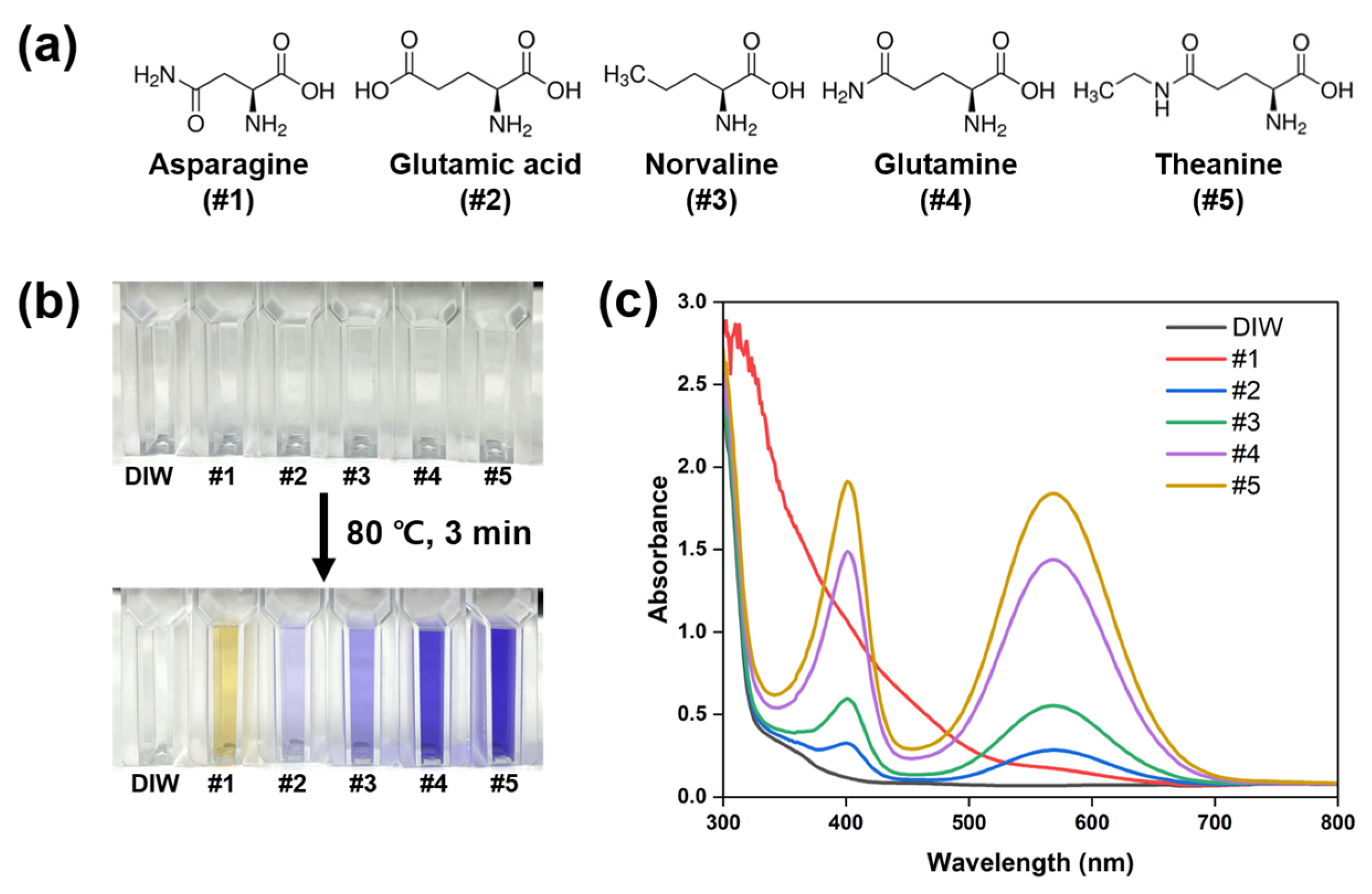
Five molecules were nominated as natural free amino acids (Figure 2a) for conventional ninhydrin solution-based assays. To determine whether the free amino acids could be detected by our ninhydrin-based colorimetric method, aqueous solutions of #1–#5 molecules were prepared; all solutions were transparent (Figure 2b). The ninhydrin aqueous solution was introduced and reacted with each amino acid, with a corresponding colorimetric response dependent on the type of amino acid (Figure 2b). The color change observed with theanine and ninhydrin incubation was the most striking, from transparent to purple. Absorption at 400 nm and 570 nm (characteristic absorptions of Ruhemann’s purple [15]) was characteristic in #2–#5 (Figure 2c). This shows distinct reactivity of the different amino acids with ninhydrin, theanine being the most reactive. Some amines react with ninhydrin considerably more slowly than others; therefore, a lower purple color yield at one timepoint may be due to an incomplete reaction. Alternatively, the ninhydrin-mediated reaction has several intermediate steps, which might determine different degrees of Ruhemann’s purple. Decarboxylation (loss of CO2) and aldehyde formation are essentially irreversible; therefore, any equilibrium before the last irreversible step (aldehyde formation) can slow the reaction rate [23]. If the test sample contained proteinogenic amino acids such as proline, the coloration would be yellow. The ninhydrin-asparagine (#1) complex resembles the enol-betaine structure of the ninhydrin-proline complex because of the circularization of decarboxylated asparagine [22]. For this reason, the ninhydrin-asparagine (#1) reaction provided a yellow-colored solution (Figure 2b).
The challenging issue of the ninhydrin solution-based assay is the difficulty of portability in point-of-care testing due to liquid-media-based detection. Additionally, the color of the ninhydrin solution is affected by the color of the analyte sample. To overcome these issues, we developed a ninhydrin-loaded alginate-based microcapsule and dipped it into an analyte solution containing free amino acids. Figure 3 shows the schematic description of the microcapsule preparation (ninhydrin-loaded microcapsules) and amino acid detection procedure. Pale white microcapsules (diameter of 3 ± 0.1 mm) were reproducibly fabricated with mass production available by ionic crosslinking between anionic alginate and calcium divalent cations (Figure 3a). The incubation conditions (80 °C for 3 min) for the ninhydrin-loaded microcapsule-based detection were defined based on the principle of ninhydrin and amino acids reaction. A colorimetric change (from pale white to purple) was anticipated for the reaction between amino acids and the microcapsules (Figure 3b).
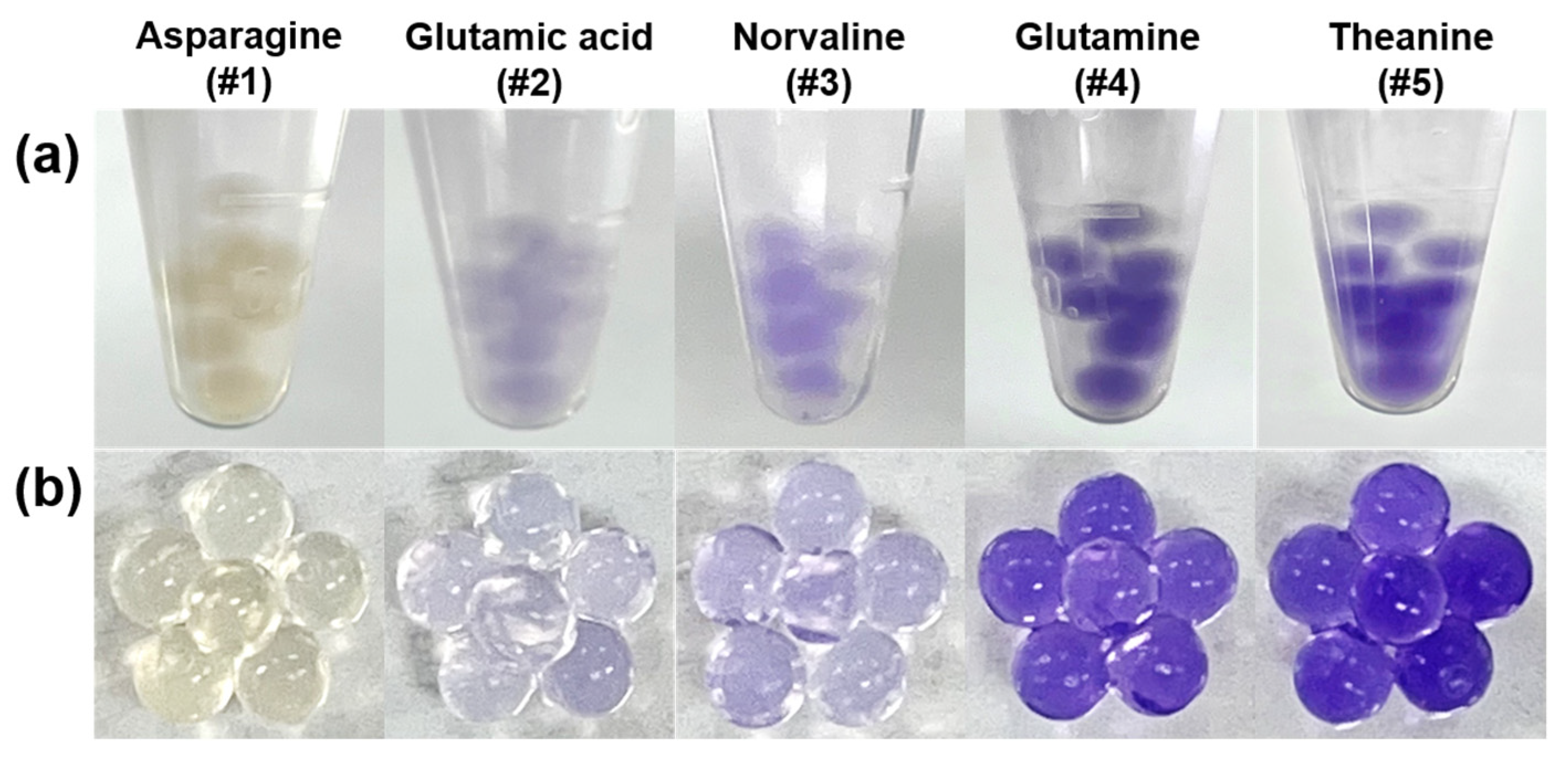
Microcapsules were dipped into five different amino acid solutions. Figure 4 shows how the individual microcapsules appear purple inside the analyte solution and how the color was retained after the incubation. The trend in the degree of colorimetric change was aligned with the results obtained with the ninhydrin solution-based assay (Figure 2b).
Because theanine showed the most apparent purple color and RGB intensity among the tested amino acids, we selected #5 (theanine) to evaluate the ninhydrin-loaded microcapsule-based detection method sensitivity. As shown in Figure 5a, a concentration-dependent colorimetric response was observed in the range of 0–300 mM. RGB (%) was calculated by the color signal intensity from the captured images of the microcapsules, providing a limit of detection of 0.826 mM. Altogether, these results show that reliable free amino acid quantification can be obtained with a simple dipping analysis using portable microcapsules.
In the tea industry, visible identification and quantification of natural free amino acid, e.g., theanine, is necessary for determining high-quality products due to its unique flavor and potential health benefits. Based on the literature [4], we recognized that theanine could exist in a maximum amount of 0.57 mM and 0.23 mM in black tea infusion and green tea infusion, respectively. Our detection method is available to detect theanine down to 0.826 mM, and therefore should have improved sensitivity for its applications.
4. Conclusions
Ninhydrin-loaded alginate microcapsules were developed to overcome the challenges of traditional ninhydrin-colorimetric assays, namely, to provide portability to the detection platform. This detection system allows naked-eye detection by simply dipping the microcapsule into the analyte solution. We consider how to improve the sensitivity of the detection method and expect to extend the applicability of this system to free amino acid quantification in samples from various food and plant leaf infusions, contributing to uncover their heathy potential. This detection system can also be extended to other fields that use ninhydrin solution-based assays, such as in protein, agricultural, biomedical, and forensic sciences.
Author Contributions
Conceptualization, S.J.; methodology, S.J.; software, S.J.; validation, S.J.; formal analysis, S.J.; investigation, S.J., Y.J., J.M. and S.M.J.; data curation, S.J.; writing—original draft preparation, S.J. and S.S.; writing—review and editing, H.L., K.C., P.-I.Y., B.-S.A. and S.S.; supervision, S.S.; project administration, S.S.; funding acquisition, S.S. All authors have read and agreed to the published version of the manuscript.
Funding
This work was supported by a 2-Year Research Grant of Pusan National University (201913770004).
Institutional Review Board Statement
Not applicable.
Informed Consent Statement
Not applicable.
Data Availability Statement
Data is contained within the article.
Conflicts of Interest
The authors declare no conflict of interest.
References
- Scharbert, S.; Hofmann, T. Molecular definition of black tea taste by means of quantitative studies, taste reconstitution, and omission experiments. J. Agric. Food Chem. 2005, 53, 5377–5384. [Google Scholar] [CrossRef]
- Yılmaz, C.; Özdemir, F.; Gökmen, V. Investigation of free amino acids, bioactive and neuroactive compounds in different types of tea and effect of black tea processing. Lwt 2020, 117, 108655. [Google Scholar] [CrossRef]
- Liu, W.; Chen, X.; Li, H.; Zhang, J.; An, J.; Liu, X. Anti-Inflammatory Function of Plant-Derived Bioactive Peptides: A Review. Foods 2022, 11, 2361. [Google Scholar] [CrossRef]
- Keenan, E.K.; Finnie, M.D.A.; Jones, P.S.; Rogers, P.J.; Priestley, C.M. How much theanine in a cup of tea? Effects of tea type and method of preparation. Food Chem. 2011, 125, 588–594. [Google Scholar] [CrossRef]
- Kimura, K.; Ozeki, M.; Juneja, L.R.; Ohira, H. l-Theanine reduces psychological and physiological stress responses. Biol. Psychol. 2007, 74, 39–45. [Google Scholar] [CrossRef]
- Rogers, P.J.; Smith, J.E.; Heatherley, S.V.; Pleydell-Pearce, C.W. Time for tea: Mood, blood pressure and cognitive performance effects of caffeine and theanine administered alone and together. Psychopharmacology 2008, 195, 569–577. [Google Scholar] [CrossRef]
- Li, M.Y.; Liu, H.Y.; Wu, D.T.; Kenaan, A.; Geng, F.; Li, H.B.; Gunaratne, A.; Li, H.; Gan, R.Y. L-Theanine: A Unique Functional Amino Acid in Tea (Camellia sinensis L.) With Multiple Health Benefits and Food Applications. Front. Nutr. 2022, 9, 853846. [Google Scholar] [CrossRef]
- Anas Sohail, A.; Ortiz, F.; Varghese, T.; Fabara, S.P.; Batth, A.S.; Sandesara, D.P.; Sabir, A.; Khurana, M.; Datta, S.; Patel, U.K. The Cognitive-Enhancing Outcomes of Caffeine and L-theanine: A Systematic Review. Cureus 2021, 13, e20828. [Google Scholar] [CrossRef]
- Li, J.; Ma, J.; Li, Q.; Fan, S.; Fan, L.; Ma, H.; Zhang, Y.; Zheng, L. Determination of 35 Free Amino Acids in Tea Using Ultra-Performance Liquid Chromatography Coupled With Quadrupole Time-of-Flight Mass Spectrometry. Front. Nutr. 2021, 8, 767801. [Google Scholar] [CrossRef]
- Bi, W.; He, C.; Ma, Y.; Shen, J.; Zhang, L.H.; Peng, Y.; Xiao, P. Investigation of free amino acid, total phenolics, antioxidant activity and purine alkaloids to assess the health properties of non-Camellia tea. Acta Pharm. Sin. B 2016, 6, 170–181. [Google Scholar] [CrossRef]
- Kazan, R.M.; Seddik, H.A.; Marstani, Z.M.; Elsutohy, M.M.; Yasri, N.G. Determination of amino acids content in tea species using liquid chromatography via pre-column fluorescence derivatization. Microchem. J. 2019, 150, 104103. [Google Scholar] [CrossRef]
- Zhou, P.; Zhao, F.; Chen, M.; Ye, N.; Lin, Q.; Ouyang, L.; Cai, X.; Meng, P.; Gong, X.; Wang, Y. Determination of 21 free amino acids in 5 types of tea by ultra-high performance liquid chromatography coupled with tandem mass spectrometry (UHPLC–MS/MS) using a modified 6-aminoquinolyl-N-hydroxysuccinimidyl carbamate (AQC) method. J. Food Compos. Anal. 2019, 81, 46–54. [Google Scholar] [CrossRef]
- How, Z.T.; Busetti, F.; Linge, K.L.; Kristiana, I.; Joll, C.A.; Charrois, J.W.A. Analysis of free amino acids in natural waters by liquid chromatography–tandem mass spectrometry. J. Chromatogr. A 2014, 1370, 135–146. [Google Scholar] [CrossRef] [Green Version]
- Choi, K.H.; Lee, J.H. Quantitative analysis of metabolites in Korean green tea using NMR. J. Korean Magn. Reson. Soc. 2018, 22, 132–138. [Google Scholar] [CrossRef]
- Friedman, M. Applications of the Ninhydrin Reaction for Analysis of Amino Acids, Peptides, and Proteins to Agricultural and Biomedical Sciences. J. Agric. Food Chem. 2004, 52, 385–406. [Google Scholar] [CrossRef]
- Das, S. Recent applications of ninhydrin in multicomponent reactions. RSC Adv. 2020, 10, 18875–18906. [Google Scholar] [CrossRef]
- Pilicer, S.L.; Wolf, C. Ninhydrin Revisited: Quantitative Chirality Recognition of Amines and Amino Alcohols Based on Nondestructive Dynamic Covalent Chemistry. J. Org. Chem. 2020, 85, 11560–11565. [Google Scholar] [CrossRef]
- Ahmad Raus, R.; Wan Nawawi, W.M.F.; Nasaruddin, R.R. Alginate and alginate composites for biomedical applications. Asian J. Pharm. Sci. 2021, 16, 280–306. [Google Scholar] [CrossRef]
- Jang, S.; Son, S.U.; Kim, J.; Kim, H.; Lim, J.; Seo, S.B.; Kang, B.; Kang, T.; Jung, J.; Seo, S.; et al. Polydiacetylene-based hydrogel beads as colorimetric sensors for the detection of biogenic amines in spoiled meat. Food Chem. 2023, 403, 134317. [Google Scholar] [CrossRef]
- Raji, M.A.; Chinnappan, R.; Shibl, A.; Suaifan, G.; Weber, K.; Cialla-May, D.; Popp, J.; El Shorbagy, E.; Al-Kattan, K.; Zourob, M. Low-cost colorimetric diagnostic screening assay for methicillin resistant Staphylococcus aureus. Talanta 2021, 225, 121946. [Google Scholar] [CrossRef]
- Bottom, C.B.; Hanna, S.S.; Siehr, D.J. Mechanism of the ninhydrin reaction. Biochem. Educ. 1978, 6, 4–5. [Google Scholar] [CrossRef]
- Sheng, S.J.; Kraft, J.J.; Schuster, S.M. Schuster A specific quantitative colorimetric assay for L-asparagine. Anal. Biochem. 1993, 211, 242–249. [Google Scholar] [CrossRef]
- Friedman, M.; David Williams, L. Stoichiometry of formation of Ruhemann’s purple in the ninhydrin reaction. Bioorganic Chem. 1974, 3, 267–280. [Google Scholar] [CrossRef]
Figure 1.
Mechanisms of reactions of α-amino acids with ninhydrin to form Ruhemann’s purple.

Figure 2.
(a) Molecular structures of five natural free amino acids (asparagine, glutamic acid, norvaline, glutamine, theanine). (b) The photograph of color change in the amino acid aqueous solution in the ninhydrin solution-based assay. (c) UV-Visible absorption spectra of the amino acid aqueous solution by the ninhydrin assay.

Figure 3.
(a) Schematic illustration of ninhydrin-loaded microcapsule preparation. (b) The microcapsule-based colorimetric detection of natural free amino acid solution.
Figure 3.
(a) Schematic illustration of ninhydrin-loaded microcapsule preparation. (b) The microcapsule-based colorimetric detection of natural free amino acid solution.

Figure 4.
(a) Photograph of ninhydrin-loaded microcapsules incubated in the analyte solution of each amino acid (100 mM). (b) Photograph of microcapsules upon retrieval from the analyte solution.
Figure 4.
(a) Photograph of ninhydrin-loaded microcapsules incubated in the analyte solution of each amino acid (100 mM). (b) Photograph of microcapsules upon retrieval from the analyte solution.

Figure 5.
(a) Photograph of incubated microcapsules in increasing concentrations of theanine solutions (0–300 mM). (b) RGB (%) of the microcapsules after incubation with different concentrations of theanine. * , ** LOD: limit of detection.
Figure 5.
(a) Photograph of incubated microcapsules in increasing concentrations of theanine solutions (0–300 mM). (b) RGB (%) of the microcapsules after incubation with different concentrations of theanine. * , ** LOD: limit of detection.

Disclaimer/Publisher’s Note: The statements, opinions and data contained in all publications are solely those of the individual author(s) and contributor(s) and not of MDPI and/or the editor(s). MDPI and/or the editor(s) disclaim responsibility for any injury to people or property resulting from any ideas, methods, instructions or products referred to in the content. |
© 2023 by the authors. Licensee MDPI, Basel, Switzerland. This article is an open access article distributed under the terms and conditions of the Creative Commons Attribution (CC BY) license (https://creativecommons.org/licenses/by/4.0/).
Share and Cite
MDPI and ACS Style
Jeong, S.; Jeon, Y.; Mun, J.; Jeong, S.M.; Liang, H.; Chung, K.; Yi, P.-I.; An, B.-S.; Seo, S. Ninhydrin Loaded Microcapsules for Detection of Natural Free Amino Acid. Chemosensors 2023, 11, 49. https://doi.org/10.3390/chemosensors11010049
AMA Style
Jeong S, Jeon Y, Mun J, Jeong SM, Liang H, Chung K, Yi P-I, An B-S, Seo S. Ninhydrin Loaded Microcapsules for Detection of Natural Free Amino Acid. Chemosensors. 2023; 11(1):49. https://doi.org/10.3390/chemosensors11010049
Chicago/Turabian StyleJeong, Suhui, Yeji Jeon, Jaehun Mun, Se Min Jeong, Huiling Liang, Kyeongwoon Chung, Pyong-In Yi, Beum-Soo An, and Sungbaek Seo. 2023. "Ninhydrin Loaded Microcapsules for Detection of Natural Free Amino Acid" Chemosensors 11, no. 1: 49. https://doi.org/10.3390/chemosensors11010049
Note that from the first issue of 2016, this journal uses article numbers instead of page numbers. See further details here.