
Enzymatic Electrochemical Biosensors for Neurotransmitters Detection: Recent Achievements and Trends


1
CenBRAIN Neurotech, School of Engineering, Westlake University, Hangzhou 310030, China
2
MiCRA Biodiagnostics Technology Gateway, Technological University Dublin (TU Dublin), D24 FKT9 Dublin 24, Ireland
3
Centre of Applied Science for Health, Technological University Dublin (TU Dublin), D24 FKT9 Dublin 24, Ireland
4
Institute of Biopharmaceutical and Health Engineering, Tsinghua Shenzhen International Graduate School, Tsinghua University, Shenzhen 518055, China
*
Authors to whom correspondence should be addressed.
Chemosensors 2023, 11(7), 388; https://doi.org/10.3390/chemosensors11070388
Submission received: 28 May 2023
/
Revised: 26 June 2023
/
Accepted: 5 July 2023
/
Published: 12 July 2023
(This article belongs to the Special Issue State-of-the-Art and Advances in Electrochemical Sensors/Biosensors)
Abstract
:Neurotransmitters (NTs) play a crucial role in regulating the behavioral and physiological functions of the nervous system. Imbalances in the concentrations of NT have been directly linked to various neurological diseases (e.g., Parkinson’s, Huntington’s, and Alzheimer’s disease), in addition to multiple psychotic disorders such as schizophrenia, depression, dementia, and other neurodegenerative disorders. Hence, the rapid and real-time monitoring of the NTs is of utmost importance in comprehending neurological functions and identifying disorders. Among different sensing techniques, electrochemical biosensors have garnered significant interest due to their ability to deliver fast results, compatibility for miniaturization and portability, high sensitivity, and good controllability. Furthermore, the utilization of enzymes as recognition elements in biosensing design has garnered renewed attention due to their unique advantages of catalytic biorecognition coupled with simultaneous signal amplification. This review paper primarily focuses on covering the recent advances in enzymatic electrochemical biosensors for the detection of NTs, encompassing the importance of electrochemical sensors, electrode materials, and electroanalytical techniques. Moreover, we shed light on the applications of enzyme-based biosensors for NTs detection in complex matrices and in vivo monitoring. Despite the numerous advantages of enzymatic biosensors, there are still challenges that need to be addressed, which are thoroughly discussed in this paper. Finally, this review also presents an outlook on future perspectives and opportunities for the development of enzyme-based electrochemical biosensors for NTs detection.

1. Introduction
The demand for efficient and prompt diagnosis of human diseases is rapidly escalating to better manage and control them. The growing human population is contributing to the increasing number of patients with various diseases and disorders [1]. Additionally, society is experiencing new health challenges associated with ecological contamination, resulting in sickness stresses, disease susceptibility, and well-being syndromes. However, the lengthy and complicated analysis procedures require the processing of large volumes of samples, which may result in economic losses and fatalities. The functioning of the human brain, responsible for consciousness and actions, depends on billions of neurons. Hence, recent developments in neuroscience have contributed significantly to our understanding of the brain and its activities. Neurotransmitters (NTs) act as chemical messengers between neurons and play a crucial role in ensuring the human brain functions properly and are essential in the diagnosis of certain diseases [2]. Additionally, NT fluctuations are responsible for many diseases, including Parkinson’s [3], Alzheimer’s [4], Huntington’s [5], schizophrenia [6], epilepsy [7], glaucoma, thyroid hormone deficiency [8], and congestive heart failure [9]. A list of key neurotransmitters is shown in Table 1 along with their corresponding blood concentration values and diseases associated with abnormal levels.
Hence, accurately detecting and monitoring NTs in real-world samples is essential for advancing neurological research. While existing technologies can diagnose patients, conventional analysis methods are time-consuming and require specialized laboratory professionals. As there is an increasing demand for personalized medicine and point-of-care approaches, there is an ultimate need for scientific diagnostic tools that are user-friendly, rapid, and capable of miniaturization. As a result, researchers are focusing on improving analytical techniques to meet these requirements. Nevertheless, the conventional in vitro techniques utilized to measure NTs levels usually rely on chromatography analysis, such as high efficiency liquid chromatography and gas chromatography [10], as well as mass spectrometry [11]. These methods are costly, time-consuming, need skilled analysts/specialists, and are impractical for preclinical diagnosis of nervous system disorders in patients who lack professional guidance [12]. Furthermore, the conventional analysis of NTs primarily takes place in cerebrospinal fluid since this is where nearly all NTs are located and are present in higher concentrations compared to other bodily fluids [13].
Currently, electrochemical biosensors dominate the market due to their prevalence and wider availability. They are popular due to their portability, ease of use, and lower cost compared to other biosensors. These features make them ideal for developing point-of-care (POC) devices. These devices possess several advantages, including their high flexibility, biocompatibility, light weight, low invasiveness, and affordability, which make them easy to manufacture and install, whether externally on human skin or internally on soft tissue, while still being connected to a system [14]. In recent years, enzymatic electrochemical biosensors have emerged as a promising alternative for neurotransmitter detection due to their high sensitivity, selectivity, and simplicity. Enzymatic electrochemical biosensors are based on the principle of enzymatic catalysis and electrochemical transduction, allowing for rapid and real-time detection of neurotransmitters with high accuracy [15]. Moreover, enzymatic biosensors can be easily integrated with microfluidic systems and wearable devices, enabling continuous and non-invasive monitoring of neurotransmitter dynamics in vivo. Given the significance of NTs biosensors, our group have published recently a thorough summary of the latest developments in biosensors and their applications for in vivo monitoring [16]. In addition, we presented in another manuscript an electrochemical biosensor specifically designed for catecholamines detection and discussed the different strategies for enhancing the sensitivity and selectivity of these biosensors, such as the use of nanomaterials and molecularly imprinted polymers [17]. On the other hand, Ramirez et al. provided general views and a comparison of enzyme-based biosensor modalities, their proposed trends, and reported applications [18]. Another comprehensive overview of biosensing approaches for in vivo screening of dopamine (DA) was presented by Ferapontova [19].
While there are a significant number of review papers discussing NTs biosensors, there is still a noticeable gap in the literature regarding enzymatic electrochemical monitoring of NTs. Furthermore, there is a particular need for emphasis on the utilization of enzymatic biosensors in complex environments and for in vivo detection. Therefore, this review provides a comprehensive overview covering the recent achievements and trends in enzymatic electrochemical biosensors for neurotransmitter detection, with a focus on dopamine, serotonin, norepinephrine, epinephrine, acetylcholine, and tyrosine. We begin by discussing the significance of electrochemical biosensors in neurotransmitter detection and the electrode materials and electroanalytical techniques associated with these sensors. Next, we examine the applications of enzyme-based biosensors for neurotransmitter detection in complex matrices, such as biological fluids, food, and environmental samples, and discuss the various enzymatic electrochemical biosensors, including enzyme-modified electrodes, nanostructured electrodes, and screen-printed electrodes. Additionally, we highlight the challenges and limitations of these biosensors and strategies to overcome them. Finally, we explore the application of enzyme-based biosensors for in vivo monitoring of neurotransmitters, including the latest developments in biosensor design, fabrication, and integration, such as microfluidic systems and wearable devices, for improved performance and practical applications.
2. Electrochemical Biosensors
2.1. Significance of Electrochemical Sensors
Electrochemical sensors possess the capability of converting system chemical interactions into exploitable electrical signals. These sensors have shown interest in wide-ranging applications in various fields, including environmental monitoring [20,21], biological research [22], and traffic management [23], making them increasingly popular as reliable analytic tools and alternatives to other complex and traditional sensing techniques. Electrochemical biosensors have gained much attention as they provide a cost-effective sensing tool with minimal training and expertise required compared to traditional sensing techniques. Moreover, these sensors exhibit exceptional sensitivity, enabling swift analysis with short response times, and perform at par with transducer microfabrication technology [24]. The constituents undergo a biological process that produces electrical signals [25,26]. As illustrated in Figure 1, sensors typically rely on four essential components: (1) an analyte support, (2) a bioreceptor, (3) a transducer, and (4) a measurement tool. Two crucial components accurately detect and interpret the sample contents, the receptor networks that interact with the analyte at the solid/liquid interface and the transducer that translates the received information into appropriate electrical signals.
2.2. Electrode Materials and Their Performances
Various electrode materials, including noble metals, carbon, and conductive polymers, are accessible for the advancement of bio (chemical) sensors. Due to their remarkable conductivity and superior electron transfer kinetics, noble metals such as platinum (Pt), gold (Au), and silver (Ag) are frequently used. Furthermore, metal-based electrodes exhibit a high level of stability and inertness. Gold is a highly favored material for biosensing applications due to its compatibility for microfabrication and immobilization techniques, and it can be used reliably in the range of −0.1 to 1.3 V [27]. One of the key advantages of using Au is its ability to immobilize bioorganic materials, such as DNA, in a stable manner while maintaining their bioactivity. This is particularly important in the development of highly sensitive biosensors. On the other hand, Au is able to facilitate electron transfer directly between redox centers and bulk electrode materials without the need for mediators. This property makes gold an excellent material for electrochemical sensing. The high stability of gold in terms of reactivity with other materials makes it a widely used material for various applications. In addition, Au possesses a high interface energy that facilitates the proximity of proteins and metals, thereby promoting effective electron transfer between redox proteins and electrode surfaces [28].
Electrode-based indium tin oxide (ITO) has garnered significant attention due to its cost-effectiveness, transparency, and ease of processing, making it a viable alternative to noble metals. Furthermore, ITO offers a broader potential range compared to gold, spanning from −0.4 to 1.9 V, albeit with a lower conductivity of 104 S cm−1 versus gold’s 107 S cm−1 and it is not stable in acidic environment [29]. Moreover, the electron transfer kinetics of ITO are typically poorer compared to gold or glassy carbon. Carbon-based electrodes comprise various materials classified based on the hybridization of carbon atoms. However, graphite is composed of sp2 hybridized carbon atoms, ranging from simple graphene sheets to highly oriented pyrolytic graphite (HOPG), whereas diamond contains sp3 hybridized carbon atoms. Therefore, doped diamond structures like boron-doped diamond are widely performed to enhance the diamond conductivity like boron-doped diamond. Glassy carbon is another commonly used carbon material for electrode applications, as it can be processed easily and has a broad potential range of use (−0.4 to 1.7 V). Although carbon electrodes perform optimally when used with organic redox molecules, they are inherently reactive, which creates a selectivity challenge in complex detection environments.
Electrodes comprised of conducting polymers such as polypyrrole (PPy), polyaniline (PANI), and poly(3,4-ethylenedioxythiophene) (PEDOT) are commonly selected for their mechanical flexibility and optical transparency. However, their electroactivity and conductivity are typically inferior to those made of carbon materials and noble metals, and they may experience issues with long-term water stability [30]. Mercury, despite being the most commonly used electrode material in polarography, has limited applications in biosensors due to its inherent toxicity [31].
Advanced and novel materials for electrode fabrication and sensor construction are in continuous demand for developing innovative technologies in this space. Among various fabrication techniques, Chemical Vapor Deposition (CVD) is widely performed due to the large area synthesis advantages, especially for carbon-based electrode materials [32]. CVD involves the deposition of a precursor gas onto a substrate, where the gas undergoes chemical reactions and forms a solid film. This method allows for the controlled growth of carbon materials with specific properties [33]. Besides noble metals and carbon-based materials, conducting polymers can also be synthesized using the CVD technique. By introducing gaseous monomers or precursors into a reactor and initiating polymerization, conducting polymers like PPy or PANI can be deposited onto electrodes. In addition, laser writing techniques have emerged as a promising method for electrode fabrication. These techniques involve the use of laser beams to directly write or pattern electrodes on various substrates [34]. Laser writing offers several advantages, including high precision, flexibility in design, and the ability to create intricate electrode patterns. This technique allows for the fabrication of custom-shaped electrodes with precise dimensions, enabling the optimization of electrode performance for specific biosensing applications. Laser writing can be applied to a wide range of electrode materials, including noble metals, carbon-based materials, and conducting polymers.
2.3. Electron Transfer Mechanisms
The analysis of electron transfer mechanisms is particularly important for the development of biosensors, which rely on the detection of specific biomolecules to provide information about biological systems. Two principal mechanisms of electron transfer are available: indirect electron transfer (IET) and direct electron transfer (DET) as presented in Figure 2 [35];
- Direct Electron Transfer (DET): DET occurs when an electron is transferred directly between an electrode and a redox-active molecule (such as a protein, enzyme, or other biomolecule) without the need for any mediator. This mechanism is typically faster than indirect electron transfer, as it avoids the need for a mediator, which can introduce additional steps and slow down the reaction. In DET, the redox-active molecule must be able to directly interact with the electrode surface, often through specific binding interactions or through a suitable electronic pathway.
- Indirect Electron Transfer (IET): The process of IET in electrochemistry involves the utilization of a mediator molecule to shuttle electrons between the electrode and the redox-active molecule. The mediator molecule can accept electrons from the electrode and transfer them to the redox-active molecule, or vice versa. This mechanism is typically slower than DET, as it introduces an additional step and requires a mediator molecule that must be carefully selected to match the redox properties of the target molecule. Nevertheless, IET can be a valuable approach in situations where the redox-active molecule is unable to interact directly with the electrode surface, or when the redox reaction occurs at a considerable distance from the electrode.
2.4. Electroanalytical Techniques
Electrochemical sensors convert the information associated with the electrochemical reactions (electrode–analyte) into an exploitable signal. They are mainly classified as amperometric/voltammetric, potentiometric, and conductometric. Electrochemical transducers are the most commonly used transducers in the construction of innovative biosensing applications. Electrochemical analysis has advantages over other measurement systems in terms of being rapid, simple, portable, and able to be easily miniaturized [36,37].
Voltammetric techniques are a prevalent type of electrochemical analysis that involves the application of a potential between a working electrode (WE) and a reference electrode (RE), resulting in a measurable circulating current between the WE and the counter electrode (CE). Typically, the voltammetric method relies on the measurement of a resulted current of oxidation/reduction process at the electrode surface. The simplest form of voltammetry involves the application of a constant potential and measuring the resulting current over a specific time (amperometry) and integrating it over a period of time (coulometry). Other advanced electroanalytical techniques, such as differential pulse voltammetry (DPV) and square-wave voltammetry (SWV), can be applied through the variation of potential over time for signal-to-noise ratio improvement or for the redox reaction analytical measurement. Cyclic voltammetry (CV) is classified as voltammetric measurement which is commonly utilized for the determination of reaction kinetics, diffusion characteristics, analyte concentrations, and electrode dimensions, where the resulting current over the linear applied potential range is directly proportional to the analyte bulk concentration [38,39,40,41].
2.4.1. Cyclic Voltammetry (CV)
Among the various electrochemical detection-based voltammetry measurements, CV is the most performed technique due to its widespread use in examining the oxidation and reduction potentials of the analyte, as well as in tracking reaction mass transfer. The shape and position of peaks observed in the resulting CV curves can be used to determine whether the response is a reversible or irreversible process. Furthermore, the electrodeposition of material on the electrode surface can also be achieved using CV [42]. It is a very good technique for characterization and analysis. Slow-scan CV is commonly performed with a scan rate below 100 mV/s for in vitro applications [43]. In a specific study, NiO nanoparticles were employed by Jamal et al. [44] to determine the levels of glutamate, achieving a sensitivity of 11 μA mM−1 cm−2 at a CV scan rate of 0.05 V/s, with a limit of detection (LOD) of 272 μM. However, conventional CV’s slow time scale renders it unsuitable for analyzing biological changes, leading to the emergence of fast-scan cyclic voltammetry (FSCV) [45,46]. FSCV is capable of completing a single scan within a few milliseconds due to its scan rates of several hundred V/s, typically around 400 V/s. Such a feature makes it possible to detect rapid changes in neurotransmitter levels in the human brain [47,48]. FSCV, in comparison to a traditional CV, boasts scan rates that are 1000 times faster, typically operating at a speed of 400 V/s with a frequency of 10 Hz, thereby enabling measurements with subsecond temporal resolution [49]. Despite its benefits, FSCV cannot be used with larger conventional electrodes such as glassy carbon electrode (GCE), which take a significant amount of time to stabilize the background current. Instead, FSCV works best with carbon fiber (CF) microelectrodes due to their short time constant and background current of several hundred nA [50].
2.4.2. Differential Pulse Voltammetry (DPV)
DPV is a sensitive analytical technique that involves the application of a linear ramp potential with a series of voltage pulses to detect multiple NTs simultaneously. To do so, a base potential is first selected, where no faradaic reaction occurs on the electrode, while the base potential is then incremented by equal amounts between pulses. The current measurements are taken immediately before and after each pulse to determine any changes in NT levels [51]. This method offers a highly sensitive means of detecting NTs as well as being able to detect multiple NTs by using a single pulse [52].
2.4.3. Square Wave Voltammetry (SWV)
To date, SWV is classified as one of the most advanced voltammetric methods widely performed in fundamental studies and analytical applications for multiple compound recognition, including pesticides, medicaments, biologically active substances, and many more [53]. Compared with other voltammetric methods, SWV possesses the advantages of a short analysis time, higher sensitivity, and a simple working set-up. Altogether, its great reduction of capacitance currents uplift it to be an attractive candidate for the quantification of important components class of heavy metals, drugs, vitamins, and proteins [54]. Basically, SWV involves the application of a staircase-like ramp with square-shaped potential pulses. Two equal potential pulses with opposite directions are applied at each ramp step, completing a single potential cycle in SWV. SWV also shows appealing performance for the detection and quantifications of NTs. Ma et al. [55] proposed a disposable SPE sensor modified with multi-walled carbon nanotubes and Nickel(II) oxide (NiO/MWNTs) for redox signal enhancement and background current improvement. The developed structure displayed an enhanced response toward norepinephrine (NE) with a LOD of 0.05 μM measured at a concentration range of 0.75 μM to 30.0 μM. Similarly, Goyal et al. [56] used the SWV to simultaneously detect adenosine and ATP using nanogold-modified ITO electrodes. The detection mechanism relied on the catalytic properties of gold nanoparticles, which facilitated adenosine oxidation and increased separation between the oxidation peaks of adenosine and ATP, enabling simultaneous detection of both compounds. The reported method displayed a LOD of 0.07 mM and 0.10 mM with a sensitivity of 22.9 nA mM−1 and 20.9 nA mM−1 for adenosine and ATP detection, respectively.
2.4.4. Amperometry
Clark introduced the first amperometry sensor that used an oxygen electrode, wherein silver was oxidized, and a corresponding amount of oxygen was reduced into the water via a gas-permeable membrane [57,58,59]. The amperometry technique involves measuring current at a constant potential produced by electrochemical reactions of oxidation or reduction of electroactive substances during a biochemical reaction. Amperometry sensors enable the alteration of analyte concentration in their proximity, as the magnitude of the current they measure is directly proportional to the concentration of the analyte. While a steady state can be reached in these sensors, full equilibrium is typically not achieved. This is commonly observed when utilizing a carbon-based working electrode and a reference electrode acting as a counter electrode to maintain a consistent potential. This technique is particularly effective in assessing the anti-interference capabilities of the sensor.
2.4.5. Chronoamperometry
Chronoamperometry measures the current response to a pulsed potential applied to the WE compared to the RE [60]. Two general forms of chronoamperometric experiments can be performed: single and double potential steps. The former involves applying a forward potential and recording the corresponding current, while the latter involves using the forward potential and returning it to the starting potential within a given time period [61]. This technique is also useful in determining the anti-interference ability of the analyte [62]. Lei et al. [63], used the chronoamperometric technique for epinephrine (EP) detection based on a thin Ni6MnO8@C nanocomposite. The Ni6MnO8@C nanocomposite was synthesized using hydrothermal and calcination processes, leading to a nanosheet structure with an average thickness of 7–20 nm and a surface area of 254.26 m2g−1. The developed epinephrine sensor showed high sensitivity to epinephrine within a linear range of 0.01 to 800 μM and LOD of 3.33 nM. The selectivity was evaluated through possible interfering substances such as glucose, lactose, folic acid, glycine, K+, Na+, and Cl−.
2.4.6. Chronocoulometry
Chronocoulometry involves measuring the adsorbed charge of electroactive species over time [64]. Generally, chronocoulometry is used to determine diffusion coefficients and for comprehending adsorption kinetics. This detection method finds wide application in various fields, including the quantification of nucleotide molecules based on the determination of the negative charge of the phosphate backbone in DNA strands through the measurement of diffusion current generated by a positively charged redox probe [65]. In addition to nucleotidic molecule quantification, chronocoulometry is also used to determine neurotransmitters in complex aqueous solutions. For example, Ortuño et al. suggested using a solvent polymeric membrane made of dibenzo-18-crown-6 (DB18C6) for the determination of 2-phenylethylamine (PEA, central nervous system stimulant) using chronocoulometry. The detection mechanism was based on the protonation of the DB18C6 membrane under the presence of PEA, whereas the LOD was reported between 1.7 and 2.7 μM using chronocoulometry [66].
2.4.7. Electrochemical Impedance Spectroscopy (EIS)
Impedance techniques refer to the resistance measurement created by a current flow at a specific applied potential within a particular frequency range. Impedance represents the vector quantity that comprises two independent scalar quantities, resistance and reactance, denoted by Z [49]. In order to assess experimental results against an equivalent electrical circuit, certain mathematical calculations are required. Electrochemical Impedance Spectroscopy (EIS) is a technique that can offer insights into multiple electrochemical properties of a system, including mass transport, conductivity, reaction rates, as well as dielectric constants, thus making EIS a useful tool for analyzing and interpreting experimental data. Impedance can be visualized in two different ways: a Nyquist plot in a semicircular shape, which describes the charge transfer process, or as a Bode plot. The Nyquist plot represents impedance data in the complex plane, typically showing a semicircular shape, and is used to analyze charge transfer processes and evaluate electrochemical properties. On the other hand, a Bode plot displays impedance magnitude and phase angle as a function of frequency, providing insights into dominant processes and phenomena occurring within the system. Together, these plots aid in understanding and interpreting impedance measurements in various applications, ranging from electrochemical analyses to material characterization and biosensing.
2.4.8. Potentiometric Techniques
Potentiometry is a technique that primarily involves measuring the potential difference between two reference electrodes without the flow of current. The most commonly used potentiometric sensors are ion-selective electrodes, which employ a membrane to generate an ion-selective response (ISE), also used for detecting gases and biomolecules [67,68]. Chen et al. [69] utilized hydrophobic macrocycle Oxatub [4] arenes as a selective ionophore in a potentiometric ISE for detecting acetylcholine. Accordingly, the suggested ionophores exhibited outstanding stability, selectivity, and a lower limit of detection LOD of 100 nM. Following the same principle, another established application named field-effect transistors (FETs) is based on the measurement of the trapped ions at the gate electrode area. However, there is some controversy regarding the validity of FET biosensors, especially in nonpolarized interfaces like graphene/electrolyte interfaces. Therefore, to ensure that the measurement results are not due to artifacts, it is essential to perform control experiments that demonstrate the actual FET sensing of the analytes [70,71,72]. In view of this, Zhao et al. [73] developed implantable aptamer–FET neuroprobes for neurotransmitter serotonin monitoring based on aptamers coupled In2O3 surfaces. The resulting aptamer-FET neuroprobes allowed for detection down to femtomolar levels of serotonin in brain tissue with minimal biofouling.
2.4.9. Conductometric Techniques
The conductometric technique dedicated to biosensor applications is based on detecting changes in conductance between a pair of metal electrodes in solution and investigated in several biosensing applications [74,75]. For instance, Fabre et al. [76] suggested a conductometric biosensor for dopamine detection using interdigitated microarray electrodes coated with poly (aniline boronic acid). Under the binds of dopamine biomolecules to the boronic acid groups, the conductivity of the polymer decreases, resulting in a noticeable decrease in drain current at a constant offset potential. The developed dopamine biosensor exhibits a LOD as low as 0.153 × 10⁻⁷ M within a concentration range from 2.0 × 10⁻⁵ M to 2.4 × 10⁻⁶ M and displays an interesting selectivity against ascorbic acid. Table 2 resumes the different electrochemical techniques performances in term of sensitivity, selectivity, response time, LOD, dynamic range, stability, and cost.
3. Applications of Enzyme-Based Biosensors for NTs Detection in Complex Matrix
Electrochemical enzyme-based sensors and biosensors have enabled the successful detection of neurotransmitters in various biological samples including cerebrospinal fluid, serum, blood, and urine. Additionally, recent research has demonstrated the feasibility of conducting preliminary neurochemical tests on saliva and sweat samples using such biosensors [77]. This suggests that biosensors could potentially serve as a valuable tool for population screening and early identification of individuals who may be underdiagnosed in the initial phases of neuro-diseases development [78]. The immobilization of enzymes is a crucial aspect to consider when designing the biorecognition component of enzymatic biosensors. Extensive literature has been reported related to enzyme immobilization techniques, such as entrapment, covalence, adsorption, affinity, and cross-linking [35,79,80,81]. The ideal technique for enzyme immobilization is determined by several factors such as the desired level of sensitivity, stability, reproducibility, and the specific application. In addition to these factors, the level of difficulty associated with the immobilization process must also be considered. The process of immobilizing enzymes poses several challenges, including the need to preserve the enzyme’s biological activity and stability after being bound to the surface, avoiding denaturation or conformational changes during immobilization, and preventing enzyme desorption during use. Additionally, the immobilization process must be carefully optimized to achieve high enzyme loading while maintaining optimal activity and stability, and the choice of immobilization method may depend on factors such as the application, enzyme properties, and the properties of the immobilization surface. Physically entrapping enzymes in a biosensor, a process known as immobilization by entrapment, is a simple and effective method. This approach allows for the simultaneous entrapment of enzymes, intermediates, and additives in a single sensing layer without modifying the biological element, thus preserving enzyme activity during immobilization.
Biosensors that use physically entrapped enzymes are typically valued for their improved operational and storage stability. However, these sensors may have limitations in terms of bioreceptor leaching and the potential for diffusion barriers to occur. Furthermore, an optimal biosensor should be durable enough for long-term use. A variety of techniques are available for immobilizing enzymes via entrapment, including carbon pastes [82], sol-gel processes [83], clay-modified electrodes [84], electropolymerization [85], and polysaccharide-based gels [86]. Immobilization by entrapment procedures are frequently used for peroxidases, polyphenol oxidases, amino oxidases, and oxidoreductases [87]. Various approaches have also been explored to immobilize enzymes using different nanomaterials [88,89,90]. Functionalized nanomaterials can increase the electroactive surface area, leading to improved sensitivity and electron transfer kinetics, and can also facilitate interactions between the electrode surface and the enzyme’s active center. We have covered the enzymatic (tyrosinase-based, laccase-based, acetylcholinesterase-choline based, glutamate oxidase-based, glucose oxidase-based, and other enzymes-based) electrochemical biosensors for neurotransmitters detection by reviewing the relevant literature and developments.
3.1. Tyrosinase-Based NTs Biosensors
Tyrosinase (Tyr) is an enzyme that catalyzes the oxidation of tyrosine to dopaquinone, a compound that can be metabolized further to produce various neurotransmitters, including norepinephrine, dopamine, and epinephrine. By measuring the amount of dopaquinone produced by the tyrosinase reaction, the concentration of these neurotransmitters can be indirectly determined. [91]. The Tyr enzyme-based biosensor holds great importance in various fields due to its ability to accurately detect and monitor neurotransmitters levels. Additionally, the inclusion of polymers and nanoparticles such as AuNPs with the tyrosinase enzyme can enhance the sensitivity, stability, and reproducibility of the biosensor, making it a highly effective and efficient tool for real-time monitoring.
In this context, Sethuraman et al. [92] have developed a biosensor for monitoring dopamine using the tyrosinase enzyme, which can utilize both electrochemical and optical signaling mechanisms. The biosensor was constructed by incorporating poly(thiophene-3-boronic acid) and AuNPs with the tyrosinase enzyme, and its effectiveness was evaluated using the differential pulse voltammetry technique. The developed biosensor demonstrated a wide detection range of 50 nM to 30 µM, with a low LOD of about 20 nM. Alternatively, by combining Tyr enzyme and nickel oxide nanoparticles (NiNPs), Roychoudhury et al. [93] have developed an enzyme-based biosensor that can detect dopamine. A sol-gel method and an ionic surfactant (sodium dodecyl sulfate) were used to synthesize controlled-sized NiNPs. Tyr was then adsorbed onto the surface of NiNPs, and the enzyme-coated nanoparticles were deposited onto a PET substrate coated with ITO. The reaction mechanism of the electrochemical detection of dopamine is illustrated in Equation (1):
The biosensor demonstrated good sensitivity of 60.2 nA/µM and a low LOD of 1.04 µM over a broad linear range of 2–100 μM. These results suggest that the biosensor could be useful as a part of a point-of-care detection system.
A recent study has reported the immobilization of tyrosinase enzyme on the surface of an indium-tin-oxide electrode modified with gold nanoparticles (AuNPs) and La2O3 nanostructures [94]. This novel approach resulted in improved long-term stability, reproducibility, and a fast response time of less than 30 s. However, the detection limit for this method was found to be in the micromolar range (0.258 μM), which is higher than previous methods. Xie’s group [95] suggested the usage of the single-atom ruthenium catalytic activity-based biomimetic enzyme for detecting both DA and AA. The enzyme-based biomimetic biosensor was further utilized to detect the DA and uric acid (UA) in biological serum within a LODs of 20 and 170 nM, respectively. Additionally, the developed structure indicates good stability and a satisfying reproducibility and stability as detecting these analytes. Fritea et al. designed two Tyr enzyme-based biosensors architectures for DA detection through the combination the Tyr enzyme specificity with the enhanced sensitivity achieved using layer-by-layer deposition of graphene oxide (GO)/β-cyclodextrin [96] and reduced graphene oxide (rGO) /polyperrole (PPy)/β-cyclodextrin nanocomposite [97]. Both detection strategies demonstrated increased electroactive surface area and high performance for DA detection, with sensitivities and LODs of 0.017 nA/µM and 3.9 µM [96], and 0.012 nA/µM and 27 nM [97], respectively.
A biosensor was created by Kisner and coworkers [98] using ion-sensitive field effect transistors with nanopores in the gates, allowing for highly selective and detectable sensing of the neurotransmitter dopamine at micromolar concentrations. By immobilizing tyrosinase onto the gates of the transistors, the researchers were able to change the acid-base behavior on their surfaces. This change in behavior was likely due to the enzyme-catalyzed reactions that occurred on the gates of the transistors, which would have altered the chemical properties of the surface. The result of this immobilization was likely a functionalized transistor that could detect dopamine or other similar molecules in a sample, potentially for use in biosensors or other analytical applications. The biosensor demonstrated a linear response to dopamine concentrations within the range of 10 μmol/L to 2 μmol/L, exhibiting a sensitivity of 76 mV/log dopamine concentration, which falls within the physiological range of dopamine released through synapses. Moreover, the sensor demonstrated an excellent selectivity toward dopamine compared to GABA and glutamic acid, which presented a crucial feature that has not been previously demonstrated in ISFETs.
A further biosensor approach has been developed to detect dopamine specifically, by modifying a GCE with tyrosinase and multi-walled carbon nanotubes that has undergone electrochemical pretreatment (tyrosinase/MWNT/GCE) [99]. In order to optimize the performance of the biosensor, temperature, scan rate, and pH were investigated. Compared to previously reported dopamine sensors that had undergone electrochemical pretreatment, the suggested tyrosinase/MWNT/GCE biosensor exhibited a higher sensitivity of 1.323 mA M−1 cm−2 with LOD around 50 μM and a long-term stability with a 90% retained signal after 30 days. Palomar and colleagues [100] utilized WS2 nanotubes functionalized with carboxyl groups to immobilize tyrosinase and create an electrochemical biosensor for dopamine detection (Figure 3a). Tyrosinase oxidized dopamine to dopamine-o-quinone, which could be detected at a potential of −0.2 V. The sensitivity of the biosensor was improved through the accumulation of dopamine facilitated by interactions between the amine in dopamine and the carboxyl groups on the nanotubes. The resulting calibration curve displayed two linear ranges, 0.5–10 μM and 10–40 μM, with the latter range exhibiting lower sensitivity. These findings suggest that enzyme-based biosensors can benefit from immobilization techniques while also emphasizing the importance of utilizing non-toxic nanomaterials and eliminating potential interference in real-world samples.
P. Gopal et al. reported a biosensor utilizing multi-walled carbon nanotubes (MWCNTs) which are physically adsorbed onto the electrode’s surface and tyrosinase enzyme immobilization was reported to detect epinephrine (Figure 3b) [101]. The authors focused on studying the electrochemical redox mechanism of epinephrine, noting two reduction peaks at 0.181 V and −0.229 V during the reduction process. They also investigated the impact of concentration on EP’s voltammetric behavior, discovering a positive linear correlation between peak currents and epinephrine concentrations. The proposed approach exhibited excellent stability and selectivity, with an estimated LOD of 0.51 μM. Additionally, Erkmen et al. [102] recently introduced a novel amperometric nanobiosensor capable of detecting catechol, epinephrine, and norepinephrine simultaneously. The nanobiosensor employed an immobilized tyrosinase enzyme on a platform composed of poly(3,4-ethylenedioxythiophene) nanoparticles decorated with graphene quantum dots, achieved through glutaraldehyde crosslinking (Figure 3c). The nanobiosensor exhibited a wide detection range, specifically, 0.005–11 μM for catechol, 0.2–12 μM for epinephrine, and 0.1–2.5 μM for norepinephrine. Notably, the limits of detection for catechol, epinephrine, and norepinephrine were determined to be 0.002 μM, 0.065 μM, and 0.035 μM, respectively. In their research, Sýs [103] and colleagues explored the catalytic properties of the tyrosinase enzyme through various immobilization methods (Figure 3d). Notably, they discovered that covalently immobilizing the enzyme onto a gold support using a self-assembly process significantly enhances its catecholase activity towards dopamine compared to physically anchored enzyme. Furthermore, Baluta et al. [104] devised a compact biosensor that incorporated tyrosinase immobilization on a gold electrode, supplemented with a coating of cysteamine and carbon nanoparticles (Figure 3e). This innovative biosensor exploited the catalytic oxidation of norepinephrine (NE) to NE quinone, quantified through voltammetric techniques like cyclic voltammetry and differential pulse voltammetry. Notably, the biosensor demonstrated highly desirable attributes, encompassing a broad linear range spanning from 1 to 200 μM, an impressive LOD of 196 nM, a limit of quantification of 312 nM, as well as exceptional selectivity and sensitivity. Remarkably, the developed method proved successful in determining NE concentrations in real sample analyses. In a recent investigation, researchers developed a disposable biosensor for rapid DA detection. The biosensor employed the immobilization of tyrosinase on a polyaniline/carbon nanotubes/cellulose nanocrystals (Tyr@PANI/CNTs/CNC) conductive film (Figure 3f) [105]. The synergistic effect of the PANI/CNTs/CNC film acted as an excellent support for tyrosinase, effectively enhancing the biocatalytic oxidation of DA to o-dopaquinone. Notably, the Tyr-based DA biosensor exhibited a remarkable low LOD of 1.57 nM within a linear concentration range of the biosensor spanned from 7 to 1000 mM, highlighting its potential for accurate and sensitive dopamine detection.
In their study, Florescu and coworkers [106] presented a tyrosinase-based biosensor that effectively detects dopamine. The biosensor features a gold electrode that has been modified with a cobalt (II)-porphyrin film and cross-linked with tyrosinase, which serves as an electrochemical mediator and enzyme support. To test the biosensor, DPV was utilized, and the results showed enhanced selectivity toward DA in the presence of AA. The biosensor demonstrated a consistent correlation between dopamine concentration and the delivered signal with a sensitivity of 1.22 µA·cm−2·µM−1 and a low LOD of 0.43 µM recorded at a linear concentration range of 2–30 µM. When tested with dopamine medication, the biosensor exhibited satisfactory performance confirmed by recovery rates and relative standard deviation values below 5%. Additionally, the biosensor was found to be suitable for use with real samples such as human urine and blood serum. The incorporation of Prussian blue modified nickel oxide nanoparticles as an interface is a compelling approach due to Prussian blue’s electrocatalytic properties, which can improve the biosensor’s sensitivity. Furthermore, the utilization of tyrosinase enzyme conjugates as the sensing component is a widely used technique in biosensor development owing to tyrosinase’s specificity for dopamine [107]. Tang and coworkers developed a sensing platform based tyrosinase and carbon dot to detect dopamine with high precision and discrimination [108]. The tyrosinase immobilized on MWCNT contributed to its long-term stability. Additionally, the interference from ascorbic acid (AA), a major interfering substance, was insignificant while monitoring 1 mM dopamine. Despite all the advantages described in the aforementioned approaches, developing enzyme biosensors poses several challenges, including identifying highly active enzymes and ensuring that the sensors are sufficiently sensitive. To address these challenges, securing firm anchoring of the active enzymes onto the semiconductor chip is crucial. Keeping the base film as thin as possible is important to reduce response time and prolong the sensor’s lifespan. Additionally, improvements are necessary to enhance the adaptability and stability of enzyme-based biosensor approaches.
3.2. Laccase-Based NTs Biosensors
Laccase is a versatile enzyme that finds extensive use in biosensors for detecting neurotransmitters. It achieves this by catalyzing the oxidation reactions of various neurotransmitters like dopamine, serotonin, and norepinephrine. The oxidation generates an electric signal that is both measurable and quantifiable by the biosensor. Laccase-based biosensors are highly advantageous owing to their high sensitivity, specificity, and stability, thereby paving the way for their extensive application in the detection of neurotransmitters. For this propose, Battista’s group [109] were able to create an organic electrochemical transistor (OECT), which was seamlessly integrated into an e-textile through an additional semiconducting polymer to a single cotton yarn. The transistor was then functionalized with fungal laccase POXA1b for direct detection of Tyrosine. This type of biosensor holds tremendous potential for non-invasive monitoring and prevention of various pathologies in patients. The ability to detect Tyr in human physiological fluids is particularly significant, as this amino acid is involved in multiple physiological processes and can aid in the diagnosis and treatment of various conditions. In addition, this development in the field of wearable technology and biosensors is promising and could potentially revolutionize healthcare by improving patient outcomes.
By modifying carbon paper electrodes with molybdenum disulfide (MoS2) in flower-shaped and ribbon-shaped morphologies, Govea and coworkers developed a new type of amperometric dopamine biosensor based on laccase-based recognition [110] (Figure 4a). Two distinct types of laccases (LacI and LacII) obtained from the Pycnoporus sanguineus CS43 fungi were utilized as biorecognition elements, and their effectiveness was evaluated in comparison to a commercially available laccase sourced from Trametes versicolor (TvL). The sensing mechanism of the MoS2-Lactase electrode involves a series of electron transfers. When dopamine is present along with oxygen, the enzyme catalyzes the oxidation of dopamine, converting it into its corresponding dopamine-o-quinone (DOQ) form. Subsequently, the DOQ species undergoes electrochemical reduction at the electrode’s surface, leading to the regeneration of dopamine (DA) and the transfer of electrons to the electrode. This electron transfer process results in the generation of an electrical current, which serves as a measurable signal. Two linear detection ranges were observed for the biosensors modified with MoS2-R and with all three enzymes, ranging from 0.1 to 0.5 µM and from 1 to 5 µM. The LacII-based dopamine biosensor showed a remarkable sensitivity of 340.3 nA/µM, and a LOD of 10 nM, recording the most superior values known for dopamine detection using laccase enzyme. In a synthetic urine sample, the optimized electrodes modified with LacII and TvL resulted in LODs below the average concentrations of dopamine in real urine samples of 0.67 and 2.67 µM, respectively.
Wu and coworkers developed an electrochemical sensor based on laccase and carbon dots to detect dopamine (Figure 4b) [111]. During electrocatalytic oxidation of dopamine, laccase has the ability to recognize and promote the electrocatalytic oxidation, while carbon dots are used to immobilize laccase as well improve its electron transfer efficiency. By performing EIS and DPV measurements on the modified electrode interface, it has been found that the dopamine sensor has improved significantly in electrocatalytic activity, exhibiting a low LOD of about 80 nM and an extensive linear range of 0.25 μM to 76.81 μM. Furthermore, the sensor demonstrates excellent selectivity, allowing specific identification of dopamine in the presence of other interferences. A practical demonstration of the usefulness of this assay is provided by the analysis of dopamine in human serum.
Coelho et al. [112] have introduced an innovative sensing platform which utilized laccase enzyme immobilized on the surface of GCE layered with MWCNTs (Figure 4c). Laccase facilitates the oxidation of dopamine, resulting in the formation of dopamine quinone as the primary product. Concurrently, molecular oxygen present in the solution is reduced to water. Following the oxidation step, the dopamine-quinone species formed can undergo electrochemical reduction on the biosensor surface at a potential of 0.23 V. This reduction process leads to the regeneration of dopamine, completing the electron transfer cycle. The developed biosensor has exhibited a linear correlation between peak current and dopamine concentration, with a detection limit of 0.127 μM, as determined by SWV. The biosensor has demonstrated excellent selectivity despite interference from substances such as ascorbic acid, uric acid, and related phenolic compounds. It has been successfully used to detect dopamine in pharmaceutical injections as well as synthetic biological samples. In terms of operational stability, the biosensor demonstrated good intraday and interday repeatability, as well as long-term storage capability. Furthermore, the biosensor was used to determine spironolactone indirectly through the use of the analytical signal of dopamine, which presented a LOD of 0.94 mg/L and satisfactory results were reported for commercial pharmaceutical samples analysis.
Decarli et al. developed a novel biosensor by combining graphite, imidazolium zwitterionic surfactants, halloysite nanotubes, and laccase [113]. This biosensor was constructed on a carbon paste electrode and exhibited promising outcomes owing to its rapid electronic charge transfer and low capacitance. Under optimal conditions, the SWV method was used to determine the level of dopamine within a linear range of 0.99 to 67.8 µM. The limit of detection was found to be 0.252 μM. Additionally, the biosensor was efficiently employed to measure dopamine concentrations in pharmaceutical samples. Silva et al. showcased the potential of utilizing poly(allylamine hydrochloride)-stabilized gold nanoparticles as a support for laccase enzyme immobilization in the creation of biosensors for dopamine detection [114]. The biosensor exhibited excellent performance features, such as a linear range for dopamine ranging from 0.49 to 23.0 μM, a limit of detection of 0.26 μM, remarkable selectivity and stability, and outstanding repeatability. Furthermore, the biosensor was effectively implemented in the determination of dopamine levels in pharmaceutical samples, and the findings were consistent with those obtained using a spectrophotometric technique. Cesarino et al. [115] developed a biosensor by electrocodepositing a polymer layer (polypyrrole), MWCNT, and laccase enzyme onto a platinum electrode surface. This simple method yielded a biosensor that can detect dopamine at concentrations as low as 0.14 µM, making it suitable for tracking dopamine in urine samples. The approach also displayed excellent reproducibility (2.9%) and repeatability (1.7%). The consistency between the results obtained by the biosensor and the conventional HPLC method confirmed the effectiveness of this biosensing architecture. Hua et al. [116] created an electrochemical nanoplatform for dopamine biosensing by immobilizing laccase onto an inclusion complex of β-cyclodextrin and benzaldehyde. The inclusion complex was generated onto screen-printed carbon electrodes modified with reduced graphene oxide to create a biosensor with enhanced electron transfer rate and excellent conductivity. This biosensor established a biocompatible microenvironment, which allowed laccase to preserve its bioactivity. The electrochemical behavior of dopamine on the modified electrode was analyzed, and the biosensor displayed impressive anti-interference capabilities for dopamine detection even in the presence of ascorbic acid. It was demonstrated that the biosensing approach was capable of detecting dopamine concentrations between 0.1 and 70 μM, with a high detection limit of 0.3 μM. Furthermore, this biosensor was used to measure dopamine levels in human urine samples, suggesting that it could add value for a range of routine sensing applications. Wang and colleagues developed a laccase biosensor for dopamine detection using silica nanoparticles that were functionalized with phytic acid (SiO2-PA NPs) [117]. The biosensor’s electrochemical properties were thoroughly investigated, and the results showed excellent electrocatalytic activity towards dopamine, ranging from 0.99 to 103 μM and an impressively low detection limit of 0.26 μM. These favorable performances are attributed to the biocompatibility and nano-effect of the SiO2-PA NPs. The researchers successfully applied the biosensor to determine dopamine in both pharmaceutical samples as well rabbit blood serum. Wardek et al. used a one-step soft plasma polymerization process to prepare laccase-based biosensors [118]. The biosensor was developed by creating a bio-recognition layer on a glassy carbon electrode modified with MWCNT, using corona discharge of cold atmospheric plasma at near-room temperature. The researchers optimized the biosensors for various parameters such as voltage value, corona discharge generation, helium flow rate, and laccase solution flow rate. The use of the MWCNT interlayer resulted in a significant increase in dopamine detection sensitivity up to 22.35 μA/μM within a linear range of 0.1 μM to 6 μM.
Recently, two different detection strategies based on silole derivatives and laccase/horseradish peroxidase-modified Pt and Au electrodes have shown an attractive sensing approach for neurotransmitters detection [119]. The developed structures were based on laccase immobilization on a poly(2,6-bis(3,4-ethylenedioxythiophene)-4-methyl-4-octyl-dithienosilole)-modified Pt electrode for serotonin (5-HT) detection, whereas for DA detection, the Au electrode was functionalized with poly(2,6-bis(selenophen-2-yl)-4-methyl-4-octyl-dithienosilole) and horseradish peroxidase (HRP). The functioning of the biosensing mechanism depends on the conversion of neurotransmitters into quinone derivatives through catalytic oxidation. Both approaches exhibited considerable sensitivity, registering values of 0.0369 and 0.0256 μA mM−1 cm−2 for serotonin and dopamine, respectively. Furthermore, they achieved detection limits of approximately 48 nM and 73 nM for serotonin and dopamine, respectively. Interestingly, this method was effective in detecting neurotransmitters in a complex detection environment at the presence of L-cysteine, UA, and AA as interfering compounds. Altogether, these results demonstrate the potential of using silole derivatives and modified electrodes for developing highly sensitive and selective biosensors for neurotransmitter detection. Tseng et al. [120] investigated various electrochemical deposition processes for selective enzyme immobilization, utilizing conducting polymers. The authors utilized chitosan as an agent for enzyme immobilization, in combination with permselective polymers such as polypyrrole and Nafion. This approach is particularly attractive due to the ease of controlling the film’s thickness and the possibility of entrapping the enzyme in a single step.
Ohshiro et al. [121] introduced a novel design based on a laccase-linked mediator modified organic field-effect transistor (OFET) for dopamine levels monitoring in urine samples. The working principle of the developed structure is illustrated in Figure 5a and involves the usage of laccase enzyme-linked self-assembled monolayers to oxidize dopamine on the surface of working electrode. The designed platform was able to selectively detect target dopamine over interferents using an enzyme-linked self-assembled monolayers modified extended electrode (Figure 5b). Spike and recovery tests were also conducted using a diluted human urine sample. In real-life scenarios, the developed OFET biosensor could be utilized for dopamine levels measurement with comparable precision to commercial urine analysis equipment, as shown in Figure 5c.
3.3. Acetylcholinesterase-Choline Based NTs Biosensors
Acetylcholinesterase (AChE) is a crucial enzyme in the nervous system responsible for regulating the neurotransmitter acetylcholine (ACh). This neurotransmitter is responsible for facilitating chemical communication between neurons at synapses located in the central and peripheral nervous systems. ACh signaling is vital for various biological processes, including the regulation of neurotransmitter levels, activation of ligand-gated ion channels, and modulation of neuronal firing patterns. When the hydrolysis and diffusion of ACh at synapses are not properly regulated, it can lead to various neurological and physiological disorders such as Alzheimer’s disease [122] and myasthenia gravis [123]. Furthermore, altered levels of ACh have been observed in individuals with behavioral, learning, and sleep disorders [124]. In the same framework, a biosensor for detecting acetylcholine has been created using electrochemically polymerized polyaniline-polyvinylsulphonate (PANI-PVS) film, onto which acetylcholinesterase-choline oxidase (AChE-ChOx) has been immobilized. This novel amperometric biosensor operates through the enzymatic oxidation of H2O2, allowing for the quantification of ACh within a linear range of 0.1 to 0.6 µM [125]. Akhtar et al. have developed a new microfluidic dual-electrode sensor for detecting acetylcholine [126]. The sensor comprises of a pair of screen-printed carbon electrodes, with one electrode (a) used for enzyme reaction and the other electrode (b) used for ACh detection. The electrodes were modified by coating them with AuNPs and a porous gold layer. Subsequently, they were electropolymerized with 2,2,5,2-terthiophene-3-(p-benzoic acid) (pTTBA). Acetylcholinesterase was immobilized on the reaction electrode, while choline oxidase and hydrazine were co-immobilized on the detection electrode. The microfluidic channel structure was precisely designed to allow for the sequential enzymatic reactions that generate H2O2, which was further reduced by hydrazine to generate an analytical signal for detection. The developed sensor exhibits excellent sensitivity and specificity for detecting ACh, with a wide dynamic range of 0.7 nM to 1.5 µM and a LOD of 0.6 nM. Furthermore, the sensor was tested for ACh detection in human plasma analysis, indicating its potential application for clinical diagnosis. However, further studies are required to validate the performance of the sensor in real-world scenarios and to assess its reliability and robustness.
Recently, a new technique was developed to detect acetylcholine using covalently immobilized acetylcholinesterase and ChOx on mesoporous magnetite nanoparticles and MWCNTs [127]. In this work, the enzymatic reaction between AChE and ChOx results in the production of H2O2, which enables the detection of ACh. Amperometric measurements were carried out for ACh detection based on modification of glassy carbon electrode with Fe3O4NPs, MWCNTs, and chitosan as shown in Figure 6a. The biosensor was found to have a LOD of 0.61 nM and two linear ranges from 0.02 to 0.11 μM and 0.11 to 1.87 μM.
A further biosensor capable of detecting acetylcholine was developed by modifying electrodes with Fe2O3 nanoparticles, acetylcholinesterase, and poly(neutral red) (PNR) films in an acid-doped ethaline deep eutectic solvent (Figure 6b) [128]. The morphology and electrochemistry of PNR films were found to be significantly impacted by the polymerization solution used. The biosensor achieved high sensitivity and rapid response time by utilizing DPV. The reported LOD for acetylcholine was 1.04 μM, and the biosensor was able to detect acetylcholine in synthetic urine.
Fenoy et al. [129] suggested a graphene-FET based enzymatic biosensor for acetylcholine detection in which electropolymerized layers were used for interfacial design improvement of the reduced-graphene-oxide (rGO) FETs (Figure 6c). The developed acetylcholinesterase-modified rGO FETs displayed as stable and reproducible with an enhanced sensitivity compared to conventional designs. The enzymatic rGO FET biosensor was able to selectively detect acetylcholine in aqueous media, with an estimated LOD of 2.3 μM. Such high performances may suggest that electropolymerized layers offer an auspicious biosensing approach for neurotransmitter detection. Later, a biosensor that can detect ACh in human plasma without an external power source has been developed (Figure 6d) [130]. The biosensor uses an immobilized acetylcholinesterase onto a highly porous Au electrode. The developed system consists of a miniature flow-through membrane-less fuel cell, where the enzymatic electrode serves as the anode, generating power in response to varying Ach concentrations. The created sensor displayed a quick average response time (3 min) with a LOD of 1 μM measured at a linear detection range of 0.24 to 1.9 µM [130].
3.4. Glutamate Oxidase-Based NTs Biosensors
Glutamate oxidase (GluOx) is an enzyme that catalyzes the oxidation of glutamate to α-ketoglutarate, producing H2O2 as a byproduct. GluOx-based biosensors take advantage of this enzymatic reaction to detect the presence and concentration of glutamate in biological samples. In this context, a biosensor was created to detect glutamate using a nanocomposite of thionine and single-walled carbon nanotubes as a mediator and enzyme immobilization matrix. The biosensor demonstrated a fast response time of only 5 s, achieved through electrocatalytic oxidation of reduced nicotinamide adenine dinucleotide (NADH). The linear detection range of the biosensor was between 0.5 to 400 µM, and a LOD of 0.1 µM, making it a highly sensitive device for detecting glutamate [131]. Carbon-based materials and polymeric films have become increasingly popular for the development of biosensors in recent years. These materials have shown great promise due to their unique physical and chemical properties, which can be tailored to meet the specific requirements of different biosensing applications. Ammam’s team detected glutamate by utilizing the high conductive performance of polypyrrole/multi-walled carbon nanotube (PPy/MWCNT) nanocomposite on platinum working electrodes in combination with the selectivity of glutamate oxidase. This approach showed great performance in detecting glutamate with minimal interference from other substances, including acetic acid, paracetamol, and uric acid, which was attributed to the presence of a selective PPy membrane. The authors claimed a LOD of 0.3 μM for glutamate using this method [132]. The unique properties of Titania and Ceria nanoparticles make them promising materials for biosensor development. Nanocomposite materials containing these nanoparticles have shown potential for creating highly sensitive and accurate biosensors. This is achieved by uniformly dispersing the nanoparticles within a semipermeable chitosan membrane. In a recent study, a nanocomposite material containing Titania and Ceria nanoparticles was co-immobilized with the GlOx enzyme onto the surface of Pt microelectrodes. Under oxygen-free conditions, amperometric measurements were conducted and the biosensor demonstrated a LOD of 0.5 μM and a sensitivity of 395 pA/μM. The response time was 5 s [133]. In addition, the incorporation of enzymes into the electrode structure results in nanocomposites that exhibit improved electrochemical performance, specifically heightened sensitivity and crucial selectivity. For this propose a biosensor with potential for use in hypoxic conditions was demonstrated through preliminary in vivo glutamate monitoring in the cortex of Sprague–Dawley rats during cerebral ischemia and reperfusion [134]. Later, a combination of MWCNT and chitosan (CHIT) was utilized to create a nanocomposite, which was then used to encapsulate glutamate dehydrogenase (GlDH) and the co-factor nicotinamide adenine dinucleotide (NAD+). This encapsulated material was then deposited onto a Meldola’s Blue (Mel B)-modified screen-printed carbon electrode. The transducer used in this process was a reagentless glutamate biosensor, and the resulting biosensor displayed a linear response in the range of 7.5 to 105 µM. The LOD was found to be 3 µM, and the biosensor exhibited a sensitivity of 0.39 nA/ µM. The response time of the biosensor was between 20–30 s (Figure 7) [134].
Another highly sensitive sensor was developed for glutamate detection using a modified glassy carbon electrode. This sensor was constructed by immobilizing Ni-Pd/core-shell NPs through thermal polymerization of acrylamide (AM). The NPs were immobilized using a polyacrylamide (PAM) film as a matrix, with the synthesized Ni-Pd/core-shell NPs serving as electrocatalysts (Figure 8a) [135]. Pundir and Batra have created a new biosensor for detecting glutamate using a combination of carboxylated MWCNTs, AuNPs, and CHIT composite film electrodeposited onto an Au electrode. The biosensor employs covalent immobilization of GlOx, resulting in an amperometric detection method [136]. Indeed, Salazar et al. [137] demonstrated that a carbon fiber electrode modified with Prussian blue (CFE/PB) can be used as a transducer in micro-biosensors to detect glutamate, instead of the traditional Pt and Pt-Ir transducers (Figure 8b). The PB-modified CFE was found to provide highly sensitive H2O2 detection at a low applied potential (0.0 V vs. SCE), and effectively prevented biofouling and interference from other electroactive compounds. The biosensor showed a high sensitivity for glutamate over a good linear range (up to 150 µM), with excellent anti-interference properties and a low detection limit.
Recently, Yang et al. [138] developed an improved l-glutamate biosensor using engineered glutamate oxidase and a chitin-binding domain as a molecular tether, which was immobilized onto a Prussian blue modified screen-printed chip with the biopolymer chitosan. This immobilization strategy, presented in Figure 8c, combined with entrapment and oriented bio-affinity, resulted in a highly sensitive biosensor with a sensitivity of 53.4 µA mM−1 cm−2 and a linear range from 25 µM to 300 µM, and a LOD of 9 µM. Compared to previous biosensors, this strategy led to a three-fold increase in sensitivity and excellent stability over 14 days, retaining over 95% of the original current response. The biosensor also demonstrated strong anti-interference ability and accurately measured l-glutamate in complex environments. In the same context, an electrode with high sensitivity to glutamate was developed through a simple electrodeposition process onto platinum wires. The incorporation of metal nanoparticles and rGO synergistically enhances electron transfer and expands the electroactive surface areas, resulting in superior performance. Additionally, chitosan demonstrates its efficacy in preserving the activity of glutamate oxidase and securely attaching the enzyme to the electrode as presented in Figure 8d [139]. The modification of the electrode resulted in outstanding electrocatalytic properties specifically tailored for detecting glutamate. With an impressive detection limit of 41.33 nM and a linear response spanning a physiologically relevant concentration range, it exhibited exceptional accuracy. Moreover, the developed biosensor demonstrated remarkable reproducibility by retaining up to 90% of its initial sensitivity for at least 14 days, remaining unaffected even after 30 consecutive operations. The integration of low applied potential and compact dimensions effectively minimized biofouling and interference from electroactive compounds. These significant findings highlight the straightforward fabrication process and suggest the potential applications of these modified electrodes in diverse neuroscience research endeavors [139].
Batra et al. [140] showed that a nanocomposite of ZnONR and PPy had a synergistic effect on the detection of glutamate at a pencil graphite electrode. They optimized an enzymatic amperometric biosensor by adjusting the pH, temperature, substrate concentration, and reaction time, achieving an impressive LOD of 0.18 nM while maintaining a linear range of 0.02–500 µM. In another study, glutamate biosensor was developed by covalently immobilizing glutamate oxidase the surface of gold microelectrode modified by polyaniline decorated with polypyrrole nanoparticles [141]. Tolosa et al. [142] introduced a microprobe based on multi-electrodes array for rapid detection of glutamate. Then, a thin film of iridium oxide was electrochemically deposited on the surface of the microprobe. This allowed for the incorporation of an IrOx reference electrode in the microprobe. It was demonstrated that the biosensors could detect a range of glutamate concentrations that were physiologically significant and were able to distinguish between interfering substances such as dopamine and ascorbic acid. Combining all the electrodes onto a single device led to a reduction in baseline noise by approximately 61% in vitro and 71% in vivo. The biosensor demonstrated a remarkable sensitivity with a LOD of 0.1 nM, alongside a linear range spanning from 0.02 to 400 μM.
3.5. Glucose Oxidase-Based NTs Biosensors
Enzymes, as natural catalysts, have facilitated the development of enzyme-based sensors that can accurately identify biological processes, networks, and neurochemicals through targeted substrate selection. Given that certain non-electroactive neurotransmitters such as glutamate, ACh, and ATP are difficult to detect using conventional electrochemical methods [143]. To address this challenge, a straightforward approach was taken in the development of a chitosan-based amperometric sensor that utilizes glucose oxidase (GOD) to generate H2O2, whose electro-oxidation allows for the detection of glutamate. With a quick response time of 2 s, the sensor showcases a LOD of 0.1 μM, a wide linear range from 5 μM to 200 μM, and an exceptional sensitivity of 85 μA μM−1 cm−2 [144]. Moreover, enzyme-based biosensors that utilize GOD and hexokinase (HEX) have been able to detect purinergic neurotransmitters such as ATP and adenosine. This approach is based on the enzymatic reaction of glucose oxidase (GOD) and hexokinase (HEX) to facilitate H2O2 oxidation on the working surface. The reaction occurs in the presence of ATP for GOD and in its absence for HEX. The achieved LOD with this method is 4 μM with a sensitivity of 200 nA mM−1 mm−2, making it an effective technique for sensitive detection of ATP [145]. According to the authors, enzyme-based biosensors may be useful in detecting certain neurotransmitters, but they are also subject to some limitations. The enzymatic biosensor’s stability is susceptible to fluctuations due to the high cost of enzymes and the inherent risk of denaturation.
3.6. Other Enzymes-Based NTs Biosensors
The fabrication process for an enzymatic biosensor for multi-detection of catecholamines NTs (DA and EP) based on copper efflux oxidase was demonstrated by Algov et al. [146] (Figure 9). With a resolution of 10 nM and a linear range extending to 100 nM in artificial sweat samples, the biosensor proves to be well-suited for real-time analysis of catecholamines in sweat samples. Their objective was to create a straightforward and economical biosensor for the detection of neurotransmitters that utilizes non-hazardous reagents. A variety of biosensors have been developed using an electroactive layer composed of carbon nanomaterials and Au nanoparticles, with immobilized enzymes. These biosensors have demonstrated the successful detection of neurotransmitters by catalyzing their oxidation and then evaluated using different electrochemical methods. Marinesco reported that carbon fiber microelectrodes can be modified with enzymes, aptamers, nanomaterials or etched to reach nanoscale dimensions for intracellular investigation to obtain sensitive and specific in situ neurotransmitter detection [147].
Table 3 presents the performance parameters s of enzyme-based electrochemical neurotransmitters biosensors. Enzymatic biosensors have proven to be invaluable in the detection of neurotransmitters, owing to their remarkable specificity and sensitivity features. To further enhance their performance (monitoring of neurotransmitters) in complex environments, researchers are actively exploring novel methods to enhance the stability, sensitivity, as well as the specificity of these biosensors. With advancements in nanotechnology and electrode materials, the biosensors could provide even more precise and accurate measurements, leading to a better understanding of neurotransmitter dynamics in the brain and their involvement in various neurological disorders. Nonetheless, employing enzymatic biosensors for in vivo monitoring of DA levels presents certain drawbacks, such as inherent instability, enzymatic activity degradation, and intricate immobilization protocols.
4. Application of Enzyme-Based NTs Biosensors: In Vivo Monitoring
The in vivo detection of neurotransmitters is an important tool in neuroscience research and can provide valuable insights into the mechanisms underlying various neurological and psychiatric disorders. To create a biosensor that could potentially be used in fermentation processes and neuroscience studies, researchers explored a method for selectively immobilizing enzymes on microelectrode arrays. Accurately capturing the comprehensive concentration profiles of neurotransmitter release and spillover into the extracellular fluid presents a significant challenge due to their fleeting nature. To achieve fast mass transport and then overcome this challenge, ultrathin electrode coatings are recommended in the literature. A microbiosensor based on the enzymatic oxidation of L-glutamate by a specific enzyme, L-glutamate oxidase, was developed by Tian et al. [153] to measure L-glutamate release from neural tissue in real time. During the design process, phenylenediamine was electropolymerized on Pt microelectrodes, and then glutamate oxidase was encapsulated in a sol-gel layer and deposited on the polymer-coated electrodes. Due to the porosity of the sol-gel layer, substrate and hydrogen peroxide diffusion was facilitated, and permselective membranes prevented the exposure of common reductive partners. The miniaturized biosensor demonstrated a rapid response time under 10 s and exhibited a linear relationship with L-glutamate concentration from 0.5 to 100 μM with high sensitivity. Additionally, the biosensor sustained excellent stability over a long period of time. A study conducted by Alizadeh’s group [154] reported the development of highly sensitive electrochemical and colorimetric sensors for determining epinephrine using CuO nanorods (NRs) supported by the lacasse enzyme. The developed bioplatform was able to oxidize EP to a colored product, without interference from major interfering agents. The electrochemical investigation revealed the remarkable electrocatalytic performance of CuO nanorods in the oxidation of catecholamine EP. The signals obtained from DPV exhibited a positive correlation with the concentration of EP within the range of 0.04 to 14 μM. Notably, the LOD for EP was determined to be 20 nM, whereas the colorimetric sensor showed a linear range of 0.6–18 μM. Additionally, the sensor demonstrated the ability to detect EP released by living PC 12 cells. The reported sensor has a high biocompatibility which makes it ideal for developing in vitro and in vivo high-performance sensors. Njagi and coworkers [155] developed an implantable biosensor for the in vivo monitoring of dopamine, utilizing a carbon fiber microelectrode with tyrosinase immobilized in a biopolymer matrix of chitosan and ceria-based metal oxides. The biosensor demonstrated excellent analytical performance, featuring a low LOD of 1 nM and a linear range from 10 nM to 220 μM, and it exhibited good selectivity against various interfering compounds. The biosensor was tested in an anesthetized rat, demonstrating the ability to continuously monitor electrically stimulated dopamine release with levels exceeding 1.69 μM. For real-time dopamine monitoring in vivo, this implantable biosensor offers a cost-effective and noninvasive alternative to FSCV.
Ganesana et al. [156] have established a glutamate biosensor based on chitosan as a matrix for immobilizing glutamate oxidase enzyme on platinum electrodes. In order to monitor glutamate levels in vivo and in brain slices, they have miniaturized the biosensor to less than 50 mm in diameter. The biosensor exhibited outstanding precision and prompt response time, taking approximately 2 s, and demonstrated resilience to external interference. With a linear detection range of 5 to 50 μM, this study highlights the biosensor’s capability to maintain stability for up to a week when kept in a dry environment at +4 °C, while it reported consistent outcomes across multiple batches. Additionally, the biosensor was effectively utilized to measure the release of stimulated glutamate in both brain slices and in vivo, yielding successful results. It may be possible to monitor glutamate levels in vivo with minimal tissue damage using this biosensor. It would be beneficial to conduct further investigations on the long-term stability of the sensor as well as its suitability for long-term in vivo applications in the future.
Bermingham et al. [157] enhanced the efficacy of a biosensor composed of a polymer and enzyme composite, designed to monitor the levels of neurochemical glutamate in vitro, by optimizing its sensitivity, selectivity, and stability. The initial version of the biosensor employed Pt/glutamate oxidase and exhibited appropriate sensitivity calculated to 90.4 nA/μM. and biocompatibility, as well as ideal stability. Repeated calibrations, electron beam sterilization, and storage at +4 °C for 28 days, as well as testing in ex vivo rodent brain tissue for 14 days, did not result in a significant decrease in response. The presence of primary electroactive interferents at extracellular levels could potentially generate non-glutamate signals, but these signals are typically less than 5% of the basal glutamate response at 10 μM. While molecular oxygen interferes with glutamate, its effects are minimal at concentrations between 10 and 100 μM. The biosensor is reported to be well-suitable for neurochemical monitoring due to its low μM limit of detection of 0.44 μM and rapid response time (approximately 1.67 ± 0.06 s). Additionally, the biosensor remains unaffected by changes in pH and temperature within relevant pH and temperature ranges. As a result, it appears that the composite biosensor is reliable at detecting L-glutamate. Further, initial experiments conducted on rats have revealed that physiological fluctuations in glutamate levels caused by behavioral or neuronal stimulation can be identified through enduring recordings over several weeks. The current methods for tracking neurotransmitter levels in real-time in living organisms have been difficult due to low spatiotemporal resolution. To better understand the neural mechanisms underlying neurological disorders, it is crucial to accurately monitor the levels of both excitatory and inhibitory neurotransmitters, such as L-glutamate and GABA, which are key players in the cortical excitation/inhibition balance. In their study, Chu et al. [158] presents a flexible electrochemical microsensor that allows for simultaneous real-time detection of both L-glutamate and GABA levels (Figure 10). A flexible polyimide substrate was used to minimize brain damage during implantation and enhanced with Pt-black nanostructures to improve sensitivity. The sensor’s performance was validated through various tests, including in vitro and in vivo experiments on neuronal cells and anesthetized rats, and it was found to be both long-lasting and biocompatible. Microprobes incorporating dual L-glutamate GABA sensors provide an unprecedented solution for monitoring excitation vs. inhibition (E:I) with precision and extended reach. With this breakthrough technology, studies can be conducted to study the neural mechanisms that underlie diseased brain function.
Implantable microelectrodes play a crucial role in analyzing neurochemical activity within the brain through in vivo electrochemical sensing. Nevertheless, the intricate and ever-changing nature of the brain’s neurochemical network poses significant challenges when developing these sensors. Overcoming these challenges necessitates achieving high sensitivity, and minimal interference with brain function. While progress has been made in the advancement of materials and innovative technologies, certain gaps persist in attaining these objectives. This comprehensive review focuses on recent endeavors to address key issues concerning enzymatic electrochemical sensors. By exploring the latest methodological advancements, we propose potential solutions for each challenge, aiming to provide practical and inspiring guidelines that can shape future research in this field [159].
5. Summary and Outlook
Enzymatic electrochemical biosensors have shown great potential in the detection of neurotransmitters in complex environments (serum, urine, sweat, and blood) as well as in vivo. On the other hand, integrating enzymatic processes with electrochemical sensing measurement has proven to be a successful strategy for detecting neurotransmitters, as enzymatic reactions can enhance the specificity and sensitivity of the biosensors. Despite the remarkable advancement in neurotransmitters quantification-based enzymatic biosensors, several challenges persist hindering their widespread usage in clinical diagnosis and real-time monitoring [160]. Therefore, the key areas of interest for establishing new sensing strategies for future fundamental research are mainly addressed to identify different schemes related to: (1) The improvement of the enzyme stability for long-term functionality and reliability, as enzymes are prone to losing activity and denaturation due to environmental factors such as temperatures and pH. (2) The enhancement of the detection sensitivity without compromising other parameters’ efficiency, such as response time and stability, following various signal amplification methods and innovative transducer designs. (3) Promoting substrate selectivity, as enzymatic biosensors rely on the specificity of enzymes to their substrates for accurate detection. However, maintaining high selectivity when dealing with complex samples containing interfering is still an active research field. (4) Upgrading the biocompatibility and biodegradability to minimize any adverse effects such as causing toxicity or triggering an immune response for a successful long-term application. (5) Optimizing the transducer integration for efficient signal transduction and minimum signal loss with optimum compatibility between the enzyme and transducer materials. (6) Reducing the cost-effectiveness and advancing the scalability are two key considerations toward commercial viability and an ongoing challenge. (7) The investigation of the miniaturization and portability possibility for point-of-care diagnostics and wearable devices through optimization of the transducers’ dimensions and ease of use.
Currently, various strategies are being explored to address these challenges, including the utilization of protective coatings and encapsulating enzymes in nanostructures [161]. In this context, using metal-organic frameworks (MOFs) as a substrate for enzyme immobilization can improve biosensor performance. MOFs provide a highly stable environment for enzyme immobilization, protecting the enzyme from degradation and improving its stability. Additionally, the porous structure of MOFs allows for a high enzyme loading capacity, increasing the amount of enzyme available for catalysis and improving biosensor sensitivity. The presence of metal ions in the MOF structure can enhance enzyme binding and activity, further increasing biosensor sensitivity. MOFs can also be functionalized with specific chemical groups to improve enzyme selectivity towards specific substrates, reducing interference from other components in the sample and improving biosensor selectivity [162,163]. Additionally, to reduce the false reading signals created by interference molecules present in complex detection environments, researchers are developing new sensing approaches based on selective membranes or coatings [164,165].
To address the biocompatibility challenge for medical diagnosis applications might be difficult to achieve, as the biosensor must be designed to operate in a specific physiological environment. Researchers are exploring biocompatible materials and coatings for biosensors, as well as developing biosensors that are specifically designed for in vivo applications. The integration of enzymatic biosensors with microfluidic devices presents a unique set of challenges, as the small dimensions and complex fluidic environments involved can significantly impact the accuracy and reliability of the device. As such, meticulous design and optimization of the microfluidic channels and sensing elements are necessary to ensure dependable and reproducible detection. This requires a thorough understanding of the interactions between the sensing elements and the microfluidic environment and the development of precise control systems for fluid flow, mixing, and transport. Advanced microfabrication techniques, such as soft lithography and microfluidic chip fabrication, have been developed to address these challenges and provide high precision and reproducibility for enzymatic biosensors integrated with microfluidic devices. However, further advancements in this field are necessary to overcome such barriers and unleash the potential of these devices for various biosensing applications [166].
Down the line, it is important to focus on the synthesis of transducers at large-scale manufacturing with improved biocompatibility for better integration with biosensors, which will ameliorate the cost-effectiveness and encourage the development of superior customized sensors. Additionally, enhancing the commercialization of enzymatic biosensors can be achieved through collaborations between electronic experts, nanomaterials scientists, and clinicians to further enhance the sensing technology and contribute to improving patients’ daily lives. On the other hand, numerous published papers lack the necessary substantial evidence of enzymatic superior analytical performance or neglect crucial experiments to demonstrate its usability under physiological conditions and stability for successful commercial biosensor development. Therefore, it is highly recommended to promote increased comprehension of the optimum operational conditions and working principles of enzymatic biosensor-based neurotransmitter monitoring to enhance the effectiveness and influence of enzymes on the delivered signal.
In summary, this review summarizes the recent advances and challenges in enzymatic electrochemical biosensors for neurotransmitters detection. Specific interest was given to the detection mechanism and optimum operational condition of enzymes via resuming various research efforts devoted to grasping the highest sensing efficiency of NTs using electrochemical detection methods. A comparative study based on a specific discussion of the performance metrics, enzyme classification, and the used electrochemical technique was here delivered to highlight the potential of these biosensors to revolutionize the field of neuroscience, clinical diagnosis, and drug discovery. In conclusion, there is still ample opportunity to improve the performance of enzymatic electrochemical biosensors for NTs detection by addressing the aforementioned limitations through interdisciplinary and multidisciplinary research conducted by scientists.
Author Contributions
Conceptualization, Z.F. and B.S.; methodology & formal analysis, Z.F., M.S. and B.S.; investigation & validation, B.S., Z.F. and M.S.; data curation & software, Z.F. and B.S.; writing—original draft preparation, Z.F., M.B. and B.S.; writing—review and editing, B.S., Z.F., M.S., M.B. and P.Q.; visualization, Z.F., M.S. and B.S.; supervision, M.S. and B.S.; resources, project administration & funding acquisition, Z.F., M.S., B.S., M.B. and P.Q. All authors have read and agreed to the published version of the manuscript.
Funding
The authors would like to acknowledge funding support from Westlake University, grant number 10318A992001; Leading Innovative and Entrepreneur Team Introduction Program of Zhejiang, grant number 2020R01005; and Research Center for Industries of the Future, Westlake University, grant number 210230006022219/001. B.S. would like to thank Enterprise Ireland Technology Gateways Programme which is co-funded by the Government of Ireland and the European Union under ERDF (European Regional Development Fund) Regional Programmes. MiCRA Biodiagnostics Technology Gateway at Technological University Dublin (TU Dublin, Ireland) is funded and supported under the Technology Gateways Programme (TG 2023-2029).
Institutional Review Board Statement
Not applicable.
Informed Consent Statement
Not applicable.
Data Availability Statement
Not applicable.
Conflicts of Interest
The authors declare no conflict of interest.
References
- Bosticardo, M.; Notarangelo, L.D. Human Thymus in Health and Disease: Recent Advances in Diagnosis and Biology. Semin. Immunol. 2023, 66, 101732. [Google Scholar] [CrossRef]
- Komoto, Y.; Ohshiro, T.; Yoshida, T.; Tarusawa, E.; Yagi, T.; Washio, T.; Taniguchi, M. Time-Resolved Neurotransmitter Detection in Mouse Brain Tissue Using an Artificial Intelligence-Nanogap. Sci. Rep. 2020, 10, 11244. [Google Scholar] [CrossRef] [PubMed]
- Yadav, D.; Kumar, P. Restoration and Targeting of Aberrant Neurotransmitters in Parkinson’s Disease Therapeutics. Neurochem. Int. 2022, 156, 105327. [Google Scholar] [CrossRef]
- Andersen, J.V.; Schousboe, A.; Verkhratsky, A. Astrocyte Energy and Neurotransmitter Metabolism in Alzheimer’s Disease: Integration of the Glutamate/GABA-Glutamine Cycle. Prog. Neurobiol. 2022, 217, 102331. [Google Scholar] [CrossRef]
- Kasper, J.; Eickhoff, S.B.; Peter, J.; Dogan, I.; Wolf, R.C.; Reetz, K.; Dukart, J.; Orth, M. Functional MRI Derived Resting-State Alterations in Huntington’s Disease Are Associated With the Distribution of Serotonergic and Dopaminergic Neurotransmitter Systems. Biol. Psychiatry 2021, 89, S172. [Google Scholar] [CrossRef]
- Hahn, L.; Raabe, F.J.; Keeser, D.; Rossner, M.J.; Vetter, C.; Hasan, A.; Papzova, I.; Kambeitz, J.; Salokangas, R.K.R.; Hietala, J.; et al. The Co-Localisation with Specific Neurotransmitter Systems Discriminates Schizophrenia Patients from Healthy Controls. Neurosci. Appl. 2023, 2, 101073. [Google Scholar] [CrossRef]
- Adotevi, N.; Su, A.; Peiris, D.; Hassan, M.; Leitch, B. Altered Neurotransmitter Expression in the Corticothalamocortical Network of an Absence Epilepsy Model with Impaired Feedforward Inhibition. Neuroscience 2021, 467, 73–80. [Google Scholar] [CrossRef] [PubMed]
- Minami, K.; Suto, H.; Sato, A.; Ogata, K.; Kosaka, T.; Hojo, H.; Takahashi, N.; Tomiyama, N.; Fukuda, T.; Iwashita, K.; et al. Feasibility Study for a Downsized Comparative Thyroid Assay with Measurement of Brain Thyroid Hormones and Histopathology in Rats: Case Study with 6-Propylthiouracil and Sodium Phenobarbital at High Dose. Regul. Toxicol. Pharmacol. 2023, 1, 105283. [Google Scholar] [CrossRef] [PubMed]
- Sinha, K.; Mukhopadhyay, C.D. Quantitative Detection of Neurotransmitter Using Aptamer: From Diagnosis to Therapeutics. J. Biosci. 2020, 45, 44. [Google Scholar] [CrossRef]
- Kim, D.-S.; Kang, E.-S.; Baek, S.; Choo, S.-S.; Chung, Y.-H.; Lee, D.; Min, J.; Kim, T.-H. Electrochemical Detection of Dopamine Using Periodic Cylindrical Gold Nanoelectrode Arrays. Sci. Rep. 2018, 8, 14049. [Google Scholar] [CrossRef] [Green Version]
- Forgacsova, A.; Galba, J.; Garruto, R.M.; Majerova, P.; Katina, S.; Kovac, A. A Novel Liquid Chromatography/Mass Spectrometry Method for Determination of Neurotransmitters in Brain Tissue: Application to Human Tauopathies. J. Chromatogr. B 2018, 1073, 154–162. [Google Scholar] [CrossRef]
- Cao, Q.; Wang, Y.; Chen, B.; Ma, F.; Hao, L.; Li, G.; Ouyang, C.; Li, L. Visualization and Identification of Neurotransmitters in Crustacean Brain via Multifaceted Mass Spectrometric Approaches. ACS Chem. Neurosci. 2019, 10, 1222–1229. [Google Scholar] [CrossRef] [PubMed]
- Lourenco, M.V.; Ribeiro, F.C.; Santos, L.E.; Beckman, D.; Melo, H.M.; Sudo, F.K.; Drummond, C.; Assunção, N.; Vanderborght, B.; Tovar-Moll, F.; et al. Cerebrospinal Fluid Neurotransmitters, Cytokines, and Chemokines in Alzheimer’s and Lewy Body Diseases. J. Alzheimers Dis. 2021, 82, 1067–1074. [Google Scholar] [CrossRef] [PubMed]
- Nemčeková, K.; Labuda, J. Advanced Materials-Integrated Electrochemical Sensors as Promising Medical Diagnostics Tools: A Review. Mater. Sci. Eng. C 2021, 120, 111751. [Google Scholar] [CrossRef]
- Yamaguchi, H.; Miyazaki, M. Enzyme-Immobilized Microfluidic Devices for Biomolecule Detection. TrAC Trends Anal. Chem. 2023, 159, 116908. [Google Scholar] [CrossRef]
- Su, Y.; Bian, S.; Sawan, M. Real-Time in Vivo Detection Techniques for Neurotransmitters: A Review. Analyst 2020, 145, 6193–6210. [Google Scholar] [CrossRef]
- Fredj, Z.; Sawan, M. Advanced Nanomaterials-Based Electrochemical Biosensors for Catecholamines Detection: Challenges and Trends. Biosensors 2023, 13, 211. [Google Scholar] [CrossRef]
- Alvarado-Ramírez, L.; Rostro-Alanis, M.; Rodríguez-Rodríguez, J.; Sosa-Hernández, J.E.; Melchor-Martínez, E.M.; Iqbal, H.M.N.; Parra-Saldívar, R. Enzyme (Single and Multiple) and Nanozyme Biosensors: Recent Developments and Their Novel Applications in the Water-Food-Health Nexus. Biosensors 2021, 11, 410. [Google Scholar] [CrossRef]
- Ferapontova, E.E. Electrochemical Analysis of Dopamine: Perspectives of Specific In Vivo Detection. Electrochim. Acta 2017, 245, 664–671. [Google Scholar] [CrossRef]
- Zina, F.; Nooredeen, N.M.; Azzouzi, S.; Ali, M.B.; Abbas, M.N.; Errachid, A. Novel Sensitive Impedimetric Microsensor for Phosphate Detection Based on a Novel Copper Phthalocyanine Derivative. Anal. Lett. 2018, 51, 371–386. [Google Scholar] [CrossRef]
- Hara, T.O.; Singh, B. Electrochemical Biosensors for Detection of Pesticides and Heavy Metal Toxicants in Water: Recent Trends and Progress. ACS ES&T Water 2021, 1, 462–478. [Google Scholar] [CrossRef]
- Bahri, M.; Amin Elaguech, M.; Nasraoui, S.; Djebbi, K.; Kanoun, O.; Qin, P.; Tlili, C.; Wang, D. Laser-Induced Graphene Electrodes for Highly Sensitive Detection of DNA Hybridization via Consecutive Cytosines (PolyC)-DNA-Based Electrochemical Biosensors. Microchem. J. 2023, 185, 108208. [Google Scholar] [CrossRef]
- Fredj, Z.; Baraket, A.; Ben Ali, M.; Zine, N.; Zabala, M.; Bausells, J.; Elaissari, A.; Benson, N.U.; Jaffrezic-Renault, N.; Errachid, A. Capacitance Electrochemical PH Sensor Based on Different Hafnium Dioxide (HfO2) Thicknesses. Chemosensors 2021, 9, 13. [Google Scholar] [CrossRef]
- Zheng, Y.; Song, X.; Fredj, Z.; Bian, S.; Sawan, M. Challenges and Perspectives of Multi-Virus Biosensing Techniques: A Review. Anal. Chim. Acta 2023, 1244, 340860. [Google Scholar] [CrossRef]
- Bahri, M.; Dermoul, I.; Getaye, M.; Ben Ali, M.; Abdelhamid, E. 2D Simulation of a Microfluidic Biosensor for CRP Detection into a Rotating Micro-Channel. SN Appl. Sci. 2019, 1, 1199. [Google Scholar] [CrossRef] [Green Version]
- Bahri, M.; Dermoul, I.; Getaye, M.; Elaguech, M.A.; Djebbi, K.; Ali, M.B.; Tlili, C.; Wang, D. CRP Binding Kinetics Enhancement Using Local Narrowing into a Bent Channel: Finite Element Analysis. Engineering 2022, 14, 62–75. [Google Scholar] [CrossRef]
- Wongkaew, N.; Simsek, M.; Griesche, C.; Baeumner, A.J. Functional Nanomaterials and Nanostructures Enhancing Electrochemical Biosensors and Lab-on-a-Chip Performances: Recent Progress, Applications, and Future Perspective. Chem. Rev. 2019, 119, 120–194. [Google Scholar] [CrossRef]
- Abdulbari, H.A.; Basheer, E.A.M. Electrochemical Biosensors: Electrode Development, Materials, Design, and Fabrication. ChemBioEng Rev. 2017, 4, 92–105. [Google Scholar] [CrossRef] [Green Version]
- Mizuhashi, M. Electrical Properties of Vacuum-Deposited Indium Oxide and Indium Tin Oxide Films. Thin Solid Films 1980, 70, 91–100. [Google Scholar] [CrossRef]
- Wen, Y.; Xu, J. Scientific Importance of Water-Processable PEDOT–PSS and Preparation, Challenge and New Application in Sensors of Its Film Electrode: A Review. J. Polym. Sci. Part A Polym. Chem. 2017, 55, 1121–1150. [Google Scholar] [CrossRef] [Green Version]
- Barek, J.; Fogg, A.G.; Muck, A.; Zima, J. Polarography and Voltammetry at Mercury Electrodes. Crit. Rev. Anal. Chem. 2001, 31, 291–309. [Google Scholar] [CrossRef]
- Hou, P.-X.; Zhang, F.; Zhang, L.; Liu, C.; Cheng, H.-M. Synthesis of Carbon Nanotubes by Floating Catalyst Chemical Vapor Deposition and Their Applications. Adv. Funct. Mater. 2022, 32, 2108541. [Google Scholar] [CrossRef]
- Bahri, M.; Gebre, S.H.; Elaguech, M.A.; Dajan, F.T.; Sendeku, M.G.; Tlili, C.; Wang, D. Recent Advances in Chemical Vapour Deposition Techniques for Graphene-Based Nanoarchitectures: From Synthesis to Contemporary Applications. Coord. Chem. Rev. 2023, 475, 214910. [Google Scholar] [CrossRef]
- Brown, M.A.; Zappitelli, K.M.; Singh, L.; Yuan, R.C.; Bemrose, M.; Brogden, V.; Miller, D.J.; Smear, M.C.; Cogan, S.F.; Gardner, T.J. Direct Laser Writing of 3D Electrodes on Flexible Substrates. Nat. Commun. 2023, 14, 3610. [Google Scholar] [CrossRef]
- Navaee, A.; Salimi, A. Chapter 7—Enzyme-Based Electrochemical Biosensors. Electrochem. Biosens. 2019, 167–211. [Google Scholar] [CrossRef]
- Baranwal, J.; Barse, B.; Gatto, G.; Broncova, G.; Kumar, A. Electrochemical Sensors and Their Applications: A Review. Chemosensors 2022, 10, 363. [Google Scholar] [CrossRef]
- Saeed, A.A.; Abbas, M.N.; El-Hawary, W.F.; Issa, Y.M.; Singh, B. A Core–Shell Au@TiO2 and Multi-Walled Carbon Nanotube-Based Sensor for the Electroanalytical Determination of H2O2 in Human Blood Serum and Saliva. Biosensors 2022, 12, 778. [Google Scholar] [CrossRef] [PubMed]
- Ensafi, A.A. Chapter 1—An Introduction to Sensors and Biosensors. In Electrochemical Biosensors; Elsevier: Amsterdam, The Netherlands, 2019; pp. 1–10. [Google Scholar] [CrossRef]
- Chaubey, A.; Malhotra, B.D. Mediated Biosensors. Biosens. Bioelectron. 2002, 17, 441–456. [Google Scholar] [CrossRef]
- Thévenot, D.R.; Toth, K.; Durst, R.A.; Wilson, G.S. Electrochemical Biosensors: Recommended Definitions and classification. Biosens. Bioelectron. 2001, 16, 121–131. [Google Scholar] [CrossRef]
- Grieshaber, D.; MacKenzie, R.; Vörös, J.; Reimhult, E. Electrochemical Biosensors—Sensor Principles and Architectures. Sensors 2008, 8, 1400–1458. [Google Scholar] [CrossRef] [Green Version]
- Fredj, Z.; Azzouzi, S.; Turner, A.P.F.; Ali, M.B.; Mak, W.C. Neutravidin Biosensor for Direct Capture of Dual-Functional Biotin-Molecular Beacon-AuNP Probe for Sensitive Voltammetric Detection of MicroRNA. Sens. Actuators B Chem. 2017, 248, 77–84. [Google Scholar] [CrossRef] [Green Version]
- Banerjee, S.; McCracken, S.; Hossain, M.F.; Slaughter, G. Electrochemical Detection of Neurotransmitters. Biosensors 2020, 10, 101. [Google Scholar] [CrossRef] [PubMed]
- Jamal, M.; Chakrabarty, S.; Shao, H.; McNulty, D.; Yousuf, M.A.; Furukawa, H.; Khosla, A.; Razeeb, K.M. A Non Enzymatic Glutamate Sensor Based on Nickel Oxide Nanoparticle. Microsyst. Technol. 2018, 24, 4217–4223. [Google Scholar] [CrossRef]
- Millar, J.; Stamford, J.A.; Kruk, Z.L.; Wightman, R.M. Electrochemical, Pharmacological and Electrophysiological Evidence of Rapid Dopamine Release and Removal in the Rat Caudate Nucleus Following Electrical Stimulation of the Median Forebrain Bundle. Eur. J. Pharmacol. 1985, 109, 341–348. [Google Scholar] [CrossRef] [PubMed]
- Jackson, B.P.; Dietz, S.M.; Wightman, R.M. Fast-Scan Cyclic Voltammetry of 5-Hydroxytryptamine. Anal. Chem. 1995, 67, 1115–1120. [Google Scholar] [CrossRef] [PubMed]
- Roberts, J.G.; Sombers, L.A. Fast-Scan Cyclic Voltammetry: Chemical Sensing in the Brain and Beyond. Anal. Chem. 2018, 90, 490–504. [Google Scholar] [CrossRef]
- Rodeberg, N.T.; Sandberg, S.G.; Johnson, J.A.; Phillips, P.E.M.; Wightman, R.M. Hitchhiker’s Guide to Voltammetry: Acute and Chronic Electrodes for in Vivo Fast-Scan Cyclic Voltammetry. ACS Chem. Neurosci. 2017, 8, 221–234. [Google Scholar] [CrossRef] [Green Version]
- Bataillard, P.; Gardies, F.; Jaffrezic-Renault, N.; Martelet, C.; Colin, B.; Mandrand, B. Direct Detection of Immunospecies by Capacitance Measurements. Anal. Chem. 1988, 60, 2374–2379. [Google Scholar] [CrossRef]
- Venton, B.J.; Cao, Q. Fundamentals of Fast-Scan Cyclic Voltammetry for Dopamine Detection. Analyst 2020, 145, 1158–1168. [Google Scholar] [CrossRef]
- Kaur, H.; Siwal, S.S.; Saini, R.V.; Singh, N.; Thakur, V.K. Significance of an Electrochemical Sensor and Nanocomposites: Toward the Electrocatalytic Detection of Neurotransmitters and Their Importance within the Physiological System. ACS Nanosci. Au 2023, 3, 1–27. [Google Scholar] [CrossRef] [PubMed]
- Fredj, Z.; Ali, M.; Singh, B.; Dempsey, E. Simultaneous Voltammetric Detection of 5-Hydroxyindole-3-Acetic Acid and 5-Hydroxytryptamine Using a Glassy Carbon Electrode Modified with Conducting Polymer and Platinised Carbon Nanofibers. Mikrochim. Acta 2018, 185, 412. [Google Scholar] [CrossRef] [PubMed] [Green Version]
- Chen, A.; Shah, B. Electrochemical Sensing and Biosensing Based on Square Wave Voltammetry. Anal. Methods 2013, 5, 2158–2173. [Google Scholar] [CrossRef]
- Mirceski, V.; Gulaboski, R.; Lovric, M.; Bogeski, I.; Kappl, R.; Hoth, M. Square-Wave Voltammetry: A Review on the Recent Progress. Electroanalysis 2013, 25, 2411–2422. [Google Scholar] [CrossRef] [Green Version]
- Ma, J.; Xiong, Y.; Wang, Y.; Hong, D.; Zhao, M. Fabrication of NiO/MWNTs Modified Screen Printed Electrodes for the Determination of Norepinephrine in the Biological Fluids. Int. J. Electrochem. Sci. 2020, 15, 9325–9334. [Google Scholar] [CrossRef]
- Goyal, R.N.; Oyama, M.; Singh, S.P. Simultaneous Determination of Adenosine and Adenosine-5′-Triphosphate at Nanogold Modified Indium Tin Oxide Electrode by Osteryoung Square-Wave Voltammetry. Electroanalysis 2007, 19, 575–581. [Google Scholar] [CrossRef]
- Lubert, K.-H.; Kalcher, K. History of Electroanalytical Methods. Electroanalysis 2010, 22, 1937–1946. [Google Scholar] [CrossRef]
- Madhurantakam, S.; Karnam, J.B.; Brabazon, D.; Takai, M.; Ahad, I.U.; Balaguru Rayappan, J.B.; Krishnan, U.M. “Nano”: An Emerging Avenue in Electrochemical Detection of Neurotransmitters. ACS Chem. Neurosci. 2020, 11, 4024–4047. [Google Scholar] [CrossRef]
- Clark, L.C.; Kaplan, S.; Matthews, E.C.; Edwards, P.K.; Helmsworth, J.A. Monitor And Control Of Blood Oxygen Tension And Ph During Total Body Perfusion. J. Thorac. Surg. 1958, 36, 488–496. [Google Scholar] [CrossRef]
- Monti, P.; Bacciu, A.; Arrigo, P.; Marceddu, S.; Migheli, Q.; Serra, P.A.; Rocchitta, G. Chronoamperometry as Effective Alternative Technique for Electro-Synthesis of Ortho-Phenylendiamine Permselective Films for Biosensor Applications. J. Appl. Polym. Sci. 2020, 137, 49172. [Google Scholar] [CrossRef]
- Evans, D.H.; Kelly, M.J. Theory for Double Potential Step Chronoamperometry, Chronocoulometry, and Chronoabsorptometry with a Quasi-Reversible Electrode Reaction. Anal. Chem. 1982, 54, 1727–1729. [Google Scholar] [CrossRef]
- Kaur, H.; Sheoran, K.; Siwal, S.S.; Saini, R.V.; Saini, A.K.; Alsanie, W.F.; Thakur, V.K. Role of Silver Nanoparticle-Doped 2-Aminodiphenylamine Polymeric Material in the Detection of Dopamine (DA) with Uric Acid Interference. Materials 2022, 15, 1308. [Google Scholar] [CrossRef]
- Lei, P.; Zhou, Y.; Zhu, R.; Dong, C.; Wu, S.; Shuang, S. Facilely Synthesized Ultrathin Ni6MnO8@C Nanosheets: Excellent Electrochemical Performance and Enhanced Electrocatalytic Epinephrine Sensing. Sens. Actuators B Chem. 2021, 326, 128863. [Google Scholar] [CrossRef]
- Hammond, J.L.; Formisano, N.; Estrela, P.; Carrara, S.; Tkac, J. Electrochemical Biosensors and Nanobiosensors. Essays Biochem. 2016, 60, 69–80. [Google Scholar] [CrossRef] [Green Version]
- Keighley, S.D.; Li, P.; Estrela, P.; Migliorato, P. Optimization of DNA Immobilization on Gold Electrodes for Label-Free Detection by Electrochemical Impedance Spectroscopy. Biosens. Bioelectron. 2008, 23, 1291–1297. [Google Scholar] [CrossRef]
- Ortuño, J.A.; Olmos, J.M.; Torralba, E.; Molina, A. Sensing and Characterization of Neurotransmitter 2-Phenylethylamine Based on Facilitated Ion Transfer at Solvent Polymeric Membranes Using Different Electrochemical Techniques. Sens. Actuators B Chem. 2016, 222, 930–936. [Google Scholar] [CrossRef]
- Meyerhoff, M.E.; Opdycke, W.N. Ion-Selective Electrodes. In Advances in Clinical Chemistry; Spiegel, H.E., Ed.; Elsevier: Amsterdam, The Netherlands, 1986; Volume 25, pp. 1–47. [Google Scholar]
- Bakker, E.; Chumbimuni-Torres, K. Modern Directions for Potentiometric Sensors. J. Braz. Chem. Soc. 2008, 19, 621–629. [Google Scholar] [CrossRef]
- Chen, Q.; Yang, L.-P.; Li, D.-H.; Zhai, J.; Jiang, W.; Xie, X. Potentiometric Determination of the Neurotransmitter Acetylcholine with Ion-Selective Electrodes Containing Oxatub[4]Arenes as the Ionophore. Sens. Actuators B Chem. 2021, 326, 128836. [Google Scholar] [CrossRef]
- Béraud, A.; Sauvage, M.; Bazán, C.M.; Tie, M.; Bencherif, A.; Bouilly, D. Graphene Field-Effect Transistors as Bioanalytical Sensors: Design, Operation and Performance. Analyst 2021, 146, 403–428. [Google Scholar] [CrossRef] [PubMed]
- Bahri, M.; Shi, B.; Djebbi, K.; Elaguech, M.A.; Zhou, D.; Ben Ali, M.; Tlili, C.; Wang, D. Toward Clean and Crackless Polymer-Assisted Transfer of CVD-Grown Graphene and Its Recent Advances in GFET-Based Biosensors. Mater. Today Chem. 2021, 22, 100578. [Google Scholar] [CrossRef]
- Bahri, M.; Shi, B.; Elaguech, M.A.; Djebbi, K.; Zhou, D.; Liang, L.; Tlili, C.; Wang, D. Tungsten Disulfide Nanosheet-Based Field-Effect Transistor Biosensor for DNA Hybridization Detection. ACS Appl. Nano Mater. 2022, 5, 5035–5044. [Google Scholar] [CrossRef]
- Zhao, C.; Cheung, K.M.; Huang, I.-W.; Yang, H.; Nakatsuka, N.; Liu, W.; Cao, Y.; Man, T.; Weiss, P.S.; Monbouquette, H.G.; et al. Implantable Aptamer–Field-Effect Transistor Neuroprobes for in Vivo Neurotransmitter Monitoring. Sci. Adv. 2021, 7, eabj7422. [Google Scholar] [CrossRef]
- Runsewe, D.; Betancourt, T.; Irvin, J.A. Biomedical Application of Electroactive Polymers in Electrochemical Sensors: A Review. Materials 2019, 12, 2629. [Google Scholar] [CrossRef] [Green Version]
- Shah, J.; Wilkins, E. Electrochemical Biosensors for Detection of Biological Warfare Agents. Electroanalysis 2003, 15, 157–167. [Google Scholar] [CrossRef]
- Fabre, B.; Taillebois, L. Poly(Aniline Boronic Acid)-Based Conductimetric Sensor of Dopamine. Chem. Commun. 2003, 2982–2983. [Google Scholar] [CrossRef]
- Vogt, N. Sensing Neurotransmitters. Nat. Methods 2019, 16, 17. [Google Scholar] [CrossRef]
- Gug, I.T.; Tertis, M.; Hosu, O.; Cristea, C. Salivary Biomarkers Detection: Analytical and Immunological Methods Overview. TrAC Trends Anal. Chem. 2019, 113, 301–316. [Google Scholar] [CrossRef]
- Sassolas, A.; Blum, L.J.; Leca-Bouvier, B.D. Immobilization Strategies to Develop Enzymatic Biosensors. Biotechnol. Adv. 2012, 30, 489–511. [Google Scholar] [CrossRef]
- Cao, L. Introduction: Immobilized Enzymes: Past, Present and Prospects. In Carrier-Bound Immobilized Enzymes; John Wiley & Sons, Ltd.: Hoboken, NJ, USA, 2005; pp. 1–52. ISBN 978-3-527-60766-2. [Google Scholar]
- Guisan, J.M.; Fernandez-Lorente, G.; Rocha-Martin, J.; Moreno-Gamero, D. Enzyme Immobilization Strategies for the Design of Robust and Efficient Biocatalysts. Curr. Opin. Green Sustain. Chem. 2022, 35, 100593. [Google Scholar] [CrossRef]
- de Brito, A.R.; Tavares, I.M.D.C.; de Carvalho, M.S.; de Oliveira, A.J.; Salay, L.C.; Santos, A.S.; dos Anjos, P.N.M.; Oliveira, J.R.; Franco, M. Study of the Interaction of the Lactase Enzyme Immobilized in a Carbon Nanotube Matrix for the Development of the Chemically Modified Carbon Paste Electrode. Surf. Interfaces 2020, 20, 100592. [Google Scholar] [CrossRef]
- Azmi, N.E.; Rashid, A.H.A.; Abdullah, J.; Yusof, N.A.; Sidek, H. Fluorescence Biosensor Based on Encapsulated Quantum Dots/Enzymes/Sol-Gel for Non-Invasive Detection of Uric Acid. J. Lumin. 2018, 202, 309–315. [Google Scholar] [CrossRef]
- An, N.; Zhou, C.H.; Zhuang, X.Y.; Tong, D.S.; Yu, W.H. Immobilization of Enzymes on Clay Minerals for Biocatalysts and Biosensors. Appl. Clay Sci. 2015, 114, 283–296. [Google Scholar] [CrossRef]
- Ammam, M.; Fransaer, J. Microbiofuel Cell Powered by Glucose/O2 Based on Electrodeposition of Enzyme, Conducting Polymer and Redox Mediators. Part II: Influence of the Electropolymerized Monomer on the Output Power Density and Stability. Electrochim. Acta 2014, 121, 83–92. [Google Scholar] [CrossRef]
- Sharma, A.; Thatai, K.S.; Kuthiala, T.; Singh, G.; Arya, S.K. Employment of Polysaccharides in Enzyme Immobilization. React. Funct. Polym. 2021, 167, 105005. [Google Scholar] [CrossRef]
- Zhang, H.; Yang, H.; Liu, P.; Qin, X.; Liu, G. Colorimetric Quantification of Sodium Benzoate in Food by Using D-Amino Acid Oxidase and 2D Metal Organic Framework Nanosheets Mediated Cascade Enzyme Reactions. Talanta 2022, 237, 122906. [Google Scholar] [CrossRef] [PubMed]
- Wu, H.; Mu, W. Application Prospects and Opportunities of Inorganic Nanomaterials for Enzyme Immobilization in the Food-Processing Industry. Curr. Opin. Food Sci. 2022, 47, 100909. [Google Scholar] [CrossRef]
- Nemiwal, M.; Zhang, T.C.; Kumar, D. Enzyme Immobilized Nanomaterials as Electrochemical Biosensors for Detection of Biomolecules. Enzym. Microb. Technol. 2022, 156, 110006. [Google Scholar] [CrossRef]
- Sharifi, M.; Sohrabi, M.J.; Hosseinali, S.H.; Hasan, A.; Kani, P.H.; Talaei, A.J.; Karim, A.Y.; Nanakali, N.M.Q.; Salihi, A.; Aziz, F.M.; et al. Enzyme Immobilization onto the Nanomaterials: Application in Enzyme Stability and Prodrug-Activated Cancer Therapy. Int. J. Biol. Macromol. 2020, 143, 665–676. [Google Scholar] [CrossRef] [PubMed]
- Fredj, Z.; Ali, M.B.; Abbas, M.N.; Dempsey, E. Simultaneous Determination of Ascorbic Acid, Uric Acid and Dopamine Using Silver Nanoparticles and Copper Monoamino-Phthalocyanine Functionalised Acrylate Polymer. Anal. Methods 2020, 12, 3883–3891. [Google Scholar] [CrossRef]
- Sethuraman, V.; Sridhar, T.M.; Sasikumar, R. Development of an Electrochemical Biosensor for Determination of Dopamine by Gold Modified Poly(Thiophene-3-Boronic Acid)-Polyphenol Oxidase Modified Electrode. Mater. Lett. 2021, 302, 130387. [Google Scholar] [CrossRef]
- Roychoudhury, A.; Basu, S.; Jha, S.K. Dopamine Biosensor Based on Surface Functionalized Nanostructured Nickel Oxide Platform. Biosens. Bioelectron. 2016, 84, 72–81. [Google Scholar] [CrossRef]
- Srivastava, A.; Mishra, G.; Singh, J.; Pandey, M.D. A Highly Efficient Nanostructured Au@La2O3 Based Platform for Dopamine Detection. Mater. Lett. 2022, 308, 131231. [Google Scholar] [CrossRef]
- Xie, X.; Wang, D.P.; Guo, C.; Liu, Y.; Rao, Q.; Lou, F.; Li, Q.; Dong, Y.; Li, Q.; Yang, H.B.; et al. Single-Atom Ruthenium Biomimetic Enzyme for Simultaneous Electrochemical Detection of Dopamine and Uric Acid. Anal. Chem. 2021, 93, 4916–4923. [Google Scholar] [CrossRef]
- Fritea, L.; Tertiș, M.; Cosnier, S.; Cristea, C.; Sandulescu, R. A Novel Reduced Graphene Oxide/β-Cyclodextrin/Tyrosinase Biosensor for Dopamine Detection. Int. J. Electrochem. Sci. 2015, 10, 7292–7302. [Google Scholar] [CrossRef]
- Fritea, L.; Le Goff, A.; Putaux, J.-L.; Tertis, M.; Cristea, C.; Săndulescu, R.; Cosnier, S. Design of a Reduced-Graphene-Oxide Composite Electrode from an Electropolymerizable Graphene Aqueous Dispersion Using a Cyclodextrin-Pyrrole Monomer. Application to Dopamine Biosensing. Electrochim. Acta 2015, 178, 108–112. [Google Scholar] [CrossRef]
- Kisner, A.; Stockmann, R.; Jansen, M.; Yegin, U.; Offenhäusser, A.; Kubota, L.T.; Mourzina, Y. Sensing Small Neurotransmitter–Enzyme Interaction with Nanoporous Gated Ion-Sensitive Field Effect Transistors. Biosens. Bioelectron. 2012, 31, 157–163. [Google Scholar] [CrossRef]
- Rahman, S.F.; Min, K.; Park, S.-H.; Park, J.-H.; Yoo, J.C.; Park, D.-H. Highly Sensitive and Selective Dopamine Detection by an Amperometric Biosensor Based on Tyrosinase/MWNT/GCE. Korean J. Chem. Eng. 2016, 33, 3442–3447. [Google Scholar] [CrossRef]
- Palomar, Q.; Gondran, C.; Lellouche, J.-P.; Cosnier, S.; Holzinger, M. Functionalized Tungsten Disulfide Nanotubes for Dopamine and Catechol Detection in a Tyrosinase-Based Amperometric Biosensor Design. J. Mater. Chem. B 2020, 8, 3566–3573. [Google Scholar] [CrossRef]
- Gopal, P.; Narasimha, G.; Reddy, T.M. Development, Validation and Enzyme Kinetic Evaluation of Multi Walled Carbon Nano Tubes Mediated Tyrosinase Based Electrochemical Biosensing Platform for the Voltammetric Monitoring of Epinephrine. Process Biochem. 2020, 92, 476–485. [Google Scholar] [CrossRef]
- Erkmen, C.; Demir, Y.; Kurbanoglu, S.; Uslu, B. Multi-Purpose Electrochemical Tyrosinase Nanobiosensor Based on Poly (3,4 Ethylenedioxythiophene) Nanoparticles Decorated Graphene Quantum Dots: Applications to Hormone Drugs Analyses and Inhibition Studies. Sens. Actuators B Chem. 2021, 343, 130164. [Google Scholar] [CrossRef]
- Sýs, M.; Obluková, M.; Kolivoška, V.; Sokolová, R.; Korecká, L.; Mikysek, T. Catalytic Properties of Variously Immobilized Mushroom Tyrosinase: A Kinetic Study for Future Development of Biomimetic Amperometric Biosensors. J. Electroanal. Chem. 2020, 864, 114066. [Google Scholar] [CrossRef]
- Baluta, S.; Lesiak, A.; Cabaj, J. Simple and Cost-Effective Electrochemical Method for Norepinephrine Determination Based on Carbon Dots and Tyrosinase. Sensors 2020, 20, 4567. [Google Scholar] [CrossRef]
- Dhanjai; Yu, N.; Mugo, S.M. Disposable Capacitive Biosensor for Dopamine Sensing. ChemistrySelect 2020, 5, 12470–12476. [Google Scholar] [CrossRef]
- Florescu, M.; David, M. Tyrosinase-Based Biosensors for Selective Dopamine Detection. Sensors 2017, 17, 1314. [Google Scholar] [CrossRef] [Green Version]
- Roychoudhury, A.; Prateek, A.; Chauhan, N.; Kumar, D.S.; Basu, S.; Jha, S.K. Tyrosinase-Conjugated Prussian Blue-Modified Nickel Oxide Nanoparticles-Based Interface for Selective Detection of Dopamine. ChemistrySelect 2017, 2, 6118–6128. [Google Scholar] [CrossRef]
- Tang, Z.; Jiang, K.; Sun, S.; Qian, S.; Wang, Y.; Lin, H. A Conjugated Carbon-Dot–Tyrosinase Bioprobe for Highly Selective and Sensitive Detection of Dopamine. Analyst 2019, 144, 468–473. [Google Scholar] [CrossRef]
- Battista, E.; Lettera, V.; Villani, M.; Calestani, D.; Gentile, F.; Netti, P.A.; Iannotta, S.; Zappettini, A.; Coppedè, N. Enzymatic Sensing with Laccase-Functionalized Textile Organic Biosensors. Org. Electron. 2017, 40, 51–57. [Google Scholar] [CrossRef]
- Rubio-Govea, R.; Hickey, D.P.; García-Morales, R.; Rodriguez-Delgado, M.; Domínguez-Rovira, M.A.; Minteer, S.D.; Ornelas-Soto, N.; García-García, A. MoS2 Nanostructured Materials for Electrode Modification in the Development of a Laccase Based Amperometric Biosensor for Non-Invasive Dopamine Detection. Microchem. J. 2020, 155, 104792. [Google Scholar] [CrossRef]
- Wu, R.; Yu, S.; Chen, S.; Dang, Y.; Wen, S.-H.; Tang, J.; Zhou, Y.; Zhu, J.-J. A Carbon Dots-Enhanced Laccase-Based Electrochemical Sensor for Highly Sensitive Detection of Dopamine in Human Serum. Anal. Chim. Acta 2022, 1229, 340365. [Google Scholar] [CrossRef] [PubMed]
- Coelho, J.H.; Eisele, A.P.P.; Valezi, C.F.; Mattos, G.J.; Schirmann, J.G.; Dekker, R.F.H.; Barbosa-Dekker, A.M.; Sartori, E.R. Exploring the Exocellular Fungal Biopolymer Botryosphaeran for Laccase-Biosensor Architecture and Application to Determine Dopamine and Spironolactone. Talanta 2019, 204, 475–483. [Google Scholar] [CrossRef]
- Decarli, N.O.; Zapp, E.; de Souza, B.S.; Santana, E.R.; Winiarski, J.P.; Vieira, I.C. Biosensor Based on Laccase-Halloysite Nanotube and Imidazolium Zwitterionic Surfactant for Dopamine Determination. Biochem. Eng. J. 2022, 186, 108565. [Google Scholar] [CrossRef]
- Silva, T.R.; Vieira, I.C. A Biosensor Based on Gold Nanoparticles Stabilized in Poly(Allylamine Hydrochloride) and Decorated with Laccase for Determination of Dopamine. Analyst 2016, 141, 216–224. [Google Scholar] [CrossRef]
- Cesarino, I.; Galesco, H.V.; Moraes, F.C.; Lanza, M.R.V.; Machado, S.A.S. Biosensor Based on Electrocodeposition of Carbon Nanotubes/Polypyrrole/Laccase for Neurotransmitter Detection. Electroanalysis 2013, 25, 394–400. [Google Scholar] [CrossRef]
- Hua, Z.; Qin, Q.; Bai, X.; Wang, C.; Huang, X. β-Cyclodextrin Inclusion Complex as the Immobilization Matrix for Laccase in the Fabrication of a Biosensor for Dopamine Determination. Sens. Actuators B Chem. 2015, 220, 1169–1177. [Google Scholar] [CrossRef]
- Wang, K.; Liu, P.; Ye, Y.; Li, J.; Zhao, W.; Huang, X. Fabrication of a Novel Laccase Biosensor Based on Silica Nanoparticles Modified with Phytic Acid for Sensitive Detection of Dopamine. Sens. Actuators B Chem. 2014, 197, 292–299. [Google Scholar] [CrossRef]
- Wardak, C.; Paczosa-Bator, B.; Malinowski, S. Application of Cold Plasma Corona Discharge in Preparation of Laccase-Based Biosensors for Dopamine Determination. Mater. Sci. Eng. C 2020, 116, 111199. [Google Scholar] [CrossRef]
- Baluta, S.; Zając, D.; Szyszka, A.; Malecha, K.; Cabaj, J. Enzymatic Platforms for Sensitive Neurotransmitter Detection. Sensors 2020, 20, 423. [Google Scholar] [CrossRef] [Green Version]
- Tseng, T.T.-C.; Yao, J.; Chan, W.-C. Selective Enzyme Immobilization on Arrayed Microelectrodes for the Application of Sensing Neurotransmitters. Biochem. Eng. J. 2013, 78, 146–153. [Google Scholar] [CrossRef]
- Ohshiro, K.; Sasaki, Y.; Minami, T. An Extended-Gate-Type Organic Transistor-Based Enzymatic Sensor for Dopamine Detection in Human Urine. Talanta Open 2023, 7, 100190. [Google Scholar] [CrossRef]
- Taly, A.; Corringer, P.-J.; Guedin, D.; Lestage, P.; Changeux, J.-P. Nicotinic Receptors: Allosteric Transitions and Therapeutic Targets in the Nervous System. Nat. Rev. Drug Discov. 2009, 8, 733–750. [Google Scholar] [CrossRef]
- Lozier, B.K.; Haven, T.R.; Astill, M.E.; Hill, H.R. Detection of Acetylcholine Receptor Modulating Antibodies by Flow Cytometry. Am. J. Clin. Pathol. 2015, 143, 186–192; quiz 305. [Google Scholar] [CrossRef] [PubMed] [Green Version]
- Luchicchi, A.; Bloem, B.; Viaña, J.N.M.; Mansvelder, H.D.; Role, L.W. Illuminating the Role of Cholinergic Signaling in Circuits of Attention and Emotionally Salient Behaviors. Front. Synaptic Neurosci. 2014, 6, 24. [Google Scholar] [CrossRef]
- Tunç, A.T.; Aynacı Koyuncu, E.; Arslan, F. Development of an Acetylcholinesterase-Choline Oxidase Based Biosensor for Acetylcholine Determination. Artif. Cells Nanomed. Biotechnol. 2016, 44, 1659–1664. [Google Scholar] [CrossRef] [Green Version]
- Akhtar, M.H.; Hussain, K.K.; Gurudatt, N.G.; Shim, Y.-B. Detection of Ca2+-Induced Acetylcholine Released from Leukemic T-Cells Using an Amperometric Microfluidic Sensor. Biosens. Bioelectron. 2017, 98, 364–370. [Google Scholar] [CrossRef]
- Bolat, E.Ö.; Tığ, G.A.; Pekyardımcı, Ş. Fabrication of an Amperometric Acetylcholine Esterase-Choline Oxidase Biosensor Based on MWCNTs-Fe3O4NPs-CS Nanocomposite for Determination of Acetylcholine. J. Electroanal. Chem. 2017, 785, 241–248. [Google Scholar] [CrossRef]
- da Silva, W.; Brett, C.M.A. Novel Biosensor for Acetylcholine Based on Acetylcholinesterase/Poly(Neutral Red)—Deep Eutectic Solvent/Fe2O3 Nanoparticle Modified Electrode. J. Electroanal. Chem. 2020, 872, 114050. [Google Scholar] [CrossRef]
- Fenoy, G.E.; Marmisollé, W.A.; Azzaroni, O.; Knoll, W. Acetylcholine Biosensor Based on the Electrochemical Functionalization of Graphene Field-Effect Transistors. Biosens. Bioelectron. 2020, 148, 111796. [Google Scholar] [CrossRef]
- Moreira, F.T.C.; Sale, M.G.F.; Di Lorenzo, M. Towards Timely Alzheimer Diagnosis: A Self-Powered Amperometric Biosensor for the Neurotransmitter Acetylcholine. Biosens. Bioelectron. 2017, 87, 607–614. [Google Scholar] [CrossRef] [PubMed] [Green Version]
- Meng, L.; Wu, P.; Chen, G.; Cai, C.; Sun, Y.; Yuan, Z. Low Potential Detection of Glutamate Based on the Electrocatalytic Oxidation of NADH at Thionine/Single-Walled Carbon Nanotubes Composite Modified Electrode. Biosens. Bioelectron. 2009, 24, 1751–1756. [Google Scholar] [CrossRef]
- Ammam, M.; Fransaer, J. Highly Sensitive and Selective Glutamate Microbiosensor Based on Cast Polyurethane/AC-Electrophoresis Deposited Multiwalled Carbon Nanotubes and Then Glutamate Oxidase/Electrosynthesized Polypyrrole/Pt Electrode. Biosens. Bioelectron. 2010, 25, 1597–1602. [Google Scholar] [CrossRef] [PubMed]
- Özel, R.E.; Ispas, C.; Ganesana, M.; Leiter, J.C.; Andreescu, S. Glutamate Oxidase Biosensor Based on Mixed Ceria and Titania Nanoparticles for the Detection of Glutamate in Hypoxic Environments. Biosens. Bioelectron. 2014, 52, 397–402. [Google Scholar] [CrossRef] [Green Version]
- Hughes, G.; Pemberton, R.M.; Fielden, P.R.; Hart, J.P. Development of a Novel Reagentless, Screen-Printed Amperometric Biosensor Based on Glutamate Dehydrogenase and NAD+, Integrated with Multi-Walled Carbon Nanotubes for the Determination of Glutamate in Food and Clinical Applications. Sens. Actuators B Chem. 2015, 216, 614–621. [Google Scholar] [CrossRef]
- Yu, H.; Ma, Z.; Wu, Z. Immobilization of Ni–Pd/Core–Shell Nanoparticles through Thermal Polymerization of Acrylamide on Glassy Carbon Electrode for Highly Stable and Sensitive Glutamate Detection. Anal. Chim. Acta 2015, 896, 137–142. [Google Scholar] [CrossRef] [PubMed]
- Batra, B.; Pundir, C.S. An Amperometric Glutamate Biosensor Based on Immobilization of Glutamate Oxidase onto Carboxylated Multiwalled Carbon Nanotubes/Gold Nanoparticles/Chitosan Composite Film Modified Au Electrode. Biosens. Bioelectron. 2013, 47, 496–501. [Google Scholar] [CrossRef] [PubMed]
- Salazar, P.; Martín, M.; O’Neill, R.D.; González-Mora, J.L. Glutamate Microbiosensors Based on Prussian Blue Modified Carbon Fiber Electrodes for Neuroscience Applications: In-Vitro Characterization. Sens. Actuators B Chem. 2016, 235, 117–125. [Google Scholar] [CrossRef]
- Yang, L.; Bai, R.; Xie, B.; Zhuang, N.; Lv, Z.; Chen, M.; Dong, W.; Zhou, J.; Jiang, M. A Biosensor Based on Oriented Immobilization of an Engineered L-Glutamate Oxidase on a Screen-Printed Microchip for Detection of l-Glutamate in Fermentation Processes. Food Chem. 2023, 405, 134792. [Google Scholar] [CrossRef]
- Chen, J.; Yu, Q.; Fu, W.; Chen, X.; Zhang, Q.; Dong, S.; Chen, H.; Zhang, S. A Highly Sensitive Amperometric Glutamate Oxidase Microbiosensor Based on a Reduced Graphene Oxide/Prussian Blue Nanocube/Gold Nanoparticle Composite Film-Modified Pt Electrode. Sensors 2020, 20, 2924. [Google Scholar] [CrossRef]
- Batra, B.; Yadav, M.; Pundir, C.S. L-Glutamate Biosensor Based on l-Glutamate Oxidase Immobilized onto ZnO Nanorods/Polypyrrole Modified Pencil Graphite Electrode. Biochem. Eng. J. 2016, 105, 428–436. [Google Scholar] [CrossRef]
- Batra, B.; Kumari, S.; Pundir, C.S. Construction of Glutamate Biosensor Based on Covalent Immobilization of Glutmate Oxidase on Polypyrrole Nanoparticles/Polyaniline Modified Gold Electrode. Enzym. Microb. Technol. 2014, 57, 69–77. [Google Scholar] [CrossRef]
- Tolosa, V.M.; Wassum, K.M.; Maidment, N.T.; Monbouquette, H.G. Electrochemically Deposited Iridium Oxide Reference Electrode Integrated with an Electroenzymatic Glutamate Sensor on a Multi-Electrode Array Microprobe. Biosens. Bioelectron. 2013, 42, 256–260. [Google Scholar] [CrossRef]
- Shadlaghani, A.; Farzaneh, M.; Kinser, D.; Reid, R.C. Direct Electrochemical Detection of Glutamate, Acetylcholine, Choline, and Adenosine Using Non-Enzymatic Electrodes. Sensors 2019, 19, 447. [Google Scholar] [CrossRef] [Green Version]
- Zhang, M.; Mullens, C.; Gorski, W. Amperometric Glutamate Biosensor Based on Chitosan Enzyme Film. Electrochim. Acta 2006, 51, 4528–4532. [Google Scholar] [CrossRef]
- Kucherenko, I.S.; Didukh, D.Y.; Soldatkin, O.O.; Soldatkin, A.P. Amperometric Biosensor System for Simultaneous Determination of Adenosine-5′-Triphosphate and Glucose. Anal. Chem. 2014, 86, 5455–5462. [Google Scholar] [CrossRef]
- Algov, I.; Feiertag, A.; Shikler, R.; Alfonta, L. Sensitive Enzymatic Determination of Neurotransmitters in Artificial Sweat. Biosens. Bioelectron. 2022, 210, 114264. [Google Scholar] [CrossRef] [PubMed]
- Marinesco, S. Micro- and Nano-Electrodes for Neurotransmitter Monitoring. Curr. Opin. Electrochem. 2021, 29, 100746. [Google Scholar] [CrossRef]
- Becerra-Hernández, A.; Galindo-de-la-Rosa, J.; Martínez-Pimentel, Y.; Ledesma-García, J.; Álvarez-Contreras, L.; Guerra-Balcázar, M.; Aguilar-Elguezabal, A.; Álvarez, A.; Chávez-Ramírez, A.U.; Vallejo-Becerra, V. Novel Biomaterial Based on Monoamine Oxidase-A and Multi-Walled Carbon Nanotubes for Serotonin Detection. Biochem. Eng. J. 2019, 149, 107240. [Google Scholar] [CrossRef]
- Miyazaki, C.M.; Pereira, T.P.; Mascagni, D.B.T.; de Moraes, M.L.; Ferreira, M. Monoamine Oxidase B Layer-by-Layer Film Fabrication and Characterization toward Dopamine Detection. Mater. Sci. Eng. C 2016, 58, 310–315. [Google Scholar] [CrossRef] [PubMed]
- Guan, H.; Liu, B.; Gong, D.; Peng, B.; Han, B.; Zhang, N. Direct Electrochemical Enhanced Detection of Dopamine Based on Peroxidase-like Activity of Fe3O4@Au Composite Nanoparticles. Microchem. J. 2021, 164, 105943. [Google Scholar] [CrossRef]
- Lete, C.; Lupu, S.; Lakard, B.; Hihn, J.-Y.; del Campo, F.J. Multi-Analyte Determination of Dopamine and Catechol at Single-Walled Carbon Nanotubes—Conducting Polymer—Tyrosinase Based Electrochemical Biosensors. J. Electroanal. Chem. 2015, 744, 53–61. [Google Scholar] [CrossRef]
- Doughty, P.T.; Hossain, I.; Gong, C.; Ponder, K.A.; Pati, S.; Arumugam, P.U.; Murray, T.A. Novel Microwire-Based Biosensor Probe for Simultaneous Real-Time Measurement of Glutamate and GABA Dynamics in Vitro and in Vivo. Sci. Rep. 2020, 10, 12777. [Google Scholar] [CrossRef]
- Tian, F.; Gourine, A.V.; Huckstepp, R.T.R.; Dale, N. A Microelectrode Biosensor for Real Time Monitoring of L-Glutamate Release. Anal. Chim. Acta 2009, 645, 86–91. [Google Scholar] [CrossRef]
- Alizadeh, N.; Ghasemi, S.; Salimi, A.; Sham, T.-K.; Hallaj, R. CuO Nanorods as a Laccase Mimicking Enzyme for Highly Sensitive Colorimetric and Electrochemical Dual Biosensor: Application in Living Cell Epinephrine Analysis. Colloids Surf. B Biointerfaces 2020, 195, 111228. [Google Scholar] [CrossRef]
- Njagi, J.; Chernov, M.M.; Leiter, J.C.; Andreescu, S. Amperometric Detection of Dopamine in Vivo with an Enzyme Based Carbon Fiber Microbiosensor. Anal. Chem. 2010, 82, 989–996. [Google Scholar] [CrossRef]
- Ganesana, M.; Trikantzopoulos, E.; Maniar, Y.; Lee, S.T.; Venton, B.J. Development of a Novel Micro Biosensor for in Vivo Monitoring of Glutamate Release in the Brain. Biosens. Bioelectron. 2019, 130, 103–109. [Google Scholar] [CrossRef]
- Bermingham, K.P.; Doran, M.M.; Bolger, F.B.; Lowry, J.P. Design Optimisation and Characterisation of an Amperometric Glutamate Oxidase-Based Composite Biosensor for Neurotransmitter l-Glutamic Acid. Anal. Chim. Acta 2022, 1224, 340205. [Google Scholar] [CrossRef]
- Chu, S.S.; Nguyen, H.A.; Lin, D.; Bhatti, M.; Jones-Tinsley, C.E.; Do, A.H.; Frostig, R.D.; Nenadic, Z.; Xu, X.; Lim, M.M.; et al. Development of Highly Sensitive, Flexible Dual L-Glutamate and GABA Microsensors for in Vivo Brain Sensing. Biosens. Bioelectron. 2023, 222, 114941. [Google Scholar] [CrossRef]
- Xu, C.; Wu, F.; Yu, P.; Mao, L. In Vivo Electrochemical Sensors for Neurochemicals: Recent Update. ACS Sens. 2019, 4, 3102–3118. [Google Scholar] [CrossRef]
- Mustafa, F.; Andreescu, S. Paper-Based Enzyme Biosensor for One-Step Detection of Hypoxanthine in Fresh and Degraded Fish. ACS Sens. 2020, 5, 4092–4100. [Google Scholar] [CrossRef] [PubMed]
- Kembaren, R.; Fokkink, R.; Westphal, A.H.; Kamperman, M.; Kleijn, J.M.; Borst, J.W. Balancing Enzyme Encapsulation Efficiency and Stability in Complex Coacervate Core Micelles. Langmuir 2020, 36, 8494–8502. [Google Scholar] [CrossRef]
- Wang, X.; Lan, P.C.; Ma, S. Metal-Organic Frameworks for Enzyme Immobilization: Beyond Host Matrix Materials. ACS Cent. Sci. 2020, 6, 1497–1506. [Google Scholar] [CrossRef] [PubMed]
- Mehta, J.; Bhardwaj, N.; Bhardwaj, S.K.; Kim, K.-H.; Deep, A. Recent Advances in Enzyme Immobilization Techniques: Metal-Organic Frameworks as Novel Substrates. Coord. Chem. Rev. 2016, 322, 30–40. [Google Scholar] [CrossRef]
- Krishnan, S.K.; Singh, E.; Singh, P.; Meyyappan, M.; Nalwa, H.S. A Review on Graphene-Based Nanocomposites for Electrochemical and Fluorescent Biosensors. RSC Adv. 2019, 9, 8778–8881. [Google Scholar] [CrossRef] [PubMed]
- Munteanu, I.G.; Apetrei, C. A Review on Electrochemical Sensors and Biosensors Used in Assessing Antioxidant Activity. Antioxidants 2022, 11, 584. [Google Scholar] [CrossRef] [PubMed]
- Liu, Y.; Shen, H.; Yang, X.; Kang, S.; Cai, L.; Tian, T.; Su, R.; Yang, C.; Zhu, Z. Recent Progress in Microfluidic Biosensors with Different Driving Forces. TrAC Trends Anal. Chem. 2023, 158, 116894. [Google Scholar] [CrossRef]
Figure 1.
Simplified components’ representation of an electrochemical biosensor.

Figure 2.
Direct and indirect electron transfer catalyzed by enzymes. Reprinted from Ref. [35], Copyright (2019), with permission from Elsevier.
Figure 2.
Direct and indirect electron transfer catalyzed by enzymes. Reprinted from Ref. [35], Copyright (2019), with permission from Elsevier.

Figure 3.
Tyrosinase-based NTs biosensors. (a) Functionalization step of tungsten disulfide modified GCE for the detection of dopamine and catechol. Reproduced from Ref. [100], Copyright (2020), with permission from the Royal Society of Chemistry. (b) Measurement setup and structural components of the voltametric epinephrine biosensor based on multiwalled carbon nanotubes mediated tyrosinase. Reprinted from Ref. [101], Copyright (2020), with permission from Elsevier. (c) Structural components and detection mechanism of the developed tyrosinase biosensor made of poly (3,4 ethylenedioxythiophene) NPs decorated graphene quantum dots. Reprinted from Ref. [102], Copyright (2021), with permission from Elsevier. (d) Schematic illustration of the modified Au working electrode and its catecholase activity at the presence of dopamine and catechol. Reprinted from Ref. [103], Copyright (2020), with permission from Elsevier. (e) Schematic description of norepinephrine measuring system based on Au/Cysteamine/Carbon dots/Tyrosinase enzyme structure. Reproduced under the terms of the CC−BY license [104], Copyright 2020, The Authors, published by MDPI. (f) Schematic illustration of the (Tyr@PANI/CNT/CNC) capacitive biosensor for dopamine quantification. Reprinted from Ref. [105], Copyright (2020), with permission from John Wiley & Sons, Inc.

Figure 4.
Laccase-based neurotransmitters biosensor. (a) Detection mechanism of the developed MoS2 nanostructured electrode for amperometric dopamine biosensor. Reprinted from Ref. [110], Copyright (2020), with permission from Elsevier. (b) electrocatalytic mechanism of the developed dopamine biosensor-based carbon dots (CDs). Reprinted from Ref. [111], Copyright (2022), with permission from Elsevier. (c) Graphical representation of the voltametric biosensor-based laccase-modified exopolysaccharide (EPS)-MWCNTs/GCE for dopamine and spironolactone through dopamine signal detection. Insets are the detection and calibrations curves of dopamine and spironolactone. Reprinted from Ref. [112], Copyright (2019), with permission from Elsevier.

Figure 5.
OFET-based enzymatic biosensor for dopamine detection in human urine: (a) Schematic illustration of the structure of the device. (b) Test of selectivity of the biosensor in the presence of five coexistent interferants. (c) Dopamine spike recovery test in the human urine detection environment. Reprinted from Ref. [121], Copyright (2023), with permission from Elsevier.

Figure 6.
Acetylcholinesterase-based neurotransmitters biosensor: (a) Stepwise microfabrication of the MWCNTs-Fe3O4NPs-CS/AChE-ChOx-based Ach biosensor. Reprinted from Ref. [127], Copyright (2017), with permission from Elsevier. (b) Graphical illustration of the developed acetylcholine biosensor based on AChE/PNR/Fe2O3NP/GCE. Reprinted from Ref. [128], Copyright (2020), with permission from Elsevier. (c) Commercial graphene field-effect transistor image and the schematic description of graphene channel functionalization for the development of acetylcholine biosensor. Reprinted from Ref. [129], Copyright (2020), with permission from Elsevier. (d) Operating principle of the AChE fuel cells’ microstructure made of hPG-coated Pt wire as anode and Pt wire as the cathode. Reprinted from Ref. [130], Copyright (2017), with permission from Elsevier.

Figure 7.
Design and working principle of the MB-SPCE-based reagentless glutamate biosensor: (a) Schematic illustration of the layer-by-layer drop coating system. (b) Schematic outline of the glutamate and the GLDH enzyme at the SPCE surface for the analytical signal generation. Reprinted from Ref. [134], Copyright (2015), with permission from Elsevier.

Figure 8.
Schematic illustration of the developed Glutamate oxidase-based neurotransmitters biosensors and their working principles. (a) Schematic representation of the developed GDH/Ni–Pd–PAM/GCE made of GDH enzyme and Ni–Pd/core-shell NP immobilized onto the GCE. Reprinted from Ref. [135], Copyright (2015), with permission from Elsevier. (b) Step-by-step carbon fiber electrodes functionalization and the final structure of the glutamate microbiosensor. Reprinted from Ref. [137], Copyright (2016), with permission from Elsevier. (c) Immobilization of engineered L-glutamate oxidase on screen-printed microchip with specific orientation for L-glutamate detection. Reprinted from Ref. [138], Copyright (2023), with permission from Elsevier. (d) Stepwise coating processes of the platinum wire for amperometric glutamate oxidase microbiosensor from GO electrodeposition to electrochemical redox of glutamate and hydrogen peroxide. Reproduced under the terms of the CC-BY license [139], Copyright 2020, The Authors, published by MDPI.

Figure 9.
Graphical representation of the copper efflux oxidase (CueO)-based biosensor for the determination of dopamine and epinephrine in artificial sweat. Reprinted from Ref. [146], Copyright (2022), with permission from Elsevier.
Figure 9.
Graphical representation of the copper efflux oxidase (CueO)-based biosensor for the determination of dopamine and epinephrine in artificial sweat. Reprinted from Ref. [146], Copyright (2022), with permission from Elsevier.

Figure 10.
Platform for simultaneous real-time detection of NTs (a) Schematic illustration and detection mechanism of L-glutamate and GABA biosensing in vivo brain. (b) L-glutamate calibration curve with 1.74 nAmm−2μM−1 at a linear range of 5–100 μM. (c) GABA calibration curve with 1.34 nAmm−2μM−1 at a linear range of 10 to 100 μM. Selectivity test of (d) L-glutamate and (e) GABA detection at the presence of numerous interferons including (ascorbic acid, dopamine, etc.). (f) In an anesthetized rat’s brain, a recording of L-glutamate and GABA detection signals using 70 mM KCl stimulation. Reprinted from Ref. [158], Copyright (2023), with permission from Elsevier.


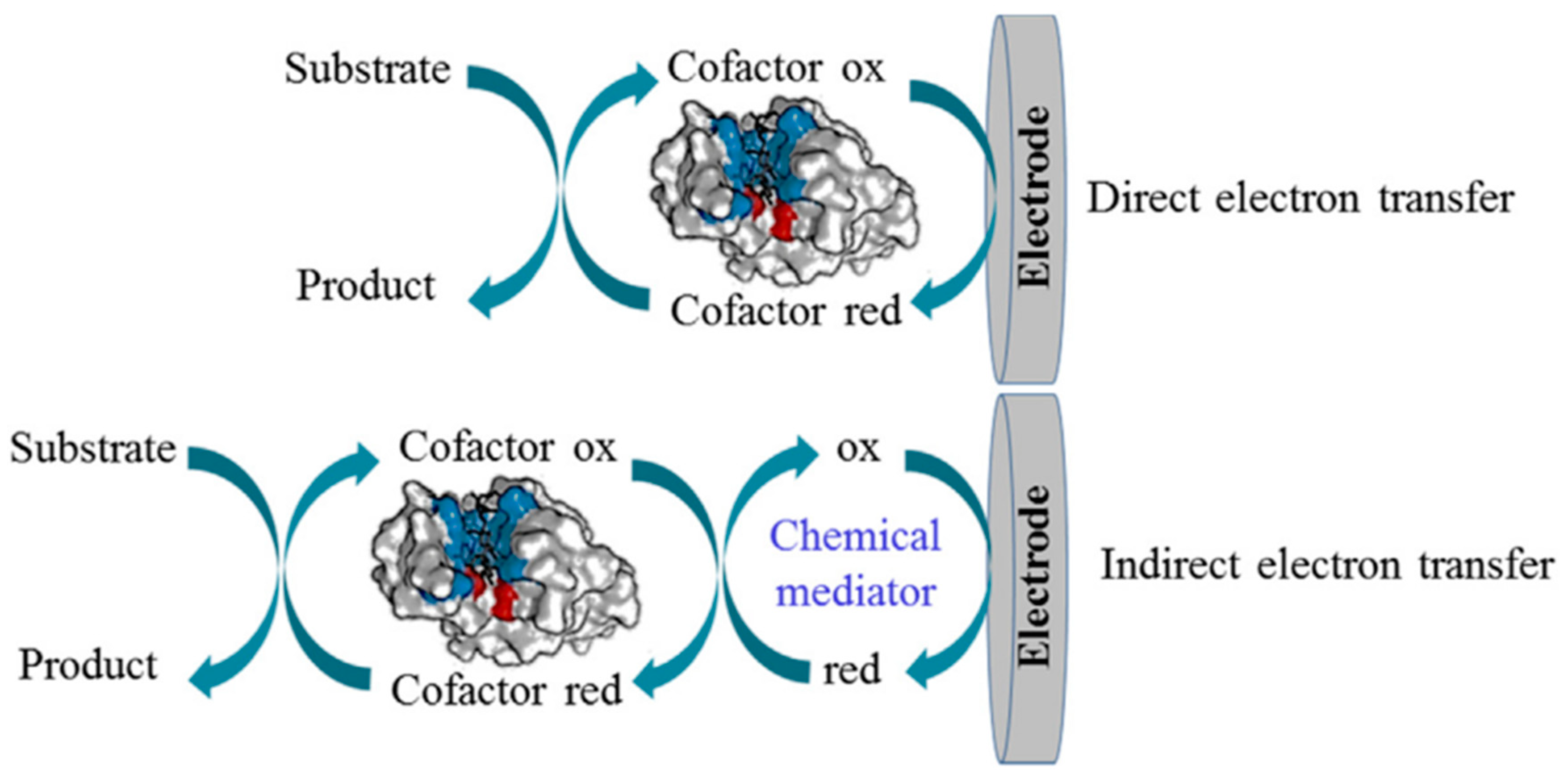
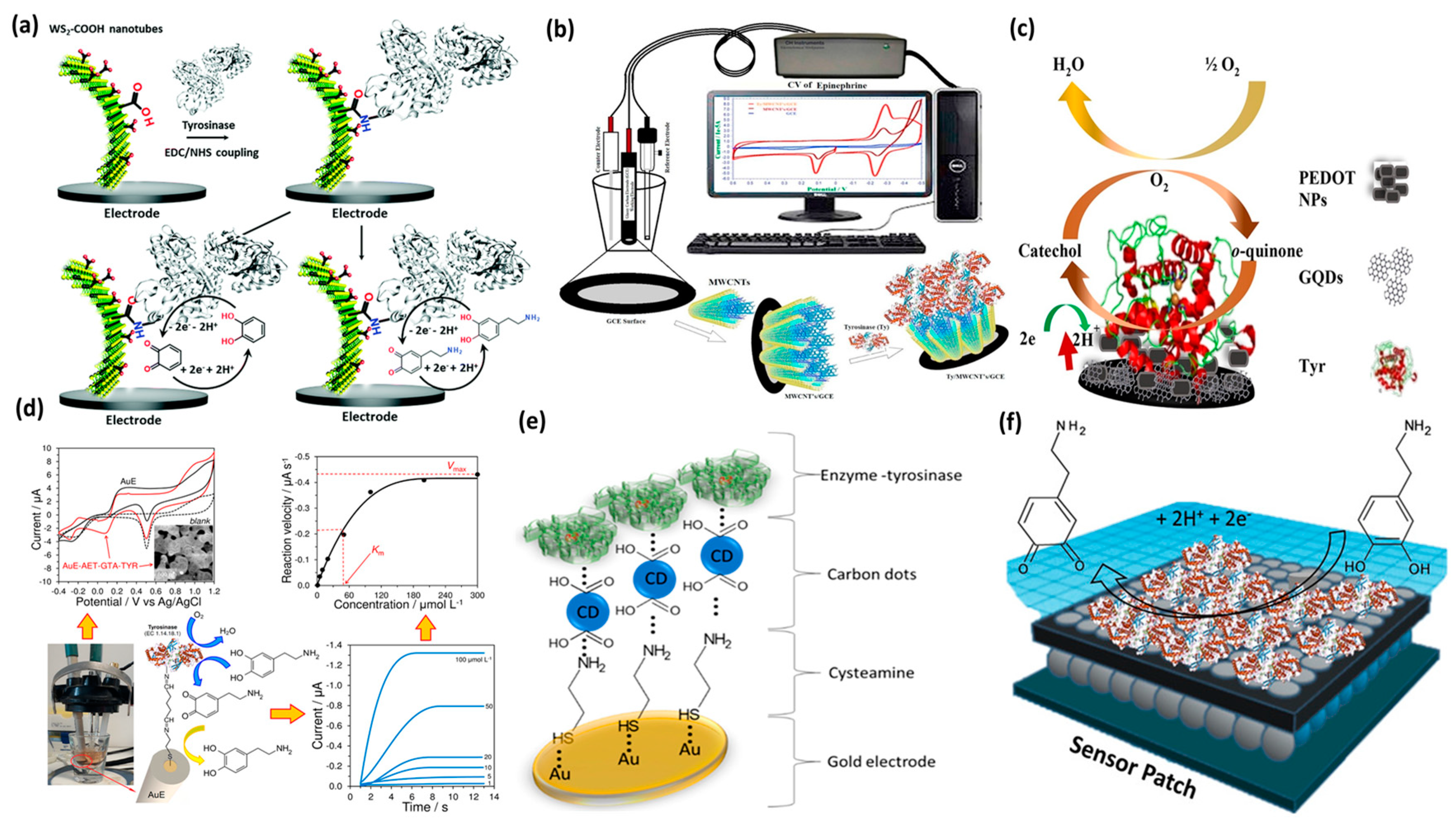
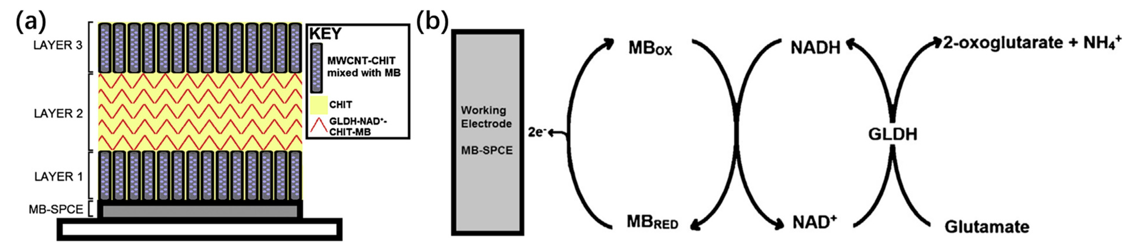
Table 1.
Common neurotransmitters, their concentration values in blood samples, and associated diseases.
Table 1.
Common neurotransmitters, their concentration values in blood samples, and associated diseases.
| Neurotransmitter | Concentration Range (Blood) | Associated Diseases |
|---|---|---|
| Dopamine | 10–480 pM | Parkinson’s disease, epilepsy |
| Acetylcholine | 7.6–9.7 nM | Alzheimer’s disease, myasthenia gravis |
| Serotonin | 0.6–1.6 μM | Depression, anxiety disorders, migraines, irritable bowel syndrome (IBS) |
| Epinephrine | 20–460 pM | Mood disorders, cardiovascular disorders |
| Norepinephrine | 0.45–2.49 nM | Post-traumatic stress disorder (PTSD), attention deficit hyperactivity disorder (ADHD) |
| Glutamate | 150–300 μM | Schizophrenia |
Table 2.
Comparative analysis of performance parameters in electrochemical sensors.
| Electrochemical Sensor | Sensitivity | Selectivity | Response Time | Detection Limit | Dynamic Range | Stability | Cost |
|---|---|---|---|---|---|---|---|
| Potentiometric Sensor | High | Moderate | Fast | Low | Wide | Good | Low |
| Amperometric Sensor | High | Low | Fast | Very Low | Narrow | Good | Moderate |
| Conductometric Sensor | Moderate | Moderate | Moderate | Moderate | Moderate | Good | Low |
| Voltammetric Sensor | High | High | Moderate | Very Low | Moderate | Good | High |
| Impedimetric Sensor | Moderate | High | Slow | Very Low | Wide | Excellent | High |
Table 3.
Performances of enzyme-based electrochemical neurotransmitters biosensors.
| Biosensor Structure | Enzyme | Analyte | Technique | Linear Range (µM) | Detection Limit (µM) | Ref. |
|---|---|---|---|---|---|---|
| Au-disk microelectrode | Tyrosinase | Dopamine | Amperometry | 20–300 | 0.24 | [141] |
| Polyphenilenediamine–Platinum disc electrodes | Adenosine deaminase | Adenosine | Amperometry | 15−100 | 7.2 | [145] |
| Pyrene-azide/GCE | Copper efflux oxidase | Dopamine Epinephrine | DPV | 10 × 10−3–0.1 | 10 × 10−3 10 × 10−3 | [146] |
| MWCNT–GCE | Monoamine oxidase | Serotonin | DPV | 0.56–2.26 | 0.2 | [148] |
| Prussian–ITO | Monoamine oxidase | Dopamine | Amperometry | 140–1000 | 86 | [149] |
| Fe3O4@AuNPs−GCE | Peroxidase | Dopamine | Voltammetry CV | 10–100 | 58 | [150] |
| SWCNT-PEDOT−Au microelectrode arrays | Tyrosinase | Dopamine | CV | 0.1–0.5 | 2.4 | [151] |
| Platinum microelectrode array | Glucose oxidase | Glutamate γ-aminobutyric acid | Amperometry | 20–1200 50–2200 | 20 50 | [152] |
| Poly[2,6-bis(3,4-ethylenedioxythiophene)-4-methyl-4-octyl-dithienosilole/Pt electrode | Laccase | Serotonin Dopamine | CV | 0.1–200 | 48 × 10−3 73 × 10−3 | [119] |
Disclaimer/Publisher’s Note: The statements, opinions and data contained in all publications are solely those of the individual author(s) and contributor(s) and not of MDPI and/or the editor(s). MDPI and/or the editor(s) disclaim responsibility for any injury to people or property resulting from any ideas, methods, instructions or products referred to in the content. |
© 2023 by the authors. Licensee MDPI, Basel, Switzerland. This article is an open access article distributed under the terms and conditions of the Creative Commons Attribution (CC BY) license (https://creativecommons.org/licenses/by/4.0/).
Share and Cite
MDPI and ACS Style
Fredj, Z.; Singh, B.; Bahri, M.; Qin, P.; Sawan, M. Enzymatic Electrochemical Biosensors for Neurotransmitters Detection: Recent Achievements and Trends. Chemosensors 2023, 11, 388. https://doi.org/10.3390/chemosensors11070388
AMA Style
Fredj Z, Singh B, Bahri M, Qin P, Sawan M. Enzymatic Electrochemical Biosensors for Neurotransmitters Detection: Recent Achievements and Trends. Chemosensors. 2023; 11(7):388. https://doi.org/10.3390/chemosensors11070388
Chicago/Turabian StyleFredj, Zina, Baljit Singh, Mohamed Bahri, Peiwu Qin, and Mohamad Sawan. 2023. "Enzymatic Electrochemical Biosensors for Neurotransmitters Detection: Recent Achievements and Trends" Chemosensors 11, no. 7: 388. https://doi.org/10.3390/chemosensors11070388
Note that from the first issue of 2016, this journal uses article numbers instead of page numbers. See further details here.