
Recent Advances in Design Strategies and Imaging Applications of Fluorescent Probes for ATP
1
Key Laboratory for Green Organic Synthesis and Application of Hunan Province, Key Laboratory of Environmentally Friendly Chemistry and Applications of Ministry of Education, College of Chemistry, Xiangtan University, Xiangtan 411105, China
2
Henan Key Laboratory of Organic Functional Molecule and Drug Innovation, Collaborative Innovation Center of Henan Province for Green Manufacturing of Fine Chemicals, Key Laboratory of Green Chemical Media and Reactions, Ministry of Education, School of Chemistry and Chemical Engineering, Henan Normal University, Xinxiang 453007, China
*
Author to whom correspondence should be addressed.
Chemosensors 2023, 11(7), 417; https://doi.org/10.3390/chemosensors11070417
Submission received: 6 June 2023
/
Revised: 19 July 2023
/
Accepted: 21 July 2023
/
Published: 24 July 2023
(This article belongs to the Special Issue A Theme Issue in Honor of Dr. Richard Horobin—Cell or Organelle Selective Fluorescent Probes: Their Design, Mechanism, Modeling and Application)
Abstract
:Adenosine 5′-triphosphate (ATP) is the energy currency in cells. It is involved in numerous cellular life activities and exhibits a close association with the development of certain diseases. Thus, the precise detection of ATP within cells holds immense significance in understanding cell biological events and related disease development. Fluorescent probes have obvious advantages in imaging ATP in cells and in vivo due to their high sensitivity, good selectivity, real-time imaging, and good biocompatibility. Thus far, an extensive array of fluorescent probes targeting ATP has been formulated to enable the visualization of ATP within cells and in vivo. This review summarizes the recent advances in ATP fluorescent probes according to different design strategies, mainly including those based on organic small molecules, metal complexes, and water-soluble conjugated polymers. In addition, the practical applications of ATP fluorescent probes in the imaging of target organelles, cell biological events, and disease markers are highlighted. Finally, the challenges and future trends of ATP detection based on fluorescent probes are discussed.
1. Introduction
Adenosine 5′-triphosphate (ATP) is an important biological molecule, which is the main source of energy for cells. It participates in a variety of life processes, including protein synthesis, ion transport in cells, neurotransmission, cell division, and glycolysis [1,2,3,4,5,6,7]. An abnormal level of ATP metabolism is related to many diseases, such as ischemia, Parkinson’s disease, cardiovascular disease, malignant tumors, and so on [8,9,10,11,12,13]. In order to study the cellular life process and explore ATP-related diseases, it is of great significance to detect the content and dynamic changes in ATP in biological samples.
Currently, ATP can be detected using various methods, including high-performance liquid chromatography (HPLC), ion chromatography, mass spectrometry, electrochemistry, and bioluminescence analysis [14,15,16,17]. However, these analytical methods have the disadvantage of low sensitivity. Fluorescence probes, with many advantages such as high sensitivity, excellent spatial and temporal resolution, and real-time in situ imaging, have emerged as crucial tools for the detection and imaging of bio-active substances during medical diagnosis [18,19,20,21,22,23,24,25,26,27,28,29,30,31]. Moreover, near-infrared (NIR) fluorescent probes have long excitation/emission wavelengths that allow deeper tissue penetration while reducing the background fluorescence of proteins and photodamage to biological samples [32,33,34,35,36,37,38,39,40,41,42,43,44].
The fluorescent probes for ATP are mainly composed of organic fluorophores and ATP recognition sites. Among them, the interaction between ATP and the recognition site causes a structural change or energy transfer of the organic fluorophore, which leads to the generation or quenching of fluorescence. The design of ATP fluorescent probes necessitates the careful consideration of several factors. The first is the excitation/emission wavelength of the probe, and longer excitation and emission wavelengths are beneficial for ATP detection in deep biological samples. The second is the selectivity of the probe, and the detection process should only respond to ATP. The third is the appropriate detection range; ATP concentration in cells is usually 1–10 mM and too low or too high a detection range will fail to dynamically image fluctuations in intracellular ATP levels. Fourth, if they are used for the detection of ATP in cells, the probes need to have good biocompatibility. Last but not least, by introducing targeting groups into the probe, it is possible for the probe to have organelle or specific cell-targeting capabilities, which allows the targeted detection of cell biological events and insight into the complex functions of ATP. So far, there has been significant progress in the design and application of ATP fluorescent probes, and many ATP fluorescent probes have been reported [45,46,47,48].
In this review, we aim to summarize recent advances in the detection of ATP with fluorescent probes. The ATP fluorescent probes in this paper are categorized into three groups based on different design strategies: organic small molecules, metal complexes, and water-soluble conjugated polymers (Figure 1). This review also highlights the application of ATP fluorescence probes in targeting organelles, imaging cell biological events, and detecting disease markers. Finally, the current challenges in ATP detection and the prospects for the development of ATP fluorescent probes are discussed.
2. The Design Strategy of Fluorescent Probes for ATP
2.1. ATP Fluorescent Probes Based on Organic Small Molecules
Fluorescent probes for ATP based on organic small molecules have been of great interest because of their good selectivity, high sensitivity, and ease of crossing the cell membrane into cells. Many organic small-molecule fluorescent probes for ATP recognition have been developed and reported. These probes have three main mechanisms, including ring-opening reaction, electrostatic interaction, and aggregation-induced luminescence (AIE).
2.1.1. ATP Fluorescent Probe Based on the Ring-Opening Reaction
Rhodamine dyes are widely utilized as fluorescent probes due to their advantageous properties, such as a high molar extinction coefficient, long emission wavelength, excellent fluorescence quantum yield, and exceptional photostability [49,50]. Rhodamine derivatives with a spirolactam ring structure exhibit non-fluorescent characteristics. When they are exposed to ATP, the spirolactam ring undergoes opening, resulting in a significant enhancement in fluorescence intensity. In recent years, the development of ATP fluorescent probes based on the rhodamine ring-opening reaction has received great attention.
In 2013, a rhodamine spirolactam derivative (probe 1, Figure 2) was developed as a colorimetric and fluorescent chemosensor for ATP [51], and the sensing process is ATP-induced spirolactam ring opening of rhodamine derivatives. Probe 1 itself has no fluorescence emission since rhodamine is in the form of a spirolactam ring in this probe. Upon the introduction of ATP, hydrogen bonds are formed between the diethylenetriamine moiety of probe 1 and the polyphosphates within ATP. Consequently, the configuration of the rhodamine molecule undergoes a transformation from a spirolactam ring to an open-ring amide structure. Thus, the fluorescence signal is activated. Probe 1 has a highly sensitive “turn-on” fluorescence response to ATP. Most importantly, probe 1 successfully overcomes the challenge of significant interference caused by other nucleoside polyphosphates (NPPs) like ADP and AMP. Moreover, this probe can be effectively employed for the fluorescence-based detection of protein kinase activity.
In 2014, a ratiometric fluorescent probe (probe 2, Figure 2) for ATP detection was developed based on the Förster resonance energy transfer (FRET) mechanism [52]. In probe 2, the naphthalimide fluorophore is incorporated as the energy donor, while rhodamine serves as the energy acceptor. Due to the spirolactam ring form of the rhodamine moiety in probe 2, only the naphthalimide moiety exhibits the emission. However, upon the addition of ATP, the formation of the probe 2–ATP complex induces a conformational change in the rhodamine moiety, transitioning it from a spirolactam ring to an open-ring amide structure. This conformational change enables a viable FRET process, facilitating energy transfer from the naphthalimide moiety (energy donor) to the rhodamine moiety (energy acceptor). Consequently, the emission from the naphthalimide decreases, while the emission from the rhodamine becomes apparent, resulting in a noticeable shift from green emission to red emission. By simultaneously interacting with the polyphosphate chain and the nucleic base group, its selectivity for ATP is unexpectedly higher than for other organophosphate anions.
In 2020, a ratiometric fluorescent probe 3 (Figure 2) was designed and synthesized for quantitatively detecting the fluctuation of mitochondrial ATP [53]. Probe 3 was designed based on the FRET mechanism. Rh6G was chosen as the donor fluorophore because of its specific localization to mitochondria and spectral overlap with the dye ACF. In addition, a multisite binding strategy was used to improve selectivity. The incorporation of either two saturated carbon linkers or a rigid piperazine ring facilitates the formation of hydrogen bonds with the phosphate group of ATP. Additionally, it promotes the π–π interaction between the nitrogenous bases and the ACF dye. This establishes the conditions for the rapid response of probe 3 to ATP. In addition, probe 3 can discriminate between normal and cancer cell lines by ATP detection.
In 2021, a near-infrared (NIR) reversible probe 4 (Figure 2) was developed for the ratiometric imaging of tumors [54]. In FRET-based probe 4, the energy donor is silicon rhodamine, while the energy acceptor is CS dye. The probe is designed with an ATP/H+ recognition unit that remains inactive until both H+ and ATP molecules are bound to the acceptor. The spirolactam of probe 4 opens after the reaction, activating both ratiometric fluorescence and photoacoustic signals. The probe demonstrates reversible activation upon the binding of both H+ and ATP molecules. In addition, probe 4 has been successfully employed in tumor photoacoustic and ratiometric fluorescence imaging, as well as fluorescence-imaging-guided tumor resection.
2.1.2. ATP Fluorescent Probe Based on Electrostatic Interaction
Electrostatic interactions involve the formation of hydrogen bonds between the oxygen atoms of the ATP phosphate group and the active hydrogen of the probe. These interactions induce structural changes in the probe, consequently impacting the fluorescence emission properties of the probe. Due to the varying number of negatively charged phosphate groups in ATP, ADP, and AMP (three, two, and one, respectively), it is feasible to design ATP probes with excellent selectivity based on electrostatic interactions.
In 2009, a pincer-like benzene-bridged probe 5 (Figure 3) was reported [4]. This probe utilized a pyrene excimer as the signal source and imidazolium as a phosphate anion receptor, enabling the detection of ATP. In probe 5, the adenine moiety of ATP can be positioned between the two pyrene moieties, causing the separation of the pyrene moieties and the production of pyrene monomer fluorescence. However, other nucleoside triphosphates such as GTP, CTP, UTP, and TTP can only interact with the already stacked pyrene–pyrene dimer of probe 5 from the outside. This interaction results in the quenching of excimer fluorescence. In addition, probe 5 can distinguish ATP from ADP and AMP, which is attributed to the variation in electrostatic interactions between the probe and phosphate anion. Two years later, a tripodal probe 6 (Figure 3) featuring three naphthoimidazolium groups and three quaternary ammonium groups was developed. Probe 6 displays diverse interactions with different nucleoside bases, leading to varying synergistic effects of π-stacking and electrostatic interactions. Notably, probe 6 exhibits substantial fluorescence enhancements upon interaction with UTP, CTP, and TTP. It demonstrates moderate fluorescence enhancements when interacting with ATP and pyrophosphate. However, a fluorescence quenching effect is observed when probe 6 interacts with GTP. This distinct response to different nucleoside bases showcases the potential of probe 6 for selective detection and discrimination among nucleoside triphosphates [55].
In 2015, a smart Off–On molecular scaffold/fluorescent probe 7 (Figure 3) was designed and synthesized [56]. The triphosphate unit of ATP interacts with the piperazine nitrogen atoms in probe 7 through hydrogen bonding and electrostatic interactions. These interactions synergistically enhance the CH–π and π–π stacking interactions between the anthracene and purine rings. As a result, a significantly enhanced “turn-on” emission is observed due to restricted photo-induced electron transfer (PET). Moreover, probe 7 exhibits outstanding photostability, cell permeability, organelle specificity, and remarkable selectivity for ATP. In the same year, a pyridinium-based probe 8 (Figure 3) was reported [57]. Probe 8 demonstrates the selective recognition of ATP by means of hydrogen bonding and electrostatic interactions at the pyridinium sites. Furthermore, probe 8 has been successfully used to detect intracellular ATP through fluorescent confocal imaging.
In 2016, fluorescence probe 9 (Figure 3) was reported [58]. Probe 9 consists of a naphthalimide fluorophore with two symmetric peptidic arms that incorporate a specifically designed anion-binding motif called guanidiniocarbonyl pyrrole. This motif enables the probe to selectively detect nucleoside triphosphates. The probe forms stable complexes with oxoanions through a salt bridge, reinforced by multiple hydrogen bonds. Each arm of the probe contains a lysine residue to facilitate additional electrostatic interactions. Moreover, the arms are functionalized with tryptophan, which has the potential to differentiate between nucleoside triphosphates based on their distinct π–π interactions with the various nucleobases. The central spacer of probe 9 is linked to a green-emitting aminonaphthalimide fluorophore, serving as the reporter unit. Upon binding to nucleoside triphosphates, especially ATP, probe 9 exhibits a significant increase in fluorescence intensity, resulting in a prominent turn-on fluorescence response. The higher fluorescence intensity observed for triphosphates compared to diphosphates or monophosphates can be attributed to the differences in electrostatic interactions with the guanidiniocarbonyl pyrrole and lysine residues, which arise from the varying anionic charges of the different phosphate groups.
2.1.3. ATP Fluorescent Probes Based on Aggregation-Induced Emission
Conventional ATP sensors often face challenges related to emission quenching caused by the aggregation-induced quenching (ACQ) effect. Mitigating this quenching typically requires using low probe concentrations, which reduces the probe-to-analyte ratio and limits practical applications. However, the use of AIE luminogens (AIEgens) offers an alternative approach. AIEgens are generally non-emissive or weakly emissive in their solution state but exhibit high emission intensity upon aggregation. This unique behavior of AIEgens effectively overcomes the fluorescence quenching issues observed with traditional fluorophores at high concentrations. At present, several AIE-based fluorescent probes for ATP detection have been reported, presenting promising solutions in the field.
In 2012, an ATP probe 10 (Figure 4) based on aggregation-induced luminescence was reported [59]. The interaction between the guanidinium and triphosphate groups plays a critical role in initiating the aggregation process, which ultimately leads to fluorescence enhancement. Additionally, the collective π–π stacking and hydrophobic interactions with the adenine moiety contribute to the cooperative self-assembly phenomenon. These interactions result in nonlinear and substantial fluorescence enhancement, significantly improving the sensitivity and selectivity of the probe for ATP detection.
In 2015, a novel thiazole orange derivative 11 (Figure 4) was developed [60]. Probe 11 demonstrates cooperative co-assembly with ATP, leading to a nonlinear and responsive AIE signal. Upon the initial addition of ATP, a 1:2 complex of ATP/11 is formed. At this stage, the dimer of probe 11 is relatively less stable, as it is modulated by only one ATP molecule, resulting in a slight enhancement of fluorescence. However, as more ATP is added, the preorganized 1:2 complexes of ATP/11 promote the formation of higher-level complexes, such as extended “oligomer-” or “polymer-”like supramolecular arrays. Consequently, the dimer of probe 11 becomes more tightly packed and stable. This leads to a faster increase in intensity at 580 nm, being attributed to the intensified mutual interactions among the colocalized thiazole orange moieties of probe 11 within the assembly.
In 2017, an AIE-based tetraphenylethylene derivative (probe 12, Figure 4) was developed for the fluorescence sensing of ATP in aqueous solution [61]. Probe 12, featuring four cationic pyridinium pendants, exhibits weak emission in HEPES buffer solution. However, upon the addition of ATP, probe 12 undergoes aggregation along the ATP molecule through electrostatic interactions and hydrophobic interactions, resulting in a significant nhancement of emission. It is worth noting that probe 12 is capable of imaging ATP in both cancer cell lines and normal cell lines, demonstrating its potential as a versatile tool for ATP imaging.
In the same year, a series of novel AIE-active fluorescence probes (probe 13–16, Figure 4) were reported [62]. These probes incorporate pyridinium and boric acid groups and have been specifically designed for ATP recognition purposes. The unique design of these probes allows for two types of interaction modes and multiple connection sites with ATP molecules, enabling the selective discrimination of ATP from other bioactive anions. As a result, these probes exhibit a significant enhancement in fluorescence emission specifically upon ATP binding. Notably, probe 16, which incorporates two types of interaction modes and multiple connection sites, demonstrates high sensitivity in recognizing ATP. The above-described probes are summarized in Table 1.
2.2. ATP Fluorescent Probes Based on Metal Complexes
Metal coordination complex-based fluorescent probes, characterized by one or two coordination sites, demonstrate a heightened affinity towards phosphate anions. This property makes them well-suited for the detection of ATP [63]. Fluorescent probes for ATP based on metal complexes have been widely studied in recent years due to their advantages of high selectivity, high sensitivity, and simple preparation.
2.2.1. ATP Fluorescent Probes Based on Zinc Complexes
Among the fluorescent probes based on metal complexes, zinc complexes have been rapidly developed. The Zn(II)-dipicolylamine (Zn-DPA) structural unit is widely used to design ATP fluorescent probes. The dipicolylamine unit can form a stable complex with Zn(II), and Zn(II) has a strong binding ability to the phosphate group of ATP. The change in the Zn(II) coordination unit after the addition of ATP causes a change in fluorescence. So far, Zn-DPA has been generally recognized as a specific recognition unit for ATP probes.
In 2002, fluorescent probe 17 (Figure 5) was developed. This probe incorporates an anthracene moiety appended with Zn-DPA, enabling efficient detection of ATP in a neutral aqueous solution. [64]. Probe 17 demonstrates a pronounced level of selectivity towards ATP, as evidenced by the observed selectivity (ATP > ADP > AMP). This selectivity can be attributed to the disparities in the respective quantities of their anionic charges. After this study, a fluorescent chemosensor (probe 18, Figure 5) for nucleoside polyphosphates such as ATP was reported in 2008 [65]. Probe 18 is designed with dual binding motifs, namely 2,2′-Dpa-Zn(II), and incorporates xanthene as a fluorescent sensing unit for nucleoside polyphosphates. The turn-on fluorescence sensing mechanism of probe 18 towards nucleoside polyphosphates can be attributed to the restoration of the conjugated structure of the xanthene ring. Initially, the fluorescence of probe 18 is suppressed due to the formation of a deconjugated structure in the xanthene fluorophore, as mentioned earlier. However, the binding of ATP to the two Zn(II)-Dpa sites disrupts the µ-oxo-dizinc(II) core of probe 18 through multiple interactions with the triphosphate group of ATP. This disruption leads to the restoration of the conjugated xanthene structure and a significant increase in fluorescence emission. Importantly, probe 18 exhibits remarkable selectivity and sensitivity for ATP detection, making it highly suitable for visualizing ATP in living cells. In 2010, ratiometric probes 19 and 20 (Figure 5) for nucleoside polyphosphates such as ATP that are based on FRET combined with a turn-on fluorescence-sensing mechanism were developed [66]. In these probes, coumarin fluorophores were introduced as the FRET donors, and a binuclear zinc complex acted as the FRET acceptor. Probes 19 and 20 exhibited the distinct dual-emission signal change upon binding with high affinity (Kapp ≈ 106–107 M−1) to nucleoside polyphosphates in aqueous solutions. Notably, no significant emission changes were observed when these probes interacted with monophosphates, phosphodiester species, or various other anions.
In 2012, a perylene diimide (PDI) ligand (probe 21, Figure 5) was developed, which was modified with a Zn2+-dipicolylethylenediamine (Zn2+-DPEN) moiety. This fluorometric chemosensor was developed with the specific aim of detecting ATP in aqueous solutions, demonstrating the selectivity over other phosphate anions. [67]. The binding stoichiometry between probe 21 and ATP has been determined to be 1:1, indicating that one molecule of probe 21 interacts with one molecule of ATP. It is noteworthy that this probe displays high sensitivity and selectivity in detecting ATP, even in a 100% aqueous solution. In the presence of ATP, probe 21 exhibits a significant fluorescence enhancement, making it a reliable and effective tool for the detection of ATP.
In 2018, a coumarin and Zn(DPA)-based fluorescent probe 22 was reported [68]. As shown in Figure 5, the Zn(II) systems within the probe exhibit an affinity for ATP and ADP. The Zn(II) center of the Zn(DPA) moiety interacts with the pendent phosphate moieties of ATP and/or ADP. This binding event is expected to reduce the interaction between DPA moiety and Zn(II), leading to the weakening of the intramolecular charge transfer (ICT) process within the complex. As a result, the observed fluorescence intensity is enhanced.
In 2018, a reversible fluorescein-based fluorescent probe 23 for Zn2+ and ATP was designed and synthesized [69]. As shown in Figure 5, the sensing mechanism of probe 23 involves a fluorescence variation caused by the opening and closing of the spiro-ring of fluorescein. In its free form, the probe exhibits minimal emission due to the closed fluorescein spirolactam structure. However, upon the introduction of Zn2+, a remarkable enhancement of fluorescence intensity at 527 nm is observed. This enhancement is attributed to the structural transformation induced by Zn2+, leading to the conversion of the colorless spirolactam ring to the colored ring-opening delocalized form of fluorescein. Upon the gradual addition of ATP to a solution containing probe 23-Zn2+, a significant decrease in fluorescence intensity at 527 nm is observed. This spectral change can be attributed to the strong binding affinity between ATP and Zn2+. The binding of ATP to Zn2+ ions results in the displacement of Zn2+ from the complex, and the compound undergoes structural recovery. Upon the further addition of Zn2+, the fluorescence is recovered, indicating the reversibility of the binding between probe 23 (23–Zn2+) and Zn2+ (ATP).
Zn2+ can combine with a variety of fluorophores to form a Zn-DPA structure for ATP detection. In 2012, a chemosensor (probe 24, Figure 5) was synthesized based on a Zn complex of 1-[bis(pyridine-2-ylmethyl)amino]methylpyrene for the detection of nucleotides in aqueous solution [70]. Probe 24 exhibited a distinct emission band ranging from 360 to 450 nm, attributed to the emission from the pyrene monomer. Upon the addition of various nucleotide anions, the fluorescence intensity of the pyrene monomer emission noticeably increased upon the addition of PPi. By contrast, only a slight fluorescence enhancement was observed when ATP and ADP were added to the solution containing probe 24. The spectra of other nucleotides displayed minimal changes, indicating limited interaction with the probe.
In 2014, a tailor-made ratiometric fluorescent probe 25 for nucleoside polyphosphates was fabricated based on a pyrene-functionalized zinc(II)–BPEA complex [71]. As shown in Figure 5, Probe 25 is composed of three components: a zinc(II)–BPEA complex (BPEA: N,N-bis(2-pyridylmethyl)ethylenediamine) serving as the ionophore, an amide residue acting as a hydrogen bond acceptor, and pyrene aromatic rings functioning as the fluorophore. These components synergistically form a fully intramolecular sandwich structure when two molecules of probe 25 interact with one molecule of ADP. This interaction is facilitated by three effects: (i) coordination of BPEA–Zn2+ with the diphosphate anion; (ii) hydrogen bonding between the amide group and ribose; (iii) π–π stacking interaction among the pyrene rings and adenine. By contrast, when two molecules of probe 25 interact with one molecule of ATP, a partial intramolecular sandwich structure is formed due to two synergistic interactions: coordination of BPEA–Zn2+ with the triphosphate anion and π–π stacking between the two pyrene rings. These distinctive binding patterns exhibited by probe 25 with ATP and ADP highlight its exceptional capability as a fluorescent probe for effectively distinguishing between ATP and ADP.
2.2.2. ATP Fluorescent Probes Based on Copper Complexes
Although Zn(II) has a good recognition affinity for ATP, Cu(II) interacts more strongly with the phosphate moiety of ATP [72]. Several ATP fluorescent probes based on Cu(II) complexes have been reported. In 2016, fluorescent probe 26 (Figure 6) with one 1,8-naphthalimide and two [12]aneN3 units was reported [73]. Probe 26 exhibited strong fluorescence emission. However, in the presence of Cu2+ ions, probe 26 formed a complex (26–Cu) with Cu2+ ions in a 1:2 ratio, leading to a significant quenching of fluorescence emission. In the presence of ATP, a strong fluorescence enhancement was observed in the 26–Cu complex. This enhancement can be attributed to the concerted coordination of the phosphates and adenosine base units of ATP to Cu(II) ions. This coordination effectively reduces the interaction between the Cu(II) ions and the amino group of the naphthalimide unit in probe 26, resulting in the activation of its fluorescence. The coordinative interactions between copper(II) ions and adenosine play a crucial role in determining the selectivity of probe 26 [74,75,76]. When ADP is present, the diminished structure matching reduces the effectiveness of the interactions, resulting in reduced effects on the fluorescence response of probe 26. On the other hand, the weak coordination of guanosine and thymidine base units to Cu2+ ions leads to poor cooperative interactions between GTP/CTP and the 26–Cu complex. As a result, there is almost no change observed in the fluorescent intensity when GTP or CTP is added to the system.
In 2018, a rhodamine-based fluorescent probe 27 (Figure 6) for Cu2+ and ATP was reported [77]. Upon the addition of Cu2+ ions, the fluorescence intensity or absorbance of the probe was significantly enhanced. This enhancement was attributed to the opening of the spiro-ring of the rhodamine moiety. However, the fluorescence or absorbance quickly returned to its original level as the probe underwent reconstruction upon reaction with ATP. The changes in fluorescence or absorbance induced by Cu2+ and ATP exhibited a good linear relationship with the concentration of Cu2+ ions in the range of 2–20 μM and ATP in the range of 0–10 μM. The detection limits for Cu2+ and ATP were determined to be 0.1 μM and 1.0 μM, respectively.
In 2020, a rhodol-based ratiometric fluorescent probe 28 (Figure 6) was designed for the reversible response of Cu2+ and ATP [78]. In the absence of Cu2+, probe 28 exhibits its maximal fluorescent intensity at 434 nm. This emission peak is attributed to the excited-state intramolecular proton transfer (ESIPT) process of the benzothiazole moiety present in the probe. However, upon the addition of Cu2+, the formation of the 28–Cu complex occurs. As a result, the maximal emission peak at around 434 nm gradually diminishes, while a new emission intensity at around 595 nm significantly enhances. This new emission is characteristic of the opening ring of the rhodol spirolactam structure formed in the presence of Cu2+. With the addition of ATP, free probe 28 is released from the 28–Cu complex. The fluorescence of 28–Cu gradually increased at 434 nm and decreased at 595 nm. Probe 28/28–Cu can selectively respond to Cu2+/ATP. Furthermore, the probe can be effectively utilized for the rapid and convenient detection of Cu2+ and ATP using filter paper and hydrogel as the detection platforms.
2.2.3. ATP Fluorescent Probes Based on Other Metal Complexes
In addition to Zn2+ and Cu2+, Ga3+ also has a strong binding affinity for diphosphate and triphosphate. Several ATP fluorescent probes based on gallium complexes have been reported. In 2014, a Ga3+ self-assembled fluorescent probe 29 (Figure 7) using the ligand of 2-(2′,3′-dihydroxyphenyl)benzoxazole (DHBO) was synthesized [79]. The probe exhibits a significant fluorescence enhancement specifically with ATP among structurally similar nucleoside triphosphates under physiological conditions. The selectivity of probe 29 towards ATP can be attributed to several factors. First, ATP is attracted to probe 29 through electrostatic interactions between the positively charged Ga3+ ion and the phosphate group of ATP. The stronger the negative charge on the phosphate groups, the stronger the intermolecular interaction, which contributes to the selective recognition of ATP over ADP and AMP. Additionally, selective π–π stacking interactions occur between the adenosine segment of ATP and DHBO ligand, further contributing to the sensing process. This π–π stacking interaction is unique to ATP and not observed with other nucleoside triphosphates or pyrophosphate, providing additional selectivity for ATP. It is worth mentioning that probe 29 can also be employed for detecting the activity of ATP-related enzymes, further demonstrating its potential utility in enzyme activity assays and biological studies.
In 2018, a naphthol-based fluorescent probe 30 (Figure 7) containing electron-donating Schiff base N, thiazole N, and phenolic OH, which provide binding sites to Ga3+, was reported [80]. The fluorescence response of probe 30 to Ga3+ is based on a combination of two mechanisms: ESIPT and CHEF (chelation-enhanced fluorescence). Probe 30 exhibits a weak emission at 518 nm due to the presence of C=N and adjacent OH groups, which are characteristic of ESIPT. Upon the binding of Ga3+ to the C=N and OH groups, the ESIPT process is further restrained. The formation of the stable complex 30–Ga3+ leads to an enhanced fluorescence at 518 nm, attributed to the CHEF effect. The recognition of ATP by probe 30 involves the extrusion of Ga3+ from the 30–Ga3+ complex, and this process is accompanied by ESIPT from the phenolic OH group to the Schiff base N in probe 30. The combination of 30–Ga3+ and ATP leads to the inhibition of CHEF and restoration of ESIPT, and thus the quenching of the bright green emission. Additionally, the 30–Ga3+ complex exhibits high selectivity and sensitivity towards ADP and ATP over anions in aqueous solution. The presence of ADP and ATP in the solution displaces the Ga3+ from the complex, leading to quenching of the fluorescence. This selective recognition and quenching mechanism make the 30–Ga3+ complex a suitable fluorescent probe for the detection of ADP and ATP.
Lanthanide complexes, particularly Eu3+ complexes, exhibit distinctive fluorescence responses in the recognition of ATP, demonstrating either a turn-on or turn-off behavior. Additionally, these complexes are known for their long fluorescence lifetimes. In 2013, fluorescent probe 31 (Figure 7) based on a Eu3+ complex was developed [81]. This probe demonstrates excellent selectivity, allowing for the effective differentiation of ATP from ADP and AMP in an aqueous solution at pH 6.8. The proposed mechanism for the enhancement of luminescence can be described as follows: First, the terpyridine arm chain of the ligand is well-suited for ATP, facilitating coordinated interactions and π–π stacking interactions with the probe. Second, the π–π stacking interactions between the terpyridine group of the ligand and ATP contribute to the reinforcement of the conjugate planes’ rigidity within the antenna groups. This interaction also restricts the rotation of C-C bonds between the aromatic rings. Additionally, compared with probes based on organic molecules and transition metal complexes, lanthanide-based probe 31 offers the advantage of an extended fluorescence lifetime.
In 2018, fluorescent probe 32 (Figure 7) based on a luminescent lanthanide complex was reported [82]. Probe 32, which contains two neutral amide donors, demonstrates a strong binding affinity towards ATP (log Ka = 5.8). This binding interaction leads to the formation of a stable ternary complex. Notably, the resulting ternary complex exhibits intense and long-lived luminescence attributed to the presence of Eu3+. Furthermore, in a competitive aqueous medium that mimics the intricate ionic environment found in cells, probe 32 displays effective discrimination between ATP, ADP, and AMP. The above-described probes are summarized in Table 2.
2.3. ATP Fluorescent Probes Based on Water-Soluble Conjugated Polymers
Water-soluble conjugated polymers have broad application prospects in the field of chemical and biological sensing [83]. The molecule of this polymer can linearly amplify the fluorescence signal to detect various analytes with high sensitivity.
In 2004, a water-soluble cationic polythiophene derivative 33 (Figure 8) was reported [84]. The probe exhibits both colorimetric and fluorescent responses to ATP. The response mechanism involves a combination of electrostatic and hydrophobic interactions. The interaction between the negatively charged triphosphate group of ATP and the positively charged ammonium group in 33 facilitates the planarization of the polythiophene backbone. As the concentration of ATP increases, a critical threshold is reached, promoting efficient π–π stacking interactions between the backbones of probe 33. These stacking interactions are induced by the synergistic effect of hydrophobic interactions between the adenine units. Consequently, these interactions cause a shift in the π–π* transition to longer wavelengths, leading to a color change from yellow to pink-red. Additionally, the fluorescence of probe 33 is quenched because of the interactions with ATP.
In 2016, a new water-soluble cationic polythiophene derivative 34 (Figure 8) was developed as a specific fluorescent probe for detecting ATP [85]. Probe 34 demonstrates a random coil conformation in water, but it undergoes conformational changes and forms aggregates upon interacting with ATP. The presence of ATP induces the aggregation of isolated probe 34 through cooperative electrostatic and hydrophobic interactions. Consequently, this conformational transition from a random coil structure to aggregates leads to the simultaneous quenching of probe 34’s fluorescence. In 2017, another water-soluble cationic polythiophene derivative (probe 35, Figure 8) for ATP sensing was reported [86]. In an aqueous solution, probe 35 adopts a random coil conformation. When it is exposed to ATP, probe 35 undergoes electrostatic and hydrophobic interactions, leading to its aggregation. This conformational transition from a random coil structure to aggregates results in the quenching of the probe’s fluorescence. Notably, probe 35 displays excellent sensitivity and selectivity towards ATP.
In 2017, a ratiometric probe 36 (Figure 8) for the detection of ATP was reported [87]. This probe utilizes a sensing mechanism that combines binding-induced modulation of FRET with aggregation-caused quenching (ACQ). To develop the ratiometric probe 36, coumarin fluorophores are introduced as the FRET donors into the side chain of polythiophene, which serves as the FRET acceptor. Upon the addition of ATP, the polymer/ATP complex is formed, resulting in conformational changes of the polythiophene backbone from a random coil structure to a planar conformation, along with aggregation. By exploiting the changes in spectral overlap before and after the addition of ATP, the ratiometric detection of ATP can be achieved by modulating the FRET efficiency between the coumarin fluorophores and the polythiophene backbone, coupled with ACQ. Importantly, probe 36 demonstrates a distinct dual-emission signal change resembling a seesaw type upon binding to ATP in an aqueous solution, exhibiting a strong affinity (Kapp = 3.12 × 10⁴ M−1). Notably, no significant emission changes are observed with ADP, AMP, or various other anions, highlighting the high selectivity of probe 36 towards ATP. The above-described probes are summarized in Table 3.
3. Imaging Applications of ATP Fluorescent Probes
In recent years, ATP fluorescent probes have made significant progress. Some ATP probes can sensitively detect the content, distribution, and dynamic changes in ATP in cells. Organelle-targeted ATP probes can accurately detect ATP in specific organelles, which helps to deepen the understanding of organelle function. Some ATP fluorescent probes can also be used to visualize the process of cellular biological events. In addition, some probes can be used for imaging changes in ATP content in diseases, which is expected to provide new tools for the diagnosis of related diseases.
3.1. ATP Fluorescent Probes for Targeting Organelles
ATP plays an important role in various life activities within cells, so it is necessary to detect ATP in specific cells or organelles. Fluorescent probes targeting cells and organelles have been a research hotspot, and ATP-based targeted fluorescent probes have also been widely studied. Among various organelles, mitochondria are the major sites of ATP production. In 2017, a fluorescent probe named 37 (Figure 9) was synthesized and used to recognize ATP in mitochondria [88]. Rhodamine, diethylenetriamine, and triphenylphosphonium were chosen as the fluorophore, reaction site, and mitochondria-targeting group, respectively. Probe 37 exhibited remarkable sensitivity to ATP. The detection range (0.1−10 mM) of the probe effectively matched the concentration levels of ATP found within the mitochondria. Notably, probe 37 exhibited exceptional selectivity towards ATP compared with other biological anions, thanks to the synergistic effect of its dual recognition sites. Additionally, this probe demonstrated specific localization within the mitochondria and proved to be a valuable tool for the real-time monitoring of changes in mitochondrial ATP concentration.
In 2017, a novel fluorescent probe 38 (Figure 9) was designed as a colorimetric and fluorescent chemosensor for ATP detection based on hydrogen bond interactions [89]. The probe exhibited a significant “turn-on” fluorescence response upon ATP binding, with a 15-fold increase in fluorescence intensity observed upon the addition of 10 equivalents of ATP. To further investigate its potential applications, colocalization experiments were conducted using MitoTracker Green, confirming the selective imaging capability of probe 38 within the mitochondria.
In 2018, a multisite-binding fluorescent probe 39 (Figure 9) for ATP was reported [90]. The fluorophore, reaction sites, and mitochondria-targeting group chosen for the design of probe 39 were rhodamine 6G, diethylenetriamine (or phenylboronic acid), and phenylboronic acid, respectively. Notably, probe 39 demonstrated excellent selectivity towards ATP, surpassing other anions, thanks to the synergistic effect of its multisite-binding recognition. Live-cell imaging experiments revealed that probe 39 exhibited preferential localization within the mitochondria maintaining good biocompatibility. Furthermore, the probe proved to be a valuable tool for real-time monitoring of changes in mitochondrial ATP concentration.
Lysosomes in cells can stimulate ATP production through Ca2+-dependent exocytosis [91], so it becomes crucial to monitor ATP levels specifically within lysosomes to gain insights into the cellular ATP dynamics and its role in various cellular processes. In 2019, a NIR fluorescent probe 40 (Figure 10) for targeting lysosomes and detecting ATP in cells was designed [92]. Probe 40, initially in a non-fluorescent closed form, exhibited a remarkable increase in fluorescence intensity upon exposure to ATP. The interaction between ATP and the probe was facilitated by the formation of hydrogen bonds between the ATP phosphate group and the diethylenetriamine moiety of the probe, along with a π–π stacking interaction between the adenine group and the xanthene moiety. Furthermore, the presence of the morpholine moiety in probe 40 conferred specificity towards lysosomes. When cells were stained with probe 40, red fluorescence was observed in the red channel, while the cells stained with lysotracker blue displayed blue fluorescence. The red fluorescent signal from probe 40 and the blue fluorescence from the lysotracker showed significant overlap, as indicated by a Pearson’s coefficient of 0.78. This overlap confirms that probe 40 primarily localizes within the lysosomes. Additionally, probe 40 demonstrated high photostability, making it suitable for long-term monitoring of ATP levels in lysosomes.
The significant role of ATP in the nucleus, acting as a hydrotrope and chelator of free Mg2+ ions, contributes to the solubilization of chromatin and nuclear macromolecules [93]. As a result, the development of a small-molecule probe capable of tracking and imaging nuclear ATP holds unique value, enabling a better understanding of its dynamics and functions within this specific cellular compartment. In 2023, probe 41 (Figure 11) was developed, which exhibits selective detection of nuclear ATP through reversible binding [94]. The Zn(II) complex probe 41 exhibits minimal fluorescence in pH 7.4 HEPES buffer. However, in the presence of ATP, probe 41 undergoes a significant fluorescence enhancement. The probe’s fluorescence signal shows a high degree of overlap with that of a nucleus tracker, Nuclear-ID Red, as evidenced by a high Pearson’s colocalization coefficient (PCC = 0.89). This indicates the probe’s ability to accurately localize within the nucleus. In addition, probe 41 demonstrates the potential to differentiate between tumor tissues and normal tissues, as well as cancer cells and normal cells, based on the varying levels of nuclear ATP. Using this probe, significantly higher nuclear ATP levels are observed in cancerous cell lines (2.1–3.3-fold higher) and tumor tissues (3.9–7.8-fold higher) compared to normal cell lines and tissues, respectively.
Cell membranes serve as protective barriers for the cytoplasm of living cells and are crucial for energy production [95]. The detection of ATP within the cell membrane is of critical importance. By understanding the levels and dynamics of membrane ATP, researchers can gain a better understanding of cellular energy metabolism, signaling pathways, and the overall functioning of the plasma membrane in different biological contexts. In 2012, fluorescent probes 42 and 43 (Figure 12) that detect the dynamics of NPPs in specific regions of living cells were reported [96]. Both probes, probe 42 and rhodamine-type complex 43, are functionalized as turn-on-type fluorescent probes specifically designed for ATP detection. In live cell imaging experiments, probe 42, which incorporates a lipid anchor, selectively localizes to the surface of the plasma membrane. On the other hand, probe 43 spontaneously localizes within the mitochondria of cells. Through the simultaneous use of probes 42 and 43, multicolor images can be obtained, enabling the detection of ATP dynamics within different cellular compartments simultaneously. Probes 42 and 43 have been employed to monitor changes in ATP levels concurrently on the plasma membrane surface and within the mitochondria of HeLa cells treated with KCN (an inhibitor of oxidative phosphorylation).
3.2. ATP Fluorescent Probes for the Imaging of Cell Biological Events
ATP and its derivatives (NPPs) are related to many cell biological events. Tracking the change of ATP content in cellular biological events is helpful in understanding the cellular life process. The use of ATP fluorescent probes to track cell life activities has been a hot topic of research. Vesicles, which are small compartments bound by lipid bilayers, play a crucial role in the secretion and uptake of cellular substances through membrane fusion processes. These membrane fusion processes are vital in various biological events, including viral infection, fertilization, and the release of neurotransmitters and hormones into the extracellular environment through exocytosis [97,98]. In 2018, a ratiometric two-photon ATP probe 44 (Figure 13) was reported, which enables the direct monitoring of membrane fusion processes between vesicles, specifically lysosomes [99]. This probe not only provides qualitative visual information but also offers quantitative data regarding ATP levels involved in the fusion processes. Probe 44 exhibits ATP sensing capabilities specifically in the acidic pH range of lysosomes, rather than the cytosolic pH range. The application of probe 44 in live-cell imaging experiments has enabled the direct visualization of the lysosomal membrane fusion process in cells. The results show that the kiss-and-run process involves repeated transient interactions between lysosomes, allowing for gradual content mixing. By contrast, the full fusion process occurs in a single event. Importantly, it is confirmed that both fusion processes maintain the conservation of the content present within the lysosomes.
Hypoxia, a common characteristic of solid tumors, arises due to insufficient blood flow and serves as a critical indicator for various medical conditions including cancer, stroke, ischemia, and cardiovascular diseases [100]. Existing evidence suggests that hypoxic cells have the propensity to upregulate the expression of nitroreductase (NTR) [101]. Nitroreductases are capable of reducing nitroaromatic compounds to their corresponding amines in the presence of reduced nicotinamide adenine dinucleotide (NADH). In 2018, a dual-function fluorescent probe 45 (Figure 14) for detecting nitroreductase (NTR) and ATP with different responses was developed [102]. Probe 45 was developed using a rhodamine/1,8-naphthalimide hybrid structure as the platform. The probe incorporates a nitro group as the reaction site for nitroreductase (NTR) and diethylenetriamine as the reaction site for ATP. This design allows probe 45 to interact with NTR, ATP, and NTR/ATP, resulting in distinct fluorescence responses. Furthermore, through cell imaging studies using probe 45, it has been observed that ATP serves as a hypoxia-sensitive species. During the hypoxic process, intracellular NTR and ATP display contrasting changes. NTR demonstrates an approximately exponential increase, while ATP shows a decrease. This inverse relationship between NTR and ATP levels can effectively indicate the degree of hypoxia in living cells.
Mitochondrial oxidative stress and energy metabolism play crucial roles in various physiological and pathological processes, including apoptosis and necrosis. One key component involved in maintaining mitochondrial redox processes and signaling mitochondrial damage is H2O2 [103,104]. Furthermore, adenosine triphosphate (ATP) acts as another important messenger in regulating mitochondrial energy metabolism [105,106]. In 2020, a single two-photon fluorescence-lifetime-based probe 46 (Figure 15) that enabled real-time imaging and simultaneous determination of mitochondrial H2O2 and ATP changes in distinct fluorescence channels without spectral crosstalk, was reported [107]. The positively charged nature of probe 46 facilitated its remarkable targeting of mitochondria. This probe was utilized to investigate the relationship between H2O2 and ATP in mitochondria and visualize the dynamic changes in their levels induced by the superoxide anion (O2•−). The researchers discovered that short-term stimulation with O2•− (8 min) transiently altered the levels of H2O2 and ATP in mitochondria, with neurons being capable of recovering to their initial state within a brief period. However, prolonged exposure to O2•− for up to 50 min resulted in permanent oxidative damage and energy deficiency. Furthermore, it was revealed that exogenous stimulation with O2•− and H2O2 had distinct impacts on the levels of mitochondrial H2O2 and ATP, with O2•− demonstrating more severe and negative consequences. This study not only provided a general molecular design methodology for imaging multiple species but also shed light on the intracellular functions related to H2O2 and ATP in mitochondria in response to oxidative stress, based on the developed probe 46. It contributes to the deep understanding of the complex interplay between H2O2, ATP, and oxidative stress within the mitochondria.
3.3. ATP Fluorescent Probes for the Imaging of Disease Markers
The development of some diseases (such as drug-induced liver injury and cancer) can cause changes in ATP levels. Therefore, ATP can be used as a biomarker for some diseases. Currently, the application of ATP fluorescent probes in disease detection is a research hotspot. Peroxynitrite (ONOO−) is a reactive nitrogen species (RNS). It is known to inhibit ATP production by oxidatively deactivating mitochondrial ATP synthase [108]. In various pathological events, correlations between ATP and peroxynitrite concentrations have been observed. [109,110]. In 2021, fluorescent probe 47 (Figure 16) was reported, which enables the simultaneous detection of ONOO− and ATP [111]. Probe 47 is designed to detect both peroxynitrite (ONOO−) and ATP through distinct fluorescence responses. The boronate pinacol ester of probe 47 undergoes selective oxidation by ONOO−, leading to the formation of a fluorescent 4-hydroxy-1,8-naphthalimide product. On the other hand, ATP binding to probe 47 triggers the opening of the spirolactam ring of rhodamine, resulting in the generation of a highly emissive product. By utilizing probe 47 and two different fluorescence channels, it becomes possible to simultaneously monitor the enhancement of ONOO− and the depletion of ATP during acetaminophen (APAP)-induced hepatotoxicity. This monitoring approach provides support for the proposed signaling pathways associated with APAP-induced liver toxicity. It is believed that the increase in ONOO− levels and the depletion of ATP play crucial roles in the development of hepatic necrosis [112].
Clinical data suggest that cancer cells exhibit increased glucose metabolism, a phenomenon known as the Warburg effect. As a result, it has been observed that the endogenous ATP concentration in cancer cells is generally higher compared to normal cells. In 2022, a tumor-targeting NIR fluorescent probe 48 (Figure 17) was developed for ATP detection in tumor cells and tissues [113]. Probe 48 consists of three main components: a si-rhodamine-based fluorophore, a diethylenetriamine-based recognition group, and a biotin-based tumor-targetable group. When probe 48 interacts with ATP, a significant increase in fluorescence emission at 675 nm is observed, making it suitable for in vivo applications in mice. One of the key advantages of probe 48 is its excellent selectivity for ATP over other potential biological analytes. The dual recognition sites present in the probe contribute to this high selectivity. Furthermore, probe 48 is equipped with a biotin-based tumor-targetable group. This enables the probe to selectively accumulate in cancer cells, guided by the presence of biotin receptors on the cell surface. As a result, probe 48 exhibits a strong fluorescence response to ATP specifically in cancer cells. It is worth noting that compared to contrast probe 49, which lacks the biotin-based tumor-targetable group, probe 48 demonstrates a higher signal-to-noise ratio and a stronger fluorescence signal for tumor imaging. This enhanced performance of probe 48 makes it a promising tool for elucidating the role of ATP in clinical cancer diagnosis. The above-described probes are summarized in Table 4.
4. Summary and Outlook
In this review, we discuss recent advances in the detection of ATP in vitro and in vivo based on fluorescent probes. The design strategies of ATP fluorescent probes are divided into three categories, which are based on organic small molecules, metal complexes, and water-soluble conjugated polymers. These probes mainly contain organic fluorophores and ATP recognition sites, among which the recognition sites can interact with ATP to cause changes in fluorophore structure or energy transfer, thus causing changes in fluorescence signals. This paper also focuses on the application of ATP fluorescence probes in the imaging of targeted organelles, cell biological events, and disease markers. Although many ATP fluorescence probes have been developed, there are still challenges in the application of ATP fluorescence probes in cells and in vivo. The following are our views on these challenges and prospects for the development of ATP fluorescent probes: (1) The excitation wavelength and emission wavelength of most existing ATP fluorescence probes are in the visible region, which makes them difficult to be applied in in vivo imaging. NIR-I and NIR-II fluorescent probes have longer excitation and emission wavelengths, thus increasing the penetration depth in biological samples and reducing the interference of spontaneous fluorescence. Therefore, it is of great significance to develop long-wavelength ATP fluorescent probes. (2) Cell life activities are often involved in a variety of bioactive substances, and it is difficult to accurately monitor such biological events with a single fluorescent probe for ATP detection. The development of fluorescent probes that simultaneously image ATP and other related signaling molecules can provide more information about relevant life activities, which is conducive to a comprehensive and accurate understanding of complex cell biological events. (3) Photoacoustic imaging uses ultrasonic waves as the photoacoustic signal and its low scattering property allows it to obtain good spatial resolution in deep tissue. The development of probes with both fluorescence and photoacoustic dual-mode imaging performance can complement the advantages of fluorescence imaging’s high sensitivity and photoacoustic imaging’s tissue penetration ability to better detect ATP in vivo.
Author Contributions
Conceptualization, Q.-S.G.; writing—original draft preparation, Q.-S.G., T.L. (Ting Li), T.L. (Ting Liu), G.Y., G.-J.M. and F.X.; project administration, writing—review and editing, C.-Y.L. All authors have read and agreed to the published version of the manuscript.
Funding
This work was supported by the National Natural Science Foundation of China (22174122, 21775133), the Hunan Provincial Natural Science Foundation (2021JJ30654), the Scientific Research Fund of the Hunan Provincial Education Department (19A479), the Open Research Fund of the School of Chemistry and Chemical Engineering, Henan Normal University (2020YB01), the Key Science Research Project of Higher Education of the Henan Province (23A150016), and the Hunan Provincial Innovation Foundation for Postgraduates (CX20220593).
Institutional Review Board Statement
Not applicable.
Informed Consent Statement
Not applicable.
Data Availability Statement
Not applicable.
Conflicts of Interest
The authors declare no conflict of interest.
References
- Patel, A.; Malinovska, L.; Saha, S.; Wang, J.; Alberti, S.; Krishnan, Y.; Hyman, A.A. ATP as a biological hydrotrope. Science 2017, 356, 753–756. [Google Scholar] [CrossRef] [PubMed]
- Fields, R.D.; Stevens, B. ATP: An extracellular signaling molecule between neurons and glia. Trends Neurosci. 2000, 23, 625–633. [Google Scholar] [CrossRef] [PubMed]
- Shen, X.; Mizuguchi, G.; Hamiche, A.; Wu, C. A chromatin remodelling complex involved in transcription and DNA processing. Nature 2000, 406, 541–544. [Google Scholar] [CrossRef]
- Xu, Z.; Singh, N.J.; Lim, J.; Pan, J.; Kim, H.N.; Park, S.; Kim, K.S.; Yoon, J. Unique sandwich stacking of pyrene-adenine-pyrene for selective and ratiometric fluorescent sensing of ATP at physiological pH. J. Am. Chem. Soc. 2009, 131, 15528–15533. [Google Scholar] [CrossRef] [PubMed]
- Impellizzeri, F.M.; Marcora, S.M.; Castagna, C.; Reilly, T.; Sassi, A.; Iaia, F.M.; Rampinini, E. Physiological and performance effects of generic versus specific aerobic training in soccer players. Int. J. Sports Med. 2006, 27, 483–492. [Google Scholar] [CrossRef] [Green Version]
- Dennis, P.B.; Jaeschke, A.; Saitoh, M.; Fowler, B.; Kozma, S.C.; Thomas, G. Mammalian TOR: A homeostatic ATP sensor. Science 2001, 294, 1102–1105. [Google Scholar] [CrossRef]
- Ashcroft, F.M.; Gribble, F.M. ATP-sensitive K+ channels and insulin secretion: Their role in health and disease. Diabetologia 1999, 42, 903–919. [Google Scholar] [CrossRef] [Green Version]
- Van Wylen, D.G.; Park, T.S.; Rubio, R.; Berne, R.M. Increases in cerebral interstitial fluid adenosine concentration during hypoxia, local potassium infusion, and ischemia. J. Cereb. Blood Flow Metab. 1986, 6, 522–528. [Google Scholar] [CrossRef] [Green Version]
- Zhu, C.; Zhao, Y.; Yan, M.; Huang, Y.; Yan, J.; Bai, W.; Chen, A. A sandwich dipstick assay for ATP detection based on split aptamer fragments. Anal. Bioanal. Chem. 2016, 408, 4151–4158. [Google Scholar] [CrossRef]
- Sweeney, M.I. Neuroprotective effects of adenosine in cerebral ischemia: Window of opportunity. Neurosci. Biobehav. Rev. 1997, 21, 207–217. [Google Scholar] [CrossRef]
- Bush, K.T.; Keller, S.H.; Nigam, S.K. Genesis and reversal of the ischemic phenotype in epithelial cells. J. Clin. Investig. 2000, 106, 621–626. [Google Scholar] [CrossRef] [PubMed] [Green Version]
- Makela, A.; Kuusi, T.; Schroder, T. Inhibition of serum phospholipase-A2 in acute pancreatitis by pharmacological agents in vitro. Scand. J. Clin. Lab. Investig. 1997, 57, 401–407. [Google Scholar] [CrossRef] [PubMed]
- Cruz-Aguado, J.A.; Chen, Y.; Zhang, Z.; Elowe, N.H.; Brook, M.A.; Brennan, J.D. Ultrasensitive ATP Detection Using Firefly Luciferase Entrapped in Sugar-Modified Sol−Gel-Derived Silica. J. Am. Chem. Soc. 2004, 126, 6878–6879. [Google Scholar] [CrossRef] [PubMed]
- Yu, P.; He, X.; Zhang, L.; Mao, L. Dual recognition unit strategy improves the specificity of the adenosine triphosphate (ATP) aptamer biosensor for cerebral ATP assay. Anal. Chem. 2015, 87, 1373–1380. [Google Scholar] [CrossRef] [PubMed]
- Huang, Y.F.; Chang, H.T. Analysis of adenosine triphosphate and glutathione through gold nanoparticles assisted laser desorption/ionization mass spectrometry. Anal. Chem. 2007, 79, 4852–4859. [Google Scholar] [CrossRef]
- Xie, P.J.; Ye, M.L.; Hu, Z.Y.; Pan, G.W.; Zhu, Y.; Zhang, J.J. Determination of levels of adenosine phosphates in blood by ion chromatography. Chin. Chem. Lett. 2011, 22, 1485–1488. [Google Scholar] [CrossRef]
- Mora, L.; Hernandez-Cazares, A.S.; Aristoy, M.C.; Toldra, F. Hydrophilic interaction chromatographic determination of adenosine triphosphate and its metabolites. Food Chem. 2010, 123, 1282–1288. [Google Scholar] [CrossRef]
- Yang, Y.M.; Zhao, Q.; Feng, W.; Li, F.Y. Luminescent chemodosimeters for bioimaging. Chem. Rev. 2013, 113, 192–270. [Google Scholar] [CrossRef]
- Li, X.; Gao, X.; Shi, W.; Ma, H. Design strategies for water-soluble small molecular chromogenic and fluorogenic probes. Chem. Rev. 2014, 114, 590–659. [Google Scholar] [CrossRef]
- Li, Z.; Liang, T.; Lv, S.W.; Zhuang, Q.G.; Liu, Z.H. A Rationally Designed Upconversion Nanoprobe for in Vivo Detection of Hydroxyl Radical. J. Am. Chem. Soc. 2015, 137, 11179–11185. [Google Scholar] [CrossRef]
- Wu, F.; Liu, J.; Tao, M.; Wang, M.; Ren, X.; Hai, Z. β-Galactosidase-Activatable Fluorescent and Photoacoustic Imaging of Tumor Senescence. Anal. Chem. 2023, 95, 10481–10485. [Google Scholar] [CrossRef] [PubMed]
- Liu, Y.; Zhou, J.; Wang, L.L.; Hu, X.X.; Liu, X.J.; Liu, M.R.; Cao, Z.H.; Shangguan, D.H.; Tan, W.H. A Cyanine Dye to Probe Mitophagy: Simultaneous Detection of Mitochondria and Autolysosomes in Live Cells. J. Am. Chem. Soc. 2016, 138, 12368–12374. [Google Scholar] [CrossRef] [PubMed]
- Zhang, X.F.; Wang, B.L.; Wang, C.; Chen, L.C.; Xiao, Y. Monitoring Lipid Peroxidation within Foam Cells by Lysosome-Targetable and Ratiometric Probe. Anal. Chem. 2015, 87, 8292–8300. [Google Scholar] [CrossRef]
- Mao, Z.Q.; Feng, W.Q.; Li, Z.; Zeng, L.Y.; Lv, W.J.; Liu, Z.H. NIR in, far-red out: Developing a two-photon fluorescent probe for tracking nitric oxide in deep tissue. Chem. Sci. 2016, 7, 5230–5235. [Google Scholar] [CrossRef] [Green Version]
- Liu, Y.C.; Teng, L.L.; Chen, L.L.; Ma, H.C.; Liu, H.W.; Zhang, X.B. Engineering of a near-infrared fluorescent probe for real-time simultaneous visualization of intracellular hypoxia and induced mitophagy. Chem. Sci. 2018, 9, 5347–5353. [Google Scholar] [CrossRef] [Green Version]
- Zhu, B.C.; Li, P.; Shu, W.; Wang, X.; Liu, C.Y.; Wang, Y.; Wang, Z.K.; Wang, Y.W.; Tang, B. Highly Specific and Ultrasensitive Two-Photon Fluorescence Imaging of Native HOCl in Lysosomes and Tissues Based on Thiocarbamate Derivatives. Anal. Chem. 2016, 88, 12532–12538. [Google Scholar] [CrossRef]
- Gao, S.T.; Tang, G.S.; Hua, D.W.; Xiong, R.H.; Han, J.Q.; Jiang, S.H.; Zhang, Q.L.; Huang, C.B. Stimuli-responsive bio-based polymeric systems and their applications. J. Mater. Chem. B 2019, 7, 709–729. [Google Scholar] [CrossRef]
- Yang, G.; Liu, Z.; Zhang, R.; Tian, X.; Chen, J.; Han, G.; Liu, B.; Han, X.; Fu, Y.; Hu, Z.; et al. A Multi-responsive Fluorescent Probe Reveals Mitochondrial Nucleoprotein Dynamics with Reactive Oxygen Species Regulation through Super-resolution Imaging. Angew. Chem. Int. Ed. 2020, 59, 16154–16160. [Google Scholar] [CrossRef] [PubMed]
- Guo, Z.; Song, N.R.; Moon, J.H.; Kim, M.; Jun, E.J.; Choi, J.; Lee, J.Y.; Bielawski, C.W.; Sessler, J.L.; Yoon, J. A Benzobisimidazolium-Based Fluorescent and Colorimetric Chemosensor for CO2. J. Am. Chem. Soc. 2012, 134, 17846–17849. [Google Scholar] [CrossRef] [PubMed]
- Li, D.; Tian, X.; Liu, Z.; Liu, J.; Han, G.; Liu, B.; Zhao, J.; Zhang, R.; Tian, Y.; Zhang, Z. Revealing Sulfur Dioxide Regulation to Nucleophagy in Embryo Development by an Adaptive Coloration Probe. Anal. Chem. 2021, 93, 13667–13672. [Google Scholar] [CrossRef]
- Yan, C.; Guo, Z.; Liu, Y.; Shi, P.; Tian, H.; Zhu, W.H. A sequence-activated AND logic dual-channel fluorescent probe for tracking programmable drug release. Chem. Sci. 2018, 9, 6176–6182. [Google Scholar] [CrossRef] [Green Version]
- Yuan, L.; Lin, W.; Zheng, K.; He, L.; Huang, W. Far-red to near infrared analyte-responsive fluorescent probes based on organic fluorophore platforms for fluorescence imaging. Chem. Soc. Rev. 2013, 42, 622–661. [Google Scholar] [CrossRef] [PubMed]
- Weissleder, R. A clearer vision for in vivo imaging. Nat. Biotechnol. 2001, 19, 316–317. [Google Scholar] [CrossRef]
- Bu, Y.; Zhu, X.; Wang, H.; Zhang, J.; Wang, L.; Yu, Z.; Tian, Y.; Zhou, H.; Xie, Y. Self-Monitoring the Endo-Lysosomal Escape and Near-Infrared-Activated Mitophagy To Guide Synergistic Type-I Photodynamic and Photothermal Therapy. Anal. Chem. 2021, 93, 12059–12066. [Google Scholar] [CrossRef]
- Kang, Z.; Wu, Q.; Guo, X.; Wang, L.; Ye, Y.; Yu, C.; Wang, H.; Hao, E.; Jiao, L. FeCl3-promoted regioselective synthesis of BODIPY dimers through oxidative aromatic homocoupling reactions. Chem. Commun. 2021, 57, 9886–9889. [Google Scholar] [CrossRef]
- Wang, T.; Wang, S.; Liu, Z.; He, Z.; Yu, P.; Zhao, M.; Zhang, H.; Lu, L.; Wang, Z.; Wang, Z.; et al. A hybrid erbium(III)-bacteriochlorin near-infrared probe for multiplexed biomedical imaging. Nat. Mater. 2021, 20, 1571–1578. [Google Scholar] [CrossRef] [PubMed]
- Yang, Q.; Jia, C.; Chen, Q.; Du, W.; Wang, Y.; Zhang, Q. A NIR fluorescent probe for the detection of fluoride ions and its application in in vivo bioimaging. J. Mater. Chem. B 2017, 5, 2002–2009. [Google Scholar] [CrossRef]
- Zhao, X.; Yao, Q.; Long, S.; Chi, W.; Yang, Y.; Tan, D.; Liu, X.; Huang, H.; Sun, W.; Du, J.; et al. An Approach to Developing Cyanines with Simultaneous Intersystem Crossing Enhancement and Excited-State Lifetime Elongation for Photodynamic Antitumor Metastasis. J. Am. Chem. Soc. 2021, 143, 12345–12354. [Google Scholar] [CrossRef] [PubMed]
- Li, H.D.; Yao, Q.C.; Fan, J.L.; Du, J.J.; Wang, J.Y.; Peng, X.J. An NIR fluorescent probe of uric HSA for renal diseases warning. Dye. Pigment. 2016, 133, 79–85. [Google Scholar] [CrossRef]
- Luo, X.; Wang, R.; Lv, C.; Chen, G.; You, J.; Yu, F. Detection of Selenocysteine with a Ratiometric near-Infrared Fluorescent Probe in Cells and in Mice Thyroid Diseases Model. Anal. Chem. 2020, 92, 1589–1597. [Google Scholar] [CrossRef]
- Yu, W.; Huang, J.; Lin, M.; Wei, G.; Yang, F.; Tang, Z.; Zeng, F.; Wu, S. Fluorophore-Dapagliflozin Dyad for Detecting Diabetic Liver/Kidney Damages via Fluorescent Imaging and Treating Diabetes via Inhibiting SGLT2. Anal. Chem. 2021, 93, 4647–4656. [Google Scholar] [CrossRef] [PubMed]
- Zhou, K.Y.; Yang, Y.T.; Zhou, T.T.; Jin, M.; Yin, C.X. Design strategy of multifunctional and high efficient hydrogen sulfide NIR fluorescent probe and its application in vivo. Dye. Pigment. 2021, 185, 108901. [Google Scholar] [CrossRef]
- Zhou, E.; Gong, S.; Xia, Q.; Feng, G. In Vivo Imaging and Tracking Carbon Monoxide-Releasing Molecule-3 with an NIR Fluorescent Probe. ACS Sens. 2021, 6, 1312–1320. [Google Scholar] [CrossRef] [PubMed]
- Zeng, Z.; Ouyang, J.; Sun, L.; Zeng, C.; Zeng, F.; Wu, S. Activatable Nanocomposite Probe for Preoperative Location and Intraoperative Navigation for Orthotopic Hepatic Tumor Resection via MSOT and Aggregation-Induced Near-IR-I/II Fluorescence Imaging. Anal. Chem. 2020, 92, 9257–9264. [Google Scholar] [CrossRef]
- Jun, Y.W.; Sarkar, S.; Kim, K.H.; Ahn, K.H. Molecular Probes for Fluorescence Imaging of ATP in Cells and Tissues. ChemPhotoChem 2019, 3, 214–219. [Google Scholar] [CrossRef]
- Huang, B.; Liang, B.; Zhang, R.; Xing, D. Molecule fluorescent probes for adenosine triphosphate imaging in cancer cells and in vivo. Coord. Chem. Rev. 2022, 452, 214302. [Google Scholar] [CrossRef]
- Sun, W.; Gu, X.; Dong, P.; Chu, L.; Zhang, Z.; Cheng, Z.; Yang, F. Cell-membrane-targeted near-infrared fluorescent probe for detecting extracellular ATP. Analyst 2022, 147, 4167–4173. [Google Scholar] [CrossRef]
- Yang, B.; Qu, W.; Guo, T.; Tian, R.; Qiu, S.; Chen, X.; Geng, Z.; Wang, Z. Tetraphenylethylene fluorophore based AIE-fluorescent probe for detection of ATP in mitochondria. Dye. Pigment. 2023, 215, 111295. [Google Scholar] [CrossRef]
- Beija, M.; Afonso, C.A.; Martinho, J.M. Synthesis and applications of Rhodamine derivatives as fluorescent probes. Chem. Soc. Rev. 2009, 38, 2410–2433. [Google Scholar] [CrossRef] [Green Version]
- Chen, X.; Pradhan, T.; Wang, F.; Kim, J.S.; Yoon, J. Fluorescent chemosensors based on spiroring-opening of xanthenes and related derivatives. Chem. Rev. 2012, 112, 1910–1956. [Google Scholar] [CrossRef]
- Li, C.Y.; Zou, C.X.; Li, Y.F.; Kong, X.F.; Zhou, Y.; Wu, Y.S.; Zhu, W.G. A colormetric and fluorescent chemosensor for adenosine-5′-triphosphate based on rhodamine derivative. Anal. Chim. Acta 2013, 795, 69–74. [Google Scholar] [CrossRef]
- Tang, J.L.; Li, C.Y.; Li, Y.F.; Zou, C.X. A ratiometric fluorescent probe with unexpected high selectivity for ATP and its application in cell imaging. Chem. Commun. 2014, 50, 15411–15414. [Google Scholar] [CrossRef] [PubMed]
- Ren, T.B.; Wen, S.Y.; Wang, L.; Lu, P.; Xiong, B.; Yuan, L.; Zhang, X.B. Engineering a Reversible Fluorescent Probe for Real-Time Live-Cell Imaging and Quantification of Mitochondrial ATP. Anal. Chem. 2020, 92, 4681–4688. [Google Scholar] [CrossRef]
- Liu, X.; Gong, X.; Yuan, J.; Fan, X.; Zhang, X.; Ren, T.; Yang, S.; Yang, R.; Yuan, L.; Zhang, X.B. Dual-Stimulus Responsive Near-Infrared Reversible Ratiometric Fluorescent and Photoacoustic Probe for In Vivo Tumor Imaging. Anal. Chem. 2021, 93, 5420–5429. [Google Scholar] [CrossRef]
- Xu, Z.; Song, N.R.; Moon, J.H.; Lee, J.Y.; Yoon, J. Bis- and tris-naphthoimidazolium derivatives for the fluorescent recognition of ATP and GTP in 100% aqueous solution. Org. Biomol. Chem. 2011, 9, 8340–8345. [Google Scholar] [CrossRef]
- Srivastava, P.; Razi, S.S.; Ali, R.; Srivastav, S.; Patnaik, S.; Srikrishna, S.; Misra, A. Highly sensitive cell imaging “Off-On” fluorescent probe for mitochondria and ATP. Biosens. Bioelectron. 2015, 69, 179–185. [Google Scholar] [CrossRef]
- Ghosh, K.; Tarafdar, D.; Samadder, A.; Khuda-Bukhsh, A.R. Pyridinum-based flexible tripodal cleft: A case of fluorescence sensing of ATP and dihydrogenphosphate under different conditions and cell imaging. RSC Adv. 2015, 5, 35175–35180. [Google Scholar] [CrossRef]
- Maity, D.; Li, M.; Ehlers, M.; Gigante, A.; Schmuck, C. A metal-free fluorescence turn-on molecular probe for detection of nucleoside triphosphates. Chem. Commun. 2016, 53, 208–211. [Google Scholar] [CrossRef] [PubMed]
- Noguchi, T.; Shiraki, T.; Dawn, A.; Tsuchiya, Y.; Lien, L.T.N.; Yamamoto, T.; Shinkai, S. Nonlinear fluorescence response driven by ATP-induced self-assembly of guanidinium-tethered tetraphenylethene. Chem. Commun. 2012, 48, 8090–8092. [Google Scholar] [CrossRef] [PubMed]
- Deng, T.; Chen, J.H.; Yu, H.; Yang, P.; Jian, Y.; Li, G.; Zhou, X.; Shen, H.Y.; Gui, J.Z. Adenosine triphosphate-selective fluorescent turn-on response of a novel thiazole orange derivative via their cooperative co-assembly. Sens. Actuators B 2015, 209, 735–743. [Google Scholar] [CrossRef]
- Jiang, G.; Zhu, W.; Chen, Q.; Shi, A.; Wu, Y.; Zhang, G.; Li, X.; Li, Y.; Fan, X.; Wang, J. A new tetraphenylethylene based AIE sensor with light-up and tunable measuring range for adenosine triphosphate in aqueous solution and in living cells. Analyst 2017, 142, 4388–4392. [Google Scholar] [CrossRef] [PubMed]
- Ma, H.; Yang, M.; Zhang, C.; Ma, Y.; Qin, Y.; Lei, Z.; Chang, L.; Lei, L.; Wang, T.; Yang, Y. Aggregation-induced emission (AIE)-active fluorescent probes with multiple binding sites toward ATP sensing and live cell imaging. J. Mater. Chem. B 2017, 5, 8525–8531. [Google Scholar] [CrossRef]
- Ngo, H.T.; Liu, X.; Jolliffe, K.A. Anion recognition and sensing with Zn(II)-dipicolylamine complexes. Chem. Soc. Rev. 2012, 41, 4928–4965. [Google Scholar] [CrossRef] [PubMed]
- Ojida, A.; Park, S.-k.; Mito-oka, Y.; Hamachi, I. Efficient fluorescent ATP-sensing based on coordination chemistry under aqueous neutral conditions. Tetrahedron Lett. 2002, 43, 6193–6195. [Google Scholar] [CrossRef]
- Ojida, A.; Takashima, I.; Kohira, T.; Nonaka, H.; Hamachi, I. Turn-on fluorescence sensing of nucleoside polyphosphates using a xanthene-based Zn(II) complex chemosensor. J. Am. Chem. Soc. 2008, 130, 12095–12101. [Google Scholar] [CrossRef] [PubMed]
- Kurishita, Y.; Kohira, T.; Ojida, A.; Hamachi, I. Rational design of FRET-based ratiometric chemosensors for in vitro and in cell fluorescence analyses of nucleoside polyphosphates. J. Am. Chem. Soc. 2010, 132, 13290–13299. [Google Scholar] [CrossRef]
- Yan, L.W.; Ye, Z.B.; Peng, C.X.; Zhang, S.H. A new perylene diimide-based fluorescent chemosensor for selective detection of ATP in aqueous solution. Tetrahedron 2012, 68, 2725–2727. [Google Scholar] [CrossRef]
- Singh, H.; Sreedharan, S.; Tiwari, R.; Walther, C.; Smythe, C.; Pramanik, S.K.; Thomas, J.A.; Das, A. A Fluorescent Chemodosimeter for Organelle-Specific Imaging of Nucleoside Polyphosphate Dynamics in Living Cells. Cryst. Growth Des. 2018, 18, 7199–7206. [Google Scholar] [CrossRef]
- Jin, X.L.; Wu, X.L.; Wang, B.; Xie, P.; He, Y.L.; Zhou, H.W.; Yan, B.; Yang, J.J.; Chen, W.X.; Zhang, X.H. A reversible fluorescent probe for Zn2+ and ATP in living cells and in vivo. Sens. Actuators B 2018, 261, 127–134. [Google Scholar] [CrossRef]
- Marbumrung, S.; Wongravee, K.; Ruangpornvisuti, V.; Tumcharern, G.; Tuntulani, T.; Tomapatanaget, B. Discrimination of nucleotides by single fluorescence sensor under solvent-dependent recognition patterns. Sens. Actuators B 2012, 171, 969–975. [Google Scholar] [CrossRef]
- Xu, Q.C.; Lv, H.J.; Lv, Z.Q.; Liu, M.; Li, Y.J.; Wang, X.F.; Zhang, Y.; Xing, G.W. A pyrene-functionalized Zinc(II)-BPEA complex: Sensing and discrimination of ATP, ADP and AMP. RSC Adv. 2014, 4, 47788–47792. [Google Scholar] [CrossRef]
- Amendola, V.; Bergamaschi, G.; Buttafava, A.; Fabbrizzi, L.; Monzani, E. Recognition and sensing of nucleoside monophosphates by a dicopper(II) cryptate. J. Am. Chem. Soc. 2010, 132, 147–156. [Google Scholar] [CrossRef] [PubMed]
- Gao, Y.G.; Tang, Q.; Shi, Y.D.; Zhang, Y.; Lu, Z.L. 1,8-naphthalimide modified [12]aneN(3) compounds as selective and sensitive probes for Cu2+ ions and ATP in aqueous solution and living cells. Talanta 2016, 152, 438–446. [Google Scholar] [CrossRef]
- Andrushchenko, V.; Bour, P. Infrared absorption detection of metal ion-deoxyguanosine monophosphate binding: Experimental and theoretical study. J. Phys. Chem. B 2009, 113, 283–291. [Google Scholar] [CrossRef] [Green Version]
- Nair, A.K.; Neelakandan, P.P.; Ramaiah, D. A supramolecular Cu(II) metallocyclophane probe for guanosine 5′-monophosphate. Chem. Commun. 2009, 42, 6352–6354. [Google Scholar] [CrossRef]
- Santangelo, M.G.; Medina-Molner, A.; Schweiger, A.; Mitrikas, G.; Spingler, B. Structural analysis of Cu(II) ligation to the 5′-GMP nucleotide by pulse EPR spectroscopy. JBIC J. Biol. Inorg. Chem. 2007, 12, 767–775. [Google Scholar] [CrossRef] [Green Version]
- Jin, X.; Gao, J.; Xie, P.; Yu, M.; Wang, T.; Zhou, H.; Ma, A.; Wang, Q.; Leng, X.; Zhang, X. Dual-functional probe based on rhodamine for sequential Cu(2+) and ATP detection in vivo. Spectrochim. Acta Part A 2018, 204, 657–664. [Google Scholar] [CrossRef]
- Jin, X.L.; Wu, X.L.; Zhang, F.; Zhao, H.Q.; Zhong, W.; Cao, Y.X.; Ma, X.H.; Leng, X.; Zhou, H.W.; She, M.Y. Cu2+/ATP reversible ratiometric fluorescent probe through strip, hydrogel, and nanofiber, and its application in living cells and edaphic ecological safety assessment. Dye. Pigment. 2020, 182, 108677. [Google Scholar] [CrossRef]
- Xiao, L.; Sun, S.; Pei, Z.; Pei, Y.; Pang, Y.; Xu, Y. A Ga(3+)self-assembled fluorescent probe for ATP imaging in vivo. Biosens. Bioelectron. 2015, 65, 166–170. [Google Scholar] [CrossRef] [PubMed]
- Zhang, X.; Jiang, Y.; Xiao, N. Monitoring ADP and ATP in vivo using a fluorescent Ga(iii)-probe complex. Chem. Commun. 2018, 54, 12812–12815. [Google Scholar] [CrossRef] [PubMed]
- Liu, X.; Xu, J.; Lv, Y.; Wu, W.; Liu, W.; Tang, Y. An ATP-selective, lanthanide complex luminescent probe. Dalton Trans. 2013, 42, 9840–9846. [Google Scholar] [CrossRef] [PubMed]
- Mailhot, R.; Traviss-Pollard, T.; Pal, R.; Butler, S.J. Cationic Europium Complexes for Visualizing Fluctuations in Mitochondrial ATP Levels in Living Cells. Chemistry 2018, 24, 10745–10755. [Google Scholar] [CrossRef] [PubMed] [Green Version]
- Ho, H.A.; Najari, A.; Leclerc, M. Optical detection of DNA and proteins with cationic polythiophenes. Acc. Chem. Res. 2008, 41, 168–178. [Google Scholar] [CrossRef] [PubMed]
- Li, C.; Numata, M.; Takeuchi, M.; Shinkai, S. A sensitive colorimetric and fluorescent probe based on a polythiophene derivative for the detection of ATP. Angew. Chem. Int. Ed. 2005, 44, 6371–6374. [Google Scholar] [CrossRef]
- Huang, B.H.; Geng, Z.R.; Ma, X.Y.; Zhang, C.; Zhang, Z.Y.; Wang, Z.L. Lysosomal ATP imaging in living cells by a water-soluble cationic polythiophene derivative. Biosens. Bioelectron. 2016, 83, 213–220. [Google Scholar] [CrossRef] [PubMed]
- Huang, B.; Geng, Z.; Yan, S.; Li, Z.; Cai, J.; Wang, Z. Water-Soluble Conjugated Polymer as a Fluorescent Probe for Monitoring Adenosine Triphosphate Level Fluctuation in Cell Membranes during Cell Apoptosis and in Vivo. Anal. Chem. 2017, 89, 8816–8821. [Google Scholar] [CrossRef]
- An, N.Q.; Zhang, Q.; Wang, J.; Liu, C.; Shi, L.Q.; Liu, L.H.; Deng, L.D.; Lu, Y. A new FRET-based ratiometric probe for fluorescence and colorimetric analyses of adenosine 5′-triphosphate. Polym. Chem. 2017, 8, 1138–1145. [Google Scholar] [CrossRef]
- Tan, K.Y.; Li, C.Y.; Li, Y.F.; Fei, J.; Yang, B.; Fu, Y.J.; Li, F. Real-Time Monitoring ATP in Mitochondrion of Living Cells: A Specific Fluorescent Probe for ATP by Dual Recognition Sites. Anal. Chem. 2017, 89, 1749–1756. [Google Scholar] [CrossRef]
- Sunnapu, O.; Kotla, N.G.; Maddiboyina, B.; Marepally, S.; Shanmugapriya, J.; Sekar, K.; Singaravadivel, S.; Sivaraman, G. Rhodamine-Based Fluorescent Turn-On Probe for Facile Sensing and Imaging of ATP in Mitochondria. ChemistrySelect 2017, 2, 7654–7658. [Google Scholar] [CrossRef]
- Xu, Z.; Zeng, G.; Liu, Y.; Zhang, X.; Cheng, J.; Zhang, J.; Ma, Z.; Miao, M.; Zhang, D.; Wei, Y. Monitoring mitochondrial ATP in live cells: An ATP multisite-binding fluorescence turn-on probe. Dye. Pigment. 2019, 163, 559–563. [Google Scholar] [CrossRef]
- Zhang, Z.; Chen, G.; Zhou, W.; Song, A.; Xu, T.; Luo, Q.; Wang, W.; Gu, X.S.; Duan, S. Regulated ATP release from astrocytes through lysosome exocytosis. Nat. Cell Biol. 2007, 9, 945–953. [Google Scholar] [CrossRef] [PubMed]
- Tikum, A.F.; Kim, G.; Nasirian, A.; Ko, J.W.; Yoon, J.; Kim, J. Rhodamine-based near-infrared probe for emission detection of ATP in lysosomes in living cells. Sens. Actuators B 2019, 292, 40–47. [Google Scholar] [CrossRef]
- Strzyz, P. Parkin and AMPK team up against necroptosis. Nat. Rev. Mol. Cell Biol. 2019, 20, 512–513. [Google Scholar] [CrossRef]
- Tamima, U.; Sarkar, S.; Islam, M.R.; Shil, A.; Kim, K.H.; Reo, Y.J.; Jun, Y.W.; Banna, H.; Lee, S.; Ahn, K.H. A Small-Molecule Fluorescence Probe for Nuclear ATP. Angew. Chem. Int. Ed. 2023, 62, e202300580. [Google Scholar] [CrossRef]
- Phillips, R.; Ursell, T.; Wiggins, P.; Sens, P. Emerging roles for lipids in shaping membrane-protein function. Nature 2009, 459, 379–385. [Google Scholar] [CrossRef] [Green Version]
- Kurishita, Y.; Kohira, T.; Ojida, A.; Hamachi, I. Organelle-localizable fluorescent chemosensors for site-specific multicolor imaging of nucleoside polyphosphate dynamics in living cells. J. Am. Chem. Soc. 2012, 134, 18779–18789. [Google Scholar] [CrossRef] [PubMed]
- Martens, S.; McMahon, H.T. Mechanisms of membrane fusion: Disparate players and common principles. Nat. Rev. Mol. Cell Biol. 2008, 9, 543–556. [Google Scholar] [CrossRef]
- White, J.M. Membrane fusion. Science 1992, 258, 917–924. [Google Scholar] [CrossRef]
- Jun, Y.W.; Wang, T.; Hwang, S.; Kim, D.; Ma, D.; Kim, K.H.; Kim, S.; Jung, J.; Ahn, K.H. A Ratiometric Two-Photon Fluorescent Probe for Tracking Lysosomal ATP: Direct In Cellulo Observation of Lysosomal Membrane Fusion Processes. Angew. Chem. Int. Ed. 2018, 57, 10142–10147. [Google Scholar] [CrossRef]
- Wilson, W.R.; Hay, M.P. Targeting hypoxia in cancer therapy. Nat. Rev. Cancer 2011, 11, 393–410. [Google Scholar] [CrossRef]
- Li, Y.; Sun, Y.; Li, J.; Su, Q.; Yuan, W.; Dai, Y.; Han, C.; Wang, Q.; Feng, W.; Li, F. Ultrasensitive near-infrared fluorescence-enhanced probe for in vivo nitroreductase imaging. J. Am. Chem. Soc. 2015, 137, 6407–6416. [Google Scholar] [CrossRef]
- Fang, Y.; Shi, W.; Hu, Y.; Li, X.; Ma, H. A dual-function fluorescent probe for monitoring the degrees of hypoxia in living cells via the imaging of nitroreductase and adenosine triphosphate. Chem. Commun. 2018, 54, 5454–5457. [Google Scholar] [CrossRef] [PubMed] [Green Version]
- Sies, H. Hydrogen peroxide as a central redox signaling molecule in physiological oxidative stress: Oxidative eustress. Redox Biol. 2017, 11, 613–619. [Google Scholar] [CrossRef]
- Brand, M.D. Mitochondrial generation of superoxide and hydrogen peroxide as the source of mitochondrial redox signaling. Free Radic. Biol. Med. 2016, 100, 14–31. [Google Scholar] [CrossRef]
- Wescott, A.P.; Kao, J.P.Y.; Lederer, W.J.; Boyman, L. Voltage-energized Calcium-sensitive ATP Production by Mitochondria. Nat. Metab. 2019, 1, 975–984. [Google Scholar] [CrossRef]
- Majdi, S.; Larsson, A.; Najafinobar, N.; Borges, R.; Ewing, A.G. Extracellular ATP Regulates the Vesicular Pore Opening in Chromaffin Cells and Increases the Fraction Released During Individual Exocytosis Events. ACS Chem. Neurosci. 2019, 10, 2459–2466. [Google Scholar] [CrossRef] [Green Version]
- Wu, Z.; Liu, M.; Liu, Z.; Tian, Y. Real-Time Imaging and Simultaneous Quantification of Mitochondrial H2O2 and ATP in Neurons with a Single Two-Photon Fluorescence-Lifetime-Based Probe. J. Am. Chem. Soc. 2020, 142, 7532–7541. [Google Scholar] [CrossRef]
- Valez, V.; Cassina, A.; Batinic-Haberle, I.; Kalyanaraman, B.; Ferrer-Sueta, G.; Radi, R. Peroxynitrite formation in nitric oxide-exposed submitochondrial particles: Detection, oxidative damage and catalytic removal by Mn-porphyrins. Arch. Biochem. Biophys. 2013, 529, 45–54. [Google Scholar] [CrossRef] [PubMed] [Green Version]
- Uribe, P.; Treulen, F.; Boguen, R.; Sanchez, R.; Villegas, J.V. Nitrosative stress by peroxynitrite impairs ATP production in human spermatozoa. Andrologia 2017, 49, e12615. [Google Scholar] [CrossRef] [PubMed]
- Graves, J.E.; Lewis, S.J.; Kooy, N.W. Role of ATP-sensitive K+ -channels in hemodynamic effects of peroxynitrite in anesthetized rats. J. Cardiovasc. Pharmacol. 2005, 46, 653–659. [Google Scholar] [CrossRef] [PubMed]
- Wu, L.; Liu, J.; Tian, X.; Groleau, R.R.; Feng, B.; Yang, Y.; Sedgwick, A.C.; Han, H.H.; Wang, Y.; Wang, H.M.; et al. Dual-Channel Fluorescent Probe for the Simultaneous Monitoring of Peroxynitrite and Adenosine-5′-triphosphate in Cellular Applications. J. Am. Chem. Soc. 2022, 144, 174–183. [Google Scholar] [CrossRef] [PubMed]
- Hinson, J.A.; Roberts, D.W.; James, L.P. Mechanisms of acetaminophen-induced liver necrosis. Handb. Exp. Pharmacol. 2010, 196, 369–405. [Google Scholar] [CrossRef] [Green Version]
- Jiang, W.L.; Wang, W.X.; Wang, Z.Q.; Tan, M.; Mao, G.J.; Li, Y.; Li, C.Y. A tumor-targeting near-infrared fluorescent probe for real-time imaging ATP in cancer cells and mice. Anal. Chim. Acta 2022, 1206, 339798. [Google Scholar] [CrossRef] [PubMed]
Figure 1.
The design strategy of fluorescent probes for ATP.

Figure 2.
Structures and response mechanisms of fluorescent probes 1–4.

Figure 3.
Structures and response mechanisms of fluorescent probes 5–9.

Figure 4.
Structures of fluorescent probes 10–16.

Figure 5.
ATP fluorescent probes 17–25 based on zinc complexes.

Figure 6.
ATP fluorescent probes 26–28 based on copper complexes.

Figure 7.
ATP fluorescent probes 29–32 based on other metal complexes.
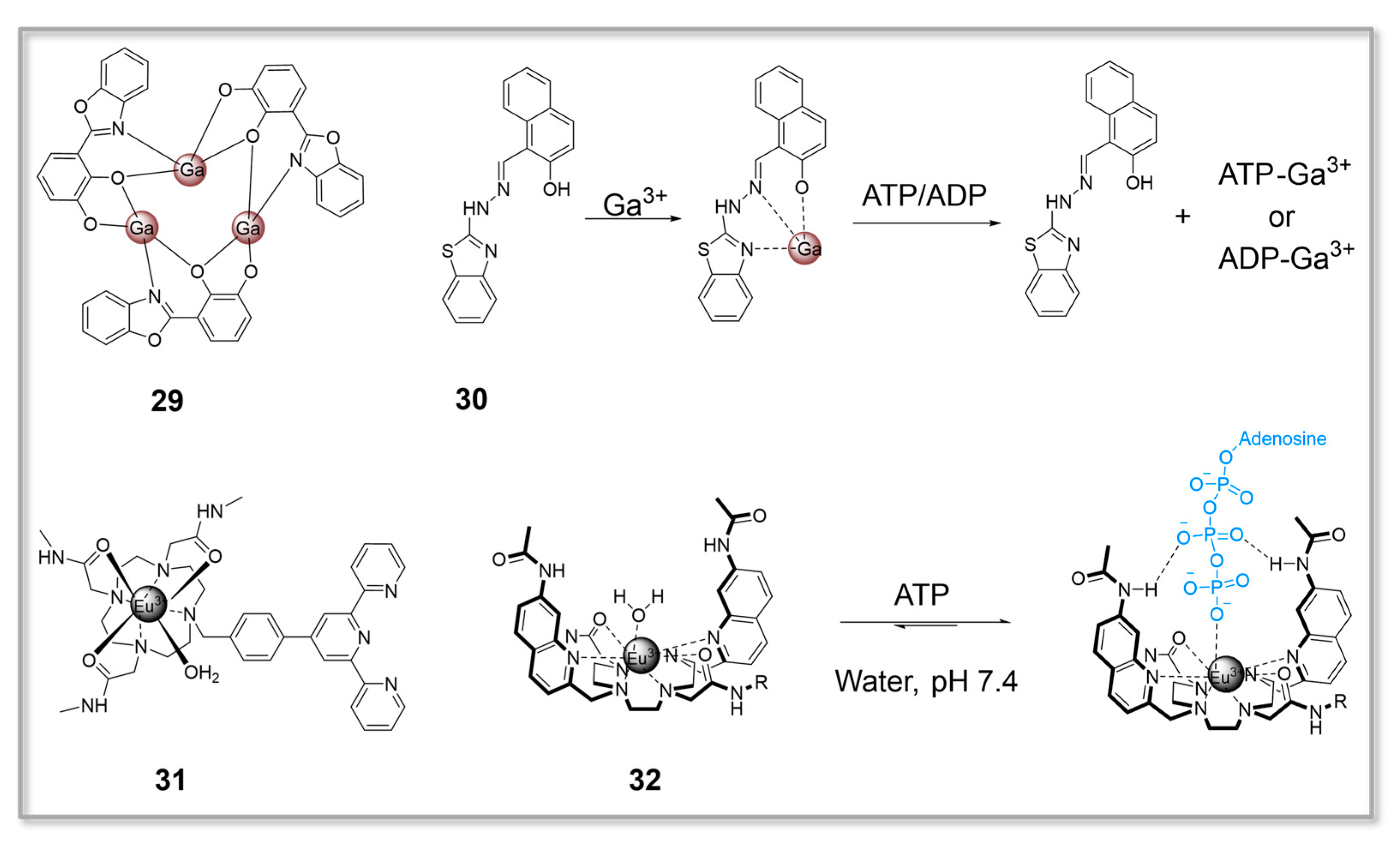
Figure 8.
ATP fluorescent probes 33–36 based on water-soluble conjugated polymers.

Figure 9.
(A) Chemical structure of probe 37. (B) Colocalization imaging of HeLa cells staining with probe 37 and MitoTracker Green. (a) The cells were stained with probe 37 (1 μM) in the red channel. (b) The cells were stained with MitoTracker Green (1 μM) in the green channel. (c) Merged image. (d) Bright-field image. (e) Intensity scatter plot of probe 37 and MitoTracker Green. (f) Fluorescence intensity profile of regions of interest (white line in parts (a,b). Scale bar, 5 μm. Reprinted with permission from [88]. Copyright 2017, American Chemical Society. (C) Mitochondria-targeting ATP fluorescent probes 38–39.
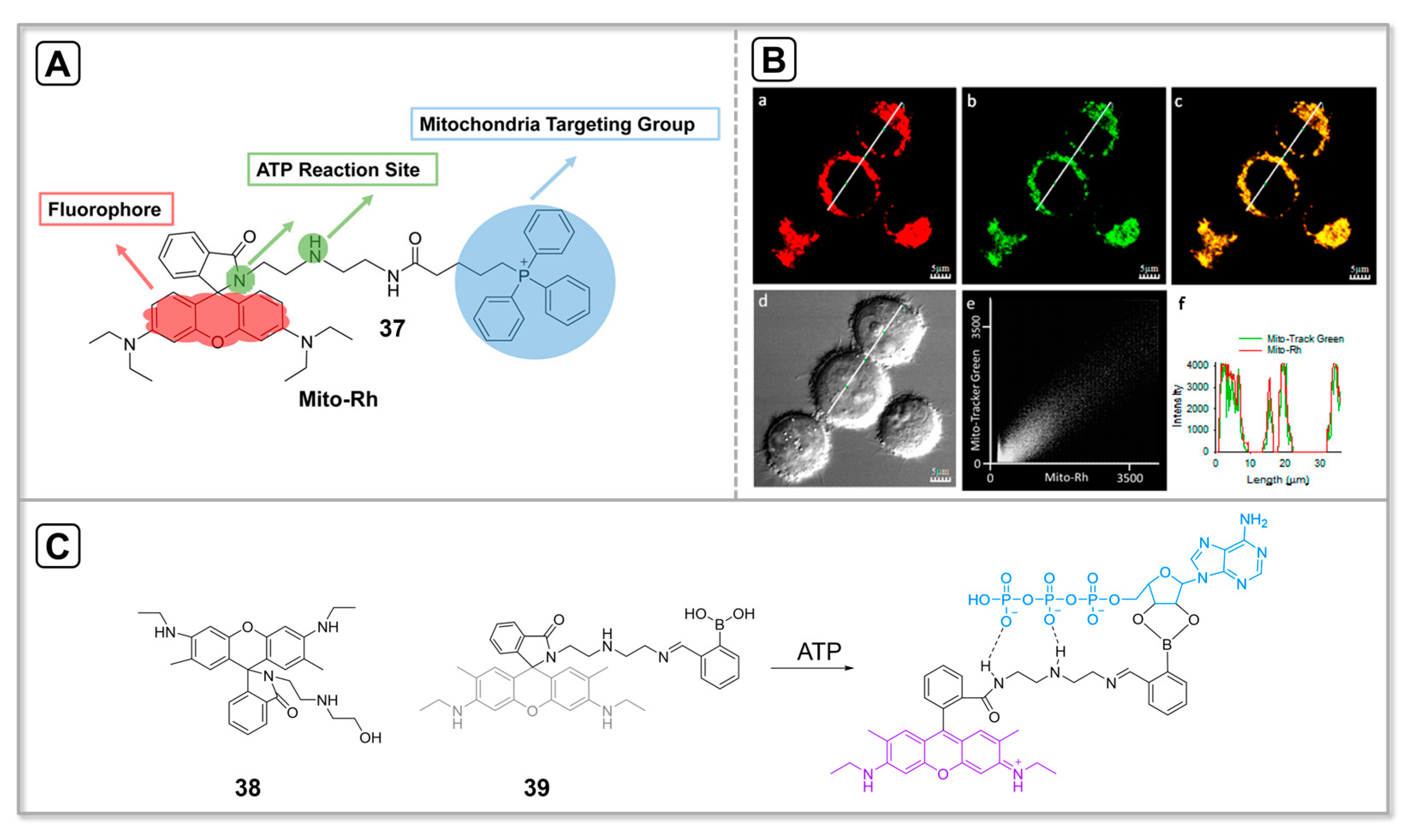
Figure 10.
(A) Lysosomes targeting ATP fluorescent probe 40. (B) HeLa cells were co-stained with 20 μM probe 40 and 200 nM Lysotracker Blue DND-22 for 30 min and fluorescence images were acquired by confocal microscopy. (a) Lysotracker Blue DND-22, (b) probe 40, (c) merged image, (d) DIC image, (e) co-localization analysis graph of Lysotracker and probe 40 (Ch 1: Lysotracker; Ch 2: probe 40), (f) Fluorescence intensity profile of ROI (region of interest) shown in (d) image: blue line, Lysotracker; red line, probe 40. Scale bar: 20 μm. Reprinted with permission from [92]. Copyright 2019, Elsevier.
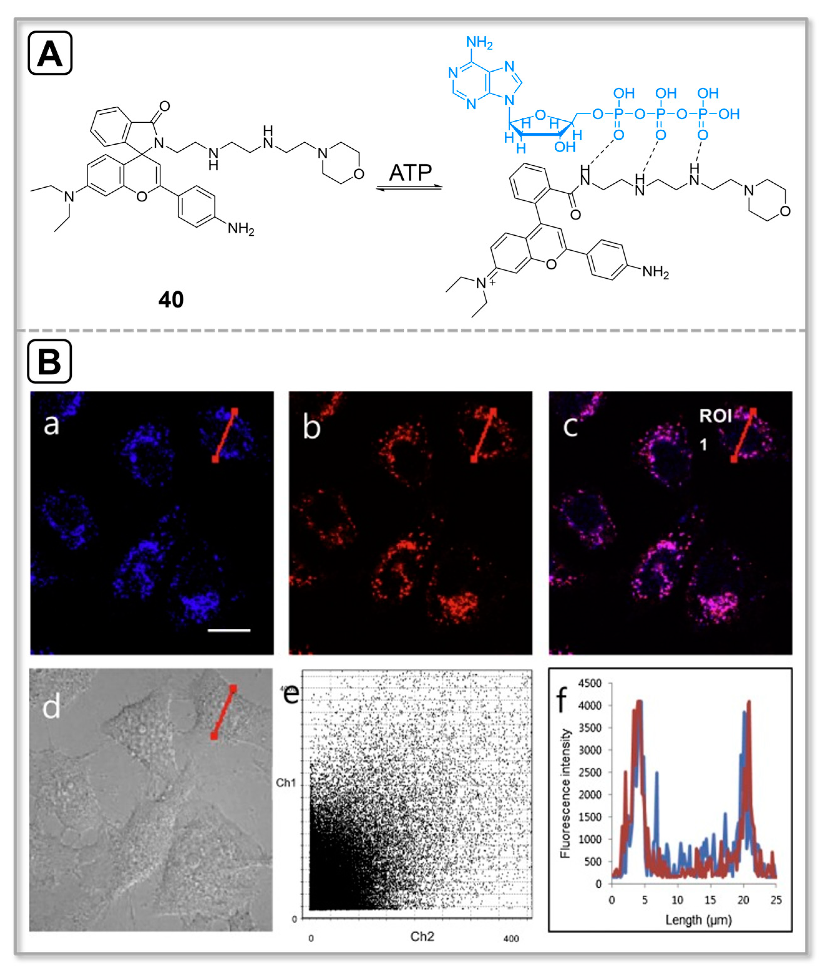
Figure 11.
(A) Nucleus-targeting ATP fluorescent probe 41. (B) Colocalization imaging of PC3 cells stained with probe 41 and Nuclear-ID Red. Reprinted with permission from [94]. Copyright 2023, Wiley-VCH.
Figure 11.
(A) Nucleus-targeting ATP fluorescent probe 41. (B) Colocalization imaging of PC3 cells stained with probe 41 and Nuclear-ID Red. Reprinted with permission from [94]. Copyright 2023, Wiley-VCH.
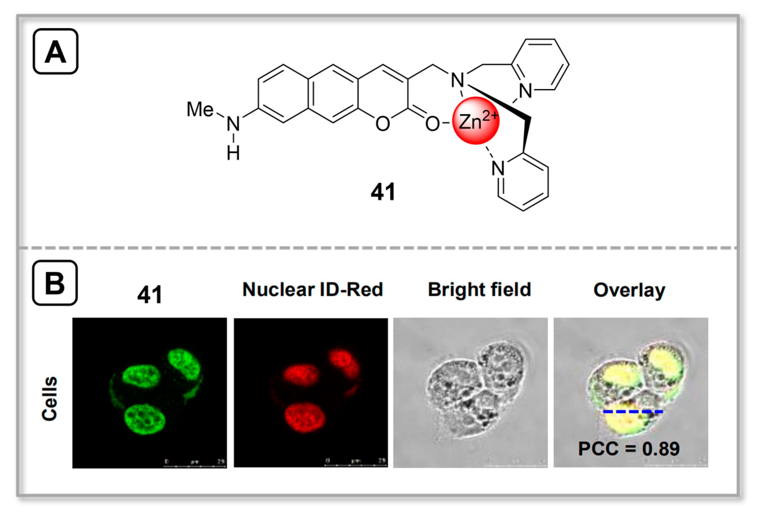
Figure 12.
(A) Structures of ATP fluorescent probes 42 and 43. (B) Confocal micrographs of HeLa cells stained with probes 42 and 43. Scale bars: 20 μm. Reprinted with permission from [96]. Copyright 2012, American Chemical Society.
Figure 12.
(A) Structures of ATP fluorescent probes 42 and 43. (B) Confocal micrographs of HeLa cells stained with probes 42 and 43. Scale bars: 20 μm. Reprinted with permission from [96]. Copyright 2012, American Chemical Society.
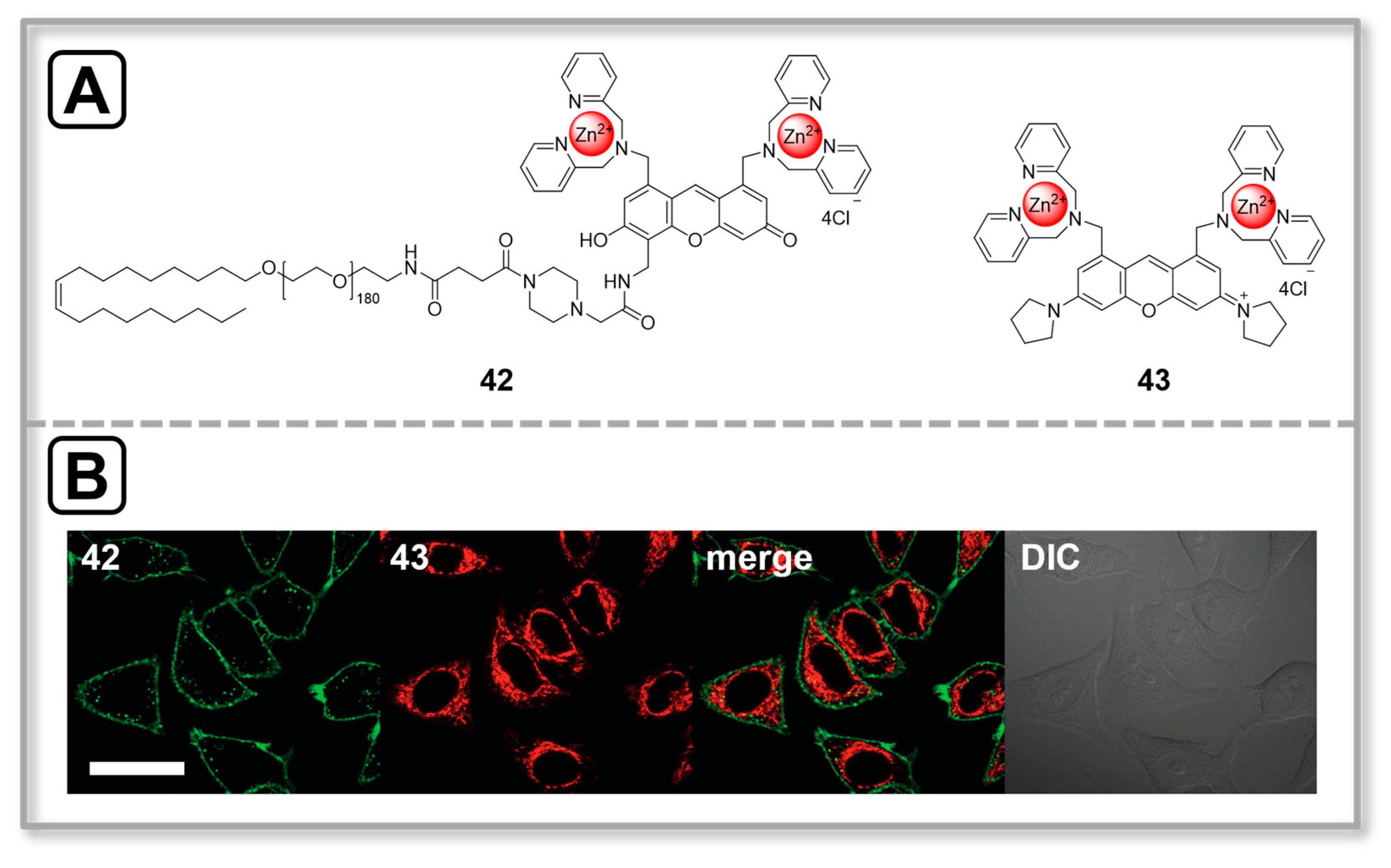
Figure 13.
(A) Mechanism of reaction between probe 44 and ATP. (B) Two-photon live-cell images of lysosomes that undergo (a) ‘kiss-and-run’ and (b) ‘full-collapse fusion’ processes, processed with pixel-to-pixel ratio images containing the quantitative information on ATP, and schematics of the two processes. HeLa cells incubated with probe 44 (10 μM) were used. The values indicate the emission intensity ratio values (IYellow/IBlue). Scale bar = 7.5 mm (a,b). Reprinted with permission from [99]. Copyright 2018, Wiley-VCH.
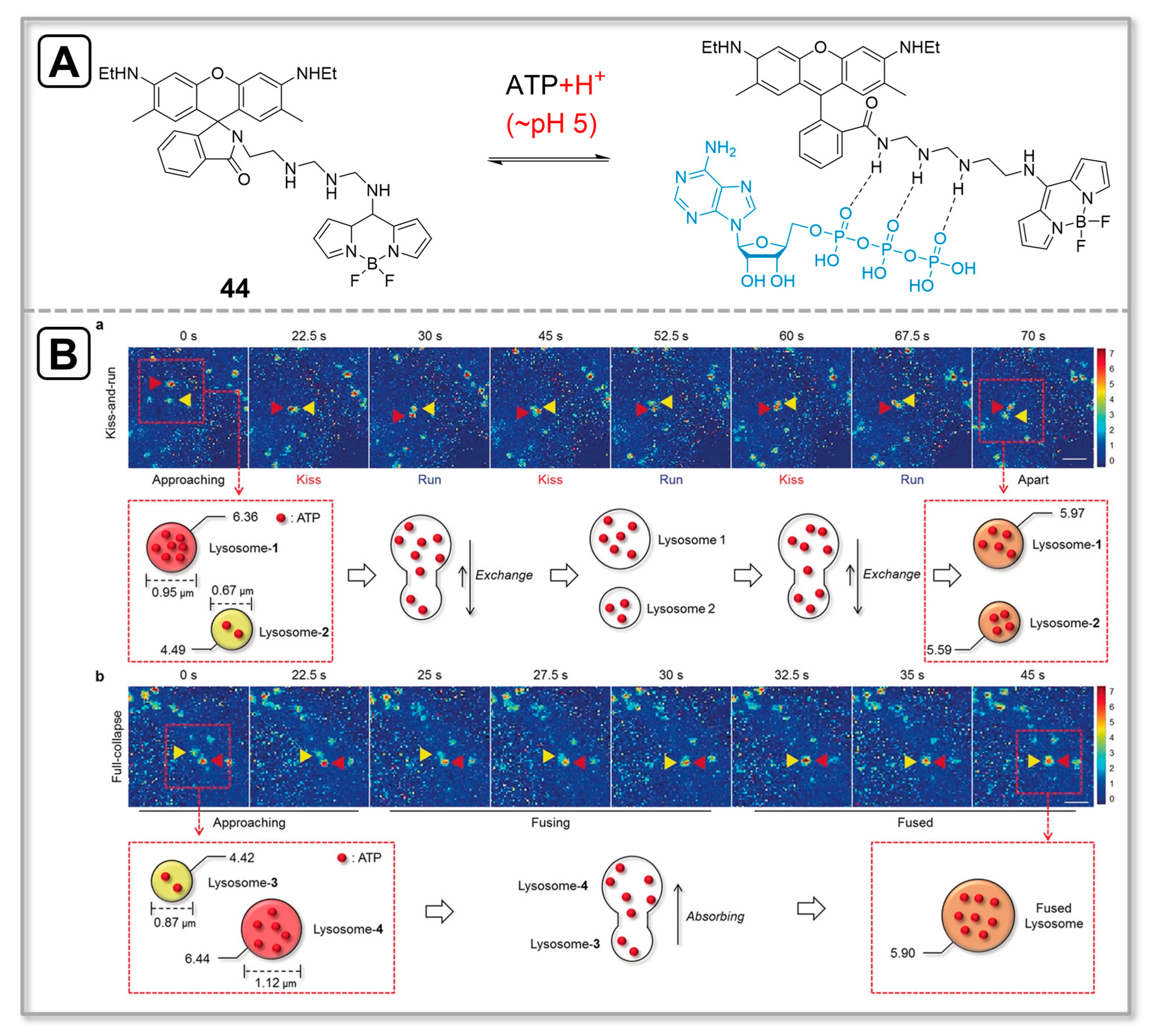
Figure 14.
(A) Structure of ATP fluorescent probe 45. (B) Confocal fluorescence images of HeLa cells under normoxic (20% O2) and different degrees of hypoxic conditions (10%, 5%, and 1%). Control (HeLa cells themselves); the cells were grown and then incubated with 10 mM probe 45 for 40 min at different O2 concentrations; green channel for NTR and red channel for ATP. Scale bar: 10 μm. Reprinted with permission from [102]. Copyright 2018, Royal Society of Chemistry.

Figure 15.
(A) The response mechanism of probe 46. (B) Fluorescence imaging and simultaneous quantification of mitochondrial H2O2 and ATP in response to exogenous O2•− stimulation. (a) Fluorescence lifetime images of neurons under O2•− stimulation. (b) Summarized data of mitochondrial levels of H2O2 and ATP in (a). (c) Fluorescence lifetime images of neurons after being treated with O2•− in the presence of DIDS or SOD. Scale bars: 10 μm for the whole images and 2 μm for ROI images. (d) Summarized data of H2O2 and ATP changes in (c). (e) Fluorescence lifetime images of neurons stimulated with O2•− for 8 min in the incubation of 46 (10 μM) at different times. Scale bar: 6 μm. (f) Fluorescence lifetime images of neurons stimulated with O2•− (100 μM) after 8, 20, 40, and 50 min. Scale bar: 6 μm. (g) Summarized data of mitochondrial H2O2 and ATP changes under O2•− stimulation at different times. The circled data represent the final levels in each stimulation, and the cyan and red dotted lines represent the initial levels of H2O2 and ATP, respectively. Error bars: SD, n = 8. Reprinted with permission from [107]. Copyright 2020, American Chemical Society.
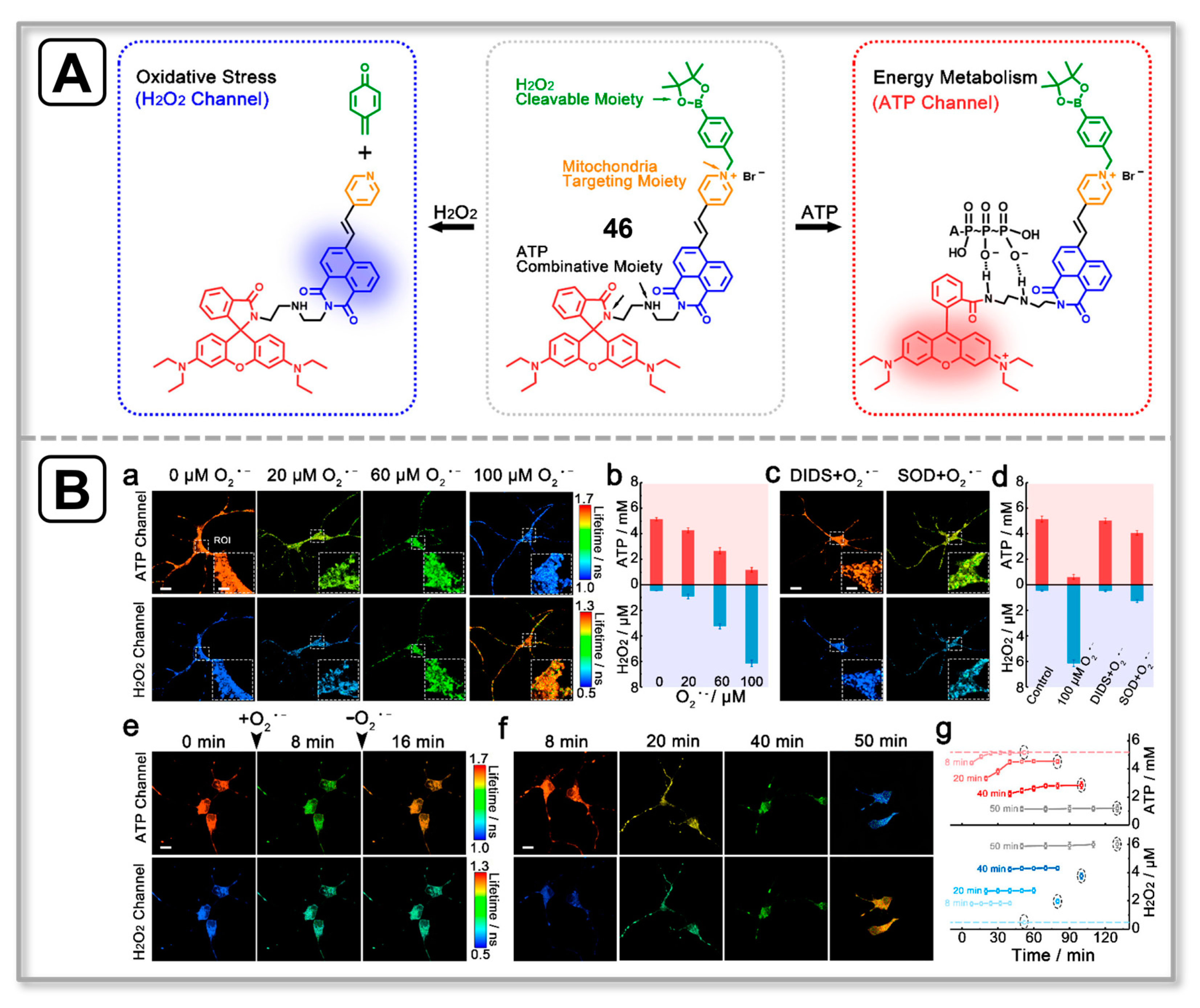
Figure 16.
(A) response mechanism of dual-analyte fluorescent probe 47. (B) Confocal images of APAP-induced liver injury and its remediation by NAC in HL-7702 cells. Green channel for ONOO− and red channel for ATP. Reprinted with permission from [111]. Copyright 2021, American Chemical Society.
Figure 16.
(A) response mechanism of dual-analyte fluorescent probe 47. (B) Confocal images of APAP-induced liver injury and its remediation by NAC in HL-7702 cells. Green channel for ONOO− and red channel for ATP. Reprinted with permission from [111]. Copyright 2021, American Chemical Society.

Figure 17.
(A) Chemical structures of probes 48 and 49. (B) Fluorescence imaging of tumor-bearing mice. The mice were given an intravenous injection of 48 or 49 at different times. Reprinted with permission from [113]. Copyright 2022, Elsevier.
Figure 17.
(A) Chemical structures of probes 48 and 49. (B) Fluorescence imaging of tumor-bearing mice. The mice were given an intravenous injection of 48 or 49 at different times. Reprinted with permission from [113]. Copyright 2022, Elsevier.


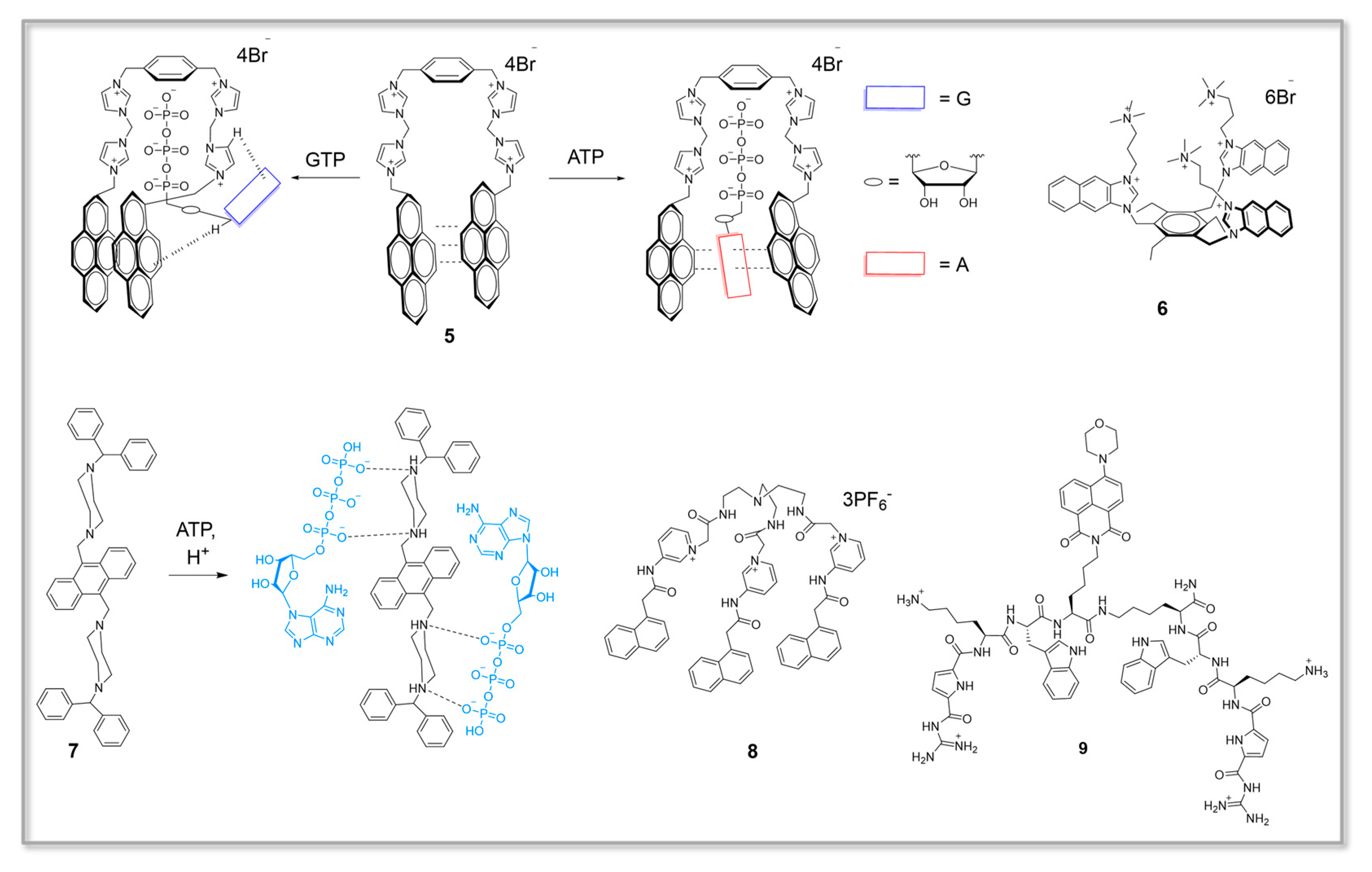
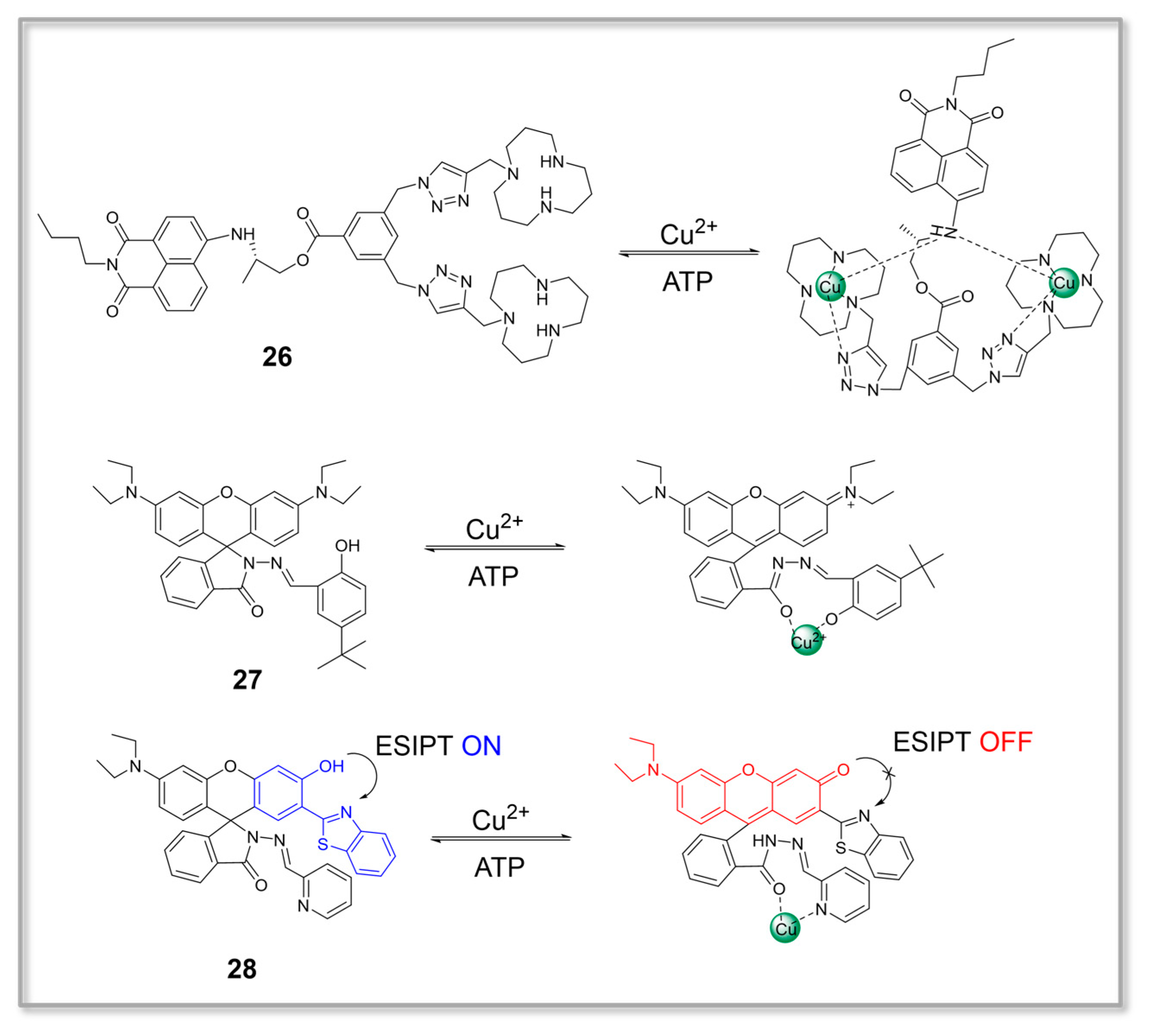
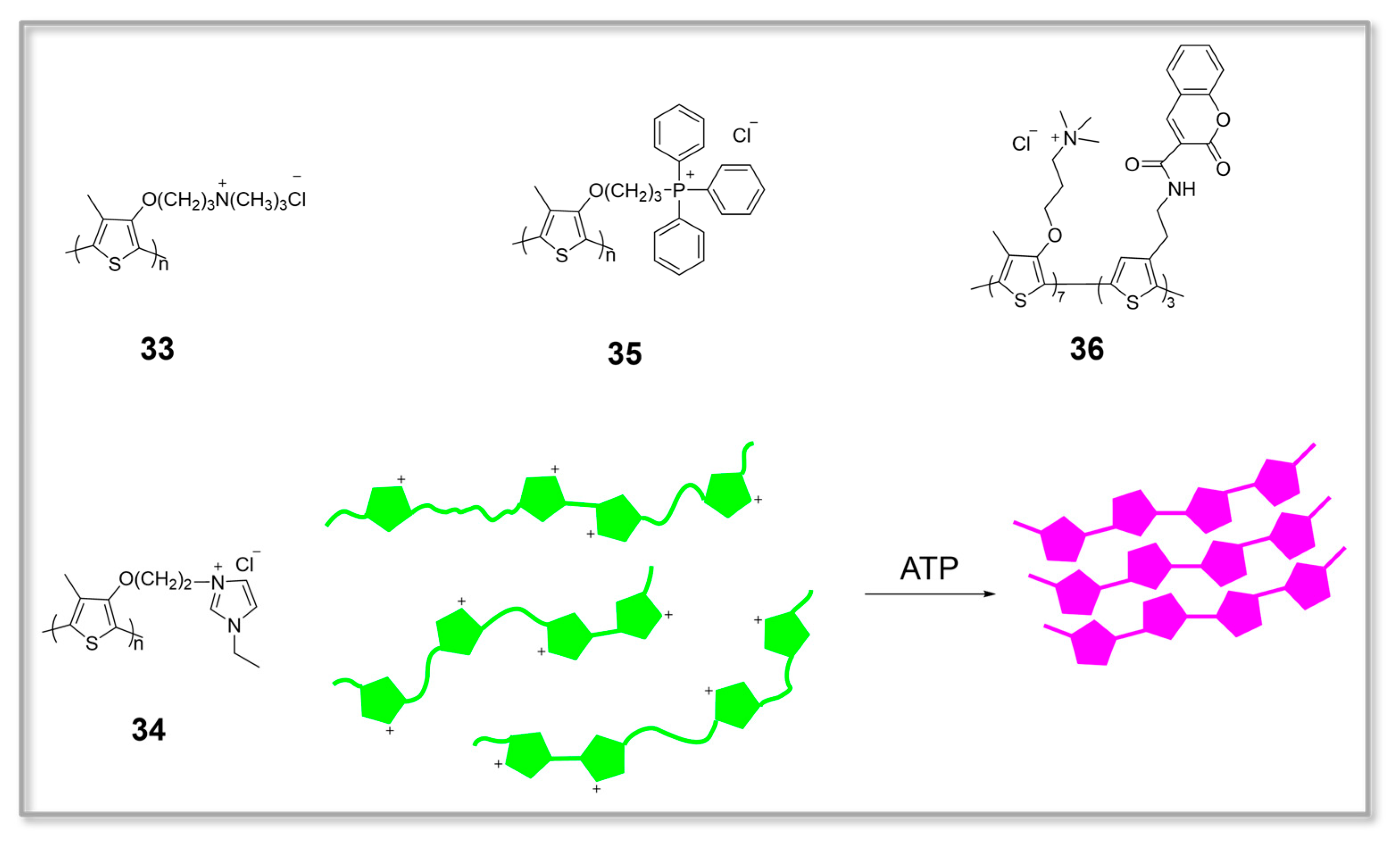
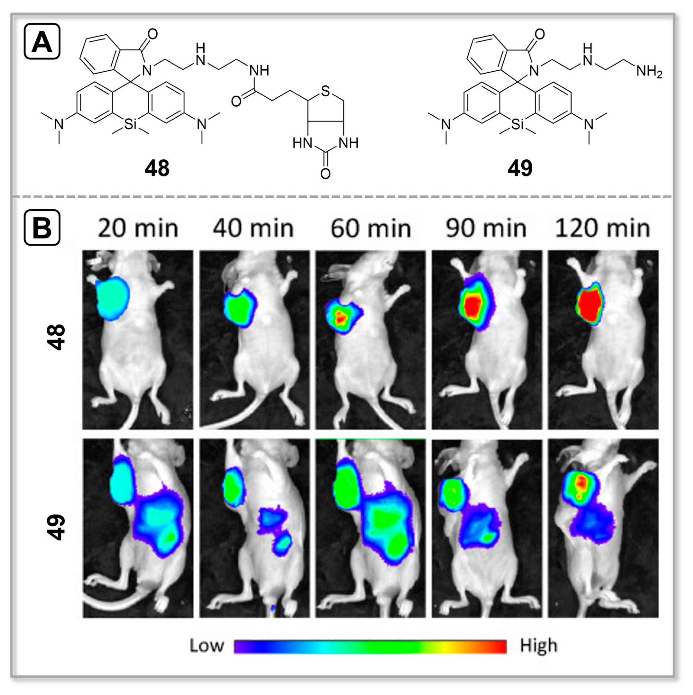
Table 1.
Summary of the ATP fluorescent probes based on organic small molecules in Section 2.1.
Table 1.
Summary of the ATP fluorescent probes based on organic small molecules in Section 2.1.
| Probe | Fluorophore | λex/λem (nm) | Detection Type | ATP Detection Limit | ATP Detection Range (mM) | Analyte | Ref. |
|---|---|---|---|---|---|---|---|
| 1 | Rhodamine | 520/583 | Fluorescence enhancement | 2.5 × 10⁻⁸ M | 1.0 × 10⁻⁴–2.0 × 10⁻¹ | ATP | [51] |
| 2 | Rhodamine, 1,8-naphthalimide | 420/530, 580 | Ratiometric | 1.0 × 10⁻⁴ M | 0–50 | ATP | [52] |
| 3 | Rhodamine | 500/560, 624 | Ratiometric | Not determined | 2.0–9.0 | ATP | [53] |
| 4 | Rhodamine | 660/700, 780 | Ratiometric | Not determined | 0–3.0 | ATP, H+ | [54] |
| 5 | Pyrene | 345/375, 487 | Ratiometric | Not determined | 0–2.0 × 10⁻¹ | ATP, GTP | [4] |
| 6 | Naphthoimidazolium | 326/~460 | Fluorescence enhancement | Not determined | 0–6.0 × 10⁻² | ATP, GTP | [55] |
| 7 | Piperazine–anthracene | 376/402, 422 | Fluorescence enhancement | 7 × 10⁻⁶ M | 0–1.0 × 10⁻¹ | H+, ATP | [56] |
| 8 | Naphthalene | 290/386 | Fluorescence enhancement | 8.44 × 10⁻⁶ M | 0–1.0 × 10⁻¹ | ATP | [57] |
| 9 | 1,8-naphthalimide | 410/540 | Fluorescence enhancement | Not determined | 0–1.4 × 10⁻² | ATP | [58] |
| 10 | Tetraphenylethene | 335/463 | Fluorescence enhancement | Not determined | 0–6.0 × 10⁻² | ATP | [59] |
| 11 | Thiazole orange | 520/580 | Fluorescence enhancement | Not determined | 0–8.0 × 10⁻² | ATP | [60] |
| 12 | Tetraphenylethylene | 365/500 | Fluorescence enhancement | 7.5 × 10⁻⁸ M | 1–4.0 × 10⁻³ | ATP | [61] |
| 13 | Triphenylamine-pyridinium | 427/613 | Fluorescence enhancement | Not determined | 0–3.0 × 10⁻¹ | ATP | [62] |
| 14 | Triphenylamine-pyridinium | 432/609 | Fluorescence enhancement | Not determined | 0–3.0 × 10⁻¹ | ATP | [62] |
| 15 | Triphenylamine-pyridinium | 437/614 | Fluorescence enhancement | Not determined | 0–3.0 × 10⁻¹ | ATP | [62] |
| 16 | Triphenylamine-pyridinium | 451/614 | Fluorescence enhancement | 0.3 ppm | 0–3.0 × 10⁻¹ | ATP | [62] |
Table 2.
Summary of the ATP fluorescent probes based on metal complexes in Section 2.2.
Table 2.
Summary of the ATP fluorescent probes based on metal complexes in Section 2.2.
| Probe | Fluorophore | λex/λem (nm) | Detection Type | ATP Detection Limit | ATP Detection Range (mM) | Analyte | Ref. |
|---|---|---|---|---|---|---|---|
| 17 | Anthracene | 380/460 | Fluorescence enhancement | Not determined | 0–1.8 × 10⁻² | ATP | [64] |
| 18 | Xanthene | 488/522 | Fluorescence enhancement | 1.0 × 10⁻⁶ M | 0–1.0 × 10⁻² | ATP | [65] |
| 19 | Xanthene, coumarin | 341/454, 525 | Ratiometric | Not determined | 0–5.0 × 10⁻³ | [ATP], [ATP] + [ADP] | [66] |
| 20 | Xanthene, coumarin | 414/477, 525 | Ratiometric | Not determined | 0–5.0 × 10⁻³ | [ATP], [ATP] + [ADP] | [66] |
| 21 | Perylene diimide | 500/559 | Fluorescence enhancement | Not determined | 0–1.0 × 10⁻¹ | ATP | [67] |
| 22 | Coumarin | 400/500 | Fluorescence enhancement | 8.4 × 10⁻⁹ M | 0–5.0 × 10⁻³ | ATP | [68] |
| 23 | Fluorescein | 420/527 | Fluorescence quenching | 5.0 × 10⁻⁷ M | 0–1.0 × 10⁻² | ATP, Zn+ | [69] |
| 24 | Pyrene | 346/383 | Fluorescence enhancement | Not determined | 1.0 × 10⁻¹ | Nucleotides | [70] |
| 25 | Pyrene | 350/390, 482 | Ratiometric | Not determined | 0–3.0 × 10⁻² | ATP, ADP, AMP | [71] |
| 26 | 1,8-naphthalimide | 451/541 | Fluorescence enhancement | 8.5 × 10⁻⁹ M | 0–1.0 × 10⁻¹ | ATP, Cu2+ | [73] |
| 27 | Rhodamine | 510/575 | Fluorescence quenching | 1.0 × 10⁻⁶ M | 0–1.0 × 10⁻² | ATP, Cu2+ | [77] |
| 28 | Benzothiazole, xanthenes | 380/434, 595 | Ratiometric | 8.0 × 10⁻⁸ M | 0–1.0 × 10⁻² | ATP, Cu2+ | [78] |
| 29 | 2-(2′,3′-dihydroxyphenyl)benzoxazole-Ga3+ complex | 360/461 | Fluorescence enhancement | 5.49 × 10⁻⁷ M | 1.0 × 10⁻³–1.0 × 10⁻² | ATP, Ga3+ | [79] |
| 30 | Naphthol-Ga3+ complex | 440/518 | Fluorescence quenching | Not determined | 0–2.75 × 10⁻² | Ga3+, ATP, ADP | [80] |
| 31 | Eu3+ | 335/591, 615 | Fluorescence enhancement | 8.87 × 10⁻⁶ M | 0–2.5 × 10⁻² | ATP | [81] |
| 32 | Eu3+ | 330/688, 697 | Fluorescence enhancement | Not determined | 0–3.0 | ATP | [82] |
Table 3.
Summary of the ATP fluorescent probes based on water-soluble conjugated polymers in Section 2.3.
Table 3.
Summary of the ATP fluorescent probes based on water-soluble conjugated polymers in Section 2.3.
| Probe | Fluorophore | λex/λem (nm) | Detection Type | ATP Detection Limit | ATP Detection Range (mM) | Analyte | Ref. |
|---|---|---|---|---|---|---|---|
| 33 | Polythiophene | 445/529 | Fluorescence quenching | 1.0 × 10⁻⁸ M | 0–5 × 10⁻¹ | ATP | [84] |
| 34 | Polythiophene | 410/520 | Fluorescence quenching | 3.6 × 10⁻¹¹ M | 0–7.5 × 10⁻⁴ | ATP | [85] |
| 35 | Polythiophene | 410/524 | Fluorescence quenching | 2.7 × 10⁻⁸ M | 0–2.5 × 10⁻³ | ATP | [86] |
| 36 | Polythiophene, coumarin | 300/410, 568 | Ratiometric | 2.9 × 10⁻⁸ M | 0–2.8 × 10⁻² | ATP | [87] |
Table 4.
Summary of the imaging applications of ATP fluorescent probes in Section 3.
Table 4.
Summary of the imaging applications of ATP fluorescent probes in Section 3.
| Probe | Fluorophore | λex/λem (nm) | Detection Type | Application | Targeting | Analyte | Ref. |
|---|---|---|---|---|---|---|---|
| 37 | Rhodamine | 520/583 | Fluorescence enhancement | Cell imaging | Mitochondria-targeting | ATP | [88] |
| 38 | Rhodamine | 527/557 | Fluorescence enhancement | Cell imaging | Mitochondria-targeting | ATP | [89] |
| 39 | Rhodamine | 510/558 | Fluorescence enhancement | Cell imaging | Mitochondria-targeting | ATP | [90] |
| 40 | Rhodamine | 480/634 | Fluorescence enhancement | Cell imaging | Lysosome-targeting | ATP | [92] |
| 41 | Benzocoumarin | 400/595 | Fluorescence enhancement | Cell imaging | Nucleus-targeting | ATP | [94] |
| 42 | Xanthene | 450/528 | Fluorescence enhancement | Cell imaging | Plasma-membrane-surface-targeting | ATP | [96] |
| 43 | Xanthene | 543/590 | Fluorescence enhancement | Cell imaging | Mitochondria-targeting | ATP | [96] |
| 44 | Rhodamine, BODIPY | 403/454, 557 | Ratiometric | Cell imaging | Lysosome-targeting | ATP, H+ | [99] |
| 45 | Rhodamine | 540/580 | Fluorescence enhancement | Cell imaging | NO | NTR, ATP | [102] |
| 46 | Rhodamine | 710/590 | Fluorescence enhancement | Cell imaging, Zebrafish imaging | Mitochondria-targeting | H2O2, ATP | [107] |
| 47 | Rhodamine | 520/587 | Fluorescence enhancement | Cell imaging | NO | ATP, ONOO− | [111] |
| 48 | Rhodamine | 640/675 | Fluorescence enhancement | Cell imaging, Mice imaging | Tumor-targeting | ATP | [113] |
| 49 | Rhodamine | Not determined | Fluorescence enhancement | Cell imaging, Mice imaging | NO | ATP | [113] |
Disclaimer/Publisher’s Note: The statements, opinions and data contained in all publications are solely those of the individual author(s) and contributor(s) and not of MDPI and/or the editor(s). MDPI and/or the editor(s) disclaim responsibility for any injury to people or property resulting from any ideas, methods, instructions or products referred to in the content. |
© 2023 by the authors. Licensee MDPI, Basel, Switzerland. This article is an open access article distributed under the terms and conditions of the Creative Commons Attribution (CC BY) license (https://creativecommons.org/licenses/by/4.0/).
Share and Cite
MDPI and ACS Style
Gu, Q.-S.; Li, T.; Liu, T.; Yu, G.; Mao, G.-J.; Xu, F.; Li, C.-Y. Recent Advances in Design Strategies and Imaging Applications of Fluorescent Probes for ATP. Chemosensors 2023, 11, 417. https://doi.org/10.3390/chemosensors11070417
AMA Style
Gu Q-S, Li T, Liu T, Yu G, Mao G-J, Xu F, Li C-Y. Recent Advances in Design Strategies and Imaging Applications of Fluorescent Probes for ATP. Chemosensors. 2023; 11(7):417. https://doi.org/10.3390/chemosensors11070417
Chicago/Turabian StyleGu, Qing-Song, Ting Li, Ting Liu, Guo Yu, Guo-Jiang Mao, Fen Xu, and Chun-Yan Li. 2023. "Recent Advances in Design Strategies and Imaging Applications of Fluorescent Probes for ATP" Chemosensors 11, no. 7: 417. https://doi.org/10.3390/chemosensors11070417
Note that from the first issue of 2016, this journal uses article numbers instead of page numbers. See further details here.