Review

Microdevices for Non-Invasive Detection of Bladder Cancer

1
Department of Bioengineering, Rice University, Houston, TX 77005, USA
2
BioSystems and Micromechanics (BioSyM) IRG, Singapore-MIT Alliance for Research and Technology (SMART) Centre, Singapore 138602, Singapore
3
Plug and Play Tech Center, Sunnyvale, CA 94043, USA
4
Department of Biochemistry Yong Loo Lin School of Medicine, National University of Singapore, Singapore 117597, Singapore
*
Author to whom correspondence should be addressed.
Chemosensors 2017, 5(4), 30; https://doi.org/10.3390/chemosensors5040030
Submission received: 27 September 2017 / Revised: 7 November 2017 / Accepted: 15 November 2017 / Published: 19 November 2017

Abstract

:
Bladder cancer holds the record for the highest lifetime cost on a per-patient basis. This is due to high recurrence rates, which necessitate invasive and costly long-term evaluation methods such as cystoscopy and imaging. Microfluidics is emerging as an important approach to contribute to initial diagnosis and follow-up, by enabling the precise manipulation of biological samples. Specifically, microdevices have been used for the isolation of cells or genetic material from blood samples, sparking significant interest as a versatile platform for non-invasive bladder cancer detection with voided urine. In this review, we revisit the methods of bladder cancer detection and describe various types of markers currently used for evaluation. We detail cutting-edge technologies and evaluate their merits in the detection, screening, and diagnosis of bladder cancer. Advantages of microscale devices over standard methods of detection, as well as their limitations, are provided. We conclude with a discussion of criteria for guiding microdevice development that could deepen our understanding of prognoses at the level of individual patients and the underlying biology of bladder cancer development. Collectively, the development and widespread application of improved microfluidic devices for bladder cancer could drive treatment breakthroughs and establish widespread, tangible outcomes on patients’ long-term survival.

1. Introduction

The human renal system is an exceptional example of biological structure and organization, efficiently filtering waste from the bloodstream while selectively retaining nutrients. However, the urothelium of the bladder is prone to transformation. Each year, over 425,000 people around the world develop urothelial bladder cancer, and more than 165,000 people die of this condition. The age-standardized rate of bladder cancer can differ by nearly ten-fold based on geography and is approximately three to five times higher in men than in women. Among the most common risk factors for future development and recurrence of bladder cancer are smoking [1], body mass [2], lack of physical activity [3], and age [4]. Furthermore, the mortality rate is highly dependent on stage at diagnosis. Patients diagnosed while cancerous cells are confined to the urothelium have a five-year relative survival rate of 95.7%, while patients who are diagnosed after metastasis have a five-year relative survival rate of just 5.0% [5].
Patients are typically diagnosed with urothelial bladder cancer following symptoms like haematuria [6], increased the urinary frequency or urgency, irritation during urination [7], and bone and/or flank pain. Patients with bladder cancer that has not spread beyond the urothelium typically undergo transurethral resection of the bladder tumor, while tumors that have spread into and beyond the muscle lining of the bladder usually require a combination of cystectomy, chemotherapy, and/or radiation therapy [8]. However, non-invasive tumors recur in roughly 70% of patients [9], necessitating long-term monitoring. A typical course of follow-up monitoring entails cystoscopy and cytology quarterly for the first two years following treatment, bi-annually for the next two years, and annually thereafter [10]. Such extensive monitoring contributes to bladder cancer’s status as the most expensive type of cancer on a per-patient basis [11,12]. The current standard of periodic cystoscopies increases the risk of urinary tract infections [13] and causes significant patient discomfort both during and after the procedure [14,15]. Likewise, the turnaround time for clinical cytology is on the order of 2–3 days, a lengthy wait for results [15].
Despite significant benefits that will arise from improving the standard of care for bladder cancer, no device for detecting bladder cancer based on biomarkers has yet achieved a recommendation for widespread clinical implementation [16,17]. The performance of existing tests varies with a number of factors, including hematuria [18,19], the specific clinician performing the assay [20], and the presence of other bladder conditions [21]. Furthermore, these tests tend to have lower sensitivity (proportion of positive test results in which a patient indeed has bladder cancer) with earlier stages of bladder cancer [22,23,24], limiting their ability to detect bladder cancer when treatment is most likely to have a positive outcome. As a result, currently available non-invasive methods are not necessarily more cost-effective than the prevailing standard of cystoscopy and cytology [25,26]. At present, no biomarker-based assay is recommended for standard clinical implementation, due to a lack of prognostic value and monitoring efficacy [16,17].
Given the shortcomings of current screening and diagnostic methods for bladder cancer, microfluidic devices hold promise for improved patient care and outcomes. In comparison to the current combination of invasive cystoscopy and slow cytology, robust devices for screening, diagnosis, and monitoring of bladder cancer will expand physicians’ ability to detect cases in early stages when survival rates are highest. The devices will also provide frequent updates on patients’ progress during treatment and beyond. In other areas of research, the development of microfluidic devices has vastly expanded the ability to probe microscale biological environments. By manipulating the small-scale flow and mixing of fluids, scientists are able to precisely perturb and monitor systems in a way not possible through standard laboratory bench techniques [27]. Areas as diverse as chemical synthesis [28], cell culture [29], and single-cell omics [30] have all benefited from microfluidics as a platform for parallelized processing, multiplexed assays, and high-throughput measurements. Specifically, in cancer research, microfluidic devices have been used for the isolation of circulating tumor cells from blood, including tumors originating in the lung [31,32], breast [33,34], and prostate [35,36].
Further strengths of microfluidic devices make them particularly well-suited to the screening and diagnosis of bladder cancer. Rather than probing individual or a select few bladder cancer biomarkers at a time, multiplexed microfluidic assays could increase the number of analytes being tested in a single protocol [37], contributing to a more sensitive and nuanced understanding of an individual patient’s case of bladder cancer. Similarly, the ability to process multiple patient samples in parallel would reduce the amount of time spent waiting on assay results [37,38] and support a faster feedback loop between patient symptoms and clinical treatment. Microfluidics’ ease of fabrication lends itself well to rapid prototyping and translation of design ideas into functional devices. Thus, the development of effective microfluidic screening and diagnostic methods for bladder cancer will likely lead to improved patient health outcomes, significant cost savings, and efficient use of clinical resources.
Here, we review recent developments in microfluidic devices that noninvasively detect clinically relevant markers of bladder cancer. We describe various approaches to the problem of early bladder cancer detection and evaluate their merits in the screening and diagnosis of bladder cancer. By compiling current methods and considering the elements necessary for the clinical implementation of a screening or diagnostic tool, we hope to provide insights into the factors most likely to lead to significant improvements in bladder cancer detection and treatment standards.

2. Standards of Detection and Evaluation

Currently, there are several options for bladder cancer detection (Table 1), of which urine cytology and cystoscopy are two of the most common clinical diagnostic approaches [39,40].

2.1. Invasive Techniques

The established guidelines for the diagnosis of non-muscle invasive bladder cancer involve cystoscopy, which allows the doctor to visualize the interior of the patient’s bladder using a cystoscope [55]. The procedure requires the insertion of a flexible cystoscope through the urethra under local anaesthesia. The cystoscope carrying a fiber optics cable attached to a CCD camera allows for a detailed inspection of the bladder wall. The detection of a tumor will require full anaesthesia for its resection necessitating a rigid cystoscope for the introduction of a surgical instrument to perform a partial or complete resection of the tumor. At first presentation, approximately 75% of cases are classified as non-muscle-invasive bladder cancer [56], which is associated with a five-year survival rate of 88–98% [57]. These tumors are classified as stage Ta if they have retained the basement membrane or as stage T1 if they have invaded the submucosal region called the chorion. However, these superficial tumors exhibit a recurrence rate as high as 50–70%, requiring routine monitoring for recurrence and progression [58]. Although cystoscopy can identify nearly all papillary and sessile lesions [59], it is an invasive and unpleasant procedure for patients and not suitable for the routine monitoring necessitated by bladder cancer [60,61]. A non-invasive assay would provide significant benefits for patient comfort, with urine as an ideal sample for bladder cancer diagnosis and follow-ups due to the ease of obtaining patient samples in a non-invasive manner [9]. Based on established guidelines, it is currently not recommended to test urinary biomarkers in place of cystoscopy [16,17,62].

2.2. Non-Invasive Techniques

Given the desirability of circumventing uncomfortable, invasive techniques and facilitating clinical decisions, research is ongoing into screening and diagnostic tools for bladder cancer based on molecular biomarkers. With a reliable and non-invasive test, clinicians could obtain meaningful information on patient cases of bladder cancer while reducing the frequency of cystoscopies.
Urine cytology is the standard non-invasive urine test that relies on visual inspection of abnormal cells in patient’s urine under a microscope [41,42]. However, it is limited by its low sensitivity, especially in the detection of low-stage, low-grade bladder cancer [40,63,64,65]. For such bladder tumors in early stages of development, the sensitivity of cytology is approximately 20% [66,67]. Furthermore, histological examinations have limited ability for prognostication as they do not reflect biological behaviour of the tumor, merely physical appearance [44]. High levels of inter-observer, intra-observer, and institutional variability also reduce the reliability of urine cytology results [40]. In addition, cytology requires highly trained personnel for sample evaluation, who may not be available in all areas [68].
Other non-invasive urine-based bladder cancer tests have been developed in the last few years, such as UroVysion fluorescence in situ hybridization (FISH) [50] and CxBladder Detect [49]. The UroVysion kit is one of the key Food and Drug Administration (FDA) -approved non-invasive devices commonly used for bladder cancer detection in actual clinical settings. CxBladder Detect is a highly sensitive multi gene urine biomarker test, targeting IGF, HOXA, MDK, CDC, and IL8R. Although both tests have much higher sensitivity than urine cytology, they are highly limited by their complexity and expense.
Despite current limitations, these non-invasive biomarker assays demonstrate increased detection sensitivity as compared to cytology and have the potential to reduce the frequency of follow-up cystoscopy (40.8% sensitivity for UroVysion with low-grade tumors [22], 69% for CxBladder Detect with low-grade tumors [69], and ~20% for cytology with low-grade tumors [66,67]). Indeed, recent reports suggest that the enhanced clinical utility of non-invasive assays such as CxBladder [49] has reduced urologists’ use of invasive diagnostic procedures.

3. Types of Urine Markers

3.1. Invasive Urine Markers

Potential biomarkers of excised cancer tissue are studied using immunohistochemical (IHC) staining of various proteins involved in cell cycle, apoptosis, and angiogenesis [44]. IHC of high-grade bladder cancer tissue may provide insight to predict progression of the disease. A combination of cell cycle proteins, p21, p53, pRB, and p27 stratified patients according to the risk of recurrence and progression. Proteins involved in apoptosis, such as Bcl-2 and caspase-3, also presented potential as survival predictors. Other studies also reported aberrant protein expression in bladder cancer tissue, such as the overexpression of SLD5 [45]. The expression levels of pAkt, PTEN, Drg-1, Cx-26, and L-plastin were often correlated with tumor grade and stage, as well as other clinicopathological features of bladder cancer. However, none of these biomarkers has been isolated as an independent predictive factor for progression-free or overall survival, while the sensitivity of immunohistochemical approaches will largely depend on the makeup of the genetic panel being assayed [46].
Recently, mRNA expression and IHC staining of tumor biopsies for the interactions of CXCL16 and CXCR6 indicated their upregulation in bladder cancer patients and correlations with cancer stage, supporting a role for CXCL16 and CXCR6 as potential therapeutic targets [47]. Upregulation of CXCR4 in invasive bladder cancer was also detected via fluorescent imaging and could likewise serve as a promising diagnostic tool [48]. The clinical significance of these markers lays in recurrence prediction upon invasive surgical methods such as transurethral resection of a bladder tumor (TURBT) and radical cystectomy (RC).

3.2. Non-Invasive Urine Markers

The ImmunoCyt non-invasive biomarker test is an assay for voided urine [70] that combines both cytology and fluorescent imaging. Three markers of the malignant urothelial cells, namely glycosylated carcinoembryonic antigen 19A211, LDQ10, and M344 are labelled with fluorescent antibodies. A sample is regarded as positive when fluorescence is observed. When used in conjunction with cytological analysis on low-grade tumors, ImmunoCyt provides a sensitivity between 80–90% and specificity (defined as the proportion of negative test results in which the patient does not have bladder cancer) between 60–70%. CxBladder kit detects the presence of five biomarker genes (IGF, HOXA, MDK, CDC, and IL8R), which are linked to elevated expression levels among bladder cancer patients; among patients with low-grade tumors, the sensitivity of the CxBladder kit was 69% [69].
Bladder Tumor Antigen (BTA) tests, such as BTA TRAK and BTA STAT, are enzyme immunoassays that detect the complement factor H-related protein (hCFHrp). BTA TRAK quantitatively measures the concentration of hCFHrp, while BTA STAT is a qualitative test; these tests have received FDA approval to complement cystoscopy. Both exhibits decreased sensitivity in patients with early stages of bladder cancer: for BTA TRAK, sensitivity decreased from 88% for high-grade tumors to 48% for low-grade, while corresponding values for BTA STAT were 89% and 55.5%. Specificities of the overall patient cohort for BTA TRAK and BTA STAT were 69% and 78.7%, respectively [71,72].
NMP22 Bladder Check Test is an FDA-approved test for the early diagnosis of bladder cancer. It tests for the nuclear matrix protein (NMP22) by using two monoclonal antibodies to target the nuclear mitotic apparatus in voided urine. In voided urine samples from bladder cancer patients, NMP22 levels are elevated by 25-fold as compared to samples from healthy controls [73]. In studies that assessed the clinical utility of NMP22 Bladder Check Test, the assay was found to have a sufficiently high sensitivity (79% for low-grade tumors) and specificity (100% for low-grade tumors) to be a possible substitute for urine cytology [74] but remained inferior to cystoscopy [75].
UroVysion is an in-situ hybridization assay that utilizes fluorescence to detect the deletion of 9p21 loci and amplification of chromosome 3, 7, and 17 in malignant urothelial cells. However, such chromosomal abnormalities observed using UroVysion were not restricted to urothelial carcinoma, for a significant possibility of false positive diagnosis [50]. In low-grade tumors, the sensitivity of UroVysion was 40.8%, while the specificity was 87.8% [22].
Such commercialized kits for urinary biomarkers have yet to establish themselves as capable of replacing cytology and cystoscopy. Motivated by the prospect of avoiding invasive cystoscopy methods and subjective cytological examinations, much research focuses on identifying novel biomarkers, such as DNA methylation, miRNA, and telomerase. New multiplexed marker panels are constantly being developed with high-resolution approaches to providing more robust solutions for effective bladder cancer detection. For example, a recent study reported a panel of five biomarkers in urine, namely Coronin-1A, Apolipoprotein A4, Semenogelin-2, Gamma synuclein, and DJ-1/PARK7 with high detection sensitivity and specificity (93.9% and 96.7%, respectively, for early-stage bladder cancer) [76]. The panel is also more specific than two prior FDA approved marker panels (NMP22 [74] and BTA [71,72]) for bladder cancer detection. These studies are currently undergoing trials for antibody validation.

4. Increasing the Detection Sensitivity of Non-Invasive Markers Using Microdevices

Microdevices’ potential for parallelized and multiplexed sample processing could improve throughput and reduce the time for diagnosis, underpinning exciting opportunities to promote medical intervention affordability and improve health outcomes (Table 2).

4.1. Cell-Based Detection

Besides molecular markers, cancer cells can also be detected for disease evaluation. The current non-invasive method to detect bladder cancer via cytological examination of urine is limited by its low sensitivity. The challenge lies in distinguishing cancer cells based on morphology in the presence of proteins, debris and other cells from the urinary tract. The utility of microdevices in cancer detection has been well-explored for the application of circulating tumor cells capture from liquid biopsies [82]. Besides cell size-based detection methods such as filtration [41,42], a wide variety of principles have been explored to detect bladder cancer based on urine.

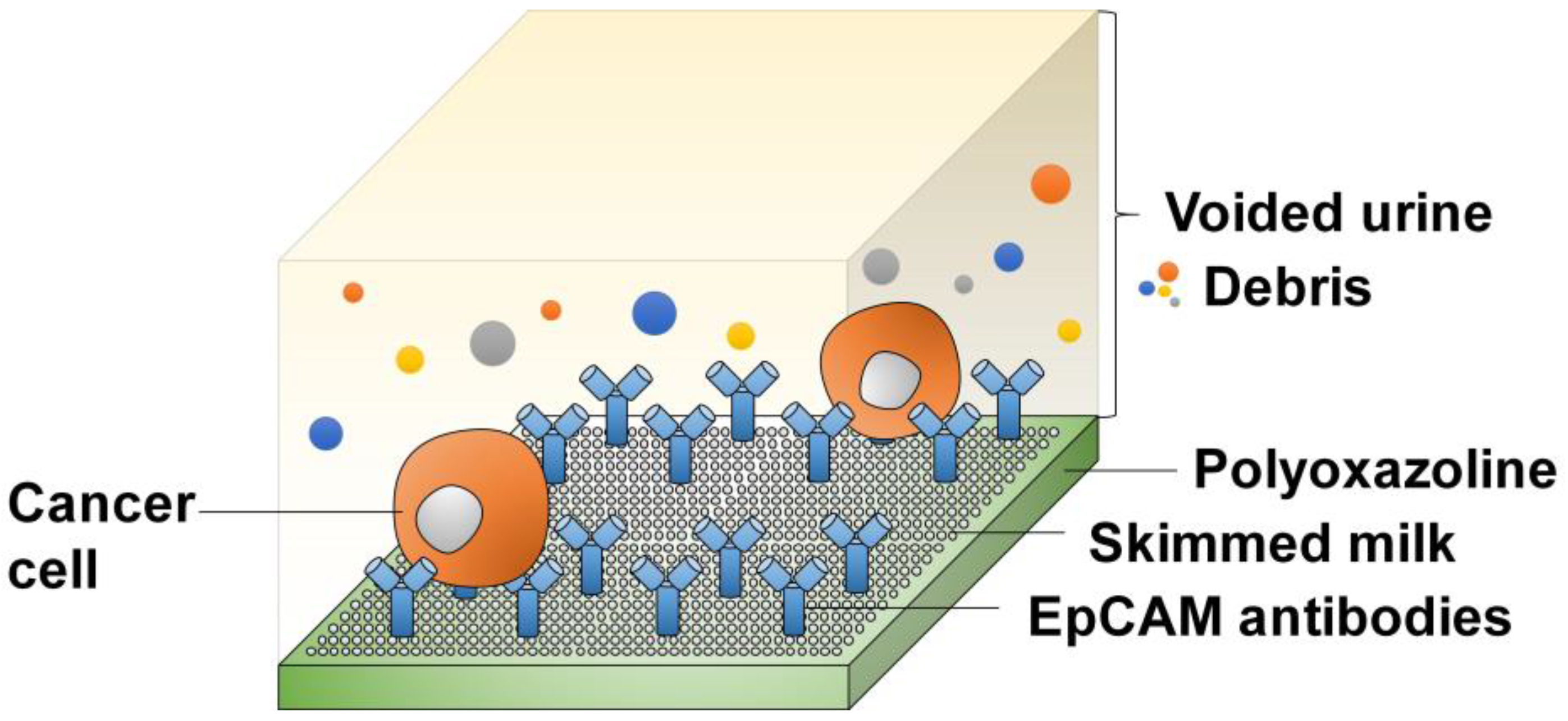
4.1.1. Affinity-Based Detection

To overcome the limitations of indistinct morphology and impure samples, a platform that selectively captures exfoliated bladder cancer cells from voided urine has been developed [80]. This device capitalized on the specific binding properties of antibodies to capture cancer cells for early detection of bladder cancer.
Antibodies against EpCAM were used to selectively bind cancer cells. EpCAM is an epithelial cell surface marker [83] shown to have prognostic significance in bladder cancer [84].
In this platform, EpCAM antibodies were immobilized on a biocompatible polyoxazoline plasma polymer by covalent bonds. Urine samples (Figure 1) were loaded into microchannels. EpCAM-positive bladder cancer cells, if present, can bind to the antibodies. Non-specific binding of debris and other cells were eliminated by blocking with skimmed milk mixture. This platform reports a specificity of 96%, sensitivity of at least 88% and enrichment (defined as the ratio of cells in the unprocessed sample over the ratio of cells after selective capture procedure) of more than 97%. There are ongoing clinical studies to further validate this platform on urinary samples from bladder cancer patients as an alternative to invasive cystoscopy and conventional cytology.

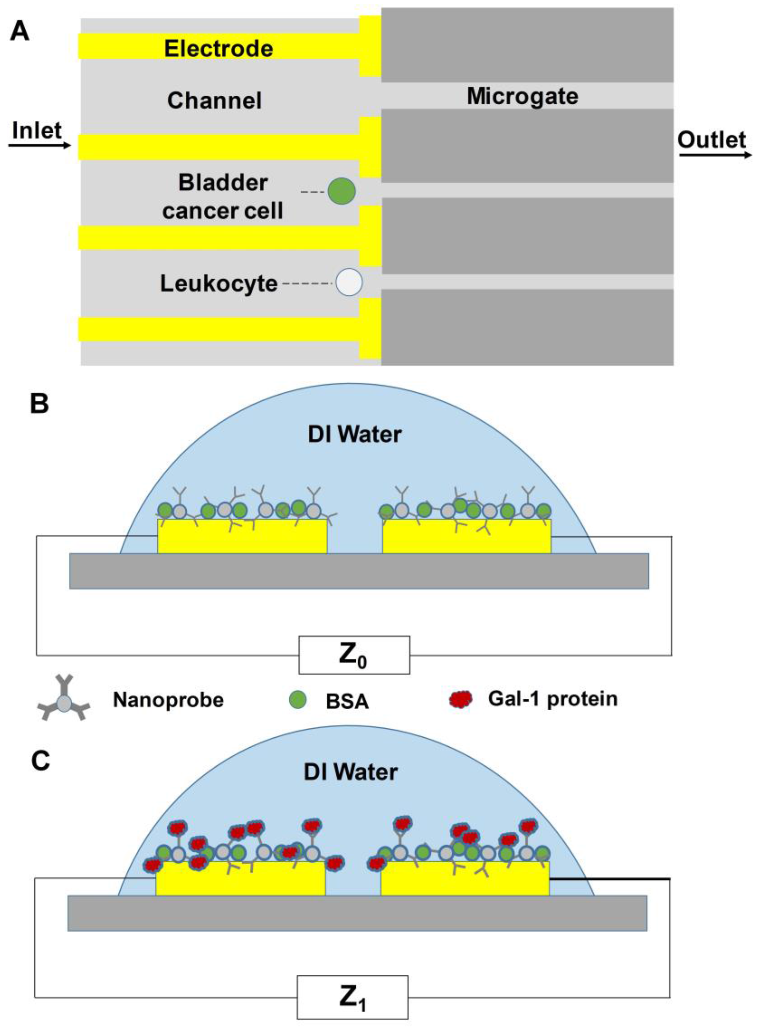
4.1.2. Detection by Membrane Capacitance

An integrated microfluidic device was developed for label-free quantification and isolation of bladder cancer cells from blood cells found in urine samples [63] based on membrane capacitance. When an analyte solution containing both bladder cancer cells (HTB-9TM human urinary bladder grade II carcinoma cell lines) and blood cells was passed through the microfluidic device, cells were captured by the silicon microchannels which served as the trapping gates (Figure 2A). Membrane capacitance of captured cells was measured using the gold electrodes patterned at the bottom of the microgates. Since bladder cancer cells and leukocytes have very different membrane capacitance, bladder cancer cells can be identified and detected through impedance measurements. Cancer cells were defined as cells which generated mean impedance changes in a channel that was higher than 60% after passing through the cell-containing solution. When the cellular response was between 20% and 35%, the trapped cell was a leukocyte. When the cellular response was below 5%, no cell was detected in the channel.
This approach is simple, non-invasive and label-free. With a flow rate of 5 mL/h, a 10 mL urine sample can be processed in 2 h, which is relatively fast as compared to other microdevices for biomarker-based bladder cancer detection. In addition, it could be used to stage bladder cancer by counting the number of bladder cancer cells present in the urine sample. However, multiple cells can be captured in a single channel greatly affecting accuracy. Lowering the flow rate could possibly mitigate this problem but could result in clogging instead. Another issue with this approach stems from the fact that trials were conducted on bladder cancer cells and blood cells in an analyte solution. However, patient urine samples also contain other urothelial cells, raising the question of determining an appropriate threshold to distinguish bladder cancer cells from healthy urothelial cells.

4.1.3. Impedance-Based Detection

An impedance-based immunosensor was designed and fabricated to quantitatively detect Galectin-1 (Gal-1) protein, a biomarker for bladder cancer [79]. To detect bladder cancer, alumina nanoparticles conjugated with the Gal-1 antibody (nanoprobes) were first trapped onto the gold microelectrode patterned on the immunosensor chip surface using programmable dielectrophoretic (DEP) manipulations. After blocking with bovine serum albumin (BSA) solution, the impedance before the immunoreaction was recorded (Z0) (Figure 2B). Then, the T24 bladder carcinoma cell spiked lysate was dropped onto the electrode for immunoreaction. This was again followed by a washing step and the impedance after immunoassay was recorded (Z1) (Figure 2C). Gal-1 protein was detected using electrochemical impedance analysis. Results showed that the normalized impedance variation increased with the concentration of T24 cell lysate.
This immunosensor chip offers several advantages as compared to traditional bladder cancer detection methods. The electrochemical detection relies only on the measurement of current or voltage to detect binding, making signal acquisition simple and label-free. With increased concentration of captured nanoprobes at the electrode surface due to DEP manipulations, the signal output is amplified for improved sensitivity. And the results showed that Grade III cell lysate had greater normalized impedance variation than Grade I cell lysate, indicating that this microdevice could be possibly used to find the grade of bladder cancer. However, this approach involves the synthesis of nanoprobes which is a complicated and time-consuming process that increases the cost and care necessary for use of this device.

4.2. Cell-Free Detection

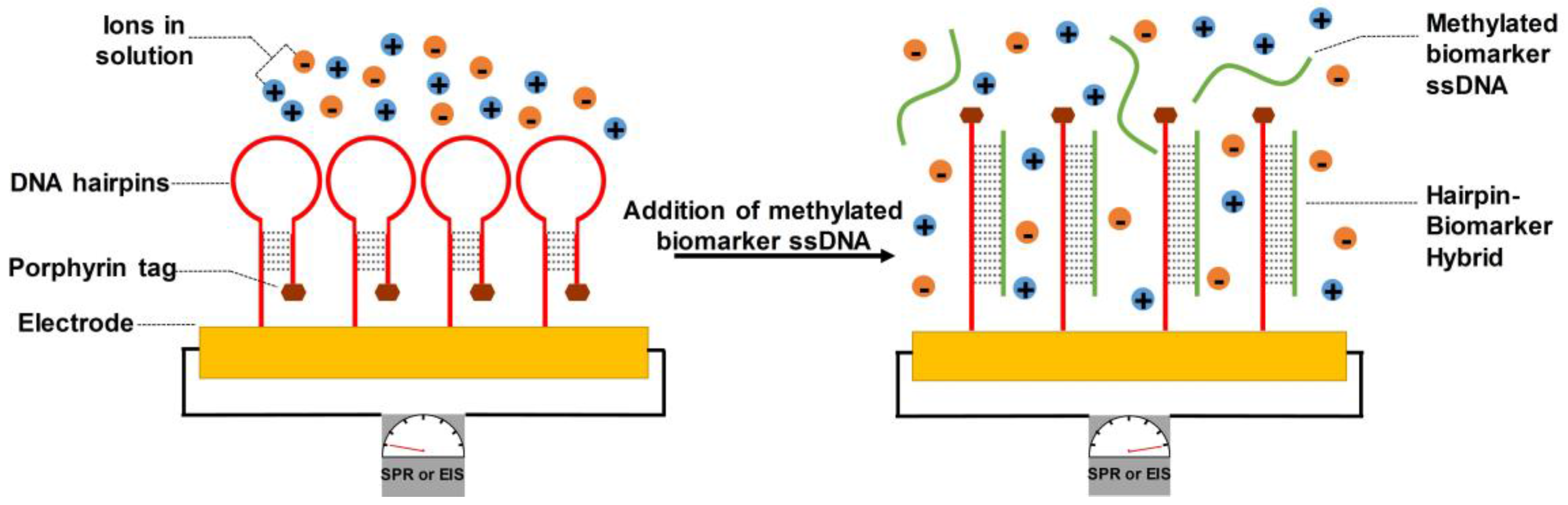
4.2.1. DNA-Bonded Substrates

In the transition from healthy to malignant tissue, tumor suppressor genes are frequently methylated and therefore silenced, as transcriptional machinery is less able to access the genes for normal regulation of cellular activity [85,86]. In addition to being present in the cell itself, methylated DNA can also be found as cell-free DNA in voided patient urine [87]. While methylation alterations can be detected by methylation-specific PCR [88], this technique may add lengthy waiting steps and can introduce artefacts through biased amplification [89]. In the context of bladder cancer, prior studies have used quantitative methylation-specific real-time PCR on patient urine samples to evaluate the degree of methylation of established marker genes. The reported sensitivities for low-grade tumor detection by methylation-specific PCR ranged from 33% to 85% [90,91], as compared to 40.8% for UroVysion, 69% for CxBladder Detect, and ~20% for cytology.
To implement assays targeting methylated DNA biomarkers of bladder cancer in the form of a microfluidic device, porphyrin-tagged DNA hairpin structures were bound to an electrode surface [78] (Figure 3). Separate hairpin loop sequences were designed to be complementary to each of three genes commonly methylated in bladder cancer patients—epithelial cadherin (CDH1), death-associated protein kinase (DAPK), and retinoic acid receptor beta (RARβ). Through incubation in a solution containing methylated biomarker DNA, the hairpin structures changed conformation to bind to the corresponding biomarker DNA, thereby causing an increase in the polarity of the porphyrin tag’s environment and in the distance from the porphyrin tag to the electrode surface. Both of these factors improved anions’ ability to reach the device electrode’s surface, contributing to a stronger electrochemical response following probe-biomarker hybridization [92] as measured by either surface plasmon resonance or electrochemical impedance spectroscopy [78]. To characterize and validate the DNA hairpin-electrode device, methylated target strands were synthesized to correspond to the sequences of CDH1, DAPK, and RARβ then spiked into a citrate buffer and a synthetic negative urine control; electrochemical responses were claimed to be identical with either solution. The device enabled measurable changes in electrode current in response to biomarker DNA concentrations as low as 2.5 × 10⁻¹⁰ M. Given that the concentration of cell-free DNA in the urine of bladder cancer patients is on the order of 10−9 M [93], these results support clinically relevant sensitivity. The device also retained performance through repeated cycles of target strand removal and addition. Furthermore, sequence specificity was validated by introducing four base pair mutations in the synthesized target strands and observing a fivefold reduction in electrochemical response. Crucially, all three target sequences could be detected simultaneously in a mixed solution, an important step toward sensitive and simultaneous targeting of multiple biomarkers.
As a next step, trials involving patient urine samples could be conducted using DNA hairpin-electrode devices. All presented data in the original study was taken in solutions of citrate buffer or synthetic negative urine control spiked with synthesized target strands, whereas clinical urine samples may vary in terms of pH, chemical component concentrations, contaminant cells, etc. Furthermore, cell-free DNA in urine exists as a mixture of many different oligo sequences and is typically double-stranded [94], driving the need for evaluating whether the urine of bladder cancer patients contains sufficient biomarker DNA that both matches the hairpin structures in the device and is single-stranded. Additionally, the device requires 20 min to process 5 μL of patient urine, limiting its applicability for applications requiring high-throughput processing of large sample volumes such as rare cell isolation.
The DNA hairpin-electrode device presents promise for the detection of single-stranded biomarkers of cancer. Multiplexed assays were demonstrated on three commonly methylated genes in bladder cancer; the presence of multiple electrodes on a single chip point toward parallel sample processing. Furthermore, the device could be targeted toward a wide range of DNA biomarkers by customizing the sequence of the hairpin structures bound to the device electrode, providing significant versatility as research into cancer biomarkers progresses. Thus, the DNA hairpin-electrode device provides a potentially versatile platform for the multiplexed capture of single-stranded DNA species present in solution.

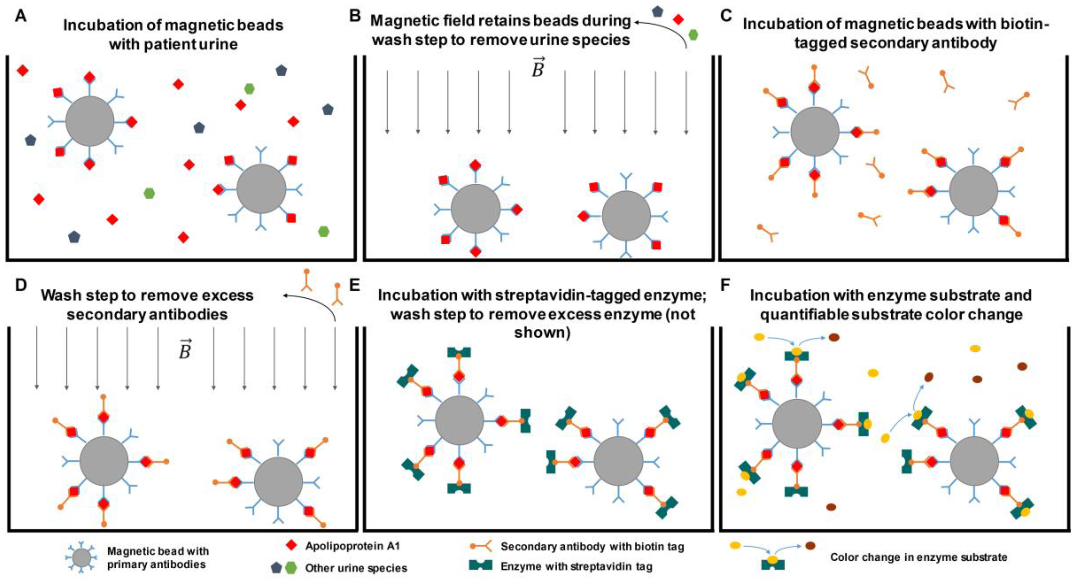
4.2.2. Antibody-Bonded Substrates

As an alternate direction to cell-free DNA as a biomarker for detection of bladder cancer, many assays and devices intended to screen cases of bladder cancer rely on the binding of antibodies to protein species present in patient urine [51,52,53,54,73,95,96]. Particularly common is plate-based ELISA, in which antibodies are coated onto wells of a plate and incubated with clinical liquid samples. As a variation on plate-based ELISA, microbeads offer improved assay sensitivity and simplification of microfluidic device design [97,98]. In a typical microbead immunoassay, primary antibodies are bound to the beads, providing a larger surface area-to-volume ratio for antigen capture as compared to the plate format of ELISA; this increased surface area-to-volume ratio improves sample mixing efficiency and antigen capture. Magnetic microbeads are particularly attractive candidates for biomarker studies, as they can be easily manipulated or trapped in device chambers through magnetic fields, reducing the need for special containment structures to avoid microbead losses during wash steps or buffer exchanges.
In 2013, a point-of-care device was presented for bladder cancer diagnosis based on magnetic microbead capture of apolipoprotein A1 (APOA1) [77], which has been demonstrated to be present at elevated concentrations in the urine of bladder cancer patients [99,100,101] (Figure 4). Magnetic microbeads bound to APOA1-targeted antibodies were incubated with a urine sample to allow binding of APOA1 to the microbead. Biotinylated secondary antibodies were then added, again targeting APOA1. Through an ELISA enzyme complexed with streptavidin, a biotin-streptavidin linkage occurred so that the enzyme was bound to the microbead if APOA1 was initially present. Finally, the enzyme’s substrate was introduced into the device chamber, causing a colour change detectable via measurements of optical density at 405 nm. The device exhibited a strong positive linear correlation between OD 405 measurements and the concentration of APOA1 in calibration solution, from 10 ng/mL to 2000 ng/mL. Given that an APOA1 concentration of 11.16 ng/mL has been proposed as a clinical cut-off for diagnosis [99], the device’s sensitivity shows promise for clinical relevance. At the upper end of the scale, patient urine APOA1 concentrations as high as 9000 ng/mL have been demonstrated, so that pre-dilutions may be necessary for accurate measurements of APOA1. However, the device retained linearity at APOA1 concentrations 100-fold higher than are possible with plate-based ELISAs (2000 ng/mL vs. 20 ng/mL, respectively), reducing the need for pre-dilution. In trials on the urine of bladder cancer patients (n = 4) and a hernia patient (non-cancer control, n = 1), measurements of APOA1 concentrations differed by less than 10% between ELISA and the APOA1-magnetic microbead device, across both patient samples and the healthy control.
However, the APOA1-magnetic microbead device presents areas that may be improved in future iterations. To mix samples during incubation and wash steps, a PDMS membrane oscillated under control of a vacuum; in the original study, a single device was used for each sample measurement to avoid membrane fatigue failure. The device’s reaction chamber for sample incubation and antibody binding accommodated 14.5 μL for a 40-min protocol, a small sample processing volume that limits extensions of the device to lower concentration biomarkers and isolation of rare cell populations. Furthermore, the device relied on antibody-based detection of biomarkers, bringing limitations due to the expense and sensitive reagents of ELISA. With the increased availability of urine samples from bladder cancer patients, the device’s performance could be evaluated with a sample size of patients larger than five individuals. If appropriately designed, such a study could also provide indications on correlations between cancer stage and APOA1 concentration, or on the prognostic value of the device’s APOA1 concentration measurements.
As compared to ELISA, the APOA1-magnetic microbead device offers significant improvements in throughput—40 min for the device vs. roughly 4 h for ELISA. Furthermore, by introducing mixtures of magnetic beads conjugated to different primary antibodies, the device presents a path to multiplexed assays that provide complementary sets of information from a single sample. In principle, the breadth of the device is limited only by the ability to produce and conjugate high-specificity primary antibodies, which would enable versatility in the detection of a variety of proteins and cellular epitopes. Overall, the APOA1-magnetic microbead device provides improved throughput and upper-end biomarker sensitivity compared to ELISA, with potential to multiplex assays for improved clinical insights.

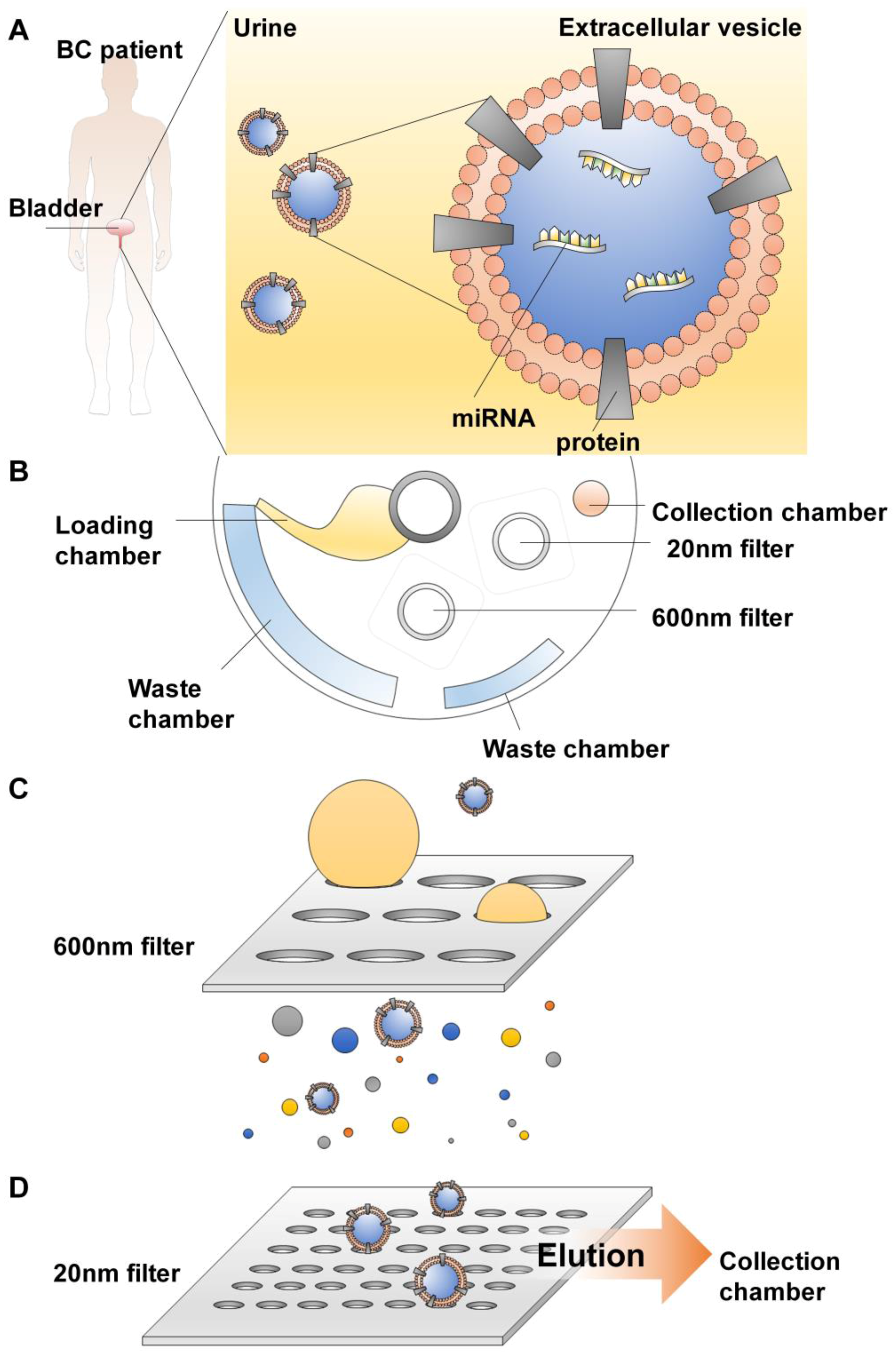
4.2.3. Extracellular Vesicles (EV) Isolation

Extracellular vesicles (EV) have emerged as a potential biomarker [102] due to their prevalence in urine and other bodily fluids, rendering them highly accessible by non-invasive liquid biopsy methods (Figure 5).
EVs are particles ranging from 30 nm to more than 5000 nm in size [103] that are secreted by cells into bodily fluids and facilitate intercellular signalling [104]. Surrounded by a lipid bilayer, EVs carry proteins, nucleic acids and lipids from their cells of origin (Figure 5), making their composition highly similar to that of the original cells, and therefore offering information about disease states. By containing a repertoire of intracellular genetic materials and proteins, EVs are a microcosm of the cancer cells from which they were released, rendering the EVs promising candidates as surrogate markers.
EVs have been known to play a role in cancer progression and metastasis via intercellular communication [105]. Indeed, the protein and miRNA composition of tumor-derived EVs found in urine have been observed to reflect the presence of cancer [43]. For example, differential protein and miRNA profiles were observed in patients with prostate cancer [105,106]. From 24 proteins known to be differentially expressed in bladder cancer patients, one (TACSTD2) was found to also be present at significantly elevated levels in EVs isolated from urine [107]. EDIL-3, another protein found to play a role in cell migration and tube formation and was present at elevated levels in bladder cancer patients [108]. Association of EDIL-3 expression in tumor tissues with EDIL-3 levels in urine exosomes posits urinary EVs as a source of biomarkers for bladder cancer detection.
However, current methods to isolate EVs present certain limitations. Various known methods of EV isolation can be laborious and time-consuming [43], with significant losses of EVs trapped in filter pores and loss of EV function upon elution [109]. The lack of standardized methods for the isolation of EVs also confounds reproducibility of results.
To circumvent these limitations of EV isolation, Exodisc has been recently developed for the isolation, enrichment, and quantification of EVs (Figure 5). The device was spun at low speed to sediment large debris. The resulting supernatant was passed through two nano filters—one filter to capture large particles and a second to enrich EVs according to their size. The first filter trapped particles larger than 600 nm, while the second filter trapped putative EVs between 20 and 600 nm. Large particles proceeded to the waste chamber while EVs were retained on the second filter. These enriched EVs were then cleansed with washing buffer and eluted into a collection chamber. Downstream protein detection was carried out by an on-disc ELISA. The Exodisc device has been demonstrated to isolate and purify EVs in 30 min, with >95% recovery of EVs and >100-fold higher concentrations of mRNA in the final sample as compared to previously-established ultracentrifugation methods for the isolation of EVs.
A key advantage of the Exodisc is the ability to directly conduct assays on raw patient urine with a timeframe of 30 min. In contrast, other EV isolation techniques require pre-processing steps (e.g., sucrose gradient, chemical sedimentation) that may add significant time (e.g., up to 12 h for chemical sedimentation) and equipment requirements (e.g., ultracentrifuges, reagents, etc.). Furthermore, the EV output of the Exodisc contains nucleic acids and proteins for subsequent omic characterization of individual patients’ cases (further discussed in “Future Perspectives” and “Concluding Remarks”). In principle, particles of other sizes can be captured through tuning and optimization of filter pore diameters, pointing toward more generalized applications for the Exodisc. However, on-chip ELISA was conducted through a custom-built system to measure the OD of the product of a horseradish peroxidase-mediated reaction. As a potential extension, it would be worthwhile to characterize the device’s compatibility with standard fluorescent-based ELISA approaches, which would enable increased sensitivity, multiplexed assays, and larger-scale implementation at other research laboratories and medical institutions. Additionally, Exodisc’s approach to EV isolation necessitates disposal and replacement of filters between trials in order to avoid inter-sample contamination. Ultimately, Exodisc presents a straightforward, robust method to isolate EVs containing rich information on the genetic, transcriptomic, and proteomic makeup of patients’ bladder cancer tumors.

5. Future Perspectives

Today, bladder cancer is one of the most common types of cancer, and demographic trends in common risk factors, such as age and smoking, point toward further increases in the number of cases worldwide. With the ageing of populations in developed nations [110,111], the proportion of individuals at increased risk of bladder cancer will also rise. Similarly, global trends in smoking hint at populations at risk of bladder cancer: the prevalence of smoking in Eastern Europe is high and stable, while in sub-Saharan Africa, the prevalence is projected to increase [112]. Given that bladder cancer treatment and long-term monitoring incur the highest costs of any cancer type on a per-patient basis, it is likely that bladder cancer will place larger and larger burdens on health care systems around the globe. To mitigate this scenario, effective detection methods for bladder cancer bear value, both for patient lives and for healthcare resources.
Future efforts should focus on formulating a set of criteria that an effective, non-invasive bladder cancer detection method should meet. As with any medical device, an ideal tool would be both low-cost and robust, such that the burden of bladder cancer is reduced without sacrificing medical insights. As promising devices progress from the lab bench to clinical trials, the metrics chosen to evaluate accuracy should cover (1) high specificity to exclude patients without bladder cancer and (2) high sensitivity to provide early detection. The aim of promoting early-stage detection would provide clinicians with the flexibility to select for cheaper and less damaging treatment options. Currently, existing monitoring methods are optimized for patients with advanced bladder cancer [22,23,113]. Given that the five-year survival rate of patients whose bladder cancer is confined to the urothelium at diagnosis is over 19 times higher than the rate for patients whose cancer has metastasized [5], screening and diagnostic devices with consistent performance across cancer stages would bring significant benefits to bladder cancer patient care.
Early detection of bladder cancer is crucial to improving patient outcomes [114]. Device performance in early-stage cases of bladder cancer could be feasibly improved through two complementary approaches: (1) development of assays that detect vanishingly small concentrations of materials linked to the presence of bladder cancer (e.g., biomarkers, exfoliated tumor cells, cell-free genetic material, etc.); and (2) development of assays targeting materials with high biological specificity to only individuals with bladder cancer (as compared to materials present in healthy individuals’ urine and exhibiting elevated concentrations in bladder cancer patients’ urine). The first approach may consist of iterative improvements upon currently-existing devices or the realization of novel techniques to assay known bladder cancer markers, while the second will likely require fundamental scientific inquiries to identify materials whose mere presence is specific and precise enough to indicate that a patient has bladder cancer.
The development of devices targeting materials with high biological specificity would provide significant benefits over currently available devices dependent on the appropriate selection of biomarker concentration cut-off values, as uniform application of cut-off values can lead to erroneous results in patients just below or above the cut-off [115,116]. For biomarkers that are typically present in the urine of healthy individuals and have elevated levels in bladder cancer patients, somewhat arbitrary choices of cut-off concentrations can lead to stark differences in medical decision-making. But on the other hand, allowing flexibility in the selection of cut-off values to account for different patient populations and medical practitioners inhibits the ability to accurately compare research results and evaluate device efficacy [117,118]. Variations in cut-off concentrations may drive potentially conflicting conclusions on the ability of a given device to accurately identify individuals with bladder cancer. Given these issues with determining appropriate concentration cutoffs, which affect all devices targeting biomarkers that are present in normal urine but have elevated levels in that of bladder cancer patients, the identification of biomarkers produced only by cancerous cells and not by other healthy cells of the bladder could strengthen medical decision-making through a more nearly binary classification of patients into healthy or cancerous. Through research into assays targeting biomarkers with high biological specificity to bladder cancer, researchers can mitigate the effects of technical noise and assay decision criteria for improved clinical insights and diagnoses.
Additionally, an emphasis should be placed on the development of screening and diagnostic methods amenable to clinical implementation. For instance, many currently available ELISA-based assays have temperature-dependent antibody behaviour and require expensive plate readers [119], factors that contribute to the recommendation against their general use in population-wide screening. In contrast, wearable sensors and phone-based detection exemplify the link between ease of implementation and breadth of use. By enabling doctors to take unobtrusive, straightforward measurements of patient physiology, such technologies have brought medical monitoring into patients’ daily lives for long-term, continuous observation of individual medical cases. Commercially available wearable sensors provide near-instant readouts on parameters such as heart rate, temperature, and oxygen saturation in a user-friendly manner [120]; even more directly relevant to bladder cancer microfluidic devices are phone-based sensors with functionalities such as calculating quantitative ELISA assay results [121] and detecting point mutations in tumor samples [122]. At a deeper level, wearable and phone-based sensors point to further guiding principles in the development of microfluidic devices to assay bladder cancer: the importance of designing devices that are compatible with hospitals’ pre-existing equipment and with patients’ daily regimens. In addition to strong screening and/or detection performance, a microfluidic device’s long-term impact on bladder cancer care will be shaped by its ability to seamlessly integrate into practitioners’ diagnostic and treatment routines.
While detecting exfoliated bladder cancer cells in voided urine provides a pain-free approach, sensitivity of cell-based tests would be limited by the number of exfoliated tumor cells. Since low-grade tumors shed few cells [113] and the presence of any circulating tumor cells confers poor prognosis [123], low numbers of exfoliated cells could limit the utility of cell-based tools for early detection. Thus, non-cell based biomarkers such as extracellular vesicles, an agent of cellular communication for budding tumors within the tumor microenvironment [124], may be a promising alternative for detection at earlier stages. Given the newfound recognition of extracellular vesicles as a source of biomarkers, methods to isolate them are varied, including size filtration, antibody capture, and precipitation [125]. However, standardization of isolation techniques must be addressed before clinical utilization, ideally achieving a balance between precision, cost, and duration of tests. Robust developments in this area would first require a unified goal of standardized EV isolation techniques. Given this need for robust EV isolation methods, the clinical utility of EV isolation remains an area of particular opportunity for future research.
In the future, screening and diagnostic tools that function without specialized equipment would provide a path toward broad acceptance, while high-throughput tools would enable rapid evaluation of cancer cases and increase the number of patients able to receive care. Furthermore, bladder cancer detection tools with simple, straightforward designs would reduce training requirements and inter-practitioner variability, thereby decreasing healthcare costs and ensuring reliability in bladder cancer screening and diagnosis. Non-invasiveness of the detection method is another area that should be taken into consideration. Invasive methods such as cystoscopy cause discomfort and pain to patients, limiting their application in early screening and routine monitoring of bladder cancer.
For us to have a more comprehensive and patient-friendly standard of care, non-invasive diagnostic methods are in great need. With a bladder cancer detection tool that provides the above traits, medical practitioners could deliver point-of-care, real-time assessments of a patient’s bladder cancer status. Conscious consideration of these features will be key to achieve sweeping improvements to the standard of care for bladder cancer patients through effective microfluidic screening and diagnostic devices.

6. Concluding Remarks

Considering the future, one can envision an even larger role for microfluidic devices in the treatment of bladder cancer. At present, research into novel cancer detection methods is ongoing in hopes of developing an alternative that surpasses invasive cystoscopy. Current FDA-approved diagnostic tests are still used in conjunction with, but never in place of, standard diagnostic tests. Building on the current standards of screening and diagnostics, non-invasive microfluidic devices could provide a platform for uncovering rich information on the diversity of individual patients’ cases. Many biomarkers of bladder cancer are present in the urine of patients, which enables the collection of large volumes of patient samples with minimal patient disturbance, particularly as compared to the volume limitations and pain of drawing blood that occurs for investigations on circulating tumor cells of other types of cancer. It is likely that microfluidic devices will have a largely complementary role to established standards such as cystoscopy and cytology (e.g., confirming the results from established standards or suggesting a need for further tests during long-term monitoring). To transition from promising results to widespread clinical implementation, early adoption of microfluidic devices will depend on partnerships between researchers and medical practitioners committed to bringing the promise of microfluidic devices to fruition. Over time, as such partnerships demonstrate the accuracy, throughput, and ease-of-use of particularly successful microfluidic devices, increasing numbers of practitioners will opt to incorporate applicable devices in their diagnosis process, as part of a self-reinforcing cycle.
Key capabilities of microdevices will provide a strong foundation for personalized care and mechanistic frameworks at the level of both individual cases and patient populations over time. The ease of obtaining patient urine samples facilitates testing and development of novel devices, but also provides ample material to detect and isolate materials indicating the presence of cancerous cells. While some screening and diagnostic devices target biomolecules or proteins, others isolate exfoliated tumor cells [80] and nucleic acid-containing vesicles [81]; this genetic material could provide a starting point for downstream genetic analyses to inform treatment decisions and provide insights into bladder cancer development and variants without requiring expensive and invasive biopsy procedures. In studies on lung [126] and breast cancer [127], microfluidic devices for mimicking tumor microenvironments provide a path toward tailored treatment approaches, while single-cell ‘omics’ analysis yields invaluable insight into the heterogeneity and molecular processes of cancer [128]. This is especially important in addressing inter-patient tumor heterogeneity [129]. For instance, by applying techniques of single-cell transcriptomics to patients’ exfoliated tumor cells, researchers could capture and sequence cellular mRNA to quantify transcription levels of individual cancer cells, enabling analysis of correlated genes, development of networks of gene co-regulation, and identification of distinct cell types with unique genetic signatures [130,131,132]. In the clinic, such fine-level knowledge of cancer cells could point toward the exact factors of dysregulation driving the development of a patient’s tumor and underpin the development of testable hypotheses for druggable mutations and effective treatment methods.
In particular, the ability to computationally cluster data from related cell populations could provide an in silico method to isolate data from rare exfoliated tumor cells (as compared to contaminant urothelial cells) [133,134,135] and could also indicate distinct subpopulations of cancerous cells that would likely benefit from treatment with a coordinated, concerted panel of drugs informed by knowledge of the characteristics of individual cells [136,137,138]. More broadly, application of single-cell transcriptomics to exfoliated bladder cancer cells from a large population of patients would provide a reference transcriptomic dataset of bladder cancer variants, useful for placing patients within a broader context of prior knowledge and for predicting efficacy of potential treatment paths based on historical data [139]. Likewise, techniques for investigating DNA methylation and changes in chromatin accessibility could provide mechanistic insight into the genomic changes that drive alterations in gene expression, cellular physiology, and progression to a cancerous state [140,141]. As a complementary direction to that of single-cell omics, clinicians could identify which drugs are most likely to be effective for a given patient through isolation, ex vivo expansion, and drug screens on a patient’s own circulating tumor cells. Such an approach has yielded prognostic information on the responses of breast cancer patients to medical treatment [142], and microfluidic devices that isolate exfoliated tumor cells could bring the power of personalized medicine to bladder cancer treatment.
Rather than being limited to monitoring bladder cancer through cell counts or biomarkers, clinicians could understand changes in genetic circuits at the scale of individual patients and identify druggable mutations for improved patient outcomes based on microfluidic devices that isolate genetic material from bladder cancer cells. Thus, the development of such microfluidic devices would provide synergistic effects on patient treatment by detecting the presence of bladder cancer and informing the selection of effective treatment options. A non-invasive method to obtain primary source human bladder cancer cells would enable the evaluation of medical treatments directly on samples from patients themselves, providing a robust test system with direct relevance to today’s bladder cancer patients and accelerating the implementation of research developments in clinical settings. Conversely, widespread clinical implementation of high-throughput assays on cancer cells isolated from patients would also bear impact on research directions, as the growth and proliferation of data on patients’ cancer cell genomics and treatment outcomes would inform our understanding of bladder cancer development and mechanisms within the larger context of multiple measurement modalities across diverse populations of patients. Thus, we anticipate that research on non-invasive devices with capabilities for both detection and an individualized analysis of bladder cancer phenotypes will yield exciting results that benefit patients, clinicians, and researchers alike.

Acknowledgments

This work is supported by the Singapore National Medical Research Council grant (NMRC) and Singapore-MIT Alliance for Research and Technology (SMART) BioSystems and Micromechanics (BioSyM) IRG, which is funded by the National Research Foundation, Prime Minister’s Office, Singapore under CREATE.

Conflicts of Interest

The authors declare no conflict of interest.

Abbreviations

APOA1Apolipoprotein A1
BTABladder tumor antigen
CDCCell division cycle
CDH1Epithelial cadherin
cxConnexin
CXCLChemokine
CXCRCXC chemokine receptor
DAPKDeath-associated protein kinase
DEPDielectrophoresis/dielectrophoretic
Drg-1Developmentally regulated GTP binding protein 1
EDIL-3EGF-like Repeats and Discoidin Domains 3
ELISAEnzyme-linked immunosorbent assay
EpCAMEpithelial cell adhesion molecule
EVExtracellular vesicles
FDAFood and Drug Administration
FISHFluorescence in situ hybridization
Gal-1Galectin-1IGF: Insulin-like growth factor
IHCImmunohistochemistry
IL8RInterleukin 8 receptor
HOXAHomeobox A
MDKMidkine
NMP22Nuclear matrix protein-22
pAktPhosphorylated protein kinase B
PDMSPolydimethylsiloxane
pRBRetinoblastoma protein
PTENPhosphatase and tensin homolog
RARβRetinoic acid receptor beta
RCRadical cystectomy
SLD5Synthetic Lethality with Dpb11-1
TACSTD2Tumor-Associated Calcium Signal Transducer 2
TURBTTransurethral resection of bladder tumor

References

  1. van Osch, F.H.M.; Jochems, S.H.J.; van Schooten, F.J.; Bryan, R.T.; Zeegers, M.P. Significant role of lifetime cigarette smoking in worsening bladder cancer and upper tract urothelial carcinoma prognosis: A meta-analysis. J. Urol. 2016, 195, 872–879. [Google Scholar] [CrossRef] [PubMed]
  2. Wyszynski, A.; Tanyos, S.A.; Rees, J.R.; Marsit, C.J.; Kelsey, K.T.; Schned, A.R.; Pendleton, E.M.; Celaya, M.O.; Zens, M.S.; Karagas, M.R.; et al. Body mass and smoking are modifiable risk factors for recurrent bladder cancer. Cancer 2014, 120, 408–414. [Google Scholar] [CrossRef] [PubMed]
  3. Koebnick, C.; Michaud, D.; Moore, S.C.; Park, Y.; Hollenbeck, A.; Ballard-Barbash, R.; Schatzkin, A.; Leitzmann, M.F. Body mass index, physical activity, and bladder cancer in a large prospective study. Cancer Epidemiol. Prev. Biomark. 2008, 17, 1214–1221. [Google Scholar] [CrossRef] [PubMed]
  4. Ploeg, M.; Aben, K.K.H.; Kiemeney, L.A. The present and future burden of urinary bladder cancer in the world. World J. Urol. 2009, 27, 289–293. [Google Scholar] [CrossRef] [PubMed]
  5. Howlader, N.; Krapcho, M.; Miller, D.; Bishop, K.; Kosary, C.L.; Yu, M.; Ruhl, J.; Tatalovich, Z.; Mariotto, A.; Lewis, D.R.; et al. Contents of the seer cancer statistics review (csr), 1975–2014. In SEER Cancer Statistics Review, 1975–2014; National Cancer Institute: Bethesda, MD, USA, 2016. [Google Scholar]
  6. Kaufman, D.S.; Shipley, W.U.; Feldman, A.S. Bladder cancer. Lancet 2009, 374, 239–249. [Google Scholar] [CrossRef]
  7. Bellmunt, J.; Orsola, A.; Leow, J.J.; Wiegel, T.; De Santis, M.; Horwich, A. Bladder cancer: Esmo practice guidelines for diagnosis, treatment and follow-up. Ann. Oncol. 2014, 25, iii40–iii48. [Google Scholar] [CrossRef] [PubMed]
  8. Miller, K.D.; Siegel, R.L.; Lin, C.C.; Mariotto, A.B.; Kramer, J.L.; Rowland, J.H.; Stein, K.D.; Alteri, R.; Jemal, A. Cancer treatment and survivorship statistics, 2016. CA Cancer J. Clin. 2016, 66, 271–289. [Google Scholar] [CrossRef] [PubMed]
  9. Goodison, S.; Rosser, C.J.; Urquidi, V. Bladder cancer detection and monitoring: Assessment of urine- and blood-based marker tests. Mol. Diagn. Ther. 2013, 17, 71–84. [Google Scholar] [CrossRef] [PubMed]
  10. Kassouf, W.; Kamat, A.M.; Zlotta, A.; Bochner, B.H.; Moore, R.; So, A.; Izawa, J.; Rendon, R.A.; Lacombe, L.; Aprikian, A.G. Canadian guidelines for treatment of non-muscle invasive bladder cancer: A focus on intravesical therapy. Can. Urol. Assoc. J. 2010, 4, 168. [Google Scholar] [CrossRef] [PubMed]
  11. Sievert, K.D.; Amend, B.; Nagele, U.; Schilling, D.; Bedke, J.; Horstmann, M.; Hennenlotter, J.; Kruck, S.; Stenzl, A. Economic aspects of bladder cancer: What are the benefits and costs? World J. Urol. 2009, 27, 295–300. [Google Scholar] [CrossRef] [PubMed]
  12. Botteman, M.F.; Pashos, C.L.; Redaelli, A.; Laskin, B.; Hauser, R. The health economics of bladder cancer: A comprehensive review of the published literature. Pharmacoeconomics 2003, 21, 1315–1330. [Google Scholar] [CrossRef] [PubMed]
  13. Herr, H.W. The risk of urinary tract infection after flexible cystoscopy in patients with bladder tumor who did not receive prophylactic antibiotics. J. Urol. 2015, 193, 548–551. [Google Scholar] [CrossRef] [PubMed]
  14. Barbadoro, P.; Labricciosa, F.M.; Recanatini, C.; Gori, G.; Tirabassi, F.; Martini, E.; Gioia, M.G.; D’Errico, M.M.; Prospero, E. Catheter-associated urinary tract infection: Role of the setting of catheter insertion. Am. J. Infect. Control 2015, 43, 707–710. [Google Scholar] [CrossRef] [PubMed]
  15. van der Aa, M.N.M.; Steyerberg, E.W.; Sen, E.F.; Zwarthoff, E.C.; Kirkels, W.J.; van der Kwast, T.H.; Essink-Bot, M.-L. Patients’ perceived burden of cystoscopic and urinary surveillance of bladder cancer: A randomized comparison. BJU Int. 2008, 101, 1106–1110. [Google Scholar] [CrossRef] [PubMed]
  16. Witjes, J.A.; Lebret, T.; Compérat, E.M.; Cowan, N.C.; De Santis, M.; Bruins, H.M.; Hernández, V.; Espinós, E.L.; Dunn, J.; Rouanne, M.; et al. Updated 2016 eau guidelines on muscle-invasive and metastatic bladder cancer. Eur. Urol. 2017, 71, 462–475. [Google Scholar] [CrossRef] [PubMed]
  17. Milowsky, M.I.; Rumble, R.B.; Booth, C.M.; Gilligan, T.; Eapen, L.J.; Hauke, R.J.; Boumansour, P.; Lee, C.T. Guideline on muscle-invasive and metastatic bladder cancer (european association of urology guideline): American society of clinical oncology clinical practice guideline endorsement. J. Clin. Oncol. 2016, 34, 1945–1952. [Google Scholar] [CrossRef] [PubMed]
  18. Hennenlotter, J.; Huber, S.; Todenhofer, T.; Kuehs, U.; Schilling, D.; Aufderklamm, S.; Gakis, G.; Schwentner, C.; Stenzl, A. Point-of-care tests for bladder cancer: The influencing role of hematuria. Adv. Urol. 2011, 2011, 1–5. [Google Scholar] [CrossRef] [PubMed]
  19. Oge, O.; Kozaci, D.; Gemalmaz, H. The bta stat test is nonspecific for hematuria: An experimental hematuria model. J. Urol. 2002, 167, 1318–1320. [Google Scholar] [CrossRef]
  20. Hautmann, S.; Toma, M.; Gomez, M.F.L.; Friedrich, M.G.; Jaekel, T.; Michl, U.; Schroeder, G.L.; Huland, H.; Juenemann, K.-P.; Lokeshwar, V.B. Immunocyt and the ha-haase urine tests for the detection of bladder cancer: A side-by-side comparison. Eur. Urol. 2004, 46, 466–471. [Google Scholar] [CrossRef] [PubMed]
  21. Sharma, S.; Zippe, C.D.; Pandrangi, L.; Nelson, D.; Agarwal, A. Exclusion criteria enhance the specificity and positive predictive value of nmp22 and bta stat. J. Urol. 1999, 162, 53–57. [Google Scholar] [CrossRef] [PubMed]
  22. Dimashkieh, H.; Wolff, D.J.; Smith, T.M.; Houser, P.M.; Nietert, P.J.; Yang, J. Evaluation of urovysion and cytology for bladder cancer detection: A study of 1835 paired urine samples with clinical and histologic correlation. Cancer Cytopathol. 2013, 121, 591–597. [Google Scholar] [CrossRef] [PubMed]
  23. Lavery, H.; Zaharieva, B.; McFaddin, A.; Heerema, N.; Pohar, K. A prospective comparison of urovysion fish and urine cytology in bladder cancer detection. BMC Cancer 2017, 17, 1–7. [Google Scholar] [CrossRef] [PubMed]
  24. Chou, R.; Dana, T. Screening adults for bladder cancer: A review of the evidence for the U.S. Preventive services task force. Ann. Intern. Med. 2010, 153, 461–468. [Google Scholar] [CrossRef] [PubMed]
  25. de Bekker-Grob, E.W.; van der Aa, M.N.M.; Zwarthoff, E.C.; Eijkemans, M.J.C.; van Rhijn, B.W.; van der Kwast, T.H.; Steyerberg, E.W. Non-muscle-invasive bladder cancer surveillance for which cystoscopy is partly replaced by microsatellite analysis of urine: A cost-effective alternative? BJU Int. 2009, 104, 41–47. [Google Scholar] [CrossRef] [PubMed]
  26. Svatek, R.S.; Sagalowsky, A.I.; Lotan, Y. Economic impact of screening for bladder cancer using bladder tumor markers: A decision analysis. Urol. Oncol. 2006, 24, 338–343. [Google Scholar] [CrossRef] [PubMed]
  27. Sackmann, E.K.; Fulton, A.L.; Beebe, D.J. The present and future role of microfluidics in biomedical research. Nature 2014, 507, 181–189. [Google Scholar] [CrossRef] [PubMed]
  28. Elvira, K.; i Solvas, X.; Wootton, R.; deMello, A. The past, present and potential for microfluidic reactor technology in chemical synthesis. Nat. Chem. 2013, 5, 905–915. [Google Scholar] [CrossRef] [PubMed]
  29. Halldorsson, S.; Lucumi, E.; Gomez-Sjoberg, R.; Fleming, R.M.T. Advantages and challenges of microfluidic cell culture in polydimethylsiloxane devices. Biosens. Bioelectron. 2015, 63, 218–231. [Google Scholar] [CrossRef] [PubMed]
  30. Prakadan, S.; Shalek, A.; Weitz, D. Scaling by shrinking: Empowering single-cell ‘omics’ with microfluidic devices. Nat. Rev. Genet. 2017, 18, 345–361. [Google Scholar] [CrossRef] [PubMed]
  31. Ebrahimi Warkiani, M.; Luan Khoo, B.; Wu, L.; Kah Ping Tay, A.; Asgar Bhagat, A.S.; Han, J.; Teck Lim, C. Ultra-fast, label-free isolation of circulating tumor cells from blood using spiral microfluidics. Nat. Protoc. 2015, 11, 134–148. [Google Scholar] [CrossRef] [PubMed]
  32. Wang, Z.; Wu, W.; Wang, Z.; Tang, Y.; Deng, Y.; Xu, L.; Tian, J.; Shi, Q. Ex vivo expansion of circulating lung tumor cells based on one-step microfluidics-based immunomagnetic isolation. Analyst 2016, 141, 3621–3625. [Google Scholar] [CrossRef] [PubMed]
  33. Ohnaga, T.; Shimada, Y.; Takata, K.; Obata, T.; Okumura, T.; Nagata, T.; Kishi, H.; Muraguchi, A.; Tsukada, K. Capture of esophageal and breast cancer cells with polymeric microfluidic devices for ctc isolation. Mol. Clin. Oncol. 2016, 4, 599–602. [Google Scholar] [CrossRef] [PubMed]
  34. Stott, S.L.; Hsu, C.-H.; Tsukrov, D.I.; Yu, M.; Miyamoto, D.T.; Waltman, B.A.; Rothenberg, S.M.; Shah, A.M.; Smas, M.E.; Korir, G.K.; et al. Isolation of circulating tumor cells using a microvortex-generating herringbone-chip. Proc. Natl. Acad. Sci. USA 2010, 107, 18392–18397. [Google Scholar] [CrossRef] [PubMed]
  35. Renier, C.; Pao, E.; Che, J.; Liu, H.E.; Lemaire, C.A.; Matsumoto, M.; Triboulet, M.; Srivinas, S.; Jeffrey, S.S.; Rettig, M.; et al. Label-free isolation of prostate circulating tumor cells using vortex microfluidic technology. NPJ Precis. Oncol. 2017, 15. [Google Scholar] [CrossRef]
  36. Ghodbane, M.; Stucky, E.C.; Maguire, T.J.; Schloss, R.S.; Shreiber, D.I.; Zahn, J.D.; Yarmush, M.L. Development and validation of a microfluidic immunoassay capable of multiplexing parallel samples in microliter volumes. Lab Chip 2015, 15, 3211–3221. [Google Scholar] [CrossRef] [PubMed]
  37. Xiong, B.; Ren, K.; Shu, Y.; Chen, Y.; Shen, B.; Wu, H. Recent developments in microfluidics for cell studies. Adv. Mater. 2014, 26, 5525–5532. [Google Scholar] [CrossRef] [PubMed]
  38. Du, G.; Fang, Q.; den Toonder, J.M.J. Microfluidics for cell-based high throughput screening platforms—A review. Anal. Chim. Acta 2016, 903, 36–50. [Google Scholar] [CrossRef] [PubMed]
  39. Chou, R.; Buckley, D.; Fu, R.; Gore, J.L.; Gustafson, K.; Griffin, J.; Grusing, S.; Selph, S. Emerging Approaches to Diagnosis and Treatment of Non-Muscle-Invasive Bladder Cancer; MD: Rockville, MA, USA, 2015. [Google Scholar]
  40. Lotan, Y.; O’Sullivan, P.; Raman, J.D.; Shariat, S.F.; Kavalieris, L.; Frampton, C.; Guilford, P.; Luxmanan, C.; Suttie, J.; Crist, H.; et al. Clinical comparison of noninvasive urine tests for ruling out recurrent urothelial carcinoma. Urol. Oncol. 2017, 35. [Google Scholar] [CrossRef] [PubMed]
  41. Andersson, E.; Steven, K.; Guldberg, P. Size-based enrichment of exfoliated tumor cells in urine increases the sensitivity for DNA-based detection of bladder cancer. PLoS ONE 2014, 9. [Google Scholar] [CrossRef] [PubMed]
  42. Deng, Y.; Yi, L.; Lin, X.; Lin, L.; Li, H.; Lin, J.M. A non-invasive genomic diagnostic method for bladder cancer using size-based filtration and microchip electrophoresis. Talanta 2015, 144, 136–144. [Google Scholar] [CrossRef] [PubMed]
  43. Verma, S.; Rajesh, A.; Prasad, S.R.; Gaitonde, K.; Lall, C.G.; Mouraviev, V.; Aeron, G.; Bracken, R.B.; Sandrasegaran, K. Urinary bladder cancer: Role of mr imaging. Radiographics 2012, 32, 371–387. [Google Scholar] [CrossRef] [PubMed]
  44. Matsushita, K.; Cha, E.K.; Matsumoto, K.; Baba, S.; Chromecki, T.F.; Fajkovic, H.; Sun, M.; Karakiewicz, P.I.; Scherr, D.S.; Shariat, S.F. Immunohistochemical biomarkers for bladder cancer prognosis. Int. J. Urol. 2011, 18, 616–629. [Google Scholar] [CrossRef] [PubMed]
  45. Yamane, K.; Naito, H.; Wakabayashi, T.; Yoshida, H.; Muramatsu, F.; Iba, T.; Kidoya, H.; Takakura, N. Regulation of sld5 gene expression by mir-370 during acute growth of cancer cells. Sci. Rep. 2016, 6. [Google Scholar] [CrossRef] [PubMed]
  46. Harris, L.D.; De La Cerda, J.; Tuziak, T.; Rosen, D.; Xiao, L.; Shen, Y.; Sabichi, A.L.; Czerniak, B.; Grossman, B.H. Analysis of the expression of biomarkers in urinary bladder cancer using a tissue microarray. Mol. Carcinog. 2008, 47, 678–685. [Google Scholar] [CrossRef] [PubMed]
  47. Lee, J.T.; Lee, S.D.; Lee, J.Z.; Chung, M.K.; Ha, H.K. Expression analysis and clinical significance of cxcl16/cxcr6 in patients with bladder cancer. Oncol. Lett. 2012, 5, 229–235. [Google Scholar] [PubMed]
  48. Nishizawa, K.; Nishiyama, H.; Oishi, S.; Tanahara, N.; Kotani, H.; Mikami, Y.; Toda, Y.; Evans, B.J.; Peiper, S.C.; Saito, R.; et al. Fluorescent imaging of high-grade bladder cancer using a specific antagonist for chemokine receptor cxcr4. Int. J. Cancer 2010, 127, 1180–1187. [Google Scholar] [CrossRef] [PubMed]
  49. Darling, D.; Luxmanan, C.; O’Sullivan, P.; Lough, T.; Suttie, J. Clinical utility of cxbladder for the diagnosis of urothelial carcinoma. Adv. Ther. 2017, 34, 1087–1096. [Google Scholar] [CrossRef] [PubMed]
  50. Reid-nicholson, M.D.; Ramalingam, P.; Adeagbo, B.; Cheng, N.; Peiper, S.C.; Terris, M.K. The use of urovysion t fluorescence in situ hybridization in the diagnosis and surveillance of non-urothelial carcinoma of the bladder. Mod. Pathol. 2009, 22, 119–127. [Google Scholar] [CrossRef] [PubMed]
  51. Gutiérrez Baños, J.L.; del Henar Rebollo Rodrigo, M.; Antolín Juárez, F.M.; García, B.M. Usefulness of the bta stat test for the diagnosis of bladder cancer. Urology 2001, 57, 685–689. [Google Scholar] [CrossRef]
  52. Van Rhijn, B.W.G.; Van Der Poel, H.G.; Van Der Kwast, T.H. Urine markers for bladder cancer surveillance: A systematic review. Eur. Urol. 2005, 47, 736–748. [Google Scholar] [CrossRef] [PubMed]
  53. Ponsky, L.E.; Sharma, S.; Pandrangi, L.; Kedia, S.; Nelson, D.; Agarwal, A.; Zippe, C.D. Screening and monitoring for bladder cancer: Refining the use of nmp22. J. Urol. 2001, 166, 75–78. [Google Scholar] [CrossRef]
  54. Moonen, P.M.J.; Kiemeney, L.A.L.M.; Witjes, J.A. Urinary nmp22® bladderchek® test in the diagnosis of superficial bladder cancer. Eur. Urol. 2005, 48, 951–956. [Google Scholar] [CrossRef] [PubMed]
  55. Natalin, R.A.; Landman, J. Where next for the endoscope? Nat. Rev. Urol. 2009, 6, 622–628. [Google Scholar] [CrossRef] [PubMed]
  56. Kamat, A.M.; Bagcioglu, M.; Huri, E. What is new in non-muscle-invasive bladder cancer in 2016? Turk. J. Urol. 2017, 43, 9–13. [Google Scholar] [CrossRef] [PubMed]
  57. Han, A.L.; Veeneman, B.A.; El-Sawy, L.; Day, K.C.; Day, M.L.; Tomlins, S.A.; Keller, E.T. Fibulin-3 promotes muscle-invasive bladder cancer. Oncogene 2017, 36, 5243–5251. [Google Scholar] [CrossRef] [PubMed]
  58. Tetu, B. Diagnosis of urothelial carcinoma from urine. Mod. Pathol. 2009, 22 (Suppl. 2), S53–S59. [Google Scholar] [CrossRef] [PubMed]
  59. Young, R.H. Tumor-like lesions of the urinary bladder. Mod. Pathol. 2009, 22 (Suppl. 2), S37–S52. [Google Scholar] [CrossRef] [PubMed]
  60. Abrol, S.; Jairath, A.; Ganpule, S.; Ganpule, A.; Mishra, S.; Sabnis, R.; Desai, M. Can ct virtual cystoscopy replace conventional cystoscopy in early detection of bladder cancer? Adv. Urol. 2015, 2015. [Google Scholar] [CrossRef] [PubMed]
  61. Skrzypczyk, M.A.; Nyk, L.; Szostek, P.; Szemplinski, S.; Borowka, A.; Dobruch, J. The role of endoscopic bladder tumour assessment in the management of patients subjected to transurethral bladder tumour resection. Eur. J. Cancer Care (Engl.) 2017, 26. [Google Scholar] [CrossRef] [PubMed]
  62. Chang, S.S.; Boorjian, S.A.; Chou, R.; Clark, P.E.; Daneshmand, S.; Konety, B.R.; Pruthi, R.; Quale, D.Z.; Ritch, C.R.; Seigne, J.D.; et al. Diagnosis and treatment of non-muscle invasive bladder cancer: Aua/suo guideline. J. Urol. 2016, 196, 1021–1029. [Google Scholar] [CrossRef] [PubMed]
  63. Hosseini, S.A.; Zanganeh, S.; Akbarnejad, E.; Salehi, F.; Abdolahad, M. Microfluidic device for label-free quantitation and distinction of bladder cancer cells from the blood cells using micro machined silicon based electrical approach; suitable in urinalysis assays. J. Pharm. Biomed. Anal. 2017, 134, 36–42. [Google Scholar] [CrossRef] [PubMed]
  64. Babjuk, M.; Burger, M.; Zigeuner, R.; Shariat, S.F.; van Rhijn, B.W.; Comperat, E.; Sylvester, R.J.; Kaasinen, E.; Bohle, A.; Palou Redorta, J.; et al. Eau guidelines on non-muscle-invasive urothelial carcinoma of the bladder: Update 2013. Eur. Urol. 2013, 64, 639–653. [Google Scholar] [CrossRef] [PubMed]
  65. Owens, C.L.; Vandenbussche, C.J.; Burroughs, F.H.; Rosenthal, D.L. A review of reporting systems and terminology for urine cytology. Cancer Cytopathol. 2013, 121, 9–14. [Google Scholar] [CrossRef] [PubMed]
  66. Bell, M.D.; Yafi, F.A.; Brimo, F.; Steinberg, J.; Aprikian, A.G.; Tanguay, S.; Kassouf, W. Prognostic value of urinary cytology and other biomarkers for recurrence and progression in bladder cancer: A prospective study. World J. Urol. 2016, 34, 1405–1409. [Google Scholar] [CrossRef] [PubMed]
  67. Turco, P.; Houssami, N.; Bulgaresi, P.; Troni, G.M.; Galanti, L.; Cariaggi, M.P.; Cifarelli, P.; Crocetti, E.; Ciatto, S. Is conventional urinary cytology still reliable for diagnosis of primary bladder carcinoma? Accuracy based on data linkage of a consecutive clinical series and cancer registry. Acta Cytol. 2011, 55, 193–196. [Google Scholar] [CrossRef] [PubMed]
  68. Yager, P.; Edwards, T.; Fu, E.; Helton, K.; Nelson, K.; Tam, M.R.; Weigl, B.H. Microfluidic diagnostic technologies for global public health. Nature 2006, 442, 412–418. [Google Scholar] [CrossRef] [PubMed]
  69. O’Sullivan, P.; Sharples, K.; Dalphin, M.; Davidson, P.; Gilling, P.; Cambridge, L.; Harvey, J.; Toro, T.; Giles, N.; Luxmanan, C.; et al. A multigene urine test for the detection and stratification of bladder cancer in patients presenting with hematuria. J. Urol. 2012, 188, 741–747. [Google Scholar] [CrossRef] [PubMed]
  70. Greene, K.L.; Berry, A.; Konety, B.R. Diagnostic utility of the immunocyt/ucyt+ test in bladder cancer. Rev. Urol. 2006, 8, 190–197. [Google Scholar] [PubMed]
  71. Oge, O.; Atsü, N.; Sahin, A.; Ozen, H. Comparison of bta stat and nmp22 tests in the detection of bladder cancer. Scand. J. Urol. Nephrol. 2000, 34, 349–351. [Google Scholar] [PubMed]
  72. Thomas, L.; Leyh, H.; Marberger, M.; Bombardieri, E.; Bassi, P.; Pagano, F.; Pansadoro, V.; Sternberg, C.N.; Boccon-Gibod, L.; Ravery, V.; et al. Multicenter trial of the quantitative bta trak assay in the detection of bladder cancer. Clin. Chem. 1999, 45, 472–477. [Google Scholar] [PubMed]
  73. Bibbo, M.; Kern, W.H. Urinary tract. In Comprehensive Cytopathology, 3rd ed.; Elsevier: Philadelphia, PA, USA, 2008. [Google Scholar]
  74. Kundal, V.K.; Pandith, A.A.; Hamid, A.; Shah, A.; Kundal, R.; Wani, S.M. Role of nmp22 bladder check test in early detection of bladder cancer with recurrence. Asian Pac. J. Cancer Prev. 2010, 141, 1279–1282. [Google Scholar]
  75. Pichler, R.; Tulchiner, G.; Fritz, J.; Schaefer, G.; Horninger, W.; Heidegger, I. Urinary ubc rapid and nmp22 test for bladder cancer surveillance in comparison to urinary cytology: Results from a prospective single-center study. Int. J. Med. Sci. 2017, 14, 811–819. [Google Scholar] [CrossRef] [PubMed]
  76. Kumar, P.; Nandi, S.; Tan, T.Z.; Ler, S.G.; Chia, K.S.; Lim, W.Y.; Butow, Z.; Vordos, D.; De la Taille, A.; Al-Haddawi, M.; et al. Highly sensitive and specific novel biomarkers for the diagnosis of transitional bladder carcinoma. Oncotarget 2015, 6, 13539–13549. [Google Scholar] [CrossRef] [PubMed]
  77. Lin, Y.-H.; Chen, Y.-J.; Lai, C.-S.; Chen, Y.-T.; Chen, C.-L.; Yu, J.-S.; Chang, Y.-S. A negative-pressure-driven microfluidic chip for the rapid detection of a bladder cancer biomarker in urine using bead-based enzyme-linked immunosorbent assay. Biomicrofluidics 2013, 7. [Google Scholar] [CrossRef] [PubMed]
  78. Pursey, J.P.; Chen, Y.; Stulz, E.; Park, M.K.; Kongsuphol, P. Microfluidic electrochemical multiplex detection of bladder cancer DNA markers. Sens. Actuators B 2017, 251, 34–39. [Google Scholar] [CrossRef]
  79. Chuang, C.H.; Du, Y.C.; Wu, T.F.; Chen, C.H.; Lee, D.H.; Chen, S.M.; Huang, T.C.; Wu, H.P.; Shaikh, M.O. Immunosensor for the ultrasensitive and quantitative detection of bladder cancer in point of care testing. Biosens. Bioelectron. 2016, 84, 126–132. [Google Scholar] [CrossRef] [PubMed]
  80. Macgregor-Ramiasa, M.; McNicholas, K.; Ostrikov, K.; Li, J.; Michael, M.; Gleadle, J.M.; Vasilev, K. A platform for selective immuno-capture of cancer cells from urine. Biosens. Bioelectron. 2017, 96, 373–380. [Google Scholar] [CrossRef] [PubMed]
  81. Woo, H.-K.; Sunkara, V.; Park, J.; Kim, T.-H.; Han, J.-R.; Kim, C.-J.; Choi, H.-I.; Kim, Y.-K.; Cho, Y.-K. Exodisc for rapid, size-selective, and efficient isolation and analysis of nanoscale extracellular vesicles from biological samples. ACS Nano 2017, 11, 1360–1370. [Google Scholar] [CrossRef] [PubMed]
  82. Khoo, B.L.; Chaudhuri, P.K.; Lim, C.T.; Warkiani, M.E. Advancing techniques and insights in circulating tumor cell (ctc) research. In Ex Vivo Engineering of the Tumor Microenvironment; Springer: Berlin/Heidelberg, Germany, 2016. [Google Scholar]
  83. Imrich, S.; Hachmeister, M.; Gires, O. Epcam and its potential role in tumor-initiating cells. Cell Adhes. Migr. 2012, 6, 30–38. [Google Scholar] [CrossRef] [PubMed]
  84. Bryan, R.T.; Shimwell, N.J.; Wei, W.; Devall, A.J.; Pirrie, S.J.; James, N.D.; Zeegers, M.P.; Cheng, K.K.; Martin, A.; Ward, D.G. Urinary epcam in urothelial bladder cancer patients: Characterisation and evaluation of biomarker potential. Br. J. Cancer 2014, 110, 679–685. [Google Scholar] [CrossRef] [PubMed]
  85. Baylin, S.B. DNA methylation and gene silencing in cancer. Nat. Clin. Pract. Oncol. 2005, 2, S4–S11. [Google Scholar] [CrossRef] [PubMed]
  86. Jones, P.A. Functions of DNA methylation: Islands, start sites, gene bodies and beyond. Nat. Rev. Genet. 2012, 13, 484–492. [Google Scholar] [CrossRef] [PubMed]
  87. Chan, M.W.Y.; Chan, L.W.; Tang, N.L.S.; Tong, J.H.M.; Lo, K.W.; Lee, T.L.; Cheung, H.Y.; Wong, W.S.; Chan, P.S.F.; Lai, F.M.M.; et al. Hypermethylation of multiple genes in tumor tissues and voided urine in urinary bladder cancer patients. Clin. Cancer Res. 2002, 8, 464–470. [Google Scholar] [PubMed]
  88. Herman, J.G.; Graff, J.R.; Myohanen, S.; Nelkin, B.D.; Baylin, S.B. Methylation-specific pcr: A novel pcr assay for methylation status of cpg islands (DNA methylation/tumor suppressor genes/pl6/p15). Proc. Natl. Acad. Sci. USA 1996, 93, 9821–9826. [Google Scholar] [CrossRef] [PubMed]
  89. Wojdacz, T.K.; Hansen, L.L.; Dobrovic, A. A new approach to primer design for the control of pcr bias in methylation studies. BMC Res. Notes 2008, 1. [Google Scholar] [CrossRef] [PubMed]
  90. Chung, W.; Bondaruk, J.; Jelinek, J.; Lotan, Y.; Liang, S.; Czerniak, B.; Issa, J.P. Detection of bladder cancer using novel DNA methylation biomarkers in urine sediments. Cancer Epidemiol. Biomark. Prev. 2011, 20, 1483–1491. [Google Scholar] [CrossRef] [PubMed]
  91. Lin, H.H.; Ke, H.L.; Huang, S.P.; Wu, W.J.; Chen, Y.K.; Chang, L.L. Increase sensitivity in detecting superficial, low grade bladder cancer by combination analysis of hypermethylation of e-cadherin, p16, p14, rassf1a genes in urine. Urol. Oncol. 2010, 28, 597–602. [Google Scholar] [CrossRef] [PubMed]
  92. Grabowska, I.; Singleton, D.G.; Stachyra, A.; Góra-Sochacka, A.; Sirko, A.; Zagórski-Ostoja, W.; Radecka, H.; Stulz, E.; Radecki, J. A highly sensitive electrochemical genosensor based on co-porphyrin-labelled DNA. Chem. Commun. 2014, 50, 4196–4199. [Google Scholar] [CrossRef] [PubMed] [Green Version]
  93. Schwarzenbach, H.; Hoon, D.S.; Pantel, K. Cell-free nucleic acids as biomarkers in cancer patients. Nat. Rev. Cancer 2011, 11, 426–437. [Google Scholar] [CrossRef] [PubMed]
  94. Volik, S.; Alcaide, M.; Morin, R.D.; Collins, C. Cell-free DNA (cfdna): Clinical significance and utility in cancer shaped by emerging technologies. Mol. Cancer Res. 2016, 14, 898–908. [Google Scholar] [CrossRef] [PubMed]
  95. Chang, Y.-H.; Wu, C.-H.; Lee, Y.-L.; Huang, P.-H.; Kao, Y.-L.; Shiau, M.-Y. Evaluation of nuclear matrix protein-22 as a clinical diagnostic marker for bladder cancer. Urology 2004, 64, 687–692. [Google Scholar] [CrossRef] [PubMed]
  96. Bhagat, A.A.; Bow, H.; Hou, H.W.; Tan, S.J.; Han, J.; Lim, C.T. Microfluidics for cell separation. Med. Biol. Eng. Comput. 2010, 48, 999–1014. [Google Scholar] [CrossRef] [PubMed]
  97. Lim, C.T.; Zhang, Y. Bead-based microfluidic immunoassays: The next generation. Biosens. Bioelectron. 2007, 22, 1197–1204. [Google Scholar] [CrossRef] [PubMed]
  98. Ng, A.H.C.; Uddayasankar, U.; Wheeler, A.R. Immunoassays in microfluidic systems. Anal. Bioanal. Chem. 2010, 397, 991–1007. [Google Scholar] [CrossRef] [PubMed]
  99. Chen, Y.-T.; Chen, C.-L.; Chen, H.-W.; Chung, T.; Wu, C.-C.; Chen, C.-D.; Hsu, C.-W.; Chen, M.-C.; Tsui, K.-H.; Chang, P.-L.; et al. Discovery of novel bladder cancer biomarkers by comparative urine proteomics using itraq technology. J. Proteom. Res. 2010, 9, 5803–5815. [Google Scholar] [CrossRef] [PubMed]
  100. Chen, Y.-T.; Chen, H.-W.; Domanski, D.; Smith, D.S.; Liang, K.-H.; Wu, C.-C.; Chen, C.-L.; Chung, T.; Chen, M.-C.; Chang, Y.-S.; et al. Multiplexed quantification of 63 proteins in human urine by multiple reaction monitoring-based mass spectrometry for discovery of potential bladder cancer biomarkers. J. Proteom. 2012, 75, 3529–3545. [Google Scholar] [CrossRef] [PubMed]
  101. Li, H.; Li, C.; Wu, H.; Zhang, T.; Wang, J.; Wang, S.; Chang, J. Identification of apo-a1 as a biomarker for early diagnosis of bladder transitional cell carcinoma. Proteom. Sci. 2011, 9. [Google Scholar] [CrossRef] [PubMed]
  102. Whiteside, T.L. Extracellular vesicles isolation and their biomarker potential: Are we ready for testing? Ann. Transl. Med. 2017, 5, 3–6. [Google Scholar] [CrossRef] [PubMed]
  103. van der Pol, E.; Boing, A.N.; Harrison, P.; Sturk, A.; Nieuwland, R. Classification, functions, and clinical relevance of extracellular vesicles. Pharmacol. Rev. 2012, 64, 676–705. [Google Scholar] [CrossRef] [PubMed]
  104. Wendler, F.; Favicchio, R.; Simon, T.; Alifrangis, C.; Stebbing, J.; Giamas, G. Extracellular vesicles swarm the cancer microenvironment: From tumor—Stroma communication to drug intervention. Nat. Publ. Group 2016, 36, 877–884. [Google Scholar] [CrossRef] [PubMed]
  105. Katsuda, T.; Kosaka, N.; Ochiya, T. The roles of extracellular vesicles in cancer biology: Toward the development of novel cancer biomarkers. Proteomics 2014, 14, 412–425. [Google Scholar] [CrossRef] [PubMed]
  106. Bryzgunova, O.E.; Zaripov, M.M.; Skvortsova, T.E.; Lekchnov, E.A. Comparative study of extracellular vesicles from the urine of healthy individuals and prostate cancer patients. PLoS ONE 2016, 11. [Google Scholar] [CrossRef] [PubMed]
  107. Chen, C.L.; Lai, Y.F.; Tang, P.; Chien, K.Y.; Yu, J.S.; Tsai, C.H.; Chen, H.W.; Wu, C.C.; Chung, T.; Hsu, C.W.; et al. Comparative and targeted proteomic analyses of urinary microparticles from bladder cancer and hernia patients. J. Proteom. Res. 2012, 11, 5611–5629. [Google Scholar] [CrossRef] [PubMed]
  108. Beckham, C.J.; Olsen, J.; Yin, P.-N.; WU, C.-H.; TING, H.-J.; Hagen, F.K.; Scosyrev, E.; Messing, E.M.; Lee, Y.-F. Bladder cancer exosomes contain edil-3/del1 and facilitate cancer progression. J. Urol. 2014, 192, 583–592. [Google Scholar] [CrossRef] [PubMed]
  109. Nawaz, M.; Camussi, G.; Valadi, H.; Nazarenko, I.; Ekström, K.; Wang, X.; Principe, S.; Shah, N.; Ashraf, N.M.; Fatima, F.; et al. The emerging role of extracellular vesicles as biomarkers for urogenital cancers. Nat. Rev. Urol. 2014, 11, 688–701. [Google Scholar] [CrossRef] [PubMed]
  110. Yancik, R.; Ries, L.A.G. Cancer in older persons: An international issue in an aging world. Semin. Oncol. 2004, 31, 128–136. [Google Scholar] [CrossRef] [PubMed]
  111. Kaeberlein, M.; Rabinovitch, P.S.; Martin, G.M. Healthy aging: The ultimate preventative medicine. Science 2015, 350, 1191–1193. [Google Scholar] [CrossRef] [PubMed]
  112. Islami, F.; Torre, L.A.; Jemal, A. Global trends of lung cancer mortality and smoking prevalence. Transl. Lung Cancer Res. 2015, 4, 327–338. [Google Scholar] [PubMed]
  113. Sullivan, P.S.; Nooraie, F.; Sanchez, H.; Hirschowitz, S.; Levin, M.; Rao, P.N.; Rao, J. Comparison of immunocyt, urovysion, and urine cytology in detection of recurrent urothelial carcinoma. Cancer Cytopathol. 2009, 117, 167–173. [Google Scholar] [CrossRef] [PubMed]
  114. Brausi, M.A. Primary prevention and early detection of bladder cancer: Two main goals for urologists. Eur. Urol. 2013, 63, 242–243. [Google Scholar] [CrossRef] [PubMed]
  115. Shariat, S.F.; Marberger, M.J.; Lotan, Y.; Sanchez-Carbayo, M.; Zippe, C.; Ludecke, G.; Boman, H.; Sawczuk, I.; Friedrich, M.G.; Casella, R.; et al. Variability in the performance of nuclear matrix protein 22 for the detection of bladder cancer. J. Urol. 2006, 176, 919–926. [Google Scholar] [CrossRef] [PubMed]
  116. Lotan, Y.; Shariat, S.F.; Schmitz-Drager, B.J.; Sanchez-Carbayo, M.; Jankevicius, F.; Racioppi, M.; Minner, S.J.; Stohr, B.; Bassi, P.F.; Grossman, H.B. Considerations on implementing diagnostic markers into clinical decision making in bladder cancer. Urol. Oncol. 2010, 28, 441–448. [Google Scholar] [CrossRef] [PubMed]
  117. Vrooman, O.P.; Witjes, J.A. Urinary markers in bladder cancer. Eur. Urol. 2008, 53, 909–916. [Google Scholar] [CrossRef] [PubMed]
  118. Budman, L.I.; Kassouf, W.; Steinberg, J.R. Biomarkers for detection and surveillance of bladder cancer. Can. Urol. Assoc. J. 2008, 2, 212–221. [Google Scholar] [CrossRef] [PubMed]
  119. Watanabe, E.; Miyake, S.; Yogo, Y. Review of enzyme-linked immunosorbent assays (elisas) for analyses of neonicotinoid insecticides in agro-environments. J. Agric. Food Chem. 2013, 61, 12459–12472. [Google Scholar] [CrossRef] [PubMed]
  120. Andreu-Perez, J.; Leff, D.R.; Ip, H.M.; Yang, G.Z. From wearable sensors to smart implants—Toward pervasive and personalized healthcare. IEEE Trans. Bio-Med. Eng. 2015, 62, 2750–2762. [Google Scholar] [CrossRef] [PubMed]
  121. Wang, S.; Zhao, X.; Khimji, I.; Akbas, R.; Qiu, W.; Edwards, D.; Cramer, D.W.; Ye, B.; Demirci, U. Integration of cell phone imaging with microchip elisa to detect ovarian cancer he4 biomarker in urine at the point-of-care. Lab. Chip. 2011, 11, 3411–3418. [Google Scholar] [CrossRef] [PubMed]
  122. Kuhnemund, M.; Wei, Q.; Darai, E.; Wang, Y.; Hernandez-Neuta, I.; Yang, Z.; Tseng, D.; Ahlford, A.; Mathot, L.; Sjoblom, T.; et al. Targeted DNA sequencing and in situ mutation analysis using mobile phone microscopy. Nat. Commun. 2017, 8. [Google Scholar] [CrossRef] [PubMed]
  123. Bidard, F.C.; Peeters, D.J.; Fehm, T.; Nole, F.; Gisbert-Criado, R.; Mavroudis, D.; Grisanti, S.; Generali, D.; Garcia-Saenz, J.A.; Stebbing, J.; et al. Clinical validity of circulating tumour cells in patients with metastatic breast cancer: A pooled analysis of individual patient data. Lancet Oncol. 2014, 15, 406–414. [Google Scholar] [CrossRef]
  124. Naito, Y.; Yoshioka, Y.; Yamamoto, Y.; Ochiya, T. How cancer cells dictate their microenvironment: Present roles of extracellular vesicles. Cell. Mol. Life Sci. 2017, 74, 697–713. [Google Scholar] [CrossRef] [PubMed]
  125. Kang, H.; Kim, J.; Park, J. Methods to isolate extracellular vesicles for diagnosis. In Micro and Nano Systems Letters; Springer: Berlin/Heidelberg, Germany, 2017. [Google Scholar]
  126. Ruppen, J.; Wildhaber, F.D.; Strub, C.; Hall, S.R.R.; Schmid, R.A.; Geiser, T.; Guenat, O.T. Towards personalized medicine: Chemosensitivity assays of patient lung cancer cell spheroids in a perfused microfluidic platform. Lab Chip 2015, 15, 3076–3085. [Google Scholar] [CrossRef] [PubMed] [Green Version]
  127. Jeon, J.S.; Bersini, S.; Gilardi, M.; Dubini, G.; Charest, J.L.; Moretti, M.; Kamm, R.D. Human 3d vascularized organotypic microfluidic assays to study breast cancer cell extravasation. Proc. Natl. Acad. Sci. USA 2015, 112, 214–219. [Google Scholar] [CrossRef] [PubMed]
  128. Genshaft, A.S.; Li, S.; Gallant, C.J.; Darmanis, S.; Prakadan, S.M.; Ziegler, C.G.K.; Lundberg, M.; Fredriksson, S.; Hong, J.; Regev, A.; et al. Multiplexed, targeted profiling of single-cell proteomes and transcriptomes in a single reaction. Nature 2016, 188. [Google Scholar] [CrossRef] [PubMed]
  129. Khoo, B.L.; Chaudhuri, P.K.; Ramalingam, N.; Tan, D.S.; Lim, C.T.; Warkiani, M.E. Single-cell profiling approaches to probing tumor heterogeneity. Int. J. Cancer 2016, 139, 243–255. [Google Scholar] [CrossRef] [PubMed]
  130. Gierahn, T.M.; Wadsworth, M.H.; Hughes, T.K.; Bryson, B.D.; Butler, A.; Satija, R.; Fortune, S.; Love, J.C.; Shalek, A.K. Seq-well: Portable, low-cost rna sequencing of single cells at high throughput. Nat. Methods 2017, 14, 395–398. [Google Scholar] [CrossRef] [PubMed]
  131. Macosko, E.Z.; Basu, A.; Satija, R.; Nemesh, J.; Shekhar, K.; Goldman, M.; Tirosh, I.; Bialas, A.R.; Kamitaki, N.; Martersteck, E.M.; et al. Highly parallel genome-wide expression profiling of individual cells using nanoliter droplets. Cell 2015, 161, 1202–1214. [Google Scholar] [CrossRef] [PubMed]
  132. Klein, A.M.; Mazutis, L.; Akartuna, I.; Tallapragada, N.; Veres, A.; Li, V.; Peshkin, L.; Weitz, D.A.; Kirschner, M.W.; Siuzdak, G.; et al. Droplet barcoding for single-cell transcriptomics applied to embryonic stem cells. Cell 2015, 161, 1187–1201. [Google Scholar] [CrossRef] [PubMed]
  133. Grün, D.; Lyubimova, A.; Kester, L.; Wiebrands, K.; Basak, O.; Sasaki, N.; Clevers, H.; van Oudenaarden, A. Single-cell messenger rna sequencing reveals rare intestinal cell types. Nature 2015, 525, 251–255. [Google Scholar] [CrossRef] [PubMed]
  134. Giustacchini, A.; Thongjuea, S.; Barkas, N.; Woll, P.S.; Povinelli, B.J.; Booth, C.A.G.; Sopp, P.; Norfo, R.; Rodriguez-Meira, A.; Ashley, N.; et al. Single-cell transcriptomics uncovers distinct molecular signatures of stem cells in chronic myeloid leukemia. Nat. Med. 2017, 23, 692–702. [Google Scholar] [CrossRef] [PubMed]
  135. Proserpio, V.; Lönnberg, T. Single-cell technologies are revolutionizing the approach to rare cells. Immun. Cell Biol. 2016, 94, 225–229. [Google Scholar] [CrossRef] [PubMed]
  136. Navin, N.E. The first five years of single-cell cancer genomics and beyond. Genome Res. 2015, 25, 1499–1507. [Google Scholar] [CrossRef] [PubMed]
  137. Saadatpour, A.; Lai, S.; Guo, G.; Yuan, G.-C. Single-cell analysis in cancer genomics. Trends Genet. 2015, 31, 576–586. [Google Scholar] [CrossRef] [PubMed]
  138. Li, H.; Courtois, E.T.; Sengupta, D.; Tan, Y.; Chen, K.H.; Goh, J.J.L.; Kong, S.L.; Chua, C.; Hon, L.K.; Tan, W.S.; et al. Reference component analysis of single-cell transcriptomes elucidates cellular heterogeneity in human colorectal tumors. Nat. Genet. 2017, 49, 708–718. [Google Scholar] [CrossRef] [PubMed]
  139. Uhlen, M.; Zhang, C.; Lee, S.; Sjöstedt, E.; Fagerberg, L.; Bidkhori, G.; Benfeitas, R.; Arif, M.; Liu, Z.; Edfors, F.; et al. A pathology atlas of the human cancer transcriptome. Science 2017, 357. [Google Scholar] [CrossRef] [PubMed]
  140. Buenrostro, J.D.; Wu, B.; Chang, H.Y.; Greenleaf, W.J. Atac-seq: A method for assaying chromatin accessibility genome-wide. Curr. Protoc. Mol. Biol. 2015, 109. [Google Scholar] [CrossRef]
  141. Buenrostro, J.D.; Giresi, P.G.; Zaba, L.C.; Chang, H.Y.; Greenleaf, W.J. Transposition of native chromatin for fast and sensitive epigenomic profiling of open chromatin, DNA-binding proteins and nucleosome position. Nat. Methods 2013, 10, 1213–1218. [Google Scholar] [CrossRef] [PubMed]
  142. Khoo, B.L.; Grenci, G.; Jing, T.; Lim, Y.B.; Lee, S.C.; Thiery, J.P.; Han, J.; Lim, C.T. Liquid biopsy and therapeutic response: Circulating tumor cell cultures for evaluation of anticancer treatment. Sci. Adv. 2016, 2. [Google Scholar] [CrossRef] [PubMed]
Figure 1. Immunocapture of EpCAM-positive bladder cancer cells in urine. Within each microchannel is a layer of polyoxazoline plasma polymer functionalized with EpCAM antibodies. Non-specific binding of other contents of urine is diminished by blocking polyoxazoline plasma polymer substrate with skimmed milk.
Chemosensors 05 00030 g001
Figure 2. Microfluidic devices capitalising on bladder cancer cell and protein detection. (A) Schematic of microchannel device with impedance detection. Bladder cancer cell (green) and leukocyte (white) are trapped at the microgate. Electrical capacitance of the trapped cells is measured by the microelectrodes; (B) Schematic of immunosensor for detection of Gal-1 protein. Both nanoprobes and BSA are trapped onto the microelectrode surface. BSA is to block non-specific interactions on the electrode surface. The impedance is measured as Z0 (before immunoreaction); (C) Gal-1 proteins from T24 cell lysate bind to nanoprobes. After 30 min of immunoreaction, the impedance is measured as Z1.
Chemosensors 05 00030 g002
Figure 3. Hybridization of methylated biomarker ssDNA to porphyrin-tagged DNA hairpins causes a measurable shift in electrochemical response. The addition of methylated biomarker ssDNA (green) results in hybridization with DNA hairpins (red) immobilized on the electrode surface (gold). The porphyrin tag (brown) experiences an increase in the polarity of its environment and in its distance to the electrode surface, improving the ability of ions in solution (blue and orange) to reach the electrode surface. This change can be quantified using either surface plasmon resonance (SPR) or electrochemical impedance spectroscopy (EIS).
Chemosensors 05 00030 g003
Figure 4. Antibody capture of bladder cancer biomarkers on magnetic beads enables quantitative colourimetric detection of biomarkers. (A) Magnetic beads (grey) coated with primary antibody targeting APOA1 (light blue) are incubated with patient urine containing APOA1 (red) as well as other species (green, dark blue); (B) A magnetic field is applied to the reaction chamber to trap the beads during wash steps, removing unwanted components of patient urine while retaining beads bound to APOA1; (C) Secondary antibodies with a biotin tag targeting APOA1 (orange) are incubated with the beads, causing binding of the secondary antibody to APOA1; (D) A wash step removes excess secondary antibody; (E) An enzyme with streptavidin (blue-green) is added to the reaction chamber, forming a biotin–streptavidin linkage that binds the enzyme to the secondary antibody. A wash step (not shown in the figure) removes excess enzyme; (F) Enzyme substrate (yellow) is added to the reaction chamber so that enzyme-mediated reactions result in a substrate colour change (yellow to brown) that is quantifiable via optical density measurements.
Chemosensors 05 00030 g004
Figure 5. Extracellular vesicles (EVs) in urine as a source of biomarkers for patients with bladder cancer. (A) Urine, obtained non-invasively from bladder cancer patients, contains extracellular vesicles. Proteins found on the surface of isolated EVs and miRNAs are encapsulated within EVs. Signature protein or miRNA profiles are used to reflect diseased states; (B) Schematic diagram of EV isolation using Exodisc. Exodisc contains nine chambers. Urine is loaded into the loading chamber; (C) The sample is filtered through a 600 nm nano-filter to remove large particles while (D) the smaller EVs are captured on the second filter which is 20 nm in pore size. Washing buffer removes impurities that proceed into waste chambers. EVs are recovered by elution into the collection chamber.
Chemosensors 05 00030 g005
Table 1. Overview of current bladder cancer diagnosis methods.
MethodsDescription of Diagnosis MethodTargeted Biomarkers/Genes
Urine cytologyExamine microscopically urinary sediment for the presence of tumor cells [41,42].N/A
CystoscopyDetect growths in the bladder and determine the need for a biopsy or surgery with the use of a cystoscope [42].N/A
Computed tomography (CT)Show abnormalities or tumors in a detailed, cross-sectional view using X-ray. Measure the tumor size [43].N/A
Magnetic resonance imaging (MRI)Produce detailed images of the tumor using a magnetic field. Measure the tumor size [43].N/A
Immunohistochemical StainingVisualize protein presence in excised tissue via primary antibodies and secondary antibodies with fluorescent tags. p21, p53, pRB, and p27 [44] Bcl-2 and caspase-3 [45] pAkt, PTEN, Drg-1, Cx-26, and L-plastin [46] CXCL16 and CXCR6 [47] CXCR4 [48]
CxBladder DetectTest for the presence of mRNA associated with cancer-linked genesIGF, HOXA, MDK, CDC, and IL8R [49]
UroVysionDetect aneuploidy and loss of loci from patient’s urine sample via fluorescence in situ hybridization (FISH).9p21 loci, amplification of chromosomes 3, 7 and 17 [50]
BTA Trak and BTA StatMeasure protein in the urine, quantitatively via ELISA (BTA Trak) or qualitatively via inspection for immobilized antibodies (BTA Stat).Human complement factor H-related protein [51,52]
BladderChekMeasure protein in urine, quantitatively via ELISA or qualitatively via immunochromatography (BladderChek).Nuclear matrix protein-22 [53,54]
ImmunoCyt/uCyt+Identify presence of high-molecular-weight glycosylated carcinoembryonic antigens and mucins via fluorescent antibody binding.19A211, LDQ10, and M344 [20]
Table 2. List of devices for the detection of bladder cancer non-invasive markers.
Ref[77][78][63][79][80][81]
Detection principleAntibody capture on magnetic microbeadDNA hairpins bound to electrodeMembrane capacitance differenceGalectin-1 protein via immunosensorAntibody captureSize filtration of EVs
Urine samplesYesNoYesYesYesYes
Processing rate/sample14.5 μL/40 min5 μL/20 min5 mL/60 min100 μL, 120 min1 mL, 15 min2 samples per run, 1 mL, 30 min
Sensitivity/%NDNDNDND>88%ND
Lower Detection Limit10 ng antibody/mL urine250 fM biomarker DNAND0.0078 mg/mL of T24 cell lysateNDND
Upper Detection Limit2000 ng antibody/mL urine100 nM biomarker DNANDNDNDND

