
Long-Term Reduction of Bacterial Adhesion on Polyurethane by an Ultra-Thin Surface Modifier


1
Department of Chemistry, University of Toronto, 80 George Street, Toronto, ON M5S 3H6, Canada
2
Department of Materials Science, University of Toronto, 184 College Street, Toronto, ON M5S 3E4, Canada
*
Author to whom correspondence should be addressed.
Biomedicines 2022, 10(5), 979; https://doi.org/10.3390/biomedicines10050979
Submission received: 14 February 2022
/
Revised: 12 April 2022
/
Accepted: 21 April 2022
/
Published: 23 April 2022
(This article belongs to the Special Issue New Techniques and Materials for Biomedical Applications)
Abstract
:Indwelling urinary catheters are employed widely to relieve urinary retention in patients. A common side effect of the use of these catheters is the formation of urinary tract infections (UTIs), which can lead not only to severe medical complications, but even to death. A number of approaches have been used to attempt reduction in the rate of UTI development in catheterized patients, which include the application of antibiotics and modification of the device surface by coatings. Many of these coatings have not seen use on catheters in medical settings due to either the high cost of their implementation, their long-term stability, or their safety. In previous work, it has been established that the simple, stable, and easily applicable sterilization surface coating 2-(3-trichlorosilylpropyloxy)-ethyl hydroxide (MEG-OH) can be applied to polyurethane plastic, where it greatly reduces microbial fouling from a variety of species for a 1-day time period. In the present work, we establish that this coating is able to remain stable and provide a similarly large reduction in fouling against Escherichia coli and Staphylococcus aureus for time periods in an excess of 30 days. This non-specific coating functioned against both Gram-positive and Gram-negative bacteria, providing a log 1.1 to log 1.9 reduction, depending on the species and day. This stability and continued efficacy greatly suggest that MEG-OH may be capable of providing a solution to the UTI issue which occurs with urinary catheters.
1. Introduction
In order to relieve urinary retention in hospitals and care home settings, indwelling urinary catheters are used, with over 100 million being sold worldwide annually [1]. Despite their widespread use, these devices are not without serious complications, with the development of nosocomial urinary tract infections (UTIs), generally known as catheter-associated UTIs (CAUTIs) being the most serious among them. Roughly 80% of catheterized patients will develop these infections, which can lead to not only severe medical complications and death, but also to significant economic cost in terms of treatment of the resulting infections [2,3,4,5]. There are several species that can cause CAUTIs, with Escherichia coli, Proteus mirabilis, Pseudomonas aeruginosa, Candida albicans, and Staphylococcus aureus being among the most common [1].
Several approaches to resolving this issue of CAUTIs have been proposed, with various surface coatings being the most common. One of these approaches is the use of polyethylene-glycol chains (PEG) [6,7,8,9,10,11,12,13], which reduce the fouling observed from bacteria by between 80 and 94%, depending on the species studied. However, these coatings have the downside of reduced long-term stability and eventual breakdown, potentially causing cytotoxic materials to enter the body [14,15,16,17,18]. Additionally, slippery liquid-infused porous surfaces (SLIPS) based coatings have also been studied [19,20,21,22], where the amount of fouling reduction depends highly on the surface roughness and oil used and shows anywhere from a 50 to 99% reduction. Despite their reasonable performance against fouling, these coatings are typically difficult to apply, and cause leeching of the infused oil [23], which has limited their use in the medical setting.
Surface bound enzymes have also been explored [24,25,26] and show high reduction of 98–99.9%, but the denaturation of proteins and their inability to be sterilized by traditional methods have also limited their use [27,28]. In addition, surface-bound nanoparticles have been extensively studied [29,30,31,32], showing a 70–90% reduction in bacterial fouling depending on the pH and particle used, but their cytotoxicity is of great concern for their use in implanted medical devices [33,34,35]. A more detailed review of these coatings and their effects has been described previously [36].
A surface coating that is low cost to produce, is stable for long periods of time, does not leach potentially toxic compounds, and can be sterilized by traditional methods is needed to solve the issue of CAUTIs. In our research, we have developed and investigated a mono-ethylene glycol-based surface coating, which uses a silane linkage, 2-(3-trichlorosilylpropyloxy)-ethyl hydroxide (MEG-OH) to modify a variety of materials (Figure 1) [37,38]. This coating has proven effective at reducing fouling from blood cells and serum proteins [39,40,41]. This coating functions by allowing a semi-crystalline hydration layer to form at the surface of the coated material, which prevents fouling [42,43].
In a recently published work, this coating was shown to reduce microbial fouling by E. coli, S. aureus, P. aeruginosa, and C. albicans for a 1-day time period when attached to polyurethane tubing [35]. In addition, it was found to retain the capability to reduce fouling after autoclaving, after long-term storage, and across a 3-day time period. Additionally, this coating is low cost to produce and easy to apply to a variety of materials as shown in our previous work. The simple structure of MEG-OH suggests that it will not cause toxic compounds to release from the surface, though cytotoxicity studies have yet to be performed. This gives MEG-OH a very significant advantage over the other discussed coatings as a potentially viable medical coating.
In order to be effective against the formation of CAUTIs in patients, MEG-OH coatings would have to be stable for several weeks on surfaces, and still provide anti-microbial fouling across that time period. In order to test that in this work, a CDC bioreactor was used to incubate modified and control polyurethane samples in a low concentration lysogenic broth medium that had been infected with either Gram-negative E. coli, or Gram-positive S. aureus bacteria, allowing for samples to be tested for bacterial fouling for a time period greater than 1 month. It is hypothesized that MEG-OH will provide a similar reduction in fouling across this time period, as was observed in those previous 1-day trials.
2. Materials and Methods
2.1. Materials
MEG-TFA was synthesized according to previously published methods [37]. Heptane was purchased from Sigma-Aldrich (St. Louis, MO, USA). Polyurethane tubing was purchased from McMaster Carr (Aurora, OH, USA). Ethanol was obtained from Caledon Laboratory Chemicals (Georgetown, ON, Canada). Broths bases, agar, and fetal bovine serum (FBS) were purchased from University of Toronto MedStore (Toronto, ON, Canada). Glutaraldehyde (GDA) and Tween-20 (T20) were purchased from Sigma-Aldrich (Mississauga, ON, Canada). Staining was performed with SYTOX green (Life Technologies, Carlsbad, CA, USA). Adhesion experiments were carried out with Escherichia coli GFP (ATCC 25922GFP) and Staphylococcus aureus KR3.
2.2. Polymer Surface Modification
Polyurethane tubing (1/16″ inner diameter, 1/8″ outer) was cut into 2.5 cm sections. These sections were then rinsed with 1% SDS, followed by 5 min of sonication in 1% SDS, and a final rinse of 1% SDS. They were then rinsed with 95% ethanol, followed by sonication in 95% ethanol, and a final rinse in 95% ethanol. After this, the samples were dried under a stream of air. Control samples were set aside at this point, while experimental samples were given 10 min of plasma cleaning under atmosphere twice, rotating the samples between cleans. The experimental samples were then stored in a humidity chamber held at 80% relative humidity overnight.
In pre-silanized glass scintillation vials, the samples were completely submerged in a MEG-TFA solution (2:1000 MEG-TFA:heptane) and placed on a rotator for 90 min under N2 atmosphere. After this, the solution was removed from each sample, followed by rinsing each with heptane, and sonicating each in heptane for 5 min. After this, each sample was again rinsed with heptane, then rinsed with 95% ethanol. The samples were then left rotating overnight in a solution of 50% ethanol in water to deprotect the surface adlayer. The MEG-OH-coated samples were rinsed with 95% ethanol, then air dried and stored until use.
2.3. Bacterial Incubation and Visualization
To verify the integrity of the surface coating, the samples were first tested for 24 h. To do so, a polyurethane control and MEG-OH-coated tubing were placed in a 6-well plate (2 tubes of the same condition in each well). The tubes were pre-fouled with fetal bovine serum (150 μL/tube) for 1 h to simulate natural protein fouling. Standard culture conditions were used to prepare all bacteria for adhesion. Pre-cultures of the tested strains were prepared by obtaining a single colony of freshly grown cells from a lysogenic broth (LB) agar media plate incubated overnight at 37 °C. This colony was inoculated into 5 mL of LB and incubated overnight at 37 °C under constant agitation. Then, 5 mL of bacterial solution was added to each well (1% S. aureus in LB, or 1% E. coli in LB). The well plate was incubated at 37 °C for 24 h without shaking. Each tube was then removed from its well and rinsed with PBS solution (3 × 10 mL), followed by pipetting 2 mL of PBS through the tube, and stored in a sterile 24-well plate.
A 1 cm section of each tube was cut from the center of the sample and placed in 10 mL of PBS solution, which was then sonicated for 5 min to release any bound organisms. This solution was then serially diluted to 100X and 1000X. For each concentration, including the original 10 mL solution, 3 spots of 20 μL each were spotted onto an LB agar plate. The plates were incubated for 18 h at 37 °C, and then the grown colonies were counted.
The remaining sections of tubing were then submerged in 1% GDA saline solution (1 mL per plate) for 30 min. After fixing the bacteria with GDA, the plates were then removed from the GDA solution and placed in a 0.05% Tween-20 in 0.9% NaCl for 10 min, then stained in the dark for 30 min with 50 µL Sytox Green in PBS buffer. The Sytox green solution was diluted from each tube with 0.9% NaCl solution (2 mL per plate). The tubes were then imaged by fluorescence microscopy (Olympus BX63, Tokyo, Japan) using 4X, 10X, and 20X dry objectives with a GFP filter (λex/λem 395/470 nm). Image analysis and filtering was performed with the Olympus cellSens imaging software (3.2, Olympus, Tokyo, Japan).
2.4. Long Term Incubation
For long-term anti-fouling studies, control and MEG-OH samples were first pre-fouled with serum as previously described. The samples were then placed in the standard CDC bioreactor sample holders by bending the tubes and pushing them into the cylindrical openings. The elastic rebound of the tubes was sufficient to hold them in place within the bioreactor. The reactor was then filled with 400 mL of bacteria solution (1% S. aureus in LB, or 1% E. coli in LB) and allowed to incubate overnight at room temperature.
Following this, a dilute medium was flowed into the reactor (2 g/L LB, 2 mL/min), and the flow was continuous for the entirety of the experiment. The solution was not stirred in order to better simulate the low-flow conditions of a catheter and bag. Two samples of each condition were taken from the bioreactor and processed using the previously described methods at various time points, up to 35 days. Statistical analysis of the agar plate counts using a two variable t-test (n = 6, df = 10) was performed for each sampled day.
3. Results
3.1. Reduction of E. coli Fouling
In order to study the long-term fouling of E. coli (Gram-negative bacteria) versus both control and MEG-OH-coated polyurethane, samples were taken from the bioreactor 24 h after incubation with a full concentration LB medium (static culture), then at days 14, 28, and 35 under the flow of 2 g/L LB medium. For these samples, a 1 cm section was cut from the middle of the sample and used for CFU counting (Figure 2), and the remainder of the sample was stained with Sytox green fluorescent stain and visualized at 20× magnification with fluorescence microscopy (Figure 3).
CFU counts and fluorescence images both show a reduction in E. coli fouling as a result of MEG-OH coatings, compared to control surfaces for the entire time period studied. After 1 day in a full concentration medium, much of the control PUR surface is covered in loosely connected bacteria and has a CFU count of 5.8 × 10⁵ CFU/cm2. Meanwhile the MEG-OH coated PUR surface shows only a handful of bacteria on the surface with no connections between them and has a CFU count of 0.4 × 10⁵ CFU/cm2, which represents a log 1.2 reduction in fouling as a result of MEG-OH, with a 98% confidence interval.
After 2 weeks in a diluted medium under flow conditions, the number of bacterial cells on the control surface appears to be reduced to some extent, and a CFU count of 2.1 × 10⁵ CFU/cm2 colonies. Still, the MEG-OH-coated surface shows an even greater reduction in bacteria, with very few visible at all across the surface, and a much lower CFU count of 0.3 × 10⁵ CFU/cm2, which is a similar log 1.1 reduction in fouling, with a 99.9% confidence interval.
Four weeks following the change to flowing diluted medium, the number of bacteria on the control surface appears to have increased compared to the 2-week samples, and interestingly, the individual bacteria appear to each be smaller. This is confirmed with a CFU count of 3.1 × 10⁵ CFU/cm2. The MEG-OH samples still show a large visual reduction in fouling, and a much lower CFU count of 0.3 × 10⁵ CFU/cm2, again, a log 1.1 reduction in fouling, with a 99% confidence interval.
Finally, after 5 weeks in the lower concentration medium, a thicker biofilm of bacteria is visible on the control samples, with brightly visible bacteria suspended throughout and a CFU count of 29.6 × 10⁵ CFU/cm2. This biofilm is non-existent on the MEG-OH samples, which do show a larger number of bacteria compared to previous weeks, it is still highly reduced compared to the control samples with a CFU count of 1.6 × 10⁵ CFU/cm2, a log 1.3 reduction, with a 99.8% confidence interval.
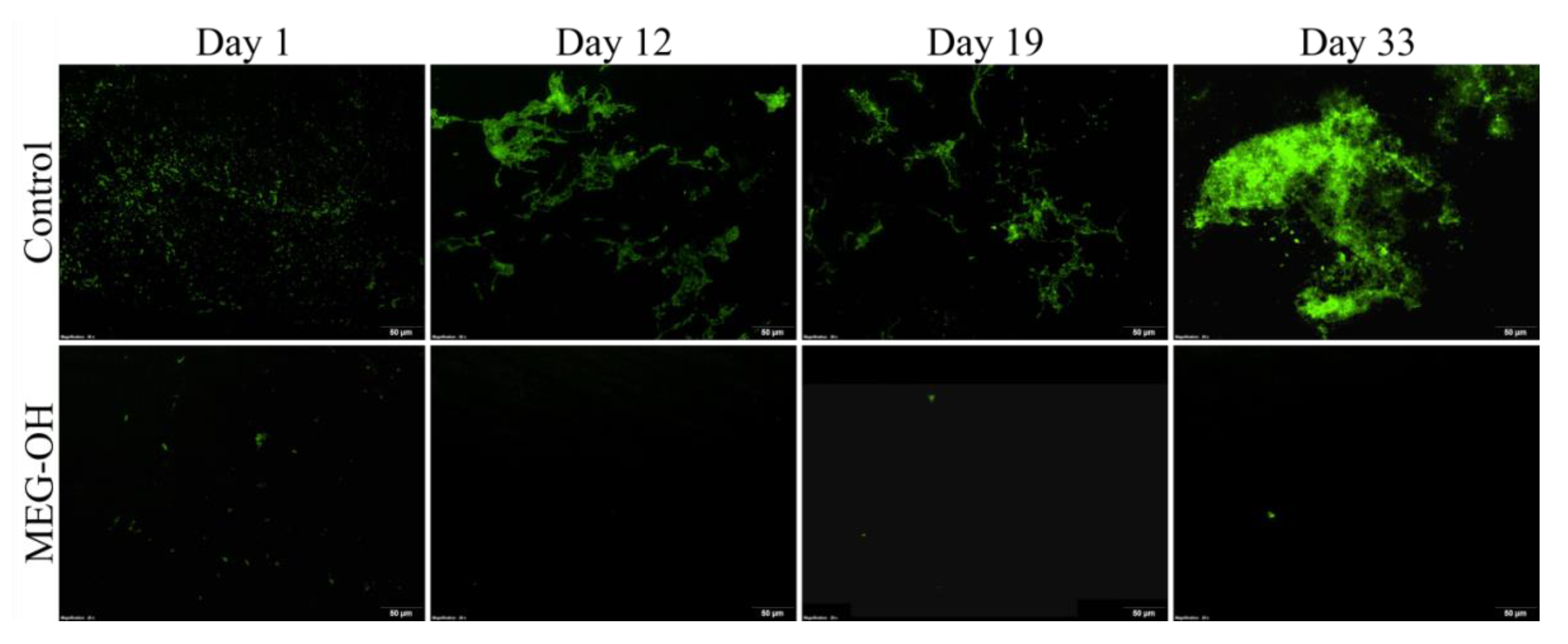
3.2. Reduction of S. aureus Fouling
As with the E. coli month-long study, an identical study was performed with S. aureus (Gram-positive bacteria) as the instigating bacteria. As with the E. coli samples, CFU counts were prepared from 1 cm of tubing (Figure 4), and the remainder was used for fluorescence imaging (Figure 5).
The day 1 counts showed a reduction from 16.6 × 10⁴ CFU/cm2 for the control samples, to 0.9 × 10⁴ CFU/cm2 for the MEG-OH modified samples, which represents a log 1.2 reduction in fouling with a confidence interval of 95%, which agrees well with the fluorescence images of these samples.
As with E. coli, there was an initial drop in colony counts at the 2 week time period, or 12 days in this case, following the reduction in media concentration. The counts decreased to 1.1 × 10⁴ CFU/cm2 for the control samples, and 0.2 × 10⁴ CFU/cm2 for the MEG-OH samples, which is a log 0.7 reduction in observed fouling with a confidence interval of 95%. Despite the overall lower counts, the fluorescence images show some biofilm formation on the control samples, which was not observed on day 1, despite the lower CFU counts.
For the following time periods, the levels of fouling for control samples increased at each timepoint to 2.3 × 10⁴ CFU/cm2 at day 19, and 3.3 × 10⁴ CFU/cm2 at day 33. However, for MEG-OH, the levels of fouling decreased across each timepoint to 0.1 × 10⁴ CFU/cm2 at day 19, and 0.5 × 10³ CFU/cm2 at day 33. This means that the reduction in anti-fouling provided by MEG-OH increased to log 1.3 at day 19, and log 1.9 at day 33, with 90% and 99% confidence intervals, respectively. These findings agree with the fluorescence images, which show very few bacteria on the MEG-OH samples, while control samples show areas of large colonies, and biofilm formation.
4. Discussion
For both experiments, beginning with either E. coli or S. aureus as the seed bacteria, MEG-OH showed a greater than log 1 reduction in fouling with a significant confidence interval across nearly every time point sampled for the more than 30 days that the experiments were performed. Additionally, both experiments showed predominantly individual bacteria across much of the control surfaces after 24 h of exposure, with E. coli showing far more bacteria in both the fluorescence images and the CFU counts. This matches well with the 24 h experiments published previously regarding MEG-OH anti-microbial fouling [36].
After the first 2 weeks in diluted media, both E. coli- and S. aureus-exposed tubing showed reductions in the colony counts for control and MEG-OH tubing. This is most likely due to the reduction in available food in the lower concentration media causing fewer bacteria to be able to survive. In the case of S. aureus, the reduction in fouling being reduced to 82% at this time point is likely due to the much lower amount of bacteria fouling the control surface as opposed to a reduction in the efficacy of MEG-OH, especially given that the percent reduction in colony counts returned to greater than 90% in subsequent weeks.
Over time, the number of bacteria fouling the control surfaces for both species began to recover, potentially due to biofilm formation. This is evidenced by the reduction in bacteria size visible for E. coli-coated control tubing at days 28 and 35, as well as additional biofilm being visible at later time points. Additional experiments would be needed to determine if this is the case, but that is beyond the scope of this investigation.
Instead, what is important is the reduction in surface-bound bacteria and biofilm formation observed on PUR tubing as a result of the MEG-OH coating. The greater than log 1.3 reduction in observed counted and observed colonies for both E. coli and S. aureus seeded bacteria after more than one month compares very favorably to PEG coatings [14], which were found to reduce E. coli biofouling approximately log 1 for a similar time period, while also showing signs of coating degradation.
These results also compare very favorably to SLIPS-coated surfaces, which typically release the majority of their compounds within the first few hours–days [19] and show biofouling along areas where the oil has defused [20]. As MEG-OH does not require any compound to elute from the surface to be effective and the monolayer itself is easy to apply and uniform, this is not an issue for this coating. Instead, the effective anti-fouling ability of the MEG-OH coating was confirmed to last for more than one month, which suggests as well that the coating is stable during this time.
This bodes well for the use of MEG-OH as a coating for urinary catheters to reduce bacterial fouling and potentially CAUTIs, as those catheters are typically not implanted for longer than one month. Since MEG-OH is able to be sterilized prior to use [36] and displays stable anti-fouling against both Gram-positive and Gram-negative seeded bacteria for longer than this time period, MEG-OH could prove to be valuable coating for urinary catheters and potentially other medical devices.
Further work is necessary to determine if MEG-OH can be used successfully on urinary catheters. Firstly, actual production Foley catheters would need to be modified with MEG-OH coatings and have their surfaces characterized to determine the extent and uniformity of the MEG-OH coating. Additionally, an in vivo animal study will need to be performed to determine if there are any adverse effects from the use of MEG-OH coatings inside the body, as well as to determine if the infection rates of catheterized animals is reduced as a result of the coating. Finally, the coating will need to be put through human trials, again, in order to determine the safety and efficacy of MEG-OH coatings in reducing infection rates in patients.
5. Conclusions
MEG-OH surface coatings were previously found to reduce microbial (E. coli, S. aureus, P. aeruginosa, and C. albicans) fouling from a variety of species across a 1-day time period but had not been tested for periods of use longer than 3 days against any species. To remedy this, MEG-OH coatings were used on polyurethane tubing to study the reduction in bacterial fouling from Gram-negative E. coli and Gram-positive S. aureus across time periods greater than 30 days. It was found that the MEG-OH coating provided a greater than log 1.3 reduction in fouling from these bacteria across this time period, showing that MEG-OH is stable and able to continue to function for at least several weeks. It also compares very well to other types of coatings, such as PEG, SLIPS, or surface-bound enzymes across this entire time period. This clearly indicates that it would be appropriate for use on urinary catheters, as these devices are generally not implanted for longer than 1 month. Before this application can be achieved, MEG-OH-coated catheters will have to be assessed in vivo via animal models to ensure that the system does not cause any adverse reactions to mammalian tissues. Additionally, MEG-OH coatings would have to be applied to full-size urinary catheters at scale for their use in medical settings. Finally, human trials need to be conducted to ensure the safety of MEG-OH attached to implanted catheters.
Author Contributions
Conceptualization, B.D.L.F., D.A., B.H. and M.T.; methodology, B.D.L.F. and D.A.; formal analysis, B.D.L.F.; investigation, B.D.L.F.; resources, B.H. and M.T.; data curation, B.D.L.F.; writing—original draft preparation, B.D.L.F.; writing—review and editing, B.D.L.F., D.A., B.H. and M.T.; supervision, B.H. and M.T.; funding acquisition, B.D.L.F. and M.T. All authors have read and agreed to the published version of the manuscript.
Funding
Support for this work was provided by MITACS, Vancouver, Canada (project number IT14364) and Econous Systems Inc. of Toronto, Canada, and in part by the Natural Sciences and Engineering Research Council of Canada (Grant 9522).
Institutional Review Board Statement
Not applicable.
Informed Consent Statement
Not applicable.
Data Availability Statement
Not applicable.
Conflicts of Interest
The authors declare no conflict of interest.
References
- Nicolle, L.E. Catheter associated urinary tract infections. Antimicrob. Resist. Infect. Control 2014, 3, 23. [Google Scholar] [CrossRef] [PubMed] [Green Version]
- Lo, E.; Nicolle, L.E.; Coffin, S.E.; Gould, C.; Maragakis, L.L.; Meddings, J.; Pegues, D.A.; Pettis, A.M.; Saint, S.; Yokoe, D.S. Strategies to Prevent Catheter-Associated Urinary Tract Infections in Acute Care Hospitals: 2014 Update. Infect. Control Hosp. Epidemiol. 2014, 35, 464–479. [Google Scholar] [CrossRef] [PubMed] [Green Version]
- Jacobsen, S.M.; Stickler, D.J.; Mobley, H.L.T.; Shirtliff, M.E. Complicated Catheter-Associated Urinary Tract Infections Due to Escherichia coli and Proteus mirabilis. Clin. Microbiol. Rev. 2008, 21, 26–59. [Google Scholar] [CrossRef] [PubMed] [Green Version]
- Umscheid, C.A.; Mitchell, M.D.; Doshi, J.A.; Agarwal, R.; Williams, K.; Brennan, P.J. Estimating the Proportion of Healthcare-Associated Infections That Are Reasonably Preventable and the Related Mortality and Costs. Infect. Control Hosp. Epidemiol. 2011, 32, 101–114. [Google Scholar] [CrossRef] [PubMed]
- Scott, R.D. The Direct Medical Costs of Healthcare-Associated Infections in US Hospitals and the Benefits of Prevention; Division of Healthcare Quality Promotion, National Center for Preparedness, Detection, and Control of Infectious Diseases, Coordinating Center for Infectious Diseases, Centers for Disease Control and Prevention: Washington, DC, USA, 2009. [Google Scholar]
- Xing, C.-M.; Meng, F.-N.; Quan, M.; Ding, K.; Dang, Y.; Gong, Y.-K. Quantitative fabrication, performance optimization and comparison of PEG and zwitterionic polymer antifouling coatings. Acta Biomater. 2017, 59, 129–138. [Google Scholar] [CrossRef]
- Konradi, R.; Acikgoz, C.; Textor, M. Polyoxazolines for nonfouling surface coatings—A direct comparison to the gold stand-ard PEG. Macromol. Rapid Commun. 2012, 33, 1663–1676. [Google Scholar] [CrossRef]
- Harris, L.; Tosatti, S.; Wieland, M.; Textor, M.; Richards, R. Staphylococcus aureus adhesion to titanium oxide surfaces coated with non-functionalized and peptide-functionalized poly(l-lysine)-grafted-poly(ethylene glycol) copolymers. Biomaterials 2004, 25, 4135–4148. [Google Scholar] [CrossRef]
- Park, K.D.; Kim, Y.S.; Han, D.K.; Kim, Y.H.; Lee, E.H.B.; Suh, H.; Choi, K.S. Bacterial adhesion on PEG modified polyurethane surfaces. Biomaterials 1998, 19, 851–859. [Google Scholar] [CrossRef]
- Pasmore, M.; Todd, P.; Smith, S.; Baker, D.; Silverstein, J.; Coons, D.; Bowman, C.N. Effects of ultrafiltration membrane surface properties on Pseudomonas aeruginosa biofilm initiation for the purpose of reducing biofouling. J. Membr. Sci. 2001, 194, 15–32. [Google Scholar] [CrossRef]
- Roosjen, A.; Busscher, H.J.; Norde, W.; van der Mei, H.C. Bacterial factors influencing adhesion of Pseudomonas aeruginosa strains to a poly(ethylene oxide) brush. Microbiology 2006, 152, 2673–2682. [Google Scholar] [CrossRef] [Green Version]
- Nejadnik, M.R.; van der Mei, H.C.; Norde, W.; Busscher, H.J. Bacterial adhesion and growth on a polymer brush-coating. Biomaterials 2008, 29, 4117–4121. [Google Scholar] [CrossRef] [PubMed]
- Chandra, J.; Patel, J.D.; Li, J.; Zhou, G.; Mukherjee, P.K.; McCormick, T.S.; Anderson, J.M.; Ghannoum, M.A. Modification of Surface Properties of Biomaterials Influences the Ability of Candida albicans To Form Biofilms. Appl. Environ. Microbiol. 2005, 71, 8795–8801. [Google Scholar] [CrossRef] [PubMed] [Green Version]
- Sharma, S.; Johnson, R.W.; Desai, T.A. Evaluation of the Stability of Nonfouling Ultrathin Poly(ethylene glycol) Films for Silicon-Based Microdevices. Langmuir 2003, 20, 348–356. [Google Scholar] [CrossRef] [PubMed]
- Pidhatika, B.; Rodenstein, M.; Chen, Y.; Rakhmatullina, E.; Muehlebach, A.; Acikgoez, C.; Textor, M.; Konradi, R. Comparative Stability Studies of Poly(2-methyl-2-oxazoline) and Poly(ethylene glycol) Brush Coatings. Biointerphases 2012, 7, 1. [Google Scholar] [CrossRef] [Green Version]
- Noguer, A.C.; Olsen, S.M.; Hvilsted, S.; Kiil, S. Long-term stability of PEG-based antifouling surfaces in seawater. J. Coatings Technol. Res. 2016, 13, 567–575. [Google Scholar] [CrossRef] [Green Version]
- Han, S.; Kim, C.; Kwon, D. Thermal/oxidative degradation and stabilization of polyethylene glycol. Polymer 1997, 38, 317–323. [Google Scholar] [CrossRef]
- Glastrup, J. Degradation of polyethylene glycol. A study of the reaction mechanism in a model molecule: Tetraethylene glycol. Polym. Degrad. Stabil. 1996, 52, 217–222. [Google Scholar] [CrossRef]
- Kratochvil, M.J.; Welsh, M.A.; Manna, U.; Ortiz, B.J.; Blackwell, H.E.; Lynn, D.M. Slippery Liquid-Infused Porous Surfaces that Prevent Bacterial Surface Fouling and Inhibit Virulence Phenotypes in Surrounding Planktonic Cells. ACS Infect. Dis. 2016, 2, 509–517. [Google Scholar] [CrossRef] [Green Version]
- Keller, N.; Bruchmann, J.; Sollich, T.; Richter, C.; Thelen, R.; Kotz, F.; Schwartz, T.; Helmer, D.; Rapp, B.E. Study of Biofilm Growth on Slippery Liquid-Infused Porous Surfaces Made from Fluoropor. ACS Appl. Mater. Interfaces 2019, 11, 4480–4487. [Google Scholar] [CrossRef]
- Epstein, A.K.; Wong, T.-S.; Belisle, R.A.; Boggs, E.M.; Aizenberg, J. Liquid-infused structured surfaces with exceptional anti-biofouling performance. Proc. Natl. Acad. Sci. USA 2012, 109, 13182–13187. [Google Scholar] [CrossRef] [Green Version]
- Deng, R.; Shen, T.; Chen, H.; Lu, J.; Yang, H.-C.; Li, W. Slippery liquid-infused porous surfaces (SLIPSs): A perfect solution to both marine fouling and corrosion? J. Mater. Chem. A 2020, 8, 7536–7547. [Google Scholar] [CrossRef]
- Mendichi, R.; Schieroni, A.G.; Piovani, D.; Allegrini, D.; Ferrara, M.; Romano, M.R. Comparative Study of Chemical Composition, Molecular and Rheological Properties of Silicone Oil Medical Devices. Transl. Vis. Sci. Technol. 2019, 8, 9. [Google Scholar] [CrossRef] [PubMed] [Green Version]
- Asker, D.; Awad, T.S.; Baker, P.; Howell, P.L.; Hatton, B.D. Non-eluting, surface-bound enzymes disrupt surface attachment of bacteria by continuous biofilm polysaccharide degradation. Biomaterials 2018, 167, 168–176. [Google Scholar] [CrossRef] [PubMed]
- Eby, D.M.; Luckarift, H.R.; Johnson, G.R. Hybrid antimicrobial enzyme and silver nanoparticle coatings for medical instruments. ACS Appl. Mater. Interfaces 2009, 1, 1553–1560. [Google Scholar] [CrossRef] [PubMed]
- Alves, D.; Pereira, M.O. Mini-review: Antimicrobial peptides and enzymes as promising candidates to functionalize biomaterial surfaces. Biofouling 2014, 30, 483–499. [Google Scholar] [CrossRef] [PubMed]
- Tanford, C. Protein denaturation. In Advances in Protein Chemistry; Anflnsen, C.B., Anson, M.L., Edsall, J.T., Richards, F.M., Eds.; Academic Press: New York, NY, USA, 1968; Volume 23, pp. 121–282. [Google Scholar]
- Klibanov, A.M. Immobilized Enzymes and Cells as Practical Catalysts. Science 1983, 219, 722–727. [Google Scholar] [CrossRef] [PubMed]
- Kheiria, S.; Liua, X.; Thompson, M. Nanoparticles at biointerfaces: Antibacterial activity and nanotoxicology. Colloids Surf. B Biointerfaces 2019, 184, 110550. [Google Scholar] [CrossRef]
- Vasudevan, R.; Kennedy, A.J.; Merritt, M.; Crocker, F.H.; Baney, R.H. Microscale patterned surfaces reduce bacterial fouling-microscopic and theoretical analysis. Colloids Surf. B Biointerfaces 2014, 117, 225–232. [Google Scholar] [CrossRef]
- Parham, S.; Wicaksono, D.H.B.; Bagherbaigi, S.; Lee, S.L.; Nur, H. Antimicrobial treatment of different metal oxide nanoparticles: A critical review. J. Chin. Chem. Soc. 2016, 63, 385–393. [Google Scholar] [CrossRef]
- Qiao, Z.; Yao, Y.; Song, S.; Yin, M.; Luo, J. Silver nanoparticles with pH induced surface charge switchable properties for antibacterial and antibiofilm applications. J. Mater. Chem. B 2018, 7, 830–840. [Google Scholar] [CrossRef]
- Albers, C.E.; Hofstetter, W.; Siebenrock, K.A.; Landmann, R.; Klenke, F.M. In vitro cytotoxicity of silver nanoparticles on osteoblasts and osteoclasts at antibacterial concentrations. Nanotoxicology 2013, 7, 30–36. [Google Scholar] [CrossRef] [PubMed]
- Lewinski, N.; Colvin, V.; Drezek, R. Cytotoxicity of nanoparticles. Small 2008, 4, 26–49. [Google Scholar] [CrossRef] [PubMed]
- Kong, B.; Seog, J.H.; Graham, L.M.; Lee, S.B. Experimental considerations on the cytotoxicity of nanoparticles. Nanomedicine 2011, 6, 929–941. [Google Scholar] [CrossRef] [PubMed] [Green Version]
- De La Franier, B.; Asker, D.; van den Berg, D.; Hatton, B.; Thompson, M. Reduction of microbial adhesion on polyurethane by a sub-nanometer covalently-attached surface modifier. Colloids Surf. B Biointerfaces 2021, 200, 111579. [Google Scholar] [CrossRef] [PubMed]
- Sheikh, S.; Yang, D.Y.; Blaszykowski, C.; Thompson, M. Single ether group in a glycol-based ultra-thin layer prevents surface fouling from undiluted serum. Chem. Commun. 2012, 48, 1305–1307. [Google Scholar] [CrossRef]
- Sheikh, S.; Sheng, J.C.-C.; Blaszykowski, C.; Thompson, M. New oligoethylene glycol linkers for the surface modification of an ultra-high frequency acoustic wave biosensor. Chem. Sci. 2010, 1, 271–275. [Google Scholar] [CrossRef]
- Fedorov, K.; Blaszykowski, C.; Sheikh, S.; Reheman, A.; Romaschin, A.; Ni, H.; Thompson, M. Prevention of thrombogenesis from whole human blood on plastic polymer by ultrathin monoethylene glycol silane adlayer. Langmuir 2014, 30, 3217. [Google Scholar] [CrossRef]
- Fedorov, K.; Jankowski, A.; Sheikh, S.; Blaszykowski, C.; Reheman, A.; Romaschin, A.; Ni, H.; Thompson, M. Prevention of surface-induced thrombogenesis on poly(vinylchloride). J. Mater. Chem. B 2015, 3, 8623. [Google Scholar] [CrossRef]
- Fedorov, K.; Sheikh, S.; Romaschin, A.; Thompson, M. Enhanced Long-term Antithrombogenicity Instigated by Covalently-Attached Surface Modifier on Biomedical Polymers. Recent Prog. Mater. 2020, 2, 16. [Google Scholar] [CrossRef]
- Pawlowska, N.M.; Fritzsche, H.; Blaszykowski, C.; Sheikh, S.; Vezvaie, M.; Thompson, M. Probing the Hydration of Ultrathin Antifouling Organosilane Adlayers using Neutron Reflectometry. Langmuir 2014, 30, 1199–1203. [Google Scholar] [CrossRef]
- Sheikh, S.; Blaszykowski, C.; Nolan, R.; Thompson, D.; Thompson, M. On the hydration of subnanometric antifouling organosilane adlayers: A molecular dynamics simulation. J. Colloid Interface Sci. 2015, 437, 197–204. [Google Scholar] [CrossRef] [PubMed]
Figure 1.
Conversion of surface bound MEG-TFA to MEG-OH.

Figure 2.
Comparison of CFU counts of E. coli growing on control (blue) and MEG-OH coated (red) polyurethane surfaces at days 1 (static culture), 14, 28, and 35 (under flow of 2 g/L LB medium). Asterisks represent the calculated confidence interval using a two variable t-test (*** = 98%, and **** = 99%+, n = 6).
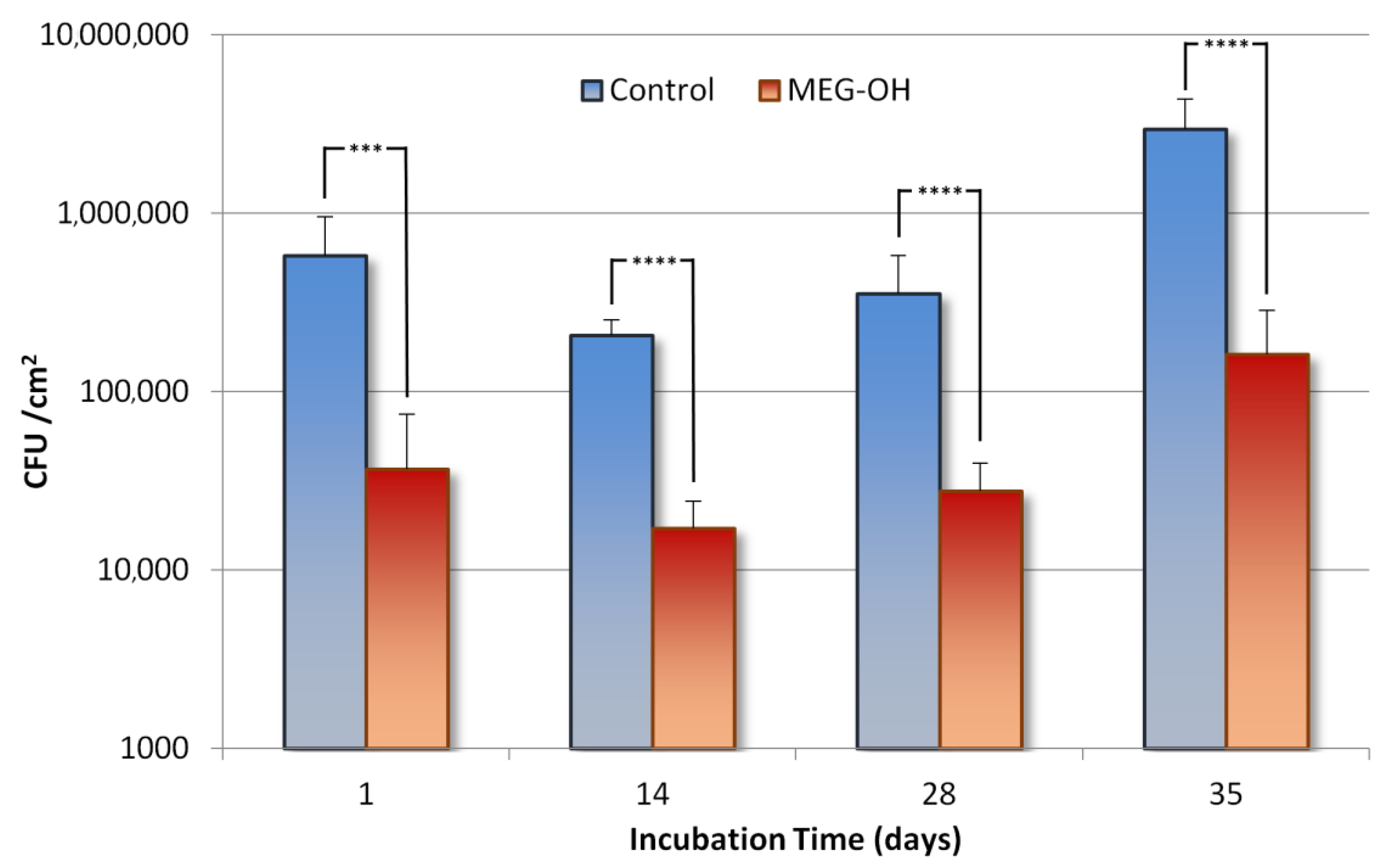
Figure 3.
Fluorescence images of control, and MEG-OH coated surfaces that were exposed to static LB containing E. coli for 1 day, then at days 14, 28, and 35 under flow of 2 g/L concentration LB medium.
Figure 3.
Fluorescence images of control, and MEG-OH coated surfaces that were exposed to static LB containing E. coli for 1 day, then at days 14, 28, and 35 under flow of 2 g/L concentration LB medium.
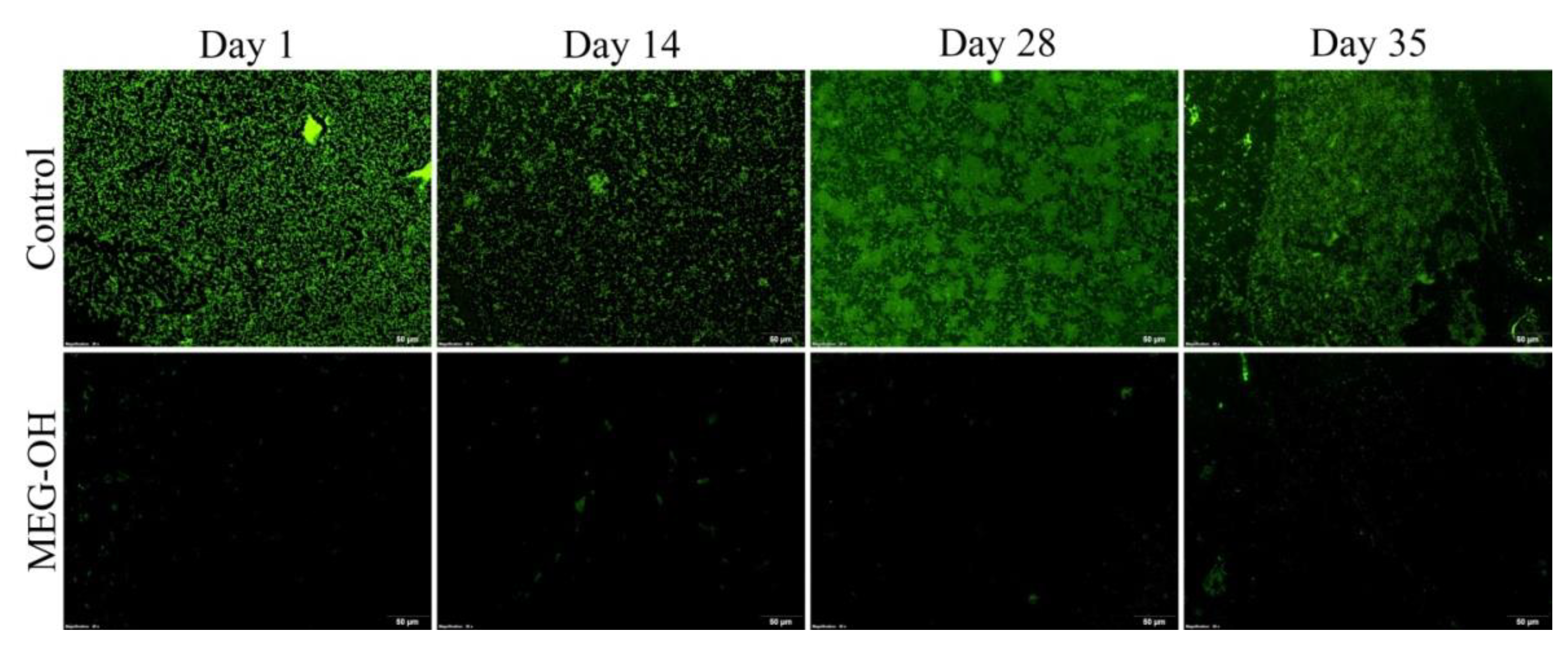
Figure 4.
Comparison of CFU counts of S. aureus growing on control (blue) and MEG-OH coated (red) polyurethane surfaces at days 1 (static culture), 12, 19, and 33 (under flow of 2 g/L LB medium). Asterisks represent the calculated confidence interval using a two variable t-test (* = 90%, ** = 95%, and **** = 99%+, n = 6).

Figure 5.
Fluorescence images of control, and MEG-OH coated surfaces that were exposed to static LB containing S. aureus for 1 day, then at days 12, 19, and 33 under flow of 2 g/L concentration LB medium.
Figure 5.
Fluorescence images of control, and MEG-OH coated surfaces that were exposed to static LB containing S. aureus for 1 day, then at days 12, 19, and 33 under flow of 2 g/L concentration LB medium.

Publisher’s Note: MDPI stays neutral with regard to jurisdictional claims in published maps and institutional affiliations. |
© 2022 by the authors. Licensee MDPI, Basel, Switzerland. This article is an open access article distributed under the terms and conditions of the Creative Commons Attribution (CC BY) license (https://creativecommons.org/licenses/by/4.0/).
Share and Cite
MDPI and ACS Style
De La Franier, B.; Asker, D.; Hatton, B.; Thompson, M. Long-Term Reduction of Bacterial Adhesion on Polyurethane by an Ultra-Thin Surface Modifier. Biomedicines 2022, 10, 979. https://doi.org/10.3390/biomedicines10050979
AMA Style
De La Franier B, Asker D, Hatton B, Thompson M. Long-Term Reduction of Bacterial Adhesion on Polyurethane by an Ultra-Thin Surface Modifier. Biomedicines. 2022; 10(5):979. https://doi.org/10.3390/biomedicines10050979
Chicago/Turabian StyleDe La Franier, Brian, Dalal Asker, Benjamin Hatton, and Michael Thompson. 2022. "Long-Term Reduction of Bacterial Adhesion on Polyurethane by an Ultra-Thin Surface Modifier" Biomedicines 10, no. 5: 979. https://doi.org/10.3390/biomedicines10050979
Note that from the first issue of 2016, this journal uses article numbers instead of page numbers. See further details here.