Review

Decoding Hidden Messengers: Proteomic Profiling of Exosomes in Mammary Cancer Research

by
Adriana Alonso Novais
1,
Guilherme Henrique Tamarindo
2,3,
Luiz Gustavo de Almeida Chuffa
4 and
Debora Aparecida Pires de Campos Zuccari
3,*
1
Health Sciences Institute (ICS), Universidade Federal de Mato Grosso (UFMT), Sinop 78550-728, Brazil
2
Brazilian Biosciences National Laboratory, Brazilian Center for Research in Energy and Materials (CNPEM), Campinas 13083-970, Brazil
3
Cancer Molecular Research Laboratory (LIMC), Department of Molecular Biology, Faculdade de Medicina de São José do Rio Preto/FAMERP (FAMERP), São José do Rio Preto 15090-000, Brazil
4
Department of Structural and Functional Biology, Institute of Biosciences, Universidade Estadual Paulista (UNESP), Botucatu 18618-689, Brazil
*
Author to whom correspondence should be addressed.
Biomedicines 2023, 11(10), 2839; https://doi.org/10.3390/biomedicines11102839
Submission received: 20 September 2023 / Revised: 11 October 2023 / Accepted: 14 October 2023 / Published: 19 October 2023

Abstract

:
Cancer is a complex and heterogeneous disease, influenced by various factors that affect its progression and response to treatment. Although a histopathological diagnosis is crucial for identifying and classifying cancer, it may not accurately predict the disease’s development and evolution in all cases. To address this limitation, liquid biopsy has emerged as a valuable tool, enabling a more precise and non-invasive analysis of cancer. Liquid biopsy can detect tumor DNA fragments, circulating tumor cells, and exosomes released by cancer cells into the bloodstream. Exosomes attracted significant attention in cancer research because of their specific protein composition, which can provide valuable insights into the disease. The protein profile of exosomes often differs from that of normal cells, reflecting the unique molecular characteristics of cancer. Analyzing these proteins can help identify cancer-associated markers that play important roles in tumor progression, invasion, and metastasis. Ongoing research and clinical validation are essential to advance and effectively utilize protein biomarkers in cancer. Nevertheless, their potential to improve diagnosis and treatment is highly promising. This review discusses several exosome proteins of interest in breast cancer, particularly focusing on studies conducted in mammary tissue and cell lines in humans and experimental animals. Unfortunately, studies conducted in canine species are scarce. This emphasis sheds light on the limited research available in this field. In addition, we present a curated selection of studies that explored exosomal proteins as potential biomarkers, aiming to achieve benefits in breast cancer diagnosis, prognosis, monitoring, and treatment.

1. Introduction

In the realm of cancer research, protein biomarkers emerged as powerful tools that unlock a deeper understanding of this complex disease. These molecular signposts found in the body offer invaluable insights into cancer detection, diagnosis, prognosis, and treatment. Their great importance relies on their ability to revolutionize personalized medicine, enhance patient outcomes, and pave the way for targeted therapies. In this review, we delve into the mammary cancer protein biomarkers described for humans and dogs from a proteomic standpoint.
In most of the past four decades and during the most recent years (2010–2019), mammary cancer incidence rates have risen by 0.5% annually [1]. Female breast cancer (BC) exceeds lung cancer statistics (11.7% vs. 11.4%) as the most frequently diagnosed cancer in women, with an estimated 2.3 million new cases yearly, although the mortality rate is much lower (6.9% vs. 18%) [2].
In veterinary medicine, canine mammary tumors (CMT) are the most common cancer in female dogs [3], although they occur mainly in countries where early female sterilization is not a current practice. Due to the high similarity of CMT to human breast cancers, it represents an excellent experimental model for BC. In addition, human BC biomarkers are frequently detectable in cases of CMT [4].
The history of BC is full of attempts to understand the wily nature of this hormone-responsive cancer [5]. A hereditary predisposition may influence screening and follow-up recommendations for high-risk patients, but a specific predisposing gene is identified in less than 30% of cases [6]. In contrast, there is an important role for microenvironmental and epigenetic changes [7].
Tissue biopsy is one of the traditional methods of detecting cancer. However, it is not comprehensive enough to detect the entire genomic scenario of breast tumors. Fortunately, improvements in new techniques, such as liquid biopsy, allowed for the improved management of breast cancer [8].
The liquid biopsy technique uses a sample of tumor cell constituents released from a tumor and/or tumor metastasis in biological substances such as blood, urine, milk, synovial fluid, and saliva. Such constituents may be circulating tumor cells (CTCs), circulating tumor DNA (ctDNA), or circulating tumor RNA (ctRNA), platelets, and exosomes [9]. When we compare liquid biopsy with tissue tumor biopsies, the ease of obtaining bioliquids makes them a very promising source. Furthermore, exosomes released from cancer cells into biofluids carry components that provide important information about tumors [10]. Therefore, liquid biopsies in breast cancers provided encouraging results, especially for monitoring treatment response and predicting disease progression or recurrence [11].
As one of the most prevalent and devastating diseases affecting women worldwide, the early detection and accurate diagnosis of mammary cancer are crucial for improving patient outcomes and implementing effective treatment strategies. Therefore, extensive research efforts are dedicated to identifying reliable biomarkers that can aid in early detection, monitoring, and treatment.

2. Breast Cancer Biomarkers

Currently, the histopathological examination of the tumor after surgical excision defines the diagnosis and prognosis of breast cancer. Unfortunately, there are still no reliable histological criteria for metastatic potential before metastasis occurs. There is a great divergence between the histological prognosis and the actual outcome of the disease [12]. The main obstacle to accurate diagnosis is due to the enormous heterogeneity of breast cancer. To overcome this obstacle, several molecular biomarkers have been recently used in clinical practice, such as hormone receptors for breast cancer subtyping and several genes involved in genome conservation, which can predict susceptibility to breast cancer [13,14].
Liquid blood biopsy can detect biomarkers and offers an alternative non-invasive strategy to improve cancer detection [15]. Various biomarkers such as proteins, autoantibodies, miRNAs, nucleic acid methylation, metabolites, lipids, and exosomes demonstrated great potential for detecting cancer, including in the early stages of the disease [15].
Clinical cancer proteomics has been used more recently to identify protein biomarkers and expand the discovery of new potential tumor biomarkers [16]. Furthermore, the evolution of early identification and diagnosis methods is bringing great progress to the treatment of breast cancer, so that each patient and their cancer will be able to receive specific (personalized) therapeutic approaches [17].

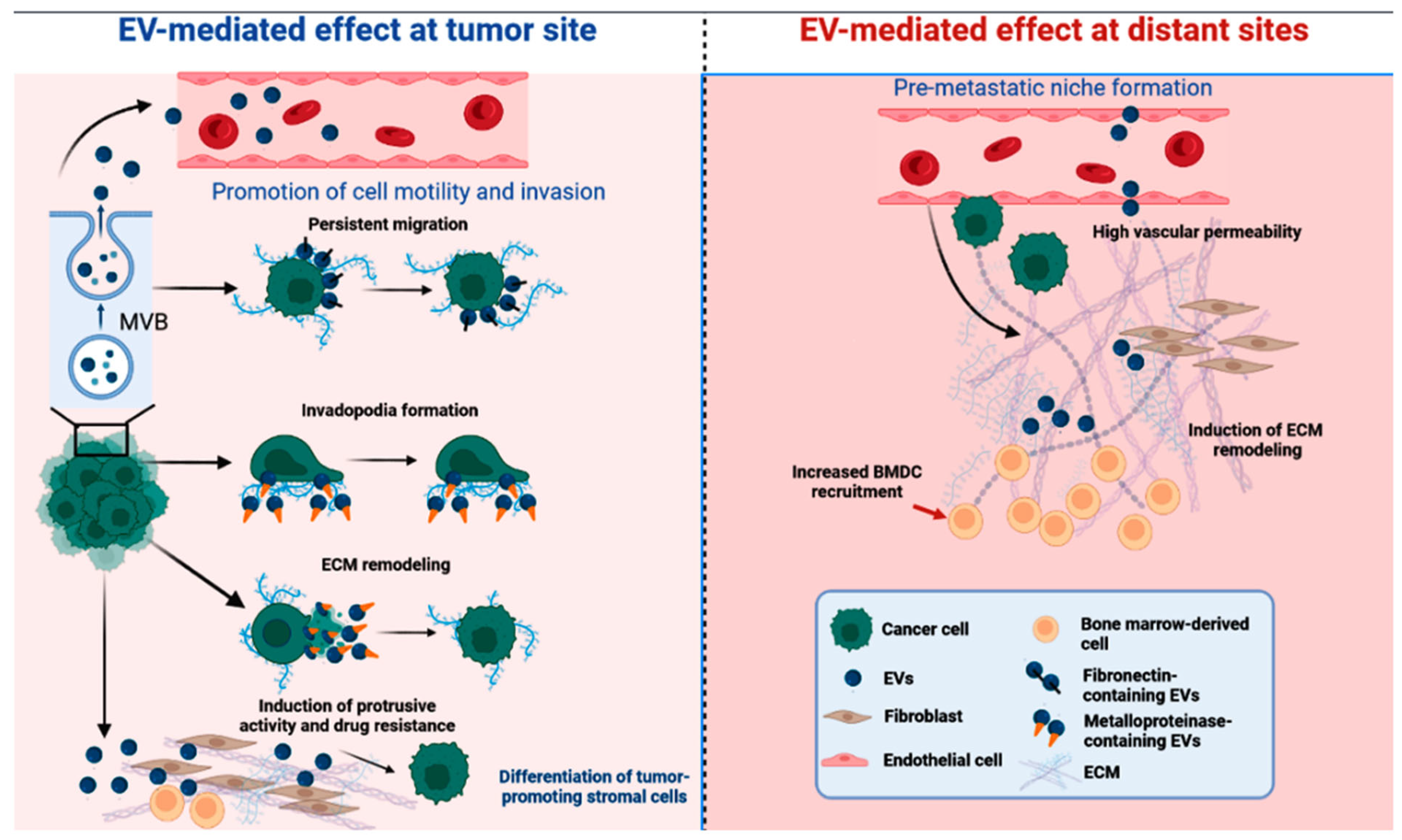
3. Small Extracellular Vesicles in Cancer Intercellular Communication

Much attention has been given to small extracellular vesicles (SEVs) released by tumor cells for their participation in the transfer of signaling proteins between cancer cells and healthy tissues, which are capable of interfering with their invasive activity [18]. SEVs, also called exosomes, are small membrane nanoparticles released by tissue cells. These small vesicles transport nucleic acids and biologically active proteins (microRNA, mRNA, non-coding RNA, DNA, transcription factors, integrins, signaling molecules, and growth factors) that contribute to cancer progression and metastasis [18,19]. SEVs from tumor cells were demonstrated to preferentially fuse with their resident destination, preparing the niche for metastasis in a process that was intensely investigated by cancer researchers and seems to be highly related to exosomal integrins. Clinical data indicated that exosomal integrins can be used to predict organ-specific metastasis [20,21] (Figure 1).

4. SEVs Shuttle Cargo Proteins That Regulate Tumorigenesis and Show Diagnostics and Prognostics Potential

Proteins are among SEV cargo molecules, and they can regulate many processes such as the tropism of cells to receive vesicles, the binding and activation of receptors on recipient cells, and the promotion of some reactions inside the SEVs [22]. However, SEVs do not contain a random array of cargo proteins but rather a specific array. The range of the SEV biocargo reported in the literature was cataloged in EVpedia (http://evpedia.info, accessed on 18 September 2023), Vesiclepedia (http://www.microvesicles.org, accessed on 18 September 2023), and ExoCarta (http://www.exocarta.org, accessed on 18 September 2023) [23].

5. Mammary Canine SEV Proteomic Studies Are Scarce

The identification of exosomal proteins involved in tumor signaling through proteomics was recently become a promising field of research. Despite the growing number of studies on exosomal proteomics in humans, particularly in in vitro studies using cell lines, research on the proteomics of exosomes in canine species is still scarce, especially in mammary carcinoma. So far, the literature reported proteins found in tissues and body fluids but not specifically inside small extracellular vesicles.
For example, Klopfleisch and colleagues (2010) [24] used 2D-DIGE and MALDI-TOF-MS and identified 21 proteins with significantly altered protein expression among metastatic canine mammary carcinoma samples. Upregulated proteins included proliferating cell nuclear antigen (PCNA), ferritin light chain (FTL), bomapin, tropomyosin 3 (TPM3), thioredoxin-containing domain containing 5 (TXNDC5), adenosine deaminase (ADA), ornithine aminotransferase (OAT), coronin 1A (CORO1A), (RANBP1) RAN-binding protein 1,3-phosphoglycerate dehydrogenase, and eukaryotic translation elongation factor 1 (eEF1). Downregulated proteins included calretinin, myosin, light chain 2, peroxiredoxin 6, maspin, the ibrinogen beta chain, vinculin, isocitrate dehydrogenase 1, tropomyosin 1, annexin A5, and Rho GTPase activating protein 1. Interestingly, 19 of these 21 proteins were also described in human breast cancer, with their expression associated with malignancy.
One year later, Suárez-Bonnet and colleagues (2011) [25] examined the expression of 14-3-3 σ, a protein related to cell cycle regulation, in normal, dysplastic, and neoplastic canine mammary tissue, to assess the capacity of this protein to act as a marker of myoepithelial cells (MECs). The findings indicated that the 14-3-3 σ protein is present in both normal and neoplastic canine mammary tissue, with high expression of this antigen in MECs. These results suggest that this protein may have a role in enhancing the spreading capacity of canine mammary tumors and can be considered both a prognostic biomarker and a therapeutic target.
Later, Jagarlamudi et al. (2014) [26] analyzed a cell-cycle-related protein called serum thymidine kinase 1 (sTK1), reporting that its levels and activity were significantly higher in CMT than in healthy dogs.
Other authors [27], in 2019, reported the detection of the biomarker BIRC5 (survivin) in dog serum, in primary culture of canine mammary tumor cells and in the canine mammary cancer cell line REM-134. They found that serum levels of this protein, which is involved in inhibiting cell death, were 109.83 ± 8.10 pg/mL in dogs with CMT, compared with 44.7 ± 2.61 pg/mL and 30.23 ± 1.32 pg/mL, respectively, in dogs with non-cancerous diseases and healthy individuals.
Fortunately, school attendance improved afterward. Fhaikrue and colleagues (2020) [28] developed a study to identify protein expression in canine mammary tumors (CMTs) using primary cell cultures from benign mixed tumors, simple carcinomas, complex carcinomas, and healthy mammary glands using a proteomic approach. Cytokeratin 5 (CK5) and transketolase (TKT) were identified in benign mixed tumor cells and complex carcinoma cells. Furthermore, cytokeratin 18 (CK18) and pyruvate kinase (PKM) were identified in simple carcinoma cells. Furthermore, they identified the tumor antigen alpha-2-HS-glycoprotein in complex carcinoma cells specifically, whereas ATP-dependent platelet-like proteins 6-phosphofructokinase and elongation factor 2 proteins were observed in benign cells. These changes in genes related to metabolism indicate that MTCs undergo metabolic reprogramming that transforms benign tumors into malignant ones, meeting the demands of proliferating cells [29].
In the same year, Park and colleagues (2020) [30] studied the proteomics of plasma from healthy dogs and dogs with cancer, focusing on uncovering biomarkers of aggressiveness of canine mammary tumors. They identified 54 proteins that were higher in cancer than in normal plasma, including SERPING1, SERPINA6, and lecithin cholesterol acyltransferase (LCAT). The authors concluded that the plasma protein LCAT could be considered a biomarker for advanced breast cancer and metastasizing breast tumors.
Shortly thereafter, Cordeiro and collaborators (2021) [31] compared the proteomic profile of canine tumor cells at different degrees of differentiation. The authors demonstrated that the malignant phenotype could be a consequence of changes in the expression of key proteins such as FNDC1, A1BG, CANX, HSPA5, and PDIA3, which could lead to tumor evasion against inflammatory cells, thus facilitating the spread of cancer.
Additionally, Yuan and colleagues (2021) [32] studied anterior gradient protein 2 (AGR2), a chaperone and p53 inhibitor involved in cell migration, transformation, and metastasis. They described that it was overexpressed in tissue samples from canine mammary malignant tumor (MMT) tissues and that high levels of AGR2 in sera from dogs with MMT were associated with the progression and remote metastasis of MMT and a low overall survival rate.
As previously documented, research on mammary tumor proteins in the exosomes of dogs is currently non-existent. As a result, there is a pressing need for further investigation in this area. Despite extensive research conducted on mammary tumors in dogs, there was relatively little focus on the proteins in the exosomes derived from these tumors. Therefore, the comprehensive characterization and exploration of protein cargo within dog mammary tumor exosomes remain largely unexplored.

6. SEV Proteomics May Identify Novel Breast Cancer Biomarkers

Extensive efforts have been dedicated to identifying reliable biomarkers that can aid in the early detection and monitoring of human mammary cancer. The proteomic analysis of exosomes has gained significant attention in mammary cancer research because of the abundance and diversity of proteins encapsulated within these vesicles. By characterizing the protein composition of exosomes derived from mammary cancer cells, researchers can uncover unique molecular signatures specific to cancerous cells.
In recent decades, advances in proteomic analysis have presented the main objective of discovering molecular biomarkers for the early detection of cancer, the characterization of the tumor profile, and the identification of new therapeutic targets [33]. A particular advantage of the proteome is that not only tissues but also body fluids such as blood, urine, and saliva can be used to investigate the molecular correlation between disease and drug action [34,35].
For example, Khan and colleagues (2014) [36] demonstrated that the protein survivin and its splice variants were present in the cargo of exosomes isolated from the serum of breast cancer patients, mimicking a pattern they had also reported in breast cancer tissue samples. They concluded that differential expression of exosomal survivin, particularly survivin-2B, could serve as a diagnostic and/or prognostic marker in patients with early-stage breast cancer (Table 1).
Harris and colleagues (2015) studied the proteomic profiling of exosomes released from three breast cancer cell lines (MCF-7, MDA-MB-231, and Rab27b) and identified 85 differentially expressed proteins. In metastatic-tumor-derived exosomes, they observed upregulation of a unique set of adhesion proteins (vimentin, galectin-3-binding protein, annexin A1, plectin, protein CYR61, EGF-like repeat and discoidin I-like domain containing protein, filamin-B, and protein-glutamine gamma-glutamyltransferase 2) [37].
Using proteomic analysis, Blomme and colleagues (2016) [38] validated a novel exosomal protein, termed myoferlin, which is related to angiogenesis, metabolism reprogramming, and epithelial–mesenchymal transition in cancer [39]. They demonstrated that myoferlin depletion in cancer cells leads to exosomes that are functionally deficient and have a significantly reduced ability to induce the migration and proliferation of those cells.
Vardaki and colleagues (2016) [40] documented significant differences not only in the number of exosomes secreted but also in the protein content of exosomes secreted by metastatic vs. non-metastatic tumors. The authors identified periostin, a protein that appears to bind to integrins in cancer cells, triggering the Akt/PKB and FAK signaling pathways and increasing angiogenesis, invasion, metastasis, and cell survival [41]. The presence of periostin was validated in a pilot cohort of samples from breast cancer patients with localized disease or lymph node metastasis.
Hurwitz and colleagues (2016) [42] used sixty National Cancer Institute cell lines (NCI-60) to provide the largest proteomic profile of SEVs in a single study, identifying a total of 6071 proteins. Only tetraspanins CD81, Alix, and HSC70 were found in all samples. The periostin protein was confirmed in two metastatic breast cancer cell lines (MDA-MB-231 and HS 578T) but was not detected in other non-metastatic breast cancer SEVs. Other proteins, such as ratilin, fibulin-7, and plasminogen activator inhibitor 1, were exclusively found in SEVs from metastatic breast cancer. The authors concluded that SEVs reliably represent their progenitor cells and are excellent candidates as biomarkers for cancer diagnosis, progression, and metastasis.
Moon et al. (2016) [43] identified fibronectin (FN), a protein that mediates the interaction of cells with the extracellular matrix (ECM) and downstream factor that promotes metastasis [44], as a biomarker candidate because of its presence on the surface of SEVs secreted from human BC cell lines. FN levels were significantly elevated at all stages of BC and returned to normal after tumor removal. At early stages of BC, another study conducted by Lee and colleagues (2017) [45] reported Developmental Endothelial Locus-1 (DEL-1) as a possible diagnostic tool to distinguish benign tumors from a healthy breast.
Gangoda et al. (2017) [46] compared exosomes isolated from several strains of genetically related mouse breast tumors with different metastatic propensities through proteomic analysis. The authors observed that the exosomes derived from metastatic cells were rich in proteins capable of promoting cell migration, proliferation, invasion, and angiogenesis. On the other hand, exosomes derived from non-metastatic cells contained proteins involved in cell–cell/cell matrix adhesion and polarity maintenance. The contents of metastatic exosomes revealed membrane proteins, including ceruloplasmin and metatherin, which could help target primary cancer cells to specific metastatic sites.
The annexin A2 protein was reported by Maji et al. (2017) [47] to be important for signaling in the breast cancer microenvironment, promoting angiogenesis and vascularization. Furthermore, annexin A2 induced the activation of macrophages, favoring breast cancer metastasis in distant organs. The authors concluded that exosomal annexin A2 may be a potential biomarker and therapeutic target for the diagnosis and treatment of metastatic breast cancer.
Rontogianni and colleagues (2019) [48] focused on defining EV-subtype-specific signatures that could play a role in non-invasive diagnostic testing. To this end, they profiled the proteomes of SEVs secreted by BC cell lines and patient serum, with a special emphasis on the TNBC and HER2 subtypes. Some representative TNBC-signature proteins included ephrin type-A receptor 2, DnaJ homolog subfamily A member 1, polyadenylate-binding protein 1, and neuropilin-1, which showed higher expression levels in the patient’s SEVs compared with the SEVs of HER2-positive patients. Similarly, receptor tyrosine protein kinase erbB-2, growth factor receptor-bound protein 7, eukaryotic translation initiation factor 3 subunit H, and brefeldin A-inhibited guanine nucleotide-exchange protein 2 were the most discriminative protein markers for serum-derived SEVs from HER2-positive patient. Their data revealed very distinct proteomic profiles across the different cell-line-derived SEVs, thus reflecting the unique biology of their breast cancer subtype.
The distinct proteomic content of SEVs was also demonstrated in invasive breast cancer cell lines compared with noninvasive breast cancer cells. SEVs produced by invasive MDA-MB231 cells were significantly enriched for proteins involved in vesicle formation, protein synthesis, proteolysis, and glycolysis. Conversely, SEVs produced by MCF10 were significantly enriched in membrane proteins, adhesion molecules, proteins involved in cellular migration, and components of the extracellular matrix (ECM). Based on these differences, the most abundant proteins uniquely identified in MDA-MB231 SEVs were those involved in transcriptional regulation (spliceosome, transcription factors, ribosomal proteins, and tRNA ligases), proteolysis (proteasome units abd pyrophosphatase), EV formation (annexin and vesicle markers LAMP-1 and EEA1), cell cycle (NUMA1), cell motility, and adherence to extracellular matrices (vitronectin, collagen, filamin proteins, and EDIL3) [20].
While examining the protein content of SEVs derived from three breast cancer cell lines (MCF-7, MDA-MB-231, and T47D), Dalla and colleagues (2020) [49] found that seven proteins were the most abundant (actin cytoplasmic 1, pyruvate kinase, glyceraldehyde-3-phosphate dehydrogenase, 60 kDa mitochondrial heat shock protein, mitochondrial ATP synthase alpha subunit, sodium/potassium transport ATPase beta-3 subunit, and voltage-dependent anion selective protein channel 2). They concluded that once the majority are proteins related to the metabolism of mitochondrial processes, their presence supports the hypothesis of the relevant role of SEVs in metabolic reprogramming.
Complementing previous studies, Risha and colleagues (2020) [50] studied SEVs’ proteomes from MDA-MB-231 and MCF-10A cell lines. They reported that 16 proteins were mainly involved in the formation of cancer metastasis. Among them, three exosomal membrane/surface proteins, glucose transporter 1 (GLUT-1), glypican 1 (GPC-1), disintegrin, and metalloproteinase domain-containing protein 10 (ADAM10), were identified as potential breast cancer biomarkers. The authors also concluded that SEVs can mediate distinct molecular mechanisms such as glucose uptake and ECM remodeling.
In the same year, Vinik and colleagues (2020) [51] isolated small fractions enriched in SEVs from the plasma of healthy controls and BC patients at different stages of the disease, before and after surgery. Proteomic analysis revealed a signature of seven proteins that differentiated BC patients (fibronectin, FAK, MEC1, B-actin, p90RSK_pT573, N-Cadherin, and C-Raf). Among them, FAK and fi-bronectin demonstrated high diagnostic accuracy.
The study on the proteomics of serum exosomes derived from 10 TNBC patients and 17 healthy donors by Li and colleagues (2021) [52] found that the expression levels of tetraspanin CD151 in TNBC-derived exosomes were significantly higher than those in healthy patient exosomes, validating their findings in 16 additional donor samples. The authors also observed that exosomal CD151 facilitated the secretion of ribosomal proteins while inhibiting the exosomal secretion of complement proteins. Notably, CD151-deleted exosomes significantly decreased the migration and invasion of TNBC cells.
In a different approach, the scientific team of Patwardhan and colleagues (2021) [53] demonstrated the role of exosomes in the stiffness of the ECM, triggering the invasiveness of breast cancer. Proteomic analysis of exosomal lysates revealed the enrichment of cell adhesion and migration proteins in exosomes from rigid ECM cultures compared with those from soft cultures. Thrombospondin-1 (THBS1) was identified as a prospective regulator of ECM stiffness-dependent cancer invasion involving matrix metalloproteinase and focal adhesion kinase.
Taken together, these data may support the development of new diagnostic tools, but further investigation is still required (Table 1).
Table 1. List of main SEV-associated proteins as potential biomarkers for BC according to publications from 2014 to 2021. Proteins highlighted in red mean upregulated, while those in blue mean downregulated.
AuthorProteinsIsolationPathologyApplicabilitySource
Khan et al. (2014) [36]BIRC5 and splice variantsUCTNBC, ER, and PR positiveDiagnosis, prognosis, and treatmentTumor
Harris et al. (2015) [37]Several proteins related with adhesion/motility/cytoskeleton, proteases, transporters, cell surface receptor, stress response proteins, small GTPases, metabolic enzymes, and RNA bindingUCTNBCTreatmentCells in vitro
Several proteins related with tetraspanin, adhesion, cell surface receptor, transporter, stress response proteins, budding vesicles, trafficking/transport, calcium binding, and small GTPaseUCLuminal
Blomme et al. (2016) [38]MYOFUCLuminal and TNBCTreatmentCells in vitro
Vardaky et al. (2016) [40]POSTNUCTNBC cells and patients with LN metastasisDiagnosisCells in vitro and tumor samples
Hurwitz et al. (2016) [42]POSTNExtraPEG and UCTNBCDiagnosisCells in vitro
RFTN1, FBLN7, and SERPINE1Metastatic breast cancer
Moon et al. (2016) [43]; Lee et al. (2017) [45]DEL-1UCEarly-stage breast cancer, several subtypesDiagnosisCells in vitro and plasma samples
Gangoda et al. (2017) [46]CP and MTDHUCTNBC, highly metastaticDiagnosisCells in vitro
Maji et al. (2017) [47]ANXA2UCMetastatic breast cancer cellsDiagnosis and treatmentCells in vitro
Rontogianni et al. (2019) [48]EPHA2, DNAJA1, PABPC1, and NRP1UCTNBCDiagnosisCells in vitro and serum samples
HER2, GRB7, EIF3H, and ARFGEF2HER2
Jordan et al. (2020) [20]Several proteins related with spliceosome, transcription factors, ribosomal proteins, tRNA ligases, proteasome units; pyrophosphatase, annexin, LAMP-1, EEA1, NUMA1, VTN, collagen, filamin proteins, and EDIL3Sepharose CL-2B SECTNBCDiagnosisCells in vitro and serum samples
Proteins from adherin family members, laminin proteins, proteoglycans, SDC1, EPCAM, b-catenin, collagen, CD109, RARRES1, PTGFRN, FAT1, S100A14, AREG, calcium-binding proteins, serine proteases, and cholesterol- and lipoprotein-binding proteinsDuctal carcinoma in situ
Dalla et al. (2020) [49]Actin cytoplasmic 1, PKM, GAPDH, HSP60, ATP1B3, and VDAC2UCShared among MCF-7, MDA-MB-231, and T47DDiagnosisCells in vitro
Risha et al. (2020) [50]GLUT1, GPC3 and ADAM10UCMDA-MB-231Diagnosis and prognosisCells in vitro
Vinik et al. (2020) [51]EGFR, FAK, fibronectin, p38_pT180_Y182, N-cadherin, E2F1, PARP, MEK1, Aurora-B, p90RSK_pT573, S6_pS240_S244SECStage IDiagnosis, and treatmentSerum samples
C-Raf, fibronectin, heregulin, FAK, MEK, β-actin, N-cadherin, FoxO3a_PS318_S231, P-cadherin, PDHK1, TAZStage IIA
Li et al. (2021) [52]CD151UCTNBCDiagnosisSerum samples
Patwardhan et al. (2021) [53]THBS1ExoEnrichTNBCTreatmentCells in vitro
Legend: BIRC5—survivin; MYOF—myoferlin; POSTN—periostin; FBLN7—fibulin 7; RFTN1—raftilin; SERPINE1—plasminogen activator inhibitor 1; DEL-1—Developmental Endothelial Locus-1; CP—ceruloplasmin; MTDH—metadherin; ANXA2—annexin A2; EPHA2—EPH receptor A2; DNAJA1—DnaJ Heat Shock Protein Family (Hsp40) Member A1; PABPC1—Poly(A) Binding Protein Cytoplasmic 1; NRP1—Neuropilin 1; HER2—Human Epidermal Growth Factor Receptor 2; GRB7—Growth Factor Receptor-Bound Protein 7; EIF3H—Eukaryotic Translation Initiation Factor 3 Subunit H; ARFGEF2—ADP Ribosylation Factor Guanine Nucleotide Exchange Factor 2; VTN—Vitronectin; EDIL3—EGF Like Repeats And Discoidin Domains 3; SDC1—Syndecan-1; EPCAM—Epithelial Cell Adhesion Molecule; RARRES1—Retinoic Acid Receptor Responder 1; PTGFRN—Prostaglandin F2 Receptor Inhibitor; FAT1—FAT Atypical Cadherin 1; S100A14—S100 Calcium Binding Protein A14; AREG—Amphiregulin; PKM—pyruvate kinase; GAPDH—Glyceraldehyde-3-Phosphate Dehydrogenase; ATP1B3—ATPase Na+/K+ transporting subunit beta 3; VDAC2—Voltage-Dependent Anion-Selective Channel Protein 2; HSP60—60 kDa heat shock protein, mitochondrial; GLUT1—glucose transporter 1; GPC3—Glypican 1; ADAM10—disintegrin and metalloproteinase domain-containing protein 10; THBS1—Thrombospondin-1; EGFR—Epidermal Growth Factor Receptor; FAK—focal adhesion kinase; E2F1—E2F Transcription Factor 1; PARP—Poly(ADP-Ribose) Polymerase; MEK1—Mitogen-Activated Protein Kinase Kinase 1; PDHK1—Pyruvate Dehydrogenase Kinase 1; TAZ—WW Domain Containing Transcription Regulator 1; CD151—Platelet-Endothelial Tetraspan Antigen 3; UC—Ultracentrifugation; SEC—size-exclusion column.

7. Concluding Remarks and Perspectives

In view of the possibility of performing early diagnosis and longitudinal prognostic evaluation, the number of studies focusing on the exosomal proteomics of patients diagnosed with breast tumor has grown. In addition, one of the most promising applications of cancer protein biomarkers relies on the development of targeted therapies. By identifying specific proteins that drive tumor growth, metastasis, or resistance to treatment, researchers can design drugs or treatment strategies that specifically target these biomarkers. In addition, the identification of a group of proteins allows us to clarify what type of biological processes are deregulated due to SEVs. In the present review, it is clear that metabolic reprogramming occurs not only at distinct points but also in several proteins that mediate cell interaction with ECM, suggesting the key role of SEVs in metastasis. This could improve advances in the personalized approach, known as precision medicine, which maximizes treatment effectiveness while minimizing side effects on healthy tissues. For obvious reasons, research in the canine species is still scarce, albeit important because CMT is considered as an excellent experimental model for BC. Due to the shorter lifespan of dogs and the rapid progression of CMTs, researchers can gain insights into tumor development, metastasis, and therapeutic responses in a relatively shorter timeframe compared with human studies. On the other hand, liquid biopsy has several advantages over traditional tissue biopsy in the context of breast cancer because it enables the identification and characterization of tumor-specific genetic alterations and mutations, even at early stages when traditional imaging techniques may not detect the disease. Its non-invasive nature makes it an attractive option for serial sampling, thereby reducing patient discomfort and facilitating longitudinal studies. Therefore, unveiling biomarker proteins carried by exosomes through liquid biopsy may provide valuable information. Indeed, if biomarker detection occurs before tumor signaling, there is a chance to interrupt the pathogenesis of the disease. Biomarker proteins can be modulated at various levels, such as by silencing genes, affecting their transcription or even the protein signaling pathway. It would be possible, for example, to block the signaling effect of exosomal metastasis-inducing proteins or even to modulate this effect by introducing protective signaling proteins into the vesicles. Cancer protein biomarkers revolutionized cancer research and patient care, ushering in a new era of precision medicine. As researchers continue to unravel the intricate landscape of cancer biology, protein biomarkers will remain invaluable in the fight against cancer, bringing hope for improved outcomes, enhanced quality of life, and, ultimately, a world free from the burden of this devastating disease.

Author Contributions

A.A.N. conceived the review, compiled the literature, and wrote the manuscript. G.H.T. compiled the literature and wrote the manuscript. L.G.d.A.C. prepared figures for the article and guided the insertions and the scope of the review. D.A.P.d.C.Z. conceived the review and checked all drafts and the final version of the report. All authors significantly contributed to and approved the final version of the manuscript. All authors have read and agreed to the published version of the manuscript.

Funding

The authors are grateful to the São Paulo Research Foundation (FAPESP) and the National Council for Scientific and Technological Development (CNPq). The authors received financial support from GHT (FAPESP grant number 2022/00890-5), DAPCZ (FAPESP grant number 2020/12970-8; CNPq process number 307288/2022-6), and LGAC (CNPq process number 304108/2020-0).

Institutional Review Board Statement

Not applicable.

Informed Consent Statement

Not applicable.

Data Availability Statement

Data sharing is not applicable.

Conflicts of Interest

The authors declare that the research was conducted in the absence of any commercial or financial relationships that could be understood as potential conflicts of interest.

References

  1. Giaquinto, A.N.; Sung, H.; Miller, K.D.; Kramer, J.L.; Newman, L.A.; Minihan, A.; Jemal, A.; Siegel, R.L. Breast cancer statistics, 2022. CA Cancer J. Clin. 2022, 72, 524–541. [Google Scholar] [CrossRef] [PubMed]
  2. Sung, H.; Ferlay, J.; Siegel, R.L.; Laversanne, M.; Soerjomataram, I.; Jemal, A.; Bray, F. Global cancer statistics 2020: GLOBOCAN estimates of incidence and mortality worldwide for 36 cancers in 185 countries. CA Cancer J. Clin. 2021, 71, 209–249. [Google Scholar] [CrossRef] [PubMed]
  3. Sorenmo, K. Canine mammary gland tumors. Vet. Clin. N. Am. Small Anim. Pract. 2003, 33, 573–596. [Google Scholar] [CrossRef] [PubMed]
  4. Kaszak, I.; Ruszczak, A.; Kanafa, S.; Kacprzak, K.; Król, M.; Jurka, P. Current biomarkers of canine mammary tumors. Acta Vet. Scand. 2018, 60, 66. [Google Scholar] [CrossRef] [PubMed]
  5. Lakhtakia, R. A Brief History of Breast Cancer: Part I: Surgical domination reinvented. Sultan Qaboos Univ. Med. J. 2014, 14, e166-9. [Google Scholar]
  6. Shiovitz, S.; Korde, L.A. Genetics of breast cancer: A topic in evolution. Ann. Oncol. 2015, 26, 1291–1299. [Google Scholar] [CrossRef]
  7. Polyak, K. Breast cancer: Origins and evolution. J. Clin. Investig. 2007, 117, 3155–3163. [Google Scholar] [CrossRef] [PubMed]
  8. Alimirzaie, S.; Bagherzadeh, M.; Akbari, M.R. Liquid biopsy in breast cancer: A comprehensive review. Clin. Genet. 2019, 95, 643–660. [Google Scholar] [CrossRef]
  9. Freitas, A.J.A.D.; Causin, R.L.; Varuzza, M.B.; Calfa, S.; Hidalgo Filho, C.M.T.; Komoto, T.T.; Souza, C.D.P.; Marques, M.M.C. Liquid biopsy as a tool for the diagnosis, treatment, and monitoring of breast cancer. Int. J. Mol. Sci. 2022, 23, 9952. [Google Scholar] [CrossRef]
  10. Halvaei, S.; Daryani, S.; Eslami-S, Z.; Samadi, T.; Jafarbeik-Iravani, N.; Bakhshayesh, T.O.; Majidzadeh-A, K.; Esmaeili, R. Exosomes in cancer liquid biopsy: A focus on breast cancer. Mol. Ther. Nucleic Acids 2018, 10, 131–141. [Google Scholar] [CrossRef]
  11. Tay, T.K.Y.; Tan, P.H. Liquid biopsy in breast cancer: A focused review. Arch. Pathol. Lab. Med. 2021, 145, 678–686. [Google Scholar] [CrossRef]
  12. da Costa, A.; Lenze, D.; Hummel, M.; Kohn, B.; Gruber, A.D.; Klopfleisch, R. Identification of six potential markers for the detection of circulating canine mammary tumour cells in the peripheral blood identified by microarray analysis. J. Comp. Pathol. 2012, 146, 143–151. [Google Scholar] [CrossRef] [PubMed]
  13. Li, G.; Hu, J.; Hu, G. Biomarker studies in early detection and prognosis of breast cancer. Adv. Exp. Med. Biol. 2017, 1026, 27–39. [Google Scholar] [CrossRef] [PubMed]
  14. Gam, L.-H. Breast cancer and protein biomarkers. World J. Exp. Med. 2012, 2, 86–91. [Google Scholar] [CrossRef]
  15. Loke, S.Y.; Lee, A.S.G. The future of blood-based biomarkers for the early detection of breast cancer. Eur. J. Cancer 2018, 92, 54–68. [Google Scholar] [CrossRef]
  16. Celis, J.E.; Gromov, P.; Cabezón, T.; Moreira, J.M.A.; Ambartsumian, N.; Sandelin, K.; Rank, F.; Gromova, I. Proteomic characterization of the interstitial fluid perfusing the breast tumor microenvironment: A novel resource for biomarker and therapeutic target discovery. Mol. Cell. Proteom. 2004, 3, 327–344. [Google Scholar] [CrossRef] [PubMed]
  17. Duan, S.; Buxton, I.L.O. Evolution of medical approaches and prominent therapies in breast cancer. Cancers 2022, 14, 2450. [Google Scholar] [CrossRef]
  18. Antonyak, M.A.; Li, B.; Boroughs, L.K.; Johnson, J.L.; Druso, J.E.; Bryant, K.L.; Holowka, D.A.; Cerione, R.A. Cancer cell-derived microvesicles induce transformation by transferring tissue transglutaminase and fibronectin to recipient cells. Proc. Natl. Acad. Sci. USA 2011, 108, 4852–4857. [Google Scholar] [CrossRef]
  19. Tkach, M.; Théry, C. Communication by extracellular vesicles: Where we are and where we need to go. Cell 2016, 164, 1226–1232. [Google Scholar] [CrossRef]
  20. Jordan, K.R.; Hall, J.K.; Schedin, T.; Borakove, M.; Xian, J.J.; Dzieciatkowska, M.; Lyons, T.R.; Schedin, P.; Hansen, K.C.; Borges, V.F. Extracellular vesicles from young women’s breast cancer patients drive increased invasion of non-malignant cells via the Focal Adhesion Kinase pathway: A proteomic approach. Breast Cancer Res. 2020, 22, 128. [Google Scholar] [CrossRef]
  21. Hoshino, A.; Costa-Silva, B.; Shen, T.-L.; Rodrigues, G.; Hashimoto, A.; Tesic Mark, M.; Molina, H.; Kohsaka, S.; Di Giannatale, A.; Ceder, S.; et al. Tumour exosome integrins determine organotropic metastasis. Nature 2015, 527, 329–335. [Google Scholar] [CrossRef]
  22. Jabalee, J.; Towle, R.; Garnis, C. The role of extracellular vesicles in cancer: Cargo, function, and therapeutic implications. Cells 2018, 7, 93. [Google Scholar] [CrossRef]
  23. Wang, H.-X.; Gires, O. Tumor-derived extracellular vesicles in breast cancer: From bench to bedside. Cancer Lett. 2019, 460, 54–64. [Google Scholar] [CrossRef]
  24. Klopfleisch, R.; Klose, P.; Weise, C.; Bondzio, A.; Multhaup, G.; Einspanier, R.; Gruber, A.D. Proteome of metastatic canine mammary carcinomas: Similarities to and differences from human breast cancer. J. Proteome Res. 2010, 9, 6380–6391. [Google Scholar] [CrossRef] [PubMed]
  25. Suárez-Bonnet, A.; Herráez, P.; Martín de las Mulas, J.; Rodríguez, F.; Déniz, J.M.; Espinosa de los Monteros, A. Expression of 14-3-3 σ protein in normal and neoplastic canine mammary gland. Vet. J. 2011, 190, 345–351. [Google Scholar] [CrossRef]
  26. Jagarlamudi, K.K.; Westberg, S.; Rönnberg, H.; Eriksson, S. Properties of cellular and serum forms of thymidine kinase 1 (TK1) in dogs with acute lymphocytic leukemia (ALL) and canine mammary tumors (CMTs): Implications for TK1 as a proliferation biomarker. BMC Vet. Res. 2014, 10, 228. [Google Scholar] [CrossRef] [PubMed]
  27. Jena, S.C.; Shrivastava, S.; Saxena, S.; Kumar, N.; Maiti, S.K.; Mishra, B.P.; Singh, R.K. Surface plasmon resonance immunosensor for label-free detection of BIRC5 biomarker in spontaneously occurring canine mammary tumours. Sci. Rep. 2019, 9, 13485. [Google Scholar] [CrossRef]
  28. Fhaikrue, I.; Srisawat, W.; Nambooppha, B.; Pringproa, K.; Thongtharb, A.; Prachasilchai, W.; Sthitmatee, N. Identification of potential canine mammary tumour cell biomarkers using proteomic approach: Differences in protein profiles among tumour and normal mammary epithelial cells by two-dimensional electrophoresis-based mass spectrometry. Vet. Comp. Oncol. 2020, 18, 787–795. [Google Scholar] [CrossRef] [PubMed]
  29. Tamarindo, G.H.; Novais, A.A.; Chuffa, L.G.A.; Zuccari, D.A.P.C. Metabolic alterations in canine mammary tumors. Animals 2023, 13, 2757. [Google Scholar] [CrossRef]
  30. Park, H.-M.; Kim, H.; Kim, D.W.; Yoon, J.-H.; Kim, B.-G.; Cho, J.-Y. Common plasma protein marker LCAT in aggressive human breast cancer and canine mammary tumor. BMB Rep. 2020, 53, 664–669. [Google Scholar] [CrossRef]
  31. Cordeiro, Y.G.; Mulder, L.M.; van Zeijl, R.J.M.; Paskoski, L.B.; van Veelen, P.; de Ru, A.; Strefezzi, R.F.; Heijs, B.; Fukumasu, H. Proteomic Analysis Identifies FNDC1, A1BG, and Antigen Processing Proteins Associated with Tumor Heterogeneity and Malignancy in a Canine Model of Breast Cancer. Cancers 2021, 13, 5901. [Google Scholar] [CrossRef] [PubMed]
  32. Yuan, S.H.-C.; Chang, S.-C.; Huang, Y.; Liu, H.-P. Serum Level of Tumor-Overexpressed AGR2 Is Significantly Associated with Unfavorable Prognosis of Canine Malignant Mammary Tumors. Animals 2021, 11, 2923. [Google Scholar] [CrossRef]
  33. Gast, M.-C.W.; Schellens, J.H.M.; Beijnen, J.H. Clinical proteomics in breast cancer: A review. Breast Cancer Res. Treat. 2009, 116, 17–29. [Google Scholar] [CrossRef]
  34. Hondermarck, H.; Tastet, C.; El Yazidi-Belkoura, I.; Toillon, R.-A.; Le Bourhis, X. Proteomics of breast cancer: The quest for markers and therapeutic targets. J. Proteome Res. 2008, 7, 1403–1411. [Google Scholar] [CrossRef] [PubMed]
  35. Latterich, M.; Abramovitz, M.; Leyland-Jones, B. Proteomics: New technologies and clinical applications. Eur. J. Cancer 2008, 44, 2737–2741. [Google Scholar] [CrossRef] [PubMed]
  36. Khan, S.; Bennit, H.F.; Turay, D.; Perez, M.; Mirshahidi, S.; Yuan, Y.; Wall, N.R. Early diagnostic value of survivin and its alternative splice variants in breast cancer. BMC Cancer 2014, 14, 176. [Google Scholar] [CrossRef]
  37. Harris, D.A.; Patel, S.H.; Gucek, M.; Hendrix, A.; Westbroek, W.; Taraska, J.W. Exosomes released from breast cancer carcinomas stimulate cell movement. PLoS ONE 2015, 10, e0117495. [Google Scholar] [CrossRef]
  38. Blomme, A.; Fahmy, K.; Peulen, O.; Costanza, B.; Fontaine, M.; Struman, I.; Baiwir, D.; de Pauw, E.; Thiry, M.; Bellahcène, A.; et al. Myoferlin is a novel exosomal protein and functional regulator of cancer-derived exosomes. Oncotarget 2016, 7, 83669–83683. [Google Scholar] [CrossRef]
  39. Zhu, W.; Zhou, B.; Zhao, C.; Ba, Z.; Xu, H.; Yan, X.; Liu, W.; Zhu, B.; Wang, L.; Ren, C. Myoferlin, a multifunctional protein in normal cells, has novel and key roles in various cancers. J. Cell. Mol. Med. 2019, 23, 7180–7189. [Google Scholar] [CrossRef]
  40. Vardaki, I.; Ceder, S.; Rutishauser, D.; Baltatzis, G.; Foukakis, T.; Panaretakis, T. Periostin is identified as a putative metastatic marker in breast cancer-derived exosomes. Oncotarget 2016, 7, 74966–74978. [Google Scholar] [CrossRef]
  41. Dorafshan, S.; Razmi, M.; Safaei, S.; Gentilin, E.; Madjd, Z.; Ghods, R. Periostin: Biology and function in cancer. Cancer Cell Int. 2022, 22, 315. [Google Scholar] [CrossRef]
  42. Hurwitz, S.N.; Rider, M.A.; Bundy, J.L.; Liu, X.; Singh, R.K.; Meckes, D.G. Proteomic profiling of NCI-60 extracellular vesicles uncovers common protein cargo and cancer type-specific biomarkers. Oncotarget 2016, 7, 86999–87015. [Google Scholar] [CrossRef]
  43. Moon, P.-G.; Lee, J.-E.; Cho, Y.-E.; Lee, S.J.; Jung, J.H.; Chae, Y.S.; Bae, H.-I.; Kim, Y.-B.; Kim, I.-S.; Park, H.Y.; et al. Identification of Developmental Endothelial Locus-1 on Circulating extracellular vesicles as a Novel Biomarker for Early Breast Cancer Detection. Clin. Cancer Res. 2016, 22, 1757–1766. [Google Scholar] [CrossRef]
  44. Lin, T.-C.; Yang, C.-H.; Cheng, L.-H.; Chang, W.-T.; Lin, Y.-R.; Cheng, H.-C. Fibronectin in cancer: Friend or foe. Cells 2019, 9, 27. [Google Scholar] [CrossRef] [PubMed]
  45. Lee, S.J.; Lee, J.; Jung, J.H.; Park, H.Y.; Moon, P.-G.; Chae, Y.S.; Baek, M.-C. Exosomal Del-1 as a Potent Diagnostic Marker for Breast Cancer: Prospective Cohort Study. Clin. Breast Cancer 2021, 21, e748–e756. [Google Scholar] [CrossRef] [PubMed]
  46. Gangoda, L.; Liem, M.; Ang, C.-S.; Keerthikumar, S.; Adda, C.G.; Parker, B.S.; Mathivanan, S. Proteomic Profiling of Exosomes Secreted by Breast Cancer Cells with Varying Metastatic Potential. Proteomics 2017, 17, 1600370. [Google Scholar] [CrossRef] [PubMed]
  47. Maji, S.; Chaudhary, P.; Akopova, I.; Nguyen, P.M.; Hare, R.J.; Gryczynski, I.; Vishwanatha, J.K. Exosomal annexin II promotes angiogenesis and breast cancer metastasis. Mol. Cancer Res. 2017, 15, 93–105. [Google Scholar] [CrossRef]
  48. Rontogianni, S.; Synadaki, E.; Li, B.; Liefaard, M.C.; Lips, E.H.; Wesseling, J.; Wu, W.; Altelaar, M. Proteomic profiling of extracellular vesicles allows for human breast cancer subtyping. Commun. Biol. 2019, 2, 325. [Google Scholar] [CrossRef]
  49. Dalla, P.V.; Santos, J.; Milthorpe, B.K.; Padula, M.P. Selectively-Packaged Proteins in Breast Cancer extracellular vesicles Involved in Metastasis. Int. J. Mol. Sci. 2020, 21, 4990. [Google Scholar] [CrossRef]
  50. Risha, Y.; Minic, Z.; Ghobadloo, S.M.; Berezovski, M.V. The proteomic analysis of breast cell line exosomes reveals disease patterns and potential biomarkers. Sci. Rep. 2020, 10, 13572. [Google Scholar] [CrossRef] [PubMed]
  51. Vinik, Y.; Ortega, F.G.; Mills, G.B.; Lu, Y.; Jurkowicz, M.; Halperin, S.; Aharoni, M.; Gutman, M.; Lev, S. Proteomic analysis of circulating extracellular vesicles identifies potential markers of breast cancer progression, recurrence, and response. Sci. Adv. 2020, 6, eaba5714. [Google Scholar] [CrossRef] [PubMed]
  52. Li, S.; Li, X.; Yang, S.; Pi, H.; Li, Z.; Yao, P.; Zhang, Q.; Wang, Q.; Shen, P.; Li, X.; et al. Proteomic Landscape of Exosomes Reveals the Functional Contributions of CD151 in Triple-Negative Breast Cancer. Mol. Cell. Proteom. 2021, 20, 100121. [Google Scholar] [CrossRef] [PubMed]
  53. Patwardhan, S.; Mahadik, P.; Shetty, O.; Sen, S. ECM stiffness-tuned exosomes drive breast cancer motility through thrombospondin-1. Biomaterials 2021, 279, 121185. [Google Scholar] [CrossRef] [PubMed]
Figure 1. Diverse cellular signaling of exosomes (SEVs) was described to influence the establishment and spread of cancer. EV-mediated effects may occur at tumor sites (e.g., persistent migration, invadopodia formation, ECM remodeling, induction of protrusive activity and drug resistance, and differentiation of tumor-promoting stromal cells) and at distant sites (e.g., high vascular permeability, induction of ECM remodeling, and increased BMDC recruitment) [19]. EV, extracellular vesicle; MVB, multivesicular bodies; ECM, extracellular matrix; BMDC, bone-marrow-derived cell.
Biomedicines 11 02839 g001
Disclaimer/Publisher’s Note: The statements, opinions and data contained in all publications are solely those of the individual author(s) and contributor(s) and not of MDPI and/or the editor(s). MDPI and/or the editor(s) disclaim responsibility for any injury to people or property resulting from any ideas, methods, instructions or products referred to in the content.

