Article

Osteoprotegerin Gene as a Biomarker in the Development of Osteoporosis in Postmenopausal Women

1
Department of Pharmacology and Pharmacoeconomics, Pomeranian Medical University in Szczecin, 71-230 Szczecin, Poland
2
Department of Personalized Medicine and Cell Therapy, Regional Blood Center, Marcelińska 44, 60-354 Poznan, Poland
3
Department of General and Dental Radiology, Pomeranian Medical University in Szczecin, al. Powstańców Wielkopolskch 72, 70-111 Szczecin, Poland
4
General, Mini-Invasive and Gastroenterogical Surgery Clinic, Pomeranian Medical University in Szczecin, 71-252 Szczecin, Poland
5
Department of Stem Cells and Regenerative Medicine, Institute of Natural Fibres and Medicinal Plants, Kolejowa 2, 62-064 Plewiska, Poland
6
Department of Orthopaedics and Traumatology, Independent Public Clinical Hospital No. 1, Pomeranian Medical University in Szczecin, Unii Lubelskiej 1, 71-252 Szczecin, Poland
7
Department of Orthopaedics, Traumatology and Orthopaedic Oncology, Pomeranian Medical University, Unii Lubelskiej 1, 71-252 Szczecin, Poland
*
Author to whom correspondence should be addressed.
Biomedicines 2023, 11(12), 3218; https://doi.org/10.3390/biomedicines11123218
Submission received: 2 November 2023 / Revised: 27 November 2023 / Accepted: 1 December 2023 / Published: 4 December 2023
(This article belongs to the Special Issue Osteoclast and Osteoblast: Current Status and Future Prospects)

Abstract

:
Osteoporosis is a multifactorial and polygenic disease caused by an imbalance between osteoclastogenesis and osteoblastogenesis, leading to a decrease in bone mineral density and the occurrence of disorders in the microarchitecture and metabolism of bone tissue. In postmenopausal women, there is a significant decrease in the production of estrogens, which play a key role in maintaining proper bone mineral density. Estrogens have an inhibitory effect on the development and activity of osteoclasts by reducing the synthesis of pro-resorption cytokines and stimulating the expression of osteoprotegerin (OPG). Osteoprotegerin is a cytokine that prevents bone loss by inhibiting the process of osteoclastogenesis, reducing bone resorption. The aim of our study was to determine the influence of the rs3102735 (−163A>G), rs3134070 (−245T>G), rs207361 (−950T>C), rs7844539 (6890A>C), and rs2073618 (1181G>C) polymorphisms of the OPG gene on the risk of osteoporosis and osteopenia in postmenopausal Polish women. The study included 802 unrelated women (osteoporosis: n = 317, osteopenia: n = 110, controls: n = 375) at postmenopausal age (54.7 ± 8.6 years). Genetic analysis was performed using real-time PCR. BMD values as well as clinical and bone parameters with the tested polymorphisms were analyzed among the study population. Analysis of the PPARG rs1801282 variants did not show any association with the risk of osteoporosis and osteopenia. However, for the OPG rs207361 polymorphism, we observed a statistically significant association with the risk of osteoporosis, suggesting that the OPG rs207361 variant may be one of the genetic markers associated with the pathogenesis of osteoporosis.

Graphical Abstract

1. Introduction

From generation to generation, the average human life expectancy is increasing. This is a result of technological progress, increasing public awareness of health, and, above all, the continuous development of medical science. Increasing life expectancy also brings with it certain difficulties and challenges for modern medicine.
Osteoporosis is a systemic, multifactorial metabolic disease of bone tissue characterized by atrophy of the microarchitecture of bones and a consequent decrease in bone mass. This results in increased fragility and loss of stability and elasticity of the skeleton, leading to fractures, even under low mechanical stress (low-energy injuries). Bone atrophy is caused by a faster breakdown of the bone’s beads and connective tissue relative to bone formation. Osteoporotic fractures significantly reduce the quality of life of patients and are associated with high morbidity, mortality, and economic burden on the health care system [1].
Osteoprotegerin (OPG) is a cytokine receptor belonging to the TNF group, but, unlike other TNFs, it lacks a hydrophobic transmembrane domain and is therefore secreted by cells as a soluble protein [2]. Its name comes from its function—to protect bone (Latin: “os”—bone and “protegere”—to protect). OPG is also known as osteoclastogenesis inhibitory factor (OCIF) or TNFRS1 1B, FDRC1, or TR1. OPG’s ligands are RANKL and tumor-necrosis-factor-related apoptosis-activating ligand (TRAIL) [3]. Osteoprotegerin is a key regulator of bone tissue remodeling. It protects bone from excessive resorption by inhibiting the final stages of osteoclastogenesis, suppressing the activation of mature osteoclasts, and further inducing their apoptosis [4]. Both OPG and RANK are receptors that show an affinity for the same ligand, RANKL. OPG is an antagonistic endogenous receptor and, upon binding to RANKL, inhibits osteoclastogenesis, thereby inhibiting the process of bone resorption [5]. This can be confirmed by the fact that transgenic mice in which the OPG gene was deleted quickly developed severe osteoporosis. They were observed to have spontaneous fractures due to the excessive formation of the RANKL–RANK complex [6].
The gene encoding OPG consists of five exons and is located on the long arm of the eighth chromosome. Osteoprotegerin is synthesized as a propeptide from which a short signal peptide is extracted to form a mature 380 amino acid homodimeric protein that contains seven domains: four cysteine-rich N-terminal domains [1,2,3,7], two apoptosis-related regions [4,8] and a C-terminal heparin-binding region, and a C-terminal heparin-binding domain [5,8].
Due to the role of osteopotegrin in the bone formation process, numerous polymorphisms of the OPG gene have become the subject of research regarding their impact on BMD (rs2073618, rs3102735) [9], the risk of osteoporosis (rs3102735, rs2073618) [10], and the response to treatment in postmenopausal osteoporosis (rs3102735, rs2073618) [11]. However, the data are often ambiguous, or even contradictory, and do not include Caucasians. There is a lack of studies focusing on postmenopausal Polish women. The aim of this study was to analyze the selected single-nucleotide polymorphisms (SNPs) of the OPG (TNFRSF11B) gene, rs3102735 (−163A>G), rs3134070 (−245T>G), rs207361 (−950T>C), rs7844539 (6890A>C), and rs2073618 (1181G>C), in the development of osteoporosis and osteopenia in postmenopausal women. The polymorphisms selected for investigation are frequently mentioned in the literature as being potentially associated with BMD, osteoporosis, or other bone mineralization disorders.

2. Materials and Methods
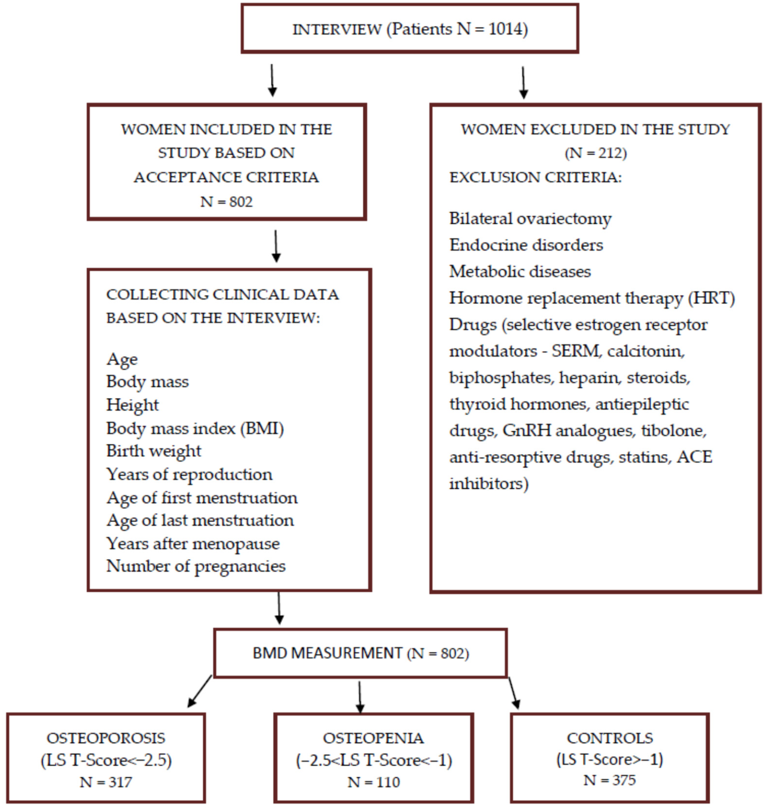
2.1. Study Group

A total of 802 unrelated postmenopausal Caucasian women in the clinical hospital of the Pomeranian Medical University in Szczecin were enrolled in the study. All patients were informed about the scope of the study before inclusion. The study was approved by the Bioethics Committee of the Pomeranian Medical University in Szczecin (no. KB-0012/100/15). The study was conducted in accordance with the Helsinki Declaration (1975, revised 2000). Written informed consent was obtained from all participants. Patients had their body weight and height measured, which made it possible to calculate body mass index (BMI). The ratios of the subjects’ average bone mineral density to the young adult (YA) average and average bone mineral density to the age-matched average (AM) were also determined.
A thorough medical history was taken with the patients qualified to participate in the experiment, including past medical history, birth weight, age of first and last menstruation, and drug treatment used, with particular emphasis on hormonal treatment and preparations from the group of drugs that affect bone mass. Postmenopausal women not taking hormone replacement therapy (HRT) and drugs such as selective estrogen receptor modulators (SERM), calcitonin, biphosphates, heparin, steroids, thyroid hormones, antiepileptic drugs, GnRH analogues, tibolone, anti-resorptive drugs, statins, and ACE inhibitors were eligible for the experiments (Figure 1). Patients with bilateral ovariectomy, endocrine disorders, metabolic diseases, and connective tissue diseases were also excluded from the experiment. Qualified women underwent densitometry using a Lunar DPX100 instrument (Lunar Corp., Madison, WI, USA), with a measurement accuracy of 0.5%. Bone mineral density (BMD) was determined at the lumbar L1–L4 using the dual X-ray beam (DXA) method. The obtained measurements were taken as absolute values (BMD (g/cm2)) and related to the mean value in the age group.
The T-score and Z-score values were then calculated, which allowed the assignment of the studied women to different groups. After analyzing the results, measurements that could correspond to degenerative changes in the lumbar spine were excluded from the study. Based on T-score, patients were divided into control group (n = 375, T-score > −1.0), women with osteopenia (n = 110, T-score between −1.0 and −2), and women with osteoporosis (n = 317, T-score < −2.5).

2.2. Determination of OPG Polymorphisms

Five OPG polymorphisms (rs3102735, rs3134070, rs207361, rs7844539, rs2073618) were analyzed in the development of osteoporosis and osteopenia in postmenopausal women. Real-time polymerase chain reaction (rt-PCR) was used for the analysis. This method uses hybridization probes labeled with a fluorescent dye that binds to DNA at a specific site. With this method, the amount of product in its exponential growth phase can be determined from successive cycle measurements so the DNA amplification process can be monitored in real-time. The LightCycler®96 instrument and LightCycler®96 Basic Software version 1.2 were used for molecular studies to analyze the results obtained. Fluorescence measured during melting curve analysis was used for genotyping. For polymorphisms, primers and probes specific for the amplified fragments were included—LightSNiP rs3102735, LightSNiP rs3134069, LightSNiP rs2073617, LightSNiP rs7844639, and LightSNiP rs2073618 (TIBMolbiol, Berlin, Germany) reagents were used. Real-time PCR reactions were performed for 45 cycles for each polymorphism tested.
All methods were carried out in accordance with relevant guidelines and regulations.

2.3. Statistical Analysis

Statistical analysis of the obtained results was carried out using the R program—version 4.1.2 (R Foundation for Statistical Computing, Vienna, Austria)—and the SNPassoc package. For quantitative variables, an analysis of conformity to the Gaussian distribution was carried out using the Shapiro–Wilk normality test and presented as mean ± standard deviation (SD). In the case of conformity of the trait distribution to the normal distribution, a one-way analysis of variance for unrelated variables (ANOVA) was used to assess the relationship between the means in the study groups, followed by the Tukey HSD post hoc test. Categorical variables were presented as numbers (percentages) and were compared according to the abundance of expected values using Pearson’s χ2 test or Fisher’s exact test. All statistical tests performed were two sided. Values of two-sided p < 0.05 were considered statistically significant. When the two analyzed characteristics were quantitative, the Pearson correlation with Holm’s correction for multiple testing was used to look for relationships between them.
Pearson’s χ2 test was used to check whether each of the studied genetic variants met the assumptions of the Hardy–Weinberg equilibrium (HWE). The associations of the studied SNPs with the risk of osteopenia and osteoporosis were assessed in five genetic models (codominant, dominant, recessive, super-dominant, and log additive) using unconditional logistic regression analysis and presented as odds ratios (OR) and associated 95% confidence intervals (95% CI). Inheritance models were selected using the Akaike information criterion (AIC).
The linkage disequilibrium (LD) parameters between the studied SNPs and haplotype frequencies were calculated using Haploview version 4.2 software. The linkage disequilibrium analysis was performed for the entire study group. The distance between the analyzed SNPs of the TNFRSF11B gene was calculated. The obtained statistics, Lewontin’s D′, logarithm of the odds (LOD), and correlation coefficients r2, between polymorphic variants are summarized in the Section 3. Associations of TNFRSF11B gene haplotypes with osteopenia and osteoporosis were performed for four polymorphic variants, excluding the rs7844539 variant due to weak coupling with the remaining polymorphisms. Statistically significant p-values were corrected using a 10,000-fold permutation test.

3. Results

3.1. Evaluation of Clinical Data

Table 1 shows the mean densitometric results of female patients in the groups listed in the study. The mean bone mineral density of the lumbar spine (L2–L4), in g/cm2, was 1.20 ± 0.10 in the control group, 0.98 ± 0.05 in women with osteopenia, and 0.82 ± 0.07 in patients with osteoporosis (< 0.001). The ratios of mean BMD to the mean for young adult women (YA) and mean BMD to the mean for age (AM) were, respectively, in percentages: 100.45 ± 8.03 and 108.52 ± 10.23 in the control group, 81.71 ± 4.43 and 89.24 ± 6.62 in osteopenia, and 68.26 ± 5.34 and 78.10 ± 7.15 in osteoporosis (p < 0.001). The mean values of T-score and Z-score parameters in the control group were 0.05 ± 0.90 and 0.64 ± 1.11, in osteopenia −1.80 ± 0.43 and −0.84 ± 0.66, and −3.16 ± 0.54 and −1.62 ± 0.72 in osteoporosis (p < 0.001).

3.2. Figures, Tables and Schemes

Analyzing the clinical data collected from medical records and patient interviews, it was observed that the average age of women with osteoporosis was statistically significantly higher than the averages in the other groups. Statistically significant differences were also observed for height, weight, and BMI. The lowest mean height was observed in the group with osteoporosis. Patients with osteoporosis also had the lowest mean body weight, and the difference was statistically significant compared to the mean of women with osteopenia (p = 0.011) and the control group (p < 0.001). The mean body mass index in the group of women with osteoporosis was significantly lower than the mean in the control group. No differences were observed between the mean years of onset of first and last menstruation and the reproductive period calculated from them. On average, the group with osteoporosis had more years since menopause than the other groups. The number of pregnancies was very similar in all groups. In contrast, the average birth weight of their children was statistically significantly highest for the controls. These results are shown in Table 2.
We also analyzed the association of the patients’ clinical and densitometric data for the different genotypes of the OPG gene variants studied in the groups of women separated by T-score. Statistically significant differences were found between the mean neonatal birth weight and genotypes of the rs3134069 in the group of women with osteoporosis and the rs2073618 genotypes in the control group. The mean T-score in patients with osteoporosis differed between genotypes of the rs2073618 polymorphism and averaged 1.21 ± 0.55 for the GG genotype and 1.83 ± 0.61 for GC heterozygotes and 1.61 ± 0.92 for the CC genotype. However, the most interesting results were obtained for rs3102735. For the entire study population of women, the average birth weight of newborns of mothers with the CC genotype was 4056.47 g, which was significantly higher than the average of 3238.33 g of women with the heterozygous TC genotype (p < 0.001) and 3177.69 g with the TT genotype. The difference between the averages for the TC and TT genotypes was not statistically significant. After dividing the groups studied in the study, differences were observed for women with osteoporosis and the control group. For patients with osteopenia, the difference was not statistically significant, but the average weight of newborns of mothers with the CC genotype was also the highest and for those with the TT genotype the lowest. Other parameters such as body mass, BMI, and BMD did not show any statistical relationship with the tested polymorphisms.

3.3. Evaluation of the Prevalence of the Polymorphisms Studied

After laboratory analysis by real-time PCR, results were obtained for 375 controls, 110 women with osteopenia, and 317 with osteoporosis. The frequencies of the polymorphic variants analyzed in the study followed the Hardy–Weinberg equilibrium (HWE) law for all groups. The Minor Allele Frequency (MAF) for the groups and the percentage of missing genotype designations are shown in Table 3.

3.4. Effect of the Prevalence of the Polymorphisms Studied on Osteopenia

Table 4 compares the allele frequencies of the polymorphic variants discussed in the paper in patients with osteopenia and the control group. The frequencies of the rarer alleles of these SNPs were comparable in both groups. Wild-type alleles of variants rs3102735, rs3134069, rs2073617, and rs2073618 were observed more frequently in women with osteopenia compared to the control group. Only for the rs7844539 polymorphism was the rarer C allele present, in 15% of patients with osteopenia and 13% of the control group (OR = 1.23; 95% PU: 0.65–2.34; p = 0.526).
In the next stage of the study, we examined the associations of the studied SNPs with the risk of osteopenia in five genetic models (codominant, dominant, recessive, super-dominant, and logarithmic) using unconditional logistic regression analysis. No statistically significant results were obtained for any of the models (Table 5).

3.5. Effect of the Prevalence of the Polymorphisms Studied on Osteoporosis

Comparing the allele frequency of the TNFRSF11B gene variants studied in the control and osteoporosis groups, a statistically significant difference was observed for rs2073617. The C allele was more common in patients with osteoporosis compared to the control group (51% vs. 46%, p = 0.041; OR = 1.249; 95% PU: 1.008–1.548). For the other polymorphic variants, there were no statistically significant differences in allele frequency between the control and osteoporosis groups (Table 6).
Also, in the association analysis of polymorphic variants of the TNFRSF11B gene with osteoporosis risk, the most interesting results were obtained for rs2073617. A predominance in the dominant model (TT vs. TC+CC) of genotypes containing the mutant C allele in the group with osteoporosis of 27.3% vs. 22.1% in controls was observed (OR = 1.35; 95% PU: 0.96–1.90; p = 0.083; AIC = 930.4). The best model for this variant was the log-additive model (OR = 1.24; 95% PU: 1.00–1.53; p = 0.046; AIC = 929.4). For the remaining SNPs, single-locus analysis showed no statistically significant association with osteoporosis (Table 7).

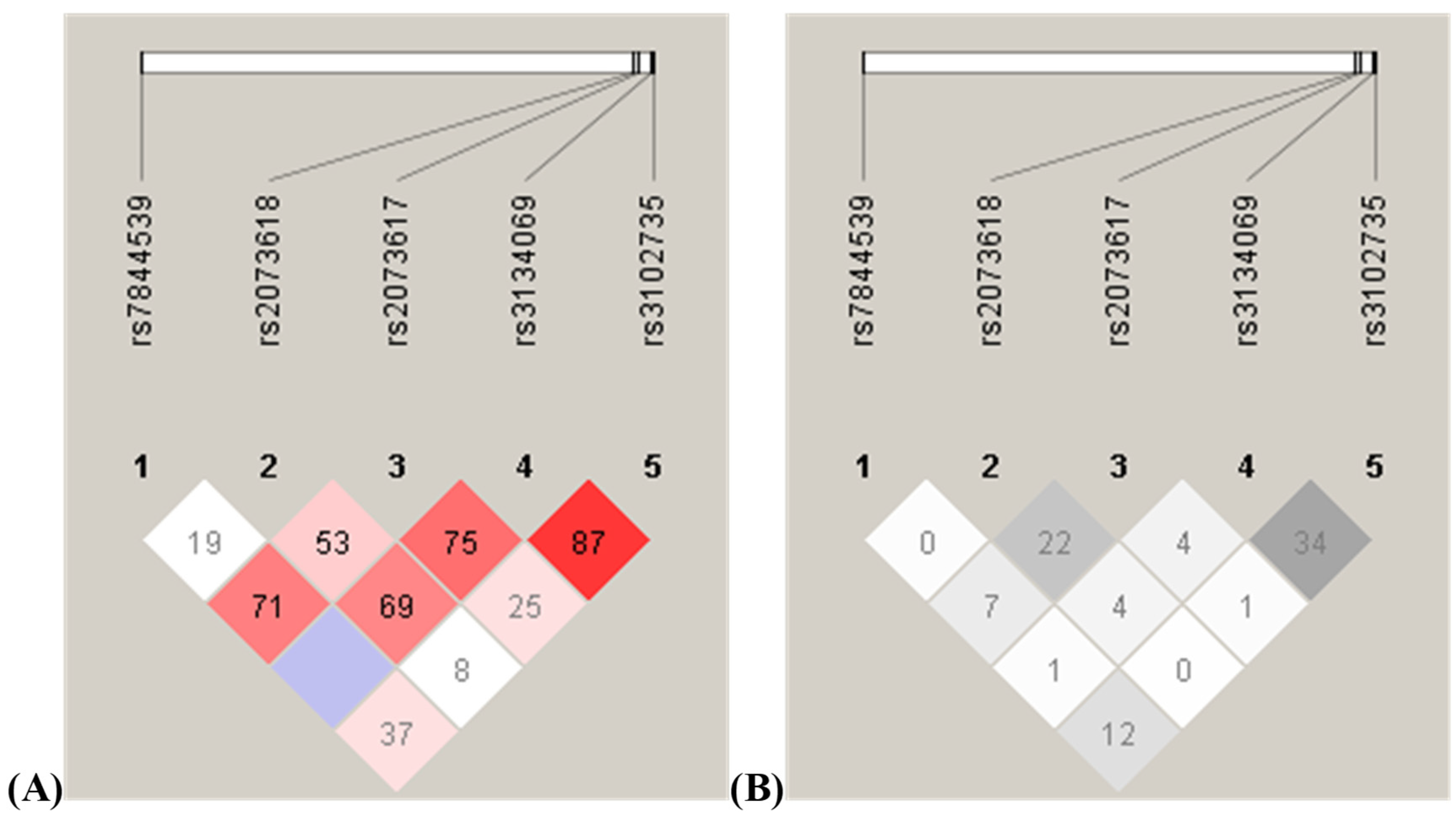
3.6. Linkage Disequilibrium Analysis and Haplotype Frequency Analysis of the Analyzed Polymorphisms

Coupling disequilibrium analysis was performed for the entire study group of 802 women. The distance between the analyzed SNPs of the TNFRSF11B gene was only 82 base pairs of the rs3134069 and rs3102735 variants, while rs7844539 and rs3102735 were separated by more than 26 kb (26,345 base pairs, to be exact). Statistics obtained, Lewontin’s D′, the logarithm of the odds (LOD), and r2 correlation coefficients, between polymorphic variants are summarized in Table 8 and shown in Figure 2.
Associations of TNFRSF11B gene haplotypes with osteopenia and osteoporosis were carried out for four polymorphic variants. The rs7844539 variant was excluded from the analysis due to its weak association with the other polymorphisms. The obtained associations are shown in Table 9 and the letters in the haplotypes indicate the first allele of the rs2073618 polymorphism, the second allele of the rs2073617 polymorphism, the third allele of the rs3134069 polymorphism, and the fourth allele of the rs3102735 polymorphism, respectively. In the control group, the CTTA haplotype was the most common. The GCTA haplotype was present in 32.4% of patients in the control group and 40.7% of those with osteoporosis (p = 0.0078). In contrast, the GTTA haplotype was less frequent in the group of patients with osteoporosis compared to the control group (7.6% vs. 12.1%, p = 0.0171). Statistically significant differences were also observed in the frequency of the CCTA haplotype between the control group and osteopenia (p = 0.0083) and osteoporosis (p = 0.0022) groups. A 10,000-fold permutation test was also performed, which raised the significance level p. The p-values obtained by this test are shown in Table 9 in parentheses next to the corresponding statistically significant values. The test confirmed statistically significant associations of CCTA and GCTA haplotypes with osteoporosis (p = 0.0132 and p = 0.0467, respectively).

4. Discussion

Osteoporosis is the most common bone disease characterized by decreased bone density and increased risk of fractures. Many studies have shown that genetics is one of the most important risk factors for this disease [12]. The OPG gene is one such gene that has been studied for its potential involvement in the etiology of osteoporosis. OPG is a protein that regulates bone resorption by inhibiting the activity of osteoclasts, the cells that break down bone tissue. Polymorphisms in the OPG gene can alter the expression or function of the OPG protein, which can affect bone remodeling and potentially lead to osteoporosis.
The author’s study showed no effect of the rs3102735 polymorphism on the incidence of osteopenia and osteoporosis. This seems to be in line with the study by Wu et al., whose study group was postmenopausal women, with a special focus on women with osteoporosis [13]. They observed that this polymorphism does not affect bone mass either in postmenopausal women with osteoporosis or in healthy postmenopausal women [13]. Similarly, García-Unzueta et al. found no relationship between the rs3102735 polymorphism and reduced bone mass in women [14]. Brambila-Tapia et al. studied the significance of the rs3102735 polymorphism in Mexican women with rheumatoid arthritis and osteoporosis [15]. They did not observe any statistically significant correlations [15]. Similarly, in a Polish study by Boroń et al., where the effect of different OPG gene polymorphisms on T-score was investigated, the rs3102735 polymorphism was not found to affect the results of the women studied [16]. The subject of a Brazilian study was the association of serum RANKL and OPG levels with vertebral fractures and bone mineral density in women with systemic lupus erythematosus. The data obtained by the researchers did not show statistically significant relationships [17]. On the other hand, the results of a study by - Blascakova et al. showed significant differences between bone mineral density among subjects with AA and GG genotypes (p < 0.05) in the control group and AA and AG genotypes (p < 0.01) in the osteoporotic group [18]. However, there was no statistical significance in the genotypes represented between the control and osteoporotic groups [18]. Similarly, Abdi et al. found that the rs3102735 polymorphism is not a potential risk factor for postmenopausal osteoporosis but correlates with the BMD of women already suffering from osteoporosis [19].
There are also several studies indicating that the rs3102735 polymorphism is linked to the incidence of osteoporosis. The first was conducted on a Danish population in 2002 by Langdahl et al., and the researchers suggested that the rs3102735 polymorphism may have an impact on both osteoporosis and fracture incidence as the presence of this polymorphism correlated with bone mass [20]. In another 2004 study of Danish postmenopausal women, Jorgensen et al. noted a significant effect of the rs3102735 polymorphism in the OPG promoter region on bone mass and fracture incidence, independent of serum osteoprotegerin levels [21]. In 2006, two groups of researchers from China looked at the problem. Hsu et al. found that the GG genotype of the rs3102735 polymorphism positively correlated with total BMD in a population-based study, but this was a study conducted among men [22]. The first Polish study on this topic was conducted in 2009 by Seremak-Mrozikiewicz et al., and 310 unrelated women were included. The researchers concluded that the variant may have a role in the development of osteoporosis in a group of osteoporotic women, but the researchers themselves acknowledged that further research in this direction is needed, with particular attention to the potential influence of other factors [23]. In contrast, a study performed on a group of 200 women in Egypt by Hussien et al. indicated that the homozygous GG genotype occurring with the rs3102735 polymorphism significantly increased the risk of osteoporosis [24]. In 2016, a Croatian study found that postmenopausal women with osteoporosis were more likely to have the AG genotype than women without osteoporosis, which may suggest that the rs3102735 polymorphism influences greater bone mass loss in postmenopausal women. However, as the authors themselves note, the number of subjects they studied was too small to draw firm conclusions [25].
A comprehensive meta-analysis performed in 2022 by Han et al., where they analyzed the results of most of the above studies as well as previously performed meta-analyses, showed that all significant associations between OPG rs3102735 polymorphisms and increased osteoporosis risk tended to be false positives, which is in line with the results obtained in this paper [10].
The authors’ study also showed no effect of the rs3134070 gene polymorphism on the incidence of osteoporosis and osteopenia. Similar conclusions were drawn by Wu et al., who examined the relationship between the amount of body fat, the rs3134070 polymorphism, and the incidence of osteoporosis. They noted that the bone mass of postmenopausal women with osteoporosis was significantly lower than that of healthy postmenopausal women. In contrast, the percentage of body fat mass of women with osteoporosis was also significantly lower than that of healthy women. However, the rs3134070 polymorphism alone does not affect BMD in either osteoporotic postmenopausal women or healthy women [26]. Similarly, a study conducted by Kim et al. on a Korean female population showed no effect of this polymorphism on either bone mass or blood osteoprotegerin levels alone [27]. Also, a 2011 study by Mencej-Bedrac et al. showed no effect of rs3134070 on serum OPG levels. This study also showed that this polymorphism cannot be considered a genetic susceptibility factor for postmenopausal osteoporosis and has no effect on bone mineral density [28]. In 2015, Zavala-Cerna et al. studied the osteoprotegerin rs3134070 polymorphism in a Mexican population with rheumatoid arthritis and osteoporosis, finding no statistically significant correlation [29]. Cvijetic et al. also found no effect of this polymorphism on either bone mass or the incidence of osteoporosis [25]. There are also several publications indicating an association of this alteration with osteoporosis and associated fractures. In the case of the rs3134070 polymorphism, the first researchers looking for a correlation between reduced bone mass and osteoporosis were Langdahl et al., who showed that this polymorphism was associated with the incidence of osteoporotic fractures [20]. Other studies showing the effect of this polymorphism on bone mineral density were those by Arko et al. [4] and Zajickova et al. [30]. Of note is the higher prevalence of the rs3134070 polymorphism in systemic lupus erythematosus patients with low BMD compared to patients with normal BMD. Bonfa et al. suggest that a possible explanation is that the G allele may induce OPG dysfunction, with adverse effects on BMD, despite normal serum OPG concentrations, or that this polymorphism may have yet another undiscovered effect on bone structure and maintenance of bone health [17].
A meta-analysis conducted in 2022 by Han et al., taking into account all studies conducted to date, also found no effect of the osteoprotegerin rs3134070 polymorphism on the incidence of osteoporosis, which is in line with the conclusions drawn from this study [10].
The study found a statistically significant difference between the incidence of the rs207361 polymorphism in the control group and the osteoporosis group. The C allele was more frequent in patients with osteoporosis compared to the control group. Moreover, in the correlation analysis of polymorphic variants, a predominance of genotypes containing the mutant C allele in the group with osteoporosis of 27.3% vs. 22.1% in controls was observed in the dominant model (TT vs. TC+CC). Similar results were obtained by Wu et al., who found that, in postmenopausal women with osteoporosis, femoral neck BMD was significantly lower in those with the CC genotype compared to those with the TT and TC genotypes. In a group of healthy postmenopausal women, there were no significant differences in BMD between the three genotypes, but the polymorphism itself did not affect the incidence of osteoporosis [31]. In contrast, Boroń et al. showed that the CC genotype may be a factor for the increased risk of faster bone mass loss and osteoporosis in Polish postmenopausal women. The authors suggest that this polymorphism may be a potential genetic marker responsible for the development of osteoporosis [16].
There are also several studies suggesting no association of this polymorphism with osteoporosis. The first report suggesting an effect of this alteration on bones was a Swedish study by Brandstrom et al., where it was suggested to have an effect on reduced bone mass among men [32]. Two years later, Langdahl et al. demonstrated a lack of effect of this polymorphism on both the incidence of osteoporosis and associated fractures. Interestingly, the same study showed that the CC genotype was associated with increased lumbar spine bone mass in osteoporosis patients [20]. However, another study conducted the same year by Wynne et al. found no effect of this polymorphism on bone mass among a population of Irish women [33]. A study by Vidal et al. found no statistically significant association between rs207361 and bone mineral density in postmenopausal women [34]. However, similar to the study by Langdahl et al. [20], CC homozygotes had higher BMD at all anatomical locations, while TT homozygotes had relatively lower BMD [34]. Interestingly, in a study by Zavala-Cerna et al., where the correlation of osteoprotegerin polymorphisms in a Mexican population with rheumatoid arthritis (RA) and osteoporosis was checked, the C allele of the rs207361 polymorphism was shown to be associated with RA, but the main premise of this study was not proven [29]. The lack of an association between rs207361 or its serum concentration and the incidence of osteoporosis in older Australian women was also demonstrated by Ueland et al. [35].
Lee et al. conducted a meta-analysis that included eight studies, and the authors noted no association between rs207361 polymorphisms and bone mineral density [36]. In contrast, the results of a meta-analysis by Guo et al. indicated that rs207361 was associated with the risk of developing osteoporosis [37]. Another meta-analysis on the association of the rs207361 gene polymorphism with osteoporosis risk was conducted by Li et al. and considered the Chinese population. Based on seven studies, which included a total of 1,850 osteoporosis cases and 3074 controls, it showed that the polymorphism was significantly associated with the risk of developing postmenopausal osteoporosis. However, the authors themselves acknowledge that, due to the limitations of this study, the results and conclusions presented should be interpreted with caution [38]. Also, a meta-analysis by Han et al. considering 12 studies, including 1610 cases and 1234 controls, and previous meta-analyses found no statistically significant correlation between the rs207361 polymorphism and osteoporosis [10].
The results of the present study also showed no effect of the rs2073618 polymorphism on the development of osteoporosis. Similarly, Wynne et al. found no significant correlation between lumbar spine BMD and the presence of C alleles [33]. A study by Vidal et al. on the effect of this polymorphism on bone mineral density in postmenopausal women in Malta also did not yield any statistically significant results [34]. Bonfa et al., investigating the effect of rs2073618 on vertebral fractures and bone mineral density in premenopausal women with systemic lupus erythematosus, found no statistically significant relationships [17]. A Mexican study by Nava-Valdivia et al. found no association between rs2073618 and bone mineral density in women with rheumatoid arthritis [39]. A Mexican study by Gonzalez-Mercado et al. that sought to determine the association of this polymorphism with bone mineral density in postmenopausal women also found no association between one and the other. Interestingly, the results of the study indicated that the GG genotype was significantly associated with lower BMI in women with osteoporosis [40]. Both this work and a large number of studies have noted an association of the C allele with higher bone mineral density. A study by Langdahl et al. found that the CC genotype of the rs2073618 polymorphism was less common in patients with osteoporosis and was associated with changes in bone mass at the lumbar spine. Those with the GG genotype had higher BMD at all locations than those with the GC genotype, but, as the authors themselves report, the difference was not statistically significant. Moreover, the polymorphism was not a significant predictor of osteoporotic fractures. These results did not provide conclusive evidence for the role of the rs2073618 polymorphism in the pathogenesis of osteoporosis [20]. Similarly, Choi et al. showed that rs2073618 was associated with the BMD of the radius bone and heel bone in postmenopausal Korean women. The mineral density of these bones in women with the CC genotype was higher than in those with the GG genotype [41]. Interestingly, Moffett et al. also found an association of GG genotype rs2073618 with lower BMD, but, at the same time, there was a lower risk of femoral neck and hip fracture in these individuals [42]. Garcia-Uzueta et al. indicated that the C allele of the rs2073618 polymorphism is associated with higher BMD at the lumbar spine in women. The researchers suggest that it affects the trabecular bone more significantly than the coccyx, as there was no significant association with hip BMD. Moreover, since the relationship is stronger in premenopausal women, it may be more relevant to peak bone mass than to hip BMD [14]. On the other hand, Kim et al., investigating the association between osteoprotegerin, RANK, and RANKL gene polymorphisms and circulating OPG, soluble RANKL, and bone mineral density in Korean postmenopausal women, noted that rs2073618, in combination with the RANKL rs2277438 polymorphism, appeared to increase lumbar spine BMD, suggesting a significant gene–gene interaction between OPG and RANKL polymorphisms [27].
A study by Arko et al. looked at eight different OPG polymorphisms [4]. According to their analysis, the rs2073618 polymorphism is associated with BMD and can therefore be considered a component of genetic susceptibility to osteoporosis [4]. In contrast, the conclusion of a study by Mencej-Bedrac et al. is that this polymorphism can be considered a risk factor for genetic susceptibility to postmenopausal osteoporosis while not affecting serum OPG levels [28]. The results of Boron et al. indicated that this polymorphism is associated with reduced bone density and increased risk of postmenopausal osteoporosis, but also with women’s body weight and birth weight [16]. A study conducted on a population of Arab postmenopausal women by Abdi et al. found a significant difference in the allelic and genotypic distribution of rs2073618 among participants with and without osteoporosis. According to the researchers, the CG heterozygote was associated with a reduced risk of postmenopausal osteoporosis. The G allele had a protective effect against the disease, which is in contrast to the aforementioned study, where it was found that women with osteoporosis were less likely to have the C allele. In addition, in the same study by Abdi et al., the polymorphism analysis showed no effect on either serum OPG, RANKL, or BMD levels [19].
A meta-analysis by Han et al. in 2022, which analyzed most of the above studies, yielding a total of 3472 cases and 3035 controls, showed that the significant results between the rs2073618 gene polymorphism and osteoporosis risk were most likely false positives, so this variation was shown to not affect the incidence of osteoporosis, which is consistent with the results of the present study [10].
The results of this study also indicate a statistically significant association between CCTA and GCTA haplotypes and osteoporosis (p = 0.0132 and p = 0.0467, respectively). Furthermore, statistically significant differences in the frequency of the CCTA haplotype were observed between the control group and individuals with osteopenia (p = 0.0083) and osteoporosis (p = 0.0022). A 10,000-fold permutation test confirmed the statistically significant associations of CCTA and GCTA haplotypes with osteoporosis (p = 0.0132 and p = 0.0467, respectively).
Moreover, it was demonstrated that, in the control group, the most prevalent haplotype was CTTA. The GCTA haplotype occurred in 32.4% of patients in the control group and 40.7% of those with osteoporosis (p = 0.0078). Conversely, the GTTA haplotype was less frequent in the osteoporosis group compared to the control group (7.6% vs. 12.1%, p = 0.0171). In the literature, results regarding the association of haplotypes with osteoporosis are inconclusive, and various polymorphisms and haplotypes are investigated, and comparing them is difficult.
No association was found of haplotype TNFRSF11B with bone mineral density (BMD) in the femoral neck among the postmenopausal Chinese women examined [43].
The study by Canto-Cetina et al. also did not demonstrate an association of the haplotype TNFRSF11B with bone mineral density (BMD) measured at three skeletal locations [44]. However, Vidal et al., in their study, demonstrated that, in the examined population of Caucasian women, the T-G-T haplotype may increase the risk of osteoporosis due to lower expression of the OPG transcript, leading to increased bone resorption [34].
On the other hand, Rojano-Mejia et al. demonstrated that, in the studied population of Mexican women, the analysis of TNFRSF11B haplotypes serves as a reliable genetic marker for changes in bone mineral density (BMD), with the A-G-T haplotype being associated with alterations in BMD in the femoral neck [45]. The suggestion is made to continue research in other populations due to ethnic diversity [45].

5. Conclusions

In conclusion, the study of OPG gene polymorphisms (rs3102735, rs3134070, rs207361, rs7844539, and rs2073618) indicated that the rs2073617 polymorphism has a statistically significant effect on the incidence of postmenopausal osteoporosis. The results indicate that there is no relationship between the rs3102735, rs3134069, rs7844639, and rs2073618 polymorphisms and the incidence of postmenopausal osteoporosis. The presence of the C allele of the rs2073618 polymorphism was also found to increase bone mineral density in postmenopausal women. In contrast, the rs2073617 polymorphism may have potential diagnostic significance in monitoring the development of postmenopausal osteoporosis.
A limitation of this study is the small number of patients needed to further validate the results. The strengths of the study concern the detailed analysis of clinical data (e.g., BMI, BMD, birth weight, reproductive years, first and last menstrual period) with the tested polymorphisms and their impact on the risk of osteoporosis. Secondly, the study examined five OPG gene polymorphisms simultaneously, which is rarely seen in other clinical studies.

Author Contributions

F.P.—analysis of data, original draft preparation. I.U.—analysis of data, original draft preparation. A.B.—methodology and software, original draft preparation. K.K.—validation and formal analysis. T.S. writing—review and editing. M.W.—collecting literature, writing—review and editing. A.K.—writing—review and editing. P.Z.—methodology and software. B.C.—project administration, leadership, and supervision. All authors have read and agreed to the published version of the manuscript.

Funding

The study was supported by statutory projects from Pomeranian Medical University in Szczecin (Poland).

Institutional Review Board Statement

Not applicable.

Informed Consent Statement

Not applicable.

Data Availability Statement

Data are available from the authors upon request.

Conflicts of Interest

The authors declare no conflict of interest.

References

  1. Sözen, T.; Özışık, L.; Başaran, N.Ç. An overview and management of osteoporosis. Eur. J. Rheumatol. 2017, 4, 46–56. [Google Scholar] [CrossRef] [PubMed]
  2. Simonet, W.S.; Lacey, D.L.; Dunstan, C.R.; Kelley, M.; Chang, M.S.; Luthy, R.; Nguyen, H.Q.; Wooden, S.; Bennett, L.; Boone, T.; et al. Osteoprotegerin: A Novel Secreted Protein Involved in the Regulation of Bone Density. Cell 1997, 89, 309–319. [Google Scholar] [CrossRef] [PubMed]
  3. Feng, X. RANKing Intracellular Signaling in Osteoclasts. IUBMB Life 2005, 57, 389–395. [Google Scholar] [CrossRef]
  4. Arko, B.; Preželj, J.; Kocijančič, A.; Komel, R.; Marc, J. Association of the osteoprotegerin gene polymorphisms with bone mineral density in postmenopausal women. Maturitas 2005, 51, 270–279. [Google Scholar] [CrossRef] [PubMed]
  5. Pettit, A.R.; Ji, H.; von Stechow, D.; Müller, R.; Goldring, S.R.; Choi, Y.; Benoist, C.; Gravallese, E.M. TRANCE/RANKL knockout mice are protected from bone erosion in a serum transfer model of arthritis. Am. J. Pathol. 2001, 159, 1689–1699. [Google Scholar] [CrossRef]
  6. Yasuda, H.; Shima, N.; Nakagawa, N.; Mochizuki, S.-I.; Yano, K.; Fujise, N.; Sato, Y.; Goto, M.; Yamaguchi, K.; Kuriyama, M.; et al. Identity of Osteoclastogenesis Inhibitory Factor (OCIF) and Osteoprotegerin (OPG): A Mechanism by which OPG/OCIF Inhibits Osteoclastogenesis in vitro. Endocrinology 1998, 139, 1329–1337. [Google Scholar] [CrossRef]
  7. Kostenuik, P.; Shalhoub, V. Osteoprotegerin A Physiological and Pharmacological Inhibitor of Bone Resorption. Curr. Pharm. Des. 2001, 7, 613–635. [Google Scholar] [CrossRef] [PubMed]
  8. Théoleyre, S.; Tat, S.K.; Vusio, P.; Blanchard, F.; Gallagher, J.; Ricard-Blum, S.; Fortun, Y.; Padrines, M.; Rédini, F.; Heymann, D. Characterization of osteoprotegerin binding to glycosaminoglycans by surface plasmon resonance: Role in the interactions with receptor activator of nuclear factor κB ligand (RANKL) and RANK. Biochem. Biophys. Res. Commun. 2006, 347, 460–467. [Google Scholar] [CrossRef]
  9. Nair, S.; Bhadricha, H.; Patil, A.; Surve, S.; Joshi, B.; Balasinor, N.; Desai, M. Association of OPG and RANKL gene polymorphisms with bone mineral density in Indian women. Gene 2022, 840, 146746. [Google Scholar] [CrossRef]
  10. Han, X.; Zheng, L.; Mu, Y.-Y.; Li, H.-Z.; He, X.-F. Association between OPG polymorphisms and osteoporosis risk: An updated meta-analysis. Front. Genet. 2022, 13, 1032110. [Google Scholar] [CrossRef] [PubMed]
  11. Tariq, S.; Tariq, S.; Abualhamael, S.A.; Baig, M.; Malik, A.A.; Shahzad, M. Osteoprotegerin genetic polymorphisms and their influence on therapeutic response to ibandronate in postmenopausal osteoporotic females. PLoS ONE 2023, 18, e0291959. [Google Scholar] [CrossRef] [PubMed]
  12. Kim, G.S.; Koh, J.M.; Chang, J.S.; Park, B.L.; Kim, L.H.; Park, E.K.; Kim, S.-Y.; Shin, H.D. Association of the OSCAR Promoter Polymorphism with BMD in Postmenopausal Women: Oscar polymorphism and BMD. J. Bone Miner. Res. 2005, 20, 1342–1348. [Google Scholar] [CrossRef]
  13. Wu, Z.Z.; Liu, M. The effect of low-body weight combined with T149C and A163G polymorphism of osteoprotegerin promoter region on osteoporosis. Chin. J. Geriatr. 2006, 25, 645–650. [Google Scholar]
  14. García-Unzueta, M.T.; Riancho, J.A.; Zarrabeitia, M.T.; Sañudo, C.; Berja, A.; Valero, C.; Pesquera, C.; Paule, B.; González-Macías, J.; Amado, J.A. Association of the 163A/G and 1181G/C Osteoprotegerin Polymorphism with Bone Mineral Density. Horm. Metab. Res. 2008, 40, 219–224. [Google Scholar] [CrossRef] [PubMed]
  15. Brambila-Tapia, A.J.L.; Durán-González, J.; Sandoval-Ramírez, L.; Mena, J.P.; Salazar-Páramo, M.; Gámez-Nava, J.I.; González-López, L.; Lazalde-Medina, B.B.; Dávalos, N.O.; Peralta-Leal, V.; et al. MTHFR C677T, MTHFR A1298C, and OPG A163G Polymorphisms in Mexican Patients with Rheumatoid Arthritis and Osteoporosis. Dis. Markers 2012, 32, 109–114. [Google Scholar] [CrossRef] [PubMed]
  16. Boroń, D.; Kotrych, D.; Bartkowiak-Wieczorek, J.; Uzar, I.; Bogacz, A.; Kamiński, A. Polymorphisms of OPG and their relation to the mineral density of bones in pre- and postmenopausal women. Int. Immunopharmacol. 2015, 28, 477–486. [Google Scholar] [CrossRef] [PubMed]
  17. Bonfá, A.C.; Seguro, L.P.C.; Caparbo, V.; Bonfá, E.; Pereira, R.M.R. RANKL and OPG gene polymorphisms: Associations with vertebral fractures and bone mineral density in premenopausal systemic lupus erythematosus. Osteoporos. Int. 2015, 26, 1563–1571. [Google Scholar] [CrossRef] [PubMed]
  18. Blaščáková, M.M.; Blaščáková, Ľ.; Poráčová, J.; Mydlár, J.; Vašková, J.; Bernasovská, J.; Boroňová, I.; Petrejčíková, E.; Bernasovský, I. Relationship between A163G osteoprotegerin gene polymorphism and other osteoporosis parameters in Roma and non-Roma postmenopausal women in eastern Slovakia. J. Clin. Lab. Anal. 2017, 31, 22093. [Google Scholar] [CrossRef] [PubMed]
  19. Abdi, S.; Binbaz, R.A.; Mohammed, A.K.; Ansari, M.G.; Wani, K.; Amer, O.E.; Alnaami, A.M.; Aljohani, N.; Al-Daghri, N.M. Association of RANKL and OPG Gene Polymorphism in Arab Women with and without Osteoporosis. Genes 2021, 12, 200. [Google Scholar] [CrossRef] [PubMed]
  20. Langdahl, B.L.; Carstens, M.; Stenkjaer, L.; Eriksen, E.F. Polymorphisms in the Osteoprotegerin Gene Are Associated With Osteoporotic Fractures. J. Bone Miner. Res. 2002, 17, 1245–1255. [Google Scholar] [CrossRef]
  21. Kusk, P.; Madsen, B.; Fenger, M.; Lauritzen, J.B.; Jørgensen, H.L. Serum osteoprotegerin (OPG) and the A163G polymorphism in the OPG promoter region are related to peripheral measures of bone mass and fracture odds ratios. J. Bone Miner. Metab. 2004, 22, 132–138. [Google Scholar] [CrossRef]
  22. Hsu, Y.-H.; Niu, T.; Terwedow, H.A.; Xu, X.; Feng, Y.; Li, Z.; Brain, J.D.; Rosen, C.J.; Laird, N.; Xu, X. Variation in genes involved in the RANKL/RANK/OPG bone remodeling pathway are associated with bone mineral density at different skeletal sites in men. Hum. Genet. 2006, 118, 568–577. [Google Scholar] [CrossRef] [PubMed]
  23. Seremak-Mrozikiewicz, A.; Tatuśko, J.; Drews, K.; Barlik, M.; Krajewski, P.; Spaczyński, M.; Mrozikiewicz, P.M. Polymorphism of osteoprotegerin gene and osteoporosis in postmenopausal women. Ginekol. Pol. 2009, 80, 354–360. [Google Scholar] [PubMed]
  24. Hussien, Y.M.; Shehata, A.; Karam, R.A.; Alzahrani, S.S.; Magdy, H.; El-Shafey, A.M. Polymorphism in vitamin D receptor and osteoprotegerin genes in Egyptian rheumatoid arthritis patients with and without osteoporosis. Mol. Biol. Rep. 2013, 40, 3675–3680. [Google Scholar] [CrossRef] [PubMed]
  25. Cvijetic, S.; Grazio, S.; Kosovic, P.; Uremovic, M.; Nemcic, T.; Bobic, J. Osteoporosis and polymorphisms of osteoprotegerin gene in postmenopausal women—A pilot study. Rheumatology 2016, 54, 10–13. [Google Scholar] [CrossRef]
  26. Wu, Z.Z.; Feng, J.Q.; Liu, M.; Lin, W.; Huang, Y.; Shi, Q.W. The relationship between body fat ratio combined with G209A and T245G polymorphism of osteoprotegerin promoter region and osteoporosis. Chin. J. Geriatr. 2007, 12, 2409–2413. [Google Scholar]
  27. Kim, J.G.; Kim, J.H.; Kim, J.Y.; Ku, S.Y.; Jee, B.C.; Suh, C.S.; Kim, S.H.; Choi, Y.M. Association between osteoprotegerin (OPG), receptor activator of nuclear factor-kappaB (RANK), and RANK ligand (RANKL) gene polymorphisms and circulating OPG, soluble RANKL levels, and bone mineral density in Korean postmenopausal women. Menopause 2007, 14, 913–918. [Google Scholar] [CrossRef]
  28. Mencej-Bedrač, S.; Preželj, J.; Marc, J. TNFRSF11B gene polymorphisms 1181G>C and 245T>G as well as haplotype CT influence bone mineral density in postmenopausal women. Maturitas 2011, 69, 263–267. [Google Scholar] [CrossRef]
  29. Zavala-Cerna, M.G.; Moran-Moguel, M.C.; Cornejo-Toledo, J.A.; Gonzalez-Montoya, N.G.; Sanchez-Corona, J.; Salazar-Paramo, M.; Nava-Zavala, A.H.; Aguilar-Chavez, E.A.; Alcaraz-Lopez, M.F.; Gonzalez-Sanchez, A.G.; et al. Osteoprotegerin Polymorphisms in a Mexican Population with Rheumatoid Arthritis and Generalized Osteoporosis: A Preliminary Report. J. Immunol. Res. 2015, 2015, 376197. [Google Scholar] [CrossRef]
  30. Zajíčková, K.; Zemanová, A.; Hill, M.; Žofková, I. Is A163G polymorphism in the osteoprotegerin gene associated with heel velocity of sound in postmenopausal women? Physiol. Res. 2008, 57 (Suppl. S1), 153–157. [Google Scholar] [CrossRef]
  31. Wu, Z.Z.; Liu, M.; Feng, J.Q. Relationships among single T~(950) -C mutation polymorphism in the promoter region of the osteoprotegerin gene, bone mineral density and metabolism. Chin. J. Geriatr. 2005, 3, 247–250. [Google Scholar]
  32. Vidal, N.; Brandstrom, H.; Jonsson, K.; Ohlsson, C. Osteoprotegerin mRNA is expressed in primary human osteoblast-like cells: Down-regulation by glucocorticoids. J. Endocrinol. 1998, 159, 191–195. [Google Scholar] [CrossRef] [PubMed]
  33. Wynne, F.; Drummond, F.; O’Sullivan, K.; Daly, M.; Shanahan, F.; Molloy, M.; Quane, K. Investigation of the Genetic Influence of the OPG, VDR (Fok1), and COLIA1 Sp1 Polymorphisms on BMD in the Irish Population. Calcif. Tissue Int. 2002, 71, 26–35. [Google Scholar] [CrossRef] [PubMed]
  34. Vidal, C.; Brincat, M.; Anastasi, A.X. TNFRSF11B gene variants and bone mineral density in postmenopausal women in Malta. Maturitas 2006, 53, 386–395. [Google Scholar] [CrossRef] [PubMed]
  35. Ueland, T.; Bollerslev, J.; Wilson, S.; Dick, I.; Islam, F.; Mullin, B.; Devine, A.; Prince, R. No associations between OPG gene polymorphisms or serum levels and measures of osteoporosis in elderly Australian women. Bone 2007, 40, 175–181. [Google Scholar] [CrossRef] [PubMed]
  36. Lee, Y.H.; Woo, J.-H.; Choi, S.J.; Ji, J.D.; Song, G.G. Associations between osteoprotegerin polymorphisms and bone mineral density: A meta-analysis. Mol. Biol. Rep. 2010, 37, 227–234. [Google Scholar] [CrossRef] [PubMed]
  37. Guo, L.; Tang, K.; Quan, Z.; Zhao, Z.; Jiang, D. Association Between Seven Common OPG Genetic Polymorphisms and Osteoporosis Risk: A Meta-Analysis. DNA Cell Biol. 2014, 33, 29–39. [Google Scholar] [CrossRef]
  38. Li, S.; Jiang, H.; Du, N. Association between osteoprotegerin gene T950C polymorphism and osteoporosis risk in the Chinese population: Evidence via meta-analysis. PLoS ONE 2017, 12, e0189825. [Google Scholar] [CrossRef]
  39. Nava-Valdivia, C.A.; Saldaña-Cruz, A.M.; Corona-Sanchez, E.G.; Murillo-Vazquez, J.D.; Moran-Moguel, M.C.; Salazar-Paramo, M.; Perez-Guerrero, E.E.; Vazquez-Villegas, M.L.; Bonilla-Lara, D.; Rocha-Muñoz, A.D.; et al. Polymorphism rs2073618 of the TNFRSF11B (OPG) Gene and Bone Mineral Density in Mexican Women with Rheumatoid Arthritis. J. Immunol. Res. 2017, 2017, 7680434. [Google Scholar] [CrossRef]
  40. González-Mercado, A.; Sánchez-López, J.Y.; Perea-Díaz, F.J.; Magaña-Torres, M.T.; Salazar-Páramo, M.; González-López, L.; González-Mercado, M.G.; Ibarra-Cortés, B. Association of polymorphisms of the TNFRSF11B and TNFSF11 genes with bone mineral density in postmenopausal women from western Mexico. Arch. Med. Sci. 2019, 15, 1352–1356. [Google Scholar] [CrossRef]
  41. Choi, J.Y.; Shin, A.; Park, S.K.; Chung, H.W.; Cho, S.; Shin, C.S.; Kim, H.; Lee, K.H.; Kang, C.; Cho, D.Y.; et al. Genetic Polymorphisms of OPG, RANK, and ESR1 and Bone Mineral Density in Korean Postmenopausal Women. Calcif. Tissue Int. 2005, 77, 152–159. [Google Scholar] [CrossRef]
  42. Moffett, S.P.; Oakley, J.I.; Cauley, J.A.; Lui, L.Y.; Ensrud, K.E.; Taylor, B.C.; Hillier, T.A.; Hochberg, M.C.; Li, J.; Cayabyab, S.; et al. Osteoprotegerin Lys3Asn Polymorphism and the Risk of Fracture in Older Women. J. Clin. Endocrinol. Metab. 2008, 93, 2002–2008. [Google Scholar] [CrossRef]
  43. Tu, P.; Duan, P.; Zhang, R.-S.; Xu, D.-B.; Wang, Y.; Wu, H.-P.; Liu, Y.-H.; Si, L. Polymorphisms in genes in the RANKL/RANK/OPG pathway are associated with bone mineral density at different skeletal sites in post-menopausal women. Osteoporos. Int. 2014, 26, 179–185. [Google Scholar] [CrossRef]
  44. Canto-Cetina, T.; Reyes, L.P.; Herrera, L.G.; Rojano-Mejía, D.; Coral-Vázquez, R.M.; Coronel, A.; Canto, P. Polymorphism of LRP5, but not of TNFRSF11B, is associated with a decrease in bone mineral density in postmenopausal maya-mestizo women. Am. J. Hum. Biol. 2013, 25, 713–718. [Google Scholar] [CrossRef]
  45. Rojano-Mejía, D.; Coral-Vázquez, R.M.; Espinosa, L.C.; Romero-Hidalgo, S.; López-Medina, G.; del Carmen Aguirre García, M.; Coronel, A.; Ibarra, R.; Canto, P. TNFRSF11B gene haplotype and its association with bone mineral density variations in postmenopausal Mexican-Mestizo women. Maturitas 2012, 71, 49–54. [Google Scholar] [CrossRef]
Figure 1. Diagram showing the process of including and excluding participants.
Biomedicines 11 03218 g001
Figure 2. Analysis of coupling disequilibrium between the five SNPs of the TNFRSF11B gene analyzed in the study in the entire group of 802 patients. The coupling between SNPs was assessed by D′ and r2 values.(A) D′ values are on the left. D′ = 1 indicates full coupling (linkage disequilibrium); D′ = 0 indicates no coupling or coupling balance (linkage equilibrium); (B) r2 is on the right side. r2 = 1 means full coupling, r2 > 0.33 means strong coupling, and r2 = 0 coupling equilibrium.
Biomedicines 11 03218 g002
Table 1. Comparison of densitometric findings for female patients.
ParameterA
Control Group
(n = 375)
B
Osteopenia
(n = 110)
C
Osteoporosis
(n = 317)
p
(ANOVA)
p
(Tukey HSD)
BMD L2–L4 (g/cm2) <0.001C-A–<0.001
mean ± SD1.20 ± 0.100.98 ± 0.050.82 ± 0.07B-A–<0.001
median (min–max)1.19 (1.08–1.47)0.97 (0.90–1.07)0.83 (0.63–0.90)B-C–<0.001
BMD L2–L4 YA (%) <0.001C-A–<0.001
mean ± SD100.45 ± 8.0381.71 ± 4.4368.26 ± 5.34B-A–<0.001
median (min–max)99.00 (90.00–123.00)81.00 (75.00–89.00)69.00 (53.00–75.00)B-C–<0.001
BMD L2–L4 AM (%) <0.001C-A–<0.001
mean ± SD108.52 ± 10.2389.24 ± 6.6278.10 ± 7.15B-A–<0.001
median (min–max)107.00 (91.00–133.00)89.00 (74.00–108.00)78.00 (60.00–92.00)B-C–<0.001
T-score <0.001C-A–<0.001
mean ± SD0.05 ± 0.90−1.80 ± 0.43−3.16 ± 0.54B-A–<0.001
median (min–max)−0.17 (−0.97–3.13)−1.89 (−2.49)–(−1.05)−3.05 (-4.73)–(−2.50)B-C–<0.001
Z-score <0.001C-A–<0.001
mean ± SD0.64 ± 1.11−0.84 ± 0.66−1.62 ± 0.72B-A–<0.001
median (min–max)0.56 (−1.85–2.65)−0.88 (−2.36–0.77)−1.62 (−3.13–0.98)B-C–<0.001
Table 2. Comparison of clinical data of female patients.
ParameterA
Control Group
(n = 375)
B
Osteopenia
(n = 110)
C
Osteoporosis
(n = 317)
p
(ANOVA)
p
(Tukey HSD)
Patient age (years) 0.007C-A–0.058
mean ± SD53.49 ± 8.2353.19 ± 8.2056.45 ± 8.83B-A–0.972
median (min–max)55.00 (51.00–71.00)53.00 (52.00–77.00)57.00 (51.00–78.00)B-C–0.010
Height (cm) <0.001C-A–0.002
mean ± SD163.17 ± 5.98162.81 ± 5.02160.18 ± 5.12B-A–0.905
median (min–max)164.00 (152.00–180.00)163.00 (150.00–175.00)160.00 (150.00–175.00)B-C–0.002
Body mass (kg) <0.001C-A–<0.001
mean ± SD68.71 ± 12.2265.51 ± 11.1460.93 ± 9.16B-A–0.148
median (min–max)66.00 (50.00–100.00)65.00 (41.00–114.00)61.00 (43.00–85.00)B-C–0.011
BMI (kg/m2) 0.004C-A–0.003
mean ± SD25.84 ± 4.5524.72 ± 3.9723.70 ± 3.10B-A–0.157
median (min–max)24.76 (18.33–37.18)24.98 (17.30–43.43)23.63 (17.10–31.63)B-C–0.168
First menstruation (years) 0.654C-A–0.630
mean ± SD13.38 ± 1.8813.12 ± 2.3912.94 ± 2.16B-A–0.854
median (min–max)14.00 (9.00–16.00)13.00 (9.00–18.00)13.00 (9.00–18.00)B-C–0.894
Last menstruation (years) 0.057C-A–0.060
mean ± SD50.17 ± 4.3949.34 ± 4.5948.06 ± 5.08B-A–0.646
median (min–max)50.00 (41.00–58.00)50.00 (38.00–60.00)49.00 (34.00–60.00)B-C–0.237
Reproductive period (years) 0.724C-A–0.766
mean ± SD36.38 ± 5.3536.20 ± 4.9335.62 ± 5.01B-A–0.986
median (min–max)37.00 (27.00–48.00)36.50 (23.00–49.00)36.00 (24.00–48.00)B-C–0.795
Years since menopause 0.001C-A–0.013
mean ± SD7.03 ± 5.597.18 ± 6.0210.63 ± 5.75B-A–0.992
median (min–max)6.50 (1.00–23.00)6.00 (0.00–25.00)10.00 (1.00–22.00)B-C–0.003
Number of pregnancies 0.852C-A–0.994
mean ± SD1.94 ± 1.221.84 ± 1.131.92 ± 1.31B-A–0.867
median (min–max)2.00 (0.00–6.00)2.00 (0.00–6.00)2.00 (0.00–7.00)B-C–0.902
Newborn’s weight (g) 0.005C-A–0.009
mean ± SD3628.95 ± 480.753226.79 ± 411.073141.25 ± 536.32B-A–0.014
median (min–max)3600 (2460–5100)3200 (2500–4500)3000 (2470–4500)B-C–0.828
Table 3. SNP analysis for the whole group.
RsnumberControl Group
N = 375
Osteopenia
N = 110
Osteoporosis
N = 317
MAFHWE
p
Missing (%)MAFHWE
p
Missing (%)MAFHWE
p
Missing (%)
rs3102735G = 14.20.281.6G = 12.60.371.8G = 14.90.510.6
rs3134069G = 7.31.003.7G = 5.31.004.6G = 6.60.4515.8
rs2073617C = 45.90.402.4C = 43.20.165.5T = 48.50.572.8
rs2073618C = 45.80.251.1C = 44.40.561.8C = 45.40.310.9
rs7844539C = 12.90.311.1C = 15.41.001.8C = 14.01.002.86
Table 4. Allele frequency of TNFRSF11B gene polymorphisms in control and osteopenia groups.
SNPGroupAllelesPercentage of People SurveyedChi2Pearson’s pOR (95% CI)
163A>G
rs3102735
AG0.980.3600.5480.87 (0.55–1.37)
osteopenia187 (0.87)27 (0.13)
control633 (0.86)105 (0.14)
245T>G
rs3134069
TG0.961.0610.3020.70 (0.36–1.38)
osteopenia197 (0.95)11 (0.05)
control669 (0.93)53 (0.07)
950T>C
rs2073617
TC0.970.4720.4910.90 (0.66–1.22)
osteopenia117 (0.57)89 (0.43)
control396 (0.54)336 (0.46)
1181G>C
rs2073618
GC0.990.1360.7110.94 (0.70–1.28)
osteopenia119 (0.56)95 (0.44)
control402 (0.54)340 (0.46)
6890A>C
rs7844539
AC0.980.4010.5261.23 (0.65–2.34)
osteopenia181 (0.85)33 (0.15)
control648 (0.87)96 (0.13)
Table 5. Association of TNFRSF11B gene variants with osteopenia.
SNPModelControlOsteopeniaOR (95% PU)pAIC
163A>G
rs3102735
AA274 (74.3)83 (77.6)1.000.752512.8
AG85 (23.0)21 (19.6)0.82 (0.48–1.40)
GG10 (2.7)3 (2.8)0.99 (0.27–3.68)
dominant95 (25.7)24 (22.4)0.83 (0.50–1.39)0.482510.8
recessive359 (97.3)104 (97.2)1.04 (0.28–3.83)0.958511.3
overdominant284 (77.0)86 (80.4)0.82 (0.48–1.39)0.451510.8
log additive369 (77.5)107 (22.5)0.88 (0.57–1.36)0.557511.0
245T>G
rs3134069
TT310 (85.9)93 (89.4)1.000.704498.6
TG49 (13.6)11 (10.6)0.75 (0.37–1.50)
GG2 (0.6)0 (0.0)
dominant51 (14.1)11 (10.6)0.72 (0.36–1.44)0.337497.4
recessive359 (99.4)104 (100.0)1.000497.3
overdominant312 (86.4)93 (89.4)0.75 (0.38–1.51)0.413497.6
log additive361 (77.6)104 (22.4)0.70 (0.36–1.38)0.704497.2
950T>C
rs2073617
TT111 (30.3)37 (35.9)1.000.504498.4
TC174 (47.5)43 (41.7)0.74 (0.45–1.22)
CC81 (22.1)23 (22.3)0.85 (0.47–1.54)
dominant255 (69.7)66 (64.1)0.78 (0.49–1.23)0.284496.6
recessive285 (77.9)80 (77.7)1.01 (0.60–1.71)0.966497.8
overdominant192 (52.5)60 (58.3)0.79 (0.51–1.23)0.297496.7
log additive366 (78.0)103 (22.0)0.90 (0.67–1.22)0.505497.3
1181G>C
rs2073618
GG103 (27.8)31 (29.0)1.000.920514.2
GC196 (52.8)57 (53.3)0.97 (0.59–1.59)
CC72 (19.4)19 (17.8)0.88 (0.46–1.67)
dominant268 (72.2)76 (71.0)0.94 (0.59–1.52)0.807512.3
recessive299 (80.6)88 (82.2)0.90 (0.51–1.57)0.700512.2
overdominant175 (47.2)50 (46.7)1.02 (0.66–1.57)0.936512.3
log additive371 (77.6)107 (22.4)0.94 (0.68–1.29)0.702512.2
6890A>C
rs7844539
AA282 (75.8)76 (71.0)1.000.795227.7
AC84 (22.6)29 (27.1)1.28 (0.61–2.67)
CC6 (1.6)2 (1.9)1.24 (0.11–14.02)
dominant90 (24.2)31 (29.0)1.28 (0.62–2.61)0.499225.7
recessive366 (98.4)105 (98.1)1.16 (0.10–13.08)0.903226.1
overdominant288 (77.4)78 (72.9)1.27 (0.61–2.65)0.513225.7
log additive372 (36.7)107 (63.3)1.24 (0.64–2.38)0.518225.7
Table 6. Allele frequency of TNFRSF11B gene polymorphisms in the control and osteoporosis groups.
SNPGroupAllelesPercentage of People with
Non-Missing Genotypes
Chi2Pearson’s pOR (95% CI)
163A>G
rs3102735
AG0.9880.1310.7171.057 (0.782~1.428)
osteoporosis536(0.85)94(0.15)
control633(0.86)105(0.14)
245T>G
rs3134069
TG0.9180.1550.6930.894 (0.514~1.556)
osteoporosis508(0.93)36(0.07)
control669(0.93)53(0.07)
950T>C
rs2073617
TC0.9734.1390.0411.249 (1.008~1.548)
osteoporosis299 (0.49)317 (0.51)
control396 (0.54)336 (0.46)
1181G>C
rs2073618
GC0.9890.0260.8700.982 (0.793~1.216)
osteoporosis343(0.55)285(0.45)
control402(0.54)340(0.46)
6890A>C
rs7844539
AC0.9620.1000.7501.101 (0.606~2.002)
osteoporosis505 (0.86)83 (0.14)
control108 (0.87)16 (0.13)
Table 7. Associations of TNFRSF11B gene variants with osteoporosis.
SNPModelControlOsteoporosisOR (95% PU)pAIC
163A>G
rs3102735
AA274 (74.3)226 (71.7)1.000.358947.9
AG85 (23.0)84 (26.7)1.20 (0.84–1.70)
GG10 (2.7)5 (1.6)0.61 (0.20–1.80)
dominant95 (25.7)89 (28.3)1.14 (0.81–1.59)0.461947.4
recessive359 (97.3)310 (98.4)0.58 (0.20–1.71)0.312946.9
overdominant284 (77.0)231 (73.3)1.21 (0.86–1.72)0.273946.8
log additive369 (53.9)315 (46.1)1.06 (0.78–1.43)0.718947.8
245T>G
rs3134069
TT310 (85.9)119 (87.5)1.000.846589.0
TG49 (13.6)16 (11.8)0.85 (0.47–1.55)
GG2 (0.6)1 (0.7)1.30 (0.12–14.50)
dominant51 (14.1)17 (12.5)0.87 (0.48–1.56)0.635587.1
recessive359 (99.4)135 (99.3)1.33 (0.12–14.78)0.820587.3
overdominant312 (86.4)120 (88.2)0.85 (0.46–1.55)0.590587.0
log additive361 (72.6)136 (27.4)0.90 (0.52–1.55)0.693587.2
950T>C
rs2073617
TT111 (30.3)75 (24.4)1.000.135931.4
TC174 (47.5)149 (48.4)1.27 (0.88–1.83)
CC81 (22.1)84 (27.3)1.53 (1.01–2.34)
dominant255 (69.7)233 (75.6)1.35 (0.96–1.90)0.083930.4
recessive285 (77.9)224 (72.7)1.32 (0.93–1.88)0.122931.0
overdominant192 (52.5)159 (51.6)1.03 (0.76–1.40)0.829933.3
log additive366 (54.3)308 (45.7)1.24 (1.00–1.53)0.046929.4
1181G>C
rs2073618
GG103 (27.8)89 (28.3)1.000.985950.8
GC196 (52.8)165 (52.5)0.97 (0.69–1.38)
CC72 (19.4)60 (19.1)0.96 (0.62–1.50)
dominant268 (72.2)225 (71.7)0.97 (0.70–1.36)0.866948.8
recessive299 (80.6)254 (80.9)0.98 (0.67–1.44)0.921948.9
overdominant175 (47.2)149 (47.5)0.99 (0.73–1.34)0.941948.9
log additive371 (54.2)314 (45.8)0.98 (0.79–1.22)0.866948.8
6890A>C
rs7844539
AA282 (75.8)217 (73,8)1.000.948290.4
AC84 (22.6)71 (24.2)1.09 (0.55–2.15)
CC6 (1.6)6 (2.0)1.30 (0.14–11.89)
dominant90 (24.2)77 (26.0)1.10 (0.57–2.14)0.773288.5
recessive366 (98.4)288 (98.0)1.27 (0.14–11.59)0.828288.5
overdominant288 (77.4)223 (76.0)1.08 (0.55–2.13)0.821288.5
log additive372 (54.0)294 (46.0)1.10 (0.61–2.00)0.750288.4
Table 8. Statistics of linkage disequilibrium strength between the analyzed variants of the TNFRSF11B gene.
SNP1SNP2D′LODr2Distance in bp
rs7844539rs20736180.1930.520.00825,327
rs7844539rs20736170.7126.470.07725,558
rs7844539rs31340691.01.080.0126,263
rs7844539rs31027350.3738.50.12426,345
rs2073618rs20736170.53942.570.224231
rs2073618rs31340690.6964.890.043936
rs2073618rs31027350.0890.180.0011018
rs2073617rs31340690.7519.30.048705
rs2073617rs31027350.2542.250.012787
rs3134069rs31027350.87641.120.34682
Table 9. Results of association of TNFRSF11B gene haplotypes with osteopenia and osteoporosis.
Haplotype *Group TurnoutOsteopenia vs. ControlOsteoporosis vs. Control
Control GroupOsteopeniaOsteoporosisChi-Squared
p
Chi-Squared
p
CTTA0.3460.3800.3580.55310.7774
GCTA0.3240.3520.4070.51910.0078 (0.0467)
GTTA0.1210.1430.0760.34540.0171 (0.0981)
GTTG0.0630.0410.0620.40690.8409
CCTA0.0550.0010.0110.0083 (0.0608)0.0022 (0.0132)
CCGG0.0480.0600.0640.61160.3317
GCTG0.0140.0250.0110.84980.5419
* rs2073618/rs2073617/rs3134069/rs3102735. In parentheses, the p-value after the 10,000-fold permutation test is shown.
Disclaimer/Publisher’s Note: The statements, opinions and data contained in all publications are solely those of the individual author(s) and contributor(s) and not of MDPI and/or the editor(s). MDPI and/or the editor(s) disclaim responsibility for any injury to people or property resulting from any ideas, methods, instructions or products referred to in the content.

