Article

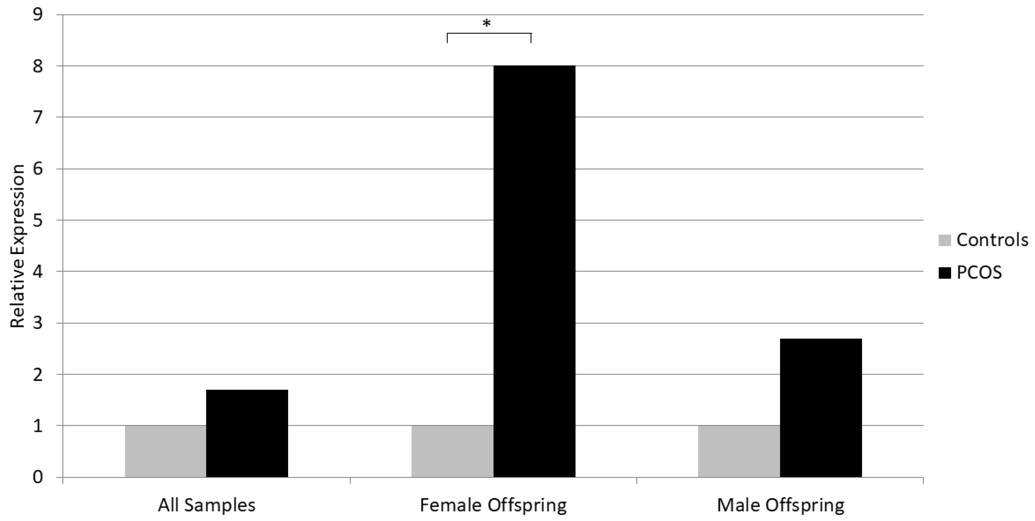
Placental mRNA Expression of Neurokinin B Is Increased in PCOS Pregnancies with Female Offspring

by
Georgios K. Markantes
1,*,
Evangelia Panagodimou
2,
Vasiliki Koika
1,
Irene Mamali
1,
Apostolos Kaponis
2,
George Adonakis
2 and
Neoklis A. Georgopoulos
1
1
Division of Endocrinology, Department of Internal Medicine, School of Health Sciences, University of Patras, 26504 Patras, Greece
2
Department of Obstetrics and Gynecology, School of Health Sciences, University of Patras, 26504 Patras, Greece
*
Author to whom correspondence should be addressed.
Biomedicines 2024, 12(2), 334; https://doi.org/10.3390/biomedicines12020334
Submission received: 5 January 2024 / Revised: 21 January 2024 / Accepted: 28 January 2024 / Published: 1 February 2024
(This article belongs to the Special Issue Molecular Research on Polycystic Ovary Syndrome (PCOS) 2.0)

Abstract

:
Current research suggests that polycystic ovary syndrome (PCOS) might originate in utero and implicates the placenta in its pathogenesis. Kisspeptin (KISS1) and neurokinin B (NKB) are produced by the placenta in high amounts, and they have been implicated in several pregnancy complications associated with placental dysfunction. However, their placental expression has not been studied in PCOS. We isolated mRNA after delivery from the placentae of 31 PCOS and 37 control women with term, uncomplicated, singleton pregnancies. The expression of KISS1, NKB, and neurokinin receptors 1, 2, and 3 was analyzed with real-time polymerase chain reaction, using β-actin as the reference gene. Maternal serum and umbilical cord levels of total testosterone, sex hormone-binding globulin (SHBG), free androgen index (FAI), androstenedione, dehydroepiandrosterone sulfate (DHEAS), Anti-Mullerian hormone (AMH), and estradiol were also assessed. NKB placental mRNA expression was higher in PCOS women versus controls in pregnancies with female offspring. NKB expression depended on fetal gender, being higher in pregnancies with male fetuses, regardless of PCOS. NKB was positively correlated with umbilical cord FAI and AMH, and KISS1 was positively correlated with cord testosterone and FAI; there was also a strong positive correlation between NKB and KISS1 expression. Women with PCOS had higher serum AMH and FAI and lower SHBG than controls. Our findings indicate that NKB might be involved in the PCOS-related placental dysfunction and warrant further investigation. Studies assessing the placental expression of NKB should take fetal gender into consideration.

1. Introduction

Polycystic ovary syndrome (PCOS) is the commonest endocrine abnormality in reproductive-aged women, affecting 5–18% of them, depending on the diagnostic criteria used [1,2]. The syndrome is characterized by considerable phenotypic heterogeneity: menstrual disturbances (mostly in the form of oligomenorrhea), the clinical manifestations of hyperandrogenemia (acne, hirsutism, androgenic alopecia), obesity, and/or insulin resistance (IR) that may affect the patients in different degrees [3]. Furthermore, women with PCOS frequently exhibit traditional risk factors for cardiovascular disease, including dyslipidemia, hypertension, metabolic syndrome, and type 2 diabetes [4]. PCOS is a major source of fertility problems, and it is the main cause of infertility related to anovulation [3]. Women with PCOS often need to use assisted reproduction technologies (ART) to achieve pregnancy and, even when this is accomplished, pregnancy is associated with an increased risk of adverse outcomes such as gestational diabetes, hypertension and preeclampsia (PE), miscarriage, and preterm delivery [5]. Anxiety, depression, and eating disorders are among the psychological comorbidities of the syndrome [6].
It is evident that PCOS is a highly prevalent disease, causing a significant burden to both patients and health care systems; hence, research aiming to understand its pathogenesis is expansive. Even though PCOS etiology is a topic attracting considerable attention, the cause of this disorder remains elusive. Several hypotheses pertaining to PCOS origins have been formulated, and most of them assume an interplay between genetic, epigenetic, and environmental factors [7]. Genome Wide Association Studies (GWAS) have identified several genetic loci with genetic susceptibility for PCOS, but the evidence supporting the relationship between these loci and the clinical manifestations of the syndrome are considered inconclusive [7]. Furthermore, it is estimated that these loci can explain less than 10% of PCOS heritability [8]. Obesity and exposure to endocrine-disrupting chemicals are also considered significant environmental contributors favoring PCOS development in genetically susceptible individuals [9]. Epigenetic mechanisms could also explain why only a subset of genetically predisposed individuals will manifest the syndrome, as it is known that factors external to the DNA can affect gene expression. A plausible theory is that the in utero hyper-exposure of the female fetus to androgens influences the expression of several genes, particularly those regulating ovarian steroid production, insulin action, and GnRH pulsatility, leading to a “re-programming” of the reproductive axis which later in life manifests as PCOS [7].
Indeed, data from animal studies have shown that prenatal exposure to excess androgen results in reproductive as well as metabolic disturbances that are typical to PCOS [10]. In humans, data are very limited but suggest that the fetuses of PCOS women are exposed to increased androgen levels in utero and this has been correlated with PCOS traits in later life [10].
The placenta, being the main steroidogenic organ in pregnancy, as well as a theoretical barrier to the overexposure of the fetus to maternally derived androgens (via androgen aromatization), could be implicated in the aforementioned theory of PCOS pathogenesis. It has been shown that women with PCOS demonstrate alterations in placental structure and function, and there is increasing evidence that androgen excess and insulin resistance, two characteristics of PCOS, may negatively affect the placenta [11]. As the placenta is a mediator of both pregnancy complications and developmental programming, mechanisms known to be involved in pregnancy complications such as PE and gestational diabetes might also be implicated in PCOS pregnancies.
Kisspeptin (KISS1) and neurokinin-B (NKB) are neuropeptides acting synergistically at the hypothalamus and stimulating pulsatile GnRH release. Their role is fundamental in puberty initiation, and they are also involved in the disturbed hypothalamic–pituitary function characterizing PCOS [12,13]. KISS1 and NKB levels increase in pregnancy due to placental production, and these peptides along with neurokinin receptors have been implicated in the pathogenesis of several pregnancy complications (PE, gestational diabetes, intrauterine growth restriction (IUGR), and preterm delivery) [14,15]. However, studies regarding the expression of these peptides in the human placenta in PCOS are lacking.
The aim of our study was to compare the placental expression of KISS1, NKB, and neurokinin receptors in women with PCOS and healthy pregnant women, and to correlate data from gene expression with the maternal and cord blood sex steroid levels.

2. Materials and Methods

2.1. Participants

This was a prospective, single-center case–control study. Participants were recruited from the Department of Obstetrics and Gynecology of the University Hospital of Patras, Greece, from January 2020 to December 2022. Only women with term, uncomplicated, singleton pregnancies, who gave birth to healthy babies were included in the study. The presence of PCOS prior to pregnancy was ascertained according to the Rotterdam criteria, when at least two of the following were present, i.e., oligo-anovulation, clinical and/or biochemical hyperandrogenemia, and polycystic ovarian morphology, after the exclusion of other pathologies with similar presentation [16]. The control group consisted of healthy women without a prior history of menstrual irregularity, clinical or biochemical hyperandrogenemia, or polycystic ovarian morphology. Exclusion criteria were any major medical condition, drug or alcohol use during pregnancy, and the use of hormonal or anti-diabetic medication up to 3 months prior to conception. Overall, 68 women participated in the study, with 31 having PCOS and 37 controls. The study was approved by the University Hospital of Patras Ethics Committee, and all participants provided written informed consent.

2.2. Placental Tissue Sampling and Gene Expression Analysis

Placental samples were collected within 15 min of placental delivery. We took three full-depth samples from each participant, sized 1 × 1 cm, from different areas at the middle point of the placental radius, away from infarcts or damage. We removed the maternal decidua and chorionic plate tissues and divided each sample into 2–3 pieces of 0.5–1 cm3. The samples were rigorously washed with normal saline solution to remove blood and were then submerged in RNAlater (QIAGEN, Hilden, Germany) solution and stored at 4 °C for 24 h and then at −20 °C until analysis.
We studied the mRNA expression of kisspeptin (KISS1), neurokinin B (NKB), and neurokinin receptors 1, 2, and 3 (NK1R, NK2R, NK3R). Total RNA was isolated from placental samples using the commercially available RNeasy Lipid Tissue Mini kit (QIAGEN, Hilden, Germany). In brief, tissue samples are homogenized in QIAzol Lysis Reagent. After the addition of chloroform, the homogenate is separated into aqueous and organic phases. The aqueous phase is collected, and ethanol is added to provide appropriate binding conditions. The sample is then subjected to a silica membrane column including a 15 min DNAse I treatment to avoid any DNA presence. Finally, RNA is eluted in RNase-free water. We estimated RNA concentration and purity by measuring the optical absorption at 260 nm and calculating the ratio 260/280 nm, respectively. Complementary DNA (cDNA) synthesis was carried out with the Transcriptor First Strand cDNA Synthesis Kit (04379012001; Roche, Basel, Switzerland) with a mixture of anchored-oligo(dT)18 primer (designed to bind at the beginning of the poly(A) tail and guarantee full-length cDNA synthesis) and 1 μg of total RNA. Quantitative real-time polymerase chain reaction (qRT-PCR) was conducted in the LightCycler 2.0 Instrument (Roche, Basel, Switzerland), using 50 ng of template cDNA and FastStart Universal SYBR Green 100 Master (Roche Hellas, Athens, Greece). PCR primers can be provided upon request for the genes ACTB (NM_001101), KISS1 (NM_002256), NKB (NM_001006667), NK1R (NM_001058), NK2R (NM_001057), and NK3R (NM_001059). Reactions were run in triplicates. We used melting curve analysis to confirm the quality of the PCR reactions and the specificity of the primers. Relative gene expression was evaluated with the ΔΔCt method, using β-actin (ACTB) as a reference gene because of its suitability for this experimental setting [17].

2.3. Hormone Measurements

At delivery, blood samples were collected from the mother (from a peripheral vein) and from the umbilical cord for hormonal determinations. Samples were immediately centrifuged (2500× g for 10 min), and the serum was collected and stored at −80 °C until analysis. Total testosterone, sex hormone-binding globulin (SHBG), androstenedione, dehydroepiandrosterone sulfate (DHEAS), Anti-Mullerian hormone (AMH), and estradiol were measured using electrochemiluminescence quantization in the Cobas e601 analyzer (Roche Diagnostics®, Mannheim, Germany). The method follows a competition principle, in which the serum is incubated with a biotinylated antibody against the measured hormone and a hormone conjugate labeled with a ruthenium complex. The biotinylated antibodies form immune complexes with both the hormone that exists in the serum and the ruthenium-labeled conjugate. After the addition of streptavidin-coated microparticles, the complexes become bound to the solid phase via the interaction of biotin and streptavidin. In the measuring cell, the microparticles are magnetically captured onto the surface of the electrode. Unbound substances are removed, and the application of a voltage to the electrode induces chemiluminescent emission, which is measured using a photomultiplier, and the hormone concentration is determined via a calibration curve. The samples were assayed in a single large batch. The intra- and inter-assay precision CV (%) values were 2.1–14.8% and 2.5%–18.1% for testosterone, 1.1–1.7% and 1.8–4.0% for SHBG, 1.8–3% and 3.7–4.6% for androstenedione, 1.5–3.2% and 2.2–2.7% for DHEAS, 0.9–1.7% and 2.7–3.5% for AMH, and 1.1–6.7% and 1.9–10.6% for estradiol. The free androgen index (FAI) was calculated according to the formula:
FAI = testosterone (nmol/L) × 100/SHBG (nmol/L)

2.4. Statistical Analysis

Data were analyzed using IBM SPSS Statistics for Windows, version 27.0 (IBM Corp., Armonk, NY, USA). Variables were tested for normality with the Kolmogorov–Smirnov test. Categorical data are presented as numbers (percentages) and continuous data as means ± standard deviations (SDs) (normally distributed variables) or as medians (interquartile ranges (IQRs)) (non-normally distributed variables). Comparisons between the two study groups were conducted using the independent samples t-test for normally distributed continuous data and the Mann–Whitney U test for non-normally distributed continuous data, while the chi-squared test was used for comparisons concerning categorical variables. Correlations were estimated by Pearson or Spearman correlation tests, as appropriate. All tests were 2-tailed, and a p-value of less than 0.05 was considered significant.

3. Results

The study included 68 women (PCOS, n = 31; controls, n = 37) with a mean age of 31.81 ± 5.49 years. The demographic data and pregnancy characteristics of the two groups are shown in Table 1. There was no significant difference between PCOS and control women regarding age, BMI, the presence of gestational diabetes, pregnancy duration, the mode of delivery, and offspring gender, weight, or length.
Women with PCOS had higher serum FAI and AMH and lower SHBG levels than controls, while no significant difference was observed between the two groups regarding serum total testosterone, androstenedione, DHEAS, and estradiol (Table 2). The umbilical cord blood hormone levels were studied separately in male and female offspring, as there are considerable differences between genders. In both the male and female subgroups, the cord blood hormone levels did not differ significantly between PCOS and control women’s offspring (Table 2).
Maternal serum total testosterone levels were strongly positively correlated with androstenedione (Spearman’s r = 0.875, p < 0.001), DHEAS (r = 0.385, p < 0.001), and AMH (r = 0.407, p < 0.001). AMH was positively correlated with testosterone, as well as with estradiol (r = 0.287, p = 0.018) and androstenedione (r = 0.454, p = 0.002). Maternal SHBG was negatively correlated with the first visit BMI (r = −0.305, p = 0.013), and positively with estradiol (r = 0.371, p = 0.002).
Umbilical cord total testosterone was positively correlated with maternal serum testosterone (r = 0.249, p = 0.043), DHEAS (r = 0.428, p < 0.001), and androstenedione (r = 0.329, p = 0.029). Cord blood DHEAS and androstenedione were also positively correlated with maternal serum androgens, while cord blood estradiol was positively correlated with maternal estradiol, testosterone, DHEAS, and androstenedione.
We did not detect a statistically significant difference in the placental mRNA expression of the studied genes between PCOS and control women, although NKB and KISS1 showed a trend for increased expression in PCOS (p = 0.16 for NKB and p = 0.12 for KISS1) (Table 3). Similarly, there was no difference in the placental gene expression between PCOS and control women in the male offspring subgroup. However, in the female offspring group, NKB expression was significantly increased in PCOS women versus controls (Median 2−ΔCt for relative expression to ACTB was 0.0008 for PCOS and 0.0001 for Controls, p = 0.021) (Table 3, Figure 1). Furthermore, placental NKB expression was higher in women with male offspring versus those with female offspring, regardless of their PCOS status (p = 0.034).
In the correlations of placental gene expression with demographic and pregnancy characteristics, NK3R expression was positively correlated with the maternal BMI at delivery (Spearman’s r = 0.409, p = 0.005). There was no correlation between placental gene expression and maternal serum hormone levels. Placental NKB expression showed a positive correlation with umbilical cord FAI (r = 0.356, p = 0.021) and AMH (r = 0.336, p = 0.028) levels, while KISS1 expression was positively correlated with cord estradiol (r = 0.324, p = 0.032), testosterone (r = 0.432, p = 0.003), and FAI (r = 0.415, p = 0.006). Lastly, NKB and KISS1 expression were strongly and positively correlated (r = 0.496, p < 0.001), while the expression of each neurokinin receptor was positively correlated with the expression of the other two (NK1R with NK2R and NK3R, and so forth).

4. Discussion

The present study showed that the placental mRNA expression of NKB is increased in women with PCOS versus controls in pregnancies with female offspring. It also confirmed that pregnant women with PCOS have higher serum AMH and higher amounts of circulating free, bioactive androgens (higher FAI and lower SHBG) compared to healthy pregnant women. Umbilical cord blood hormones did not differ between the groups in neither male nor female offspring. Notably, our study included only women with term, uncomplicated, singleton pregnancies.
The presence of a genetic component in PCOS has been anticipated since the 1960s, with studies showing the familial segregation of the syndrome [18]. Since then, several reports have confirmed these findings, demonstrating a high prevalence of PCOS in the first-degree relatives of the probands [19], as well as a high concordance in twins [8]. However, family and twin studies have failed to provide a consistent pattern of inheritance, while genetic loci identified using GWAS are estimated to explain only a small part of PCOS heritability [7,19].
The Barker or thrifty phenotype hypothesis was introduced in the 1990s, after a study showed that IUGR and low birth weight were correlated with cardiovascular disease that the fetus will develop when it becomes middle-aged [20]. This hypothesis supports that the intrauterine environment can “program” the future health of the fetus and determine the risk of chronic diseases, by inducing epigenetic changes in the fetal DNA. In the context of PCOS, this theory proposes that a hyperandrogenic intrauterine environment has epigenetic consequences affecting the expression of genes that regulate the future endocrine and metabolic functions of the fetus (i.e., GnRH pulsatility, folliculogenesis, the ovarian production of sex steroids, insulin resistance). This theory for PCOS inheritance has been supported by animal studies showing that the administration of androgens during pregnancy leads to the manifestation of PCOS-like phenotypes in adult life in the female offspring; results have been consistent across a variety of species, such as rodents, sheep, and monkeys [21,22,23]. These studies have proven that a prenatal androgen excess produces reproductive defects, such as morphological changes/increased kisspeptin expression and reduced sex steroid feedback in the hypothalamus and an increased LH secretion, leading to increased ovarian androgen production, anovulation, and infertility [10,23,24,25,26,27]. It has also been shown that an in utero androgen excess is associated with metabolic perturbations in adult life, i.e., beta cell dysfunction, insulin resistance, hyperinsulinemia, increased adiposity, and obesity have been reported [28,29,30]. In humans, obviously, there are and will be no studies exploring the effect of exogenous androgen administration in pregnancy. Some studies have longitudinally followed-up the offspring of hyperandrogenic mothers with PCOS and showed that the daughters of PCOS women exhibit PCOS features, like an elevated testosterone, an increased LH, and an increased ovarian volume [31]. Furthermore, the daughters of mothers with congenital adrenal hyperplasia (CAH), another disease state of intrauterine hyperandrogenemia, frequently manifest a PCOS phenotype [32]. Although data regarding the longitudinal metabolic phenotyping of PCOS or CAH offspring is sparse, insulin resistance, dyslipidemia, and an increased hospitalization for metabolic disorders have been reported in this population [33,34,35].
The placenta is the source of oxygen and nutrients for the fetus, and also the major producer of the steroid hormones required for the maintenance of pregnancy. As the main determinant of the intrauterine hormonal and metabolic/nutritional milieu, it is consequently implicated in the hypothesis linking intrauterine hyperandrogenemia with PCOS inheritance. In support of this view, placental insufficiency leads to infants born small for gestational age (SGA), which has been shown to be a predisposing factor for PCOS [36]. There is a great body of evidence exposing the central role of the placenta in a variety of pregnancy complications (spontaneous abortion, PE, preterm labor, IUGR, SGA) [11]. PCOS pregnancies are characterized by increased rates of such adverse outcomes which are directly related to placental dysfunction [5]. Therefore, it is possible that there are common pathogenic mechanisms/placental alterations underlying both these complications and PCOS. Khan et al. showed, for example, that there is an overlap of five proteomic biomarkers between PCOS and PE [37]; however, there are several mediators of placental pathophysiology that are still not fully investigated in PCOS [11].
There is no doubt that the process of placentation can be problematic in PCOS, as evidenced by studies demonstrating an increased occurrence of structural abnormalities in the placenta of women suffering from the syndrome. Palomba et al. found that PCOS placentae had significantly lower weight, thickness, density, and volume and an increased frequency of lesions such as fibrosis compared to those of healthy pregnant women [38]. Another study showed that PCOS was characterized by a higher incidence of placental anomalies associated with an increased hypoxic state, such as chorioamnionitis, funisitis, villitis, vascular thrombosis, infarction, and villous immaturity [39]. Notably, in both of these studies, the increased rate of placental anomalies in PCOS was not accounted for by other pregnancy complications, as the former study included only uncomplicated pregnancies, while in the latter, the adjustment for pregnancy complications was performed. Finally, a recent study examining the placentas of women with PCOS who underwent in vitro fertilization revealed that PCOS was associated with important anatomic as well as vascular placental abnormalities [40].
Apart from anomalies in placental structure, it seems that placental steroidogenic function is also impaired in PCOS. Androgen levels rise during physiologic pregnancy, serving as estrogen precursors but also in the initiation of parturition. Normally, maternal and fetal over-exposure to androgen is hampered by a significant increase in SHBG production by the maternal liver and a high level of placental aromatase activity [10]. However, it is possible that in pregnant PCOS women, the supra-normal ovarian androgen production exceeds the capacity of these mechanisms, facilitating the hyperexposure of the fetus to maternal androgens. Indeed, increased 3b-hydroxysteroid dehydrogenase and decreased aromatase activities have been observed in placental tissue from PCOS women; this was correlated with increased maternal serum androgens [41]. Furthermore, PCOS pregnancies are characterized by elevated androgen levels in the maternal serum [42,43,44], as well as in the amniotic fluid [45] compared to controls; studies investigating umbilical cord blood androgen levels and their correlation with maternal androgens have yielded conflicting results [11,44]. In our study, pregnant women with PCOS had significantly higher serum FAI, hence, an increased concentration of free androgens relative to controls; the levels of total testosterone, androstenedione, and DHEAS did not differ between the groups. Maternal serum testosterone was positively correlated with the other maternal androgens, a finding that could reflect the coordinated increase in all steps of steroidogenesis characterizing the end of gestation [10]. The positive correlation between maternal AMH and testosterone has been reported before in pregnant and non-pregnant women [46,47]. Although a causal relationship between AMH and testosterone has not been established, it has been suggested that testosterone could affect follicular growth [46], and that AMH might induce gestational hyperandrogenemia [47]. Not surprisingly, maternal SHBG concentrations were positively correlated with maternal estradiol and negatively correlated with maternal BMI; it is well known that estradiol increases and obesity decreases the hepatic SHBG production. With regard to umbilical cord hormone levels, there was no significant difference between the groups, regardless of the offspring gender. However, cord blood androgens were positively correlated with maternal serum androgens, underlining the orchestrated steroidogenesis of the maternal–placental–fetal unit, or even implying that increased maternal androgen levels could favor placental androgen production (as shown in animals) [10]. Aside from an altered steroidogenesis, the PCOS status affects placental function in a more generalized way, as proteomic analysis showed the differential expression between PCOS and control women in 258 placental proteins [44].
Little is known concerning the mechanisms involved in the placental dysfunction complicating PCOS, and most data come from animal models. Both animal and human placentae express the androgen receptor and are therefore susceptible to androgen effects [48]. The exogenous administration of androgens during gestation in the animal models of PCOS leads to decreased placental weight and fetal growth restriction [49,50,51,52,53,54], implying a negative effect of hyperandrogenemia on placental function and efficiency. The altered regulation of placental nutrient transport (decreased free fatty acid and amino acid transport and increased signal transducer and activator of transcription 3 (STAT3) expression) has been shown in prenatally androgenized rodents and monkeys [49,54,55]. Increased placental STAT3 signaling has also been found in humans with PCOS [56]. Despite increased free serum androgens, our PCOS women did not differ from controls in terms of the offspring weight and length; however, our study included only uncomplicated pregnancies, and therefore, the cases of placental insufficiency, IUGR, or SGA were excluded.
Furthermore, androgen administration results in the increased placental expression of estrogen and androgen receptors in rodents [51,57], further enhancing the actions of steroid hormones. Gestational hyperandrogenism has also been linked with placental vasculopathy; treatment with testosterone led to decreased uterine artery blood flow, elevated vascular resistance, and the increased expression of hypoxia-responsive genes in rats [52,53], while in sheep, it led to increased placental mRNA and protein expression of vascular endothelial growth factor (VEGF) [50]. Insulin resistance is another cardinal characteristic of PCOS, which might adversely affect placentation. Insulin resistance impairs human trophoblast invasion in vitro, while insulin sensitizers promote appropriate trophoblast migration and invasion [58]. Hyperandrogenic and insulin-resistant rats show disrupted trophoblast invasion/differentiation, associated with placental mitochondrial dysfunction and the over-production of reactive oxygen species [59]. Insulin resistance predisposes a woman with PCOS to gestational diabetes mellitus (GDM), which commonly complicates PCOS pregnancies [5]. GDM is in turn associated with placental dysfunction, via several mechanisms such as reduced placental apoptosis, impaired vasculogenesis, increased ischemia, and villous immaturity [11]. A small number of women with GDM were included in our study (four PCOS women and four controls). These women were diagnosed by an oral glucose tolerance test conducted at the 24th–28th week of pregnancy, none received insulin treatment, and all achieved excellent glycemic control with diet only. This mild abnormality in glucose metabolism would be expected to have minor, if any, effects on the placenta. The placental expression of the studied genes did not differ between women with and without GDM.
Another potential factor implicated in PCOS heritability and acting, at least in part, at the level of the placenta is AMH. The administration of AMH to pregnant mice led to increased maternal GnRH activity, increased LH and testosterone, and decreased estradiol and progesterone; at the placenta level, AMH treatment induced a decrease in aromatase and 3b-hydroxysteroid dehydrogenase and an increase in LH receptor expression; the female offspring of AMH-treated mice demonstrated an altered GnRH activity and the phenotypic features of PCOS [47]. In humans, AMH levels are normally low in pregnancy, reflecting ovarian suppression. However, in PCOS, high AMH levels persist and even increase in pregnancy [47]. It is therefore possible that an increased AMH contributes to maternal hyperandrogenemia via GnRH/LH activation. Moreover, since the human placenta expresses the AMH receptor type 2, an increased maternal AMH might block placental aromatase and contribute to fetal androgen overexposure. Our study confirmed that PCOS is characterized by the non-suppression of AMH during pregnancy, since we found that serum AMH levels measured at delivery are significantly higher in PCOS women versus controls.
KISS1 and NKB are increased in pregnancy as a result of placental production. Although the role of kisspeptin in regulating trophoblast invasion and embryo implantation is established [15], little is known regarding the function of NKB in normal pregnancy [14]. Kisspeptins may have a role in the pathogenesis of GDM, as lower KISS1 plasma levels and higher placental protein expression of KISS1 and the kisspeptin-1 receptor have been found in women with GDM [60,61]. KISS1 levels are elevated in preeclamptic compared to healthy pregnant women, and its concentrations correlate with the severity of PE [62,63], while KISS1 mRNA expression is increased in the preeclamptic placenta [64,65]. Furthermore, a decreased maternal KISS1 has been correlated with fetal growth restriction [66]. KISS1 might also exert antiapoptotic effects on the placenta: in hypothyroid pregnant rats, the administration of kisspeptin suppresses the apoptotic effect of hypothyroidism, by blocking the activation of the inflammasome NLRP3 pathway [67].
It is well established that PE is characterized by significantly increased maternal NKB levels and by increased placental NKB mRNA and protein expression; the placental expression of neurokinin receptors is also increased in PE [14,68]. The chronic infusion of high-dose NKB in rats induces hypertension [69], implying a possible etiologic role of NKB in PE development. The concentration of NKB is increased in the maternal plasma of IUGR pregnancies [70], while an increased placental mRNA expression of NKB has been shown in preterm labor [71].
Although KISS1 and NKB are implicated in PCOS pathogenesis (both at the level of the hypothalamus [12,13] and at the level of the ovary [72]) and in several pregnancy complications characterized by placental dysfunction, data concerning their placental expression in PCOS are lacking. To our knowledge, our study was the first to assess the expression of these peptides in PCOS placentae. We found that the mRNA expression of NKB was increased in the placenta of women with PCOS and female offspring compared to controls. This is a very interesting finding, as altered NKB expression might be involved in adverse pregnancy outcomes complicating PCOS but could also be implicated in the syndrome’s heritability. When all women were examined together regardless of the offspring gender, there was a trend for increased placental expression of NKB and KISS1, but this did not reach statistical significance. NKB expression was higher in women with male versus those with female offspring, regardless of their PCOS status. Furthermore, NKB demonstrated a positive correlation with umbilical cord FAI and AMH levels, while KISS1 was positively correlated with cord testosterone and FAI. These findings demonstrate that the placental expression of NKB and KISS1 either depends on fetal androgen and/or AMH levels, or it is regulated by factors that also influence fetal hormones. In support of the former possibility, studies in animals have shown that intrauterine hyperandrogenemia induces morphological changes in kisspeptin/neurokinin B/dynorphin neurons and increases kisspeptin-positive cells in the hypothalamus [24,25]; furthermore, the exogenous AMH administration during gestation alters the hypothalamic GnRH network [47] (in which NKB and KISS1 are major components). Maybe the fact that the percentage of male offspring was higher in our control group (57% vs. 45% in PCOS) attenuated the difference in NKB and KISS1 expression between PCOS and control women in the whole group analysis. The notion that fetal sex might affect the placental adaptation to external insults is not new; testosterone administration in pregnant sheep leads to fetal growth restriction more frequently in female fetuses, underscoring the possibility of sex-specific placental alterations in PCOS pregnancies [73]. In our study, the placental expression of the neurokinin receptors 1, 2, and 3 appeared to be higher in pregnancies with male versus female offspring, although this did not reach statistical significance. This finding should be carefully interpreted, as the placental expression of neurokinin receptors expression was very low relative to the reference gene (ACTB). Finally, we observed a strong positive correlation in the placental expression of NKB and KISS1, compatible with data from studies in rat placental cells showing that NKB upregulates KISS1 mRNA expression [74].
Our study does not lack limitations. We used the Rotterdam criteria to diagnose PCOS, which led to the inclusion of all PCOS phenotypes in our cohort, increasing the heterogeneity of this subgroup and possibly affecting the results; for example, since increased maternal androgens are a potential mediator of placental dysfunction, placental alterations might differ in hyperandrogenic versus normoandrogenic PCOS women. PCOS was retrospectively diagnosed in this study, increasing the risk of recall bias. Furthermore, our study cannot answer whether the observed increase in placental NKB expression contributes to PCOS-related placental dysfunction, or it is a compensatory mechanism. In support of the former, NKB suppresses several proteins involved in antioxidant defense and in the inhibition of intravascular coagulation in human cytotrophoblast cells [75], suggesting that NKB might be a crucial mediator in placental dysfunction.
In conclusion, our study showed that NKB placental mRNA expression is increased in women with PCOS versus controls in pregnancies with female offspring. NKB expression depends on fetal gender, and it is positively correlated with placental KISS1 expression as well as with umbilical cord blood FAI and AMH levels. More studies are needed to clarify the potential role of NKB in the placental abnormalities characterizing PCOS.

Author Contributions

Conceptualization, G.K.M.; methodology, G.K.M., V.K., A.K., G.A. and N.A.G.; software, G.K.M.; formal analysis, G.K.M.; investigation, E.P., V.K., I.M., A.K. and G.A.; resources, A.K., G.A. and N.A.G.; data curation, G.K.M. and E.P.; writing—original draft preparation, G.K.M.; writing—review and editing, E.P., V.K., I.M., A.K., G.A. and N.A.G.; supervision, G.K.M. and N.A.G.; project administration, G.K.M. and N.A.G.; funding acquisition, G.K.M. and N.A.G. All authors have read and agreed to the published version of the manuscript.

Funding

This research is co-financed by Greece and the European Union (European Social Fund—ESF) through the Operational Programme “Human Resources Development, Education and Lifelong Learning 2014–2020” in the context of the project “Exploring the developmental theory for polycystic ovary syndrome: the role of alterations in placental gene expression and in fetal DNA methylation (MIS: 5047128)”. The APC (publication fees) of this manuscript have been financed by the Research Council of the University of Patras.

Institutional Review Board Statement

The study was conducted in accordance with the Declaration of Helsinki and approved by the Institutional Review Board of the University Hospital of Patras, Greece (protocol code: 679-15/10/2019).

Informed Consent Statement

Informed consent was obtained from all subjects involved in the study.

Data Availability Statement

Data are available upon request.

Conflicts of Interest

The authors declare no conflicts of interest.

References

  1. Azziz, R.; Woods, K.S.; Reyna, R.; Key, T.J.; Knochenhauer, E.S.; Yildiz, B.O. The prevalence and features of the polycystic ovary syndrome in an unselected population. J. Clin. Endocrinol. Metab. 2004, 89, 2745–2749. [Google Scholar] [CrossRef] [PubMed]
  2. March, W.A.; Moore, V.M.; Willson, K.J.; Phillips, D.I.W.; Norman, R.J.; Davies, M.J. The prevalence of polycystic ovary syndrome in a community sample assessed under contrasting diagnostic criteria. Hum. Reprod. 2010, 25, 544–551. [Google Scholar] [CrossRef]
  3. The Amsterdam ESHRE/ASRM-Sponsored 3rd PCOS Consensus Workshop Group. Consensus on women’s health aspects of polycystic ovary syndrome (PCOS). Hum. Reprod. 2012, 27, 14–24. [Google Scholar] [CrossRef]
  4. Wild, R.A.; Carmina, E.; Diamanti-Kandrarakis, E.; Dokras, A.; Escobar-Morreale, H.F.; Futterweit, W.; Lobo, R.; Norman, R.J.; Talbott, E.; Dumesic, D.A. Assessment of cardiovascular risk and prevention of cardiovascular disease in women with the polycystic ovary syndrome: A consensus statement by the Androgen Excess and Polycystic Ovary Syndrome (AE-PCOS) Society. J. Clin. Endocrinol. Metab. 2010, 95, 2038–2049. [Google Scholar] [CrossRef] [PubMed]
  5. Yu, H.F.; Chen, H.S.; Rao, D.P.; Gong, J. Association between polycystic ovary syndrome and the risk of pregnancy complications: A PRISMA-compliant systematic review and meta-analysis. Medicine 2016, 95, e4863. [Google Scholar] [CrossRef] [PubMed]
  6. Rassi, A.; Veras, A.B.; dos Reis, M.; Pastore, D.L.; Bruno, L.M.; Bruno, R.V.; de Ávila, M.A.P.; Nardi, A.E. Prevalence of psychiatric disorders in patients with polycystic ovary syndrome. Compr. Psychiatry 2010, 51, 599–602. [Google Scholar] [CrossRef]
  7. Raperport, C.; Homburg, R. The Source of Polycystic Ovarian Syndrome. Clin. Med. Insights Reprod. Health 2019, 13, 1179558119871467. [Google Scholar] [CrossRef] [PubMed]
  8. Crespo, R.P.; Bachega, T.A.; Mendonça, B.B.; Gomes, L.G. An Update of Genetic Basis of PCOS Pathogenesis. Arch. Endocrinol. Metab. 2018, 62, 352–361. [Google Scholar] [CrossRef]
  9. Palioura, E.; Diamanti-Kandarakis, E. Polycystic ovary syndrome (PCOS) and endocrine disrupting chemicals (EDCs). Rev. Endocr. Metab. Disord. 2015, 16, 365–371. [Google Scholar] [CrossRef]
  10. Hakim, C.; Padmanabhan, V.; Vyas, A.K. Gestational Hyperandrogenism in Developmental Programming. Endocrinology 2017, 158, 199–212. [Google Scholar] [CrossRef] [PubMed]
  11. Kelley, A.S.; Smith, Y.R.; Padmanabhan, V. A Narrative Review of Placental Contribution to Adverse Pregnancy Outcomes in Women With Polycystic Ovary Syndrome. Clin. Endocrinol. Metab. 2019, 104, 5299–5315. [Google Scholar] [CrossRef]
  12. Gorkem, U.; Togrul, C.; Arslan, E.; Sargin Oruc, A.; Buyukkayaci Duman, N. Is there a role for kisspeptin in pathogenesis of polycystic ovary syndrome? Gynecol. Endocrinol. 2018, 34, 157–160. [Google Scholar] [CrossRef]
  13. George, J.T.; Kakkar, R.; Marshall, J.; Scott, M.L.; Finkelman, R.D.; Ho, T.W.; Veldhuis, J.; Skorupskaite, K.; Anderson, R.A.; McIntosh, S.; et al. Neurokinin B Receptor Antagonism in Women With Polycystic Ovary Syndrome: A Randomized, Placebo-Controlled Trial. J. Clin. Endocrinol. Metab. 2016, 101, 4313–4321. [Google Scholar] [CrossRef]
  14. Page, N.M. Neurokinin B and pre-eclampsia: A decade of discovery. Reprod. Biol. Endocrinol. 2010, 8, 4. [Google Scholar] [CrossRef]
  15. Szydełko-Gorzkowicz, M.; Poniedziałek-Czajkowska, E.; Mierzyński, R.; Sotowski, M.; Leszczyńska-Gorzelak, B. The Role of Kisspeptin in the Pathogenesis of Pregnancy Complications: A Narrative Review. Int. J. Mol. Sci. 2022, 23, 6611. [Google Scholar] [CrossRef]
  16. The Rotterdam ESHRE/ASRM—Sponsored PCOS Consensus Workshop Group 2004. Revised 2003 consensus on the diagnostic criteria and long term health risks related to polycystic ovary syndrome. Fertil. Steril. 2003, 81, 19–25. [Google Scholar]
  17. Panagodimou, E.; Koika, V.; Markatos, F.; Kaponis, A.; Adonakis, G.; Georgopoulos, N.A.; Markantes, G.K. Expression stability of ACTB, 18S, and GAPDH in human placental tissues from subjects with PCOS and controls: GAPDH expression is increased in PCOS. Hormones 2022, 21, 329–333. [Google Scholar] [CrossRef] [PubMed]
  18. Cooper, H.E.; Spellacy, W.E.; Prem, K.A.; Cohen, W.D. Hereditary factors in Stein-Leventhal syndrome. Am. J. Obstet. Gynecol. 1968, 100, 371–382. [Google Scholar] [CrossRef]
  19. Jones, M.R.; Goodarzi, M.O. Genetic Determinants of Polycystic Ovary Syndrome: Progress and Future Directions. Fertil. Steril. 2016, 106, 25–32. [Google Scholar] [CrossRef] [PubMed]
  20. Barker, D.J. The fetal and infant origins of adult disease. Br. Med. J. 1990, 301, 1111. [Google Scholar] [CrossRef] [PubMed]
  21. Abbott, D.H.; Dumesic, D.A.; Levine, J.E.; Dunaif, A.; Padmanabhan, V. Animal models and fetal programming of PCOS. In Contemporary Endocrinology: Androgen Excess Disorders in Women: Polycystic Ovary Syndrome and Other Disorders; Azziz, J.E., Nestler, J.E., Dewailly, D., Eds.; Humana Press: Totowa, NJ, USA, 2006; pp. 259–272. [Google Scholar]
  22. Franks, S. Animal models and the developmental origins of polycystic ovary syndrome: Increasing evidence for the role of androgens in programming reproductive and metabolic dysfunction. Endocrinology 2012, 153, 2536–2538. [Google Scholar] [CrossRef]
  23. Padmanabhan, V.; Veiga-Lopez, A. Sheep models of polycystic ovary syndrome phenotype. Mol. Cell. Endocrinol. 2013, 373, 8–20. [Google Scholar] [CrossRef]
  24. Cernea, M.; Padmanabhan, V.; Goodman, R.L.; Coolen, L.M.; Lehman, M.N. Prenatal testosterone treatment leads to changes in the morphology of KNDy neurons, their inputs, and projections to GnRH cells in female sheep. Endocrinology 2015, 156, 3277–3291. [Google Scholar] [CrossRef]
  25. Kondo, M.; Osuka, S.; Iwase, A.; Nakahara, T.; Saito, A.; Bayasula; Nakamura, T.; Goto, M.; Kotani, T.; Kikkawa, F. Increase of kisspeptin-positive cells in the hypothalamus of a rat model of polycystic ovary syndrome. Metab. Brain Dis. 2016, 31, 673–681. [Google Scholar] [CrossRef]
  26. Dumesic, D.A.; Abbott, D.H.; Eisner, J.R.; Goy, R.W. Prenatal exposure of female rhesus monkeys to testosterone propionate increases serum luteinizing hormone levels in adulthood. Fertil. Steril. 1997, 67, 155–163. [Google Scholar] [CrossRef]
  27. Foecking, E.M.; Szabo, M.; Schwartz, N.B.; Levine, J.E. Neuroendocrine consequences of prenatal androgen exposure in the female rat: Absence of luteinizing hormone surges, suppression of progesterone receptor gene expression, and acceleration of the gonadotropin-releasing hormone pulse generator. Biol. Reprod. 2005, 72, 1475–1483. [Google Scholar] [CrossRef]
  28. Padmanabhan, V.; Veiga-Lopez, A.; Abbott, D.H.; Recabarren, S.E.; Herkimer, C. Developmental programming: Impact of prenatal testosterone excess and postnatal weight gain on insulin sensitivity index and transfer of traits to offspring of overweight females. Endocrinology 2010, 151, 595–605. [Google Scholar] [CrossRef]
  29. Lu, C.; Cardoso, R.C.; Puttabyatappa, M.; Padmanabhan, V. Developmental programming: Prenatal testosterone excess and insulin signaling disruptions in female sheep. Biol. Reprod. 2016, 94, 113. [Google Scholar] [CrossRef]
  30. Eisner, J.R.; Dumesic, D.A.; Kemnitz, J.W.; Colman, R.J.; Abbott, D.H. Increased adiposity in female rhesus monkeys exposed to androgen excess during early gestation. Obes. Res. 2003, 11, 279–286. [Google Scholar] [CrossRef]
  31. Sir-Petermann, T.; Codner, E.; Pérez, V.; Echiburú, B.; Maliqueo, M.; Ladrón de Guevara, A.; Preisler, J.; Crisosto, N.; Sánchez, F.; Cassorla, F.; et al. Metabolic and reproductive features before and during puberty in daughters of women with polycystic ovary syndrome. J. Clin. Endocrinol. Metab. 2009, 94, 1923–1930. [Google Scholar] [CrossRef]
  32. Hague, W.M.; Adams, J.; Rodda, C.; Brook, C.G.; de Bruyn, R.; Grant, D.B.; Jacobs, H.S. The prevalence of polycystic ovaries in patients with congenital adrenal hyperplasia and their close relatives. Clin. Endocrinol. 1990, 33, 501–510. [Google Scholar] [CrossRef] [PubMed]
  33. Boomsma, C.M.; Eijkemans, M.J.; Hughes, E.G.; Visser, G.H.; Fauser, B.C.; Macklon, N.S. A meta-analysis of pregnancy outcomes in women with polycystic ovary syndrome. Hum. Reprod. Update 2006, 12, 673–683. [Google Scholar] [CrossRef]
  34. Recabarren, S.E.; Smith, R.; Rios, R.; Maliqueo, M.; Echiburú, B.; Codner, E.; Cassorla, F.; Rojas, P.; Sir-Petermann, T. Metabolic profile in sons of women with polycystic ovary syndrome. J. Clin. Endocrinol. Metab. 2008, 93, 1820–1826. [Google Scholar] [CrossRef]
  35. Speiser, P.W.; Serrat, J.; New, M.I.; Gertner, J.M. Insulin insensitivity in adrenal hyperplasia due to nonclassical steroid 21-hydroxylase deficiency. J. Clin. Endocrinol. Metab. 1992, 75, 1421–1424. [Google Scholar] [PubMed]
  36. de Zegher, F.; Reinehr, T.; Malpique, R.; Darendeliler, F.; López-Bermejo, A.; Ibáñez, L. Reduced Prenatal Weight Gain and/or Augmented Postnatal Weight Gain Precedes Polycystic Ovary Syndrome in Adolescent Girls. Obesity 2017, 25, 1486–1489. [Google Scholar] [CrossRef]
  37. Khan, G.H.; Galazis, N.; Docheva, N.; Layfield, R.; Atiomo, W. Overlap of proteomics biomarkers between women with pre-eclampsia and PCOS: A systematic review and biomarker database integration. Hum. Reprod. 2015, 30, 133–148. [Google Scholar] [CrossRef] [PubMed]
  38. Palomba, S.; Russo, T.; Falbo, A.; Di Cello, A.; Tolino, A.; Tucci, L.; La Sala, G.B.; Zullo, F. Macroscopic and microscopic findings of the placenta in women with polycystic ovary syndrome. Hum. Reprod. 2013, 28, 2838–2847. [Google Scholar] [CrossRef]
  39. Koster, M.P.; de Wilde, M.A.; Veltman-Verhulst, S.M.; Houben, M.L.; Nikkels, P.G.; van Rijn, B.B.; Fauser, B.C. Placental characteristics in women with polycystic ovary syndrome. Hum. Reprod. 2015, 30, 2829–2837. [Google Scholar] [CrossRef]
  40. Hochberg, A.; Mills, G.; Volodarsky-Perel, A.; Nu, T.N.T.; Machado-Gedeon, A.; Cui, Y.; Shaul, J.; Dahan, M.H. The impact of polycystic ovary syndrome on placental histopathology patterns in in-vitro fertilization singleton live births. Placenta 2023, 139, 12–18. [Google Scholar] [CrossRef]
  41. Maliqueo, M.; Lara, H.E.; Sánchez, F.; Echiburú, B.; Crisosto, N.; Sir-Petermann, T. Placental steroidogenesis in pregnant women with polycystic ovary syndrome. Eur. J. Obstet. Gynecol. Reprod. Biol. 2013, 166, 151–155. [Google Scholar] [CrossRef]
  42. Sir-Petermann, T.; Maliqueo, M.; Angel, B.; Lara, H.E.; Pérez-Bravo, F.; Recabarren, S.E. Maternal serum androgens in pregnant women with polycystic ovarian syndrome: Possible implications in prenatal androgenization. Hum. Reprod. 2002, 17, 2573–2579. [Google Scholar] [CrossRef] [PubMed]
  43. Glintborg, D.; Jensen, R.C.; Bentsen, K.; Schmedes, A.V.; Brandslund, I.; Kyhl, H.B.; Bilenberg, N.; Andersen, M.S. Testosterone levels in third trimester in polycystic ovary syndrome. Odense Child Cohort. J. Clin. Endocrinol. Metab. 2018, 103, 3819–3827. [Google Scholar] [CrossRef] [PubMed]
  44. Sun, M.; Sun, B.; Qiao, S.; Feng, X.; Li, Y.; Zhang, S.; Lin, Y.; Hou, L. Elevated maternal androgen is associated with dysfunctional placenta and lipid disorder in newborns of mothers with polycystic ovary syndrome. Fertil. Steril. 2020, 113, 1275–1285.e2. [Google Scholar] [CrossRef]
  45. Palomba, S.; Marotta, R.; Di Cello, A.; Russo, T.; Falbo, A.; Orio, F.; Tolino, A.; Zullo, F.; Esposito, R.; La Sala, G.B. Pervasive developmental disorders in children of hyperandrogenic women with polycystic ovary syndrome: A longitudinal case-control study. Clin. Endocrinol. 2012, 77, 898–904. [Google Scholar] [CrossRef] [PubMed]
  46. Lv, P.P.; Jin, M.; Rao, J.P.; Chen, J.; Wang, L.Q.; Huang, C.C.; Yang, S.Q.; Yao, Q.P.; Feng, L.; Shen, J.M.; et al. Role of anti-Müllerian hormone and testosterone in follicular growth: A cross-sectional study. BMC Endocr. Disord. 2020, 20, 101. [Google Scholar] [CrossRef]
  47. Tata, B.; Mimouni, N.E.H.; Barbotin, A.L.; Malone, S.A.; Loyens, A.; Pigny, P.; Dewailly, D.; Catteau-Jonard, S.; Sundström-Poromaa, I.; Piltonen, T.T.; et al. Elevated prenatal anti-Müllerian hormone reprograms the fetus and induces polycystic ovary syndrome in adulthood. Nat. Med. 2018, 24, 834–846. [Google Scholar] [CrossRef]
  48. Hsu, T.Y.; Lan, K.C.; Tsai, C.C.; Ou, C.Y.; Cheng, B.H.; Tsai, M.Y.; Kang, H.Y.; Tung, Y.H.; Wong, Y.H.; Huang, K.E. Expression of androgen receptor in human placentas from normal and preeclamptic pregnancies. Taiwan. J. Obstet. Gynecol. 2009, 48, 262–267. [Google Scholar] [CrossRef]
  49. Sathishkumar, K.; Elkins, R.; Chinnathambi, V.; Gao, H.; Hankins, G.D.; Yallampalli, C. Prenatal testosterone-induced fetal growth restriction is associated with down-regulation of rat placental amino acid transport. Reprod. Biol. Endocrinol. 2011, 9, 110. [Google Scholar] [CrossRef]
  50. Cleys, E.R.; Halleran, J.L.; Enriquez, V.A.; da Silveira, J.C.; West, R.C.; Winger, Q.A.; Anthony, R.V.; Bruemmer, J.E.; Clay, C.M.; Bouma, G.J. Androgen receptor and histone lysine demethylases in ovine placenta. PLoS ONE 2015, 10, e0117472. [Google Scholar] [CrossRef]
  51. Sun, M.; Maliqueo, M.; Benrick, A.; Johansson, J.; Shao, R.; Hou, L.; Jansson, T.; Wu, X.; Stener-Victorin, E. Maternal androgen excess reduces placental and fetal weights, increases placental steroidogenesis, and leads to long-term health effects in their female offspring. Am. J. Physiol. Endocrinol. Metab. 2012, 303, E1373–E1385. [Google Scholar] [CrossRef]
  52. Gopalakrishnan, K.; Mishra, J.S.; Chinnathambi, V.; Vincent, K.L.; Patrikeev, I.; Motamedi, M.; Saade, G.R.; Hankins, G.D.; Sathishkumar, K. Elevated testosterone reduces uterine blood flow, spiral artery elongation, and placental oxygenation in pregnant rats. Hypertension 2016, 67, 630–639. [Google Scholar] [CrossRef]
  53. Chinnathambi, V.; Blesson, C.S.; Vincent, K.L.; Saade, G.R.; Hankins, G.D.; Yallampalli, C.; Sathishkumar, K. Elevated testosterone levels during rat pregnancy cause hypersensitivity to angiotensin II and attenuation of endothelium-dependent vasodilation in uterine arteries. Hypertension 2014, 64, 405–414. [Google Scholar] [CrossRef]
  54. Hu, M.; Richard, J.E.; Maliqueo, M.; Kokosar, M.; Fornes, R.; Benrick, A.; Jansson, T.; Ohlsson, C.; Wu, X.; Skibicka, K.P.; et al. Maternal testosterone exposure increases anxiety-like behavior and impacts the limbic system in the offspring. Proc. Natl. Acad. Sci. USA 2015, 112, 14348–14353. [Google Scholar] [CrossRef] [PubMed]
  55. Abbott, D.H.; Cristin, R.; Bruns, C.R.; Barnett, D.K.; Dunaif, A.; Theodore, L.; Goodfriend, T.L.; Daniel, A.; Tarantal, D.; Tarantal, A. Experimentally induced gestational androgen excess disrupts glucoregulation in rhesus monkey dams and their female offspring. Am. J. Physiol. Endocrinol. Metab. 2010, 299, E741–E751. [Google Scholar] [CrossRef]
  56. Maliqueo, M.; Sundström Poromaa, I.; Vanky, E.; Fornes, R.; Benrick, A.; Åkerud, H.; Stridsklev, S.; Labrie, F.; Jansson, T.; Stener-Victorin, E. Placental STAT3 signaling is activated in women with polycystic ovary syndrome. Hum. Reprod. 2015, 30, 692–700. [Google Scholar] [CrossRef]
  57. Fornes, R.; Maliqueo, M.; Hu, M.; Hadi, L.; Jimenez-Andrade, J.M.; Ebefors, K.; Nyström, J.; Labrie, F.; Jansson, T.; Benrick, A.; et al. The effect of androgen excess on maternal metabolism, placental function and fetal growth in obese dams. Sci. Rep. 2017, 7, 8066. [Google Scholar] [CrossRef]
  58. Mayama, R.; Izawa, T.; Sakai, K.; Suciu, N.; Iwashita, M. Improvement of insulin sensitivity promotes extravillous trophoblast cell migration stimulated by insulin-like growth factor-I. Endocr. J. 2013, 60, 359–368. [Google Scholar] [CrossRef]
  59. Zhang, Y.; Zhao, W.; Xu, H.; Hu, M.; Guo, X.; Jia, W.; Liu, G.; Li, J.; Cui, P.; Lager, S.; et al. Hyperandrogenism and insulin resistance-induced fetal loss: Evidence for placental mitochondrial abnormalities and elevated reactive oxygen species production in pregnant rats that mimic the clinical features of polycystic ovary syndrome. J. Physiol. 2019, 597, 3927–3950. [Google Scholar] [CrossRef] [PubMed]
  60. Kapustin, R.V.; Drobintseva, A.O.; Alekseenkova, E.N.; Onopriychuk, A.R.; Arzhanova, O.N.; Polyakova, V.O.; Kvetnoy, I.M. Placental protein expression of kisspeptin-1 (KISS1) and the kisspeptin-1 receptor (KISS1R) in pregnancy complicated by diabetes mellitus or preeclampsia. Arch. Gynecol. Obstet. 2020, 301, 437–445. [Google Scholar] [CrossRef]
  61. Bowe, J.E.; Hill, T.G.; Hunt, K.F.; Smith, L.I.; Simpson, S.J.; Amiel, S.A.; Jones, P.M. A role for placental kisspeptin in β cell adaptation to pregnancy. JCI Insight 2019, 4, e124540. [Google Scholar] [CrossRef] [PubMed]
  62. Fang, L.; Gao, Y.; Wang, Z.; Li, Y.; Yan, Y.; Wu, Z.; Cheng, J.-C.; Sun, Y.-P. EGF stimulates human trophoblast cell invasion by downregulating ID3-mediated KISS1 expression. Cell Commun. Signal. 2021, 19, 101. [Google Scholar] [CrossRef] [PubMed]
  63. Adali, E.; Kurdoglu, Z.; Kurdoglu, M.; Kamaci, M.; Kolusari, A.; Yildizhan, R. Metastin levels in pregnancies complicated by pre-eclampsia and their relation with disease severity. J. Matern. Fetal Neonatal Med. 2012, 25, 2671–2675. [Google Scholar] [CrossRef]
  64. Qiao, C.; Wang, C.; Zhao, J.; Liu, C.; Shang, T. Elevated expression of KiSS-1 in placenta of Chinese women with early-onset preeclampsia. PLoS ONE 2012, 7, e48937. [Google Scholar] [CrossRef]
  65. Vazquez-Alaniz, F.; Galaviz-Hernandez, C.; Marchat, L.A.; Salas-Pacheco, J.M.; Chairez-Hernandez, I.; Guijarro-Bustillos, J.J.; Mireles-Ordaz, A. Comparative expression profiles for KiSS-1 and REN genes in preeclamptic and healthy placental tissues. Eur. J. Obstet. Gynecol. Reprod. Biol. 2011, 159, 67–71. [Google Scholar] [CrossRef] [PubMed]
  66. Smets, E.M.L.; Deurloo, K.L.; Go, A.T.J.I.; van Vugt, J.M.G.; Blankenstein, M.A.; Oudejans, C.B.M. Decreased plasma levels of metastin in early pregnancy are associated with small for gestational age neonates. Prenat. Diagn. 2008, 28, 299–303. [Google Scholar] [CrossRef] [PubMed]
  67. Santos, B.R.; Cordeiro, J.M.D.A.; Santos, L.C.; Santana, L.D.S.; Nascimento, A.E.J.; Silva, J.F. Kisspeptin Suppresses Inflammasome-NLRP3 Activation and Pyroptosis Caused by Hypothyroidism at the Maternal-Fetal Interface of Rats. Int. J. Mol. Sci. 2023, 24, 6820. [Google Scholar] [CrossRef]
  68. Liu, Y.; Chen, X.; Chen, H. Placental and umbilical cord levels of neurokinin B and neurokinin B receptor in pre-eclampsia. Int. J. Gynecol. Obstet. 2009, 107, 58–59. [Google Scholar] [CrossRef] [PubMed]
  69. Yang, J.; Dhawan, V.; Morrish, D.W.; Kaufman, S. Bimodal effects of chronically administered neurokinin B (NKB) on in vivo and in vitro cardiovascular responses in female rats. Regul. Pept. 2007, 143, 136–142. [Google Scholar] [CrossRef] [PubMed]
  70. D'Anna, R.; Baviera, G.; Corrado, F.; Crisafulli, A.; Ientile, R.; Buemi, M.; Squadrito, F. Neurokinin B and nitric oxide plasma levels in pre-eclampsia and isolated intrauterine growth restriction. BJOG 2004, 111, 1046–1050. [Google Scholar] [CrossRef]
  71. Torricelli, M.; Giovannelli, A.; Leucci, E.; Florio, P.; De Falco, G.; Torres, P.B.; Reis, F.M.; Leoncini, L.; Petraglia, F. Placental neurokinin B mRNA expression increases at preterm labor. Placenta 2007, 28, 1020–1023. [Google Scholar] [CrossRef] [PubMed]
  72. Blasco, V.; Pinto, F.M.; Fernández-Atucha, A.; Prados, N.; Tena-Sempere, M.; Fernández-Sánchez, M.; Candenas, L. Altered expression of the kisspeptin/KISS1R and neurokinin B/NK3R systems in mural granulosa and cumulus cells of patients with polycystic ovarian syndrome. J. Assist. Reprod. Genet. 2019, 36, 113–120. [Google Scholar] [CrossRef] [PubMed]
  73. Beckett, E.M.; Astapova, O.; Steckler, T.L.; Veiga-Lopez, A.; Padmanabhan, V. Developmental programing: Impact of testosterone on placental differentiation. Reproduction 2014, 148, 199–209. [Google Scholar] [CrossRef] [PubMed]
  74. Oride, A.; Kanasaki, H.; Mijiddorj, T.; Sukhbaatar, U.; Ishihara, T.; Kyo, S. Regulation of kisspeptin and gonadotropin-releasing hormone expression in rat placenta: Study using primary cultures of rat placental cells. Reprod. Biol. Endocrinol. 2015, 13, 90. [Google Scholar] [CrossRef]
  75. Sawicki, G.; Dakour, J.; Morrish, D.W. Functional proteomics of neurokinin B in the placenta indicates a novel role in regulating cytotrophoblast antioxidant defences. Proteomics 2003, 3, 2044–2051. [Google Scholar] [CrossRef] [PubMed]
Figure 1. Relative placental mRNA expression of NKB in PCOS and control women. The expression level in controls is considered as reference. * p < 0.05 (Mann–Whitney U test).
Biomedicines 12 00334 g001
Table 1. Demographic data and pregnancy characteristics of the PCOS and control women included in the study. VD: vaginal delivery; CS: cesarean section; M: male; and F: female. Categorical data are presented as number (percentage) and continuous data as mean ± SD.
PCOS (n = 31)Controls (n = 37)p Value
Age (years)31.52 ± 5.3232.06 ± 5.780.677
BMI at first visit (kg/m2)26.81 ± 5.0625.27 ± 4.440.191
BMI at delivery (kg/m2) 31.98 ± 5.4929.84 ± 4.440.105
Gestational diabetes4 (12.9%)4 (10.8%)0.790
Delivery week39 (2)39 (2)0.785
Mode of delivery (VD/CS)15 (48.4%)/16 (51.6%)19 (51.4%)/18 (48.6%)0.808
Offspring gender (M/F)14 (45.2%)/17 (54.8%)21 (56.8%)/16 (43.2%)0.274
Offspring weight (g)3161.33 ± 555.143330.27 ± 462.900.179
Offspring length (cm)49.75 ± 2.4450.88 ± 2.270.139
Table 2. Maternal serum and umbilical cord blood hormone levels in PCOS and control women. SHBG: sex hormone-binding globulin; FAI: free androgen index; DHEAS: dehydroepiandrosterone sulfate; and AMH: Anti-Mullerian hormone. Data are presented as mean ± SD (normally distributed variables) or as median (IQR) (non-normally distributed variables).
Maternal Serum PCOS (n = 31)Controls (n = 37)p Value
Total testosterone (ng/dL)88.29 (98.77)91.27 (70.99)0.538
SHBG (nmol/L)415.99 ± 135.61478.20 ± 120.470.049
FAI0.68 (0.40)0.56 (0.67)0.048
Androstenedione (ng/mL)2.19 (2.10)1.64 (2.12)0.387
DHEAS (μg/dL)105.97 ± 54.46120.99 ± 72.820.347
AMH (pmol/L)7.23 (5.41)3.84 (6.07)0.012
Estradiol (pg/mL)8860 (16,301)6698 (15,510)0.310
Umbilical cord blood
Female OffspringPCOS (n = 17)Controls (n = 16)
Total testosterone (ng/dL)142.54 ± 56.71130.97 ± 37.270.501
SHBG (nmol/L)32.16 (18.15)32.50 (23.31)0.624
FAI14.57 (9.37)12.69 (11.52)0.468
Androstenedione (ng/mL)0.52 ± 0.120.44 ± 0.110.185
DHEAS (μg/dL)425.58 ± 194.81353.89 ± 151.840.255
AMH (pmol/L)1.50 (2.03)1.19 (1.56)0.624
Estradiol (pg/mL)2577.19 ± 929.942959.82 ± 1095.360.295
Male OffspringPCOS (n = 14)Controls (n = 21)
Total testosterone (ng/dL)168.76 ± 69.34166.62 ± 56.620.923
SHBG (nmol/L)33.54 (9.02)37.06 (12.43)0.255
FAI17.14 (9.13)16.12 (6.73)0.893
Androstenedione (ng/mL)0.56 ± 0.240.48 ± 0.170.341
DHEAS (μg/dL)394.01 ± 166.72349.44 ± 161.110.449
AMH (pmol/L)165.84 (80.60)180.10 (90.90)0.439
Estradiol (pg/mL)3266.23 ± 1375.432730.19 ± 1769.120.363
Statistically significant p values are shown in bold.
Table 3. Placental mRNA expression of NKB, NK1R, NK2R, NK3R, and KISS1 genes in PCOS and control women. Results are expressed as the median (IQR) relative expression to ACTB according to the ΔΔCt method.
All SamplesPCOS (n = 31)Controls (n = 37)p Value
NKB0.0017 (0.04)0.0010 (0.02)0.160
NK1R2.58 × 10−5 (5.3 × 10⁻⁵)2.44 × 10⁻⁵ (9.0 × 10⁻⁵)0.514
NK2R3.66 × 10−6 (9 × 10⁻⁶)4.14 × 10⁻⁶ (14 × 10−6)0.298
NK3R5.41 × 10−5 (18.8 × 10⁻⁵)2.80 × 10⁻⁵ (7.4 × 10⁻⁵)0.394
KISS10.0160 (0.07)0.0079 (0.07)0.120
Female OffspringPCOS (n = 17)Controls (n = 16)
NKB0.0008 (0.03)0.0001 (0.0001)0.021
NK1R1.51 × 10−5 (2.9 × 10⁻⁵)2.43 × 10⁻⁵ (7.0 × 10⁻⁵)0.762
NK2R0.77 × 10−6 (24 × 10−6)1.57 × 10⁻⁶ (4 × 10⁻⁶)0.579
NK3R1.66 × 10−5 (37.0 × 10⁻⁵)1.42 × 10⁻⁵ (6.3 × 10⁻⁵)0.631
KISS10.0136 (0.03)0.0022 (0.03)0.315
Male OffspringPCOS (n = 14)Controls (n = 21)
NKB0.0135 (0.07)0.0050 (0.02)0.586
NK1R5.62 × 10−5 (9.4 × 10⁻⁵)3.56 × 10⁻⁵ (31.4 × 10⁻⁵)0.867
NK2R4.63 × 10−6 (12 × 10−6)2.97 × 10⁻⁶ (5 × 10⁻⁶)0.660
NK3R8.40 × 10−5 (18.7 × 10⁻⁵)2.20 × 10⁻⁵ (6.8 × 10⁻⁵)0.363
KISS10.0435 (0.26)0.0089 (0.08)0.135
Statistically significant p values are shown in bold.
Disclaimer/Publisher’s Note: The statements, opinions and data contained in all publications are solely those of the individual author(s) and contributor(s) and not of MDPI and/or the editor(s). MDPI and/or the editor(s) disclaim responsibility for any injury to people or property resulting from any ideas, methods, instructions or products referred to in the content.

