Review

HGF/Met-Signaling Contributes to Immune Regulation by Modulating Tolerogenic and Motogenic Properties of Dendritic Cells

by
Jessica Hübel
1,2 and
Thomas Hieronymus
1,2,*
1
Department of Cell Biology, Institute for Biomedical Engineering, Medical Faculty, RWTH Aachen University, Pauwelsstrasse 30, 52074 Aachen, Germany
2
Helmholtz Institute for Biomedical Engineering, RWTH Aachen University, Pauwelsstrasse 20, 52074 Aachen, Germany
*
Author to whom correspondence should be addressed.
Biomedicines 2015, 3(1), 138-148; https://doi.org/10.3390/biomedicines3010138
Submission received: 23 December 2014 / Revised: 6 February 2015 / Accepted: 13 February 2015 / Published: 3 March 2015
(This article belongs to the Special Issue New aspects of the Hepatocyte Growth Factor/c-Met System)

Abstract

:
Hepatocyte growth factor (HGF)-signaling via Met can induce mitogenic, morphogenic, and motogenic activity in various cell types. Met expression in the immune system is limited to cells with antigen-presenting capacities, including dendritic cells (DCs). Thus, it appears highly conceivable that Met-signaling impacts on adaptive immune responses. However, the mechanisms by which HGF imparts its effects on immunological responses are not yet fully understood. DCs possess unique functionalities that are critically involved in controlling both tolerance and immunity. HGF conveys immunoregulatory functions, which strongly correlate with that of DCs orchestrating the apt immune response in inflammation. Therefore, this review focuses on the current knowledge of Met-signaling in DCs with specific emphasis on the morphogenic and motogenic activities. HGF has been identified to play a role in peripheral immune tolerance by directing DC differentiation towards a tolerogenic phenotype. In skin immunity, Met-signaling was shown to drive mobilization of DCs by regulating matrix metalloproteinase activities. This is strikingly reminiscent of the role of Met for regulating a cell fate program during embryonic development, wound healing, and in tumor invasion known as epithelial-mesenchymal transition (EMT). Thus, the concept emerges that an EMT program is executed by Met-signaling in DCs, which will be also discussed.

Graphical Abstract

1. Introduction on the Immunoregulatory Function of HGF/Met

Met, a high affinity transmembrane tyrosine kinase receptor for hepatocyte growth factor (HGF) was originally identified as proto-oncogene [1]. HGF was first described as a mitogenic factor for hepatocytes [2] and independently as scatter factor (SF), a motility factor for epithelial cells [3,4] that express Met in various organs during embryonic mouse development [5]. It was demonstrated that conventional null mutations of Hgf or Met in mice are embryonically lethal, emphasizing the role of HGF/Met-signaling for a proper development [6,7,8]. Later during lifetime, HGF/Met has a vital role in liver [9,10] and pancreatic β-cell [11] regeneration, and in wound healing [12,13,14]. Met-signaling leads to the transient conversion of cells from an epithelial to a mesenchymal phenotype (epithelial-to-mesenchymal transition, EMT) followed by well-controlled cell migration [15,16]. Similarly, aberrant Met-signaling contributes to tumorigenesis and induces invasive growth and metastasis [17,18].
The tissue regenerative function of HGF/Met correlates directly with immune regulation and inflammation [19]. For example, HGF is produced amongst others by platelets [2], neutrophils, and mast cells [20] and acts on various cell types involved in regenerative processes. A role of HGF/Met in autoimmune diseases has been also suggested [21,22]. Consequently, HGF/Met has been considered as a potential target for immune-based clinical therapies [19].
While Met is predominantly expressed by epithelial cells, various hematopoietic cells are in addition able to acquire Met expression. In the hematopoietic compartment and therewith in the immune system, Met expression is limited to hematopoietic progenitor cells and their progenies with antigen-presenting capacities, including monocytes/macrophages, B cells, and dendritic cells (DCs). Met-signaling was found to play a role in monocyte-macrophage development [23] and in regulating B cell homing to the lymph nodes [24]. Likewise, Met expression was shown to regulate splenic DC function [25,26]. Met expression was found on bone marrow (BM)-derived DCs and dermal DCs (dDCs) and Langerhans cells (LCs; the epidermal contingent of DCs), which play a pivotal role in skin immunity [27,28]. Consequently, it appears highly conceivable that HGF-signaling via Met impacts on adaptive immune responses.

2. Dendritic Cells Are Key Regulators of the Immune System

DCs are the most professional antigen-presenting cells (APC) of the immune system. They are part of the innate immune system and are distributed in lymphoid and non-lymphoid organs throughout the body where they serve as sentinels of the immune surveillance system [29].
DCs originate from self-renewing hematopoietic stem cells (HSCs) in the BM that develop into progenitor cells. To fulfill their function progenitor cells are obliged to migrate via the peripheral blood towards lymphoid and non-lymphoid tissues where they differentiate into immature DCs [30,31]. Upon encountering foreign pathogens DCs take them up and process them, which includes degradation and peptide/antigen loading on major histocompatibility complex class I or II (MHCI or MHCII) molecules depending on the endogenous or exogenous origin of the pathogen, respectively. MHC and antigen complexes are expressed on the cell surface for recognition by CD4+ (MHCII) and CD8+ (MHCI) T cells [32]. Thereby, DCs are the key initiators and regulators of adaptive immune responses. Interestingly, DCs exhibit the unique property to process exogenous antigens and load them on MHCI through a process referred to as cross-presentation. This represents an important mechanism to act against intracellular pathogens (which are not present in DCs themselves) or tumor cells by induction of a cytotoxic CD8+ T cell response [33,34].
Hence, DCs possess unique functionalities that are critically involved in controlling and maintaining the balance between tolerance and immunity, thus developing an effective immune response and at the same time limit self-damage. This makes DCs attractive targets for immunotherapies, such as vaccines against cancer [35], treatment of graft-versus-host disease following transplantations, and in infectious or autoimmune diseases [36].
In this respect it is interesting that HGF mediates immunoregulatory activities, which correlate with that of DCs orchestrating an appropriate adaptive immune response. Moreover, although the mechanisms by which HGF/Met effects immunological functions are not yet fully understood an impact of HGF/Met-signaling on DC functions in splenic DCs [25], as well as on dDCs and LCs was identified [27,28].

3. HGF Mediates Development of Tolerogenic Dendritic Cells (DCs)

In peripheral immune tolerance HGF has been identified to play a role by impairment of DC activation characterized by an obstructed antigen presenting capacity [26,37]. In the study by Okunishi et al. [26] no detectable interleukin (IL)-10 secretion, a cytokine that suppresses DC function in an autocrine manner [38], was found. Other studies however showed the contrary result, where HGF inhibited immunogenic DC function by stimulating autocrine IL-10 secretion [39,40].
In murine autoimmune models and in human monocyte derived DCs, HGF imparts DCs with suppressive competence resulting in effective induction of regulatory T cells (Tregs) [25,39,41]. Supplementation of differentiating DCs with HGF directed DC development towards a tolerance inducing DC phenotype that increases their capacity of Treg induction. With the enhancement of HGF induced Tregs, other effector T cell subsets, such as T helper (Th)1 and Th17 cells, were concurrently down regulated.
HGF treated DCs were found to be characterized by an increased expression of programmed-death ligand 1 (PD-L1) and IL-27, factors mediating DC-driven Treg generation [41]. In addition to PD-L1 and IL-27, DCs also exhibited HGF-induced increase in glucocorticoid-induced leucine zipper (GILZ) expression [41], which was found to mechanistically impair DC function by interfering with NF-κB-signaling [41,42]. Notably, GILZ expression was shown in previous studies to be a common property of tolerogenic DCs preventing their immunostimulatory activity [43,44]. The interference of HGF on NF-κB-signaling was also shown before to involve activation of Bruton’s tyrosine kinase, a proximal component of the Met-signaling pathway and the PI3K/Akt pathway [39]. These findings indicate that HGF exerts its potent immunoregulatory function by directing DC differentiation towards a tolerogenic phenotype using specific signaling pathways. These findings open the way for drug-mediated interventions on Met-signaling for immune-based therapies.

4. Met-Signaling Impacts on DC Migration

4.1. Met-Signaling in Monocytes

Monocytes are a leukocyte sub-population with the capacity to differentiate into tissue macrophages and DCs under inflammatory conditions [30]. In recent years evidence accumulated that HGF regulates monocyte and macrophage function including cellular migration. First studies showed that in activated human monocytes under conditions resembling inflammation, Met expression is upregulated [23,45]. Further studies confirmed enhanced Met expression upon monocyte activation by inflammatory signals, which made them accessible for HGF and its functional effects. Stimulation of monocytes with HGF induced increased matrigel invasion and upregulation of proinflammatory cytokines and chemokines, such as IL-4, Il-1β, GM-CSF, and MIP-1β suggesting a proinflammatory role of HGF [46]. Such a role was later confirmed in a murine model of experimental autoimmune encephalomyelitis [47]. It was further reported that inflammatory signals induce activity of cellular pro-HGF convertase that activates the inactive HGF pro-peptide into its active form by proteolytic cleavage and thus increasing monocyte responsiveness to HGF. Furthermore, it was shown that activated monocytes secrete HGF assuming autocrine signaling [48]. Most interestingly, functional migration assays revealed a potent function of HGF to induce directional migration in monocytes [46,48].

4.2. Met-Signaling in DC Motility and Migration

HGF-induced migration of monocytes resembles the crucial motile property of DCs for executing their immune function. The motogenic capacities of activated DC are required for emigration form peripheral tissues and migration towards lymphoid organs where they instruct antigen-specific T cells. For this purpose, DC subpopulations in peripheral tissues, such as the skin, constitute a first immunological barrier to the external environment. In skin, dDCs and LCs were shown to express Met, and binding of its ligand HGF effectively activated Met-signaling [27,28]. This augmented DC adhesion to laminin, an extracellular matrix component without affecting APC function [27]. Additionally, skin explant experiments demonstrated that Met activation induces dDC and LC emigration from skin [27,28].
These findings were supported using a conditional Met deficient mouse model where activated skin resident DCs failed to migrate towards the skin-draining lymph nodes despite an activated phenotype [28]. This establishes Met-signaling as a critical determinant of DC/LC detachment from the peripheral tissue and emigration from the skin upon stimulation. Moreover, the data suggest that Met-signaling drives mobilization of LCs and dDCs via matrix metalloproteinases (MMP) activation. It was found that Met is essential for DC migration through the extracellular matrix, which requires MMP activities for matrix degradation. Indeed, both MMP-2 and MMP-9 activity were found to be regulated by Met in DCs [28]. This is in line with previous studies that showed that MMP-2 and MMP-9 were critically involved in DC migration [49,50,51]. Similar to the effect of Met-signaling on DCs, HGF-mediated migration of human corneal epithelial cells [52] and keratinocytes in wound healing was dependent on MMP-9 activity [53]. The concurrence of identical Met-driven mechanisms in different cellular systems such as epithelial cells and DCs point further to the direct correlation of tissue regeneration with inflammatory responses.

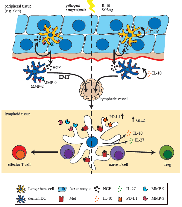
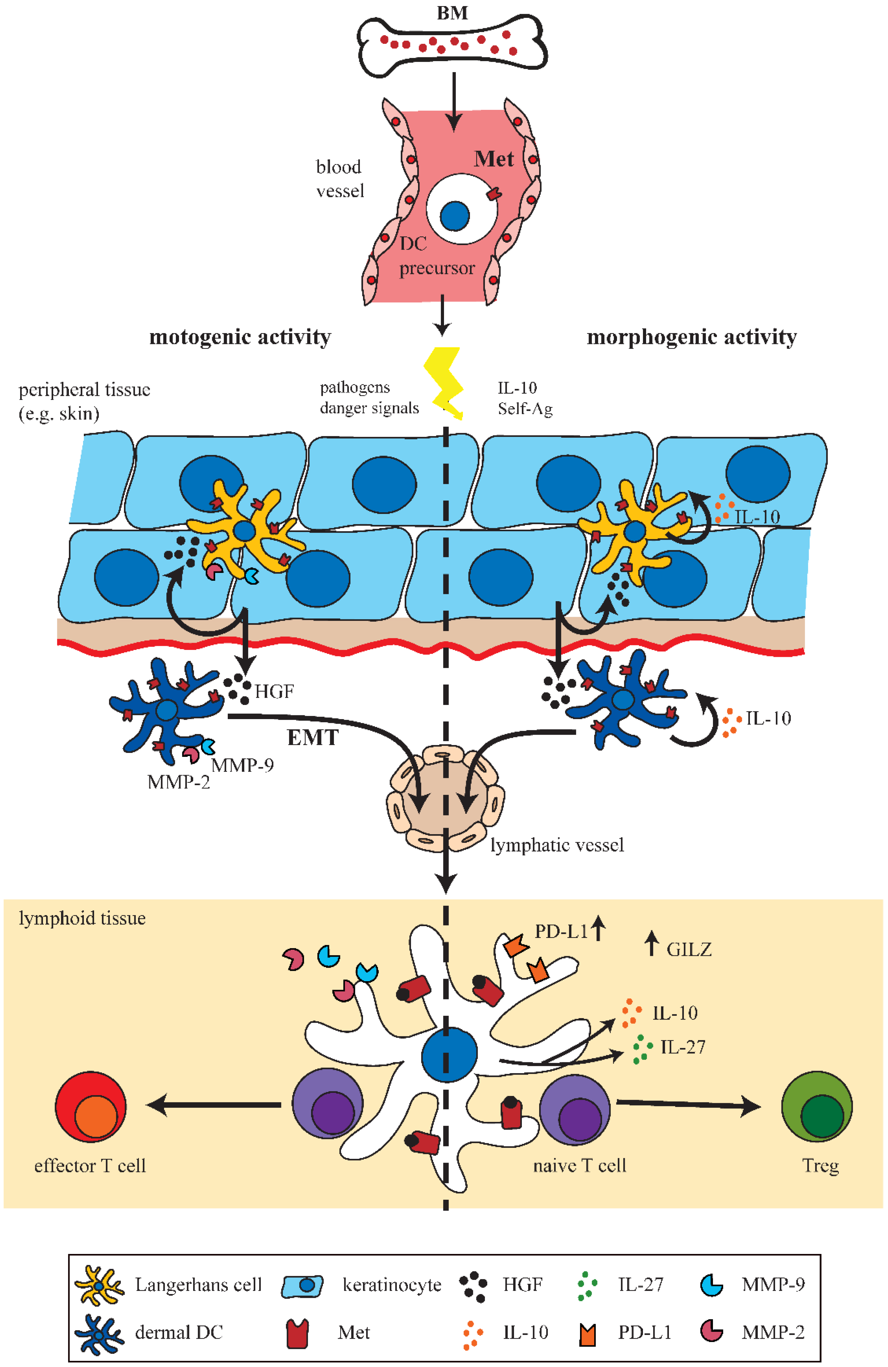
5. A Met-Driven Program of Epithelial-Mesenchymal Transition in DCs?

HGF-induced DC mobilization and migration upon activation is associated with various phenotypic transformations that includes for example MMP activation and detachment from surrounding tissue. This is strikingly reminiscent of a Met-signaling driven mechanism seen during embryonic development, wound healing, and invasive growth of tumors known as epithelial-mesenchymal transition (EMT). In this process activated immobile epithelial cells convert transiently into a migratory mesenchymal phenotype [15,16]. Thus, the concept emerges that an EMT (or EMT-like) program is executed by Met-signaling in DCs. Similar to epithelial cells, where this program was shown to be Met driven, dDCs and LCs need to disrupt their physical contact to neighboring cells mediated by adherens and tight junctions [54,55]. EMT is characterized by downregulation of components that form adherens and tight junction complexes, including EpCAM, occludins, claudins, zonula occludens (ZO) proteins, and cytokeratins and thereby decomposes cell-to-cell contacts. Simultaneously, cells gain a mesenchymal phenotype, such as expression of N-cadherin, vimentin, integrins, and MMPs and reorganization of the cytoskeleton, which altogether facilitate cell migration. Again, the proteolytic activity of MMPs, stimulated by Met-signaling was shown to facilitate tumor cell dissociation and scattering [54,56]. Another hallmark of EMT is the loss of E-cadherin expression, which is regulated by the zinc finger and E-box binding transcription factors (ZEB) 1 and 2 [55,56,57].
LCs in skin were shown to express E-cadherin and various other epithelial-like junctional proteins, including claudin-1, EpCAM/TROP1, TROP2, ZO-1, occludin, JAM1, and cytokeratins, which allows them to functionally integrate into the keratinocyte layer. Moreover, activation of LC was found to induce expression of the EMT regulators ZEB1 and ZEB2 and downregulation of E-cadherin and EpCAM, accompanied by upregulated expression of N-cadherin [58]. Together with the above described regulation of MMPs in DCs, these findings strongly supports the concept that a Met-signaling initiated EMT-like program is executed in DCs upon activation.

6. Conclusions and Perspectives

Altogether this review summarizes the pivotal role of HGF/Met-signaling in DC mediated immunity (Figure 1). HGF/Met-signaling enables DCs to be directed into two distinct immunomodulatory pathways, either conferring immune tolerance or immunity. In the case of tolerogenic DCs, the influence of HGF has been extensively studied indicating that HGF triggers the IL-10-dependent mechanism of tolerance induction. Furthermore, several proteins restricted to tolerogenic DC were identified as specific HGF/Met-signaling pathway components. Accumulating data indicate that HGF protects against autoimmunity via DC-dependent mechanisms in a number of studies, for example in a murine lupus nephritis model of chronic graft-versus-host disease [59], collagen-induced arthritis [60], experimental autoimmune encephalomyelitis [25,41] and chronic airway inflammation [26].
In contrast, the role of HGF/Met-signaling for DC migration is less well established. Initial studies highlighted the importance of Met-signaling for control of DC motility and migration. First fundaments towards understanding Met-dependent mechanisms in DC migration were established by elucidating MMP regulation by Met-signaling. However, knowledge about intracellular signaling transduction pathways that control DC migration following Met activation is not yet fully exploited. Based on the notion that in wound healing keratinocyte and DC migration are regulated by the same Met-driven mechanisms, it is highly conceivable that similar protein signaling network dynamics exist in DCs and keratinocytes [15,54,61].
Figure 1. Hepatocyte growth factor (HGF)/Met-signaling in dendritic cells (DCs). Schematic representation of the motogenic and morphogenic activities of Met-signaling on DCs in peripheral and lymphoid tissues. DC precursors originating from hematopoietic stem cells (HSC) in bone marrow (BM) migrate towards peripheral tissues such as the skin. Upon activation DCs/Langerhans cells (LCs) migrate via lymphatics to draining lymph nodes to present antigens to naive T cells. Met-signaling induces LC and DC emigration from skin in an epithelial-mesenchymal transition (EMT)-like process, including matrix metalloproteinases (MMP) activation. HGF-induced morphogenic activities include induction of a tolerogenic phenotype of DCs by IL-10 secretion and upregulated expression of, e.g., glucocorticoid-induced leucine zipper (GILZ) and programmed-death ligand 1 (PD-L1), which finally results in enhanced numbers of Tregs.
Biomedicines 03 00138 g001
Met has been examined in numerous cancer-based studies as a critical factor for tumor invasion and metastasis and thus is highly considered as a drug target for tumor therapies. Accordingly, control of DC function by HGF/Met-signaling as a crux for DC-based immunity represents a potential starting-point for immune therapy against cancer and autoimmune diseases. However, means to target HGF/Met in order to favor immune tolerance might at the same time boost potential tumor cell towards invasive growth and metastasis. The other way round, blocking Met-induced migration of tumor cells in patients might alter their immune function towards development of autoimmune reactions. Nevertheless, this would be a matter of considering the pro and cons, depending on the required therapy and taking into account whether therapies are only temporarily applied.
In summary, the knowledge of intracellular HGF/Met-signaling mechanisms in DCs is still incomplete and needs to be expanded by further research to allow save Met-based therapies in the future. Elucidation of downstream signaling pathways of Met involved in DC migration will also reveal whether a Met-driven EMT program is in fact operating in DCs.

Acknowledgments

This work was supported by the START-program of the Faculty of Medicine, RWTH Aachen.

Conflicts of Interest

The authors declare no conflict of interest.

References

  1. Park, M.; Dean, M.; Cooper, C.S.; Schmidt, M.; O’Brien, S.J.; Blair, D.G.; vande Woude, G.F. Mechanism of met oncogene activation. Cell 1986, 45, 895–904. [Google Scholar] [CrossRef] [PubMed]
  2. Nakamura, T.; Teramoto, H.; Ichihara, A. Purification and characterization of a growth factor from rat platelets for mature parenchymal hepatocytes in primary cultures. Proc. Natl. Acad. Sci. USA 1986, 83, 6489–6493. [Google Scholar] [CrossRef] [PubMed]
  3. Stoker, M.; Gherardi, E.; Perryman, M.; Gray, J. Scatter factor is a fibroblast-derived modulator of epithelial cell mobility. Nature 1987, 327, 239–242. [Google Scholar] [CrossRef] [PubMed]
  4. Weidner, K.M.; Arakaki, N.; Hartmann, G.; Vandekerckhove, J.; Weingart, S.; Rieder, H.; Fonatsch, C.; Tsubouchi, H.; Hishida, T.; Daikuhara, Y.; et al. Evidence for the identity of human scatter factor and human hepatocyte growth factor. Proc. Natl. Acad. Sci. USA 1991, 88, 7001–7005. [Google Scholar] [CrossRef]
  5. Sonnenberg, E.; Meyer, D.; Weidner, K.M.; Birchmeier, C. Scatter factor/hepatocyte growth factor and its receptor, the c-Met tyrosine kinase, can mediate a signal exchange between mesenchyme and epithelia during mouse development. J. Cell Biol. 1993, 123, 223–235. [Google Scholar] [CrossRef] [PubMed]
  6. Bladt, F.; Riethmacher, D.; Isenmann, S.; Aguzzi, A.; Birchmeier, C. Essential role for the c-Met receptor in the migration of myogenic precursor cells into the limb bud. Nature 1995, 376, 768–771. [Google Scholar] [CrossRef] [PubMed]
  7. Schmidt, C.; Bladt, F.; Goedecke, S.; Brinkmann, V.; Zschiesche, W.; Sharpe, M.; Gherardi, E.; Birchmeier, C. Scatter factor/hepatocyte growth-factor is essential for liver development. Nature 1995, 373, 699–702. [Google Scholar] [CrossRef] [PubMed]
  8. Uehara, Y.; Minowa, O.; Mori, C.; Shiota, K.; Kuno, J.; Noda, T.; Kitamura, N. Placental defect and embryonic lethality in mice lacking hepatocyte growth factor/scatter factor. Nature 1995, 373, 702–705. [Google Scholar] [CrossRef] [PubMed]
  9. Block, G.D.; Locker, J.; Bowen, W.C.; Petersen, B.E.; Katyal, S.; Strom, S.C.; Riley, T.; Howard, T.A.; Michalopoulos, G.K. Population expansion, clonal growth, and specific differentiation patterns in primary cultures of hepatocytes induced by HGF/SF, EGF and TGF alpha in a chemically defined (HGM) medium. J. Cell Biol. 1996, 132, 1133–1149. [Google Scholar] [CrossRef] [PubMed]
  10. Borowiak, M.; Garratt, A.N.; Wustefeld, T.; Strehle, M.; Trautwein, C.; Birchmeier, C. Met provides essential signals for liver regeneration. Proc. Natl. Acad. Sci. USA 2004, 101, 10608–10613. [Google Scholar] [CrossRef] [PubMed]
  11. Alvarez-Perez, J.C.; Ernst, S.; Demirci, C.; Casinelli, G.P.; Mellado-Gil, J.M.; Rausell-Palamos, F.; Vasavada, R.C.; Garcia-Ocana, A. Hepatocyte growth factor/c-Met signaling is required for beta-cell regeneration. Diabetes 2014, 63, 216–223. [Google Scholar] [CrossRef] [PubMed]
  12. Nayeri, F.; Xu, J.; Abdiu, A.; Nayeri, T.; Aili, D.; Liedberg, B.; Carlsson, U. Autocrine production of biologically active hepatocyte growth factor (HGF) by injured human skin. J. Dermatol. Sci. 2006, 43, 49–56. [Google Scholar] [CrossRef] [PubMed]
  13. Chmielowiec, J.; Borowiak, M.; Morkel, M.; Stradal, T.; Munz, B.; Werner, S.; Wehland, J.; Birchmeier, C.; Birchmeier, W. c-Met is essential for wound healing in the skin. J. Cell Biol. 2007, 177, 151–162. [Google Scholar] [CrossRef] [PubMed]
  14. Cowin, A.J.; Kallincos, N.; Hatzirodos, N.; Robertson, J.G.; Pickering, K.J.; Couper, J.; Belford, D.A. Hepatocyte growth factor and macrophage-stimulating protein are upregulated during excisional wound repair in rats. Cell Tissue Res. 2001, 306, 239–250. [Google Scholar] [CrossRef] [PubMed]
  15. Birchmeier, C.; Birchmeier, W.; Gherardi, E.; vande Woude, G.F. Met, metastasis, motility and more. Nat. Rev. Mol. Cell Biol. 2003, 4, 915–925. [Google Scholar] [CrossRef] [PubMed]
  16. Boccaccio, C.; Comoglio, P.M. Invasive growth: A Met-driven genetic programme for cancer and stem cells. Nat. Rev. Cancer 2006, 6, 637–645. [Google Scholar] [CrossRef] [PubMed]
  17. Rong, S.; Bodescot, M.; Blair, D.; Dunn, J.; Nakamura, T.; Mizuno, K.; Park, M.; Chan, A.; Aaronson, S.; vande Woude, G.F. Tumorigenicity of the Met proto-oncogene and the gene for hepatocyte growth factor. Mol. Cell. Biol. 1992, 12, 5152–5158. [Google Scholar] [PubMed]
  18. Kanda, H.; Tajima, H.; Lee, G.H.; Nomura, K.; Ohtake, K.; Matsumoto, K.; Nakamura, T.; Kitagawa, T. Hepatocyte growth factor transforms immortalized mouse liver epithelial cells. Oncogene 1993, 8, 3047–3053. [Google Scholar] [PubMed]
  19. Nakamura, T.; Sakai, K.; Nakamura, T.; Matsumoto, K. Hepatocyte growth factor twenty years on: Much more than a growth factor. J. Gastroen. Hepatol. 2011, 26, 188–202. [Google Scholar] [CrossRef]
  20. Jiang, W.G.; Martin, T.A.; Parr, C.; Davies, G.; Matsumoto, K.; Nakamura, T. Hepatocyte growth factor, its receptor, and their potential value in cancer therapies. Crit. Rev. Oncol. Hematol. 2005, 53, 35–69. [Google Scholar] [CrossRef] [PubMed]
  21. Ruggeri, R.M.; Sciacchitano, S.; Vitale, A.; Cardelli, P.; Galletti, M.; Vitarelli, E.; Barresi, G.; Benvenga, S.; Trimarchi, F.; Trovato, M. Serum hepatocyte growth factor is increased in hashimoto’s thyroiditis whether or not it is associated with nodular goiter as compared with healthy non-goitrous individuals. J. Endocrinol. Investig. 2009, 32, 465–469. [Google Scholar] [CrossRef]
  22. Ruggeri, R.M.; Vitarelli, E.; Barresi, G.; Trimarchi, F.; Benvenga, S.; Trovato, M. The tyrosine kinase receptor c-met, its cognate ligand HGF and the tyrosine kinase receptor trasducers STAT3, PI3K and RHO in thyroid nodules associated with Hashimoto’s thyroiditis: An immunohistochemical characterization. Eur. J. Histochem. 2010, 54, e24. [Google Scholar] [CrossRef] [PubMed]
  23. Beilmann, M.; Odenthal, M.; Jung, W.; vande Woude, G.F.; Dienes, H.P.; Schirmacher, P. Neoexpression of the c-Met/hepatocyte growth factor-scatter factor receptor gene in activated monocytes. Blood 1997, 90, 4450–4458. [Google Scholar] [PubMed]
  24. Van der Voort, R.; Taher, T.E.; Keehnen, R.M.; Smit, L.; Groenink, M.; Pals, S.T. Paracrine regulation of germinal center B cell adhesion through the c-met-hepatocyte growth factor/scatter factor pathway. J. Exp. Med. 1997, 185, 2121–2131. [Google Scholar] [CrossRef] [PubMed]
  25. Benkhoucha, M.; Santiago-Raber, M.L.; Schneiter, G.; Chofflon, M.; Funakoshi, H.; Nakamura, T.; Lalive, P.H. Hepatocyte growth factor inhibits CNS autoimmunity by inducing tolerogenic dendritic cells and CD25+Foxp3+ regulatory T cells. Proc. Natl. Acad. Sci. USA 2010, 107, 6424–6429. [Google Scholar] [CrossRef] [PubMed]
  26. Okunishi, K.; Dohi, M.; Nakagome, K.; Tanaka, R.; Mizuno, S.; Matsumoto, K.; Miyazaki, J.; Nakamura, T.; Yamamoto, K. A novel role of hepatocyte growth factor as an immune regulator through suppressing dendritic cell function. J. Immunol. 2005, 175, 4745–4753. [Google Scholar] [CrossRef] [PubMed]
  27. Kurz, S.M.; Diebold, S.S.; Hieronymus, T.; Gust, T.C.; Bartunek, P.; Sachs, M.; Birchmeier, W.; Zenke, M. The impact of c-Met/scatter factor receptor on dendritic cell migration. Eur. J. Immunol. 2002, 32, 1832–1838. [Google Scholar] [CrossRef] [PubMed]
  28. Baek, J.H.; Birchmeier, C.; Zenke, M.; Hieronymus, T. The HGF receptor/Met tyrosine kinase is a key regulator of dendritic cell migration in skin immunity. J. Immunol. 2012, 189, 1699–1707. [Google Scholar] [CrossRef] [PubMed]
  29. Banchereau, J.; Steinman, R.M. Dendritic cells and the control of immunity. Nature 1998, 392, 245–252. [Google Scholar] [CrossRef] [PubMed]
  30. Geissmann, F.; Manz, M.G.; Jung, S.; Sieweke, M.H.; Merad, M.; Ley, K. Development of monocytes, macrophages, and dendritic cells. Science 2010, 327, 656–661. [Google Scholar] [CrossRef] [PubMed]
  31. Alvarez, D.; Vollmann, E.H.; von Andrian, U.H. Mechanisms and consequences of dendritic cell migration. Immunity 2008, 29, 325–342. [Google Scholar] [CrossRef] [PubMed]
  32. Banchereau, J.; Briere, F.; Caux, C.; Davoust, J.; Lebecque, S.; Liu, Y.J.; Pulendran, B.; Palucka, K. Immunobiology of dendritic cells. Annu. Rev. Immunol. 2000, 18, 767–811. [Google Scholar] [CrossRef] [PubMed]
  33. Stoitzner, P.; Tripp, C.H.; Eberhart, A.; Price, K.M.; Jung, J.Y.; Bursch, L.; Ronchese, F.; Romani, N. Langerhans cells cross-present antigen derived from skin. Proc. Natl. Acad. Sci. USA 2006, 103, 7783–7788. [Google Scholar] [CrossRef] [PubMed]
  34. Burgdorf, S.; Scholz, C.; Kautz, A.; Tampe, R.; Kurts, C. Spatial and mechanistic separation of cross-presentation and endogenous antigen presentation. Nat. Immunol. 2008, 9, 558–566. [Google Scholar] [CrossRef] [PubMed]
  35. Dhodapkar, M.V.; Sznol, M.; Zhao, B.; Wang, D.; Carvajal, R.D.; Keohan, M.L.; Chuang, E.; Sanborn, R.E.; Lutzky, J.; Powderly, J.; et al. Induction of antigen-specific immunity with a vaccine targeting NY-ESO-1 to the dendritic cell receptor DEC-205. Sci. Transl. Med. 2014, 6, 232ra251. [Google Scholar] [CrossRef]
  36. Steinman, R.M.; Banchereau, J. Taking dendritic cells into medicine. Nature 2007, 449, 419–426. [Google Scholar] [CrossRef] [PubMed]
  37. Singhal, E.; Sen, P. Hepatocyte growth factor-induced c-Src-phosphatidylinositol 3-kinase-AKT-mammalian target of rapamycin pathway inhibits dendritic cell activation by blocking IkappaB kinase activity. Int. J. Biochem. Cell Biol. 2011, 43, 1134–1146. [Google Scholar] [CrossRef] [PubMed]
  38. Corinti, S.; Albanesi, C.; la Sala, A.; Pastore, S.; Girolomoni, G. Regulatory activity of autocrine IL-10 on dendritic cell functions. J. Immunol. 2001, 166, 4312–4318. [Google Scholar] [CrossRef] [PubMed]
  39. Singhal, E.; Kumar, P.; Sen, P. A novel role for bruton’s tyrosine kinase in hepatocyte growth factor-mediated immunoregulation of dendritic cells. J. Biol. Chem. 2011, 286, 32054–32063. [Google Scholar] [CrossRef] [PubMed]
  40. Rutella, S.; Bonanno, G.; Procoli, A.; Mariotti, A.; de Ritis, D.G.; Curti, A.; Danese, S.; Pessina, G.; Pandolfi, S.; Natoni, F.; et al. Hepatocyte growth factor favors monocyte differentiation into regulatory interleukin (IL)-10++IL-12low/neg accessory cells with dendritic-cell features. Blood 2006, 108, 218–227. [Google Scholar] [CrossRef] [PubMed]
  41. Benkhoucha, M.; Molnarfi, N.; Dunand-Sauthier, I.; Merkler, D.; Schneiter, G.; Bruscoli, S.; Riccardi, C.; Tabata, Y.; Funakoshi, H.; Nakamura, T.; et al. Hepatocyte growth factor limits autoimmune neuroinflammation via glucocorticoid-induced leucine zipper expression in dendritic cells. J. Immunol. 2014, 193, 2743–2752. [Google Scholar] [CrossRef] [PubMed]
  42. Ayroldi, E.; Migliorati, G.; Bruscoli, S.; Marchetti, C.; Zollo, O.; Cannarile, L.; D’Adamio, F.; Riccardi, C. Modulation of T-cell activation by the glucocorticoid-induced leucine zipper factor via inhibition of nuclear factor kappaB. Blood 2001, 98, 743–753. [Google Scholar] [CrossRef] [PubMed]
  43. Cohen, N.; Mouly, E.; Hamdi, H.; Maillot, M.C.; Pallardy, M.; Godot, V.; Capel, F.; Balian, A.; Naveau, S.; Galanaud, P.; et al. Gilz expression in human dendritic cells redirects their maturation and prevents antigen-specific T lymphocyte response. Blood 2006, 107, 2037–2044. [Google Scholar] [CrossRef] [PubMed]
  44. Hamdi, H.; Godot, V.; Maillot, M.C.; Prejean, M.V.; Cohen, N.; Krzysiek, R.; Lemoine, F.M.; Zou, W.; Emilie, D. Induction of antigen-specific regulatory T lymphocytes by human dendritic cells expressing the glucocorticoid-induced leucine zipper. Blood 2007, 110, 211–219. [Google Scholar] [CrossRef] [PubMed]
  45. Chen, Q.; DeFrances, M.C.; Zarnegar, R. Induction of met proto-oncogene (hepatocyte growth factor receptor) expression during human monocyte-macrophage differentiation. Cell Growth Differ. 1996, 7, 821–832. [Google Scholar] [PubMed]
  46. Beilmann, M.; Vande Woude, G.F.; Dienes, H.P.; Schirmacher, P. Hepatocyte growth factor-stimulated invasiveness of monocytes. Blood 2000, 95, 3964–3969. [Google Scholar] [PubMed]
  47. Moransard, M.; Sawitzky, M.; Fontana, A.; Suter, T. Expression of the HGF receptor c-Met by macrophages in experimental autoimmune encephalomyelitis. Glia 2010, 58, 559–571. [Google Scholar] [PubMed]
  48. Galimi, F.; Cottone, E.; Vigna, E.; Arena, N.; Boccaccio, C.; Giordano, S.; Naldini, L.; Comoglio, P.M. Hepatocyte growth factor is a regulator of monocyte-macrophage function. J. Immunol. 2001, 166, 1241–1247. [Google Scholar] [CrossRef] [PubMed]
  49. Ratzinger, G.; Stoitzner, P.; Ebner, S.; Lutz, M.B.; Layton, G.T.; Rainer, C.; Senior, R.M.; Shipley, J.M.; Fritsch, P.; Schuler, G.; et al. Matrix metalloproteinases 9 and 2 are necessary for the migration of Langerhans cells and dermal dendritic cells from human and murine skin. J. Immunol. 2002, 168, 4361–4371. [Google Scholar] [CrossRef] [PubMed]
  50. Yen, J.H.; Khayrullina, T.; Ganea, D. PGE2-induced metalloproteinase-9 is essential for dendritic cell migration. Blood 2008, 111, 260–270. [Google Scholar] [CrossRef] [PubMed]
  51. Saalbach, A.; Klein, C.; Schirmer, C.; Briest, W.; Anderegg, U.; Simon, J.C. Dermal fibroblasts promote the migration of dendritic cells. J. Investig. Dermatol. 2010, 130, 444–454. [Google Scholar] [CrossRef] [PubMed]
  52. Daniels, J.T.; Limb, G.A.; Saarialho-Kere, U.; Murphy, G.; Khaw, P.T. Human corneal epithelial cells require MMP-1 for HGF-mediated migration on collagen I. Investig. Ophthalmol. Vis. Sci. 2003, 44, 1048–1055. [Google Scholar] [CrossRef]
  53. McCawley, L.J.; O’Brien, P.; Hudson, L.G. Epidermal growth factor (EGF)- and scatter factor/hepatocyte growth factor (SF/HGF)-mediated keratinocyte migration is coincident with induction of matrix metalloproteinase (MMP)-9. J. Cell. Physiol. 1998, 176, 255–265. [Google Scholar] [CrossRef] [PubMed]
  54. Trusolino, L.; Comoglio, P.M. Scatter-factor and semaphorin receptors: Cell signalling for invasive growth. Nat. Rev. Cancer 2002, 2, 289–300. [Google Scholar] [CrossRef] [PubMed]
  55. Thiery, J.P.; Acloque, H.; Huang, R.Y.; Nieto, M.A. Epithelial-mesenchymal transitions in development and disease. Cell 2009, 139, 871–890. [Google Scholar] [CrossRef] [PubMed]
  56. Christofori, G. New signals from the invasive front. Nature 2006, 441, 444–450. [Google Scholar] [CrossRef] [PubMed]
  57. Peinado, H.; Olmeda, D.; Cano, A. Snail, Zeb and bHLH factors in tumour progression: An alliance against the epithelial phenotype? Nat. Rev. Cancer 2007, 7, 415–428. [Google Scholar] [CrossRef] [PubMed]
  58. Hieronymus, T.; Zenke, M.; Baek, J.H.; Sere, K. The clash of Langerhans cell homeostasis in skin: Should I stay or should I go? Semin. Cell Dev. Biol. 2014. [Google Scholar] [CrossRef]
  59. Kuroiwa, T.; Iwasaki, T.; Imado, T.; Sekiguchi, M.; Fujimoto, J.; Sano, H. Hepatocyte growth factor prevents lupus nephritis in a murine lupus model of chronic graft-versus-host disease. Arthritis Res. Ther. 2006, 8, R123. [Google Scholar] [CrossRef] [PubMed]
  60. Okunishi, K.; Dohi, M.; Fujio, K.; Nakagome, K.; Tabata, Y.; Okasora, T.; Seki, M.; Shibuya, M.; Imamura, M.; Harada, H.; et al. Hepatocyte growth factor significantly suppresses collagen-induced arthritis in mice. J. Immunol. 2007, 179, 5504–5513. [Google Scholar] [CrossRef] [PubMed]
  61. Singh, A.; Nascimento, J.M.; Kowar, S.; Busch, H.; Boerries, M. Boolean approach to signalling pathway modelling in HGF-induced keratinocyte migration. Bioinformatics 2012, 28, i495–i501. [Google Scholar] [CrossRef] [PubMed]

