Advances in Ultrafast Fiber Lasers for Multiphoton Microscopy in Neuroscience


Department of Neurosciences, Lerner Research Institute, Cleveland Clinic, Cleveland, OH 44195, USA
*
Author to whom correspondence should be addressed.
Photonics 2023, 10(12), 1307; https://doi.org/10.3390/photonics10121307
Submission received: 7 September 2023
/
Revised: 8 November 2023
/
Accepted: 23 November 2023
/
Published: 26 November 2023
(This article belongs to the Special Issue Recent Advances in Multiphoton Microscopy)
Abstract
:Multiphoton microscopy (MPM) has emerged as a vital tool in neuroscience, enabling deeper imaging with a broader field of view, as well as faster and sub-cellular resolution. Recent innovations in ultrafast fiber laser technology have revolutionized MPM applications in living brains, offering advantages like cost-effectiveness and user-friendliness. In this review, we explore the progress in ultrafast fiber laser technology, focusing on its integration into MPM for neuroscience research. We also examine the utility of femtosecond fiber lasers in fluorescence and label-free two- and three-photon microscopy applications within the field. Furthermore, we delve into future possibilities, including next-generation fiber laser designs, novel laser characteristics, and their potential for achieving high spatial and temporal resolution imaging. We also discuss the integration of fiber lasers with implanted microscopes, opening doors for clinical and fundamental neuroscience investigations.
1. Introduction
Multi-photon microscopy (MPM) is a powerful technique for the minimally invasive imaging of live brains with subcellular resolution and for performing three-dimensional imaging with depths ranging from a few hundred microns to a few millimeters using two- and three-photon excitation processes [1,2,3,4,5,6]. Because of the nonlinear interaction between ultrashort pulses and the brain at the near-infrared wavelengths, MPM can provide the high-resolution imaging, as well as deeper penetration depth. The distinctive advantages of MPM adoption in neuroscience include the ability to visualize the brain activity and structure with subcellular resolution (even imaging fine structures such as spine and axons) in vivo at millimeter depths with or without labeling the different cell types in the brain. Although confocal and light sheet microscopy also provides subcellular resolution, they are only capable of imaging shallower depths compared to multiphoton microscopy. Because it is critical to understand the connectivity between neurons at distinct cortical layers when they process sensory and other kinds of information, multiphoton microscopy is particularly useful in neuroscience to address these types of questions.
A femtosecond pulsed laser is used as a laser source for the MPM. Generally, the ultrashort laser pulses can be generated either by using bulk or fiber-type laser systems. Titanium-doped Sapphire (Ti:S) lasers, which provide stability, short pulse widths in the femtosecond region, and extended wavelength tunability, are the most frequent conventional lasers used in MPM. Ti-S lasers produce wavelengths ranging from 700 to 1300 nm at a repetition rate of 80 MHz. In general, femtosecond laser pulses with this spectral range and repetition rate are appropriate for exciting most fluorophores for structural and functional brain imaging via two-photon interactions [7]. When performing three-photon imaging of the brain, the laser characteristics differ dramatically from those used for two-photon microscopy. For three-photon microscopy, the number of photons that are absorbed during this process can be described as follows [5,6]:
where the terms are explained as follows: n: number of excited photons, : excitation cross-section, P: average power, : pulse width, R: repetition rate, NA: numerical aperture, h: Planck ‘s constant, c: speed of light, and : wavelength.
Therefore, it is important to increase the laser power and lower the repetition rate and the pulse width in order to increase the number of photons excited in a three-photon process while keeping the numerical aperture and the wavelength constant. Specifically, the wavelength of the laser has been chosen at longer wavelength ranges, such as 1300 and 1700 nm. The laser average power is the key player in three-photon microscopy for brain imaging, because, the high average power can result in (a) tissue heating through linear absorption of longer excitation wavelengths, (b) nonlinear damage at the focal plane due to high peak power/intensity, (c) saturation of fluorophores due to the high pulse energy. Due to these effects, it is crucial to consider laser parameters and perform control experiments to make sure that it is safe to perform 3p imaging in the living brain [6,8]. Also, the repetition rates must be decreased to 0.5 to 2 MHz, and the pulse widths must be less than 50 fs [5,8,9]. Significant progress has recently been made in the development of optical parametric amplifiers (OPAs) that can produce longer excitation wavelengths with moderate repetition rates at short pulse widths. Although these commercial lasers offer several multiphoton imaging choices in the brain [10], they have several drawbacks, including high costs and sophisticated and bulky equipment. As a result, democratizing these imaging techniques in neuroscience labs is extremely difficult.
As an alternative to conventional ultrafast lasers, great progress has been made in fiber lasers, which can overcome various disadvantages of conventional lasers. In particular, fiber lasers are very small and compact, can be operated with air cooling, have a high pump-to-signal conversion efficiency, have a lower overall cost than conventional solid-state lasers, and can provide unconventional laser characteristics such as longer wavelengths, high repetition rates, variable pulse energy, and narrow pulse widths. Thus, in the recent decade, the use of fiber lasers for neuroscience applications has emerged. The simplest way to generate multiple wavelengths in a fiber laser is to adapt various types of gain fibers to achieve the required wavelength of light [11,12]. The ytterbium (Yb) fiber is employed to generate 1000 nm wavelengths [13]. Similarly, the laser wavelength spectrum of 1550 nm is achieved by using Erbium (Er)-doped fiber as the gain medium [14,15] and the thulium (Tm)-doped fiber emits a laser wavelength spectrum of 1700 nm [16]. Recent advancements in femtosecond fiber lasers have been reported in fiber laser oscillators as well as fiber amplifiers. In laser oscillators, important advancements have focused on the development of novel saturable absorbers (SAs), various cavity designs, similaritons, all-normal dispersion (ANDi) configurations, dual-wavelength generation, variable repetition rates, and multimode fiber laser oscillators [17]. Similarly, innovative fiber amplifiers concentrate on chirped pulse amplification and direct amplification approaches for increasing the energy of ultra-short laser pulses. Eventually, the average power of the laser pulses increased to over 1 Watts [18] at MHz repetition rates, which is essential for achieving multi-photon effects such as two- and three-photon fluorescence, as well as label-free imaging in the brain [19].
In this review, we focus on recent advances in the development of ultrafast fiber lasers for neuroscience applications, with a specific emphasis on two- and three-photon fluorescence and label-free microscopy for structural and functional brain imaging. As a future perspective, we propose that new breakthroughs in fiber laser technology will cover a wide range of applications in the field of neuroscience. We believe that this review is critical for neuroscientists and laser scientists to understand the current state-of-the-art fiber lasers for performing multiphoton imaging in living brains, as well as to see potential new developments in fiber lasers to democratize their use in neuroscience labs.
The rest of this paper is organized as follows: In Section 2, multiphoton microscopy for neuronal imaging is discussed. In Section 3, the advances in ultrafast lasers for neural imaging are presented. In Section 4, fiber laser-based multiphoton microscopy (FL- MPM) applications in neuroscience are discussed. Finally, the future directions and outlook, as well as the conclusion are presented in Section 5 and Section 6, respectively.
2. Multiphoton Microscopy (MPM) for Neuronal Imaging
Conventionally, several imaging techniques, including positron emission tomography (PET), ultrasonography, and functional magnetic resonance imaging (fMRI), have been proposed for structural and functional brain imaging. However, they are limited in terms of spatial and temporal resolution [20]. These disadvantages can be solved by optical imaging techniques, such as confocal, light-sheet, two-photon (2P), and three-photon (3P) microscopes. The main advantage of these technologies is to have the capability of imaging the structure and function of the brain at sub-cellular and greater temporal resolutions. When we compare the spatial and temporal resolution capabilities of multiphoton, light sheet, and confocal microscopy techniques, they all exhibit almost the same performance. On the other hand, light sheet and confocal microscopy enable brain imaging at shorter depths, but 2P and 3P microscopy techniques can image all cortical layers, white matter, and even subcortical regions in intact brains [5,6,21]. There are several advantages and challenges of 2P and 3P microscopy for performing structural and functional brain imaging. In minimally invasive 2P microscopy for functional brain imaging, it is very feasible to perform high-speed (kHz rate) and larger field of view (several millimeters) functional imaging mostly at superficial cortical layers [22,23,24,25]. However, it is challenging to perform 2P microscopy in deeper regions of the brain due to high out-of-focus signal generation. When compared to 2P microscopy, 3P microscopy may readily minimize the challenge of deep brain functional imaging by utilizing longer excitation wavelengths (1300, and 1700 nm) at moderate imaging speeds (5–10 Hz) and a smaller field of view [4,5,6]. Therefore, the challenges of functional brain imaging with 3P microscopy are (a) to increase the imaging speed to kHz range at deeper brain regions, (b) to increase the imaging depth to subcortical regions, (c) to increase the field of view to image multiple brain regions simultaneously. For structural brain imaging, two-photon fluorescence microscopy (2PEF), three-photon fluorescence microscopy (3PEF), second-harmonic generation (SHG), and third-harmonic generation (THG) microscopy are utilized. The challenges of 2PEF, 3PEF, SHG, and THG microscopy for brain imaging include developing fluorophores with greater absorption peaks at longer excitation wavelengths and developing PMT modules that can have higher sensitivity outside the visible range of light to perform deeper brain imaging. A typical MPM experimental setup and different kinds of MPM modalities are explained in Figure 1.
A typical MPM setup is shown in Figure 1a. In a typical multiphoton microscope, a femtosecond pulsed fiber or a conventional laser is used as a laser source. The collimated light is directed to the pair of Galvo mirrors for scanning the beam in the two-dimensional space. Then, the light beam is passed through a scan and tube lens, which magnifies the beam to fill the back aperture of the microscope objective. Finally, the laser beam is transmitted by the dichroic and focused on the tissue through the objective with a beam size in the range of micrometers. The reflected light is directed to the same dichroic mirror which sends the beam to the highly sensitive photon multiplier tubes (PMTs). In front of each PMT, there are band-pass filters to select an appropriate emission wavelength for different kinds of MPM techniques (Figure 1a). There are numerous varieties of MPM based on the degree of the nonlinear excitation process and the type of contrast mechanism. Two-photon excited fluorescence (2PEF), second-harmonic generation (SHG), three-photon excited fluorescence (3PEF), and third-harmonic generation (THG) microscopy techniques are described in Figure 1b. When two lower energy photons are absorbed at the same time, electrons shift from the ground to the excitation energy state, resulting in two-photon excited fluorescence (2PEF). The electrons then lose some energy as they travel to a lower energy ground state level and produce another photon with a wavelength more than half that of the incident photons (Figure 1b). Second-harmonic generation (SHG) occurs when two photons of identical energy excite an electron to a virtual energy state. When the electrons return to the ground state, they emit a photon with half the wavelength of the incident photons (Figure 1b). In terms of three-photon interactions, three-photon excited fluorescence (3PEF) occurs when electrons absorb three photons simultaneously and move to the excitation state. The electrons then lose energy as they migrate from the higher energy excitation states to the ground state, releasing another photon with a wavelength greater than one-third of the excitation of the incident photons. On the other hand, third-harmonic generation (THG) is a parametric harmonic generation process similar to SHG in which three photons of the same energy are involved in the excitation process of electrons to a virtual energy state, and when the excited electrons return to the ground state, they emit photons at one-third of the excitation wavelength of incident photons. The important challenges of 2PEF, 3PEF, SHG, and THG microscopy for brain imaging include developing fluorophores with greater absorption peaks at longer excitation wavelengths and developing PMT modules that can have higher sensitivity outside the visible range of light to perform deeper brain imaging. Besides, femtosecond laser sources should possess versatile characteristics in order to enhance the MPM performance for neuroimaging applications.
3. The Advances in Ultrafast Fiber Lasers for MPM
The technology developments in ultrafast fiber lasers for MPM mainly focus on extending the excitation wavelengths (>1000 nm) to perform structural and functional brain imaging [26,27]. These applications require moderate repetition rates (<80 MHz) and higher pulse energies (>10 nJ) compared to conventional ultrafast lasers [26,27]. Finally, it is demonstrated that signal generation in two-photon (2p) and three-photon (3p) microscopy is inversely proportional to the pulse width [6,8]; thus, it is very advantageous to reduce the pulse width (<100 fs) in order to generate a stronger signal with these microscopy techniques.
The widespread adoption of two-photon microscopy was aided by the development of reliable near-infrared femtosecond-pulse sources, such as the titanium–sapphire laser. The benefits of using longer excitation wavelengths were confirmed using a Ti:S-pumped optical parametric oscillator (OPO) [17]. But, the complete system is expensive, occupies more space, is complex, and requires expertise to handle the laser. This has led researchers to focus on the development of fiber lasers that possess all the merits of conventional laser systems, along with easy handling and low operational costs. Table 1 summarizes the advantages and disadvantages of fiber lasers and traditional lasers. To be specific, the advantages of usage of fiber laser for neuroscience applications are as follows:
- In fiber laser-based sources, the signal propagates through the fiber’s core, eliminating the need for alignment while maintaining the same beam quality;
- By combining the fiber laser oscillator with a fiber amplifier, the average power can be scaled up;
- The laser’s output travels through the fiber and it is easily connected to a miniscope or an endoscope;
- The components used to construct the fiber lasers are commercially available, and they can be easily replaced, reducing the overall cost to an order of magnitude that of a commercial laser.
In this section, we focus on the principles of ultrafast lasers, various configurations of fiber lasers, fiber amplifiers, and fiber laser characteristics.
3.1. The Recent Developments in Ultrafast Fiber Lasers
3.1.1. Mechanism of Mode-Locking Pulse Generation
Generally, a femtosecond fiber laser consists of a fiber laser oscillator connected to one or more fiber amplifiers to reach the required pulse energy and pulse duration for MPM applications. The laser oscillator works on the principle of mode-locking to produce ultra-short laser pulses. In Figure 2a, a typical fiber laser configuration is exemplified. The mode-locking oscillator consists of a continuous wave (CW) pump source which operates at 975 nm and emits a pump power of 500 mW. Based on the active fiber selection and its fiber emission spectrum as a gain medium, (for example ‘Yb’, ‘Er’, and ‘Tm’), the laser operating regime is determined. A wavelength division multiplexer (WDM) is utilized to combine the pump signal (975 nm) and the gain medium light (1064 nm). An isolator is also used to prevent cavity damage from backward reflection and to assure unidirectional operation. The light is then sent through a saturable absorber, which dampens low-intensity pulses while amplifying higher-intensity pulses across several round trips. To evaluate the laser properties inside the oscillator, a coupler is employed to split a part of the laser signal. In the last two decades, the advancements were more focused on the configuration of the laser cavity and enhancing the nonlinear behavior of the saturable absorber [28].
The inset of Figure 2a depicts the concept of mode-locking that produces ultra-short pulses by locking longitudinal modes in-phase with each other, which are equally spaced by c/2L inside the laser cavity. The modes whose corresponding frequencies fall within the gain bandwidth of the laser are amplified and lased out.
3.1.2. Various Operating Regimes
Figure 2b shows the various operating regimes of fiber laser oscillator which includes soliton, stretched pulse, similariton, and all-normal dispersion (ANDi) [29,30,31]. Among them, soliton regimes are widely adopted in both conventional and fiber lasers for mode-locking the laser pulses. Soliton pulses are generated by balancing anomalous dispersion with Kerr non-linearity that arises in the fiber [32]. Although the soliton pulses are very stable, they are limited by the smaller pulse energies that can be calculated by the soliton area theorem (here, is the pulse energy, is the pulse duration, is the nonlinear coefficient, and the group velocity dispersion is denoted as ), which are normally in the range of pJ. In order to meet higher pulse energy requirements, the similariton and ANDi regimes were discovered. “Similariton” laser pulses are parabolic in nature and can be generated when an arbitrary pulse propagates through a longer length of gain medium with normal dispersion inside the laser cavity. It possesses unique properties, including being free from wave breaking under strong nonlinearity and offering transform-limited compression. Consequently, high-energy pulses can be generated from the similariton laser cavity with good stability [33].
3.1.3. Types of Saturable Absorbers
Another advancement in laser oscillator technology is the discovery of innovative saturable absorber technologies based on optical phenomena and materials, as shown in Figure 2c. The saturable absorber is required in ultrafast lasers to initiate the mode-locking operation. Over the previous two decades, two types of SAs have been deployed. Real saturable absorbers comprise two main types: bulk semiconductor saturable absorber mirrors (SESAMs) [34] and nanomaterial-based saturable absorbers (SAs). The latter category includes quantum dots [35], single-walled carbon nanotubes [36], graphene [37], transition metal dichalcogenides (TMDs) like MoS2 and WS2 [38], as well as topological insulators (TIs), black phosphorus (BP), and MXenes [32], which are all subtypes of TMDs. In mode-locked lasers, SESAMs have constraints such as high costs and narrow bandwidth. On the other hand, nanomaterial-based SAs require a complicated fabrication technique and have a low damage threshold. Nonlinear polarization rotation (NPR), nonlinear optical loop mirror (NOLM), and nonlinear magnifying annular mirror (NALM) SAs [32] from the second group can overcome these constraints. These SAs are highly resistant to high power and are commonly utilized in lasers of various wavelengths. These SAs have the disadvantage of being affected by environmental perturbations, and eventually, mode-locking is disturbed. The most recent development in SAs, based on nonlinear multimode interference (NL-MMI), presents an all-fiber, compact solution that is less affected by environmental effects [39]. Among these, graphene and CNT SA-based ultrafast fiber laser research is particularly appealing for neuroscience applications due to its capacity to tune the laser across a wider range of wavelengths, achieve short pulse durations, and generate stable laser pulses. In MPM, a CNT-based femtosecond laser has recently been used to image biosamples using SHG, THG, 2PFE, and 3PFE [40]. The advantages of a CNT-based femtosecond laser for MPM applications include lower costs and a longer wavelength of operation. In the artificial SA category, nonlinear polarization rotation SA-based lasers are widely used for MPM applications because of a stable generation of laser pulses with requisite energy and narrow pulse width [26].
3.1.4. Tunable Repetition Rate and Wavelength
The predominant share of optogenetics and imaging research in neuroscience is heavily dependent on femtosecond lasers with pulse energies ranging from 500 nJ to 100 J and repetition rates spanning from a few hundred kHz to a few MHz for two-photon optogenetics [41], a higher repetition rate (80 MHz) and lower pulse energies of (10–50 nJ) for two-photon imaging [21], and a lower repetition rate (0.5–4 MHz) and higher pulse energies (1–2 J) for three-photon imaging [6].
The mode-locked oscillator’s repetition rate is determined by its entire cavity length. Most fiber oscillator designs use long optical fibers with typical repetition rates in the 10 MHz range. The repetition rate can be lowered by inserting more passive fibers into the cavity. However, lowering the repetition rate of a ring-cavity oscillator below 1 MHz necessitates a large cavity length of approximately 200 m, which is impractical. Therefore, the most feasible technique to generate ultrashort pulses with a repetition rate of less than 10 MHz is to use an external acousto-optic pulse picker, as shown in Figure 2d. By implementing this method, we can achieve a repetition rate of down to the kHz range. Another significant advancement in ultrafast fiber laser technology is the generation of longer wavelengths (>1050 nm), which can be accomplished by utilizing the soliton self-frequency shift (SSFS) [42], supercontinuum generation, and multi-wavelength generation lasers. SSFS is effectively achieved by utilizing the intra-pulse Raman scattering effect in an optical fiber, which results in shifted longer wavelengths of above 1300 nm from the shorter pump wavelength. This is accomplished by moving energy from a higher frequency to a lower frequency. In order to achieve SSFS for the excitation wavelengths below 1300 nm, photonic crystal fibers (PCFs) are usually employed because their dispersion can be flexibly engineered. Furthermore, when suitable PCFs are pumped by a Yb-fiber laser, SSFS can lead to a soliton pulse with the center wavelength shifted beyond 1300 nm, even reaching 1700 nm [43,44]. The researchers’ focus has recently switched to constructing tunable ultrafast fiber laser sources for imaging multiple fluorophores in the brain. Therefore, they have been developing nonlinear fiber-optic technologies to develop tunable-wavelength ultrashort pulses from a fiber laser source. Supercontinuum (or) white light generation is normally caused by the interaction of the nonlinear effect of self-phase modulation and linear dispersion. As a result, the pulse leading edge has red-shifted components, and the trailing edge has blue-shifted components. Further, normal dispersion leads to increased separation between these red and blue components in the temporal domain, causing the pulse to broaden, whereas anomalous dispersion cancels it entirely and induces pulse stabilization in the form of an optical soliton, depending on the properties of the fiber used and the pump source wavelength [45,46]. Recently, this type of supercontinuum has been used in neuroscience imaging and optogenetic stimulation. A recent study revealed the development of a supercontinuum laser for imaging mouse brain tissue with GCaMP6s as well as perturbing it with optogenetic stimulation with C1V1-mCherry opsin [47]. Multi-wavelength generation sources are necessary in neuroscience to perform two- and three-photon brain imaging with the same laser source. Furthermore, these laser sources allow us to perform optogenetic stimulation too [48,49].
3.2. Various Configurations of Ultrafast Lasers
All-normal dispersion (ANDi) laser and similariton laser configurations are well suited for MPM applications since the characteristics of the laser pulses meet the requirement of MPM for neuroscience applications. Figure 3a depicts a schematic of an ANDi fiber laser implemented in a multiphoton microscope for neuronal imaging. An ANDi laser oscillator was recently built using multimode high-power pump diodes, along with passive and gain fibers. Without using any of the fiber amplifiers, this setup produced a pulse energy of 30 nJ with 81 fs pulse width and a repetition rate of 40 MHz. As a result, additional noises that are generated by fiber amplifiers can be avoided [26]. Figure 3b shows a successful demonstration of employing an ultrafast source at 1550 nm, a repetition rate of 1 MHz, and a pulse duration of 500 fs that was used to generate an SSFS at 1617 nm using a photonic crystal rod integrated into a multiphoton microscope. The produced soliton pulse has a pulse width of 74.5 fs and a pulse energy of 141 nJ [50]. Another way of generating a longer wavelength for three-photon microscopy was to utilize a material like Cr-forsterite (Cr-F) in the laser medium. Figure 3c depicts a schematic experimental view of a Cr:F laser oscillator. The cavity configuration was a z-fold design achieved by inserting concave mirrors. They used a Yb-doped fiber laser as a pump source, with a maximum pump power of 14 W. An ultrashort pulse with a pulse width of 32 fs at 1260 nm, a repetition rate of 24 MHz, and a pulse energy of 22 nJ was produced by this cavity configuration, which operates in the soliton regime. The total cavity length was 6.22 m. Even though they can generate a pulse energy of 22 nJ without an amplifier stage, this pulse energy was insufficient for deep tissue imaging of the mouse brain [51]. Very recently, a Raman shifter was used to shift the excitation wavelength to longer wavelengths for performing three- and four-photon microscopy in the brain. The schematic representation of the 1800 nm femtosecond fiber laser system is shown in Figure 3d. Here, an Er-doped fiber laser oscillator was used, which produced laser pulses with a pulse duration of 492 fs, a repetition rate of 50 MHz, and an output power of 1 mW. The demonstrated system at 1800 nm was able to produce a 150 fs pulse duration. The pulses were amplified using a two-stage Tm: ZBLAN fiber amplifier. After the final stage, they increased pulse energy up to 1 J at 0.5 MHz [16].
3.3. Fiber Amplifiers
3.3.1. Chirped Pulse Fiber Amplifier (CPA)
The schematic representation of CPA is shown in Figure 4a, which consists of (1) an ultrafast mode-locked laser oscillator that generates seeding pulses; (2) a pulse stretcher that extends the seeding pulse to the picosecond level; (3) pre-amplifiers and power amplifiers; (4) a pulse picker used to adjust the repetition rate; and (5) a pulse compressor that dechirps the amplified pulse to nearly transform-limited pulse width [15,52].
3.3.2. Master-Oscillator Power-Amplifier (MOPA)
The schematic representation of MOPA system is depicted in Figure 4b. Generally, the picosecond and nanosecond laser pulses are amplified with high average output power and peak power using MOPA. In this scheme, an ultrafast fiber oscillator generates stable pulses, which are then amplified by subsequent fiber amplifiers to increase average power and pulse energy. Chirped output pulses of the oscillator are directly fed to an amplifier without any stretcher unit. The master oscillator in a MOPA system only needs to provide low-power/energy pulses and can thus be carefully engineered to achieve low noise, high compactness, and extreme robustness. In subsequent fiber amplifiers, the weak seeding pulses are amplified by orders of magnitude in pulse energy or average power. In order to reduce the nonlinear effects, large-mode area fiber or double-clad gain medium is used in the power-amplifier stage [53,54].
3.3.3. Gain-Managed Nonlinear (GMN) Fiber Amplifier
Figure 4c shows a schematic illustration of GMN. Amplification is carried out in a new gain-managed regime that can provide pulses with high energy and a spectrum wider than the gain bandwidth. In this approach, a single-stage amplifier is sufficient to provide maximum pulse energy in the J range while laser pulses can be as short as 50 fs. Recently, using a gain-managed nonlinear fiber amplifier, researchers were able to achieve a maximum pulse energy of 58 nJ, a laser repetition rate of 31 MHz, and a compressed pulse duration of 33 fs [18].
3.4. Ultrafast Fiber Laser Characteristics
The repetition rate, pulse energy, pulse width, peak power, average power, spectral bandwidth, and central wavelength are all measurable properties of an ultrafast fiber laser system. In practice, tuning of these parameters is required for optimized laser and microscopy performance while performing wider, deeper, and faster brain imaging [55] and optogenetics. Because of technical limits, maximizing one parameter always compromises another. Figure 5 shows example measurements of some of these fiber laser parameters.
The wavelength spectrum is centered at 1033.2 nm as shown in Figure 5a. The mode-locked laser oscillator pulse train is depicted in Figure 5b, and the time interval between the single pulse is measured to be 46.2 ns. Figure 5c shows the radio frequency (RF) spectrum of the laser output peak, which is centered at 21.35 MHz and has a signal-to-noise ratio (SNR) of 58 dB. Figure 5d presents the measured autocorrelation trace of the mode-locked laser with pulse durations of 2 ps (inset) and 300 fs before and after the compression, respectively.
Figure 6a shows the laser pre-amplifier spectrum, which is centered at 1033.2 nm. Figure 6b presents the power-amplifier output spectrum at 11 W average power. These two figures (Figure 6a,b) show that there is a slight change in the spectral profile of the amplifier output, which could be due to the occurrence of a nonlinear effect. Amplified spontaneous emission (ASE) is normally present in any fiber amplifier system and this emission occurs due to the spontaneous emission during the amplification of laser pulses. In a high-power amplifier, this is considered as the amplifier noise, and it can limit the gain of the amplification. Therefore, the ASE should be within a particular limit to achieve high power amplification. In order to verify the presence of an ASE signal while scaling the power in the power amplifier, the output spectrum of the laser signal is measured and the pump spectrum exactly peaks at 975 nm and the strength of the ASE spectrum is very low, which is within the acceptable limit (Figure 6c). Figure 6d represents the output power versus input pump power of the power amplifier’s simulated and experimental characteristics [52].
4. Fiber Laser-Based Multiphoton Microscopy (FL-MPM) Applications in Neuroscience
Despite significant advancements in fiber laser technologies, their use in neuroscience research has been limited. In this section, we will present research studies that used fiber lasers to accomplish fluorescence and label-free two- and three-photon imaging in diverse species’ brains. Ultrafast fiber lasers are also used in temporal focusing (TF) microscopy for two- and three-photon excitations, where the axial modulation is carried out to increase the axial confinement in a wide field without point-scanning which eventually enhances the optical sectioning in multiphoton microscopy. The diffraction gratings are used to separate the spatial frequencies and temporally focus the light. As a result, there is no nonlinear effect until the light reaches the focal plane where there is a spatiotemporal focusing of the beam. Recently, temporal focusing has been shown to perform functional and structural brain imaging via two- and three-photon excitation [10]. In vivo multiphoton microscopy is an emerging imaging tool for performing structural and functional brain imaging within intact animals at cellular resolution. Moreover, 2PEF and SHG microscopy techniques have been used to perform structural and functional brain imaging in shallower depths. TPEF has been used to perform functional brain imaging via calcium indicators [21] and structural brain imaging of elements such as neurons and microvasculature with fluorescent dyes [26]. SHG microscopy is a label-free technique and is sensitive to structural features with broken inversion symmetry, such as collagen fibers in the skull and meninges [56] and microtubules [57]. On the other hand, 3PEF and THG microscopy techniques have been used to perform structural and functional brain imaging in deeper regions of the brain. More specifically, 3PEF microscopy has been used to perform functional brain imaging with calcium indicators [5,6], as well as to perform structural imaging of microvasculature, neurons, astrocytes with fluorescent dyes [6,9,16]. THG is sensitive to the index of refraction and third-order susceptibility changes in the brain. Therefore, THG has been used to image the microvasculature as well as myelinated axonal tracts [5,6,58].
4.1. Two-Photon Fluorescence Microscopy
4.1.1. Functional Imaging
Two-photon microscopy has been the workhorse of functional brain imaging with animal models at sub-cellular resolution. The majority of commercial Ti:sapphire oscillators with high repetition rates are used in two-photon brain imaging applications. The main advantage of commercial two-photon lasers involves their high wavelength tunability, which allows for the use of various calcium indicators for functional brain imaging. There has recently been development in producing calcium indicators that can be excited using standard fiber laser excitation wavelengths. In a recent study, researchers developed a jYCaMP, a calcium indicator suited for excitation wavelengths around 1030 nm [27].
At layer 2/3 of the primary visual cortex, they utilized a commercial Ytterbium fiber laser (Spark Lasers) at 1040 nm with 31.25 MHz to accomplish dual color neuronal (Thy1:(jRGECO1a transgenic mice)) and axonal (jYCaMP) imaging as illustrated in Figure 7a. Although commercial lasers have a high repetition rate, they can cause damage to the living brain since significant power is required to accomplish high-resolution deep brain imaging. Recently, an adaptive fiber laser source was developed to excite a region of interest, resulting in a 30-fold reduction in the power demand of two-photon functional brain imaging in awake mice with traditional calcium indicators such as GCaMP6s [59]. Researchers developed a custom−made fiber laser based on SSFS at 1840 nm and used a BIBO crystal to generate a 920 nm wavelength at a repetition rate of 32 MHz. They compared the spontaneous activity of neurons at a 680 m depth with point scanning standard 2-photon imaging and an adaptive excitation source (AES), as shown in Figure 7b. The field of view was 700 × 700 m, and the frame rate at 512 × 512 pixels was 30 Hz. They demonstrated that the photon number per neuron is 7 times greater than without AES, although using 4.5 times less average power with AES.
4.1.2. Structural Imaging
Since the calcium indicator development at fiber laser excitation wavelengths is still in its early stages, most of the early fiber laser research focused on structural fluorescence brain imaging with two-photon microscopy. At 40 MHz repetition rate, a seminal work used a multimode pump and double-clad fiber design to generate 30 nJ energy pulses with uncompressed average outputs of 1.2 W [26].
The authors injected the Texas Red fluorophore retro-orbitally into transgenic YFP mice, which expressed yellow fluorescent protein in layer V pyramidal neurons. Consequently, they could observe the structure of layer V pyramidal neurons, including their apical dendrites and the microvasculature, up to a depth of 700–800 m, as shown in Figure 8a. At the surface and the 750 m depth, the imaging power ranged from 20 to 200 mW. They also compared the structural imaging performance of their custom-made fiber laser to that of a commercial Ti:S laser at 920 nm. They used 380 mW of average power, as opposed to 200 mW, to image the neuronal soma at a depth of 900 m with the commercial laser and the fiber laser, respectively, under identical imaging settings. They were able to resolve neuronal soma with the fiber laser but not with the commercial laser at a 900 m depth. They also demonstrated that the fiber laser could resolve dendritic spines (about 1 m in size) up to a 200 m depth. Another work used a fiber laser with a Yb fiber amplifier and a diamond Raman laser to generate two wavelengths of 1060 and 1250 nm simultaneously [60]. They used these two lasers to do neuronal and microvasculature imaging with a galvo–galvo scanner. The Raman laser was used to image the brain vasculature tagged with Alexafluor 647, and the Yb fiber laser was utilized to image the neuronal somata labeled with tdTomato up to a depth of 750 m as illustrated in Figure 8b. Another seminal paper focused on the development of a fiber laser using circularly polarized soliton-frequency shift (SSFS) at 1617 nm to perform two-photon imaging of brain vasculature labeled with indocyanine green (IGC), as shown in Figure 8c [50]. At the laser exit, they generated pulses of 147 nJ pulse energy and 74.5 fs pulse width at a repetition rate of 1 MHz. They then demonstrated the ability to image the brain vasculature to a depth of 2 mm with a maximum pulse energy of 56 nJ as demonstrated in Figure 8c.
4.2. Two-Photon Label-Free Imaging
Second-Harmonic Generation Microscopy
In addition to performing structural two-photon fluorescence microscopy in the brain, researchers also made significant advances in performing label-free second-harmonic generation (SHG) microscopy for performing structural two-photon microscopy in the brain. In particular, SHG contrasts only structural features with broken inversion symmetry, making it a suitable probe for buried interfaces, inducible molecular dipoles, polarized structures, and field-induced anisotropy. In a recent study, researchers developed a custom-made Cr:forsterite laser to perform both second- and third-harmonic generation imaging of fixed mouse brain acquired by a 3xTg mouse utilized as a mouse model for Alzheimer’s disease (AD). The laser generated a 1262 nm wavelength at a 105 MHz repetition rate. The pulse width was 38 fs and the power on the sample varied between 20 to 30 mW [61]. Their goal was to achieve label-free imaging of amyloid beta plaques (A) and neurofibrillary tangles (NFT), which are hallmarks of Alzheimer’s disease. They discovered that A plaques generated both SHG and THG signals, whereas NFT produced predominantly SHG signals, and axons/dendrites produced mostly THG signals. They also showed that they could track the evolution of AD disease by comparing fixed mouse brain slices from different ages of 3xTg mice to age-matched samples from control mice. In another study, researchers developed an extended-cavity Cr:forsterite laser integrated with a photonic-crystal fiber solution frequency shifter and a periodically poled lithium niobate spectrum compressor for simultaneous harmonic generation and coherent Raman brain imaging in fixed mouse brain tissues [62]. They developed a home-built Cr:forsterite laser oscillator with a central wavelength of 1250 nm and a pulse width of 40 fs. This combination produced laser pulses with an energy of 18 nJ at a repetition rate of 20 MHz, equal to 360 mW average power. They then used second- and third-harmonic generation microscopy with an average laser power of 20–30 mW to image brain slices from C57BL/6 mice. SHG microscopy was used in this study to visualize cell bodies as black patches representing the lowest density of interfaces and the highest optical homogeneity of the medium within the cell bodies. A smaller scale texture between the cell bodies, related to nerve fibers and dendritic structure, effectively generating a formed surface, is particularly efficient in SHG. In this study, they were able to visualize a distinctive layered structure of the hippocampus, matching the stratum moleculare, stratum granulosum, and polymorphic layer by using SHG microscopy. Table 2 shows the fiber laser parameters that have been used for performing functional and structural imaging and their corresponding imaging depth achieved in 2PEF microscopy. Further, ultrafast fiber laser parameters used for SHG are also listed for the comparison with 2PEF microscopy. From the comparison, it can be concluded that the SHG requires less pulse energy with shallow depth imaging when compared to 2PEF, and the imaging is label free.
4.3. Three-Photon Fluorescence Microscopy
4.3.1. Functional Imaging
Although two-photon microscopy has shown significant potential for structural and functional brain imaging, it has major limits for deep-brain functional imaging due to high optical intensities and a poor signal-to-background ratio. Three-photon microscopy mitigates these challenges and has been used to image structural and functional features of deep cortical layers in the mouse brain [6,58] at subcellular resolution in our own studies. Nonetheless, these experiments were carried out using commercial solid-state lasers. Recent improvements have been achieved in the development of custom-built fiber lasers to perform three-photon microscopy for functional brain imaging. In a recent study, a Cr-forsterite oscillator with a center wavelength of 1260 nm was developed to perform 30-Hz functional three-photon brain imaging of Drosophila [51].
The laser repetition rate in that study was 24 MHz, the hyperbolic secant pulse width was 32 fs, and the laser output power was 530 mW, equivalent to a pulse energy of 22 nJ. They monitored the neuronal activity of Kenyon cells tagged with GCaMP7f while electric shocking the Drosophila as shown in Figure 9a. In another study [59], researchers used transgenic jRGECO1a mice to perform 30 Hz functional three-photon imaging at a depth of 750 m with adaptive excitation source (AES) based on a self-soliton frequency shift at 1700 nm. They compared the performance of the fiber laser system with and without AES and they found that the photon number per neuron with AES is 30 times higher than that without AES, although the same power of 35 mW was used. The spontaneous activity of neurons was recorded and a signal-to-background ratio with AES was significantly higher compared to that without AES, as illustrated in Figure 9b.
4.3.2. Structural Imaging
Similar to three-photon fluorescence functional imaging studies, in very recent studies, structural three-photon imaging in mouse brains has been performed. In a very recent study, researchers developed a custom-made fiber laser consisting of an Er-doped silica fiber laser and a Raman shift fiber in order to generate a wavelength of 1800 nm with 150 fs pulse width, and 1 J pulse energy at 0.5 MHz on the mouse brain [16].
To demonstrate the performance of their system in vivo, they labeled the excitatory neurons of the mouse brain with TurboFP636 virally in the somatosensory cortex (S1). They varied the laser power between 50–85 mW while they were imaging from the surface to a 700 m depth, as shown in Figure 10a. Their acquisition speed was 1 Hz at 512 × 512 pixels. In another study, researchers developed a two-color fiber laser at 1300 and 1700 nm wavelengths with optical parametric chirped pulse amplification (OPCPA). They used a Yb-doped fiber amplifier (YDFA) as a pump source at 1030 nm to generate a signal beam at 1700 nm as well as an idler beam at 2600 nm. They frequency-doubled 2600 nm to achieve 1300 nm wavelength output. This system generated ultrashort pulses at 69 fs with 0.71 J at 1300 nm and 65 fs with 3.1 J at 1700 nm output wavelengths at 1.25 MHz repetition rate, respectively. They imaged a fixed mouse brain up to a depth of 600 m first to show that their 3-photon dual-color system worked. Then, they performed dual-color three-photon imaging with HEK cells, chicken embryo spinal cord, and live adult zebrafish brain, as shown in Figure 10b [49].
4.4. Three-Photon Label-Free Imaging
Third-Harmonic Generation Microscopy
Although most previous studies in neuroscience focused on fluorescence three-photon microscopy with exogenous fluorophores, three-photon microscopy also provides label-free brain imaging via third-harmonic generation (THG), which provides contrast from myelinated axons and brain microvasculature.
In a recent study, a fiber laser with a master-oscillator power-amplifier (MOPA) at 1650–1700 nm wavelength range, 50 MHz repetition rate, 70 fs pulse width, and 400 mW average power was developed [63]. Researchers used this fiber laser to image the fixed Rainbow-3 mouse cortex and brain stem. At depths of 20–150 m, they performed three-photon fluorescence (3PEF) imaging of tdTomato-labeled brain microvasculature and THG imaging of myelinated axons as presented in Figure 11. They also demonstrated simultaneous 3PEF and THG imaging of a living brain with developing Drosophila embryos [63]. THG has also been shown to provide pathological information regarding tumor-affected human brain tissues. Table 3 shows the fiber laser parameters that have been used for performing functional and structural imaging and their corresponding imaging depth achieved in 3PEF microscopy. Further, ultrafast fiber laser parameters used for THG are also listed for the comparison with 3PEF microscopy. From the comparison, it can be concluded that the THG requires fewer energy pulses compared to 3PEF. On the other hand, THG offers label-free imaging. THG provides real-time images of unstained human brain tissue, highlighting features such as increased cellularity, nuclear pleomorphism, and rarefaction of neuropils [64].
5. Future Direction and Outlook
In this section, we will discuss the various innovations and developments that can be expected in the next-generation fiber lasers for neuroscience applications in the near future. We expect to see the development of new ultrafast fiber lasers offering exciting advancements in saturable absorbers, new gain mediums, fiber-based adaptive optics modules for high-resolution imaging; and exciting pulse characteristics such as longer excitation wavelengths, tunable repetition rates, shorter pulse widths; and full fiber-based implanted and endoscopic systems for performing brain imaging with freely-moving animals as well as with human brain tissues as illustrated in Figure 12. In the following sections, we will briefly explain these advancements that we are hoping to see in the near future.
5.1. Saturable Absorber
The saturable absorber (SA) is a critical component of the laser cavity that generates ultrashort pulses. Based on its physical appearance and optical dynamics, it is classified as artificial or real SAs, as shown in Figure 12. SESAMs and nanomaterial-based SAs are examples of real SAs. On the downside, these SAs have limitations such as limited bandwidth, low damage thresholds, and difficult fabrication procedures. These constraints can be overcome by using artificial SA, such as nonlinear polarization rotation (NPR), nonlinear optical loop mirror (NOLM), and nonlinear magnifying annular mirror (NALM). It has been recently discovered that artificial SAs have limitations in terms of stability and broadband laser development. Currently, the focus has shifted to developing new broadband SAs based on different fiber structures, such as no core (NC), graded-index multimode (GIM), and step-index multimode (SIM) [52] fibers, as well as extraordinary 2D materials, such as black phosphorus (BP), MXene, and transition metal dichalcogenides (TMDs) [65] that have emerged with their own set of advantages. These SAs can be made with 3D printing technology. In the near future, we expect to see novel designs and implementations of SAs for generating different wavelengths.
5.2. Laser Characteristics
The most popular lasers used in neuroscience applications for two-photon microscopy are Ti:sapphire and solid-state lasers. Thus, neuroscientists want to ensure that fiber lasers have similar laser characteristics to commercial lasers before employing them in their research. The most essential laser properties are the capacity to tune the repetition rate and the wavelength, to lower the pulse duration, and to increase the pulse energy.
For two-photon microscopy, most of the commercial lasers rely on high repetition rates (80 MHz) and low pulse energies (10–40 nJ) resulting in 1–3 W average laser power. Therefore, two-photon microscopy with these lasers is limited to imaging shallower depths in the brain. To bridge the gap between high pulse energy and lower repetition rate requirements for deep brain imaging, fiber lasers can be utilized. A fiber laser operating in the all-normal dispersion regime is well-suited for this two-photon application, as it operates at a 1000 nm wavelength and can be tuned across a band of 100 nm at longer wavelengths (1000–2500 nm) with high average power (>5 W) [66]. To make these wavelengths useful for two-photon brain imaging, these wavelengths can be divided into half by using second-harmonic generation crystals like beta barium borate (BBO) [44]. For three-photon microscopy, commercial lasers rely on nonlinear optical parametric amplification (NOPA) and they provide tunable wavelengths (1200–2400 nm) with low repetition rate (<4 MHz) and high pulse energies (>1 J) [6]. These laser specifications are good enough to perform functional brain imaging with moderate imaging speeds. In order to increase the imaging speed, fiber lasers can be utilized to generate longer excitation wavelengths, such as 1300 nm at high repetition rates (>4 MHz) to perform deep and high-speed three-photon microscopy [51].
Most popular commercial lasers for two-photon brain imaging have wavelength tunability in the range of 700–1300 nm, a constant repetition rate of 80 MHz, pulse width tunability from 80 to 200 fs, and pulse energy tunability in the range of 10 to 50 nJ. Current fiber lasers can provide reasonable wavelength tunability, as well as tunable repetition rates ranging from 1 to 100 MHz and pulse energy tunability ranging from 50 nJ to 5 J. The only disadvantage of current fiber laser technology is that it has longer pulse widths (>200 fs) than commercial lasers for two-photon imaging. As a result, larger wavelength and repetition rate tunability, higher pulse energy, and shorter pulse width advancements are expected in the fiber lasers for two-photon microscopy in the near future. The required laser characteristics for deep brain imaging using three-photon microscopy include a lower repetition rate (0.5 to 2 MHz) combined with longer wavelengths, such as 1300 and 1700 nm, substantial pulse energies (0.5–5 J), and shorter pulse durations (<60 fs). Recently, several commercial lasers have offered these laser characteristics. However, these lasers are more expensive and require more space. Currently, most commercial lasers have either a fixed or extremely limited repetition rate tunability. These constraints can be circumvented by adding acousto-optical modulators (AOMs) into the laser system, which allows for repetition rate tuning. Using novel gain mediums and high-power diodes, similar pulse energies can be achieved with fiber lasers. Furthermore, by using other gain mediums such as fluoride and ZBLAN fibers, the laser wavelength can be extended up to 2.2 m. Shorter pulse widths for three-photon microscopy can be achieved with new fiber-based pulse compressors, double-chirped mirrors, and novel saturable absorbers (SA), such as BP, MXene, and fiber-based SA. A recent study, for example, demonstrated functional three-photon brain imaging of Drosophila brain using a home-made Cr-forsterite oscillator and double-chirped mirrors at a center wavelength of 1260 nm and a pulse width of 32 fs [51].
5.3. Multi-Wavelength Emission
The multi-wavelength laser emissions enable simultaneous stimulation of various fluorophores and opsins for multi-color imaging and/or all-optical interrogation using integrated imaging and optogenetics. In general, commercial lasers typically provide wide wavelength tunability in one output and a fixed wavelength (about 1040 nm) in the second output for two-photon microscopy. As a result, imaging several fluorophores simultaneously with two-photon microscopy is difficult. Fiber lasers can solve this problem by generating wavelengths of interest based on fluorophores at various repetition rates, allowing them to deliver more emitted signals than conventional two-photon lasers. A similar problem exists for three-photon microscopy. Researchers can achieve dual color imaging with GFP and RFP-based fluorophores using a laser illuminating the spectral window 1300 nm and 1700 nm. Recently, many companies and studies have reported dual laser emission-based cavities for three-photon microscopy [49]. With fiber lasers, similar wavelengths are directly generated from the mode-locking cavity, which minimizes costs and enables for optimization based on user requirements. As a result, we expect to see multiple wavelength emission fiber lasers to perform multi-color imaging and all-optical interrogation in living brains in the near future.
5.4. High Resolution Imaging
Due to the spatial and temporal pulse properties of fiber lasers, obtaining high-resolution images is one of the most difficult challenges. Furthermore, there is little evidence in the literature of enhancing their resolution by using adaptive optics (AO). The optical resolution of MPM is reduced by tissue scattering and aberration in the brain as well as in the microscopes. Optical resolution of MPM in deep regions of the mouse brain has increased using AO elements, such as spatial light modulators (SLMs), deformable mirrors (DMs), and wavefront sensors [67,68]. As a result, we anticipate enhanced spatial and temporal pulse characteristics when using fiber lasers coupled with AO elements inside fiber lasers, resulting in user-friendly, alignment-free, optically aberration-free, vignetting-free, and cost-effective systems capable of producing high-resolution images of the brain.
5.5. Implanted Microscope
5.5.1. Animal
Currently, all the implanted miniaturized MPM to the freely moving animals require external, free-space coupling of the commercial lasers to the miniaturized microscopes to perform two- or three-photon functional brain imaging [69,70,71,72]. This external coupling can cause alignment instabilities, preventing researchers from performing long-term multiphoton brain imaging. Despite the difficulties, we anticipate progress in splicing fiber laser outputs with unique nonlinear fibers such as Kagome hollow-core and photonic bandgap fibers in order to produce all-fiber and alignment-free systems. Integrating a fiber laser with MPM without using any free-space coupling will allow for longer-term and more stable multiphoton brain imaging of freely moving animals.
5.5.2. Clinical Application
The ultrafast fiber lasers, which are small and portable, can be employed in two ways in the clinic: First, these fiber lasers can be paired with table-top multiphoton microscopes to provide multiphoton imaging of live, excised brain tissues in the operating room, providing the surgeon with rapid feedback on the condition of the biopsy tissues. As a result, there will be no need to submit these tissues to pathologists to determine whether they are healthy, benign, or malignant. Second, these fiber lasers can be used in conjunction with endoscopes equipped with miniaturized microscopes to accomplish label-free and fluorescent multiphoton imaging in the human brain. In this configuration, fiber lasers can be directly coupled with endoscopes without the requirement for external coupling, which improves the system’s sturdiness and steadiness when combined with surgical robots. Surgeons can use this imaging system to do either superficial or deep human brain imaging in order to differentiate between healthy and malignant cells and enhance defining surgical margins. This configuration will revolutionize brain surgery by eliminating the need for biopsies because in vivo multiphoton imaging can offer similar biopsy information in situ.
6. Conclusions
In this review paper, we reviewed ultrafast fiber laser sources and their applications in the field of neuroscience. Because of its outstanding heat dissipation capability, high single-pass gain, robustness, compact size, cost-effectiveness, portability, and simple operational operations, ultrafast fiber laser sources are particularly appealing in the neuroscience field. Ultrafast fiber lasers, in line with the advancements in genetically encoded sensors for in vivo functional imaging and the development of next-generation multiphoton microscopes, are poised to become more common in neuroscience labs and clinics.
Author Contributions
Conceptualization, T.S., M.Y.; writing—original draft preparation, T.S. and M.Y.; writing—review and editing, T.S. and M.Y.; visualization, T.S. and M.Y.; supervision, M.Y.; funding acquisition, M.Y. All authors have read and agreed to the published version of the manuscript.
Funding
This research was funded by National Institute of Biomedical Imaging and Bioengineering: R00EB027706.
Institutional Review Board Statement
Not applicable.
Informed Consent Statement
Not applicable.
Data Availability Statement
Not applicable.
Conflicts of Interest
The authors declare no conflict of interest.
References
- Svoboda, K.; Helmchen, F.; Denk, W.; Tank, D.W. Spread of dendritic excitation in layer 2/3 pyramidal neurons in rat barrel cortex in vivo. Nat. Neurosci. 1999, 2, 65–73. [Google Scholar] [CrossRef] [PubMed]
- Stosiek, C.; Garaschuk, O.; Holthoff, K.; Konnerth, A. In vivo two-photon calcium imaging of neuronal networks. Proc. Natl. Acad. Sci. USA 2003, 100, 7319–7324. [Google Scholar] [CrossRef] [PubMed]
- Xiao, Y.; Deng, P.; Zhao, Y.; Yang, S.; Li, B. Three-photon excited fluorescence imaging in neuroscience: From principles to applications. Front. Neurosci. 2023, 17, 1085682. [Google Scholar] [CrossRef]
- Horton, N.G.; Wang, K.; Kobat, D.; Clark, C.G.; Wise, F.W.; Schaffer, C.B.; Xu, C. In vivo three-photon microscopy of subcortical structures within an intact mouse brain. Nat. Photonics 2013, 7, 205–209. [Google Scholar] [CrossRef] [PubMed]
- Ouzounov, D.G.; Wang, T.; Wang, M.; Feng, D.D.; Horton, N.G.; Cruz-Hernández, J.C.; Cheng, Y.T.; Reimer, J.; Tolias, A.S.; Nishimura, N.; et al. In vivo three-photon imaging of activity of GCaMP6-labeled neurons deep in intact mouse brain. Nat. Methods 2017, 14, 388–390. [Google Scholar] [CrossRef] [PubMed]
- Yildirim, M.; Sugihara, H.; So, P.T.; Sur, M. Functional imaging of visual cortical layers and subplate in awake mice with optimized three-photon microscopy. Nat. Commun. 2019, 10, 177. [Google Scholar] [CrossRef]
- Chen, T.W.; Wardill, T.J.; Sun, Y.; Pulver, S.R.; Renninger, S.L.; Baohan, A.; Schreiter, E.R.; Kerr, R.A.; Orger, M.B.; Jayaraman, V.; et al. Ultrasensitive fluorescent proteins for imaging neuronal activity. Nature 2013, 499, 295–300. [Google Scholar] [CrossRef]
- Wang, T.; Xu, C. Three-photon neuronal imaging in deep mouse brain. Optica 2020, 7, 947–960. [Google Scholar] [CrossRef]
- Yildirim, M.; Delepine, C.; Feldman, D.; Pham, V.A.; Chou, S.; Ip, J.; Nott, A.; Tsai, L.H.; Ming, G.L.; So, P.T.; et al. Label-free three-photon imaging of intact human cerebral organoids for tracking early events in brain development and deficits in Rett syndrome. eLife 2022, 11, e78079. [Google Scholar] [CrossRef]
- Weisenburger, S.; Tejera, F.; Demas, J.; Chen, B.; Manley, J.; Sparks, F.T.; Traub, F.M.; Daigle, T.; Zeng, H.; Losonczy, A.; et al. Volumetric Ca2+ imaging in the mouse brain using hybrid multiplexed sculpted light microscopy. Cell 2019, 177, 1050–1066. [Google Scholar] [CrossRef]
- Fermann, M.E.; Hartl, I. Ultrafast fibre lasers. Nat. Photonics 2013, 7, 868–874. [Google Scholar] [CrossRef]
- Wang, Z.; Zhang, B.; Liu, J.; Song, Y.; Zhang, H. Recent developments in mid-infrared fiber lasers: Status and challenges. Opt. Laser Technol. 2020, 132, 106497. [Google Scholar] [CrossRef]
- Davoudzadeh, N.; Ducourthial, G.; Spring, B.Q. Custom fabrication and mode-locked operation of a femtosecond fiber laser for multiphoton microscopy. Sci. Rep. 2019, 9, 4233. [Google Scholar] [CrossRef] [PubMed]
- Chung, H.Y.; Liu, W.; Cao, Q.; Kärtner, F.X.; Chang, G. Er-fiber laser enabled, energy scalable femtosecond source tunable from 1.3 to 1.7 μm. Opt. Express 2017, 25, 15760–15771. [Google Scholar] [CrossRef] [PubMed]
- Elahi, P.; Kalaycıoğlu, H.; Li, H.; Akçaalan, Ö.; Ilday, F.Ö. 175 fs-long pulses from a high-power single-mode Er-doped fiber laser at 1550 nm. Opt. Commun. 2017, 403, 381–384. [Google Scholar] [CrossRef]
- Murakoshi, H.; Ueda, H.H.; Goto, R.; Hamada, K.; Nagasawa, Y.; Fuji, T. In vivo three-and four-photon fluorescence microscopy using a 1.8 μm femtosecond fiber laser system. Biomed. Opt. Express 2023, 14, 326–334. [Google Scholar] [CrossRef]
- Fu, W.; Wright, L.G.; Sidorenko, P.; Backus, S.; Wise, F.W. Several new directions for ultrafast fiber lasers. Opt. Express 2018, 26, 9432–9463. [Google Scholar] [CrossRef]
- Sidorenko, P.; Buttolph, M.; Mejooli, M.; Eom, C.Y.; Schaffer, C.B.; Wise, F. Evaluation of a gain-managed nonlinear fiber amplifier for multiphoton microscopy. Biomed. Opt. Express 2023, 14, 2324–2332. [Google Scholar] [CrossRef]
- Hsiao, Y.T.; Huang, Y.F.; Borah, B.J.; Chen, S.K.; Sun, C.K. Single-laser-based simultaneous four-wavelength excitation source for femtosecond two-photon fluorescence microscopy. Biomed. Opt. Express 2021, 12, 4661–4679. [Google Scholar] [CrossRef] [PubMed]
- Prasad, A.; Chaichi, A.; Kelley, D.P.; Francis, J.; Gartia, M.R. Current and future functional imaging techniques for post-traumatic stress disorder. RSC Adv. 2019, 9, 24568–24594. [Google Scholar] [CrossRef]
- Rikhye, R.V.; Yildirim, M.; Hu, M.; Breton-Provencher, V.; Sur, M. Reliable sensory processing in mouse visual cortex through cooperative interactions between somatostatin and parvalbumin interneurons. J. Neurosci. 2021, 41, 8761–8778. [Google Scholar] [CrossRef]
- Zhang, T.; Hernandez, O.; Chrapkiewicz, R.; Shai, A.; Wagner, M.J.; Zhang, Y.; Wu, C.H.; Li, J.Z.; Inoue, M.; Gong, Y.; et al. Kilohertz two-photon brain imaging in awake mice. Nat. Methods 2019, 16, 1119–1122. [Google Scholar] [CrossRef]
- Wu, J.; Liang, Y.; Chen, S.; Hsu, C.L.; Chavarha, M.; Evans, S.W.; Shi, D.; Lin, M.Z.; Tsia, K.K.; Ji, N. Kilohertz two-photon fluorescence microscopy imaging of neural activity in vivo. Nat. Methods 2020, 17, 287–290. [Google Scholar] [CrossRef]
- Stirman, J.N.; Smith, I.T.; Kudenov, M.W.; Smith, S.L. Wide field-of-view, multi-region, two-photon imaging of neuronal activity in the mammalian brain. Nat. Biotechnol. 2016, 34, 857–862. [Google Scholar] [CrossRef]
- Sofroniew, N.J.; Flickinger, D.; King, J.; Svoboda, K. A large field of view two-photon mesoscope with subcellular resolution for in vivo imaging. eLife 2016, 5, e14472. [Google Scholar] [CrossRef]
- Perillo, E.P.; McCracken, J.E.; Fernée, D.C.; Goldak, J.R.; Medina, F.A.; Miller, D.R.; Yeh, H.C.; Dunn, A.K. Deep in vivo two-photon microscopy with a low cost custom built mode-locked 1060 nm fiber laser. Biomed. Opt. Express 2016, 7, 324–334. [Google Scholar] [CrossRef] [PubMed]
- Mohr, M.A.; Bushey, D.; Aggarwal, A.; Marvin, J.S.; Kim, J.J.; Marquez, E.J.; Liang, Y.; Patel, R.; Macklin, J.J.; Lee, C.Y.; et al. jYCaMP: An optimized calcium indicator for two-photon imaging at fiber laser wavelengths. Nat. Methods 2020, 17, 694–697. [Google Scholar] [CrossRef] [PubMed]
- Wise, F.W. Femtosecond fiber lasers based on dissipative processes for nonlinear microscopy. IEEE J. Sel. Top. Quantum Electron. 2011, 18, 1412–1421. [Google Scholar] [CrossRef] [PubMed]
- Chong, A.; Buckley, J.; Renninger, W.; Wise, F. All-normal-dispersion femtosecond fiber laser. Opt. Express 2006, 14, 10095–10100. [Google Scholar] [CrossRef] [PubMed]
- Chong, A.; Renninger, W.H.; Wise, F.W. Properties of normal-dispersion femtosecond fiber lasers. JOSA B 2008, 25, 140–148. [Google Scholar] [CrossRef]
- Song, Y.; Jia, X.; Lin, Q.; Yan, L.; Hou, L.; Lu, B.; Bai, J. Tunable all-normal-dispersion femtosecond Yb: Fiber laser with biased nonlinear amplifying loop mirror. Appl. Phys. Express 2021, 14, 102002. [Google Scholar] [CrossRef]
- Han, Y.; Guo, Y.; Gao, B.; Ma, C.; Zhang, R.; Zhang, H. Generation, optimization, and application of ultrashort femtosecond pulse in mode-locked fiber lasers. Prog. Quantum Electron. 2020, 71, 100264. [Google Scholar] [CrossRef]
- Oktem, B.; Ülgüdür, C.; Ilday, F.Ö. Soliton–similariton fibre laser. Nat. Photonics 2010, 4, 307–311. [Google Scholar] [CrossRef]
- Wang, Q.; Geng, J.; Jiang, Z.; Luo, T.; Jiang, S. Mode-Locked Tm–Ho-Codoped Fiber Laser at 2.06 μm. IEEE Photonics Technol. Lett. 2011, 23, 682–684. [Google Scholar] [CrossRef]
- Du, J.; Zhang, M.; Guo, Z.; Chen, J.; Zhu, X.; Hu, G.; Peng, P.; Zheng, Z.; Zhang, H. Phosphorene quantum dot saturable absorbers for ultrafast fiber lasers. Sci. Rep. 2017, 7, 42357. [Google Scholar] [CrossRef] [PubMed]
- Solodyankin, M.A.; Obraztsova, E.D.; Lobach, A.S.; Chernov, A.I.; Tausenev, A.V.; Konov, V.I.; Dianov, E.M. Mode-locked 1.93 μm thulium fiber laser with a carbon nanotube absorber. Opt. Lett. 2008, 33, 1336–1338. [Google Scholar] [CrossRef] [PubMed]
- Boguslawski, J.; Sotor, J.; Sobon, G.; Kozinski, R.; Librant, K.; Aksienionek, M.; Lipinska, L.; Abramski, K.M. Graphene oxide paper as a saturable absorber for Er-and Tm-doped fiber lasers. Photonics Res. 2015, 3, 119–124. [Google Scholar] [CrossRef]
- Kong, L.; Xie, G.; Yuan, P.; Qian, L.; Wang, S.; Yu, H.; Zhang, H. Passive Q-switching and Q-switched mode-locking operations of 2 μm Tm: CLNGG laser with MoS2 saturable absorber mirror. Photonics Res. 2015, 3, A47–A50. [Google Scholar] [CrossRef]
- Thulasi, S.; Sivabalan, S. Hybrid structure based on no-core and graded-index multimode fibers as saturable absorber for a self-starting mode-locked Yb-doped fiber laser. Appl. Opt. 2020, 59, 7357–7363. [Google Scholar] [CrossRef]
- Kieu, K.; Mehravar, S.; Gowda, R.; Norwood, R.A.; Peyghambarian, N. Label-free multi-photon imaging using a compact femtosecond fiber laser mode-locked by carbon nanotube saturable absorber. Biomed. Opt. Express 2013, 4, 2187–2195. [Google Scholar] [CrossRef]
- Oron, D.; Papagiakoumou, E.; Anselmi, F.; Emiliani, V. Two-photon optogenetics. Prog. Brain Res. 2012, 196, 119–143. [Google Scholar] [PubMed]
- Lee, J.H.; van Howe, J.; Xu, C.; Liu, X. Soliton self-frequency shift: Experimental demonstrations and applications. IEEE J. Sel. Top. Quantum Electron. 2008, 14, 713–723. [Google Scholar] [CrossRef] [PubMed]
- van Howe, J.; Lee, J.H.; Zhou, S.; Wise, F.; Xu, C.; Ramachandran, S.; Ghalmi, S.; Yan, M.F. Demonstration of soliton self-frequency shift below 1300nm in higher-order mode, solid silica-based fiber. Opt. Lett. 2007, 32, 340–342. [Google Scholar] [CrossRef] [PubMed]
- Li, B.; Wang, M.; Xu, C. Generation of high-pulse energy, wavelength-tunable, femtosecond pulse at 1600–2520 nm and its second-harmonic for multiphoton imaging. In Proceedings of the 2017 Conference on Lasers and Electro-Optics (CLEO), San Jose, CA, USA, 14–19 May 2017; IEEE: Piscataway, NJ, USA, 2017; pp. 1–2. [Google Scholar]
- Valliammai, M.; Sivabalan, S. Wide-band supercontinuum generation in mid-IR using polarization maintaining chalcogenide photonic quasi-crystal fiber. Appl. Opt. 2017, 56, 4797–4806. [Google Scholar] [CrossRef] [PubMed]
- Husakou, A.; Herrmann, J. Supercontinuum generation of higher-order solitons by fission in photonic crystal fibers. Phys. Rev. Lett. 2001, 87, 203901. [Google Scholar] [CrossRef] [PubMed]
- Liu, Y.Z.; Renteria, C.; Courtney, C.D.; Ibrahim, B.; You, S.; Chaney, E.J.; Barkalifa, R.; Iyer, R.R.; Zurauskas, M.; Tu, H.; et al. Simultaneous two-photon activation and imaging of neural activity based on spectral–temporal modulation of supercontinuum light. Neurophotonics 2020, 7, 045007. [Google Scholar] [CrossRef]
- Hontani, Y.; Xia, F.; Xu, C. Multicolor three-photon fluorescence imaging with single-wavelength excitation deep in mouse brain. Sci. Adv. 2021, 7, eabf3531. [Google Scholar] [CrossRef]
- Guesmi, K.; Abdeladim, L.; Tozer, S.; Mahou, P.; Kumamoto, T.; Jurkus, K.; Rigaud, P.; Loulier, K.; Dray, N.; Georges, P.; et al. Dual-color deep-tissue three-photon microscopy with a multiband infrared laser. Light Sci. Appl. 2018, 7, 12. [Google Scholar]
- Cheng, H.; Tong, S.; Deng, X.; Liu, H.; Du, Y.; He, C.; Qiu, P.; Wang, K. Deep-brain 2-photon fluorescence microscopy in vivo excited at the 1700 nm window. Opt. Lett. 2019, 44, 4432–4435. [Google Scholar] [CrossRef]
- Chou, L.T.; Wu, S.H.; Hung, H.H.; Jang, J.C.; Chen, C.M.; Chang, T.C.; Lin, W.Z.; Chu, L.A.; Sun, C.K.; Kartner, F.X.; et al. Video-rate three-photon imaging in deep Drosophila brain based on a single Cr: Forsterite oscillator. bioRxiv 2023. [Google Scholar] [CrossRef]
- Thulasi, S.; Sivabalan, S. High power chirped pulse Yb fiber amplifier seeded by mode-locked fiber laser oscillator with multimode interference based saturable absorber. Opt. Laser Technol. 2023, 157, 108626. [Google Scholar]
- Galvanauskas, A.; Cheng, M.Y.; Hou, K.C.; Liao, K.H. High peak power pulse amplification in large-core Yb-doped fiber amplifiers. IEEE J. Sel. Top. Quantum Electron. 2007, 13, 559–566. [Google Scholar] [CrossRef]
- Zheng, C.; Zhang, H.; Cheng, W.; Liu, M.; Yan, P.; Gong, M. 11-mJ pulse energy wideband Yb-doped fiber laser. Opt. Commun. 2012, 285, 3623–3626. [Google Scholar] [CrossRef]
- Xu, C. Imaging deeper, wider, and faster (Conference Presentation). In Proceedings of the Multiphoton Microscopy in the Biomedical Sciences XX, San Francisco, CA, USA, 1–6 February 2020; SPIE: Bellingham, WA, USA, 2020; Volume 11244, p. 1124407. [Google Scholar]
- Esquibel, C.R.; Wendt, K.D.; Lee, H.C.; Gaire, J.; Shoffstall, A.; Urdaneta, M.E.; Chacko, J.V.; Brodnick, S.K.; Otto, K.J.; Capadona, J.R.; et al. Second Harmonic Generation Imaging of Collagen in Chronically Implantable Electrodes in Brain Tissue. Front. Neurosci. 2020, 14, 95. [Google Scholar] [CrossRef] [PubMed]
- Dombeck, D.A.; Kasischke, K.A.; Vishwasrao, H.D.; Ingelsson, M.; Hyman, B.T.; Webb, W.W. Uniform polarity microtubule assemblies imaged in native brain tissue by second-harmonic generation microscopy. Proc. Natl. Acad. Sci. USA 2003, 100, 7081–7086. [Google Scholar] [CrossRef] [PubMed]
- Yildirim, M.; Hu, M.; Le, N.M.; Sugihara, H.; So, P.T.; Sur, M. Quantitative third-harmonic generation imaging of mouse visual cortex areas reveals correlations between functional maps and structural substrates. Biomed. Opt. Express 2020, 11, 5650–5673. [Google Scholar] [CrossRef]
- Li, B.; Wu, C.; Wang, M.; Charan, K.; Xu, C. An adaptive excitation source for high-speed multiphoton microscopy. Nat. Methods 2020, 17, 163–166. [Google Scholar] [CrossRef]
- Engelmann, S.A.; Zhou, A.; Hassan, A.M.; Williamson, M.R.; Jarrett, J.W.; Perillo, E.P.; Tomar, A.; Spence, D.J.; Jones, T.A.; Dunn, A.K. Diamond Raman laser and Yb fiber amplifier for in vivo multiphoton fluorescence microscopy. Biomed. Opt. Express 2022, 13, 1888–1898. [Google Scholar] [CrossRef]
- Chakraborty, S.; Chen, S.T.; Hsiao, Y.T.; Chiu, M.J.; Sun, C.K. Additive-color multi-harmonic generation microscopy for simultaneous label-free differentiation of plaques, tangles, and neuronal axons. Biomed. Opt. Express 2020, 11, 571–585. [Google Scholar] [CrossRef]
- Doronina-Amitonova, L.V.; Lanin, A.A.; Ivashkina, O.I.; Zots, M.A.; Fedotov, A.B.; Anokhin, K.V.; Zheltikov, A.M. Nonlinear-optical brain anatomy by harmonic-generation and coherent Raman microscopy on a compact femtosecond laser platform. Appl. Phys. Lett. 2011, 99, 231109. [Google Scholar] [CrossRef]
- Cadroas, P.; Abdeladim, L.; Kotov, L.; Likhachev, M.; Lipatov, D.; Gaponov, D.; Hideur, A.; Tang, M.; Livet, J.; Supatto, W.; et al. All-fiber femtosecond laser providing 9 nJ, 50 MHz pulses at 1650 nm for three-photon microscopy. J. Opt. 2017, 19, 065506. [Google Scholar] [CrossRef]
- Kuzmin, N.; Wesseling, P.; de Witt Hamer, P.; Noske, D.; Galgano, G.; Mansvelder, H.; Baayen, J.; Groot, M. Third harmonic generation imaging for fast, label-free pathology of human brain tumors. Biomed. Opt. Express 2016, 7, 1889–1904. [Google Scholar] [CrossRef] [PubMed]
- Mohanraj, J.; Velmurugan, V.; Sivabalan, S. Transition metal dichalcogenides based saturable absorbers for pulsed laser technology. Opt. Mater. 2016, 60, 601–617. [Google Scholar] [CrossRef]
- Dupriez, P. Ultrafast Fiber Lasers for Multiphoton Microscopy: High-resolution microscopy using ultrafast fiber lasers. PhotonicsViews 2019, 16, 70–73. [Google Scholar] [CrossRef]
- Streich, L.; Boffi, J.C.; Wang, L.; Alhalaseh, K.; Barbieri, M.; Rehm, R.; Deivasigamani, S.; Gross, C.T.; Agarwal, A.; Prevedel, R. High-resolution structural and functional deep brain imaging using adaptive optics three-photon microscopy. Nat. Methods 2021, 18, 1253–1258. [Google Scholar] [CrossRef]
- Wang, K.; Sun, W.; Richie, C.T.; Harvey, B.K.; Betzig, E.; Ji, N. Direct wavefront sensing for high-resolution in vivo imaging in scattering tissue. Nat. Commun. 2015, 6, 7276. [Google Scholar] [CrossRef] [PubMed]
- Zong, W.; Wu, R.; Chen, S.; Wu, J.; Wang, H.; Zhao, Z.; Chen, G.; Tu, R.; Wu, D.; Hu, Y.; et al. Miniature two-photon microscopy for enlarged field-of-view, multi-plane and long-term brain imaging. Nat. Methods 2021, 18, 46–49. [Google Scholar] [CrossRef]
- Zong, W.; Obenhaus, H.A.; Skytøen, E.R.; Eneqvist, H.; de Jong, N.L.; Vale, R.; Jorge, M.R.; Moser, M.B.; Moser, E.I. Large-scale two-photon calcium imaging in freely moving mice. Cell 2022, 185, 1240–1256. [Google Scholar] [CrossRef]
- Klioutchnikov, A.; Wallace, D.J.; Frosz, M.H.; Zeltner, R.; Sawinski, J.; Pawlak, V.; Voit, K.M.; Russell, P.S.J.; Kerr, J.N. Three-photon head-mounted microscope for imaging deep cortical layers in freely moving rats. Nat. Methods 2020, 17, 509–513. [Google Scholar] [CrossRef]
- Klioutchnikov, A.; Wallace, D.J.; Sawinski, J.; Voit, K.M.; Groemping, Y.; Kerr, J.N. A three-photon head-mounted microscope for imaging all layers of visual cortex in freely moving mice. Nat. Methods 2023, 20, 610–616. [Google Scholar] [CrossRef]
Figure 1.
Fundamentals and types of multiphoton microscopy (MPM). (a) A femtosecond laser generates ultrashort pulses, which are sent to galvanometric (xy) scanning mirrors to scan the beam on the back aperture of the objective with the help of scan and tube lenses. The dichroic mirror helps to transmit the excitation wavelength and reflects the emitted light from the mouse brain. Since the beam fills most of the back aperture of the objective lens, the beam is focused to a micrometer level beam size on the focal plane of the objective. The emitted light is directed to the highly sensitive photon multiplier tubes (PMTs) by using two dichroic mirrors and two band-pass filters depending on the type of MPM. (b) Energy-level diagrams of different kinds of MPMs, such as 2PEF, SHG, 3PEF, and THG. is the excitation photon energy, denotes wavelength.

Figure 2.
The recent developments in the field of ultra-fast fiber lasers. (a) Ring laser cavity design. Insets: Longitudinal modes in a simple laser cavity: gain medium of the laser, cavity longitudinal modes are equally spaced by , the modes whose corresponding frequencies fall within the gain bandwidth of the laser will be amplified and lased out. (b) Pulse shaping regimes of the mode-locked fiber lasers. (c) Different categories of saturable absorbers: Optical phenomena-based, and material-based. (d) Tunable repetition rate and wavelength source for MPM imaging.

Figure 3.
Various configurations of ultrafast lasers. (a) Schematic representation of the mode-locked Yb-doped fiber laser ring cavity. The total cavity length is 4.8 m. A polarizing beam splitter (PBS) is used to split the fast and slow axis light and helps to measure the pulsed output. Half-wave and quarter-wave plates are used to varying the polarization state of the light which helps to obtain the phase and mode-locking. The use of the birefringent filter plate (BRF) allows for control of the spectral bandwidth in the cavity. Col.1 and Col.2 indicate the collimators [26]. (b) Experimental configuration of circularly polarized solitons generation: QWP, quarter-wave plate; L, lens; LPF, long-pass filter [50]. (c) Experimental view of Cr:F laser setup. FC: fiber collimator; PL: pump lens; CM: Concave mirror; DCM: double-chirped mirror; PL: pump lens; OC: output coupler; BD: beam dump [51]. (d) Schematic of the 1.8 m femtosecond fiber laser system, Stage 1: Er-doped amplifier, Stage 2: Tm:ZBLAN pre-amplifier and power amplifier [16].

Figure 4.
Different configurations of fiber amplifiers. (a) System diagram for a basic CPA system (b) different building blocks of the MOPA system, (c) schematic representation of GMNA.
Figure 4.
Different configurations of fiber amplifiers. (a) System diagram for a basic CPA system (b) different building blocks of the MOPA system, (c) schematic representation of GMNA.

Figure 5.
All−fiber mode−locked oscillator output characteristics. (a) Wavelength spectrum of the laser. (b) Mode−locked laser pulse train. (c) Measured RF spectrum repetition rate centered at 21.35 MHz. (d) Oscillator output before and after compression. Adapted from [52].
Figure 5.
All−fiber mode−locked oscillator output characteristics. (a) Wavelength spectrum of the laser. (b) Mode−locked laser pulse train. (c) Measured RF spectrum repetition rate centered at 21.35 MHz. (d) Oscillator output before and after compression. Adapted from [52].

Figure 6.
Characteristics of different stage amplifier output. (a) Optical spectrum of pre−amplifier. (b) Optical spectrum of the power−amplifier. (c) The power amplifier ASE spectrum. (d) The relationship between input and output power. Adapted from [52].
Figure 6.
Characteristics of different stage amplifier output. (a) Optical spectrum of pre−amplifier. (b) Optical spectrum of the power−amplifier. (c) The power amplifier ASE spectrum. (d) The relationship between input and output power. Adapted from [52].

Figure 7.
Two-photon functional mouse brain imaging with fiber lasers. (a) Dual color neuronal (jRGECO1a) and axonal (jYCaMP1s) imaging at 1040 nm wavelength with 31.25 MHz repetition rate. (Top left) The average image of both colors, (top right) pixel-wise covariance of response amplitudes in each color. (Bottom) jYCaMP (yellow) and jRGECO (red) recordings from the button site. Gray lines represent the visual stimulus onsets. Adapted from [27]. (b) Comparison of spontaneous activity of neurons at a 680 m depth with or without AES by two-photon microscopy. (Top left) Structural image of neurons without AES. (Top right) structural image of neurons with AES. (Bottom left) Spontaneous activity traces acquired without AES. (Bottom right) Spontaneous activity traces acquired with AES. Adapted from [59].

Figure 8.
Two-photon fluorescence structural mouse brain imaging with fiber lasers. (a) Dual color neuronal (green, YFP mice) and microvasculature (red, Texas Red dye) imaging at 1060 nm wavelength with 40 MHz repetition rate up to a 750 m depth. (left) Microvasculature imaging with Texas-red dye, (right) neuronal imaging with YFP transgenic mice. Adapted from [26]. (b) Neuronal and brain vasculature imaging by exciting the mouse brain with a Yb amplifier-based fiber laser and a Raman laser simultaneously. (Top) Maximum intensity projections ranging from the surface to a 750 m depth for neurons (left, tdTomato, 1060 nm excitation), vasculature (right, Alexafluor 647, 1250 nm excitation), and overlaid to each other (middle). (Bottom) Side view for the same stacks from neurons (left), vasculature (right), and overlaid to each other (middle) Adapted from [60]. (c) (Left) 3D reconstruction of 2-photon fluorescence image stack of mouse brain vasculature labeled with ICG (red), and third-harmonic generation images of myelinated axons (green) delineating the white matter. (Right) Representative 2D two-photon fluorescence images at the depth from the brain surface, as indicated. Adapted from [50].

Figure 9.
Three-photon fluorescence functional mouse and Drosophila brain imaging with fiber lasers. (a) Video-rate imaging of neural activity in Kenyon cells labeled with GCaMP7f during electric shocks. The blue and orange lines show single Kenyon cell activity under rest and electric shock, respectively. Red shade areas represent the periods of electric shocks. Inset: a single Kenyon cell. Scale bar is 80 m. Adapted from [51]. (b) (Top) Structural images of neurons without AES (left), and with AES (right). (Bottom) Spontaneous activity of neurons without AES (left), and with AES (right). With AES, the spontaneous traces have higher SNR than those without AES. Reproduced with permission from [59].

Figure 10.
Examples of three-photon structural brain imaging. (a) Schematic illustration of in vivo imaging of neurons expressing TurboFP635. TurboFP635 was expressed in cortical neurons of a mouse (C57BL/6N) and was excited by 3-photon excitation at 1.8 m (left), 3D reconstruction of 700 m z-stack (right). Reproduced with permission from Ref. [16]. (b) Comparison of 3P and 2P excitation for imaging mouse brain tissue (left); 3PEF (top) and 2PEF (bottom) imaging of a tdTomato-labeled fixed mouse brain cortex at depths of 200 and 600 m. (right) Dual-color 3PEF imaging for several combinations of green–red fluorescent proteins in HEK cells (top) and mouse brain tissue at a depth of 500 m (bottom). Reproduced with permission from Ref. [49].

Figure 11.
Simultaneous three-photon fluorescence (3PEF) and third-harmonic generation (THG) imaging of fixed Rainbow-3 mouse brain cortex and brain stem. (a–c) Simultaneous THG (a) and 3PEF (b) imaging of a fixed mouse brain cortex. (c) In the cortex, THG imaging provides contrast similar to that of brain vasculature, as confirmed by overlaying the 3PEF signal acquired through tdTomato. (d–f) Simultaneous THG (d) and 3PEF (e) imaging of a fixed mouse brain stem; (f) 3PEF highlights the brain vasculature and THG highlights myelinated axons and cell bodies so that the overlapping of these two signals is minimal. Reproduced with permission from [63].

Figure 12.
Several fiber laser advancement prospects as well as potential future innovations.


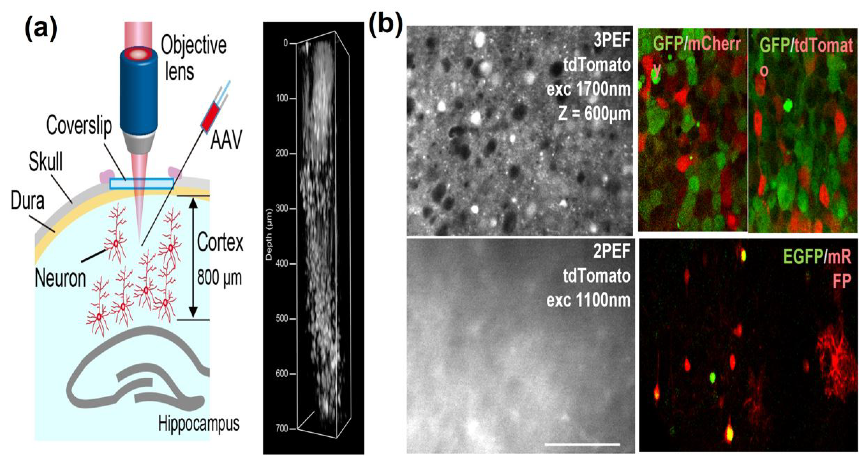
Table 1.
Comparison between ultrafast fiber lasers and traditional lasers.
| Laser Type | Advantages | Disadvantages |
|---|---|---|
| Ultrafast traditional lasers | Shorter pulse width High repetition rate for fast imaging Low repetition rate for deep tissue imaging Larger wavelength tunability (e.g., 700–2000 nm) | More space requirement Higher cost Sensitive to environmental changes Higher level maintenance required. Water cooling is required. Moderate level of average power (1 to 3 W) |
| Ultrafast fiber lasers | Lower cost Compact and portable Air cooling is sufficient. Moderate and high repetition rate is feasible. Low level of maintenance is sufficient. Less sensitive to environmental conditions High average power capabilities (>10 W) | Larger pulse width Low repetition rate is challenging. Smaller wavelength tunability |
Table 2.
Comparison of laser parameters to perform 2PEF and SHG microscopy in the brain.
| Imaging Techniques | Laser Parameters | Indicators | Imaging Depth | Ref. | |||||
|---|---|---|---|---|---|---|---|---|---|
|
Wavelength Range |
Average Power |
Pulse Width |
Pulse Energy |
Repetition Rate | |||||
| 2PEF | Functional Imaging | 1040 nm | - | <150 fs | - | 31.25 MHz | jRGECO1a and jYCaMP1s | 200 m | [27] |
| 920 nm | 800 mW | 70 fs | 110 nJ | 32 MHz | GCaMP6s | 680 m | [59] | ||
| Structural Imaging | 1060 nm | 1.2 W | - | 30 nJ | 40 MHz | Texas-red dye, YFP | 900 m | [26] | |
| 1060 and 1250 nm | - | - | - | - | Alexafluor 647, tdTomato | 750 m | [60] | ||
| 1617 nm | 74.5 fs | 147 nJ | 1 MHz | Indocyanine green (IGC) | 2 mm | [50] | |||
| SHG | Structural Imaging | 1262 nm | - | 38 fs | - | 105 MHz | label-free | - | [61] |
| 1250 nm | 360 mW | 40 fs | 18 nJ | 20 MHz | label-free | - | [62] | ||
Table 3.
Comparison of laser parameters to perform 3PEF and THG microscopy in the brain.
| Imaging Techniques | Laser Parameters | Indicators | Imaging Depth | Ref. | |||||
|---|---|---|---|---|---|---|---|---|---|
|
Wavelength Range |
Average Power |
Pulse Width |
Pulse Energy |
Repetition Rate | |||||
| 3PEF | Functional Imaging | 1260 nm | 530 mW | 32 fs | 22 nJ | 24 MHz | GCaMP7f | 150 m | [51] |
| 1700 nm | - | 64 fs | 140 nJ | 32 MHz | jRGECO1a | 750 m | [59] | ||
| Structural Imaging | 1800 nm | - | 150 fs | 1 J | 0.5 MHz | TurboFP636 | 700 m | [16] | |
| 1700 nm | - | 65 fs | 3.1 J | 1.25 MHz | tdTomato and GFP | 600 m | [49] | ||
| THG | Structural Imaging | 1650–1700 nm | 400 mW | 70 fs | 9 nJ | 50 MHz | label-free | 20–150 m | [63] |
Disclaimer/Publisher’s Note: The statements, opinions and data contained in all publications are solely those of the individual author(s) and contributor(s) and not of MDPI and/or the editor(s). MDPI and/or the editor(s) disclaim responsibility for any injury to people or property resulting from any ideas, methods, instructions or products referred to in the content. |
© 2023 by the authors. Licensee MDPI, Basel, Switzerland. This article is an open access article distributed under the terms and conditions of the Creative Commons Attribution (CC BY) license (https://creativecommons.org/licenses/by/4.0/).
Share and Cite
MDPI and ACS Style
Srinivasan, T.; Yildirim, M. Advances in Ultrafast Fiber Lasers for Multiphoton Microscopy in Neuroscience. Photonics 2023, 10, 1307. https://doi.org/10.3390/photonics10121307
AMA Style
Srinivasan T, Yildirim M. Advances in Ultrafast Fiber Lasers for Multiphoton Microscopy in Neuroscience. Photonics. 2023; 10(12):1307. https://doi.org/10.3390/photonics10121307
Chicago/Turabian StyleSrinivasan, Thulasi, and Murat Yildirim. 2023. "Advances in Ultrafast Fiber Lasers for Multiphoton Microscopy in Neuroscience" Photonics 10, no. 12: 1307. https://doi.org/10.3390/photonics10121307
Note that from the first issue of 2016, this journal uses article numbers instead of page numbers. See further details here.