Communication

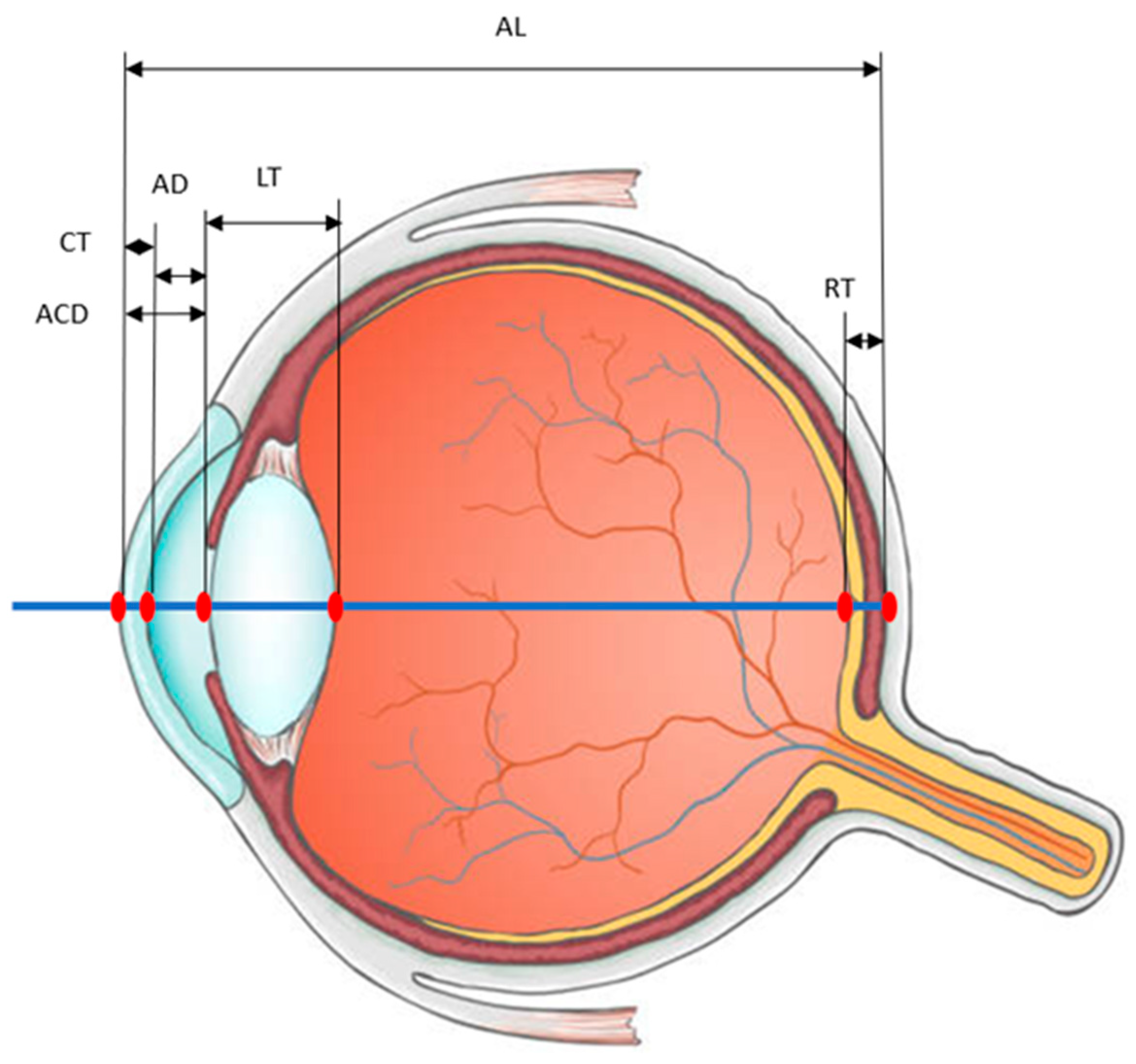
Variations in Lens Thickness Affecting the Anterior Chamber Length and Their Potential Measurement Using a Biometer

by
F. Javier Povedano-Montero
1,2,*,
Ricardo Bernardez-Villaboa
1,
Gema Martínez-Florentín
1,
Francisco López-Muñoz
2,3 and
Juan E. Cedrún-Sánchez
1
1
Optometry and Vision Department, Faculty of Optics and Optometry, Complutense University of Madrid, 28037 Madrid, Spain
2
Hospital Doce de Octubre Research Institute (i+12), 28041 Madrid, Spain
3
Health Sciences Faculty, Camilo José Cela University, 28692 Madrid, Spain
*
Author to whom correspondence should be addressed.
Photonics 2023, 10(12), 1351; https://doi.org/10.3390/photonics10121351
Submission received: 16 November 2023 / Revised: 28 November 2023 / Accepted: 6 December 2023 / Published: 7 December 2023
(This article belongs to the Special Issue Latest Developments in Ocular Biometry)

Abstract

:
Biometry is a critical aspect of ophthalmology, since it facilitates the measurement of several ocular parameters and aids in the diagnosis of conditions like glaucoma. The advent of the IOLMaster in 1999 marked a pivotal moment in biometry by introducing non-contact and highly precise measurements that revolutionized the field. Low-coherence optical reflectometry devices such as Lenstar LS900 and Aladdin have further advanced biometry, due to the exceptional accuracy they offer. Axial length, a fundamental measurement in biometry, directly correlates with conditions like myopia and glaucoma. The accurate measurement of axial length is crucial for diagnosis and treatment planning. Biometry also guides intraocular lens power calculation during cataract surgery, relying on factors like axial length, anterior chamber depth, lens thickness, and effective lens position (ELP). Ensuring precision in these measurements is essential for optimal surgical outcomes. While several studies have explored biometric parameters, dynamic changes in crystalline lens thickness during rest or accommodation have received little attention. These changes may have a significant effect on the measurement of the anterior chamber length, and consequently impact the overall biometric assessment. This study delves into dynamic biometry, particularly in the context of age-related presbyopia, and aims to assess the feasibility of incorporating into the biometric process a specialized device capable of accurately considering crystalline lens changes during different states like rest and accommodation. This exploration seeks to enhance the understanding of ocular dynamics and contribute to improving the precision of diagnostic and surgical techniques. It underscores the importance of staying at the forefront of biometric research, especially in the context of emerging technologies and their potential to transform ophthalmology.

1. Introduction

Biometry is a fundamental part of a patient’s ophthalmologic examination and is essential for calculating intraocular lens power, monitoring myopia, and detecting certain conditions such as glaucoma [1,2,3,4].
Several techniques have been used to measure ocular biometry, such as ophthalmophakometry and A-scan ultrasonography. The latter method has emerged as a potential tool for the early detection of ocular pathologies such as glaucoma [5,6].
However, the introduction of the IOLMaster (Carl Zeiss Meditec AG) in 1999, based on partial coherence interferometry, marked a significant improvement in ocular biometry. It made the technique non-contact and provided higher measurement resolution (about ±0.02 mm compared to ± 0.15 mm with ultrasound) [7]. The IOL-Master700 has established itself as a highly effective tool for evaluating ocular biometry parameters, demonstrating its capability even in challenging situations like the presence of a dense nucleus [8]. Since then, partial coherence laser interferometry (PCLI) has become a widely used method for ocular biometry [7,9,10,11].
Optical interferometry, used in ocular biometry, is a sophisticated and precise method for measuring the internal dimensions of the eye. This advanced technique uses the principles of light wave interference to provide highly accurate measurements of the eye’s structure, which is essential for tailoring treatments in ophthalmology (Figure 1).
This method is critical to several aspects of ophthalmology, most notably the calculation of intraocular lens (IOL) power for cataract surgery and the assessment of refractive errors [4,8,12,13]. It is based on the principle of superimposing two or more sources of coherent light (typically lasers) to create an interference pattern. This pattern can be analyzed to determine the wavelength of the light and the phase differences between the beams, thus allowing extremely high-precision distance measurements. In ocular biometry, devices like the IOLMaster use partial coherence interferometry (PCI) or optical low-coherence interferometry (OLCI) to measure the axial length of the eye, with the remarkable advantage of being non-contact.
Currently, devices based on optical low-coherence reflectometry (OLCR) are available, including the Lenstar LS900 (Haag-Streit Köniz), Aladdin (Topcon), AL-Scan (Nidek), and OA-2000 (Tomey) [13,14,15,16]. The accuracy and repeatability of measurements with these instruments are excellent and have been verified in various studies, where the values were similar to the values obtained from these instruments [9,11,17,18]. The ability to accurately measure various components of the eyeball, as shown in Figure 2, plays a critical role in providing reliable and robust data in ophthalmic biometry. This improved accuracy is critical to advancing our understanding and treatment of a range of eye diseases, with significant implications for eye care and research.
It is well established that there is a direct relationship between axial length and spherical ametropia [19]. Increasing myopia leads to the elongation of the eyeball and a reduced expansion in the vertical height of the eye [20,21,22]; therefore, detecting changes in the axial length of the eye is essential for carrying out any interventions to control it.
There are pathologic conditions in which the axial length of the eye is involved, most notably in glaucoma. There is evidence that this length is involved in primary open-angle glaucoma [23], and it is associated with a short axial length and shallow anterior chamber in closed-angle glaucoma [24].
Another specific function of biometry is the calculation of the power of intraocular lenses to be implanted in cataract surgery, as it determines the expected postoperative outcome and the visual quality of the patient [25,26]. Three parameters are essential for the calculation of IOL power: axial length, anterior chamber depth, and lens thickness. With the development of fourth- and fifth-generation IOL calculation formulas, lens thickness has gained significant interest due to the introduction of the concept of effective lens position (ELP) [27].
There have been studies worldwide that have analyzed the axial length of the eye, anterior chamber depth, and lens thickness [28,29,30]. However, there is no device that can analyze the changes that occur in lens thickness at rest or in the accommodative state and, therefore, the consequences that this may have on the anterior chamber length.
Research into biometric variations of the crystalline lens and ciliary body during dynamic accommodation is crucial for understanding its process, as age-related alterations lead to presbyopia.
Some studies have shown that the anterior surface of the lens moves and increases its thickness during accommodation. Measurements of anterior chamber depth, lens thickness, and anterior segment length have even been correlated using techniques such as A-scan ultrasonography or partial coherence interferometry, with refraction measured simultaneously in the same eye or in the contralateral eye [31,32].
The main objective of this study is to assess the feasibility of incorporating the objective accommodation measurement device into the biometric process, capable of accurately considering crystalline lens changes during different states like rest and accommodation.

2. Results

The objective accommodation measurement device represents a theoretical advancement in optical instrumentation. It is meticulously designed with an advanced set of internal lenses whose main function is to generate a detailed and specific ray tracing pattern. The unique nature of this pattern lies in its ability to capture highly versatile images, which can be precisely modified to adapt to a wide range of focal planes. Figure 3 illustrates this instrument that can be integrated as an additional tool, like those currently used to perform topography with the Lenstar LS900 biometer (Haag-Streit, Wedel, Germany). This visual representation helps to understand how the instrument complements and enhances the existing capabilities of the Lenstar LS900, and provides a clearer insight into its practical application in ocular measurement.
One of the most notable features of the device is the incorporation of a semi-silvered sheet, strategically placed with meticulous precision and tilted at 45 degrees. Theoretically, this sheet can project images perpendicular to the line of sight of the eye, which maximizes the accuracy of the analysis. The device’s innovative design contemplates the use of toothed discs, which house lenses of both positive and negative powers. These discs, when manipulated and combined, have the theoretical potential to offer an optical power spectrum ranging from −20 to +20 diopters.
Within the design scheme, an illuminated sheet is positioned, which synchronizes perfectly with the semi-silvered sheet. This versatile natured sheet can display images ranging from optotype letters to other graphical representations. As the image moves through this intricate lens system, another set of lenses specifically designed to adjust and compensate for any ametropic condition the eye may present is activated. This auxiliary lens system is of notable complexity, as it can integrate various combinations that include spherical and cylindrical components, all structured in a dual plate design.
Additionally, a high compatibility of the device with a wide variety of biometers currently on the market has also been considered. In order to ensure seamless integration, the incorporation of an anchor ring supplement has been theorized, which would fit perfectly in the specific dimensions of the biometer’s objective lens in question.
Finally, one of the primary objectives of the theoretical design is the ability to manipulate the relative distance of the image using lenses. This manipulation seeks to alter the perceived size of the image, which theoretically could induce variations in aspects such as pupil size and lens characteristics, like its curvature and thickness. Through this operation, valuable information about the structure and behavior of the lens could be obtained, thereby allowing for a precise evaluation of the accommodation amplitude and its adequacy in different visual contexts.

3. Discussion

Ocular biometry has undergone a major transformation with the introduction of devices based on partial coherence interferometry and optical low-coherence interferometry. These technological advances provide accurate and non-invasive measurements of crucial ocular parameters, such as lens thickness, anterior chamber depth, and axial length. However, a gap exists in the ability of these devices to measure dynamic changes in lens thickness during resting and accommodation states. These dynamic lens thickness changes may have substantial implications for the anterior chamber length, a parameter that can influence diagnostics and treatments.
To understand this, it is essential to comprehend the accommodation mechanism that depends on the lens’s mechanical capabilities and shape changes during the process. The increase in lens thickness during accommodation is greater than the decrease in anterior chamber depth, which suggests that the posterior surface of the lens moves backward during accommodation. During accommodation, the lens’s anterior surface becomes more hyperbolic [33].
An initial attempt has already been made to relate these properties to a mechanism for the young human lens using certain simplifying assumptions such as spherical curvature on the lens’s anterior surface and elastic isotropy [34]; however, it has been shown that the lens behaves as an anisotropic body [35].
Dynamic accommodation measurements have been conducted in previous studies that used monkeys as research subjects [36]. These studies revealed accommodative changes in the anterior and posterior lens curvature radii using a Purkinje imaging method. Regarding curvature changes per diopter of accommodation, slopes of 0.006 mm −1/D and 0.00485 mm −1/D were observed for the anterior and posterior lenses, respectively.
Furthermore, research in humans has produced similar results. In a study involving a 19-year-old subject, an average change of −0.33 mm/D and 0.15 mm/D was observed in the anterior and posterior lens curvature radii, respectively [37]. Other studies have also documented changes in anterior lens curvature, reporting variations up to −0.62 mm and 0.17 mm/D, with a change in radius per diopter of the anterior surface 4.7 times greater than the posterior surface [33,38].
The results highlight the importance of understanding and quantifying changes in lens curvature during human accommodation, and contribute to achieving a more comprehensive grasp of ocular dynamics and its potential clinical implications. These findings further underscore our research’s relevance, which demonstrates that ocular accommodation is a highly dynamic process. Understanding variations in lens thickness during accommodation may have a significant impact on diagnosing and treating ocular disorders, thus emphasizing the importance of continuing to explore this area of study to advance ophthalmology.
Additionally, the device’s integration with existing biometers offers a practical solution for clinics and hospitals already invested in biometric technology. The device’s modular design would allow for easy adaptation to various biometer models, enhancing its applicability and versatility.
However, it is essential to acknowledge potential challenges and limitations associated with implementing the proposed device.

4. Conclusions

The present study underscores the need to address current limitations in ocular biometry, particularly in measuring dynamic lens thickness changes. The introduction of the objective accommodation measurement device may be a significant step toward enhancing biometric measurements’ accuracy and comprehensiveness.
The proposed device not only promises to enhance the precision of accommodation measurements but also holds the potential to influence how various ocular conditions are diagnosed and treated. The ability to measure dynamic changes in lens thickness may offer new insights into age-related presbyopia and other accommodation disorders.
In summary, the objective accommodation measurement device introduced in this study has the potential to revolutionize ocular biometry, since it provides more detailed and accurate measurements that can influence the diagnosis and treatment of various eye conditions. Future studies are anticipated to validate the device’s efficacy and precision in clinical settings.

Author Contributions

All authors have contributed to this work. Conceptualization, F.J.P.-M., R.B.-V. and J.E.C.-S.; writing—original draft preparation, F.J.P.-M., R.B.-V. and J.E.C.-S.; writing—review and editing F.J.P.-M., R.B.-V., G.M.-F., F.L.-M. and J.E.C.-S.; supervision, F.J.P.-M., R.B.-V. and J.E.C.-S. All authors have read and agreed to the published version of the manuscript.

Funding

This research received no external funding.

Institutional Review Board Statement

Not applicable.

Informed Consent Statement

Not applicable.

Data Availability Statement

Data are contained within the article.

Conflicts of Interest

The authors declare no conflict of interest.

References

  1. Dervisogullari, M.S.; Totan, Y.; Guragac, B. Comparison of anterior chamber depth measurements of Nidek AL-Scan and Galilei Dual Scheimpflug Analyzer. Contact Lens Anterior Eye 2015, 38, 85–88. [Google Scholar] [CrossRef] [PubMed]
  2. Chamarty, S.; Verkicharla, P.K. Accuracy and Precision of New Optical Biometer Designed for Myopia Management in Measurement of Ocular Biometry. Optom. Vis. Sci. Off. Publ. Am. Acad. Optom. 2023. [Google Scholar] [CrossRef] [PubMed]
  3. Chandran, P.; Sneha, C.; Subramanian, S.; Raman, G.V. Comparison between ocular biometry parameters in patients with unilateral congenital glaucoma. Indian J. Ophthalmol. 2023, 71, 2962–2966. [Google Scholar] [CrossRef] [PubMed]
  4. Lee, A.C.; Qazi, M.A.; Pepose, J.S. Biometry and intraocular lens power calculation. Curr. Opin. Ophthalmol. 2008, 19, 13–17. [Google Scholar] [CrossRef] [PubMed]
  5. Savur, S.; Kaup, S.; Dinesh, A.; Shivalli, S.; Kondal, D. Can ultrasonic biometric indices with optimal cut-offs be a potential screening tool for primary angle closure disease? A case-control study. Eye 2023, 37, 1284–1289. [Google Scholar] [CrossRef] [PubMed]
  6. Yong, K.L.; Gong, T.; Nongpiur, M.E.; How, A.C.; Lee, H.K.; Cheng, L.; Perera, S.A.; Aung, T. Myopia in asian subjects with primary angle closure: Implications for glaucoma trends in East Asia. Ophthalmology 2014, 121, 1566–1571. [Google Scholar] [CrossRef] [PubMed]
  7. Santodomingo-Rubido, J.; Mallen, E.A.; Gilmartin, B.; Wolffsohn, J.S. A new non-contact optical device for ocular biometry. Br. J. Ophthalmol. 2002, 86, 458–462. [Google Scholar] [CrossRef] [PubMed]
  8. Pakuliene, G.; Zimarinas, K.; Nedzelskiene, I.; Siesky, B.; Kuzmiene, L.; Harris, A.; Januleviciene, I. Anterior segment optical coherence tomography imaging and ocular biometry in cataract patients with open angle glaucoma comorbidity. BMC Ophthalmol. 2021, 21, 127. [Google Scholar] [CrossRef]
  9. Holzer, M.P.; Mamusa, M.; Auffarth, G.U. Accuracy of a new partial coherence interferometry analyser for biometric measurements. Br. J. Ophthalmol. 2009, 93, 807–810. [Google Scholar] [CrossRef]
  10. Kaswin, G.; Rousseau, A.; Mgarrech, M.; Barreau, E.; Labetoulle, M. Biommetry and intraocular lens power calculation results with a new optical biometry device: Comparison with the gold standard. J. Cataract Refract. Surg. 2014, 40, 593–600. [Google Scholar] [CrossRef]
  11. Huang, J.H.; Savini, G.; Wu, F.; Yu, X.X.; Yang, J.; Yu, A.; Yu, Y.; Wang, Q.M. Repeatability and reproducibility of ocular biometry using a new noncontact optical low-coherence interferometer. J. Cataract Refract. Surg. 2015, 41, 2233–2241. [Google Scholar] [CrossRef] [PubMed]
  12. Upasna; Singh, V.K.; Rana, J.; Kumar, S.; Singh, A.; Singh, K. An evaluation of intraoperative and postoperative outcomes of phacoemulsification surgery in eyes with shallow anterior chamber. Indian J. Ophthalmol. 2021, 69, 1346–1347. [Google Scholar] [CrossRef] [PubMed]
  13. El-Haddad, N.; Abd Elwahab, A.; Shalaby, S.; Farag, M.M.A.; Alkassaby, M.; Ahmed, S.; Shawky, S. Comparison between open-angle glaucoma and angle-closure glaucoma regarding the short-term optic disc vessel density changes after trabeculectomy. Lasers Med. Sci. 2023, 38, 246. [Google Scholar] [CrossRef] [PubMed]
  14. Noya-Padin, V.; Garcia-Queiruga, J.; Iacubitchii, M.; Giraldez, M.J.; Yebra-Pimentel, E.; Pena-Verdeal, H. Lenstar LS900 vs EchoScan US-800: Comparison between optical and ultrasound biometry with and without contact lenses and its relationship with other biometric parameters. Expert Rev. Med. Devices 2023, 20, 681–690. [Google Scholar] [CrossRef] [PubMed]
  15. Bhullar, H.; Dhurjon, L.; Francis, C.; Bhambhwani, V. Comparative Analysis of Axial Length Measurements by Optical Biometers Based on Partial Coherence Interferometry versus Optical Low-Coherence Interferometry: An Office Audit. Cureus 2022, 14, e21883. [Google Scholar] [CrossRef] [PubMed]
  16. Xiong, Y.; Lin, Y.; Zhao, Z.; Wang, H.; Zhang, G. Evaluation and comparison of ocular biometric parameters obtained with Tomey OA-2000 in silicone oil-filled aphakic eyes. BMC Ophthalmol. 2023, 23, 218. [Google Scholar] [CrossRef] [PubMed]
  17. Shammas, H.J.; Hoffer, K.J. Repeatability and Reproducibility of Biometry and Keratometry Measurements Using a Noncontact Optical Low-Coherence Reflectometer and Keratometer. Am. J. Ophthalmol. 2012, 153, 55–61. [Google Scholar] [CrossRef] [PubMed]
  18. Mandal, P.; Berrow, E.J.; Naroo, S.A.; Wolffsohn, J.S.; Uthoff, D.; Holland, D.; Shah, S. Validity and repeatability of the Aladdin ocular biometer. Br. J. Ophthalmol. 2014, 98, 256–258. [Google Scholar] [CrossRef]
  19. Strang, N.C.; Schmid, K.L.; Carney, L.G. Hyperopia is predominantly axial in nature. Curr. Eye Res. 1998, 17, 380–383. [Google Scholar] [CrossRef]
  20. Wallman, J. Nature and nurture of myopia. Nature 1994, 371, 201–202. [Google Scholar] [CrossRef]
  21. Harrington, S.; O’Dwyer, V. The association between time spent on screens and reading with myopia, premyopia and ocular biometric and anthropometric measures in 6-to 7-year-old schoolchildren in Ireland. Ophthalmic Physiol. Opt. 2023, 43, 505–516. [Google Scholar] [CrossRef] [PubMed]
  22. Atchison, D.A.; Smith, G. Possible errors in determining axial length changes during accommodation with the IOLMaster. Optom. Vis. Sci. 2004, 81, 283–286. [Google Scholar] [CrossRef] [PubMed]
  23. Manandhar, N.; Pradhan, C.; Joshi, P.; Subedi, P.; Shrestha, P. Comparison of Ocular Biometry between Primary Open Angle Glaucoma Patients and Normal subjects. Nepal. J. Ophthalmol. 2021, 13, 62–68. [Google Scholar] [CrossRef] [PubMed]
  24. Oku, Y.; Oku, H.; Park, M.; Hayashi, K.; Takahashi, H.; Shouji, T.; Chihara, E. Long axial length as risk factor for normal tension glaucoma. Graefe’s Arch. Clin. Exp. Ophthalmol. 2009, 247, 781–787. [Google Scholar] [CrossRef] [PubMed]
  25. Hrebcová, J.; Skorkovská, S.; Vasků, A. Comparison of contact and immersion techniques of ultrasound biometry in terms of target postoperative refraction. Czech Slovak Ophthalmol. 2009, 65, 143–146. [Google Scholar]
  26. Norrby, S. Sources of error in intraocular lens power calculation. J. Cataract Refract. Surg. 2008, 34, 368–376. [Google Scholar] [CrossRef] [PubMed]
  27. Olsen, T.; Hoffmann, P. C constant: New concept for ray tracing-assisted intraocular lens power calculation. J. Cataract Refract. Surg. 2014, 40, 764–773. [Google Scholar] [CrossRef]
  28. Liang, Y.B.; Friedman, D.S.; Zhou, Q.; Yang, X.H.; Sun, L.P.; Guo, L.X.; Chang, D.S.; Lian, L.Y.; Wang, N.L.; Handan Eye Study, G. Prevalence and Characteristics of Primary Angle-Closure Diseases in a Rural Adult Chinese Population: The Handan Eye Study. Investig. Ophthalmol. Vis. Sci. 2011, 52, 8672–8679. [Google Scholar] [CrossRef]
  29. Wang, D.D.; Huang, W.Y.; Li, Y.T.; Zheng, Y.F.; Foster, P.J.; Congdon, N.; He, M.G. Intraocular Pressure, Central Corneal Thickness, and Glaucoma in Chinese Adults: The Liwan Eye Study. Am. J. Ophthalmol. 2011, 152, 454–462. [Google Scholar] [CrossRef]
  30. Ogbeide, O.U.; Omoti, A.E. Ultrasonographic ocular diameters in Nigerians. West Afr. J. Med. 2009, 28, 97–101. [Google Scholar]
  31. Bolz, M.; Prinz, A.; Drexler, W.; Findl, O. Linear relationship of refractive and biometric lenticular changes during accommodation in emmetropic and myopic eyes. Br. J. Ophthalmol. 2007, 91, 360–365. [Google Scholar] [CrossRef] [PubMed]
  32. Ostrin, L.; Kasthurirangan, S.; Win-Hall, D.; Glasser, A. Simultaneous measurements of refraction and A-scan biometry during accommodation in humans. Optom. Vis. Sci. 2006, 83, 657–665. [Google Scholar] [CrossRef] [PubMed]
  33. Dubbelman, M.; Van der Heijde, G.L.; Weeber, H.A. Change in shape of the aging human crystalline lens with accommodation. Vis. Res. 2005, 45, 117–132. [Google Scholar] [CrossRef] [PubMed]
  34. Koretz, J.F.; Handelman, G.H. Model of the accommodative mechanism in the human eye. Vis. Res. 1982, 22, 917–927. [Google Scholar] [CrossRef] [PubMed]
  35. Koretz, J.F.; Handelman, G.H. A model for accommodation in the young human eye: The effects of lens elastic anisotropy on the mechanism. Vis. Res. 1983, 23, 1679–1686. [Google Scholar] [CrossRef] [PubMed]
  36. Rosales, P.; Wendt, M.; Marcos, S.; Glasser, A. Changes in crystalline lens radii of curvature and lens tilt and decentration during dynamic accommodation in rhesus monkeys. J. Vis. 2008, 8, 18.1–18.12. [Google Scholar] [CrossRef] [PubMed]
  37. Koretz, J.F.; Handelman, G.H.; Brown, N.P. Analysis of human crystalline lens curvature as a function of accommodative state and age. Vis. Res. 1984, 24, 1141–1151. [Google Scholar] [CrossRef]
  38. Garner, L.F. Mechanisms of accommodation and refractive error. Ophthalmic Physiol. Opt. 1983, 3, 287–293. [Google Scholar]
Figure 1. Interferogram: detailed visualization of interference patterns.
Photonics 10 01351 g001
Figure 2. Detailed biometer measurement parameters. AL (Axial Length)—distance from cornea to retina; LT (Lens Thickness)—eye’s lens thickness; RT (Retinal Thickness)—thickness of the retina; CT (Corneal Thickness)—cornea’s thickness; AD (Aqueous Depth)—space between cornea and lens. ACD (Anterior Chamber Depth)—space between the cornea and lens plus corneal thickness.
Photonics 10 01351 g002
Figure 3. Objective accommodation measurement device: a graphical overview of the design and functionality of the instrument.
Photonics 10 01351 g003
Disclaimer/Publisher’s Note: The statements, opinions and data contained in all publications are solely those of the individual author(s) and contributor(s) and not of MDPI and/or the editor(s). MDPI and/or the editor(s) disclaim responsibility for any injury to people or property resulting from any ideas, methods, instructions or products referred to in the content.

