Review

Genome Studies on Nematophagous and Entomogenous Fungi in China

State Key Laboratory of Mycology, Institute of Microbiology, Chinese Academy of Sciences, No. 3 Park 1, Beichen West Rd., Chaoyang District, Beijing 100101, China
*
Author to whom correspondence should be addressed.
J. Fungi 2016, 2(1), 9; https://doi.org/10.3390/jof2010009
Submission received: 9 December 2015 / Revised: 24 January 2016 / Accepted: 29 January 2016 / Published: 5 February 2016
(This article belongs to the Special Issue Fungal '-Omics': Is the Best Yet to Come?)

Abstract

:
The nematophagous and entomogenous fungi are natural enemies of nematodes and insects and have been utilized by humans to control agricultural and forestry pests. Some of these fungi have been or are being developed as biological control agents in China and worldwide. Several important nematophagous and entomogenous fungi, including nematode-trapping fungi (Arthrobotrys oligospora and Drechslerella stenobrocha), nematode endoparasite (Hirsutella minnesotensis), insect pathogens (Beauveria bassiana and Metarhizium spp.) and Chinese medicinal fungi (Ophiocordyceps sinensis and Cordyceps militaris), have been genome sequenced and extensively analyzed in China. The biology, evolution, and pharmaceutical application of these fungi and their interacting with host nematodes and insects revealed by genomes, comparing genomes coupled with transcriptomes are summarized and reviewed in this paper.

1. Introduction

Nematophagous fungi infect their hosts using traps and other devices such as adhesive conidia and parasitic hyphae tips [1,2]. Entomogenous fungi are associated with insects, mainly as pathogens or parasites [3]. Both nematophagous and entomogenous fungi are important biocontrol resources [1,3]. Cordyceps spp. are ancient resources in Chinese medicine [4]. Biological control is an economically and ecologically friendly approach for controlling nematode and insect pests that have a negative economic impact in agriculture [5]. Nematophagous and entomopathogenic fungi have been used with various strategies for almost a century and, for instance, mass production of entomopathogenic fungi were utilized for insect-pest control in the field while nematophagous fungi were released to the soil for nematode control [6,7]. There is also a long history of utilizing these fungi for the control of agricultural and forestry pests in China [8].
Among these fungi, Arthrobotrys oligospora (adhesive network traps) and Drechslerella stenobrocha (constricting ring trap) are well-known nematode trapping fungi in the Orbiliales (Orbiliomycetes and Ascomycota) [9,10,11]; Hirsutella minnesotensis, as a nematode endoparasite, responds to the natural suppressiveness of soybean cyst nematode [12,13,14]; Ophiocordyceps sinensis, a famous fungus used as a traditional Chinese medicine, belongs to Ophiocordycipitaceae, Hypocreales (Sordariomycetes, Ascomycota) [15]; Cordyceps militaris as medicinal fungus and Beauveria bassiana as biocontrol agent of insect are representatives of Cordycipitaceae, Hypocreales (Sordariomycetes, Ascomycota) [16,17]; and Metarhizium spp. in Clavicipitaceae, Hypocreales (Sordariomycetes, Ascomycota) are important entomopathogens [18]. The biology, evolution, interaction with hosts, biocontrol application for pests, and medicinal use of these fungi have been extensively studied by Chinese mycologists [19,20].
Recently, development of next generation sequencing technology allows the easy obtention of high quality eukaryotic genome sequences [21,22]. A number of nematophagous and entomogenous fungi have been genome sequenced. Genomic surveys have provided new strategies to answer biological issues of those fungi and their sophisticated mechanisms for interacting with their invertebrate hosts. A number of nematophagous and entomogenous fungi extensively studied in China have been genome sequenced by Chinese mycologists [9,10,13,15,16,17,18]. The approaches obtained from analysis of genomes, and comparing genomes and transcriptomes of these fungi, provide comprehensive understanding of their biology and interaction with invertebrate hosts, and furthermore, enhance their application in pest control and medicine utilization.

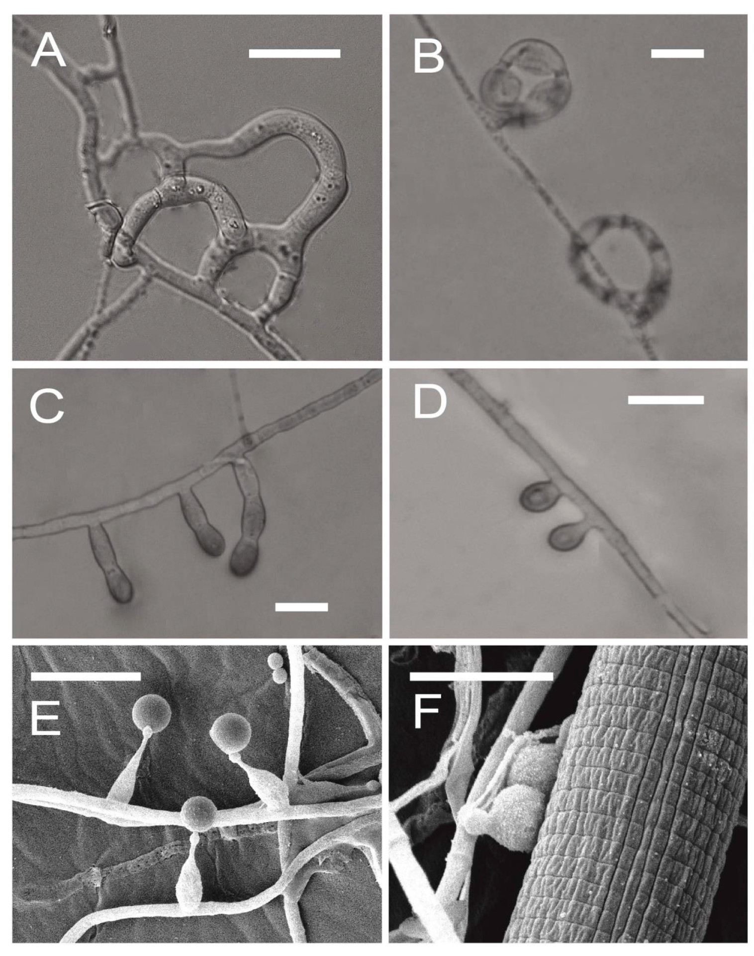
2. Nematophagous Fungi

Nematophagous fungi can use specialized traps to capture nematodes, conidia to adhere to nematodes, hyphae tips to parasitize nematode females and eggs, or produce toxins to attack nematodes [2]. Nematode-trapping fungi and nematode endoparasitic fungi are two important groups of nematophagous fungi. Predation is one of the fungal life-strategies to destroy nematodes [1,23] by capturing nematodes with sophisticated trapping structures, including constricting rings and several other types of adhesive traps (sessile adhesive knobs, stalked adhesive knobs, adhesive nets, adhesive columns, and non-constricting rings) (Figure 1A–D) [24,25]. The traps are usually produced from hyphae and induced by nematodes, peptides, and nematode extracts [26,27]. Most of the nematode-trapping fungi belong to Orbiliales (Ascomycota). Among these fungi, Arthrobotrys oligospora forming adhesive nets was first genome sequenced to understand the molecular basis of the mechanisms for capturing nematodes (Table 1) [28,29]. A proposed model containing multiple fungal signal transduction pathways associated with diverse cellular processes such as energy metabolism, biosynthesis of the cell wall and adhesive proteins, cell division, glycerol accumulation, and peroxisome biogenesis [29]. The following sequenced genome by Chinese mycologists was Drechslerella stenobrocha, which forms constricting rings to actively capture nematodes. Comparative genomic analysis provided support for the hypothesis that nematode trapping fungi evolved from saprophytic fungi in a high carbon and low nitrogen environment and revealed the transition between saprophagy and predation for further understanding of the interaction between nematode-trapping fungi and nematodes [30].
Figure 1. Morphology of traps and adhesive conidia produced by nematophagous fungi: (A) Adhesive nets (Arthrobotrys oligospora); (B) Constricting rings (Drechslerella stenobrocha); (C) Adhesive columns (Gamsylella cionopaga); (D) Adhesive knobs (Gamsylella robusta); and (E, F) Adhesive conidia (Hirsutella minnesotensis). Bars = 10 μm.
Jof 02 00009 g001
Table 1. Available genomic sequences nematophagous fungi in China.
FeaturesNematode-Trapping FungiNematode Endoparasitic FungiEntomogenous Fungi
Drechslerella stenobrochaArthrobotrys oligosporaHirsutella minnesotensisMetarhizium robertsiiMetarhizium acridumBeauveria bassianaCordyceps militarisOphiocordyceps sinensis
Assembled size (Mb)29.0240.0251.439.0438.0533.732.2~120
Protein-coding genes778111,47912,70210,582984910,36696846792
Coverage (fold)80×-12810010776.6147241
Number of scaffolds142-967174241242147-
Scaffold N50 (kb)434.4-382.419603307304550-
G + C content (%)52.545.252.151.4949.9151.551.446.1
Simple repeat rate (%)0.92-1.330.981.522.033.0437.98
TEs (%)--34.67-----
Gene density (genes per Mb)268271247.1271.1258.830825787
Exons per gene3.53.32.52.82.72.73.02.6
tRNA genes82149145141122113136-
ReferencesLiu et al. 2014 [10]Yang et al. 2011 [9]Lai et al. 2014 [13]Gao et al. 2011 [18]Gao et al. 2011 [18]Xiao et al. 2012 [17]Zheng et al. 2011 [12]Hu et al. 2013 [15]
Unlike nematode-trapping fungi, Hirsutella spp. are representatives of nematode endoparasitic fungi in Hypocreales (Ascomycota). Hirsutella minnesotensis and Hirsutella rhossiliensis, as parasites on cyst nematodes, can suppress the population of soybean cyst nematodes (SCN, Heterodera glycines) and are widely distributed in the northeast of China and Minnesota in the United States [12,29]. H. minnesotensis is the first genome sequenced nematode endoparasitic fungus [13]. It can parasitize nematodes by means of adhesive spores (Figure 1E,F). Genome of H. minnesotensis is different from those of nematode-trapping fungi but similar to those of entomopathogenic fungi. It has fewer genes encoding lectins for adhesion and glycoside hydrolases (GHs) for cellulose degradation, but more genes for protein degradation, signal transduction, and secondary metabolism [13].

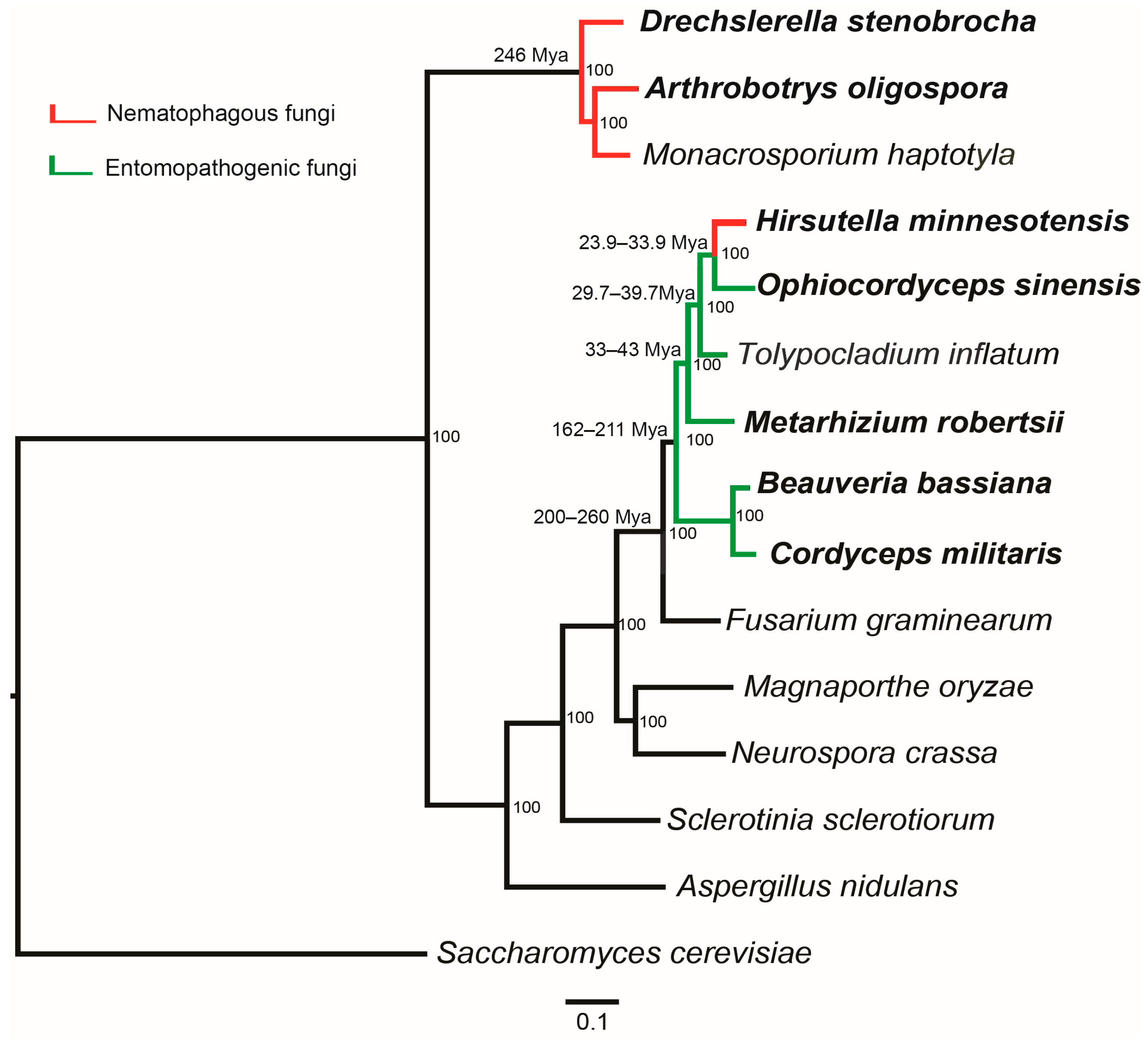
2.1. Different Origin of Nematode-Trapping and Endoparasitic Fungi

As a special living strategy of fungi, the origin and development of predation are an essential and attractive topic in evolutionary biology of fungi. It was deduced that predation was evolved to obtain the nitrogen from nematode in the environments that are rich in carbon but poor in nitrogen [30]. Direct capture of nitrogen from small animals would provide predatory fungi with a competitive advantage over strictly saprophytic fungi [31]. A previous study based on the multigene phylogeny estimated that the lineage of nematode-trapping fungi in Ascomycota diverged at 246 million years ago (Mya), five million years after the Permian–Triassic extinction (251.4 Mya) [30,32]. Other clades of fungi that form adhesive traps diverged around 200 Mya, coinciding with the Triassic–Jurassic extinction (201.4 Mya) [30]. However, still little is known about the origin of predatory fungi for the lack of strong direct evidence.
Currently, available genomic sequences of nematode-trapping fungi have made it possible for the evolutionary study at genomic level. Phylogenomic relationships constructed via the genomic sequences confirmed that both D. stenobrocha and A. oligospora are affiliated with the family Orbiliaceae (Figure 2) [9,10]. The analysis indicated that the nematode-trapping fungi were efficient saprobes. The identified large numbers of enzymes involved in the process of saprophytic degradation suggested that nematode-trapping fungi are more similar to fungal saprobes rather than animal pathogens or plant pathogens [10]. Among these enzymes, the largest number of proteins are carbohydrate binding module 1 (CBM1)-containing cellulases, previously found to be associated with cellulose degradation in the typical rot fungus (Phanerochaete chrysosporium) and the coprophilic Podospora anserina [33,34]. Thus, nematode-trapping fungi are suggested to be efficient saprobes and this provides more evidence for the hypothesis that nematode-trapping fungi might have originated in an environment rich in carbon resources but lacking nitrogen.
As a nematode endoparasite in the order Hypocreales, phylogenomic analysis indicated that H. minnesotensis is clustered to the caterpillar fungus O. sinensis, an insect pathogen. H. minnesotensis diverged from O. sinensis around 23.9–33.9 Mya and from Tolypocladium inflatum around 29.7–39.7 Mya (Figure 2) [13]. This suggests that the speciation of nematode endoparasites is independent with the mass extinction but more likely originated from entomopathogens.

2.2. Lectins and Other Adhesive Proteins Involved in Adhering to Nematode

One of the important mechanisms of fungi recognizing their nematode host is mediated by lectins. The traps of A. oligospora that are challenged by GalNAc (N-acetylgalactosamine) lost the ability to capture nematodes [35]. Other monosaccharaides such as glucose and mannose can also inhibit the capacity of the trapping-fungi to recognize nematodes [36]. However, the knocked out lectin-coding gene in A. oligospora did not affect the trapping ability or vegetative growth [37]. Moreover, lectins were not detected to be differentially expressed during nematode trapping [9]. Thus, the genomic and transcriptomic data did not support the previous hypothesis of lectin-mediated nematode recognition [9].
Figure 2. Genome-based phylogenetic tree of nematophagous fungi and other fungi. Bootstrap values and divergence time are indicated beside the nodes; color of branches corresponds to the lifestyle character states as follows: red, nematophagous fungi; green, entomopathogenic fungi; black, other fungi.
Jof 02 00009 g002
The lectin-coding genes identified in the genomes of nematode-trapping fungi suggest that lectins might play a more important role in the adhesive networks than in the constricting rings. More putative lectin-coding genes were identified in the genome of A. oligospora than in D. stenobrocha, especially in the families of fucose-specific and H-type lectins [9,10]. It is also possible that the recognition of nematodes is executed by multiple lectin-encoding genes, and this would explain why the deleted lectin gene did not affect the nematode capture capacity of the fungi. Compared with trapping fungi, H-type and fucose-specific lectins are absent in the H. minnesotensis genome and this would indicate that nematode endoparasitic fungus has evolved a different mechanism to adhere to the nematode hosts [13]. Furthermore, expression of lectin-encoding genes of H. minnesotensis is significantly up-regulated during the infection to nematode. Based on the genomic data, pretreating the nematodes with different lectins and their mixtures can also decrease the adhesion rate of H. minnesotensis [13]. Coupled with previous studies, it is still possible that lectin is important to mediate the nematode recognition by fungi.
In addition to the lectins, other adhesive proteins were also suggested to contribute to the adhesion of fungi to nematodes. For instance, numerous CFEM-containing proteins have been identified in the both nematode-trapping and endoparasitic fungal genomes [9,10,13]. The function of CFEM-containing adhesive proteins was confirmed by the transcriptomic analysis of H. minnesotensis and the qPCR analysis of A. oligospora during nematode infection [9,13]. Other supposedly adhesive proteins such as GLEYA-containing proteins have also been found in the nematode-trapping fungi [9,10]. However, most of the adhesive proteins are still functionally unknown and should be investigated in detail in the future.

2.3. Formation of Traps and Infection Pegs

Traps and infection pegs are the only specialized hyphae by which the nematode-trapping and endoparasitic fungi infect and penetrate nematode cuticle. The processes to form these sophisticated hyphal structures are very complicated and poorly understood. Comparing the expressing patterns between the saprophytic and predatory lifestyles based on the proteomic analysis of A. oligospora predicted from its genome indicated that multiple fungal pathways are proposed to be involved in the trap formation of A. oligospora, including mitogen activated protein kinase (MAPK) pathway, cell division, energy metabolism, biosynthesis of the cell wall and adhesive proteins, glycerol accumulation, and peroxisome biogenesis [9]. Similar results are found from the transcriptomic analysis of D. stenobrocha during the trap formation and infection of nematodes. In addition, Zn(2)-C6 type transcription factors might also be associated with signal transduction [10]. Though the formation of infection pegs of H. minnesotensis is quite different from the traps of nematode-trapping fungi, the protein kinase C (PKC) signal transduction pathway is shared by both predatory fungi and endoparasites. Meanwhile, cyclic AMP-protein kinase A pathway and STM1-like G protein-coupled receptor (GPCR) responsible for filamentation are also thought to be involved in nematode infection [13].

2.4. Virulence Factors Associated with Nematode Infection

2.4.1. Extracellular Enzymes

Penetrating and digesting the nematode cuticles play key roles during the fungal infection and the consumption of nematodes. Enzymes such as proteases, collagenases, and chitinases are the virulence factors of nematophagous fungi [9]. Serine proteases are the extensively studied enzymes and, more than 20 of them have been cloned or purified from the nematophagous fungi and are considered as virulent factors [20,38,39]. In addition, the improved pathogenicity by overexpressing serine protease also confirms its role in virulence function [40].
Genome sequencing has revealed a large number of proteases associated with the degradation of the nematode cuticle. Subtilisins, as one of the most important families belonging to serine proteases, namely S08, are abundant in the nematophagous fungi and totally 43 in A. oligospora, 18 in D. stenobrocha, and 21 in H. minnesotensis have been annotated [9,10,13]. However, only two subtilisin-coding genes in A. oligospora and one in D. stenobrocha were found up-regulated when they were challenged by nematode or nematode extracts (NE) [9,10]. Although five subtilisins were up-regulated that might be associated with infection processes of H. minnesotensis, the function of other subtilisins predicted in the genomes of nematophagous fungi is still unknown [13]. Besides the subtilisins, other proteases are highly expressed during the nematode infection, such as protease S28 (DRE_00914) and S33 (DRE_00834) in D. stenobrocha as well as endopeptidases (M4 and M43) and exopeptidases (M14 and M28) in H. minnesotensis [10,13]. This indicates that the degradation of nematode cuticles is not only associated with subtilisins but also a series of other secreted enzymes. Further investigation on these secreted enzymes should be performed to fully understand the molecular mechanisms of fungi infecting nematodes and will provide new insight to improve the pathogenicity of nematophagous fungi.

2.4.2. Secondary Metabolites

It has been long known that most of the compounds against nematodes are produced by toxin-producing fungi that can use them to immobilize nematodes prior to infection [41]. Although the secondary metabolism was investigated more than thirty year ago in the nematode-trapping fungi, only 10–19 genes involved in secondary metabolisms are found in trapping fungi [42,43]. Genomics provides a powerful tool for the study of secondary metabolites: consequently 94 secondary metabolite gene clusters and 101 core genes have been recently identified in the H. minnesotensis genome while only one to four core genes were found in the trapping fungi [13]. The transcriptomic profiling of H. minnesotensis has indicated that the genes encoding secondary metabolites are up-regulated before and during nematode penetration, and mycelial growth for consuming the nematode body, which suggested those compounds could be virulent factors [13]. Further investigation on the secondary metabolites of H. minnesotensis found that some compounds have nematicidal activity [44].

3. Entomopathogenic Fungi

Entomopathogenic fungi are natural enemies to manage insect pests in the ecosystems and more than 700 species have been discovered so far [45]. Some of these fungi have been developed as biological control agents and widely used to control insect pests [46]. Metarhizium robertsii and B. bassiana are two well-known biocontrol agents that have been approved by the US Environmental Protection Agency (EPA). Both species are the members of Hypocreales (Ascomycota), an order including a large number of pathogenic fungi of insects, plants, and other fungi, as well as medicinal fungi [47,48]. The biology of those two fungi and their interaction with host insects have been extensively studied and most of the studies focus on the pathogenic processes. Genes involved in regulating expression of virulent factors, the formation of infection structures, and adhesive proteins have been functionally identified in Metarhizium spp. [49,50,51]. Similarly, virulence-related genes have been also characterized in B. bassiana, such as MAP kinases and the neuronal calcium sensor contributing to virulence [52,53,54]. Likewise, the secondary metabolites with various biological activities such as insecticidal, antibiotic, and anti-tumor have also been investigated in these two fungi [55,56,57,58]. Therefore, M. robertsii (formerly known as M. anisopliae var. anisopliae), and M. acridum are distributed worldwide and extensively studied entomopathogenic fungi. Cordyceps spp. have been used as traditional Chinese medicine for centuries and the fungal products are a big industry in China. A number of medicinal and health products have been developed and extensively commercialized from natural C. militaris, O. sinensis (anamorph: Hirsutella sinensis), and other Cordyceps spp. [4]. The market demand of O. sinensis is huge while the resource in nature is limited and the artificial cultivation of the fruit body has not been reported. Some ghost moths have been successfully raised in large scale while the infection rate of the ghost moths by H. sinensis was quite low and this might due to the poor understanding of its infective mechanisms [4]. These fungal species have been genome sequenced by Chinese mycologists [15,16,17,18]. Several biological issues have been comprehensively understood from the genome data.

3.1. Divergence and Origin of Insect Pathogens

Hypocreales is one of the most important orders in Ascomycota and its members include grass endophytes as well as parasites of plants, insects, and fungi [59]. The entomopathogenic fungi with genome sequenced mainly belong to this order (Figure 2) including C. militaris and B. bassiana in Cordycipitaceae, Metarhizium spp. in Clavicepitaceae, and O. sinensis in Ophiocordycipitaceae [47]. Hypocrealean species exhibit a broad range of nutritional modes based on plants, animals, and other fungi, while the Hypocreales order is suggested to origin from the plant-based nutritional mode [59]. C. militaris diverged 166–211 Mya, closely related to the wheat pathogen Fusarium graminearum (diverged 200–260 Mya) [60]. Compared with C. militaris, the divergence of Metarhizium spp. (33–43 Mya) is much later while O. sinensis (diverged 23.9–33.9 Mya) is closely related to nematophagous H. minnesotensis [13,15,18]. Thus, O. sinensis was hypothesized to origin from C. militaris and H. minnesotensis might evolve from entomopathogens [15].
Metarhizium spp. are closely related with grass endophytes Epichlöe spp. [61] and are most abundant in grass root soils [62,63], indicating that the habitat of some Metarhizium spp. is not only insects but also the rhizosphere. The adhesive proteins, MAD1 and MAD2, as well as other genes involved in colonizing the root rhizosphere are presented in Metarhizium spp. [50,64]. Almost all families of the enzymes involved in plant cell wall degradation are presented in the genomes of Metarhizium, even some of them are absent in the plant colonizer Trichoderma reesei [18,65]. By screening the genes against the pathogen–host interaction base (PHI) among Metarhizium and plant-associated F. graminearum and Magnaporthe oryzae, PHI genes related with plant host are identified to be highly homologous shared by these fungi living on different strategies, whereas fewer Metarhizium orthologs were shared with animal pathogen Candida albicans [18]. Although Metarhizium spp. are well recognized as insect pathogens, they are more closely related to plant pathogens than to animal pathogens, suggesting their origin from plant-associated fungi. Metarhizium spp. have been found to be more abundant in agricultural than in forestry fields and the application of M. robertsii has shown beneficial effects on plants [66,67].

3.2. Host Recognition and Signal Transduction

Recognition and adaption to nutrient availability of hosts are mediated by signal transduction. Pth11-like GPCR (PHI-base acc: 404) was identified in Magnaporthe to mediate cell responses to inductive cues [68]. By comparing the transcriptional responses to hosts, it was found as a key signal receptor widely shared by the entomopahogens and up-regulated in M. robertsii, M. acridum, and B. bassiana during the host recognition stage [17,18]. However, it was not detected to be differentially expressed during the infection to nematode in H. minnesotensis [13]. The PKC belonging to the MAPK pathway signal transduction pathway is strongly activated during the infection of both nematodes and insects [9,10,13,17,18]. Moreover, STM1-like GPCR was characterized to adapt to nitrogen starvation in fission yeast Schizosaccharomyces pombe [69]. The STM1-like GPCRs were up-regulated during the whole pathogenic processes of H. minnesotensis as well as during the infection of Metarhizium spp. [9,10]. Thus, the signal transductions during the infection of nematode and insect are suggested to be analogous whereas differ in the recognition processes.

3.3. Secreted Enzymes Involved in Penetration of Insect Cuticles

To penetrate the protein-chitin rich insect cuticles and utilize host tissues as nutritional resources, entomopathogenic fungi were characterized to secrete large numbers of degradation enzymes as virulence factors. Representatively, M. robertsii and M. acridum have more genes encoding secreted proteases than any other sequenced fungus (93) with the number of 132 and 104, respectively [18]. The number of glycoside hydrolases (GH) possessed by M. robertsii (156) and M. acridum (140) is close to the average for plant pathogenic fungi (150) [18]. Similarly, totally 145 GHs are produced by B. bassiana [17]. However, O. sinensis has only 66 GHs and misses the enzymes devoted to degradation of plant materials [15].
Though the entomopathogenic fungi of Cordycipitaceae and Clavicipitaceae diverge into quite different lineages, similar enzymes are shared in these fungi suggesting that they all have potential targets in insect hosts [16,17]. Consistent with the previously characterized enzymes, numerous subtilisins, aspartyl proteases, chitinases, and lipases have also been identified as key enzymes responsible for the degradation of protein-chitin rich insect exoskeleton [18]. Chitin is a major component of insect cuticle that is more crucial for entomopathogens to penetrate [70]. Among the putative GHs, GH18 chitinases are predicted to be involved in the digestion of insect cuticle chitin [71]. The necessity to degrade chitin is reflected in the larger number of chitinases represented in Metarhizium (30 in M. robertsii and 21 in M. acridum), B. bassiana (20), and C. militaris (20) than in the compared plant pathogens (average 11) [16,17,18]. Phylogenetic analyses of the chitinase coding genes revealed that the duplication events occurred after the speciation of B. bassiana and C. militaris, Metarhizium spp., and, Trichoderma spp., respectively [17]. Thus the abundance of chitinases might be due to convergent evolution [17]. In contrast, only nine chitinase coding genes were identified in O. sinensis, indicating the weaker capacity to breach insect cuticle [15].
Proteases are another kind of enzymes predicted to degrade the protein-chitin rich insect cuticle. The entomopathogenic fungi code for large numbers of proteases required during infection. The most significant expansion of proteolytic enzymes occurs in the trypsin family (protease S01) [17,18]. Six to ten times the trypsins were predicted in the genomes of entomopathogens (12–32) than that in plant pathogens (4 or less) [16,17,18]. The distribution of trypsins might be caused by the specific duplication detected in the M. robertsii genome and horizontal transfer in the B. bassiana genome from bacteria [17,18]. In addition, the vital function was also indicated by the high expression during the infection process of M. robertsii [18]. Consistent with the overall loss of enzymes in the O. sinensis genome, only two trypsins were identified [15].
Subtilisins belonging to protease S08 are shared by entomopathogens and nematophagous fungi as virulence factors. A total of 55 subtilisins in M. robertsii and 43 in M. acridum were identified, similar to that in B. bassiana (42), while there were more than that in O. sinensis (17) [15,17,18]. However, similar to nematophagous fungi, most of the subtilisins were not detected to be abundantly expressed [10,13]. Overall, the reduction of virulence factors in O. sinensis suggests that O. sinensis might be selected to avoid host defenses, while other entomopathogens evolved to be highly parasitic to hosts [15].

3.4. Secondary Metabolites

Secondary metabolites in entomogenous fungi have been extensively investigated. They are not only insecticidal such as destruxins but also active against bacteria or tumor cells [55,58]. Cordycepin is one of the main active components in C. militaris for medicinal use [72]. Other compounds, such as bauvericin, tenellin, bassiatin, oosporein, and destruxin have already been identified, however, the gene clusters that involved in their biosynthesis were rarely known before the genomic sequencing [73,74,75,76]. On the other hand, secondary metabolites are also involved in the host range of entomopathogenic fungi. M. roberstii as a broad host range fungus has a larger potential to produce secondary metabolites for killing the hosts via toxins and grow saprophytically comparing to the acridid-specific M. acridum to kill the hosts by systemic infection [77]. More putative core genes associated with the production of secondary metabolites have been identified in M. roberstii (43 core genes) than in M. acridum (20 core genes). [18]. Gain and loss of destruxin gene clusters is closely related to the host specificity in Metarhizium [78]. Some of the genes involved in insecticidal activities are highly homologous, and M. robertsii also possesses a putative bassianolide synthetase, a crucial virulence factor identified in B. bassiana [18]. In agreement with its use as a medicinal fungus, genes encoding mycotoxins are not present in C. militaris, whereas they are abundant (total number of 45 core genes) in B. bassiana [16,17]. The adaptation of pathogenesis and human utilization of entomogenous fungi then could be associated with their secondary metabolites.

3.5. Mechanisms of Fungal Pathogen Speciation and Host Adaptation

Since genomic data have provided the comprehensive understanding on mechanisms of fungal pathogenicity against insect hosts, comparing the fungi with narrow and wide host ranges provided new insights into the mechanisms of host adaption. Typically, gene expansion is significant in the wide host range fungi. For instance, the wide host range fungi M. robertsii and B. bassiana have more proteases with a higher proportion in the families of trypsins (average 28) and subtilisin (average 49) than in the narrow host range fungi M. acridum and C. militaris (averaged 14 trypsins and 43 subtilisins) [16,17,18]. Gene duplication or horizontal transfer might lead to the expansion of proteases as virulence factors [18]. At the same time, the lineage-specific expanded proteases might be responsible for the adaptation to a broad host range [17,18].
The 12 Metarhizium species described so far include those with wide insect host range, transitional species, and those with specialized hosts, providing excellent materials to research the speciation and evolution of fungi interacting with host insects [79,80,81,82]. Seven Metarhizium species were genome sequenced with different host ranges. Significantly, more proteins involved in both primary and secondary metabolism were identified in the species with wide insect host range than the specialist species (M. album and M. acridum) and these proteins could contribute to virulence and host range through production of mycotoxins or detoxification of host metabolites [79]. On the other side, the genes as virulence factors in Metarhizium spp. including serine proteases, chitinases, and CFEM domain-containing proteins as virulence determinants were detected to be positively selected, suggesting the Metarhizium spp. are undergoing rapid evolution during the host adaption [79].
O. sinensis can be found as a caterpillar fungus in Tibetan Plateau alpine ecosystems and is highly specialized to infect moth larvae (caterpillar fungus). The dependence on the nutritional insect-based resources leads to an overall loss of carbohydrate degrading enzymes with fewer glycoside hydrolases [15]. The genome is shaped by retrotransposon-driven expansions which is corresponding to the close interaction with host. The changes in gene content are very different from those species of Cordyceps spp. and Metarhizium spp. [15,16,18]. The entomopathogens provide a trade-off between the advantages of increased genetic variation and deletion of genes dispensable due to a specialized pathogenic lifestyle [15].

4. Conclusions

The improvements of next generation sequencing technologies of the high quality eukaryotic genome sequences have led to the availability of a large number of fungal genomes. Based on fungal genomics, the biology of nematophagous and entomogenous fungi is much more comprehensively understood, including the origin and evolution, mechanisms of interaction of the fungus and invertebrate, host specificity, and secondary metabolites. Transcriptomics and proteomics are essential supplements for genomics. “-Omics” have been a powerful tool for biological and ecological research. The following main points should be emphasized based on “-omics” in the future studies for nematophagous and entomogenous fungi.
  • Nematophagous and entomogenous fungi include many important species that impact ecology and human life. Origination and differentiation of some species such as O. sinensis or H. minnesotensis have been studied based on multigene analysis [83,84]. Availability of de novo genome sequences of these important species has made it possible for population genomics analysis that could provide a more comprehensive understanding of their origination, differentiation, and speciation.
  • Phylogeny based on single gene or multigenes has been extensively applied in the systematics and evolution of fungi. Origination and evolution of trapping devices of nematode-trapping fungi have been deduced to result from the mass extinct events based on the multigene analysis [25,30]. Since a number of fungal genomes have been sequenced, phylogenomics has become a new strategy for the systematic and evolutionary study.
  • Many nematophagous and entomopathogenic fungi have been developed/are being developed as biocontrol agents. The molecular mechanisms of fungi interacting with nematodes and insects are essential for utilization of these fungi in agricultural pest management. “-Omics” should be more efficient for finding and identifying the functional genes and pathways involved in the interaction between fungi and host pests that will help in the development of biocontrol agents.
  • Secondary metabolites are not only involved in the interaction between fungi and their host pests, but also the precursors to develop nematicides, insecticides and drugs. Destruxins were identified in 1990s while their biosynthetic puzzle was not solved until the genomes of Metarhizium spp. were sequenced [75,76]. “-Omics” have provided an efficient strategy to decipher and identify gene clusters encoding secondary metabolites.

Acknowledgments

We apologize to all the investigators whose research could not be appropriately cited because of space limitations. This work was jointly supported by Key Project 728, the National Natural Science Foundation of China (grant# 31430071) and 973 Program 729 (2013CB127506).

Author Contributions

Weiwei Zhang summarized the “Nematophagous fungi” part and wrote the manuscript; Xiaoli Cheng summarized the “Entomopathogenic fungi” part; and Xingzhong Liu and Meichun Xiang designed the outline of the review and prepared the manuscript.

Conflicts of Interest

The authors declare no conflict of interest.

References

  1. Barron, G.L. The Nematode-Destroying Fungi; Canadian Biological Publications Ltd.: Guelph, ON, Canada, 1977. [Google Scholar]
  2. Liu, X.; Xiang, M.; Che, Y. The living strategy of nematophagous fungi. Mycoscience 2009, 50, 20–25. [Google Scholar] [CrossRef]
  3. Roberts, D.W.; St Leger, R.J. Metarhizium spp., cosmopolitan insect-pathogenic fungi: Mycological aspects. Adv. Appl. Microbiol. 2004, 54, 1–70. [Google Scholar] [PubMed]
  4. Dong, C.; Guo, S.; Wang, W.; Liu, X. Cordyceps industry in China. Mycology 2015, 6, 1–9. [Google Scholar] [CrossRef]
  5. Eilenberg, J.; Hajek, A.; Lomer, C. Suggestions for unifying the terminology in biological control. Biocontrol 2001, 46, 387–400. [Google Scholar] [CrossRef]
  6. Cobb, N.A. The monochs (Mononchus Bastian, 1866) a genus of free living predatory nematodes. Soil Sci. 1917, 3, 431–486. [Google Scholar] [CrossRef]
  7. Krassilstschik, I.M. La production industrielle des parasites vegetaux pour la destruction des insects nuisibles. Bull. Sci. Fr. Belg. 1988, 19, 461–472. (in French). [Google Scholar]
  8. Liu, X.Z.; Li, S. Fungal secondary metabolites in biological control of crop pests. In Handbook of Industrial Mycology; An, Z., Ed.; Marcel Dekker Inc.: New York, NY, USA, 2004; pp. 723–747. [Google Scholar]
  9. Yang, J.; Wang, L.; Ji, X.; Feng, Y.; Li, X.; Zou, C.; Xu, J.; Ren, Y.; Mi, Q.; Wu, J.; et al. Genomic and proteomic analyses of the fungus Arthrobotrys oligospora provide insights into nematode-trap formation. PLoS Pathog. 2011, 7, e1002179. [Google Scholar] [CrossRef] [PubMed]
  10. Liu, K.; Zhang, W.; Lai, Y.; Xiang, M.; Wang, X.; Zhang, X.; Xiang, M.; Liu, X. Drechslerella stenobrocha genome illustrates the mechanism of constricting rings and the origin of nematode predation in fungi. BMC Genom. 2014, 15, 452–457. [Google Scholar] [CrossRef] [PubMed]
  11. Pramer, D. Nematode-trapping fungi. Science 1964, 144, 382–388. [Google Scholar] [CrossRef] [PubMed]
  12. Chen, S.; Chen, F.; Liu, X. Hirsutella minnesotensis sp nov., a new pathogen of the soybean cyst nematode. Mycologia 2000, 92, 819–824. [Google Scholar] [CrossRef]
  13. Lai, Y.; Liu, K.; Zhang, X.; Zhang, X.; Li, K.; Wang, N.; Shu, C.; Wu, Y.; Wang, C.; Bushly, K.E.; et al. Comparative genomics and transcriptomics analyses reveal divergent lifestyle features of nematode endoparasitic fungus Hirsutella minnesotensis. Genome Biol. Evol. 2014, 6, 3077–3093. [Google Scholar] [CrossRef] [PubMed]
  14. Borneman, J.; Becker, J.O. Identifying microorganisms involved in specific pathogen suppression in soil. Annu. Rev. Phytopathol. 2007, 45, 153–172. [Google Scholar] [PubMed]
  15. Hu, X.; Zhang, Y.J.; Xiao, G.H.; Peng, Z.; Xia, Y.L.; Zhang, X.Y.; St Leger, R.J.; Liu, X.; Wang, C. Genome survey uncovers the secrets of sex and lifestyle in caterpillar fungus. Chin. Sci. Bull. 2013, 58, 2846–2854. [Google Scholar] [CrossRef]
  16. Zheng, P.; Xia, Y.; Xiao, G.; Xiong, C.; Hu, X.; Zhang, S.; Zheng, H.; Huang, Y.; Zhou, Y.; Wang, S.; et al. Genome sequence of the insect pathogenic fungus Cordyceps militaris, a valued traditional chinese medicine. Genome Biol. 2011, 12, 287–302. [Google Scholar] [CrossRef] [PubMed]
  17. Xiao, G.; Ying, S.H.; Zheng, P.; Wang, Z.L.; Zhang, S.; Xie, X.Q.; Shang, Y.; St Leger, R.J.; Zhao, G.P.; Wang, C.; et al. Genomic perspectives on the evolution of fungal entomopathogenicity in Beauveria bassiana. Sci. Rep. 2012, 2, 483. [Google Scholar] [CrossRef] [PubMed]
  18. Gao, Q.; Jin, K.; Ying, S.H.; Zhang, Y.; Xiao, G.; Shang, Y.; Duan, Z.; Hu, X.; Xie, X.; Zhou, G.; et al. Genome sequencing and comparative transcriptomics of the model entomopathogenic fungi Metarhizium anisopliae and M. acridum. PLoS Genet. 2011, 7, e1001264. [Google Scholar] [CrossRef] [PubMed]
  19. Wang, C.; St Leger, R.J. Genomics of entomopathogenic fungi. In The Ecological Genomics of Fungi; Martin, F., Ed.; John Wiley & Sons: Hoboken, NJ, USA, 2014; pp. 243–260. [Google Scholar]
  20. Li, J.; Zou, C.; Xu, J.; Ji, X.; Niu, X.; Yang, J.; Huang, X.; Zhang, Q. Molecular mechanisms of nematode-nematophagous microbe interactions: Basis for biological control of plant-parasitic nematodes. Annu. Rev. Phytopathol. 2015, 53, 67–95. [Google Scholar] [CrossRef] [PubMed]
  21. Abad, P.; Gouzy, J.; Aury, J.M.; Castagnone-Sereno, P.; Danchin, E.G.; Deleury, E. Genome sequence of the metazoan plant-parasitic nematode Meloidogyne incognita. Nat. Biotechnol. 2008, 26, 909–915. [Google Scholar] [CrossRef] [PubMed]
  22. Nowrousian, M.; Stajich, J.E.; Chu, M.; Engh, I.; Espagne, E.; Halliday, K.; Kamerewerd, J.; Kempken, F.; Knab, B.; Kuo, H.C.; et al. De novo assembly of a 40 Mb eukaryotic genome from short sequence reads: Sordaria macrospora, a model organism for fungal morphogenesis. PLoS Genet. 2010, 6, e1000891. [Google Scholar] [CrossRef] [PubMed]
  23. Ahrén, D.; Tholander, M.; Fekete, C.; Rajashekar, B.; Friman, E.; Johansson, T.; Tunlid, A. Comparison of gene expression in trap cells and vegetative hyphae of the nematophagous fungus Monacrosporium haptotylum. Microbiology 2005, 151, 789–803. [Google Scholar]
  24. Nordbring-Hertz, B.; Jansson, H.B.; Tunlid, A. Nematophagous fungi. In Encyclopedia of Life Sciences; John Wiley & Sons, Ltd: Alicante, Spain, 2006. [Google Scholar]
  25. Yang, Y.; Yang, E.; An, Z.; Liu, X. Evolution of nematode-trapping cells of predatory fungi of the Orbiliaceae based on evidence from rRNA-encoding DNA and multiprotein sequences. Proc. Natl. Acad. Sci. USA 2007, 104, 8379–8384. [Google Scholar] [CrossRef] [PubMed]
  26. Nordbring-Hertz, B. Nematode-induced morphogenesis in the predacious fungus Arthrobotrys oligospora. Nematologica 1977, 23, 443–451. [Google Scholar] [CrossRef]
  27. Liu, K.; Tian, J.; Xiang, M.; Liu, X. How carnivorous fungi use three-celled constricting rings to trap nematodes. Protein Cell 2012, 3, 325–328. [Google Scholar] [CrossRef] [PubMed]
  28. Niu, X.; Zhang, K. Arthrobotrys oligospora: A model organism for understanding the interaction between fungi and nematodes. Mycology 2011, 2, 59–78. [Google Scholar] [CrossRef]
  29. Jaffee, B.A.; Zehr, E.I. Parasitism of the Nematode Criconemella xenoplax by the Fungus Hirsutella rhossiliensis. Phytopathology 1982, 72, 1378–1381. [Google Scholar] [CrossRef]
  30. Yang, E.; Xu, L.; Yang, Y.; Zhang, X.; Xiang, M.; Wang, C.; An, Z.; Liu, X. Origin and evolution of carnivorism in the Ascomycota (fungi). Proc. Natl Acad Sci USA 2012, 109, 10960–10965. [Google Scholar] [CrossRef] [PubMed]
  31. Barron, G.L. Predatory fungi, wood decay, and the carbon cycle. Biodivers 2003, 4, 3–9. [Google Scholar] [CrossRef]
  32. Jin, Y.G.; Wang, Y.; Wang, W.; Shang, Q.H.; Cao, C.Q.; Erwin, D.H. Pattern of marine mass extinction near the Permian-Triassic boundary in South China. Science 2000, 289, 432–436. [Google Scholar] [CrossRef] [PubMed]
  33. Espagne, E.; Lespinet, O.; Malagnac, F.; Silva, C.D.; Jaillon, O.; Porcel, B.M.; Couloux, A.; Aury, J.; Ségurens, B.; Poulain, J.; et al. The genome sequence of the model ascomycete fungus Podospora anserina. Genome Biol. 2008, 9, R77. [Google Scholar] [CrossRef] [PubMed]
  34. Floudas, D.; Hibbett, D.S. The Paleozoic origin of enzymatic lignin decomposition reconstructed from 31 fungal genomes. Science 2012, 336, 1715–1719. [Google Scholar] [CrossRef] [PubMed] [Green Version]
  35. Nordbring-Hertz, B.; Mattiasson, B. Action of a nematode-trapping fungus shows lectin-mediated host–microorganism interaction. Nature 1979, 5731, 477–479. [Google Scholar] [CrossRef]
  36. Rosenzweig, W.D.; Ackroyd, D. Binding characteristics of lectins involved in the trapping of nematodes by fungi. Appl. Environ. Microbiol. 1983, 46, 1093–1096. [Google Scholar] [PubMed]
  37. Balogh, J.; Tunlid, A.; Rosén, S. Deletion of a lectin gene does not affect the phenotype of the nematode-trapping fungus Arthrobotrys oligospora. Fungal Genet. Biol. 2003, 39, 128–135. [Google Scholar] [CrossRef]
  38. Wang, B.; Wu, W.; Liu, X. Purification and characterization of a neutral serine protease with nematicidal activity from Hirsutella rhossiliensis. Mycopathologia 2007, 163, 169–176. [Google Scholar] [CrossRef] [PubMed]
  39. Wang, B.; Liu, X.; Wu, W.; Liu, X.; Li, S. Purification, characterization, and gene cloning of an alkaline serine protease from a highly virulent strain of the nematode-endoparasitic fungus Hirsutella rhossiliensis. Microbiol. Res. 2009, 164, 665–673. [Google Scholar] [CrossRef] [PubMed]
  40. Åhman, J.; Johansson, T.; Olsson, M.; Punt, P.J.; van den Hondel, J.; Tunlid, A. Improving the pathogenicity of a nematode-trapping fungus by genetic engineering of a subtilisin with nematotoxic activity. Appl. Environ. Microbiol. 2002, 68, 3408–3415. [Google Scholar]
  41. Lopez-Llorca, L.V.; Maciá-Vicente, J.G.; Jansson, H.B. Mode of action and interactions of nematophagous fungi. In Integrated Management and Biocontrol of Vegetable and Grain Crops Nematodes; Springer Netherlands: Dordrecht, The Netherlands, 2008; pp. 51–76. [Google Scholar]
  42. Anke, H.; Stadler, M.A.; Sterner, O. Linoleic acid—The nematicidal principle of several nematophagous fungi and its production in trap-forming submerged cultures. Arch. Microbiol. 1993, 160, 401–405. [Google Scholar]
  43. Stadler, M.; Sterner, O.; Anke, H. New biologically active compounds from the nematode-trappmg fungus Arthrobotrys oligospora fresen. Z. Naturforschung 2014, 48, 843–850. [Google Scholar]
  44. Lai, Y. Molecular mechanism of Hirsutella spp. parasitizing on soybean cyst nematodes & a novel high-throughput nematicidal assay using embryo cells and larvae of Caenorhabditis elegans. In preparation.
  45. Roberts, D.W.; Humber, R.A. Entomogenous fungi. In Biology of Conidial Fungi; Academic Press: New York, NY, USA, 1981; pp. 201–236. [Google Scholar]
  46. De Faria, M.R.; Wraight, S.P. Mycoinsecticides and mycoacaricides: A comprehensive list with worldwide coverage and international classification of formulation types. Biol. Control 2007, 43, 237–256. [Google Scholar] [CrossRef]
  47. Sung, G.H.; Hywel-Jones, N.L.; Sung, J.M.; Luangsa-Ard, J.J.; Shrestha, B.; Spatafora, J.W. Phylogenetic classification of Cordyceps and the clavicipitaceous fungi. Stud. Mycol. 2007, 57, 5–59. [Google Scholar] [CrossRef] [PubMed]
  48. Kepler, R.M.; Sung, G.H.; Ban, S.; Nakagiri, A.; Chen, M.J.; Huang, B.; Li, Z.; Spatafora, J.W. New teleomorph combinations in the entomopathogenic genus Metacordyceps. Mycologia 2012, 104, 182–197. [Google Scholar] [CrossRef] [PubMed]
  49. Wang, C.; St Leger, R.J. The Metarhizium anisopliae perilipin homolog MPL1 regulates lipid metabolism, appressorial turgor pressure, and virulence. J. Biol. Chem. 2007, 282, 21110–21115. [Google Scholar] [CrossRef] [PubMed]
  50. Wang, C.; St Leger, R.J. The MAD1 adhesin of Metarhizium anisopliae links adhesion with blastospore production and virulence to insects, and the MAD2 adhesin enables attachment to plants. Eukaryot. Cell 2007, 6, 808–816. [Google Scholar] [CrossRef] [PubMed]
  51. Fang, W.; Pava-Ripoll, M.; Wang, S.; St Leger, R.J. Protein kinase a regulates production of virulence determinants by the entomopathogenic fungus, Metarhizium anisopliae. Fungal Genet. Biol. 2009, 46, 277–285. [Google Scholar] [CrossRef] [PubMed]
  52. Zhang, Y.; Zhang, J.; Jiang, X.; Wang, G.; Luo, Z.; Fan, Y.; Wu, Z.; Pei, Y. Requirement of a mitogen-activated protein kinase for appressorium formation and penetration of insect cuticle by the entomopathogenic fungus Beauveria bassiana. Appl. Microbiol. Biotechnol. 2010, 76, 2262–2270. [Google Scholar] [CrossRef] [PubMed]
  53. Fan, Y.; Borovsky, D.; Hawking, C.; Ortiz-Urquiza, A.; Keyhani, N. Exploiting host molecules to augment mycoinsecticide virulence. Nat. Biotechnol. 2012, 30, 35–37. [Google Scholar] [CrossRef] [PubMed]
  54. Luo, X.; Keyhani, N.O.; Yu, X.; He, Z.; Luo, Z. The MAP kinase Bbslt2 controls growth, conidiation, cell wall integrity, and virulence in the insect pathogenic fungus Beauveria bassiana. Fungal Genet. Biol. 2012, 49, 544–555. [Google Scholar] [CrossRef] [PubMed]
  55. Isaka, M.; Kittakoop, P.; Kirtikara, K.; Hywel-Jones, N.L.; Thebtaranonth, Y. Bioactive substances from insect pathogenic fungi. Acc. Chem. Res. 2005, 38, 813–823. [Google Scholar] [CrossRef] [PubMed]
  56. Pereira, J.L.; Noronha, E.F.; Miller, R.N.G.; Franco, O.L. Novel insights in the use of hydrolytic enzymes secreted by fungi with biotechnological potential. Lett. Appl. Microbiol. 2007, 44, 573–581. [Google Scholar] [CrossRef] [PubMed]
  57. Silva, W.O.B.; Santi, L.; Berger, M.; Pinto, A.F.M.; Guimarães, J.A.; Schrank, A. Characterization of a spore surface lipase from the biocontrol agent Metarhizium anisopliae. Process Biochem. 2009, 44, 829–834. [Google Scholar] [CrossRef]
  58. Kim, H.G.; Song, H.; Yoon, D.H.; Song, B.W.; Park, S.M. Cordyceps pruinosa extracts induce apoptosis of HeLa cells by a caspase dependent pathway. J. Ethnopharmacol. 2010, 128, 342–351. [Google Scholar] [CrossRef] [PubMed]
  59. Sung, G.; Poinar, G.; Spatafora, J. The oldest fossil evidence of animal parasitism by fungi supports a cretaceous diversification of fungal-arthropod symbioses. Mol. Phylogenet. Evol. 2008, 49, 495–502. [Google Scholar] [CrossRef] [PubMed]
  60. Cuomo, C.A.; Güldener, U.; Xu, J.; Trail, F.; Turgeon, B.G.; Pietro, A.D.; Walton, J.D.; Ma, L.; Baker, S.E.; Rep, M.; et al. The Fusarium graminearum genome reveals a link between localized polymorphism and pathogen specialization. Science 2008, 317, 1400–1402. [Google Scholar] [CrossRef] [PubMed]
  61. Spatafora, J.; Sung, G.; Sung, J.; Hywel-Jones, N.; White, J. Phylogenetic evidence for an animal pathogen origin of ergot and the grass endophytes. Mol. Ecol. 2007, 16, 1701–1711. [Google Scholar] [CrossRef] [PubMed]
  62. Hu, G.; St Leger, R.J. Field studies using a recombinant mycoinsecticide (Metarhizium anisopliae) reveal that it is rhizosphere competent. Appl. Environ. Microbiol. 2002, 68, 6383–6387. [Google Scholar] [CrossRef] [PubMed]
  63. Milner, R.J.; Lim, R.P.; Hunter, D.M. Risks to the aquatic ecosystem from the application of Metarhizium anisopliae for locust control in Australia. Pest Manag. Sci. 2002, 58, 718–723. [Google Scholar] [CrossRef] [PubMed]
  64. Fang, W.; St Leger, R.J. Mrt, a gene unique to fungi, encodes an oligosaccharide transporter and facilitates rhizosphere competency in Metarhizium robertsii. Plant Physiol. 2010, 154, 1549–1557. [Google Scholar] [CrossRef] [PubMed]
  65. Martinez, D.; Berka, R.M.; Henrissat, B.; Saloheimo, M.; Arvas, M.; Baker, S.E.; Chapman, J.; Chertkov, O.; Coutinho, P.M.; Cullen, D.; et al. Genome sequencing and analysis of the biomassdegrading fungus Trichoderma reesei (syn. Hypocrea jecorina). Nat. Biotechnol. 2008, 26, 553–560. [Google Scholar] [CrossRef] [PubMed]
  66. Sun, B.D.; Liu, X.Z. Occurrence and diversity of insect-associated fungi in natural soils in china. Appl. Soil Ecol. 2008, 39, 100–108. [Google Scholar] [CrossRef]
  67. Kabaluk, J.T.; Ericsson, T.D. Environmental and behavioral constraints on the infection of wireworms by Metarhizium anisopliae. Environ. Entomol. 2007, 36, 1415–1420. [Google Scholar] [CrossRef]
  68. Kulkarni, R.D.; Thon, M.R.; Pan, H.; Dean, R.A. Novel G-protein-coupled receptor-like proteins in the plant pathogenic fungus Magnaporthe grisea. Genome Biol. 2005, 6, R24. [Google Scholar] [CrossRef] [PubMed] [Green Version]
  69. Chung, K.; Won, M.; Lee, S.; Jang, Y.; Hoe, K.; Kim, D.; Lee, J.; Kim, K.; Yoo, H. Isolation of a novel gene from schizosaccharomyces pombe: Stm1+ encoding a seven-transmembrane loop protein that may couple with the heterotrimeric Gα 2 protein, Gpa2. J. Biol. Chem. 2001, 276, 40190–40201. [Google Scholar] [CrossRef] [PubMed]
  70. Thomas, M.B.; Read, A.F. Can fungal biopesticides control malaria? Nat. Rev. Microbiol. 2007, 5, 377–383. [Google Scholar] [CrossRef] [PubMed]
  71. Fan, Y.; Fang, W.; Guo, S.; Pei, X.; Zhang, Y.; Xiao, Y.; Li, D.; Jin, K.; Bidochka, M.J.; Pei, Y. Increased insect virulence in Beauveria bassiana strains overexpressing an engineered chitinase. Appl. Environ. Microbiol. 2007, 73, 295–302. [Google Scholar] [CrossRef] [PubMed]
  72. Ahn, Y.J.; Park, S.J.; Lee, S.G.; Shin, S.C.; Choi, D.H. Cordycepin: Selective growth inhibitor derived from liquid culture of Cordyceps militaris against Clostridium spp. J. Agric. Food Chem. 2000, 48, 2744–2748. [Google Scholar] [CrossRef] [PubMed]
  73. Vining, L.C.; Kelleher, W.J.; Schwarting, A.E. Oosporein production by a strain of Beauveria bassiana originally identified as amanita muscaria. Can. J. Microbiol. 1962, 8, 931–933. [Google Scholar] [CrossRef]
  74. Wat, C.T.; Mcinnes, A.G.; Smith, D.G.; Wright, J.; Vining, C.L. Yellow pigments of Beauveria species. Structures of tenellin and bassianin. Can. J. Chem. 1977, 55, 4090–4098. [Google Scholar] [CrossRef]
  75. Kagamizono, T.; Nishino, E.; Matsumoto, K.; Kawashima, A.; Kishimoto, M.; Sakai, N.; He, B.M.; Chen, Z.X.; Adachi, T.; Morimoto, S.; et al. Bassiatin, a new platelet aggregation inhibitor produced by Beauveria bassiana K-717. J. Antibiot. 1995, 48, 1407–1412. [Google Scholar] [CrossRef] [PubMed]
  76. Wu, C.C.; Chen, T.H.; Liu, B.L.; Wu, L.C.; Chen, Y.C.; Tzeng, Y.M.; Hsu, S.L. Destruxin B isolated from entomopathogenic fungus Metarhizium anisopliae induces apoptosis via a Bcl-2 family-dependent mitochondrial pathway in human nonsmall cell lung cancer cells. Evid. Based Complement. Alternat. Med. 2013, 2013, 548929. [Google Scholar] [CrossRef] [PubMed]
  77. Kershaw, M.J.; Moorhouse, E.R.; Bateman, R.; Reynolds, S.E.; Charnley, A.K. The role of destruxins in the pathogenicity of Metarhizium anisopliae for three species of Insect. J. Invertebr. Pathol. 1999, 74, 213–223. [Google Scholar] [CrossRef] [PubMed]
  78. Wang, B.; Kang, Q.; Lu, Y.; Bai, L.; Wang, C. Unveiling the biosynthetic puzzle of destruxins in Metarhizium species. Proc. Natl. Acad. Sci. USA 2012, 109, 1287–1292. [Google Scholar] [CrossRef] [PubMed]
  79. Hu, X.; Xiao, G.; Zheng, P.; Shang, Y.; Su, Y.; Zhang, X.; Liu, X.; Zhan, S.; St Leger, R.J.; Wang, C. Trajectory and genomic determinants of fungal-pathogen speciation and host adaptation. Proc. Natl. Acad. Sci. USA 2014, 111, 16796–16801. [Google Scholar] [CrossRef] [PubMed]
  80. Driver, F.; Milner, R.J.; Trueman, J.W.H. A taxonomic revision of Metarhizium based on a phylogenetic analysis of rDNA sequence data. Mycol. Res. 2000, 104, 134–150. [Google Scholar] [CrossRef]
  81. Bidochka, M.J.; Kamp, A.M.; Lavender, T.M.; Dekoning, J.; Croos, J.N.D. Habitat association in two genetic groups of the insect-pathogenic fungus Metarhizium anisopliae: Uncovering cryptic species? Appl. Environ. Microbiol. 2001, 67, 1335–1342. [Google Scholar] [CrossRef] [PubMed]
  82. Bischoff, J.F.; Rehner, S.A.; Humber, R.A. A multilocus phylogeny of the Metarhizium anisopliae lineage. Mycologia 2009, 101, 512–530. [Google Scholar] [CrossRef] [PubMed]
  83. Shu, C.; Jiang, X.; Cheng, X.; Wang, N.; Chen, S.; Xiang, M.; Liu, X. Genetic structure and parasitization-related ability divergence of a nematode fungal pathogen Hirsutella minnesotensis following founder effect in China. Fungal Genet. Biol. 2015, 97, 212–220. [Google Scholar] [CrossRef] [PubMed]
  84. Zhang, Y.; Zhang, S.; Li, Y.; Ma, S.; Wang, C.; Xiang, M. Phylogeography and evolution of a fungal-insect association on the Tibetan Plateau. Mol. Ecol. 2014, 23, 5337–5355. [Google Scholar] [CrossRef] [PubMed]

