Article

Identification of Cryptic Species of Four Candida Complexes in a Culture Collection

1
Microbiology Research Institute, Universidad Nacional Autónoma de Honduras, Tegucigalpa 11101, Honduras
2
Instituto Hondureño de Seguridad Social, Tegucigalpa 11101, Honduras
*
Author to whom correspondence should be addressed.
J. Fungi 2019, 5(4), 117; https://doi.org/10.3390/jof5040117
Submission received: 23 November 2019 / Revised: 13 December 2019 / Accepted: 15 December 2019 / Published: 17 December 2019

Abstract

:
Candida spp. are one of the most common causes of fungal infections worldwide. The taxonomy of Candida is controversial and has undergone recent changes due to novel genetically related species. Therefore, some complexes of cryptic species have been proposed. In clinical settings, the correct identification of Candida species is relevant since some species are associated with high resistance to antifungal drugs and increased virulence. This study aimed to identify the species of four Candida complexes (C. albicans, C. glabrata, C. parapsilosis, and C. haemulonii) by molecular methods. This is the first report of six cryptic Candida species in Honduras: C. dubliniensis, C. africana, C. duobushaemulonii, C. orthopsilosis, and C. metapsilosis, and it is also the first report of the allele hwp1-2 of C. albicans sensu stricto. It was not possible to demonstrate the existence of C. auris among the isolates of the C. haemulonii complex. We also propose a simple method based on PCR-RFLP for the discrimination of the multi-resistant pathogen C. auris within the C. haemulonii complex.

1. Introduction

Candida species are one of the most important pathogens in medical mycology [1], and the incidence of candidiasis in recent years seems to be on the rise [2,3]. These microorganisms are a frequent cause of bloodstream [4] and vulvovaginal infections [5]. Some species have also been described as important nosocomial pathogens in newborns [6], the elderly [7], transplant recipients [8], and immunocompromised patients [9], among others. Candida albicans is the most virulent and commonly isolated species from clinical samples, even though the reports of non-albicans species are increasing [10,11,12]. Candidiasis is mainly caused by four Candida species comprising C. albicans, C. glabrata, C. parapsilosis, and C. tropicalis [13,14].
The taxonomy of some species of the genus Candida has undergone recent changes due to the description of new genetically related species. Given the difficulties in determining their precise identification, they are currently grouped in complexes of cryptic species [15]. The C. albicans complex includes C. albicans sensu stricto (s.s.), C. dubliniensis [16], and C. africana [17]. The Candida glabrata complex comprises C. glabrata s.s., C. bracarensis [18], and C. nivariensis [19]. Candida parapsilosis s.s., C. orthopsilosis, and C. metapsilosis form the complex C. parapsilosis [20]. A fourth complex of cryptic species includes C. haemulonii s.s., C. haemulonii var. vulnera, and C. duobushaemulonii [21]. Other phylogenetically closely related species to the C. haemulonii complex have been described, namely C. pseudohaemulonii, C. ruelliae, C. heveicola, and C. auris [22,23].
The correct identification of cryptic species in a clinical setting is relevant from an epidemiological and medical point of view. It is also important to better understand the evolution of antifungal resistance. Perhaps the most notable example of the importance of identifying cryptic species is the appearance and rapid dispersion of C. auris. This species is considered a serious threat to public health worldwide due to frequent relapses and treatment failures [24,25]. The emergence of new cryptic species of Candida poses a challenge for clinical laboratories because it is not always possible to have updated methodologies for their correct identification, particularly in low-income countries. Conventional methods based on the assimilation of carbohydrates or chromogenic media are designed to identify the most common species of yeasts, but do not detect all cryptic species. The introduction of matrix-assisted laser desorption ionisation time-of-flight mass spectrometry (MALDI-TOF-MS) in the clinical laboratory has considerably improved the identification of fungi [26]. As a result of the introduction of this approach, it is possible to identify most cryptic species, however, this technology is still expensive, requires constant database updates, and its use is restricted to high-income countries or third-/fourth-level hospitals.
A growing list of molecular methods for the specific discrimination of cryptic Candida species has been proposed [15,27,28,29,30,31]. These molecular methods are intended to be cost-effective alternatives for implementation into routine microbiological diagnosis. However, the disadvantage is that they have not been commercially standardized or approved by regulatory agencies. With increased demand, the implementation of a fast and simple method based on real-time multiplex PCR may be available to most clinical laboratories at a reasonable cost in the near future [32]. Meanwhile, and due to the lack of information in Honduras about cryptic species circulating in the country, this study aimed to identify the species of four Candida complexes by five molecular methods. We also describe a simple method based on PCR-RFLP for the discrimination of C. auris within the C. haemulonii complex.

2. Materials and Methods

2.1. Yeast Isolates and Phenotypic Identification

This study analyzed 108 yeast isolates from a collection of fungi previously obtained from different clinical samples such as urine, sputum, vaginal swabs, blood, catheters, stool/rectal swabs, cutaneous secretion, otic secretion, oral swabs, and cerebrospinal fluid (CSF) [33]. Only cryptic Candida species that belonged to one of the four common complexes were included: C. albicans (n = 66), C. glabrata (n = 24), C. parapsilosis (n = 15), and C. haemulonii (n = 3). Yeasts species were identified by a phenotypic approach and five simple molecular methods. The first identification method was based on a phenotypic approach through the chromogenic medium HardyCHROM® (CRITERION®, Hardy Diagnostics, Santa Maria, CA, USA). According to the manufacturer, colonies of the C. albicans complex showed a dark metallic green color; C. glabrata complex produced medium size, smooth, pink colored colonies, often with a darker mauve center; while the dry and dark purple colonies were assigned to C. parapsilosis complex. Other species produced small, white-to-pink colored colonies. The species of the C. haemulonii complex were only identified by molecular approaches since the chromogenic method is not a robust approach for identifying species within this complex [34].

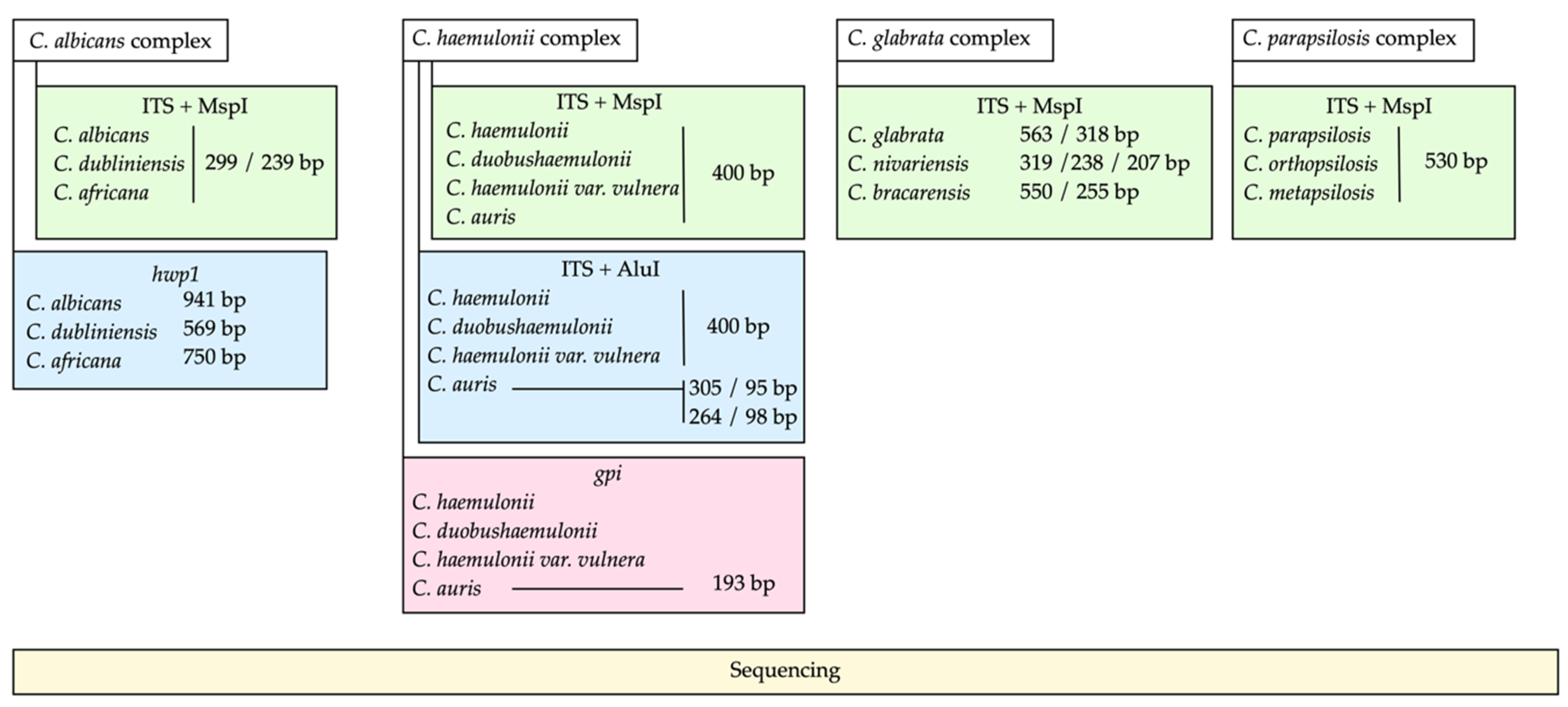
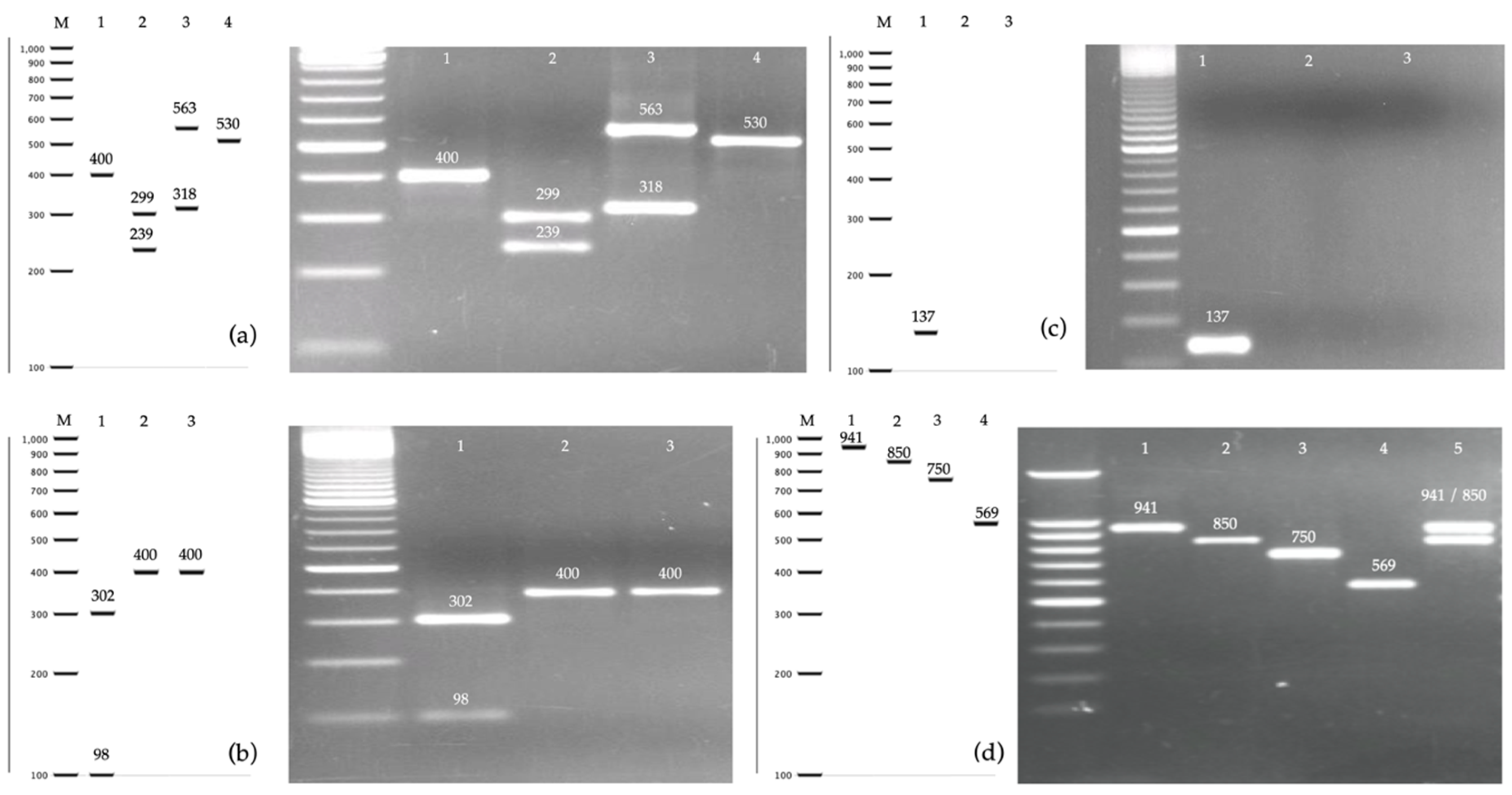
2.2. PCR-RFLP of the ITS Region and MspI

DNA from the axenic cultures was extracted following a previously described protocol using a BeadBeater® system (Bio Spec products Inc., Bartlesville, OK, USA) with glass beads, and phenol-chloroform [35]. The four Candida complexes were identified by PCR-RFLP, amplifying the internal transcribed sequence (ITS) of the ribosomal region and performing DNA digestion using the enzyme MspI [35,36]. According to this method, the species within the complexes C. albicans, C. parapsilosis, and C. haemulonii cannot be differentiated from each other, since they produce similar restriction patterns. All species of the C. albicans complex produced a restriction pattern of 299 and 239 bp; the four species of the C. haemulonii complex produced a pattern of 374–403 bp; and the three species of the C. parapsilosis complex produced a unique 530 bp band; whereas, the three species of the C. glabrata complex produce three characteristic restriction profiles (C. glabrata: 563/318 bp, C. nivariensis: 319/238/207 bp, C. bracarensis: 550/255 bp). Therefore, this is the only Candida out of the four complexes whose species can be identified by this method.
PCR conditions were carried out in a volume of 50 µL, with 25 µL of 2× PCR Master Mix (Promega Corp. Madison, WI, USA), 1 µL of each primer at 10 µM, 22 µL of water, and 1 µL of DNA (40 ng/µL). The primers used were: ITS1: 5′-TCCGTAGGTGAACCTGCGG-3′ and ITS4: 5′-TCCTCCGCTTATTGATATGC-3′ The amplification program was as follows: 95 °C for 5 min, 37 cycles of 95 °C for 30 s, 56 °C for 30 s, and 72 °C for 30 s, and an extension at 72 °C for 5 min. After confirming the amplification, 10 µL of the amplification product was digested with 0.5 µL of MspI (10 U/µL) at 37 °C for 2 h, 2 µL of buffer, and 0.2 µL of 10 µg/µL acetylated bovine serum albumin (BSA), for 2 h at 37 °C. The PCR products and digested PCR products were separated and visualized on a 1.5% agarose gel with ethidium bromide. A working algorithm of the molecular methods used in this study can be seen in Figure 1.

2.3. PCR of the hwp1 Gene for Identification of Species from C. albicans Complex

A method used to differentiate cryptic species within the C. albicans complex was based on the amplification of the hyphal wall protein 1 gene (hwp1) [37]. This method was originally proposed to discriminate C. africana from the other species of the C. albicans complex through length polymorphisms within these sequences. The three species generate different band sizes: C. africana (750 bp), C. albicans (941 and 850 bp), and C. dubliniensis (569 bp) [5]. DNA was amplified in a 50 µL volume, with 25 µL of 2X PCR Master Mix, 2 µL of each primer at 10 µM, 19 µL of water, and 2 µL of DNA (40 ng/µL). The sequences of the primers were: CR-f: 5′-GCTACCACTTCAGAATCATCA TC-3′ and CR-r: 5′-GCACCTTCAGTCGTAGAGACG-3′. PCR reaction conditions were as follows: denaturation at 95 °C for 5 min, 35 cycles of denaturation at 94 °C for 45 s, annealing at 58 °C for 40 s, and extension at 72 °C for 55 s, with a final extension at 72 °C for 10 min. All the PCR products and digested PCR products were separated and visualized on a 1.5% agarose gel with ethidium bromide.

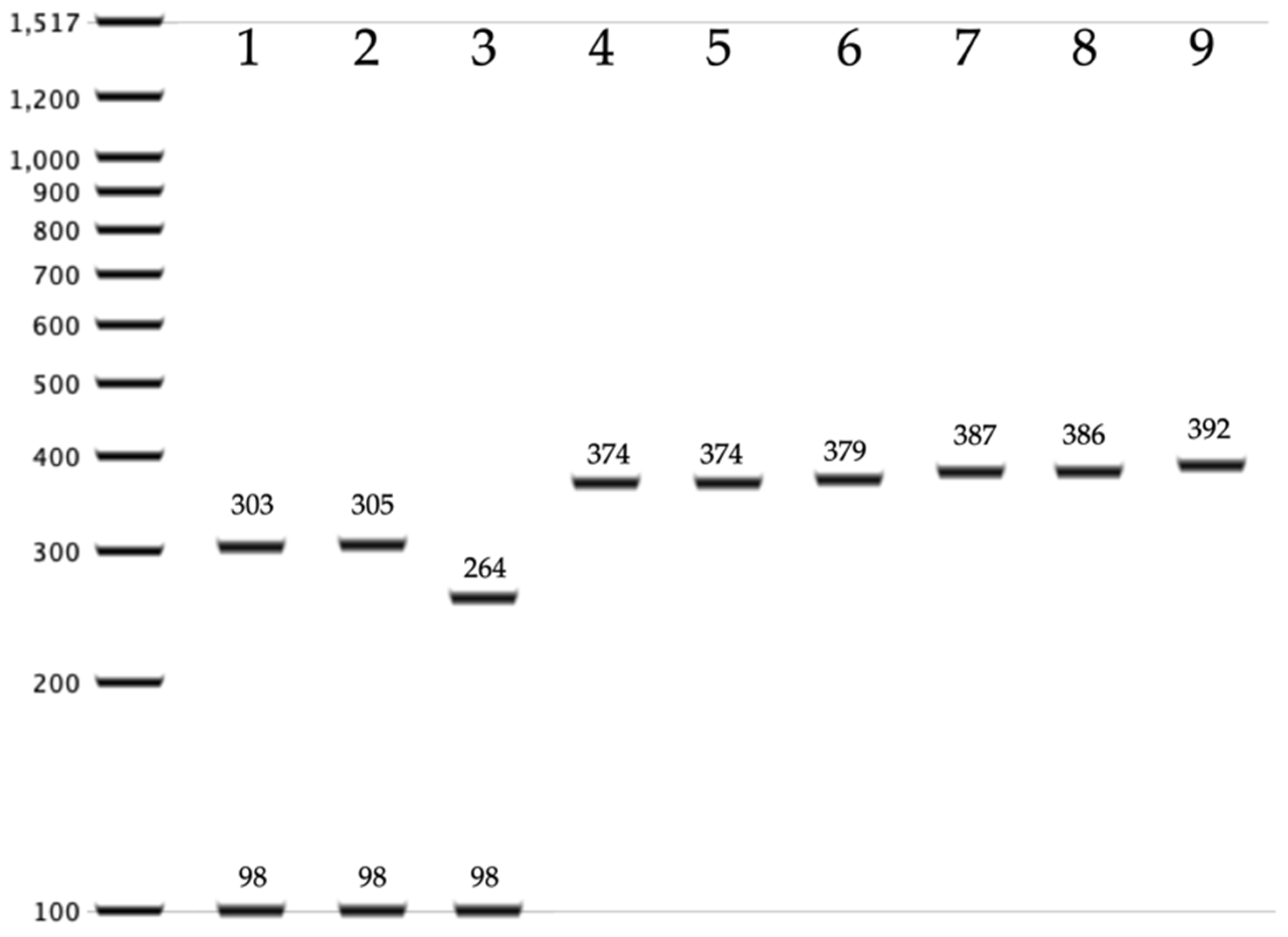
2.4. PCR-RFLP for the Identification of C. auris within the C. haemulonii Complex

Because the species of the C. haemulonii complex cannot be identified by amplification of the ITS region and digestion with MspI, a simple method was designed to differentiate C. auris from other species phylogenetically related to the C. haemulonii complex: C. haemulonii, C. haemulonii var. vulnera, C. duobushaemulonii, C. pseudohaemulonii, C. ruelliae, and C. heveicola [23]. The ITS region was amplified for subsequent digestion with the restriction enzyme AluI. To assess whether the restriction target for AluI was a conserved site, sequences available in NCBI of each of the seven species were analyzed. DNA sequences of C. haemulonii complex species isolated from countries on four continents were obtained (Americas, Europe, Asia, and Oceania). According to the in silico analysis (Figure 2), C. auris exhibited one or two restriction targets for the AluI enzyme and generates a two- or three-band restriction profile (305/95 bp; or 264/98/36 bp), while the rest of the species showed a single-band pattern between 374 and 392 bp. The amplification conditions were the same as described in the Section 2.2. Ten microliters of the amplification products of the ITS region were digested in a volume of 20 µL, with 0.5 µL of the AluI enzyme (10 U/µL), 2 µL of buffer, 17.3 µL of water, and 0.2 µL of acetylated bovine serum albumin (BSA) (10 µg/µL) for 2 h at 37 °C. A previously characterized strain of C. auris was used as a control in all assays.

2.5. PCR of the gpi Gene for Identification of C. auris

A PCR assay described for the rapid identification of C. auris was also performed on the isolates of the C. haemulonii complex. This technique amplified a unique glycosylphosphatidylinositol (GPI) protein-encoding gene originally described as QG37_03410 [38]. A standard PCR mix was prepared with 12.5 µL of 2× PCR Master Mix, 1 µL of each primer: 03410_F: 5′-GCCGCTAGATTGATCACCGT-3′ and 03410_R: 5′-TAGGTGTGGGTACCCTTGGT-3′, 9 µL of water, and 1.5 µL of DNA template. The PCR program consisted of 1 cycle at 94 °C for 3 min, 35 cycles of 30 s at 94 °C, 30 s at 60 °C, 30 s at 72 °C, and a final cycle at 72 °C for 3 min. A size band of 137 bp was expected for C. auris and no amplification for the rest of the species of the C. haemulonii complex. A reference strain was used as control.

2.6. Sequencing of PCR Products

To confirm the correct identification of cryptic species of the four complexes, five amplification products of the ITS region and four sequences of the hwp1 gene were sequenced on both strands using the same pair of primers used for amplification. Sequencing services were provided by Psomagen® (www.macrogenusa.com). The sequences were edited with the Geneious® 9.1.7 software and deposited into the NCBI GenBank.

3. Results

A total of 108 isolates were examined in this study. All isolates were previously deposited in the yeast collection of the National University of Honduras and were identified as cryptic species of one of the four complexes: C. albicans (n = 66), C. glabrata (n = 24), C. parapsilosis (n = 15), and C. haemulonii (n = 3) (Table 1). Phenotypic identification was based on the characteristics of the colony in chromogenic medium and the isolates were subsequently identified by PCR-RFLP by digesting the amplified fragment of the ITS ribosomal region with MspI (Figure 3a). The isolates were obtained from ten different types of clinical samples—mainly urine, sputum, and vaginal swabs.

3.1. Identification of Cryptic Species within the C. albicans Complex

Candida species that were identified as C. albicans complex by PCR-RFLP were also identified by a length polymorphism in the hwp1 gene. Of the 66 isolates, 52 (78.8%) were C. albicans sensu stricto with a 941 bp homozygous allele hwp1-1, 3 (4.5%) isolates were C. dubliniensis, 2 (3%) were C. africana, and one isolate showed an homozygous form of C. albicans with the rare allele hwp1-2 (1.5%), described in 2009 [39]. Eight isolates (12.1%) showed a two-band pattern compatible with a heterozygous strain of C. albicans (Figure 3d). In total, 61 (92.4%) of the isolates were identified as C. albicans sensu stricto regardless of the hwp1 gene allele, and 5 (7.6%) were identified as non-albicans cryptic species. To our knowledge, this is the first study that reports C. dubliniensis, C. africana, and a heterozygous hwp1 variant of C. albicans in Honduras. Candida dubliniensis isolates were obtained from sputum or pharyngeal exudates. Both isolates of C. africana came from vaginal swabs and urine, while the eight strains with a heterozygous profile were isolated from urine, sputum, oral swab, and vaginal swab.
Two specimens of each species were randomly selected and the fragments of the hwp1 gene were sequenced. Individuals with a two-band pattern (C. albicans, hwp1-1/hwp1-2) could not be sequenced. The identity of all isolates was confirmed through the NCBI BLAST tool. The sequences obtained were deposited in Genbank with the accession numbers: MN719370-3.

3.2. Identification of Cryptic Species of the C. haemulonii Complex

Three isolates showed an amplicon of approximately 400 bp when amplifying the ITS region and none of them showed restriction fragments with target sites for the MspI enzyme. This pattern was consistent with the C. haemulonii complex. To identify these species, three molecular methods were used. The first method was based on the digestion with AluI of the ITS region. According to an extensive in silico analysis, only C. auris has restriction target site for AluI, unlike the other six species (C. haemulonii, C. haemulonii var. vulnera, C. duobushaemulonii, C. pseudohaemulonii, C. ruelliae, and C. heveicola) that do not have a restriction target site for the enzyme to cleave and therefore maintain a band of around 400 bp. According to this analysis, none of the three isolates were identified as C. auris. A reference strain was used as a positive control in all assays (Figure 3b). The second method used to identify C. auris was a specific amplification of the gpi gene [38]. This method did not reveal the presence of C. auris among the three isolates (Figure 3c). The ITS regions of the three isolates were sequenced and all were identified as C. duobushaemulonii (accession number: MN699480). These species were isolated from two vaginal swabs and a cutaneous secretion. This is the first report of C. duobushaemulonii being identified in Honduras.

3.3. Identification of Cryptic Species of the C. glabrata and C. parapsilosis Complexes

All of the 24 isolates identified as C. glabrata complex showed a restriction pattern compatible with C. glabrata sensu stricto (Figure 3a). Sequencing of the ITS region confirmed this result (accession number MN699479). Therefore, it was not possible to identify C. nivariensis or C. bracarensis in this study. Most of the C. glabrata isolates were derived from urine samples (66.7%) (Table 1). With respect to the 15 isolates of the C. parapsilosis complex, the sequencing results of the ITS region showed that 11 were C. parapsilosis s.s., three were C. orthopsilosis and one isolate was identified as C. metapsilosis (accession numbers MN699481-3). This would also be the first report of the two less-frequent cryptic species of the C. parapsilosis complex in Honduras.

4. Discussions

Identification of yeasts at the species level is a challenge for clinical microbiologists who work for health centers in low-income countries due to the lack of appropriate technologies. Conventional methods based on phenotypic or biochemical characteristics are sometimes insufficient to provide an accurate identification of the etiological agent of an infection [40,41]. Reports of novel Candida species could be clinically relevant because they could differ both in virulence and in the spectrum of antifungal resistance [42]. Consequently, the lack of specific microbiological data could force physicians to empirically treat life-threatening mycoses with broad-spectrum antifungal medications, which would impact existing issues with antifungal resistance. Therefore, this study aimed to identify archived cryptic specimens belonging to four complexes of the genus Candida isolated from clinical samples from a hospital in Honduras in order to demonstrate the existence of novel species that the laboratory does not routinely identify and report. One hundred and eight isolates of the C. albicans, C. glabrata, C. parapsilosis, and C. haemulonii complexes were analyzed by molecular methods.
C. albicans complex spp. are the most frequently isolated from clinical samples [35,43]. However, cryptic species within this complex, such as C. dubliniensis and C. africana, are routinely misidentified or unidentified. Thus, this bias could slightly overestimate the number of C. albicans that is reported [5,44,45,46]. In this study, 66 isolates of the albicans complex were analyzed by amplification of the hwp1 gene and the result was confirmed by sequencing. More than 92% of the individuals were identified as C. albicans sensu stricto, and 7.6% were identified as C. dubliniensis or C. africana. This proportion is higher than reported in similar studies. A study from Argentina analyzed isolates obtained from patients with vulvovaginal candidiasis and showed that only two infections (3.8%) were caused by C. dubliniensis. Candida africana was not found among those isolates [5]. In Turkey, the reexamination of 376 yeasts obtained from vaginal infections showed 1.6% prevalence of C. dubliniensis and C. africana [44]. Another study carried out in China with more than one thousand isolates showed 1.5% prevalence of C. africana but did not report C. dubliniensis [47].
An interesting result of our study is the finding of 12.12% of heterozygous strains (hwp1-1/hwp1-2) and 1.5% of homozygous strains (hwp1-2/hwp1-2) of C. albicans. A shorter allele of 850 bp (hwp1-2) was initially described in 2009 [39] as a novel version of the wild-type allele of 941 bp (hwp1-1), and three deletions were described within the gene. Since then, there have been numerous reports of this genotype worldwide [5,47,48]. More recently, authors in Cameroon reported five genotypes composed of the combination of four alleles of the hwp1 gene (941, 1080, 850, and 1200 bp) [49]. Hence, our results support the high diversity of this gene in C. albicans, but despite these polymorphisms, the hwp1 gene has been shown to be a good marker for the identification of cryptic species of this complex. As far as we know, this would be the first report of C. dubliniensis and C. africana in Honduras. Both strains of C. africana were isolated from genital samples. This is concordant with that described by other authors [48,50,51]. Although some authors argue that there are no compelling reasons to separate C. dubliniensis or C. africana from C. albicans routinely [52,53], some studies show that the susceptibility of non-albicans species to antifungals could be lower than that of C. albicans [54]. In addition to the implications for clinical practice, the correct identification of cryptic species contributes to the knowledge of the epidemiology of candidiasis in Latin America [55].
One of the main reasons that motivated this study was the search for C. auris in the country, where it has not yet been reported. Candida auris is currently considered as one of the most important emerging microorganisms for public health, due to its virulence, its multiple resistance against different classes of antifungal drugs and high mortality rate [25]. There are reports of this yeast in neighboring countries such as Panama [56,57] and Colombia [58], and consequently there are high probabilities that this species will be dispersed to the rest of Central America in the near future. Phenotypic methods available in Honduras would not allow the identification of C. auris if it were to occur. Especially as its pattern of amplification of the ribosomal region is 400 bp, which resembles that of other species of the C. haemulonii complex [35]. In this study, we analyzed three yeasts with a restriction pattern congruent with C. haemulonii complex. Species identification was carried out by specific amplification of the gpi gene, and a simple PCR-RFLP technique was designed that allows C. auris to be discriminated from the rest of the species of the haemulonii complex. Both approaches were concordant that none of the three strains were C. auris. Sequencing of the ribosomal region revealed that the three isolates were C. duobushaemulonii. In the short term, it will be necessary to expand the search for C. auris with a greater number of strains.
It is rare to isolate species of the C. haemulonii complex from clinical samples. A study in Brazil identified 40 (0.3%) strains of the C. haemulonii complex from a collection of 14,632 isolates [59]. However, a considerable number of isolates suspected to be C. auris in Panama were further identified as C. duobushaemulonii and some of them were responsible for bloodstream infections and showed elevated minimal inhibitory concentrations for fluconazole, voriconazole, and amphotericin B [56]. Other Brazilian studies reported C. haemulonii complex isolates with low susceptibility to fluconazole or amphotericin B [60,61]. These data suggest that although these species are rare, they could be clinically relevant, and their correct identification is recommended.
The third complex evaluated was C. glabrata. Twenty-four isolates of C. glabrata sensu lato were analyzed by PCR-RFLP and sequencing of the ribosomal ITS and all the strains were identified as C. glabrata sensu stricto. Several studies have investigated the presence of rare cryptic species of the glabrata complex and have not been able to prove their presence [62,63,64]. However, other studies have reported C. nivariensis and C. bracarensis but with very low prevalences [28,65,66]. In relation to the 15 strains of the C. parapsilosis complex, the ITS sequence showed that the majority of the isolates were C. parapsilosis s.s., wherein three were identified as C. orthopsilosis and one of them was C. metapsilosis. It seems that the distribution of these cryptic species is also consistent with what was reported before in some countries [62,67,68,69]. This is the first description of cryptic species of the C. parapsilosis complex in Honduras, and since these species are frequently associated with invasive infections and exhibit differences in susceptibility to antifungals [70], this information will be useful to improve the species-specific identification of Candida.

5. Conclusions

This study has demonstrated the presence of cryptic species from four Candida complexes that are not routinely reported in Honduras’s hospitals. Additionally, the presence of C. dubliniensis, C. africana, C. duobushaemulonii, C. orthopsilosis, C. metapsilosis, and the hwp1-2 allele of C. albicans is described for the first time in Honduras. At present, due to the limitation of molecular technologies in Honduras, a simple method based on PCR-RFLP with AluI restriction enzyme is proposed for the identification of C. auris among other species of the C. haemulonii complex.

Author Contributions

S.B., B.O., K.M. and G.F. conceptualized the study; C.G. and S.B., obtained the yeast isolates; K.M. performed the experiments; K.M. and G.F. organized and cured the data; writing and original draft preparation, G.F.; all the authors contributed with writing, review, and editing the manuscript; supervision, project administration, and funding acquisition, G.F.

Funding

This research received no external funding. The experiments were conducted with the means provided by the Microbiology Research Institute of the National University of Honduras.

Acknowledgments

The authors acknowledge the support given by the staff of the IHSS hospital in Tegucigalpa. The authors thank Alireza Abdolrasouli of Charing Cross Hospital, London.

Conflicts of Interest

The authors declare no conflict of interest.

References

  1. Quindos, G.; Marcos-Arias, C.; San-Millan, R.; Mateo, E.; Eraso, E. The continuous changes in the aetiology and epidemiology of invasive candidiasis: From familiar Candida albicans to multiresistant Candida auris. Int. Microbiol. 2018, 21, 107–119. [Google Scholar] [CrossRef] [PubMed]
  2. Chapman, B.; Slavin, M.; Marriott, D.; Halliday, C.; Kidd, S.; Arthur, I.; Bak, N.; Heath, C.H.; Kennedy, K.; Morrissey, C.O.; et al. Changing epidemiology of candidaemia in Australia. J. Antimicrob. Chemother. 2017, 72, 1103–1108. [Google Scholar] [CrossRef] [PubMed] [Green Version]
  3. Quindos, G. Epidemiology of candidaemia and invasive candidiasis. A changing face. Rev. Iberoam. Micol. 2014, 31, 42–48. [Google Scholar] [CrossRef] [PubMed]
  4. Santolaya, M.E.; Thompson, L.; Benadof, D.; Tapia, C.; Legarraga, P.; Cortes, C.; Rabello, M.; Valenzuela, R.; Rojas, P.; Rabagliati, R.; et al. A prospective, multi-center study of Candida bloodstream infections in Chile. PLoS ONE 2019, 14, e0212924. [Google Scholar] [CrossRef] [Green Version]
  5. Mucci, M.J.; Cuestas, M.L.; Landanburu, M.F.; Mujica, M.T. Prevalence of Candida albicans, Candida dubliniensis and Candida africana in pregnant women suffering from vulvovaginal candidiasis in Argentina. Rev. Iberoam. Micol. 2017, 34, 72–76. [Google Scholar] [CrossRef]
  6. Liu, M.; Huang, S.; Guo, L.; Li, H.; Wang, F.; Zhang, Q.I.; Song, G. Clinical features and risk factors for blood stream infections of Candida in neonates. Exp. Ther. Med. 2015, 10, 1139–1144. [Google Scholar] [CrossRef] [Green Version]
  7. Sato, T.; Kishi, M.; Suda, M.; Sakata, K.; Shimoda, H.; Miura, H.; Ogawa, A.; Kobayashi, S. Prevalence of Candida albicans and non-albicans on the tongue dorsa of elderly people living in a post-disaster area: A cross-sectional survey. BMC Oral Health 2017, 17, 51. [Google Scholar] [CrossRef] [Green Version]
  8. Diba, K.; Makhdoomi, K.; Nasri, E.; Vaezi, A.; Javidnia, J.; Gharabagh, D.J.; Jazani, N.H.; Reza Chavshin, A.; Badiee, P.; Badali, H.; et al. Emerging Candida species isolated from renal transplant recipients: Species distribution and susceptibility profiles. Microb. Pathog. 2018, 125, 240–245. [Google Scholar] [CrossRef]
  9. Pendleton, K.M.; Dickson, R.P.; Newton, D.W.; Hoffman, T.C.; Yanik, G.A.; Huffnagle, G.B. Respiratory Tract Colonization by Candida species Portends Worse Outcomes in Immunocompromised Patients. Clin. Pulm. Med. 2018, 25, 197–201. [Google Scholar] [CrossRef]
  10. Gomez, J.; Garcia-Vazquez, E.; Espinosa, C.; Ruiz, J.; Canteras, M.; Hernandez-Torres, A.; Banos, V.; Herrero, J.A.; Valdes, M. Nosocomial candidemia at a general hospital: The change of epidemiological and clinical characteristics. A comparative study of 2 cohorts (1993–1998 versus 2002–2005). Rev. Iberoam. Micol. 2009, 26, 184–188. [Google Scholar] [CrossRef]
  11. Pfaller, M.A.; Andes, D.R.; Diekema, D.J.; Horn, D.L.; Reboli, A.C.; Rotstein, C.; Franks, B.; Azie, N.E. Epidemiology and outcomes of invasive candidiasis due to non-albicans species of Candida in 2496 patients: Data from the Prospective Antifungal Therapy (PATH) registry 2004–2008. PLoS ONE 2014, 9, e101510. [Google Scholar] [CrossRef] [PubMed] [Green Version]
  12. Pfaller, M.A.; Diekema, D.J.; Turnidge, J.D.; Castanheira, M.; Jones, R.N. Twenty Years of the SENTRY Antifungal Surveillance Program: Results for Candida Species From 1997–2016. Open Forum Infect. Dis. 2019, 6, S79–S94. [Google Scholar] [CrossRef] [PubMed] [Green Version]
  13. Guinea, J. Global trends in the distribution of Candida species causing candidemia. Clin. Microbiol. Infect. 2014, 20 (Suppl. 6), 5–10. [Google Scholar] [CrossRef] [PubMed] [Green Version]
  14. Fuller, J.; Dingle, T.C.; Bull, A.; Shokoples, S.; Laverdiere, M.; Baxter, M.R.; Adam, H.J.; Karlowsky, J.A.; Zhanel, G.G.; Canadian Antimicrobial Resistance Alliance (CARA); et al. Species distribution and antifungal susceptibility of invasive Candida isolates from Canadian hospitals: Results of the CANWARD 2011-16 study. J. Antimicrob. Chemother. 2019, 74, iv48–iv54. [Google Scholar] [CrossRef] [PubMed]
  15. Criseo, G.; Scordino, F.; Romeo, O. Current methods for identifying clinically important cryptic Candida species. J. Microbiol. Methods 2015, 111, 50–56. [Google Scholar] [CrossRef] [PubMed]
  16. Sullivan, D.J.; Westerneng, T.J.; Haynes, K.A.; Bennett, D.E.; Coleman, D.C. Candida dubliniensis sp. nov.: Phenotypic and molecular characterization of a novel species associated with oral candidosis in HIV-infected individuals. Microbiology 1995, 141 Pt 7, 1507–1521. [Google Scholar] [CrossRef] [Green Version]
  17. Tietz, H.J.; Hopp, M.; Schmalreck, A.; Sterry, W.; Czaika, V. Candida africana sp. nov., a new human pathogen or a variant of Candida albicans? Mycoses 2001, 44, 437–445. [Google Scholar] [CrossRef]
  18. Correia, A.; Sampaio, P.; James, S.; Pais, C. Candida bracarensis sp. nov., a novel anamorphic yeast species phenotypically similar to Candida glabrata. Int. J. Syst. Evol. Microbiol. 2006, 56, 313–317. [Google Scholar] [CrossRef] [Green Version]
  19. Alcoba-Florez, J.; Mendez-Alvarez, S.; Cano, J.; Guarro, J.; Perez-Roth, E.; del Pilar Arevalo, M. Phenotypic and molecular characterization of Candida nivariensis sp. nov., a possible new opportunistic fungus. J. Clin. Microbiol. 2005, 43, 4107–4111. [Google Scholar] [CrossRef] [Green Version]
  20. Tavanti, A.; Davidson, A.D.; Gow, N.A.; Maiden, M.C.; Odds, F.C. Candida orthopsilosis and Candida metapsilosis spp. nov. to replace Candida parapsilosis groups II and III. J. Clin. Microbiol. 2005, 43, 284–292. [Google Scholar] [CrossRef] [Green Version]
  21. Cendejas-Bueno, E.; Kolecka, A.; Alastruey-Izquierdo, A.; Theelen, B.; Groenewald, M.; Kostrzewa, M.; Cuenca-Estrella, M.; Gomez-Lopez, A.; Boekhout, T. Reclassification of the Candida haemulonii complex as Candida haemulonii (C. haemulonii group I), C. duobushaemulonii sp. nov. (C. haemulonii group II), and C. haemulonii var. vulnera var. nov.: Three multiresistant human pathogenic yeasts. J. Clin. Microbiol. 2012, 50, 3641–3651. [Google Scholar] [CrossRef] [Green Version]
  22. Sugita, T.; Takashima, M.; Poonwan, N.; Mekha, N. Candida pseudohaemulonii Sp. Nov., an amphotericin B-and azole-resistant yeast species, isolated from the blood of a patient from Thailand. Microbiol. Immunol. 2006, 50, 469–473. [Google Scholar] [CrossRef]
  23. Satoh, K.; Makimura, K.; Hasumi, Y.; Nishiyama, Y.; Uchida, K.; Yamaguchi, H. Candida auris sp. nov., a novel ascomycetous yeast isolated from the external ear canal of an inpatient in a Japanese hospital. Microbiol. Immunol. 2009, 53, 41–44. [Google Scholar] [CrossRef]
  24. Snyder, G.M.; Wright, S.B. The Epidemiology and Prevention of Candida auris. Curr. Infect. Dis. Rep. 2019, 21, 19. [Google Scholar] [CrossRef]
  25. Rhodes, J.; Fisher, M.C. Global epidemiology of emerging Candida auris. Curr. Opin. Microbiol. 2019, 52, 84–89. [Google Scholar] [CrossRef]
  26. Haas, M.; Grenouillet, F.; Loubersac, S.; Ariza, B.; Pepin-Puget, L.; Alvarez-Moreno, C.A.; Valderrama-Beltran, S.L.; Lavergne, R.A.; Le Pape, P.; Morio, F. Identification of cryptic Candida species by MALDI-TOF mass spectrometry, not all MALDI-TOF systems are the same: Focus on the C. parapsilosis species complex. Diagn. Microbiol. Infect. Dis. 2016, 86, 385–386. [Google Scholar] [CrossRef]
  27. Gamarra, S.; Dudiuk, C.; Mancilla, E.; Vera Garate, M.V.; Guerrero, S.; Garcia-Effron, G. Molecular tools for cryptic Candida species identification with applications in a clinical laboratory. Biochem. Mol. Biol. Educ. 2013, 41, 180–186. [Google Scholar] [CrossRef]
  28. Reyes-Montes, M.D.R.; Acosta-Altamirano, G.; Duarte-Escalante, E.; Salazar, E.G.; Martinez-Herrera, E.; Arenas, R.; Gonzalez, G.; Frias-De-Leon, M.G. Usefulness of a multiplex PCR for the rapid identification of Candida glabrata species complex in Mexican clinical isolates. Rev. Inst. Med. Trop. Sao Paulo 2019, 61, e37. [Google Scholar] [CrossRef] [Green Version]
  29. Neji, S.; Hadrich, I.; Ilahi, A.; Trabelsi, H.; Chelly, H.; Mahfoudh, N.; Cheikhrouhou, F.; Sellami, H.; Makni, F.; Ayadi, A. Molecular Genotyping of Candida parapsilosis Species Complex. Mycopathologia 2018, 183, 765–775. [Google Scholar] [CrossRef]
  30. Dudiuk, C.; Morales-Lopez, S.E.; Podesta, V.; Macedo, D.; Leonardelli, F.; Vitale, R.G.; Tosello, M.E.; Cabeza, M.S.; Biasoli, M.; Gamarra, S.; et al. Multiplex PCR designed to differentiate species within the Candida glabrata complex. Rev. Iberoam. Micol. 2017, 34, 43–45. [Google Scholar] [CrossRef]
  31. Fidler, G.; Leiter, E.; Kocsube, S.; Biro, S.; Paholcsek, M. Validation of a simplex PCR assay enabling reliable identification of clinically relevant Candida species. BMC Infect. Dis. 2018, 18, 393. [Google Scholar] [CrossRef]
  32. Arastehfar, A.; Fang, W.; Pan, W.; Liao, W.; Yan, L.; Boekhout, T. Identification of nine cryptic species of Candida albicans, C. glabrata, and C. parapsilosis complexes using one-step multiplex PCR. BMC Infect. Dis. 2018, 18, 480. [Google Scholar] [CrossRef]
  33. Montes, K.; Ortiz, B.; Galindo, C.; Figueroa, I.; Braham, S.; Fontecha, G. Identification of Candida Species from Clinical Samples in a Honduran Tertiary Hospital. Pathogens 2019, 8, 237. [Google Scholar] [CrossRef] [Green Version]
  34. Kumar, A.; Sachu, A.; Mohan, K.; Vinod, V.; Dinesh, K.; Karim, S. Simple low cost differentiation of Candida auris from Candida haemulonii complex using CHROMagar Candida medium supplemented with Pal’s medium. Rev. Iberoam. Micol. 2017, 34, 109–111. [Google Scholar] [CrossRef]
  35. Ortiz, B.; Perez-Aleman, E.; Galo, C.; Fontecha, G. Molecular identification of Candida species from urinary infections in Honduras. Rev. Iberoam. Micol. 2018, 35, 73–77. [Google Scholar] [CrossRef]
  36. Mirhendi, H.; Makimura, K.; Khoramizadeh, M.; Yamaguchi, H. A one-enzyme PCR-RFLP assay for identification of six medically important Candida species. Nippon Ishinkin Gakkai Zasshi 2006, 47, 225–229. [Google Scholar] [CrossRef] [Green Version]
  37. Romeo, O.; Criseo, G. First molecular method for discriminating between Candida africana, Candida albicans, and Candida dubliniensis by using hwp1 gene. Diagn. Microbiol. Infect. Dis. 2008, 62, 230–233. [Google Scholar] [CrossRef]
  38. Ruiz-Gaitan, A.C.; Fernandez-Pereira, J.; Valentin, E.; Tormo-Mas, M.A.; Eraso, E.; Peman, J.; de Groot, P.W.J. Molecular identification of Candida auris by PCR amplification of species-specific GPI protein-encoding genes. Int. J. Med. Microbiol. 2018, 308, 812–818. [Google Scholar] [CrossRef]
  39. Padovan, A.C.; Chaves, G.M.; Colombo, A.L.; Briones, M.R. A novel allele of HWP1, isolated from a clinical strain of Candida albicans with defective hyphal growth and biofilm formation, has deletions of Gln/Pro and Ser/Thr repeats involved in cellular adhesion. Med. Mycol. 2009, 47, 824–835. [Google Scholar] [CrossRef] [Green Version]
  40. Sadrossadati, S.Z.; Ghahri, M.; Imani Fooladi, A.A.; Sayyahfar, S.; Beyraghi, S.; Baseri, Z. Phenotypic and genotypic characterization of Candida species isolated from candideamia in Iran. Curr. Med. Mycol. 2018, 4, 14–20. [Google Scholar] [CrossRef]
  41. Ambaraghassi, G.; Dufresne, P.J.; Dufresne, S.F.; Vallieres, E.; Munoz, J.F.; Cuomo, C.A.; Berkow, E.L.; Lockhart, S.R.; Luong, M.L. Identification of Candida auris using the updated 8.01 VITEK(R)2 yeast identification system: A multi-laboratory evaluation study. J. Clin. Microbiol. 2019. [Google Scholar] [CrossRef]
  42. Brilhante, R.S.N.; Sales, J.A.; da Silva, M.L.Q.; de Oliveira, J.S.; Pereira, L.A.; Pereira-Neto, W.A.; Cordeiro, R.A.; Sidrim, J.J.C.; Castelo-Branco, D.; Rocha, M.F.G. Antifungal susceptibility and virulence of Candida parapsilosis species complex: An overview of their pathogenic potential. J. Med. Microbiol. 2018, 67, 903–914. [Google Scholar] [CrossRef]
  43. Bineshian, F.; Yadegari, M.H.; Sharifi, Z.; Akbari Eidgahi, M.; Nasr, R. Identification of Candida Species Using MP65 Gene and Evaluation of the Candida albicans MP65 Gene Expression in BALB/C Mice. Jundishapur J. Microbiol. 2015, 8, e18984. [Google Scholar] [CrossRef] [Green Version]
  44. Hazirolan, G.; Altun, H.U.; Gumral, R.; Gursoy, N.C.; Otlu, B.; Sancak, B. Prevalence of Candida africana and Candida dubliniensis, in vulvovaginal candidiasis: First Turkish Candida africana isolates from vulvovaginal candidiasis. J. Mycol. Med. 2017, 27, 376–381. [Google Scholar] [CrossRef]
  45. Hu, Y.; Yu, A.; Chen, X.; Wang, G.; Feng, X. Molecular Characterization of Candida africana in Genital Specimens in Shanghai, China. BioMed Res. Int. 2015, 2015, 185387. [Google Scholar] [CrossRef] [Green Version]
  46. Romeo, O.; Criseo, G. Molecular epidemiology of Candida albicans and its closely related yeasts Candida dubliniensis and Candida africana. J. Clin. Microbiol. 2009, 47, 212–214. [Google Scholar] [CrossRef] [Green Version]
  47. Shan, Y.; Fan, S.; Liu, X.; Li, J. Prevalence of Candida albicans-closely related yeasts, Candida africana and Candida dubliniensis, in vulvovaginal candidiasis. Med. Mycol. 2014, 52, 636–640. [Google Scholar] [CrossRef] [Green Version]
  48. Ngouana, T.K.; Krasteva, D.; Drakulovski, P.; Toghueo, R.K.; Kouanfack, C.; Ambe, A.; Reynes, J.; Delaporte, E.; Boyom, F.F.; Mallie, M.; et al. Investigation of minor species Candida africana, Candida stellatoidea and Candida dubliniensis in the Candida albicans complex among Yaounde (Cameroon) HIV-infected patients. Mycoses 2015, 58, 33–39. [Google Scholar] [CrossRef]
  49. Ngouana, T.K.; Drakulovski, P.; Krasteva, D.; Toghueo, R.K.; Kouanfack, C.; Reynes, J.; Delaporte, E.; Boyom, F.F.; Mallie, M.; Bertout, S. Genetic diversity of the Hwp1 gene and HIS3, EF3, CDC3 microsatellites and antifungal susceptibility profiles of Candida albicans isolates from Yaounde HIV-infected patients. Med. Mycol. 2017, 55, 546–554. [Google Scholar]
  50. Naeimi, B.; Mirhendi, H.; Khamisipour, G.; Sadeghzadeh, F.; Ahmadi, B. Candida africana in recurrent vulvovaginal candidiasis (RVVC) patients: Frequency and phenotypic and genotypic characteristics. J. Med. Microbiol. 2018, 67, 1601–1607. [Google Scholar] [CrossRef]
  51. Dieng, Y.; Sow, D.; Ndiaye, M.; Guichet, E.; Faye, B.; Tine, R.; Lo, A.; Sylla, K.; Ndiaye, M.; Abiola, A.; et al. Identification of three Candida africana strains in Senegal. J. Mycol. Med. 2012, 22, 335–340. [Google Scholar] [CrossRef] [PubMed]
  52. Odds, F.C. Molecular phylogenetics and epidemiology of Candida albicans. Future Microbiol. 2010, 5, 67–79. [Google Scholar] [CrossRef] [PubMed]
  53. Brandt, M.E.; Lockhart, S.R. Recent Taxonomic Developments with Candida and Other Opportunistic Yeasts. Curr. Fungal Infect. Rep. 2012, 6, 170–177. [Google Scholar] [CrossRef] [PubMed]
  54. Gil-Alonso, S.; Jauregizar, N.; Canton, E.; Eraso, E.; Quindos, G. Comparison of the in vitro activity of echinocandins against Candida albicans, Candida dubliniensis, and Candida africana by time-kill curves. Diagn. Microbiol. Infect. Dis. 2015, 82, 57–61. [Google Scholar] [CrossRef] [PubMed]
  55. Nucci, M.; Queiroz-Telles, F.; Alvarado-Matute, T.; Tiraboschi, I.N.; Cortes, J.; Zurita, J.; Guzman-Blanco, M.; Santolaya, M.E.; Thompson, L.; Sifuentes-Osornio, J.; et al. Epidemiology of candidemia in Latin America: A laboratory-based survey. PLoS ONE 2013, 8, e59373. [Google Scholar] [CrossRef] [PubMed] [Green Version]
  56. Ramos, R.; Caceres, D.H.; Perez, M.; Garcia, N.; Castillo, W.; Santiago, E.; Borace, J.; Lockhart, S.R.; Berkow, E.L.; Hayer, L.; et al. Emerging Multidrug-Resistant Candida duobushaemulonii Infections in Panama Hospitals: Importance of Laboratory Surveillance and Accurate Identification. J. Clin. Microbiol. 2018, 56, e00371-18. [Google Scholar] [CrossRef] [Green Version]
  57. Arauz, A.B.; Caceres, D.H.; Santiago, E.; Armstrong, P.; Arosemena, S.; Ramos, C.; Espinosa-Bode, A.; Borace, J.; Hayer, L.; Cedeno, I.; et al. Isolation of Candida auris from 9 patients in Central America: Importance of accurate diagnosis and susceptibility testing. Mycoses 2018, 61, 44–47. [Google Scholar] [CrossRef] [Green Version]
  58. Armstrong, P.A.; Rivera, S.M.; Escandon, P.; Caceres, D.H.; Chow, N.; Stuckey, M.J.; Diaz, J.; Gomez, A.; Velez, N.; Espinosa-Bode, A.; et al. Hospital-Associated Multicenter Outbreak of Emerging Fungus Candida auris, Colombia, 2016. Emerg. Infect. Dis. 2019, 25, 1339. [Google Scholar] [CrossRef] [Green Version]
  59. De Almeida, J.N., Jr.; Assy, J.G.; Levin, A.S.; Del Negro, G.M.; Giudice, M.C.; Tringoni, M.P.; Thomaz, D.Y.; Motta, A.L.; Abdala, E.; Pierroti, L.C.; et al. Candida haemulonii Complex Species, Brazil, January 2010-March 2015. Emerg. Infect. Dis. 2016, 22, 561–563. [Google Scholar] [CrossRef] [Green Version]
  60. Boatto, H.F.; Cavalcanti, S.D.; Del Negro, G.M.; Girao, M.J.; Francisco, E.C.; Ishida, K.; Gompertz, O.F. Candida duobushaemulonii: An emerging rare pathogenic yeast isolated from recurrent vulvovaginal candidiasis in Brazil. Mem. Inst. Oswaldo Cruz 2016, 111, 407–410. [Google Scholar] [CrossRef] [Green Version]
  61. Ramos, L.S.; Figueiredo-Carvalho, M.H.; Barbedo, L.S.; Ziccardi, M.; Chaves, A.L.; Zancope-Oliveira, R.M.; Pinto, M.R.; Sgarbi, D.B.; Dornelas-Ribeiro, M.; Branquinha, M.H.; et al. Candida haemulonii complex: Species identification and antifungal susceptibility profiles of clinical isolates from Brazil. J. Antimicrob. Chemother. 2015, 70, 111–115. [Google Scholar] [CrossRef] [PubMed] [Green Version]
  62. Miranda-Zapico, I.; Eraso, E.; Hernandez-Almaraz, J.L.; Lopez-Soria, L.M.; Carrillo-Munoz, A.J.; Hernandez-Molina, J.M.; Quindos, G. Prevalence and antifungal susceptibility patterns of new cryptic species inside the species complexes Candida parapsilosis and Candida glabrata among blood isolates from a Spanish tertiary hospital. J. Antimicrob. Chemother. 2011, 66, 2315–2322. [Google Scholar] [CrossRef] [PubMed] [Green Version]
  63. Esposto, M.C.; Prigitano, A.; Romeo, O.; Criseo, G.; Trovato, L.; Tullio, V.; Fadda, M.E.; Tortorano, A.M.; Group, F.W. Looking for Candida nivariensis and C. bracarensis among a large Italian collection of C. glabrata isolates: Results of the FIMUA working group. Mycoses 2013, 56, 394–396. [Google Scholar] [CrossRef] [PubMed]
  64. Asadzadeh, M.; Alanazi, A.F.; Ahmad, S.; Al-Sweih, N.; Khan, Z. Lack of detection of Candida nivariensis and Candida bracarensis among 440 clinical Candida glabrata sensu lato isolates in Kuwait. PLoS ONE 2019, 14, e0223920. [Google Scholar] [CrossRef] [PubMed]
  65. Lockhart, S.R.; Messer, S.A.; Gherna, M.; Bishop, J.A.; Merz, W.G.; Pfaller, M.A.; Diekema, D.J. Identification of Candida nivariensis and Candida bracarensis in a large global collection of Candida glabrata isolates: Comparison to the literature. J. Clin. Microbiol. 2009, 47, 1216–1217. [Google Scholar] [CrossRef] [Green Version]
  66. Morales-Lopez, S.E.; Taverna, C.G.; Bosco-Borgeat, M.E.; Maldonado, I.; Vivot, W.; Szusz, W.; Garcia-Effron, G.; Cordoba, S.B. Candida glabrata species complex prevalence and antifungal susceptibility testing in a culture collection: First description of Candida nivariensis in Argentina. Mycopathologia 2016, 181, 871–878. [Google Scholar] [CrossRef]
  67. Romeo, O.; Delfino, D.; Costanzo, B.; Cascio, A.; Criseo, G. Molecular characterization of Italian Candida parapsilosis isolates reveals the cryptic presence of the newly described species Candida orthopsilosis in blood cultures from newborns. Diagn. Microbiol. Infect. Dis. 2012, 72, 234–238. [Google Scholar] [CrossRef]
  68. Lovero, G.; Borghi, E.; Balbino, S.; Cirasola, D.; De Giglio, O.; Perdoni, F.; Caggiano, G.; Morace, G.; Montagna, M.T. Molecular Identification and Echinocandin Susceptibility of Candida parapsilosis Complex Bloodstream Isolates in Italy, 2007–2014. PLoS ONE 2016, 11, e0150218. [Google Scholar] [CrossRef]
  69. Asadzadeh, M.; Ahmad, S.; Hagen, F.; Meis, J.F.; Al-Sweih, N.; Khan, Z. Simple, Low-Cost Detection of Candida parapsilosis Complex Isolates and Molecular Fingerprinting of Candida orthopsilosis Strains in Kuwait by ITS Region Sequencing and Amplified Fragment Length Polymorphism Analysis. PLoS ONE 2015, 10, e0142880. [Google Scholar] [CrossRef] [Green Version]
  70. Lockhart, S.R.; Messer, S.A.; Pfaller, M.A.; Diekema, D.J. Geographic distribution and antifungal susceptibility of the newly described species Candida orthopsilosis and Candida metapsilosis in comparison to the closely related species Candida parapsilosis. J. Clin. Microbiol. 2008, 46, 2659–2664. [Google Scholar] [CrossRef] [Green Version]
Figure 1. Flow diagram for the identification of cryptic species within four complexes of the genus Candida by molecular methods.
Jof 05 00117 g001
Figure 2. In silico analysis of a PCR-RFLP method of the ITS region digested with AluI for identification of species from Candida haemulonii complex. Lanes 1–3: Candida auris; Lanes 4,5: C. haemulonii and C. haemulonii var. vulnera; Lane 6: Candida ruelliae; Lane 7: Candida duobushaemulonii; Lane 8: Candida pseudohaemulonii; Lane 9: Candida heveicola.
Jof 05 00117 g002
Figure 3. In silico analysis and amplification profiles of four molecular methods for the identification of Candida species. (a) PCR of the ITS region of rDNA digested with MspI. Lane 1: C. haemulonii complex, Lane 2: C. albicans complex, Lane 3: C. glabrata sensu stricto, Lane 4: C. parapsilosis complex; (b) PCR of the ITS region of rDNA digested with AluI for identification of C. auris within the C. haemulonii complex. Lane 1: C. auris, Lanes 2,3: C. haemulonii; (c) PCR of the gpi gene for identification of C. auris within the C. haemulonii complex. Lane 1: C. auris, lanes 2,3: C. haemulonii; (d) PCR of the hwp1 gene for identification of species within the C. albicans complex. Lane 1: C. albicans sensu stricto hwp1-1, Lane 2: C. albicans hwp1-2, Lane 3: C. africana, Lane 4: C. dubliniensis, Lane 5: heterozygous isolate showing a two-band profile hwp1-1/hwp1-2.
Jof 05 00117 g003
Table 1. Cryptic species of Candida according to clinical sample and identified by PCR-RFLP of the ITS region and MspI.
Clinical SampleNo. of Samples (%)C. albicans ComplexC. glabrata ComplexC. parapsilosis ComplexC. haemulonii Complex
Urine38 (35.19)21161-
Sputum28 (25.93)2512-
Vaginal swab16 (14.81)104-2
Blood7 (6.48)2-5-
Catheter7 (6.48)313-
Stool2 (1.85)--2-
Cutaneous secretion5 (4.63)1211
Otic secretion1 (0.93)--1-
Oral swab2 (1.85)2---
Cerebrospinal fluid (CSF)2 (1.85)2---
Total (%)108 (100%)66 (61.11%)24 (22.22%)15 (13.88%)3 (2.78%)

