Review

Interactions between Bacteria and Aspergillus fumigatus in Airways: From the Mycobiome to Molecular Interactions

by
Anne Debourgogne
1,
Lorra Monpierre
2,3,
Khadeeja Adam Sy
3,4,
Isabel Valsecchi
3,
Jean-Winoc Decousser
3,5 and
Françoise Botterel
2,3,*
1
UR 7300, Stress Immunité Pathogène, Université de Lorraine, 54000 Vandoeuvre les Nancy, France
2
Unité de Parasitologie-Mycologie, Département de Prévention, Diagnostic et Traitement des Infections, CHU Henri Mondor, Assistance Publique des Hôpitaux de Paris (APHP), 94000 Créteil, France
3
UR DYNAMYC 7380, Faculté de Santé, Univ Paris-Est Créteil (UPEC), Ecole Nationale Vétérinaire d’Alfort (ENVA), USC Anses, 94700 Créteil, France
4
Institut National de la Santé et de la Recherche Médicale (Inserm) U955, 94010 Créteil, France
5
Department of Infection Control, University Hospital Henri Mondor, Assistance Publique—Hôpitaux de Paris, 94000 Créteil, France
*
Author to whom correspondence should be addressed.
J. Fungi 2023, 9(9), 900; https://doi.org/10.3390/jof9090900
Submission received: 31 July 2023 / Revised: 26 August 2023 / Accepted: 30 August 2023 / Published: 1 September 2023
(This article belongs to the Special Issue Fungal Biofilms, 2nd Edition)

Abstract

:
Interactions between different kingdoms of microorganisms in humans are common but not well described. A recent analysis of the mycobiome has described the presence of different fungi and their positive and/or negative interactions with bacteria and other fungi. In chronic respiratory diseases, these different microorganisms form mixed biofilms to live inside. The interactions between Gram-negative bacteria and filamentous fungi in these biofilms have attracted more attention recently. In this review, we analyse the microbiota of the respiratory tract of healthy individuals and patients with chronic respiratory disease. Additionally, we describe the regulatory mechanisms that rule the mixed biofilms of Aspergillus fumigatus and Gram-negative bacteria and the effects of this biofilm on clinical presentations.

1. Introduction

A biofilm is a community of more or less complex microorganisms adhered to each other and to a given surface by a characteristically adhesive and protective extracellular matrix they secrete [1]. Depending on the microorganisms involved, biofilms are implicated in both the healthy microbiome and disease states. In this polymicrobial environment, microorganisms have developed complex mechanisms to promote their survival, in particular, to defend themselves against competing organisms [1]. Describing such microbial defensive mechanisms provides a better understanding of their pathogenesis and helps identify which lines of research to follow for the development of antibiofilm therapeutics.
Polymicrobial biofilms and their environment facilitate microbial interactions, cooperative metabolic functions, and cell–cell communications [2]. However, such cooperative or antagonistic microbial interactions can significantly affect microbial virulence, host interaction, and antimicrobial resistance [3,4]. Their growth mode confers phenotypic traits that favour microbial survival in hostile conditions facing antimicrobial drugs and host immunity. For instance, biofilm cells can tolerate antimicrobial and biocide concentrations of 100 to 1000 times higher than what planktonic cells can resist [5]. This tolerance is phenotypic and can be reversible or not, i.e., when biofilm cells disperse and resume a planktonic state (without biofilm) they lose such tolerance [3,6]. Biofilms are also remarkably resistant to host defences and environmental stress conditions, allowing them to persist in different environments [3].
Since the early 1980s, the role of biofilms in microbial pathogenesis has become increasingly evident and the current estimation indicates that up to 60–70% of nosocomial infections are associated with biofilms [3] often formed of bacteria species or even yeasts and bacteria [7,8]. In humans, biofilms build up mostly on epithelial, dental surfaces, or abiotic surfaces such as external devices. Sometimes the microorganisms form aggregates that colonise the lumen of the respiratory tract, for example in chronic respiratory infections such as asthma, chronic obstructive pulmonary disease (COPD), cystic fibrosis (CF), bronchiectasis …) [3].
In chronic respiratory diseases, Aspergillus fumigatus remains the most filamentous fungus detected and is almost always accompanied by bacteria, especially Gram-negative bacilli, such as Pseudomonas aeruginosa and others.
In this review, we will focus on mycobiota analysis in chronic respiratory diseases and the different mechanisms involved in bacteria–fungi interactions, particularly those between Gram-negative bacilli and A. fumigatus.

2. Pulmonary Mycobiota and Chronic Respiratory Infections

The lung microbiome, believed to be stable or at least transient in healthy individuals, is now considered a polymicrobial component contributing to dysbiosis and potentially to disease pathogenesis. Advances in culture-independent methods, especially next-generation sequencing, helped reveal the significant impact of lung mycobiome on the clinical presentation of chronic respiratory diseases, such as asthma, COPD, CF, and bronchiectasis [9]. By interacting with the bacteriome and/or virome, the respiratory mycobiome appears to be a cofactor of inflammation and host immune response and therefore, may contribute to lung function impairment and disease progression [9].
The high similarity of the fungal composition of the lower respiratory tract of healthy individuals with that of their dwellings is consistent with the hypothesis that the domestic environment is one of the most significant determinants of the human mycobiome composition [10,11]. The indoor environmental mycobiome is a mix of outdoor fungal components and human (and pet) mycobiome. The human–environment exchange of fungi depends on the specific factors the fungi encounter in each environment (temperature, nutrients, pH, immune response, etc.). Therefore, some fungi are more prevalent in the lungs than in the environment, like Rhodotorula mucilaginosa, Cryptococcus neoformans, or Aspergillus spp., whereas others are more abundant in the air or house dust, such as Malassezia restricta, and Capnodiales [9]. The health status of individuals, in particular their inflammatory status, may influence the diversity and richness of the mycobiota rather than its composition [10].
More and more studies are focusing on the composition of microbiota as a new element to consider in chronic respiratory infections and diseases. In asthma, there is strong evidence that the pulmonary and intestinal mycobiomes play a crucial role, highlighting the importance of multi-kingdom microbial interactions in the pathophysiology of asthma [12].
A recent study has shown that a highly variable fungal community dominated by A. fumigatus and Candida albicans, the two main components of the normal airway mycobiota, colonises the airways. However, notable shifts in the fungal balance within communities and between lung compartments were associated with asthma status (asthmatics, with and without fungal sensitisation, and healthy controls), longer asthma duration, and biomarkers of inflammation. The asthmatics’ airway mycobiota was dominated by a relatively small number of species, mainly A. fumigatus, C. albicans, and Mycosphaerella tassiana, and was distinct from the oropharyngeal mycobiota and air samples [13].
In CF, many bacteria and fungi are implicated in respiratory infections. Two recent studies from the prospective multicentre cohort of French CF patients (MucoFong) showed an interest in studying the mycobiome and bacterial–fungal interactions. The respiratory tract of CF patients is now considered a polymicrobial niche, and advances in next-generation sequencing have made it possible to analyse its microbiota and mycobiota. It has been known for many years that the decline in respiratory function is associated with a decrease in bacterial diversity [14]. More recently, a similar link has been demonstrated with fungi [15]. The inter-kingdom network showed three main clusters organised around Aspergillus, Candida, and Scedosporium genera. Aspergillus and Malassezia as relevantly associated with CF pulmonary exacerbation, and Scedosporium plus Pseudomonas with a decline in lung function [14]. Patients with fungal bronchitis in CF exacerbations had the highest fungal biomass dominated by communities of emerging filamentous pathogens, such as Exophiala dermatitidis and Scedosporium complex, compared with subjects without CF. The clinical subgroup of fungal bronchitis was often characterised by severe symptoms that were difficult to treat [16]. CF prognosis has dramatically improved over the past few decades, hence the urgent need to re-evaluate the prognostic factors for CF progression and to include a systematic assessment of fungal colonisation and/or bacteria-associated infections. For instance, P. aeruginosa, Achromobacter xylosoxidans, Stenotrophomonas maltophilia, and C. albicans have been associated with increased disease severity. Such results highlight (i) the potential pathogenicity of species often derived from the oral cavity of CF patients and usually considered clinically insignificant, and (ii) the polymicrobial signature of CF airway colonisation which may contribute as a collective entity to lung function decline [17].
In COPD, the mycobiome has been little explored so far. A distinct mycobiome profile is observed in patients compared with controls, as evidenced by increased α-diversity. Significant airway mycobiome differences, including greater interfungal interaction (by co-occurrence), were detected in patients with very frequent COPD exacerbations (three or more exacerbations per year). The airway mycobiome in COPD is characterised by specific fungal genera, Aspergillus, Curvularia and Penicillium, which were associated with exacerbations and increased mortality [18]. A longitudinal study demonstrated that the pulmonary mycobiome was less stable than the oral mycobiome, and neither COPD diagnosis nor intercurrent antibiotic use seemed to influence stability [19].
All these articles have explored the impact of these diseases on or in association with the lung mycobiome. The authors of these articles describe a reduction in bacterial and fungal diversity during the course of these chronic respiratory diseases, which is proportional to the patient’s respiratory function. For fungi, but this is also true for bacteria, reduced diversity may be caused by an overgrowth of a single fungal species, or by the loss of rare species that comes with a reduction in overall fungal abundance [15,20]. The airway mycobiome in COPD would be most useful in identifying patients most at risk of long-term adverse consequences, including risk of frequent exacerbations and higher mortality [18]. Available data could suggest that specific mycobiome profiles do exist in cases of chronic respiratory disease, but these data should be treated with extreme caution, due to the heterogeneity of the studies. Moreover, no significant changes were seen in airway mycobiome profiles, alpha diversity, or beta diversity, suggesting that treatment for an acute exacerbation did not influence the lung mycobiome [18].

3. Limitations of Studies and Laboratory Methodologies

Our review mainly described in vitro interactions between A. fumigatus and Gram-negative bacteria. However, the microbiome is a complex entity of different bacterial species, yeasts, and filamentous fungi (and even viruses) determining a new environment on the basis of their mutual interactions combined with many host factors (immune modulators, virulence …). This complexity introduces many biases into the current research.
Lack of Standardisation and Reproducibility: One of the primary concerns in microbiome studies is the lack of standardised methodologies, as described by our team in 2020 about DNA extraction [21]. Each study may use different sampling techniques, especially for sputa, sequencing platforms, and data analysis pipelines, leading to significant variations in results. Multiple gene loci have also been targeted for fungal gene profiling including 18S ribosomal rRNA (small subunit) and 28S rRNA region (large subunit), even if the two internal transcribed spacer units 1 and 2 (ITS1/ITS2) are used most commonly to detect fungi. Similarly, there are variations in micro and mycobiome analysis between individuals and protocols. To date, there is no established gold standard protocol to evaluate the inter-kingdom interactions between bacteria and fungi [22]. Moreover, replicating findings from one study to another has often proved challenging, raising questions about the reliability and reproducibility of microbiome research.
Correlation vs. Causation: Microbiome studies frequently highlight correlations between specific microbial compositions and various health conditions. However, it is crucial to recognise that correlation does not imply causation. Disentangling the cause-and-effect relationship between microbiota and health outcomes remains a formidable task. Many studies lack the rigorous experimental designs necessary to establish causality, leaving room for misinterpretation of the findings. Indeed, an important shortcoming of all studies is their retrospective or cross-sectional design, which is not appropriate to draw any conclusion regarding mycobiome as a causal factor. Despite difficulties in performing such studies, more prospective, multicentric, large, and longitudinal studies over a long time period are required to shed light on the role of the composition of fungal or other microbiota in different chronic respiratory diseases.
Confounding Factors: Human microbiomes are influenced by a myriad of variables, including age, diet, lifestyle, geography, genetics, and medication usage. Untangling the impact of each factor on microbial communities presents a significant challenge. The complexity of these interactions can often lead to spurious associations or confounding effects, obscuring the actual relationships between microbiota and health.
Sample Size and Selection Bias: Numerous microbiome studies suffer from small sample sizes, making it difficult to draw robust conclusions. In this case, the statistical power to detect any differences in the mycobiota between cases and controls is low. Additionally, the process of selecting study participants can introduce bias, which may not be adequately addressed in the analysis. These limitations can affect the generalisability of results and hinder the broader applicability of the findings to diverse populations.
Publication Bias: Like any other field of research, microbiome studies are susceptible to publication bias, where positive or significant results are more likely to be published than negative or non-significant ones. This bias can skew the perception of the overall evidence, leading to an overestimation of the impact of microbiota on human health.
Overemphasis on Specific Microbes: Some studies tend to overemphasise the role of individual microbial species or groups without considering the intricate interactions within the entire microbial community. This reductionist approach may oversimplify the complex dynamics of the microbiome, potentially overlooking the broader ecological significance of the entire ecosystem.
The strength of interactions between microorganisms is therefore difficult to analyse with current bioinformatics tools, especially between bacteria and fungi. Another way to analyse these interactions is to do so in vitro in pairs. The most studied interaction between A. fumigatus and bacteria, in the context of chronic respiratory infections, is that between A. fumigatus and P. aeruginosa in cellular models and/or in vivo models. In this case, interactions were often analysed as mixed biofilm or polymicrobial biofilms.

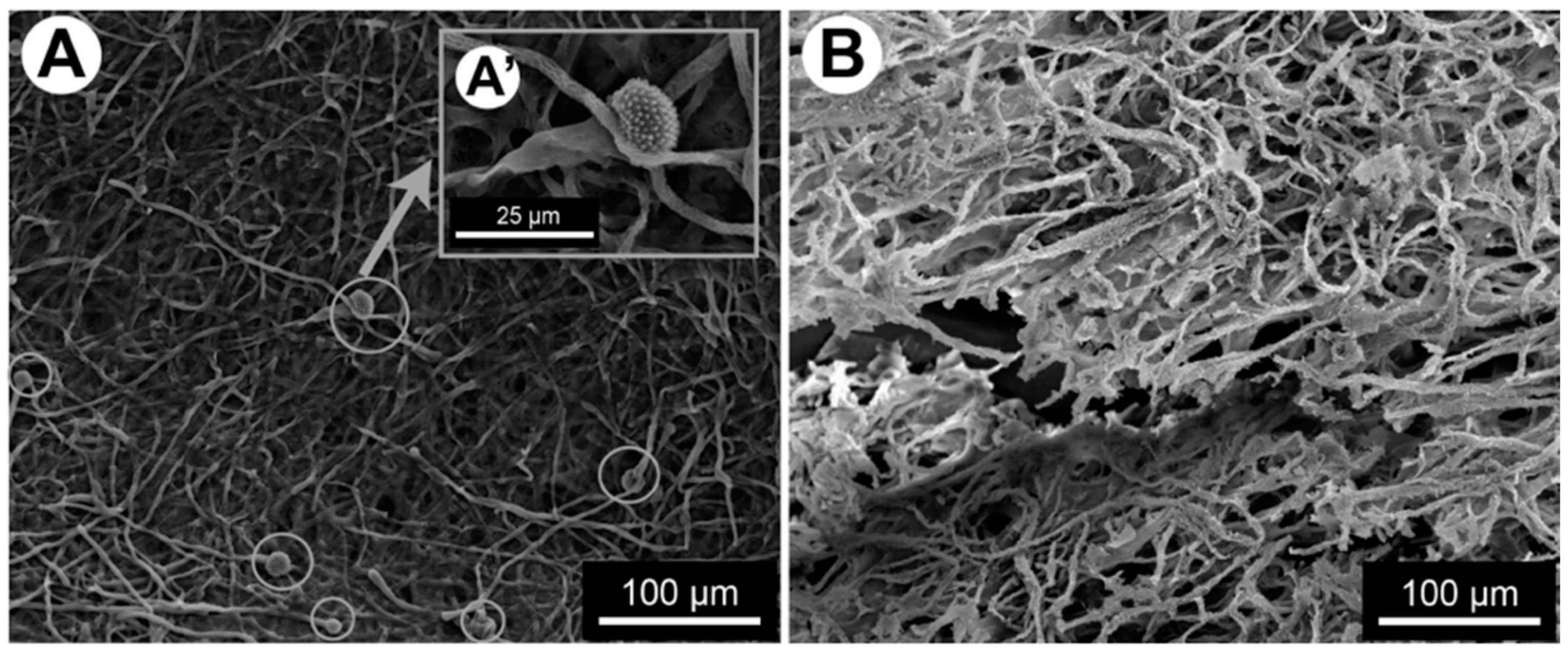
4. Formation of Mixed Filamentous Fungi–Bacteria Biofilm

A study compared a 24 h A. fumigatus biofilm alone or in the presence of P. aeruginosa and showed that A. fumigatus produced extensive and highly adherent mycelial growth with extensively branched hyphae forming a three-dimensional network of mycelium. In contrast, the monomicrobial biofilm culture of P. aeruginosa was weakly adherent. The mixed biofilm of A. fumigatus and P. aeruginosa showed a hazy background in which many P. aeruginosa bacteria were embedded in a network of filaments and mesh-like material [23]. In another study on the same pair of microorganisms, the fungal biomass was significantly reduced by 14.5% (p < 0.001) compared with the no-bacteria control [24]. Similarly, the interaction between S. maltophilia and A. fumigatus gave close biomass data and slowed down the growth of A. fumigatus in the mixed biofilm and that occurred during the growth phase of S. maltophilia causing a significant fungal biomass reduction of 54% between 12 and 16 h [25]. These differences were observed during the maturation phase of the biofilm after about 12 h and up to 24 h. When the fungus was in contact with the bacteria, the hyphal growth was delayed, and the thickness of the mixed biofilm was significantly less than that of the fungal biofilm alone. The hyphal network was less dense in the mixed biofilm and no A. fumigatus conidiation was observed at 24 h. Furthermore, the hyphae were atypical, showing atrophied and highly branched structures with shorter branching at the tips [25] (Figure 1).
Different microscopic approaches (SEM, TEM) showed that S. maltophilia cells were in contact with the A. fumigatus cell wall and embedded in the extracellular matrix, revealing a direct interaction between the bacterium and the fungus via the extracellular matrix [25] (Figure 2).
In the in vitro mixed biofilm between A. fumigatus and Streptococcus pneumoniae, the bacteria would disrupt the preformed biofilms of A. fumigatus and form a pneumococcal-dominated biofilm. S. pneumoniae could suppress both fungal proliferation and mycelium formation in the mixed biofilm without completely killing A. fumigatus. When a 48 h preformed fungal biofilm was co-cultured with pneumococci for 24 h, its hyphae were incorporated into the pneumococcal biofilm instead of making the fungal extracellular matrix, and many pneumococci adhered to the hyphae. And when a 24 h preformed fungal biofilm was co-cultured with pneumococci for 48 h, the hyphae were progressively disrupted into fine filaments [26].
Similarly, other filamentous fungi would suffer a negative impact, called antibiosis, if were grown in a mixed biofilm with bacteria. Also, inhibition of fungal growth was also observed in co-cultures of P. aeruginosa and Scedosporium aurantiacum. Kinetically, the bacteria adhered to the hyphae quite rapidly after co-culture, and within 8 h the bacteria aligned themselves along the length of the fungal hyphae. After 24 h, large clusters of P. aeruginosa cells were observed on the hyphal filaments of S. aurantiacum. The growth and thickness of hyphae were reduced compared with the control S. aurantiacum (i.e., without the bacteria) [27]. In the mixed Staphylococcus aureus–Fusarium falciform biofilm, the fungal hyphae and conidia were short and thin, and the number of fungal cells was clearly reduced. As for S. aureus, they aggregated in clusters around the fungal structures and some of them were observed on the hyphae, suggesting contact–contact interactions in the mixed biofilm. The biomass of the mixed biofilm was higher than the biomass of the S. aureus mono-biofilm (p < 0.001), and lower than the biomass of the fungal mono-biofilm (p < 0.001) [28]. In another mixed biofilm formed by A. fumigatus and S. aureus, the electron microscope revealed differences in the texture and distribution of the extracellular matrix (ECM) when compared with single biofilms. In some fragments, the appearance of ECM was porous or condensed or like a film covering the surface of large clusters of cocci [29].

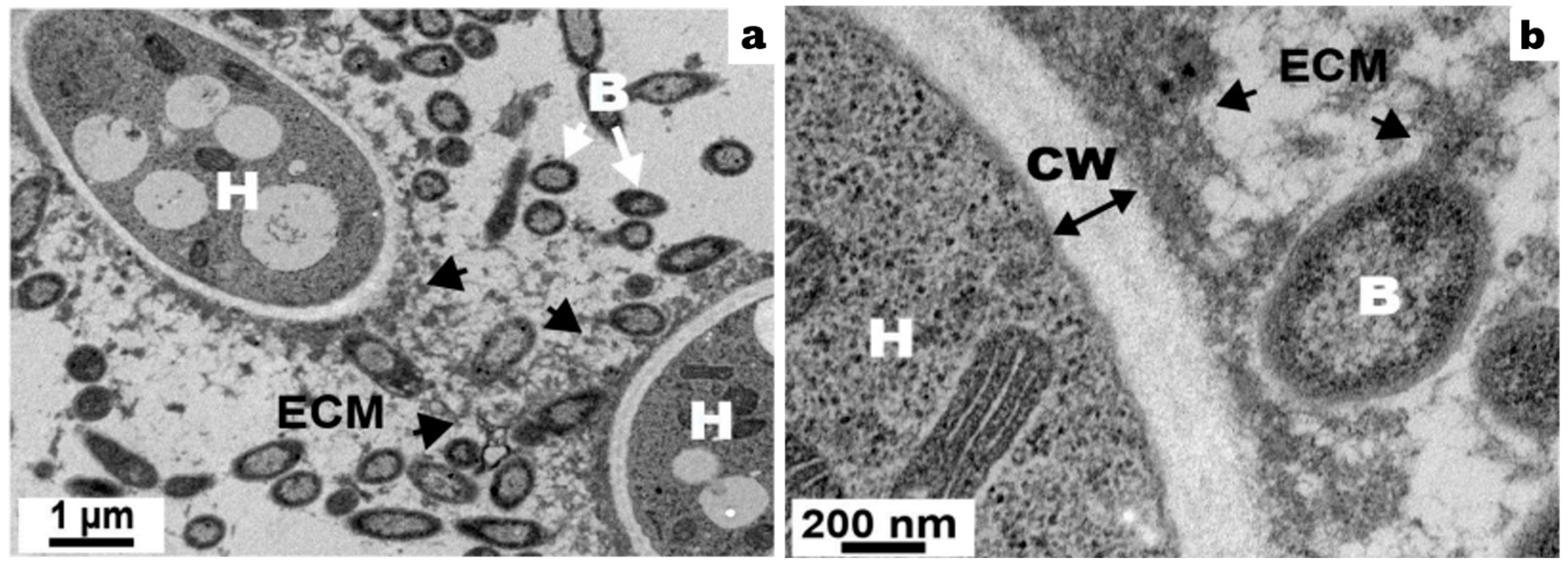
5. Extracellular Matrix Production

Biofilms produce their own characteristic extracellular matrix (ECM) that surrounds the biofilm cells and ensures the structural integrity of the microbial community [3]. ECM is usually composed of exopolysaccharides, extracellular DNA, and proteins, although its composition varies considerably depending on growth conditions and microbial species. Exopolysaccharides are the main components of many microbial species that can bind or repel antimicrobial agents through charge and hydrophobicity interactions, thus limiting their intracellular penetration [3].
The exopolysaccharides found in A. fumigatus biofilms include galactomannan, galactosaminogalactan (GAG), and α-1,3-glucan [30,31]. GAG is produced by A. fumigatus during its vegetative growth to mediate hyphal adhesion to biotic and abiotic surfaces, maintain matrix integrity, and ensure biofilm formation [32,33,34]. Additionally, GAG is a bioactive polymer involved in interactions with host cells. GAG interacts with several immune cells causing masking of pathogen-associated molecular patterns (PAMPs), neutrophil apoptosis, activation of platelet, NK cells, and macrophages [33], and induction of NLRP3-dependent inflammasome in macrophages [34]. In some contexts, GAG is also an anti-inflammatory molecule responsible for IL-1Ra secretion [35,36]. GAG participates in the formation of the extracellular matrix of biofilms, as described by Loussert et al. in a single A. fumigatus biofilm of aspergilloma, and in vivo in a mouse model of invasive aspergillosis [30]. Briard et al. documented that GAG was crucial for the interaction between A. fumigatus and P. aeruginosa since it was responsible for the binding of P. aeruginosa to the hyphae and in the biofilm [32]. Overall, GAG is a linear polysaccharide composed of galactose, N-acetylgalactosamine (GalNAc), and its de-N-acetylated form, the galactosamine (GalN). The percentage of each of these components in its sequence is highly variable. Its biosynthesis is carried out by a cluster of five genes (UGE3, SPH3, EGD3, AGD3, and GBT3/GT4C) [33,34]. The biological properties of GAG, involved in bacteria–fungi interactions or immune cell stimulation, are dependent on its de-N-acetylation. In other words, the percentage of galactosamine (GalN) in GAG would be a key factor in determining A. fumigatus virulence [35].
On the other hand, in bacteria, exopolysaccharides are high molecular weight carbohydrate biopolymers, often secreted by cells into the extracellular environment to ensure a variety of functions useful for the bacteria. For instance, P. aeruginosa produces three exopolysaccharides: Pel, a cationic polymer of partially deacetylated N-acetylgalactosamine and N-acetylglucosamine (PNAG); Psl, a pentamer of mannose, glucose, and rhamnose; and alginate, an anionic polymer of uronic acids. Pel and Psl play a key role in P. aeruginosa biofilm formation by promoting adhesion to surfaces and other cells and by interacting with extracellular DNA and host molecules [29,37,38]. Many Staphylococcus species produce deacetylated poly-N-PNAG polysaccharides, which mediate cell-to-cell adhesion and participate in biofilm formation in vitro and in vivo [39]. Deacetylation of hexosamine sugars is required to ensure the adhesive function of GAGs, PNAG, and Pel [40].

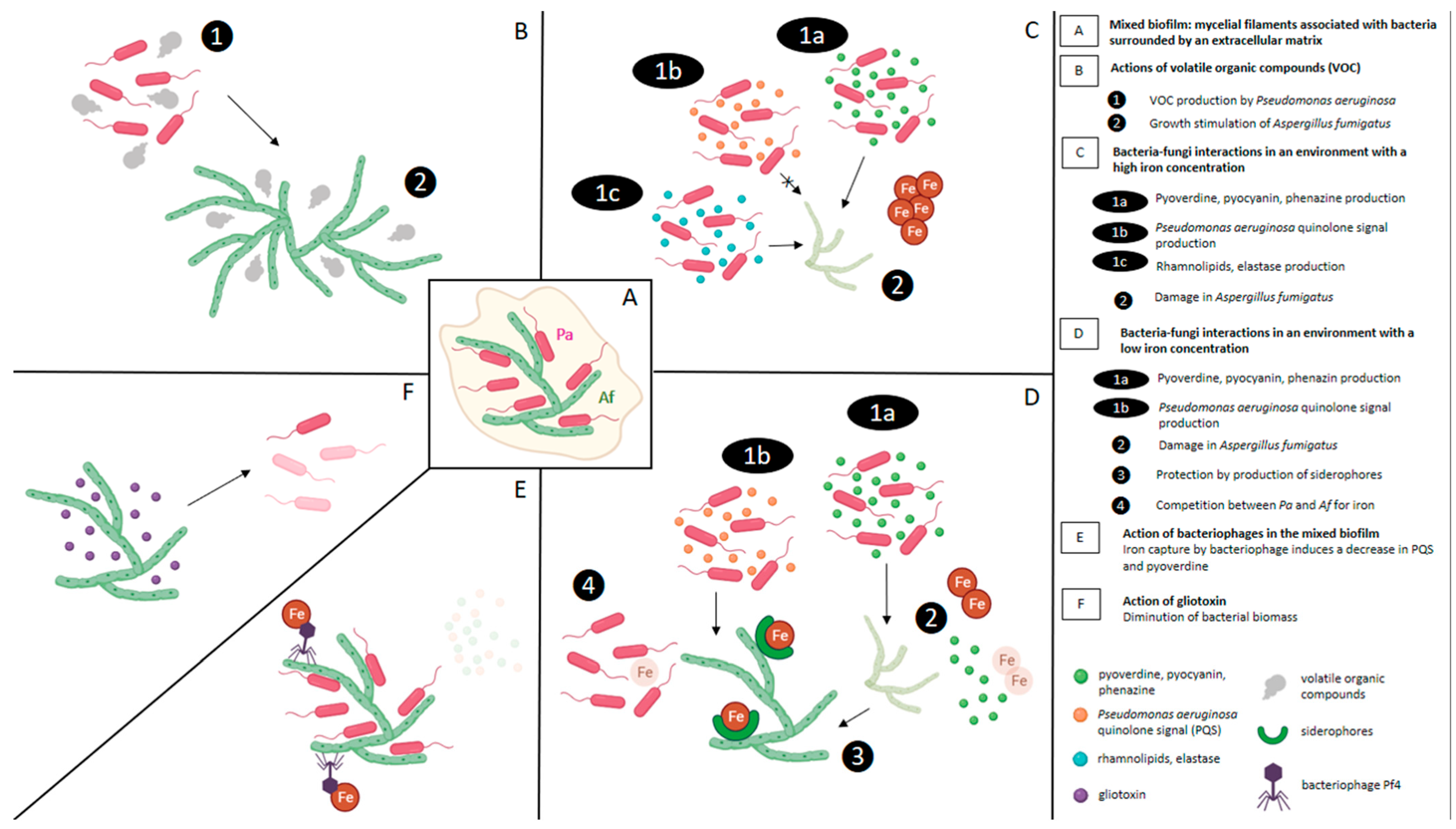
6. Social Interactions between Gram-Negative Bacteria and A. fumigatus in Mixed Biofilm

6.1. Antibiosis: The Most Common Social Interaction

There are many social interactions described for bacteria and fungi, such as: (i) physical association that can be planktonic, mixed biofilm, or intrahyphal colonisation and (ii) molecular communications such as antibiosis, signalling and chemotaxis, physiochemical changes, protein secretion, metabolite exchange, metabolite conversion, adhesion, or genetic exchange [1].
Among these social interactions, some are more common, e.g., antibiosis, in mixed biofilms of Gram-negative bacteria and A. fumigatus. Antibiosis, as described above, is a biological interaction between two or more organisms brought by the production of a metabolic substance by one of the partners that is prejudicial to the other partner, in terms of growth, morphology or structure.
Antibiosis between different pairs of bacteria and fungi has already been demonstrated and is always detrimental to the bacteria, e.g., P. aeruginosa and Scedosporium/Lomentospora [36,41], P. aeruginosa and A. fumigatus [23,40], S. maltophilia and A. fumigatus [25,42], or Klebsiella pneumonia and Aspergillus spp. [37]. This inhibitory effect is directly proportional to the number of bacteria [23] or to the distance between the two colonies of microorganisms in the culture [40].

6.2. Effect of Antibiosis Induced by the Bacteria on the Development of the Fungi

In the interactions between P. aeruginosa and A. fumigatus, Manavathu et al. showed that while hyphae were refractory to the fungicidal effect of the bacteria, conidia and spores were sensitive to it and were completely killed.
In contrast, Nogueira et al. showed that K. pneumoniae inhibited spore germination and hyphal development of Aspergillus species, suggesting that the inhibitory effect of K. pneumoniae on Aspergillus species is independent of the fungal growth stage. The inhibitory effect was reversible since the fungus resumed its growth upon eliminating the bacteria by antibiotics [37]. A mechanistic hypothesis proposes that in order to withstand the interaction with K. pneumoniae, Aspergillus adopted a dormant or standby (sleep) mode, during which the genes regulating hyphal development were downregulated, conferring a low energy supply requirement for survival [37].

6.3. Strain-Dependent Interaction Effect

Bacteria–fungus interactions can be modulated by factors intrinsic to the isolates present in each association. The antibiosis effect of P. aeruginosa or S. maltophilia on A. fumigatus did not depend on the strain of the fungus [38,42]. However, the inhibition of A. fumigatus was related to the source and phenotype of the bacteria isolate. The inhibitory effect of mucoid or non-mucoid CF P. aeruginosa isolates was higher than the effect of the non-CF isolates [39,43]. In contrast, small colony variants of P. aeruginosa appeared so variable in their ability to inhibit A. fumigatus [44]. The antibiosis effect of S. maltophilia strains on A. fumigatus depended on the bacterial strain and its genotype. S. maltophilia strains of genotype 6 isolated from patients, in particular from cystic fibrosis patients, were more virulent on A. fumigatus than animal or environmental strains, even at high bacterial concentrations [42].
Many models of bacteria–filamentous fungi interactions have shown that inhibition can also be observed with culture filtrates or supernatants [26,27,28,39,41,43,44,45]. The results suggested that cell–cell contact is not necessary for bacteria to inhibit fungal growth, and those bacterial metabolites and/or extracellular signalling molecules might be involved in this inhibition.
The different mechanisms described in these bacteria—A. fumigatus interactions are developed in the following paragraphs and summarised in Figure 3.

7. Effect of Extracellular Soluble Molecules of Bacteria on A. fumigatus Development

7.1. Role of P. aeruginosa Pyoverdine and Phenazines

Interactions between A. fumigatus and P. aeruginosa have revealed the antifungal role of the bacteria and the interference with fungal metabolism or growth via molecules the bacteria secrete like the major siderophores, pyoverdine, and phenazines, such as pyocyanin (5-N-methyl-1-hydroxyphenazine), 1-hydroxyphenazine, phenazine-1-carboxamide, phenazine-1-carboxylic acid, and di-rhamnolipids [46]. Upon using pyoverdine-defective mutants, it was possible to prove the key inhibitory role of this siderophore. Studies exploring pure pyoverdine effects corroborated these findings, including the restoration of inhibition by the pyoverdine deletion mutants. A correlation was also observed between the concentration of the produced pyoverdine and the resulting antifungal activity in clinical isolates of P. aeruginosa taken from the lungs of CF patients [44,45,47,48]. Pyocyanin exerted a similar inhibitory effect on A. fumigatus but not on S. aurantiacum [27,45]. A synergistic antifungal activity was described by adding voriconazole combined with P. aeruginosa supernatants. The inhibitory effect was mediated by pyoverdine and pyochelin under low iron conditions and pyocyanin under high iron conditions [46]. The key inhibitory mechanism of pyoverdine relied on iron chelation and iron denial to A. fumigatus [48].

7.2. Role of P. aeruginosa Quinolone Signal (PQS)

PQS 3,4-dihydroxy-alkyl-quinolone is a quorum-sensing molecule of P. aeruginosa that plays a coordinating role in many bacterial functions. It is involved in inter-microbial interactions, iron chelation (specifically Fe3+), and Fe delivery to the cell membrane in conjunction with siderophores. Under low Fe conditions, PQS inhibited the formation of A. fumigatus biofilms. Inversely, in an iron-rich environment, both biofilm formation and A. fumigatus growth significantly increased as compared with an iron-poor environment. This phenotype was not observed with an Aspergillus mutant not expressing siderophores. PQS production by P. aeruginosa could have favourable consequences for A. fumigatus in CF patients’ airways where Fe homeostasis is impaired, thus aggravating the disease [49].

7.3. Role of Rhamnolipids and Elastase

Elastase and rhamnolipids are major antifungal molecules of P. aeruginosa. Both act by increasing cell wall thickness and inhibiting fungal growth. They are controlled by the quorum sensing system and could help P. aeruginosa in iron acquisition [50]. Elastase is a metalloprotease that hydrolyses extracellular matrix proteins and degrades components of the host immune response, including lysozyme and proteins involved in bacterial opsonisation. The presence of a fungus stimulates the production of this important virulence factor by P. aeruginosa [40].

7.4. Role of Volatile Organic Compounds

Communication between microbial species involves not only the release of water-soluble compounds but also the release and detection of volatile organic compounds. Briard et al. were able to demonstrate an unexpected stimulatory effect of P. aeruginosa on the growth of A. fumigatus. The effect was mediated by dimethyl sulphide, which is a volatile gaseous compound [51,52]. The same observation was documented in the Scedosporium species [41].

7.5. Role of Bacteriophages

The filamentous bacteriophage Pf, of genus Inovirus, is abundantly produced by P. aeruginosa biofilm in clinical CF isolates. Bacteriophage Pf4 inhibits the metabolic activity of A. fumigatus biofilms. This phage-mediated inhibition would be dose-dependent, reversed by phage denaturation, and would be more pronounced against pre-formed A. fumigatus biofilms than against forming biofilms. However, planktonic conidial growth is unaffected. Pf4 can attach itself to A. fumigatus hyphae avidly and for a prolonged period of time. Given that Pf4 usually binds to iron, it denies A. fumigatus a crucial resource. This inhibition is reversible and could be overcome with supplemental ferric iron [53]. Infection of P. aeruginosa by Pf bacteriophage significantly reduced the production of quorum-regulated or unregulated antimicrobial molecules and pyoverdine [54].

8. Effect of Extracellular Soluble Molecules of Fungi on Bacterial Development (Figure 3)

8.1. Role of Gliotoxin

A. fumigatus and P. aeruginosa have mutual antagonistic effects. A. fumigatus supernatants showed strong anti-Pseudomonas activity in which gliotoxin is the major active agent, a fact supported by the inability of a gliotoxin-mutant strain of A. fumigatus to inhibit P. aeruginosa biofilm formation. Moreover, gliotoxin reduces bacterial biomass. Based on these facts, a broad-spectrum antibiofilm activity was described against other bacteria commonly found in CF airways [55].

8.2. Role of A. fumigatus Siderophores

Aspergillus retaliates by releasing hydroxamate siderophores that play a central role in the competition for iron between A. fumigatus and P. aeruginosa [48,56]. Sass et al. studied a supernatant of A. fumigatus mutant lacking sidA and saw that fungal siderophores were able to protect A. fumigatus biofilms from P. aeruginosa’s antifungal activity [56]. A lack of iron was the signal to increase the production of fungal siderophores, molecules that specifically chelate ferric iron with high affinity.

9. Role of Iron in Biofilm Interactions

Iron is an essential nutrient for both pathogens; hence, the competition between them to acquire it. In pathological situations such as CF, lung mucus acts as an iron chelator, creating an iron-limiting environment for pathogens living in the lungs. However, there are situations where iron availability is not limited, such as in microhaemorrhages and haemoptysis. Under iron-limiting conditions, P. aeruginosa released pyoverdine to deny iron to the fungus, thus interfering with fungal biofilm metabolism. Under non-iron limiting conditions, where pyoverdine was no longer produced, P. aeruginosa were still able to affect A. fumigatus biofilms by releasing pyocyanin toxin. Thus, the bacteria can use different molecules depending on the encountered inter-microbial competition [50].
There is a paradoxical microbial interaction between P. aeruginosa and A. fumigatus ruled by the amount of iron present in the medium. In an iron-poor environment, A. fumigatus biofilm formation was inhibited, whereas, in an iron-rich environment, both fungal biofilm formation and A. fumigatus growth were boosted [50].

10. Other Mechanisms

10.1. Apoptosis and ROS Production

After 24 h of P. aeruginosaA. fumigatus interaction, apoptotic effects characterised by elevated intracellular ROS levels, increased depolarisation of mitochondrial potential, and DNA fragmentation mediated by metacaspase activation were observed [43]. The release of phenazines and gliotoxin was associated with ROS production. By inducing differential levels of oxidative stress controlled by metabolites’ redox properties and their environment-dependent activities, phenazines and gliotoxin play a dual role; as toxins with antifungal activities (high levels) and as a conidiation signal in Aspergillus development (moderate levels), which was fine-tuned by NapA oxidative stress response pathway [57].

10.2. Role of Oxygen and pH Gradients

Biofilm formation establishes an environment with stable gradients of oxygen, nutrients, and pH that provide different localised habitats on a small scale [58]. Such gradients are probably important in chronic respiratory diseases, e.g., CF, where the apical environment of epithelial cells is hypoxic and rather anaerobic. In this setting, P. aeruginosa growth is reduced by the anaerobic conditions, and bacterial filtrates are less effective against hypoxic A. fumigatus. Without the mixed biofilm, P. aeruginosa would therefore be less active in such environments, as would be the case in CF [59].

11. Susceptibility of Polymicrobial Biofilms

The polymicrobial biofilm of P. aeruginosa and A. fumigatus showed differential susceptibility to antimicrobial drugs, whereas this susceptibility did not change in the A. fumigatus monomicrobial biofilm. For instance, tobramycin alone and in combination with posaconazole were highly effective against P. aeruginosa monomicrobial and polymicrobial biofilms, respectively, whereas cefepime alone and in combination with posaconazole showed excellent activity against P. aeruginosa monomicrobial biofilms, and less activity against polymicrobial biofilms, respectively. A. fumigatus polymicrobial and monomicrobial biofilms showed similar susceptibility to posaconazole with and without the antibacterial drug [23]. In another study on microbial interactions of A. fumigatus and S. maltophilia, Roisin et al. demonstrated that polymicrobial biofilms could modulate the antimicrobial response of pathogens. S. maltophilia could increase the antifungal susceptibility of A. fumigatus to amphotericin B, whereas A. fumigatus could protect S. maltophilia from levofloxacin [60].
Biofilm growth generates microenvironments that host physiologically heterogeneous cell populations of different growth and metabolic states, or cells subjected to different nutritional or oxidative stresses [61,62]. Metabolically quiescent cells or those expressing adaptive stress responses are less susceptible to antimicrobial killing. Finally, recent studies have reported that Gram-negative bacteria and fungi overexpress antimicrobial efflux pumps. The relative contribution of these mechanisms to biofilm antimicrobial tolerance varies between individual organisms and antimicrobials [3]. For example, the inhibitory effect of S. maltophilia on A. fumigatus growth was more pronounced in CF strains than in reference strains [60].

12. Polymicrobial Biofilms Evasion of Immune System

Microorganisms growing in biofilms readily evade the host immune system through different mechanisms. In vitro studies suggested that microorganisms in biofilms are less recognised by the immune system [63,64,65] and thus resistant to phagocytosis and killing by neutrophils [66,67]. For example, the P. aeruginosa exopolysaccharide Psl reduces opsonophagocytosis by inhibiting complement molecules deposition at the surface of the bacteria and promotes intracellular bacterial survival, whereas alginate protects against phagocyte activation, uptake, and killing [68,69]. Similarly, exopolysaccharide GAGs mask b-glucans from recognition by Dectin-1, a receptor of innate immunity, and protect hyphae from neutrophil extracellular traps [35,70].

13. Conclusions

Microbes are sometimes colloquially described as “friends”, and even “frenemies” (“Frenemies are enemies who act like friends”), as mentioned by Justine Dees in the ASM editorial brief of 1 February 2019. The word “frenemy” is particularly suited to relationships that defy the limits of commensalism, mutualism, and parasitism. Frenemies perfectly describe some of the relationships between microbes that coexist in an infection, such as bacteria and filamentous fungi. These relationships emerge in mixed biofilms and are mediated by complex regulatory mechanisms controlled especially by iron and other metals. In chronic lung infections, this balance between filamentous fungi may partly explain some fungal complications. While microbiome studies offer exciting prospects for medical and scientific advancements, they are not immune to challenges and limitations. As the field continues to evolve, researchers must address these issues head-on and adhere to rigorous scientific practices to ensure the validity and reliability of their findings. Collaboration among researchers, standardisation of methodologies, and a critical mindset are paramount to realising the full potential of microbiome research and its practical applications in the future.

Author Contributions

Conceptualisation, F.B. and A.D.; methodology, I.V., F.B. and A.D.; writing—original draft preparation A.D. and F.B.; writing—review and editing, A.D., L.M., K.A.S., I.V., J.-W.D. and F.B. All authors have read and agreed to the published version of the manuscript.

Funding

This research received no external funding.

Data Availability Statement

Data sharing not applicable.

Acknowledgments

We gratefully acknowledge Suhad Assad for her critical linguistic review.

Conflicts of Interest

Over the past five years, F.B. has received travel grants from Gilead and Pfizer, and payment for lectures from Gilead. The funders had no role in the design of the study; in the collection, analyses, or interpretation of data; in the writing of the manuscript; or in the decision to publish the results.

References

  1. Peleg, A.Y.; Hogan, D.A.; Mylonakis, E. Medically Important Bacterial–Fungal Interactions. Nat. Rev. Microbiol. 2010, 8, 340–349. [Google Scholar] [CrossRef] [PubMed]
  2. Wolcott, R.; Costerton, J.W.; Raoult, D.; Cutler, S.J. The Polymicrobial Nature of Biofilm Infection. Clin. Microbiol. Infect. 2013, 19, 107–112. [Google Scholar] [CrossRef]
  3. Boisvert, A.-A.; Cheng, M.P.; Sheppard, D.C.; Nguyen, D. Microbial Biofilms in Pulmonary and Critical Care Diseases. Ann. Am. Thorac. Soc. 2016, 13, 1615–1623. [Google Scholar] [CrossRef]
  4. Burmølle, M.; Ren, D.; Bjarnsholt, T.; Sørensen, S.J. Interactions in Multispecies Biofilms: Do They Actually Matter? Trends Microbiol. 2014, 22, 84–91. [Google Scholar] [CrossRef]
  5. Fux, C.A.; Costerton, J.W.; Stewart, P.S.; Stoodley, P. Survival Strategies of Infectious Biofilms. Trends Microbiol. 2005, 13, 34–40. [Google Scholar] [CrossRef]
  6. Sauer, K.; Stoodley, P.; Goeres, D.M.; Hall-Stoodley, L.; Burmølle, M.; Stewart, P.S.; Bjarnsholt, T. The Biofilm Life Cycle: Expanding the Conceptual Model of Biofilm Formation. Nat. Rev. Microbiol. 2022, 20, 608–620. [Google Scholar] [CrossRef]
  7. Costerton, J.W.; Stewart, P.S.; Greenberg, E.P. Bacterial Biofilms: A Common Cause of Persistent Infections. Science 1999, 284, 1318–1322. [Google Scholar] [CrossRef]
  8. Parsek, M.R.; Singh, P.K. Bacterial Biofilms: An Emerging Link to Disease Pathogenesis. Annu. Rev. Microbiol. 2003, 57, 677–701. [Google Scholar] [CrossRef]
  9. Nguyen, L.D.N.; Viscogliosi, E.; Delhaes, L. The Lung Mycobiome: An Emerging Field of the Human Respiratory Microbiome. Front. Microbiol. 2015, 6, 89. [Google Scholar] [CrossRef] [PubMed]
  10. Rubio-Portillo, E.; Orts, D.; Llorca, E.; Fernández, C.; Antón, J.; Ferrer, C.; Gálvez, B.; Esteban, V.; Revelles, E.; Pérez-Martín, C.; et al. The Domestic Environment and the Lung Mycobiome. Microorganisms 2020, 8, 1717. [Google Scholar] [CrossRef] [PubMed]
  11. Rothschild, D.; Weissbrod, O.; Barkan, E.; Kurilshikov, A.; Korem, T.; Zeevi, D.; Costea, P.I.; Godneva, A.; Kalka, I.N.; Bar, N.; et al. Environment Dominates over Host Genetics in Shaping Human Gut Microbiota. Nature 2018, 555, 210–215. [Google Scholar] [CrossRef]
  12. van Tilburg Bernardes, E.; Gutierrez, M.W.; Arrieta, M.-C. The Fungal Microbiome and Asthma. Front. Cell. Infect. Microbiol. 2020, 10, 583418. [Google Scholar] [CrossRef]
  13. Rick, E.; Woolnough, K.F.; Seear, P.J.; Fairs, A.; Satchwell, J.; Richardson, M.; Monteiro, W.R.; Craner, M.; Bourne, M.; Wardlaw, A.J.; et al. The Airway Fungal Microbiome in Asthma. Clin. Exp. Allergy 2020, 50, 1325–1341. [Google Scholar] [CrossRef]
  14. The Mucofong Investigation Group; Soret, P.; Vandenborght, L.-E.; Francis, F.; Coron, N.; Enaud, R.; Avalos, M.; Schaeverbeke, T.; Berger, P.; Fayon, M.; et al. Respiratory Mycobiome and Suggestion of Inter-Kingdom Network during Acute Pulmonary Exacerbation in Cystic Fibrosis. Sci. Rep. 2020, 10, 3589. [Google Scholar] [CrossRef]
  15. Delhaes, L.; Monchy, S.; Fréalle, E.; Hubans, C.; Salleron, J.; Leroy, S.; Prevotat, A.; Wallet, F.; Wallaert, B.; Dei-Cas, E.; et al. The Airway Microbiota in Cystic Fibrosis: A Complex Fungal and Bacterial Community—Implications for Therapeutic Management. PLoS ONE 2012, 7, e36313. [Google Scholar] [CrossRef]
  16. Cuthbertson, L.; Felton, I.; James, P.; Cox, M.J.; Bilton, D.; Schelenz, S.; Loebinger, M.R.; Cookson, W.O.C.; Simmonds, N.J.; Moffatt, M.F. The Fungal Airway Microbiome in Cystic Fibrosis and Non-Cystic Fibrosis Bronchiectasis. J. Cyst. Fibros. 2021, 20, 295–302. [Google Scholar] [CrossRef] [PubMed]
  17. Francis, F.; Enaud, R.; Soret, P.; Lussac-Sorton, F.; Avalos-Fernandez, M.; MucoFong Investigation Group; Bui, S.; Fayon, M.; Thiébaut, R.; Delhaes, L. New Insights in Microbial Species Predicting Lung Function Decline in CF: Lessons from the MucoFong Project. J. Clin. Med. 2021, 10, 3725. [Google Scholar] [CrossRef]
  18. Tiew, P.Y.; Dicker, A.J.; Keir, H.R.; Poh, M.E.; Pang, S.L.; Mac Aogáin, M.; Chua, B.Q.Y.; Tan, J.L.; Xu, H.; Koh, M.S.; et al. A High-Risk Airway Mycobiome Is Associated with Frequent Exacerbation and Mortality in COPD. Eur. Respir. J. 2021, 57, 2002050. [Google Scholar] [CrossRef] [PubMed]
  19. Martinsen, E.M.H.; Eagan, T.M.L.; Wiker, H.G.; Leiten, E.O.; Husebø, G.R.; Knudsen, K.S.; Tangedal, S.; Sanseverino, W.; Paytuví-Gallart, A.; Nielsen, R. A Longitudinal Study of the Pulmonary Mycobiome in Subjects with and without Chronic Obstructive Pulmonary Disease. PLoS ONE 2022, 17, e0267195. [Google Scholar] [CrossRef]
  20. Charlson, E.S.; Bittinger, K.; Chen, J.; Diamond, J.M.; Li, H.; Collman, R.G.; Bushman, F.D. Assessing Bacterial Populations in the Lung by Replicate Analysis of Samples from the Upper and Lower Respiratory Tracts. PLoS ONE 2012, 7, e42786. [Google Scholar] [CrossRef]
  21. Angebault, C.; Payen, M.; Woerther, P.-L.; Rodriguez, C.; Botterel, F. Combined Bacterial and Fungal Targeted Amplicon Sequencing of Respiratory Samples: Does the DNA Extraction Method Matter? PLoS ONE 2020, 15, e0232215. [Google Scholar] [CrossRef]
  22. Enaud, R.; Vandenborght, L.-E.; Coron, N.; Bazin, T.; Prevel, R.; Schaeverbeke, T.; Berger, P.; Fayon, M.; Lamireau, T.; Delhaes, L. The Mycobiome: A Neglected Component in the Microbiota-Gut-Brain Axis. Microorganisms 2018, 6, 22. [Google Scholar] [CrossRef] [PubMed]
  23. Manavathu, E.K.; Vager, D.L.; Vazquez, J.A. Development and Antimicrobial Susceptibility Studies of in Vitro Monomicrobial and Polymicrobial Biofilm Models with Aspergillus Fumigatus and Pseudomonas Aeruginosa. BMC Microbiol. 2014, 14, 53. [Google Scholar] [CrossRef]
  24. Mowat, E.; Rajendran, R.; Williams, C.; McCulloch, E.; Jones, B.; Lang, S.; Ramage, G. Pseudomonas Aeruginosa and Their Small Diffusible Extracellular Molecules Inhibit Aspergillus Fumigatus Biofilm Formation: Pseudomonas Inhibits Aspergillus Biofilms. FEMS Microbiol. Lett. 2010, 313, 96–102. [Google Scholar] [CrossRef]
  25. Melloul, E.; Luiggi, S.; Anaïs, L.; Arné, P.; Costa, J.-M.; Fihman, V.; Briard, B.; Dannaoui, E.; Guillot, J.; Decousser, J.-W.; et al. Characteristics of Aspergillus Fumigatus in Association with Stenotrophomonas Maltophilia in an In Vitro Model of Mixed Biofilm. PLoS ONE 2016, 11, e0166325. [Google Scholar] [CrossRef] [PubMed]
  26. Iwahashi, J.; Kamei, K.; Watanabe, H. Disruption of Aspergillus Fumigatus Biofilm by Streptococcus Pneumoniae: Mycelial Fragmentation by Hydrogen Peroxide. J. Infect. Chemother. 2020, 26, 831–837. [Google Scholar] [CrossRef]
  27. Kaur, J.; Pethani, B.P.; Kumar, S.; Kim, M.; Sunna, A.; Kautto, L.; Penesyan, A.; Paulsen, I.T.; Nevalainen, H. Pseudomonas Aeruginosa Inhibits the Growth of Scedosporium Aurantiacum, an Opportunistic Fungal Pathogen Isolated from the Lungs of Cystic Fibrosis Patients. Front. Microbiol. 2015, 6, 866. [Google Scholar] [CrossRef]
  28. Bautista-Hernández, L.A.; Gómez-Olivares, J.L.; Buentello-Volante, B.; Dominguez-Lopez, A.; Garfias, Y.; Acosta-García, M.C.; Calvillo-Medina, R.P.; Bautista-de Lucio, V.M. Negative Interaction of Staphylococcus Aureus on Fusarium Falciforme Growth Ocular Isolates in an in Vitro Mixed Biofilm. Microb. Pathog. 2019, 135, 103644. [Google Scholar] [CrossRef] [PubMed]
  29. Ramírez Granillo, A.; Canales, M.G.M.; Espíndola, M.E.S.; Martínez Rivera, M.A.; de Lucio, V.M.B.; Tovar, A.V.R. Antibiosis Interaction of Staphylococccus Aureus on Aspergillus Fumigatus Assessed in Vitro by Mixed Biofilm Formation. BMC Microbiol. 2015, 15, 33. [Google Scholar] [CrossRef]
  30. Loussert, C.; Schmitt, C.; Prevost, M.-C.; Balloy, V.; Fadel, E.; Philippe, B.; Kauffmann-Lacroix, C.; Latgé, J.P.; Beauvais, A. In Vivo Biofilm Composition of Aspergillus Fumigatus. Cell. Microbiol. 2010, 12, 405–410. [Google Scholar] [CrossRef]
  31. Reichhardt, C.; Ferreira, J.A.G.; Joubert, L.-M.; Clemons, K.V.; Stevens, D.A.; Cegelski, L. Analysis of the Aspergillus Fumigatus Biofilm Extracellular Matrix by Solid-State Nuclear Magnetic Resonance Spectroscopy. Eukaryot. Cell 2015, 14, 1064–1072. [Google Scholar] [CrossRef]
  32. Briard, B.; Rasoldier, V.; Bomme, P.; ElAouad, N.; Guerreiro, C.; Chassagne, P.; Muszkieta, L.; Latgé, J.-P.; Mulard, L.; Beauvais, A. Dirhamnolipids Secreted from Pseudomonas Aeruginosa Modify Anjpegungal Susceptibility of Aspergillus Fumigatus by Inhibiting Β1,3 Glucan Synthase Activity. ISME J. 2017, 11, 1578–1591. [Google Scholar] [CrossRef]
  33. Lee, M.J.; Geller, A.M.; Bamford, N.C.; Liu, H.; Gravelat, F.N.; Snarr, B.D.; Le Mauff, F.; Chabot, J.; Ralph, B.; Ostapska, H.; et al. Deacetylation of Fungal Exopolysaccharide Mediates Adhesion and Biofilm Formation. mBio 2016, 7, e00252-00216. [Google Scholar] [CrossRef]
  34. Briard, B.; Fontaine, T.; Samir, P.; Place, D.E.; Muszkieta, L.; Malireddi, R.K.S.; Karki, R.; Christgen, S.; Bomme, P.; Vogel, P.; et al. Galactosaminogalactan Activates the Inflammasome to Provide Host Protection. Nature 2020, 588, 688–692. [Google Scholar] [CrossRef]
  35. Lee, M.J.; Liu, H.; Barker, B.M.; Snarr, B.D.; Gravelat, F.N.; Al Abdallah, Q.; Gavino, C.; Baistrocchi, S.R.; Ostapska, H.; Xiao, T.; et al. The Fungal Exopolysaccharide Galactosaminogalactan Mediates Virulence by Enhancing Resistance to Neutrophil Extracellular Traps. PLoS Pathog. 2015, 11, e1005187. [Google Scholar] [CrossRef]
  36. Chen, S.C.-A.; Patel, S.; Meyer, W.; Chapman, B.; Yu, H.; Byth, K.; Middleton, P.G.; Nevalainen, H.; Sorrell, T.C. Pseudomonas Aeruginosa Inhibits the Growth of Scedosporium and Lomentospora In Vitro. Mycopathologia 2018, 183, 251–261. [Google Scholar] [CrossRef]
  37. Homa, M.; Sándor, A.; Tóth, E.; Szebenyi, C.; Nagy, G.; Vágvölgyi, C.; Papp, T. In Vitro Interactions of Pseudomonas Aeruginosa With Scedosporium Species Frequently Associated With Cystic Fibrosis. Front. Microbiol. 2019, 10, 441. [Google Scholar] [CrossRef]
  38. Smith, K.; Rajendran, R.; Kerr, S.; Lappin, D.F.; Mackay, W.G.; Williams, C.; Ramage, G. Aspergillus Fumigatus Enhances Elastase Production in Pseudomonas aeruginosa Co-Cultures. Med. Mycol. 2015, 53, 645–655. [Google Scholar] [CrossRef]
  39. Melloul, E.; Roisin, L.; Durieux, M.-F.; Woerther, P.-L.; Jenot, D.; Risco, V.; Guillot, J.; Dannaoui, E.; Decousser, J.-W.; Botterel, F. Interactions of Aspergillus Fumigatus and Stenotrophomonas Maltophilia in an in Vitro Mixed Biofilm Model: Does the Strain Matter? Front. Microbiol. 2018, 9, 2850. [Google Scholar] [CrossRef]
  40. Nogueira, M.F.; Pereira, L.; Jenull, S.; Kuchler, K.; Lion, T. Klebsiella Pneumoniae Prevents Spore Germination and Hyphal Development of Aspergillus Species. Sci. Rep. 2019, 9, 218. [Google Scholar] [CrossRef]
  41. Nazik, H.; Moss, R.B.; Karna, V.; Clemons, K.V.; Banaei, N.; Cohen, K.; Choudhary, V.; Stevens, D.A. Are Cystic Fibrosis Aspergillus Fumigatus Isolates Different? Intermicrobial Interactions with Pseudomonas. Mycopathologia 2017, 182, 315–318. [Google Scholar] [CrossRef]
  42. Ferreira, J.A.G.; Penner, J.C.; Moss, R.B.; Haagensen, J.A.J.; Clemons, K.V.; Spormann, A.M.; Nazik, H.; Cohen, K.; Banaei, N.; Carolino, E.; et al. Inhibition of Aspergillus Fumigatus and Its Biofilm by Pseudomonas Aeruginosa Is Dependent on the Source, Phenotype and Growth Conditions of the Bacterium. PLoS ONE 2015, 10, e0134692. [Google Scholar] [CrossRef]
  43. Shirazi, F.; Ferreira, J.A.G.; Stevens, D.A.; Clemons, K.V.; Kontoyiannis, D.P. Biofilm Filtrates of Pseudomonas Aeruginosa Strains Isolated from Cystic Fibrosis Patients Inhibit Preformed Aspergillus Fumigatus Biofilms via Apoptosis. PLoS ONE 2016, 11, e0150155. [Google Scholar] [CrossRef]
  44. Anand, R.; Moss, R.B.; Sass, G.; Banaei, N.; Clemons, K.V.; Martinez, M.; Stevens, D.A. Small Colony Variants of Pseudomonas Aeruginosa Display Heterogeneity in Inhibiting Aspergillus Fumigatus Biofilm. Mycopathologia 2018, 183, 263–272. [Google Scholar] [CrossRef]
  45. Wurster, S.; Sass, G.; Albert, N.D.; Nazik, H.; Déziel, E.; Stevens, D.A.; Kontoyiannis, D.P. Live Imaging and Quantitative Analysis of Aspergillus fumigatus Growth and Morphology during Inter-Microbial Interaction with Pseudomonas aeruginosa. Virulence 2020, 11, 1329–1336. [Google Scholar] [CrossRef]
  46. Sass, G.; Shrestha, P.; Stevens, D.A. Pseudomonas Aeruginosa Virulence Factors Support Voriconazole Effects on Aspergillus Fumigatus. Pathogens 2021, 10, 519. [Google Scholar] [CrossRef]
  47. Sass, G.; Nazik, H.; Penner, J.; Shah, H.; Ansari, S.R.; Clemons, K.V.; Groleau, M.-C.; Dietl, A.-M.; Visca, P.; Haas, H.; et al. Studies of Pseudomonas Aeruginosa Mutants Indicate Pyoverdine as the Central Factor in Inhibition of Aspergillus Fumigatus Biofilm. J. Bacteriol. 2018, 200, 24. [Google Scholar] [CrossRef]
  48. Sass, G.; Nazik, H.; Penner, J.; Shah, H.; Ansari, S.R.; Clemons, K.V.; Groleau, M.-C.; Dietl, A.-M.; Visca, P.; Haas, H.; et al. Aspergillus-Pseudomonas Interaction, Relevant to Competition in Airways. Med. Mycol. 2019, 57, S228–S232. [Google Scholar] [CrossRef]
  49. Nazik, H.; Sass, G.; Ansari, S.R.; Ertekin, R.; Haas, H.; Déziel, E.; Stevens, D.A. Novel Intermicrobial Molecular Interaction: Pseudomonas Aeruginosa Quinolone Signal (PQS) Modulates Aspergillus Fumigatus Response to Iron. Microbiology 2020, 166, 44–55. [Google Scholar] [CrossRef]
  50. Sass, G.; Nazik, H.; Chatterjee, P.; Stevens, D.A. Under Nonlimiting Iron Conditions Pyocyanin Is a Major Antifungal Molecule, and Differences between Prototypic Pseudomonas aeruginosa Strains. Med. Mycol. 2021, 59, 453–464. [Google Scholar] [CrossRef]
  51. Briard, B.; Heddergott, C.; Latgé, J.-P. Volatile Compounds Emitted by Pseudomonas Aeruginosa Stimulate Growth of the Fungal Pathogen Aspergillus Fumigatus. mBio 2016, 7, e00219-16. [Google Scholar] [CrossRef] [PubMed]
  52. Briard, B.; Mislin, G.L.A.; Latgé, J.-P.; Beauvais, A. Interactions between Aspergillus Fumigatus and Pulmonary Bacteria: Current State of the Field, New Data, and Future Perspective. J. Fungi 2019, 5, 48. [Google Scholar] [CrossRef] [PubMed]
  53. Penner, J.C.; Ferreira, J.A.G.; Secor, P.R.; Sweere, J.M.; Birukova, M.K.; Joubert, L.-M.; Haagensen, J.A.J.; Garcia, O.; Malkovskiy, A.V.; Kaber, G.; et al. Pf4 Bacteriophage Produced by Pseudomonas Aeruginosa Inhibits Aspergillus Fumigatus Metabolism via Iron Sequestration. Microbiology 2016, 162, 1583–1594. [Google Scholar] [CrossRef]
  54. Secor, P.R.; Sass, G.; Nazik, H.; Stevens, D.A. Effect of Acute Predation with Bacteriophage on Intermicrobial Aggression by Pseudomonas Aeruginosa. PLoS ONE 2017, 12, e0179659. [Google Scholar] [CrossRef]
  55. Reece, E.; Doyle, S.; Greally, P.; Renwick, J.; McClean, S. Aspergillus Fumigatus Inhibits Pseudomonas Aeruginosa in Co-Culture: Implications of a Mutually Antagonistic Relationship on Virulence and Inflammation in the CF Airway. Front. Microbiol. 2018, 9, 1205. [Google Scholar] [CrossRef]
  56. Sass, G.; Ansari, S.R.; Dietl, A.-M.; Déziel, E.; Haas, H.; Stevens, D.A. Intermicrobial Interaction: Aspergillus Fumigatus Siderophores Protect against Competition by Pseudomonas Aeruginosa. PLoS ONE 2019, 14, e0216085. [Google Scholar] [CrossRef]
  57. Zheng, H.; Kim, J.; Liew, M.; Yan, J.K.; Herrera, O.; Bok, J.W.; Kelleher, N.L.; Keller, N.P.; Wang, Y. Redox Metabolites Signal Polymicrobial Biofilm Development via the NapA Oxidative Stress Cascade in Aspergillus. Curr. Biol. 2015, 25, 29–37. [Google Scholar] [CrossRef]
  58. Flemming, H.-C.; Wingender, J.; Szewzyk, U.; Steinberg, P.; Rice, S.A.; Kjelleberg, S. Biofilms: An Emergent Form of Bacterial Life. Nat. Rev. Microbiol. 2016, 14, 563–575. [Google Scholar] [CrossRef]
  59. Anand, R.; Clemons, K.V.; Stevens, D.A. Effect of Anaerobiasis or Hypoxia on Pseudomonas Aeruginosa Inhibition of Aspergillus Fumigatus Biofilm. Arch. Microbiol. 2017, 199, 881–890. [Google Scholar] [CrossRef]
  60. Roisin, L.; Melloul, E.; Woerther, P.-L.; Royer, G.; Decousser, J.-W.; Guillot, J.; Dannaoui, E.; Botterel, F. Modulated Response of Aspergillus Fumigatus and Stenotrophomonas Maltophilia to Antimicrobial Agents in Polymicrobial Biofilm. Front. Cell. Infect. Microbiol. 2020, 10, 574028. [Google Scholar] [CrossRef]
  61. Stewart, P.S.; Franklin, M.J. Physiological Heterogeneity in Biofilms. Nat. Rev. Microbiol. 2008, 6, 199–210. [Google Scholar] [CrossRef] [PubMed]
  62. Serra, D.O.; Hengge, R. Stress Responses Go Three Dimensional—The Spatial Order of Physiological Differentiation in Bacterial Macrocolony Biofilms. Environ. Microbiol. 2014, 16, 1455–1471. [Google Scholar] [CrossRef]
  63. Thurlow, L.R.; Hanke, M.L.; Fritz, T.; Angle, A.; Aldrich, A.; Williams, S.H.; Engebretsen, I.L.; Bayles, K.W.; Horswill, A.R.; Kielian, T. Staphylococcus Aureus Biofilms Prevent Macrophage Phagocytosis and Attenuate Inflammation In Vivo. J. Immunol. Baltim. Md 1950 2011, 186, 6585–6596. [Google Scholar] [CrossRef]
  64. Jesaitis, A.J.; Franklin, M.J.; Berglund, D.; Sasaki, M.; Lord, C.I.; Bleazard, J.B.; Duffy, J.E.; Beyenal, H.; Lewandowski, Z. Compromised Host Defense on Pseudomonas Aeruginosa Biofilms: Characterization of Neutrophil and Biofilm Interactions. J. Immunol. Baltim. Md 1950 2003, 171, 4329–4339. [Google Scholar] [CrossRef] [PubMed]
  65. Xie, Z.; Thompson, A.; Sobue, T.; Kashleva, H.; Xu, H.; Vasilakos, J.; Dongari-Bagtzoglou, A. Candida Albicans Biofilms Do Not Trigger Reactive Oxygen Species and Evade Neutrophil Killing. J. Infect. Dis. 2012, 206, 1936–1945. [Google Scholar] [CrossRef] [PubMed]
  66. Günther, F.; Wabnitz, G.H.; Stroh, P.; Prior, B.; Obst, U.; Samstag, Y.; Wagner, C.; Hänsch, G.M. Host Defence against Staphylococcus Aureus Biofilms Infection: Phagocytosis of Biofilms by Polymorphonuclear Neutrophils (PMN). Mol. Immunol. 2009, 46, 1805–1813. [Google Scholar] [CrossRef]
  67. Staudinger, B.J.; Muller, J.F.; Halldórsson, S.; Boles, B.; Angermeyer, A.; Nguyen, D.; Rosen, H.; Baldursson, O.; Gottfreðsson, M.; Guðmundsson, G.H.; et al. Conditions Associated with the Cystic Fibrosis Defect Promote Chronic Pseudomonas Aeruginosa Infection. Am. J. Respir. Crit. Care Med. 2014, 189, 812–824. [Google Scholar] [CrossRef]
  68. Mishra, M.; Byrd, M.S.; Sergeant, S.; Azad, A.K.; Parsek, M.R.; McPhail, L.; Schlesinger, L.S.; Wozniak, D.J. Pseudomonas Aeruginosa Psl Polysaccharide Reduces Neutrophil Phagocytosis and the Oxidative Response by Limiting Complement-Mediated Opsonization. Cell. Microbiol. 2012, 14, 95–106. [Google Scholar] [CrossRef]
  69. Leid, J.G.; Willson, C.J.; Shirtliff, M.E.; Hassett, D.J.; Parsek, M.R.; Jeffers, A.K. The Exopolysaccharide Alginate Protects Pseudomonas Aeruginosa Biofilm Bacteria from IFN-Gamma-Mediated Macrophage Killing. J. Immunol. 2005, 175, 7512–7518. [Google Scholar] [CrossRef]
  70. Gravelat, F.N.; Beauvais, A.; Liu, H.; Lee, M.J.; Snarr, B.D.; Chen, D.; Xu, W.; Kravtsov, I.; Hoareau, C.M.Q.; Vanier, G.; et al. Aspergillus Galactosaminogalactan Mediates Adherence to Host Constituents and Conceals Hyphal β-Glucan from the Immune System. PLoS Pathog. 2013, 9, e1003575. [Google Scholar] [CrossRef]
Figure 1. Conidiation and phenotype of A. fumigatus in the mixed biofilm visualised on SEM. (A) 24-h-old single A. fumigatus biofilm (A’) zoom on the presence of conidial head (B) 24-h-old mixed biofilm of A. fumigatus and S. maltophilia [25].
Jof 09 00900 g001
Figure 2. Cell wall thickness of A. fumigatus in mixed biofilms. (a) scale bar = 1 μm; (b) scale bar = 200 nm. Observation on 24 h ± old mixed biofilm by TEM. H = hyphae, B = bacteria, CW = cell wall, ECM = extracellular matrix (indicated by arrow) [25].
Jof 09 00900 g002
Figure 3. Synthesis of different mechanisms involved in bacteria–fungi interactions, particularly those between Gram-negative bacilli and Aspergillus fumigatus.
Jof 09 00900 g003
Disclaimer/Publisher’s Note: The statements, opinions and data contained in all publications are solely those of the individual author(s) and contributor(s) and not of MDPI and/or the editor(s). MDPI and/or the editor(s) disclaim responsibility for any injury to people or property resulting from any ideas, methods, instructions or products referred to in the content.

