
The Potential microRNA Prognostic Signature in HNSCCs: A Systematic Review
 , , , , , ,
  and
and
Abstract
:1. Introduction
2. Results
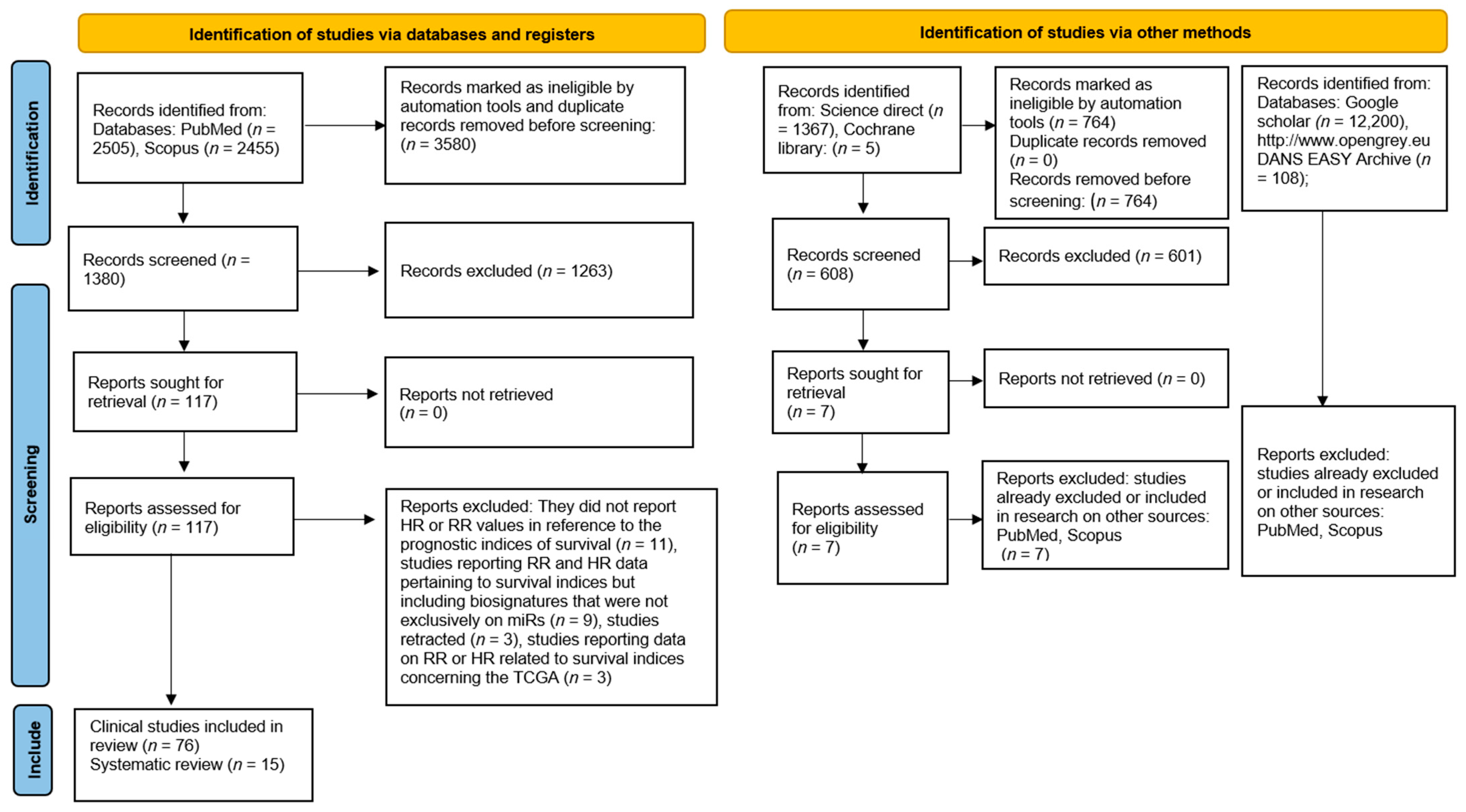
2.1. Study Selection
2.2. Data Characteristics: Systematic Review
2.3. Data Characteristics: Clinical study
2.4. Risk of Bias
- ➢
- Irimie-Aghiorghiese et al., 2019 [15]: Study eligibility criteria (?): The protocol number with which the systematic review was registered was not reported. Identification and selection of studies (?): The selection was performed only on two databases (PubMed and Embase), and the number of authors who conducted the research was not specified, nor were the start or end dates in which the review was conducted.
- ➢
- Lubov et al., 2017 [16]: Study eligibility criteria (?): The protocol number with which the systematic review was registered was not reported. Identification and selection of studies (?): The number of authors who selected the articles and the start or end dates of the review were not reported. Data collection and study appraisal (?): The number of authors who performed the data extraction and the methods of data extraction were not stated. The manuscript is both a systematic review and a retrospective study of 100 patients.
- ➢
- Xie and Wu, 2017 [17]: Study eligibility criteria (?): The protocol number with which the systematic review was registered was not reported.
- ➢
- Wang et al., 2019 [18]: Study eligibility criteria (?): The protocol number with which the systematic review was registered was not reported.
- ➢
- Li et al., 2019 [19]: Study eligibility criteria (?): The protocol number with which the systematic review was registered was not reported. Identification and selection of studies (?): The start or end dates of the review were not specified. Data collection and study appraisal (?): The risk of bias was not formally assessed using an appropriate scale or tool. A bioinformatic analysis was also performed.
- ➢
- Huang et al., 2021 [21]: Study eligibility criteria (?): The protocol number with which the systematic review was registered was not reported. Identification and selection of studies (?): The start and end dates of the review were not specified.
- ➢
- Jamali et al., 2015 [22]: Study eligibility criteria (?): The protocol number with which the systematic review was registered was not reported.
- ➢
- Troiano et al., 2018 [23]: Study eligibility criteria (?): The protocol number with which the systematic review was registered was not reported.
- ➢
- Dioguardi et al., 2023 [9]: Synthesis and findings (?): The obtained results were excessively emphasized in the conclusions.
3. Materials and Methods
3.1. Protocol
3.2. Eligibility Criteria
- Choice of reviewers (M.D. and A.B.) and a third reviewer (F.S.) as a supervisor in case of conflict regarding the studies to be included, choice of outcomes to identify, choice of databases and k words used, choice of criteria of admissibility, choice of data to be extracted and methods of synthesis and registration of the protocol on PROSPERO;
- Identification of records and selection of studies through databases with the removal of duplicates performed manually or by software (EndNote 8.0), performed independently and subsequently comparison of selected studies and decision of studies to be included;
- Independently performed table data extraction and subsequent data comparison to minimize the risk of error in reporting information.
3.3. Sources of Information, Research, and Selection
Translations
3.4. Risk of Bias, Bioinformatic Analysis
4. Discussion
5. Conclusions
Supplementary Materials
Author Contributions
Funding
Institutional Review Board Statement
Informed Consent Statement
Data Availability Statement
Conflicts of Interest
References
- Johnson, D.E.; Burtness, B.; Leemans, C.R.; Lui, V.W.Y.; Bauman, J.E.; Grandis, J.R. Head and neck squamous cell carcinoma. Nat. Rev. Dis. Primers 2020, 6, 92. [Google Scholar] [CrossRef] [PubMed]
- Marur, S.; D’Souza, G.; Westra, W.H.; Forastiere, A.A. HPV-associated head and neck cancer: A virus-related cancer epidemic. Lancet Oncol. 2010, 11, 781–789. [Google Scholar] [CrossRef] [PubMed]
- Ringash, J. Survivorship and Quality of Life in Head and Neck Cancer. J. Clin. Oncol. 2015, 33, 3322–3327. [Google Scholar] [CrossRef] [PubMed]
- John, K.; Wu, J.; Lee, B.W.; Farah, C.S. MicroRNAs in Head and Neck Cancer. Int. J. Dent. 2013, 2013, 650218. [Google Scholar] [CrossRef]
- Yashooa, R.K.; Nabi, A.Q. The miR-146a-5p and miR-125b-5p levels as biomarkers for early prediction of Alzheimer’s disease. Hum. Gene 2022, 34, 201129. [Google Scholar] [CrossRef]
- Dioguardi, M.; Spirito, F.; Sovereto, D.; Alovisi, M.; Aiuto, R.; Garcovich, D.; Crincoli, V.; Laino, L.; Cazzolla, A.P.; Caloro, G.A.; et al. The Prognostic Role of miR-31 in Head and Neck Squamous Cell Carcinoma: Systematic Review and Meta-Analysis with Trial Sequential Analysis. Int. J. Environ. Res. Public Health 2022, 19, 5334. [Google Scholar] [CrossRef]
- Dioguardi, M.; Spirito, F.; Sovereto, D.; Alovisi, M.; Troiano, G.; Aiuto, R.; Garcovich, D.; Crincoli, V.; Laino, L.; Cazzolla, A.P.; et al. MicroRNA-21 Expression as a Prognostic Biomarker in Oral Cancer: Systematic Review and Meta-Analysis. Int. J. Environ. Res. Public Health 2022, 19, 3396. [Google Scholar] [CrossRef]
- Dioguardi, M.; Spirito, F.; Sovereto, D.; La Femina, L.; Campobasso, A.; Cazzolla, A.P.; Di Cosola, M.; Zhurakivska, K.; Cantore, S.; Ballini, A.; et al. Biological Prognostic Value of miR-155 for Survival Outcome in Head and Neck Squamous Cell Carcinomas: Systematic Review, Meta-Analysis and Trial Sequential Analysis. Biology 2022, 11, 651. [Google Scholar] [CrossRef]
- Dioguardi, M.; Spirito, F.; Caloro, G.A.; Lo Muzio, L.; Cantore, S.; Ballini, A.; Scacco, S.; Malcangi, A.; Sembronio, S.; Cascardi, E.; et al. Is the Non-Coding RNA miR-195 a Biodynamic Marker in the Pathogenesis of Head and Neck Squamous Cell Carcinoma? A Prognostic Meta-Analysis. J. Pers. Med. 2023, 13, 275. [Google Scholar] [CrossRef]
- Subha, S.T.; Chin, J.W.; Cheah, Y.K.; Mohtarrudin, N.; Saidi, H.I. Multiple microRNA signature panel as promising potential for diagnosis and prognosis of head and neck cancer. Mol. Biol. Rep. 2022, 49, 1501–1511. [Google Scholar] [CrossRef]
- Guan, J.; Liu, X.; Wang, K.; Jia, Y.; Yang, B. Identification of a novel necroptosis-associated miRNA signature for predicting the prognosis in head and neck squamous cell carcinoma. Open Med. 2022, 17, 1682–1698. [Google Scholar] [CrossRef] [PubMed]
- Tomczak, K.; Czerwińska, P.; Wiznerowicz, M. The Cancer Genome Atlas (TCGA): An immeasurable source of knowledge. Contemp. Oncol. 2015, 19, A68–A77. [Google Scholar] [CrossRef] [PubMed]
- Dioguardi, M.; Cantore, S.; Sovereto, D.; La Femina, L.; Spirito, F.; Caloro, G.A.; Caroprese, M.; Maci, M.; Scacco, S.; Lo Muzio, L.; et al. Does miR-197 Represent a Valid Prognostic Biomarker in Head and Neck Squamous Cell Carcinoma (HNSCC)? A Systematic Review and Trial Sequential Analysis. J. Pers. Med. 2022, 12, 1436. [Google Scholar] [CrossRef] [PubMed]
- Dioguardi, M.; Cantore, S.; Sovereto, D.; La Femina, L.; Caloro, G.A.; Spirito, F.; Scacco, S.; Di Cosola, M.; Lo Muzio, L.; Troiano, G.; et al. Potential Role of miR-196a and miR-196b as Prognostic Biomarkers of Survival in Head and Neck Squamous Cell Carcinoma: A Systematic Review, Meta-Analysis and Trial Sequential Analysis. Life 2022, 12, 1269. [Google Scholar] [CrossRef]
- Irimie-Aghiorghiesei, A.I.; Pop-Bica, C.; Pintea, S.; Braicu, C.; Cojocneanu, R.; Zimța, A.-A.; Gulei, D.; Slabý, O.; Berindan-Neagoe, I. Prognostic Value of MiR-21: An Updated Meta-Analysis in Head and Neck Squamous Cell Carcinoma (HNSCC). J. Clin. Med. 2019, 8, 2041. [Google Scholar] [CrossRef]
- Lubov, J.; Maschietto, M.; Ibrahim, I.; Mlynarek, A.; Hier, M.; Kowalski, L.P.; Alaoui-Jamali, M.A.; da Silva, S.D. Meta-analysis of microRNAs expression in head and neck cancer: Uncovering association with outcome and mechanisms. Oncotarget 2017, 8, 55511–55524. [Google Scholar] [CrossRef]
- Xie, Y.; Wu, J. MicroRNA-21 as prognostic molecular signatures in oral cancer: A meta-analysis. Int. J. Clin. Exp. Med. 2017, 10, 9848–9856. [Google Scholar]
- Wang, P.; Xu, L.; Li, L.; Ren, S.; Tang, J.; Zhang, M.; Xu, M. The microRNA-375 as a potentially promising biomarker to predict the prognosis of patients with head and neck or esophageal squamous cell carcinoma: A meta-analysis. Eur. Arch. Otorhinolaryngol. 2019, 276, 957–968. [Google Scholar] [CrossRef]
- Li, M.-w.; Gao, L.; Dang, Y.-w.; Li, P.; Li, Z.-y.; Chen, G.; Luo, D.-z. Protective potential of miR-146a-5p and its underlying molecular mechanism in diverse cancers: A comprehensive meta-analysis and bioinformatics analysis. Cancer Cell Int. 2019, 19, 167. [Google Scholar] [CrossRef]
- Qiu, K.; Song, Y.; Rao, Y.; Liu, Q.; Cheng, D.; Pang, W.; Ren, J.; Zhao, Y. Diagnostic and Prognostic Value of MicroRNAs in Metastasis and Recurrence of Head and Neck Squamous Cell Carcinoma: A Systematic Review and Meta-Analysis. Front. Oncol. 2021, 11, 711171. [Google Scholar] [CrossRef]
- Huang, Y.; Gu, M.; Tang, Y.; Sun, Z.; Luo, J.; Li, Z. Systematic review and meta-analysis of prognostic microRNA biomarkers for survival outcome in laryngeal squamous cell cancer. Cancer Cell Int. 2021, 21, 316. [Google Scholar] [CrossRef] [PubMed]
- Jamali, Z.; Asl Aminabadi, N.; Attaran, R.; Pournagiazar, F.; Ghertasi Oskouei, S.; Ahmadpour, F. MicroRNAs as prognostic molecular signatures in human head and neck squamous cell carcinoma: A systematic review and meta-analysis. Oral Oncol. 2015, 51, 321–331. [Google Scholar] [CrossRef] [PubMed]
- Troiano, G.; Mastrangelo, F.; Caponio, V.C.A.; Laino, L.; Cirillo, N.; Lo Muzio, L. Predictive Prognostic Value of Tissue-Based MicroRNA Expression in Oral Squamous Cell Carcinoma: A Systematic Review and Meta-analysis. J. Dent. Res. 2018, 97, 759–766. [Google Scholar] [CrossRef] [PubMed]
- Jung, H.M.; Phillips, B.L.; Patel, R.S.; Cohen, D.M.; Jakymiw, A.; Kong, W.W.; Cheng, J.Q.; Chan, E.K.L. Keratinization-associated miR-7 and miR-21 regulate tumor suppressor reversion-inducing cysteine-rich protein with kazal motifs (RECK) in oral cancer. J. Biol. Chem. 2012, 287, 29261–29272. [Google Scholar] [CrossRef] [PubMed]
- Kawakita, A.; Yanamoto, S.; Yamada, S.; Naruse, T.; Takahashi, H.; Kawasaki, G.; Umeda, M. MicroRNA-21 promotes oral cancer invasion via the Wnt/beta-catenin pathway by targeting DKK2. Pathol. Oncol. Res. 2014, 20, 253–261. [Google Scholar] [CrossRef]
- Hedbäck, N.; Jensen, D.H.; Specht, L.; Fiehn, A.M.K.; Therkildsen, M.H.; Friis-Hansen, L.; Dabelsteen, E.; Von Buchwald, C. miR-21 expression in the tumor stroma of oral squamous cell carcinoma: An independent biomarker of disease free survival. PLoS ONE 2014, 9, e95193. [Google Scholar] [CrossRef]
- Yu, E.H.; Tu, H.F.; Wu, C.H.; Yang, C.C.; Chang, K.W. MicroRNA-21 promotes perineural invasion and impacts survival in patients with oral carcinoma. J. Chin. Med. Assoc. 2017, 80, 383–388. [Google Scholar] [CrossRef]
- Supic, G.; Zeljic, K.; Rankov, A.D.; Kozomara, R.; Nikolic, A.; Radojkovic, D.; Magic, Z. miR-183 and miR-21 expression as biomarkers of progression and survival in tongue carcinoma patients. Clin. Oral. Investig. 2018, 22, 401–409. [Google Scholar] [CrossRef]
- Jakob, M.; Mattes, L.M.; Küffer, S.; Unger, K.; Hess, J.; Bertlich, M.; Haubner, F.; Ihler, F.; Canis, M.; Weiss, B.G.; et al. MicroRNA expression patterns in oral squamous cell carcinoma: Hsa-mir-99b-3p and hsa-mir-100-5p as novel prognostic markers for oral cancer. Head Neck 2019, 41, 3499–3515. [Google Scholar] [CrossRef]
- Li, S.; Zhou, X.; Zhang, Q.; Zhang, L. Abnormal expression of STAT3 and miRNA-21 in oral squamous cell carcinoma. Chin. J. Clin. Oncol. 2013, 40, 323–327. [Google Scholar] [CrossRef]
- Zheng, G.; Li, N.; Jia, X.; Peng, C.; Luo, L.; Deng, Y.; Yin, J.; Song, Y.; Liu, H.; Lu, M.; et al. MYCN-mediated miR-21 overexpression enhances chemo-resistance via targeting CADM1 in tongue cancer. J. Mol. Med. 2016, 94, 1129–1141. [Google Scholar] [CrossRef]
- Li, J.; Huang, H.; Sun, L.; Yang, M.; Pan, C.; Chen, W.; Wu, D.; Lin, Z.; Zeng, C.; Yao, Y.; et al. MiR-21 indicates poor prognosis in tongue squamous cell carcinomas as an apoptosis inhibitor. Clin. Cancer Res. 2009, 15, 3998–4008. [Google Scholar] [CrossRef]
- Ganci, F.; Sacconi, A.; Manciocco, V.; Sperduti, I.; Battaglia, P.; Covello, R.; Muti, P.; Strano, S.; Spriano, G.; Fontemaggi, G.; et al. MicroRNA expression as predictor of local recurrence risk in oral squamous cell carcinoma. Head Neck 2016, 38 (Suppl. 1), E189–E197. [Google Scholar] [CrossRef]
- Wang, L.-L.; Li, H.-X.; Yang, Y.-Y.; Su, Y.-L.; Lian, J.-S.; Li, T.; Xu, J.; Wang, X.-N.; Jin, N.; Liu, X.-F. MiR-31 is a potential biomarker for diagnosis of head and neck squamous cell carcinoma. Int. J. Clin. Exp. Pathol. 2018, 11, 4339–4345. [Google Scholar]
- Qiang, H.; Zhan, X.; Wang, W.; Cheng, Z.; Ma, S.; Jiang, C. A Study on the Correlations of the miR-31 Expression with the Pathogenesis and Prognosis of Head and Neck Squamous Cell Carcinoma. Cancer Biother. Radiopharm. 2019, 34, 189–195. [Google Scholar] [CrossRef]
- Tu, H.-F.; Liu, C.-J.; Hung, W.-W.; Shieh, T.-M. Co-upregulation of miR-31 and its host gene lncRNA MIR31HG in oral squamous cell carcinoma. J. Dent. Sci. 2022, 17, 696–706. [Google Scholar] [CrossRef]
- Hess, A.K.; Müer, A.; Mairinger, F.D.; Weichert, W.; Stenzinger, A.; Hummel, M.; Budach, V.; Tinhofer, I. MiR-200b and miR-155 as predictive biomarkers for the efficacy of chemoradiation in locally advanced head and neck squamous cell carcinoma. Eur. J. Cancer 2017, 77, 3–12. [Google Scholar] [CrossRef] [PubMed]
- Zhao, X.; Zhang, W.; Ji, W. YB-1 promotes laryngeal squamous cell carcinoma progression by inducing miR-155 expression via c-Myb. Future Oncol. 2018, 14, 1579–1589. [Google Scholar] [CrossRef] [PubMed]
- Baba, O.; Hasegawa, S.; Nagai, H.; Uchida, F.; Yamatoji, M.; Kanno, N.I.; Yamagata, K.; Sakai, S.; Yanagawa, T.; Bukawa, H. MicroRNA-155-5p is associated with oral squamous cell carcinoma metastasis and poor prognosis. J. Oral Pathol. Med. 2016, 45, 248–255. [Google Scholar] [CrossRef] [PubMed]
- Shi, L.-J.; Zhang, C.-Y.; Zhou, Z.-T.; Ma, J.-Y.; Liu, Y.; Bao, Z.-X.; Jiang, W.-W. MicroRNA-155 in oral squamous cell carcinoma: Overexpression, localization, and prognostic potential. Head Neck 2015, 37, 970–976. [Google Scholar] [CrossRef]
- Kim, H.; Yang, J.M.; Ahn, S.H.; Jeong, W.J.; Chung, J.H.; Paik, J.H. Potential Oncogenic Role and Prognostic Implication of MicroRNA-155-5p in Oral Squamous Cell Carcinoma. Anticancer Res. 2018, 38, 5193–5200. [Google Scholar] [CrossRef] [PubMed]
- Bersani, C.; Mints, M.; Tertipis, N.; Haeggblom, L.; Näsman, A.; Romanitan, M.; Dalianis, T.; Ramqvist, T. MicroRNA-155, -185 and -193b as biomarkers in human papillomavirus positive and negative tonsillar and base of tongue squamous cell carcinoma. Oral Oncol. 2018, 82, 8–16. [Google Scholar] [CrossRef] [PubMed]
- Wu, M.; Duan, Q.; Liu, X.; Zhang, P.; Fu, Y.; Zhang, Z.; Liu, L.; Cheng, J.; Jiang, H. MiR-155-5p promotes oral cancer progression by targeting chromatin remodeling gene ARID2. Biomed. Pharmacother. 2020, 122, 109696. [Google Scholar] [CrossRef] [PubMed]
- Shuang, Y.; Li, C.; Zhou, X.; Huang, Y.; Zhang, L. MicroRNA-195 inhibits growth and invasion of laryngeal carcinoma cells by directly targeting DCUN1D1. Oncol. Rep. 2017, 38, 2155–2165. [Google Scholar] [CrossRef]
- Ding, D.; Qi, Z. Clinical significance of miRNA-195 expression in patients with laryngeal carcinoma. J. Buon 2019, 24, 315–322. [Google Scholar]
- Jia, L.-f.; Wei, S.-b.; Gong, K.; Gan, Y.-h.; Yu, G.-y. Prognostic implications of micoRNA miR-195 expression in human tongue squamous cell carcinoma. PLoS ONE 2013, 8, e56634. [Google Scholar] [CrossRef]
- Qin, X.; Guo, H.; Wang, X.; Zhu, X.; Yan, M.; Wang, X.; Xu, Q.; Shi, J.; Lu, E.; Chen, W.; et al. Exosomal miR-196a derived from cancer-associated fibroblasts confers cisplatin resistance in head and neck cancer through targeting CDKN1B and ING5. Genome Biol. 2019, 20, 12. [Google Scholar] [CrossRef]
- Liu, C.J.; Tsai, M.M.; Tu, H.F.; Lui, M.T.; Cheng, H.W.; Lin, S.C. miR-196a overexpression and miR-196a2 gene polymorphism are prognostic predictors of oral carcinomas. Ann. Surg. Oncol. 2013, 20 (Suppl. 3), S406–S414. [Google Scholar] [CrossRef]
- Maruyama, T.; Nishihara, K.; Umikawa, M.; Arasaki, A.; Nakasone, T.; Nimura, F.; Matayoshi, A.; Takei, K.; Nakachi, S.; Kariya, K.-I.; et al. MicroRNA-196a-5p is a potential prognostic marker of delayed lymph node metastasis in early-stage tongue squamous cell carcinoma. Oncol. Lett. 2018, 15, 2349–2363. [Google Scholar] [CrossRef]
- Zhao, X.; Zhang, W.; Ji, W. miR-196b is a prognostic factor of human laryngeal squamous cell carcinoma and promotes tumor progression by targeting SOCS2. Biochem. Biophys. Res. Commun. 2018, 501, 584–592. [Google Scholar] [CrossRef]
- Luo, M.; Sun, G.; Sun, J.W. MiR-196b affects the progression and prognosis of human LSCC through targeting PCDH-17. Auris Nasus Larynx 2019, 46, 583–592. [Google Scholar] [CrossRef] [PubMed]
- Ahn, H.; Yang, J.M.; Kim, H.; Chung, J.H.; Ahn, S.H.; Jeong, W.J.; Paik, J.H. Clinicopathologic implications of the miR-197/PD-L1 axis in oral squamous cell carcinoma. Oncotarget 2017, 8, 66178–66194. [Google Scholar] [CrossRef]
- Hudcova, K.; Raudenska, M.; Gumulec, J.; Binkova, H.; Horakova, Z.; Kostrica, R.; Babula, P.; Adam, V.; Masarik, M. Expression profiles of miR-29c, miR-200b and miR-375 in tumour and tumour-adjacent tissues of head and neck cancers. Tumour Biol. 2016, 37, 12627–12633. [Google Scholar] [CrossRef]
- Kang, Y.; Zhang, Y.; Sun, Y. MicroRNA-198 suppresses tumour growth and metastasis in oral squamous cell carcinoma by targeting CDK4. Int. J. Oncol. 2021, 59, 39. [Google Scholar] [CrossRef] [PubMed]
- Bonnin, N.; Armandy, E.; Carras, J.; Ferrandon, S.; Battiston-Montagne, P.; Aubry, M.; Guihard, S.; Meyronet, D.; Foy, J.P.; Saintigny, P.; et al. MiR-422a promotes loco-regional recurrence by targeting NT5E/CD73 in head and neck squamous cell carcinoma. Oncotarget 2016, 7, 44023–44038. [Google Scholar] [CrossRef] [PubMed]
- Ganci, F.; Sacconi, A.; Bossel Ben-Moshe, N.; Manciocco, V.; Sperduti, I.; Strigari, L.; Covello, R.; Benevolo, M.; Pescarmona, E.; Domany, E.; et al. Expression of TP53 mutation-associated microRNAs predicts clinical outcome in head and neck squamous cell carcinoma patients. Ann. Oncol. 2013, 24, 3082–3088. [Google Scholar] [CrossRef]
- Harris, T.; Jimenez, L.; Kawachi, N.; Fan, J.B.; Chen, J.; Belbin, T.; Ramnauth, A.; Loudig, O.; Keller, C.E.; Smith, R.; et al. Low-level expression of miR-375 correlates with poor outcome and metastasis while altering the invasive properties of head and neck squamous cell carcinomas. Am. J. Pathol. 2012, 180, 917–928. [Google Scholar] [CrossRef]
- Ahmad, P.; Sana, J.; Slavik, M.; Gurin, D.; Radova, L.; Gablo, N.A.; Kazda, T.; Smilek, P.; Horakova, Z.; Gal, B.; et al. MicroRNA-15b-5p Predicts Locoregional Relapse in Head and Neck Carcinoma Patients Treated With Intensity-modulated Radiotherapy. Cancer Genom. Proteom. 2019, 16, 139–146. [Google Scholar] [CrossRef]
- Rajthala, S.; Dongre, H.; Parajuli, H.; Min, A.; Nginamau, E.S.; Kvalheim, A.; Lybak, S.; Sapkota, D.; Johannessen, A.C.; Costea, D.E. Combined In Situ Hybridization and Immunohistochemistry on Archival Tissues Reveals Stromal microRNA-204 as Prognostic Biomarker for Oral Squamous Cell Carcinoma. Cancers 2021, 13, 1307. [Google Scholar] [CrossRef]
- Song, J.; Zhang, N.; Cao, L.; Xiao, D.; Ye, X.; Luo, E.; Zhang, Z. Down-regulation of miR-200c associates with poor prognosis of oral squamous cell carcinoma. Int. J. Clin. Oncol. 2020, 25, 1072–1078. [Google Scholar] [CrossRef]
- Zhao, X.; Zhang, W.; Ji, W. MYO5A inhibition by miR-145 acts as a predictive marker of occult neck lymph node metastasis in human laryngeal squamous cell carcinoma. OncoTargets Ther. 2018, 11, 3619–3635. [Google Scholar] [CrossRef] [PubMed]
- Li, M.; Tian, L.; Ren, H.; Chen, X.; Wang, Y.; Ge, J.; Wu, S.; Sun, Y.; Liu, M.; Xiao, H. MicroRNA-101 is a potential prognostic indicator of laryngeal squamous cell carcinoma and modulates CDK8. J. Transl. Med. 2015, 13, 271. [Google Scholar] [CrossRef] [PubMed]
- de Jong, M.C.; Ten Hoeve, J.J.; Grénman, R.; Wessels, L.F.; Kerkhoven, R.; Te Riele, H.; van den Brekel, M.W.; Verheij, M.; Begg, A.C. Pretreatment microRNA Expression Impacting on Epithelial-to-Mesenchymal Transition Predicts Intrinsic Radiosensitivity in Head and Neck Cancer Cell Lines and Patients. Clin. Cancer Res. 2015, 21, 5630–5638. [Google Scholar] [CrossRef] [PubMed]
- Fang, R.; Huang, Y.; Xie, J.; Zhang, J.; Ji, X. Downregulation of miR-29c-3p is associated with a poor prognosis in patients with laryngeal squamous cell carcinoma. Diagn. Pathol. 2019, 14, 109. [Google Scholar] [CrossRef]
- He, F.Y.; Liu, H.J.; Guo, Q.; Sheng, J.L. Reduced miR-300 expression predicts poor prognosis in patients with laryngeal squamous cell carcinoma. Eur. Rev. Med. Pharmacol. Sci. 2017, 21, 760–764. [Google Scholar]
- Re, M.; Çeka, A.; Rubini, C.; Ferrante, L.; Zizzi, A.; Gioacchini, F.M.; Tulli, M.; Spazzafumo, L.; Sellari-Franceschini, S.; Procopio, A.D.; et al. MicroRNA-34c-5p is related to recurrence in laryngeal squamous cell carcinoma. Laryngoscope 2015, 125, E306–E312. [Google Scholar] [CrossRef]
- Xu, Y.; Lin, Y.P.; Yang, D.; Zhang, G.; Zhou, H.F. Clinical Significance of miR-149 in the Survival of Patients with Laryngeal Squamous Cell Carcinoma. Biomed Res. Int. 2016, 2016, 8561251. [Google Scholar] [CrossRef]
- Tian, L.; Li, M.; Ge, J.; Guo, Y.; Sun, Y.; Liu, M.; Xiao, H. MiR-203 is downregulated in laryngeal squamous cell carcinoma and can suppress proliferation and induce apoptosis of tumours. Tumour Biol. 2014, 35, 5953–5963. [Google Scholar] [CrossRef]
- Zhao, X.; Zhang, W.; Ji, W. miR-181a targets GATA6 to inhibit the progression of human laryngeal squamous cell carcinoma. Future Oncol. 2018, 14, 1741–1753. [Google Scholar] [CrossRef]
- Guan, G.F.; Zhang, D.J.; Wen, L.J.; Xin, D.; Liu, Y.; Yu, D.J.; Su, K.; Zhu, L.; Guo, Y.Y.; Wang, K. Overexpression of lncRNA H19/miR-675 promotes tumorigenesis in head and neck squamous cell carcinoma. Int. J. Med. Sci. 2016, 13, 914–922. [Google Scholar] [CrossRef]
- Avissar, M.; McClean, M.D.; Kelsey, K.T.; Marsit, C.J. MicroRNA expression in head and neck cancer associates with alcohol consumption and survival. Carcinogenesis 2009, 30, 2059–2063. [Google Scholar] [CrossRef] [PubMed]
- Wu, S.; Jia, S.; Xu, P. MicroRNA-9 as a novel prognostic biomarker in human laryngeal squamous cell carcinoma. Int. J. Clin. Exp. Med. 2014, 7, 5523–5528. [Google Scholar] [PubMed]
- Wu, T.Y.; Zhang, T.H.; Qu, L.M.; Feng, J.P.; Tian, L.L.; Zhang, B.H.; Li, D.D.; Sun, Y.N.; Liu, M. MiR-19a is correlated with prognosis and apoptosis of laryngeal squamous cell carcinoma by regulating TIMP-2 expression. Int. J. Clin. Exp. Pathol. 2014, 7, 56–63. [Google Scholar] [PubMed]
- Zhang, X.W.; Liu, N.; Chen, S.; Wang, Y.; Zhang, Z.X.; Sun, Y.Y.; Qiu, G.B.; Fu, W.N. High microRNA-23a expression in laryngeal squamous cell carcinoma is associated with poor patient prognosis. Diagn. Pathol. 2015, 10, 22. [Google Scholar] [CrossRef]
- Hu, A.; Huang, J.J.; Xu, W.H.; Jin, X.J.; Li, J.P.; Tang, Y.J.; Huang, X.F.; Cui, H.J.; Sun, G.B.; Li, R.L.; et al. MiR-21/miR-375 ratio is an independent prognostic factor in patients with laryngeal squamous cell carcinoma. Am. J. Cancer Res. 2015, 5, 1775–1785. [Google Scholar]
- Re, M.; Magliulo, G.; Gioacchini, F.M.; Bajraktari, A.; Bertini, A.; Çeka, A.; Rubini, C.; Ferrante, L.; Procopio, A.D.; Olivieri, F. Expression Levels and Clinical Significance of miR-21-5p, miR-let-7a, and miR-34c-5p in Laryngeal Squamous Cell Carcinoma. Biomed Res. Int. 2017, 2017, 3921258. [Google Scholar] [CrossRef]
- Shen, Z.; Zhan, G.; Ye, D.; Ren, Y.; Cheng, L.; Wu, Z.; Guo, J. MicroRNA-34a affects the occurrence of laryngeal squamous cell carcinoma by targeting the antiapoptotic gene survivin. Med. Oncol. 2012, 29, 2473–2480. [Google Scholar] [CrossRef]
- Maia, D.; de Carvalho, A.C.; Horst, M.A.; Carvalho, A.L.; Scapulatempo-Neto, C.; Vettore, A.L. Expression of miR-296-5p as predictive marker for radiotherapy resistance in early-stage laryngeal carcinoma. J. Transl. Med. 2015, 13, 262. [Google Scholar] [CrossRef]
- Ogawa, T.; Saiki, Y.; Shiga, K.; Chen, N.; Fukushige, S.; Sunamura, M.; Nagase, H.; Hashimoto, S.; Matsuura, K.; Saijo, S.; et al. miR-34a is downregulated in cis-diamminedichloroplatinum treated sinonasal squamous cell carcinoma patients with poor prognosis. Cancer Sci. 2012, 103, 1737–1743. [Google Scholar] [CrossRef]
- Pantazis, T.L.; Giotakis, A.I.; Karamagkiolas, S.; Giotakis, I.; Konstantoulakis, M.; Liakea, A.; Misiakos, E.P. Low expression of miR-20b-5p indicates favorable prognosis in laryngeal squamous cell carcinoma, especially in patients with non-infiltrated regional lymph nodes. Am. J. Otolaryngol. 2020, 41, 102563. [Google Scholar] [CrossRef]
- Childs, G.; Fazzari, M.; Kung, G.; Kawachi, N.; Brandwein-Gensler, M.; McLemore, M.; Chen, Q.; Burk, R.D.; Smith, R.V.; Prystowsky, M.B.; et al. Low-level expression of microRNAs let-7d and miR-205 are prognostic markers of head and neck squamous cell carcinoma. Am. J. Pathol. 2009, 174, 736–745. [Google Scholar] [CrossRef] [PubMed]
- Ko, Y.H.; Won, H.S.; Sun, D.S.; An, H.J.; Jeon, E.K.; Kim, M.S.; Lee, H.H.; Kang, J.H.; Jung, C.K. Human papillomavirus-stratified analysis of the prognostic role of miR-21 in oral cavity and oropharyngeal squamous cell carcinoma. Pathol. Int. 2014, 64, 499–507. [Google Scholar] [CrossRef] [PubMed]
- Arantes, L.M.; Laus, A.C.; Melendez, M.E.; de Carvalho, A.C.; Sorroche, B.P.; De Marchi, P.R.; Evangelista, A.F.; Scapulatempo-Neto, C.; de Souza Viana, L.; Carvalho, A.L. MiR-21 as prognostic biomarker in head and neck squamous cell carcinoma patients undergoing an organ preservation protocol. Oncotarget 2017, 8, 9911–9921. [Google Scholar] [CrossRef] [PubMed]
- Chang, C.C.; Yang, Y.J.; Li, Y.J.; Chen, S.T.; Lin, B.R.; Wu, T.S.; Lin, S.K.; Kuo, M.Y.; Tan, C.T. MicroRNA-17/20a functions to inhibit cell migration and can be used a prognostic marker in oral squamous cell carcinoma. Oral Oncol. 2013, 49, 923–931. [Google Scholar] [CrossRef]
- Gee, H.E.; Camps, C.; Buffa, F.M.; Patiar, S.; Winter, S.C.; Betts, G.; Homer, J.; Corbridge, R.; Cox, G.; West, C.M.; et al. hsa-mir-210 is a marker of tumor hypoxia and a prognostic factor in head and neck cancer. Cancer 2010, 116, 2148–2158. [Google Scholar] [CrossRef]
- Jia, L.-F.; Wei, S.-B.; Gan, Y.-H.; Guo, Y.; Gong, K.; Mitchelson, K.; Cheng, J.; Yu, G.-Y. Expression, regulation and roles of miR-26a and MEG3 in tongue squamous cell carcinoma. Int. J. Cancer 2014, 135, 2282–2293. [Google Scholar] [CrossRef]
- Liao, L.; Wang, J.; Ouyang, S.; Zhang, P.; Wang, J.; Zhang, M. Expression and clinical significance of microRNA-1246 in human oral squamous cell carcinoma. Med. Sci. Monit. 2015, 21, 776–781. [Google Scholar] [CrossRef]
- Liu, N.; Jiang, N.; Guo, R.; Jiang, W.; He, Q.M.; Xu, Y.F.; Li, Y.Q.; Tang, L.L.; Mao, Y.P.; Sun, Y.; et al. MiR-451 inhibits cell growth and invasion by targeting MIF and is associated with survival in nasopharyngeal carcinoma. Mol. Cancer 2013, 12, 123. [Google Scholar] [CrossRef]
- Liu, C.J.; Shen, W.G.; Peng, S.Y.; Cheng, H.W.; Kao, S.Y.; Lin, S.C.; Chang, K.W. miR-134 induces oncogenicity and metastasis in head and neck carcinoma through targeting WWOX gene. Int. J. Cancer 2014, 134, 811–821. [Google Scholar] [CrossRef]
- Luo, Z.; Dai, Y.; Zhang, L.; Jiang, C.; Li, Z.; Yang, J.; McCarthy, J.B.; She, X.; Zhang, W.; Ma, J.; et al. miR-18a promotes malignant progression by impairing microRNA biogenesis in nasopharyngeal carcinoma. Carcinogenesis 2013, 34, 415–425. [Google Scholar] [CrossRef]
- Peng, S.C.; Liao, C.T.; Peng, C.H.; Cheng, A.J.; Chen, S.J.; Huang, C.G.; Hsieh, W.P.; Yen, T.C. MicroRNAs MiR-218, MiR-125b, and Let-7g predict prognosis in patients with oral cavity squamous cell carcinoma. PLoS ONE 2014, 9, e102403. [Google Scholar] [CrossRef]
- Sasahira, T.; Kurihara, M.; Bhawal, U.K.; Ueda, N.; Shimomoto, T.; Yamamoto, K.; Kirita, T.; Kuniyasu, H. Downregulation of miR-126 induces angiogenesis and lymphangiogenesis by activation of VEGF-A in oral cancer. Br. J. Cancer 2012, 107, 700–706. [Google Scholar] [CrossRef] [PubMed]
- Tu, H.F.; Chang, K.W.; Cheng, H.W.; Liu, C.J. Upregulation of miR-372 and -373 associates with lymph node metastasis and poor prognosis of oral carcinomas. Laryngoscope 2015, 125, E365–E370. [Google Scholar] [CrossRef] [PubMed]
- Wu, D.W.; Chuang, C.Y.; Lin, W.L.; Sung, W.W.; Cheng, Y.W.; Lee, H. Paxillin promotes tumor progression and predicts survival and relapse in oral cavity squamous cell carcinoma by microRNA-218 targeting. Carcinogenesis 2014, 35, 1823–1829. [Google Scholar] [CrossRef] [PubMed]
- Xu, Q.; Sun, Q.; Zhang, J.; Yu, J.; Chen, W.; Zhang, Z. Downregulation of miR-153 contributes to epithelial-mesenchymal transition and tumor metastasis in human epithelial cancer. Carcinogenesis 2013, 34, 539–549. [Google Scholar] [CrossRef]
- Zhang, B.; Li, Y.; Hou, D.; Shi, Q.; Yang, S.; Li, Q. MicroRNA-375 Inhibits Growth and Enhances Radiosensitivity in Oral Squamous Cell Carcinoma by Targeting Insulin Like Growth Factor 1 Receptor. Cell. Physiol. Biochem. 2017, 42, 2105–2117. [Google Scholar] [CrossRef]
- Jia, L.; Huang, Y.; Zheng, Y.; Lyu, M.; Zhang, C.; Meng, Z.; Gan, Y.; Yu, G. miR-375 inhibits cell growth and correlates with clinical outcomes in tongue squamous cell carcinoma. Oncol. Rep. 2015, 33, 2061–2071. [Google Scholar] [CrossRef]
- Hu, A.; Huang, J.J.; Xu, W.H.; Jin, X.J.; Li, J.P.; Tang, Y.J.; Huang, X.F.; Cui, H.J.; Sun, G.B. miR-21 and miR-375 microRNAs as candidate diagnostic biomarkers in squamous cell carcinoma of the larynx: Association with patient survival. Am. J. Transl. Res. 2014, 6, 604–613. [Google Scholar]
- Gu, Y.; Liu, H.; Kong, F.; Ye, J.; Jia, X.; Zhang, Z.; Li, N.; Yin, J.; Zheng, G.; He, Z. miR-22/KAT6B axis is a chemotherapeutic determiner via regulation of PI3k-Akt-NF-kB pathway in tongue squamous cell carcinoma. J. Exp. Clin. Cancer Res. 2018, 37, 164. [Google Scholar] [CrossRef]
- Győrffy, B. Discovery and ranking of the most robust prognostic biomarkers in serous ovarian cancer. Geroscience 2023, 45, 1889–1898. [Google Scholar] [CrossRef]
- Lánczky, A.; Győrffy, B. Web-Based Survival Analysis Tool Tailored for Medical Research (KMplot): Development and Implementation. J. Med. Internet Res. 2021, 23, e27633. [Google Scholar] [CrossRef]
- Benjamini, Y.; Hochberg, Y. Controlling the false discovery rate: A practical and powerful approach to multiple testing. J. R. Stat. Soc. Ser. B (Methodol.) 1995, 57, 289–300. [Google Scholar] [CrossRef]
- Liberati, A.; Altman, D.G.; Tetzlaff, J.; Mulrow, C.; Gøtzsche, P.C.; Ioannidis, J.P.; Clarke, M.; Devereaux, P.J.; Kleijnen, J.; Moher, D. The PRISMA statement for reporting systematic reviews and meta-analyses of studies that evaluate healthcare interventions: Explanation and elaboration. BMJ 2009, 339, b2700. [Google Scholar] [CrossRef] [PubMed]
- Whiting, P.; Savović, J.; Higgins, J.P.; Caldwell, D.M.; Reeves, B.C.; Shea, B.; Davies, P.; Kleijnen, J.; Churchill, R. ROBIS: A new tool to assess risk of bias in systematic reviews was developed. J. Clin. Epidemiol. 2016, 69, 225–234. [Google Scholar] [CrossRef] [PubMed]
- Sauerbrei, W.; Taube, S.E.; McShane, L.M.; Cavenagh, M.M.; Altman, D.G. Reporting Recommendations for Tumor Marker Prognostic Studies (REMARK): An Abridged Explanation and Elaboration. J. Natl. Cancer Inst. 2018, 110, 803–811. [Google Scholar] [CrossRef] [PubMed]
- Pozniak, T.; Shcharbin, D.; Bryszewska, M. Circulating microRNAs in Medicine. Int. J. Mol. Sci. 2022, 23, 3996. [Google Scholar] [CrossRef] [PubMed]
- Chamorro Petronacci, C.M.; García García, A.; Padín Iruegas, E.; Rivas Mundiña, B.; Lorenzo Pouso, A.I.; Pérez Sayáns, M. Identification of Prognosis Associated microRNAs in HNSCC Subtypes Based on TCGA Dataset. Medicina 2020, 56, 535. [Google Scholar] [CrossRef]
- Vahabi, M.; Blandino, G.; Di Agostino, S. MicroRNAs in head and neck squamous cell carcinoma: A possible challenge as biomarkers, determinants for the choice of therapy and targets for personalized molecular therapies. Transl. Cancer Res. 2021, 10, 3090–3110. [Google Scholar] [CrossRef] [PubMed]
- Muzio, L.L.; Ballini, A.; Cantore, S.; Bottalico, L.; Charitos, I.A.; Ambrosino, M.; Nocini, R.; Malcangi, A.; Dioguardi, M.; Cazzolla, A.P.; et al. Overview of Candida albicans and Human Papillomavirus (HPV) Infection Agents and their Biomolecular Mechanisms in Promoting Oral Cancer in Pediatric Patients. BioMed Res. Int. 2021, 2021, 7312611. [Google Scholar] [CrossRef]
- Supic, G.; Stefik, D.; Ivkovic, N.; Sami, A.; Zeljic, K.; Jovic, S.; Kozomara, R.; Vojvodic, D.; Stosic, S. Prognostic impact of miR-34b/c DNA methylation, gene expression, and promoter polymorphism in HPV-negative oral squamous cell carcinomas. Sci. Rep. 2022, 12, 1296. [Google Scholar] [CrossRef]
- House, R.; Majumder, M.; Janakiraman, H.; Ogretmen, B.; Kato, M.; Erkul, E.; Hill, E.; Atkinson, C.; Barth, J.; Day, T.A.; et al. Smoking-induced control of miR-133a-3p alters the expression of EGFR and HuR in HPV-infected oropharyngeal cancer. PLoS ONE 2018, 13, e0205077. [Google Scholar] [CrossRef]
- Miladinovic, B.; Mhaskar, R.; Hozo, I.; Kumar, A.; Mahony, H.; Djulbegovic, B. Optimal information size in trial sequential analysis of time-to-event outcomes reveals potentially inconclusive results because of the risk of random error. J. Clin. Epidemiol. 2013, 66, 654–659. [Google Scholar] [CrossRef]
- Kwan, J.; Psarianos, P.; Bruce, J.; Yip, K.; Liu, F.-f. The complexity of microRNAs in human cancer. J. Radiat. Res. 2016, 57, rrw009. [Google Scholar] [CrossRef]
- How, C.; Pintilie, M.; Bruce, J.P.; Hui, A.B.; Clarke, B.A.; Wong, P.; Yin, S.; Yan, R.; Waggott, D.; Boutros, P.C.; et al. Developing a prognostic micro-RNA signature for human cervical carcinoma. PLoS ONE 2015, 10, e0123946. [Google Scholar] [CrossRef] [PubMed]
- Kumar, S.; Patnaik, S.; Dixit, A. Predictive models for stage and risk classification in head and neck squamous cell carcinoma (HNSCC). PeerJ 2020, 8, e9656. [Google Scholar] [CrossRef] [PubMed]
- Bauman, J.E.; Austin, M.C.; Schmidt, R.; Kurland, B.F.; Vaezi, A.; Hayes, D.N.; Mendez, E.; Parvathaneni, U.; Chai, X.; Sampath, S.; et al. ERCC1 is a prognostic biomarker in locally advanced head and neck cancer: Results from a randomised, phase II trial. Br. J. Cancer 2013, 109, 2096–2105. [Google Scholar] [CrossRef] [PubMed]
- Psyrri, A.; Lee, J.W.; Pectasides, E.; Vassilakopoulou, M.; Kosmidis, E.K.; Burtness, B.A.; Rimm, D.L.; Wanebo, H.J.; Forastiere, A.A. Prognostic biomarkers in phase II trial of cetuximab-containing induction and chemoradiation in resectable HNSCC: Eastern cooperative oncology group E2303. Clin. Cancer Res. 2014, 20, 3023–3032. [Google Scholar] [CrossRef]
- Zhao, G.; Li, C.X.; Guo, C.; Zhu, H. MicroRNA model that can predict the prognosis of oral squamous cell carcinoma based on bioinformatics analysis. Hua Xi Kou Qiang Yi Xue Za Zhi 2020, 38, 622–627. [Google Scholar] [CrossRef]
- Dioguardi, M.; Caloro, G.A.; Laino, L.; Alovisi, M.; Sovereto, D.; Crincoli, V.; Aiuto, R.; Coccia, E.; Troiano, G.; Lo Muzio, L. Circulating miR-21 as a Potential Biomarker for the Diagnosis of Oral Cancer: A Systematic Review with Meta-Analysis. Cancers 2020, 12, 936. [Google Scholar] [CrossRef]


| First Author, Data | Country | miR | Studies and Reports Included | Patients/Sample Total Included | HR and RR Data Extract | Risk of Bias |
|---|---|---|---|---|---|---|
| Dioguardi et al., 2023 [9] | Italy | miR-195 | 3 | 81 TSCC, 304 LSCC | OS, RR = 0.36 95% CI: [0.25, 0.51]; | REMARK |
| Dioguardi et al., 2022 [13] | Italy | miR-197 | 1 + TCGA | 68 OSCC | OS, HR = 1.01, 95% CI: [1.00, 1.02]; | REMARK |
| Dioguardi et al., 2022 [14] | Italy | miR-196a, miR-196b | 5 | 417 HNSCC (105 TSCC, 3 OPSCC, 116 OSCC and others, 192 LSCC) | OS, HR = 1.67, 95% CI: [1.16, 2.49]; DFS, HR= 1.39, 95% CI: [0.33 5.52]; | REMARK |
| Dioguardi et al., 2022 [8] | Italy | miR-155 | 8 | 709 HNSCC (120 LSCC) | OS, HR = 1.40, 95% CI: [1.13, 1.75]; DFS, HR = 1.36, 95% CI: [0.65 2.83]; PFS, HR = 1.09, 95% CI: [0.53 5.15] | REMARK |
| Dioguardi et al., 2022 [6] | Italy | miR-31 | 4 | 240 HNSCC | OS, HR = 1.58, 95% CI: [1.21, 2.06] | REMARK |
| Dioguardi et al., 2022 [7] | Italy | miR-21 | 8 | 351 OSCC | OS, HR = 1.29, 95% CI: [1.16, 1.4]; DFS, HR = 2, 95% CI: [1.35, 2.95]; CSS, HR = 1.19, 95% CI: [0.72, 1.97]; RFS, HR = 1.41, 95% CI: [0.48–4.15] | REMARK |
| Irimie-Aghiorghiese et al., 2019 [15] | Romania | miR-21 | 7 | 757 HNSCC | OS, HR = 1.719, 95% CI: [1.402, 2.109] | \ |
| Lubov et al., 2017 [16] | Brazil, Canada | miR-21 | 4 | 456 HNSCC | OS, HR= 1.81, 95% CI: [0.66, 2.95] | QUADAS-2 |
| Xie and Wu, 2017 [17] | China | miR-21 | 9 | 777 Oral Cancer | OS, HR = 1.71, 95% CI: [1.20, 2.44]; CSS, HR = 2.63, 95% CI: [1.25, 5.51]; RFS, HR = 2.04, 95% CI: [1.09, 3.80]; DFS, HR= 2.70, 95% CI: [1.08, 6.76] | MOOSE |
| Wang et al., 2019 [18] | China | miR-375 | 13 (5 HNSCC) | 1340 patients (294 HNSCC) | HNSCC; OS, HR= 1.59, 95% CI [1.16, 2.18] | NOS, MOOSE |
| Li et al., 2019 [19] | China | miR-146ª | 10 cancers (1 HNSCC + TGCA) | \ | HNSCC; OS, HR = 0.734, 95% CI: [0.572, 0.941] | \ |
| Qiu et al., 2021 [20] | China | miR-205, miR-429, miR-21, miR-331, miR-200a-3p, miR-19a, miR-21-5p, miR-151a, miR-17, miR-18b, miR-324, miR-96, miR-29c, miR-200b, miR-375, miRNA-204, miR−200c, miR-130a, miR-15b | 10 | 1093 HNSCC | RFS, HR= 2.51, 95% CI: [2.13, 2.96] | NOS, QUADAS-2 |
| Huang et al., 2021 [21] | China | miR-203, miR-195, miR-29c, miR-300, miR-146, miR-155, miR-200b, miR-16-2, miR-10a, miR-100, miR-101, miR-34c, miR-125, miR-149, miR-145, miR-181a, let-7a, miR-21, miR-494, miR-720, miR-675, miR-137, miR-31, miR-9, miR-19a, miR-424, miR-23a, miR-196b. | 36 | 3020 LSCC | OS, HR = 1.10 95% CI: [1.00,1.20] downregolator; OS, HR = 1.13, 95% CI: [1.06–1.20] upregolator; DFS, HR = 2.57, 95% CI: [1.56–4.23]. | NHLBI |
| Jamali et al., 2015 [22] | Iran | miR-34a, miR-375, mir-155, let-7g, miR-210, miR-20a, miR-126, miR-21, miR-205, miR-203, miR-19a, miR-134a, miR-200c, let-7b, miR-153, miR-18a, miR-17a, miR-451, miR-193b. | 21 | HNSCC | miR-21 OS, HR = 1.57, 95% CI [1.22–2.02]; | MOOSE |
| Troiano et al. [23] | Italy | miR-21, miR-455-5p, miiR-155-5p, miR-372, miR-373, miR29b, miR-1246, miR-196a, miR-181. miR-204, miR-101, miR-32, miR-20a, miR-16, miR-17, miR-125b. | 15 | 1200 OSCC | OS, HR = 2.65 95% CI: [2.07, 3.39]; DFS, HR 1.95 95% CI: [1.28, 2.98]. | NOS |
| First Author, Date | Country | Study Design | Tumor Type/Tumor Site | miR | Follow-Up Months | Patient (Ma, Fe) | Age (Years) | Smoking | Alcohol | HPV | Staging |
|---|---|---|---|---|---|---|---|---|---|---|---|
| Jung et al., 2012 [24] | USA | RT | 17 OSCC (Tongue base 6, Tongue anterior 3, Tongue border 2, Tongue ventral, Mouth 1, Oropharynx 1, Tongue unspecified 2) | miR-7, miR-21, miR-424 | 180 | 17 Ma | 34–81 | \ | \ | HPV +10, −7 | pTNM stage I 1, II 5, IV 8, 3 N\A |
| Kawakita et al., 2014 [25] | Japan | RT | 79 OTSCC | miR-21 | 60 | 44 Ma, 35 Fe | ≥67 y 47, <67 y 32 | \ | \ | \ | T1 + T2 47, T3 + T4 32 |
| Hedbäck et al., 2014 [26] | Denmark | RT | 86 OSCC (Tongue 21, Mouth floor 65) | miR-21 | 60 | 63 Ma, 23 Fe | \ | Sm yes 86 | \ | \ | ? |
| Yu et al., 2017 [27] | Taiwan | RT | 100 OSCC (Buccal 37, Tongue 35, Mouth floor 12, Others 16) | miR-21 | 100 | 92 Ma, 8 Fe | 55.3, ≤55 y 56, >55 y 44 | \ | \ | \ | Stage I + II 23, III + IV 77 |
| Supic et al., 2018 [28] | Serbia | RT | 60 OTSCC | miR-183, miR-21 | 80 | 47 Ma, 13 Fe | 58, 43–82, <58 y 28, ≥58 y 32 | Sm Never/former 18, current 42; | Alc low 39, Alc high 21 | \ | Stage II 15, III 45 |
| Jakob et al., 2019 [29] | Germany | RT | 36 OSCC (Mouth floor 6, Tongue 25, Palate 5) | miR-21, miR-29, miR-31, miR-99a, miR-99b, miR-100, miR-143, miR-155 | 58 | 27 Ma, 9 Fe | 59, 23–84 | Sm yes 28 | Alc Yes 21 | \ | Stage I + II 10, III + IV 26 |
| Li et al., 2013 [30] | China | RT | 63 OSCC (Tongue) | miR-21 | 150 | 63 Ma | 54, 35–72 | \ | \ | \ | \ |
| Zheng et al., 2016 [31] | China | RT | 84 Tongue cancer (72 OTSCC) | miR-21 | 90 | \ | \ | \ | \ | \ | \ |
| Li et al., 2009 [32] | China | RT | 103 OTSCC | miR-21 | 70 | 56 Ma, 47 Fe | <50 y 47, ≥50 y 56 | \ | \ | \ | Clinical Stage I + II 60, III + IV 43 |
| Ganci et al., 2016 [33] | Italy | RT | 92 OSCC | miR-130b, miR-141, miR-21, miR-96 | 60 | 57 Ma, 35 Fe | <64 y 48, >64 y 44 | Sm never 22, Sm or ex 53 | Alc no 31, Alc yes 43 | HPV +1 | T1 + T2 50, T3 + T4 42 |
| Wang et al., 2018 [34] | China | RT | 118 HNSCC | miR-31 | 60 | 65 Ma, 53 Fe | <56 y 51, ≥56 y 67 | Sm yes 76, Sm no 42 | Alc no 46, Alc yes 71 | \ | TNM stage I + II 33, III + IV 85 |
| Qiang et al., 2019 [35] | China | RT | 56 HNSCC (21 Hypopharynx, 25 Larynx) | miR-31 | 60 | 32 Ma, 24 Fe | ≥60 y 29, <60 y 27 | \ | \ | \ | Clinical stage T1 + T2 21, T3 + T4 35 |
| Tu et al., 2021 [36] | Taiwan | RT | 40 OSCC | miR-31 | 160 | 36 Ma 4 Fe | 57.53 ± 1.58 y | Sm yes 30 | \ | \ | Stage I + III 12, IV 28 |
| Hess et al., 2017 [37] | Germany | RT | 149 HNSCC (Oropharynx 78, Hypopharynx 71) | miR-155, miR-200b, miR-146a | 61 | 123 Ma, 26 Fe | 57, 38–71 | Ex Sm+Sm no 54, Sm yes 92 | \ | HPV-16 +12 | \ |
| Zhao et al., 2018 [38] | China | RT | 120 LSCC (Glottic 74, Supraglottic 46) | miR-155 | 79 | 107 Ma, 13 Fe | ≥60 y 63, <60 y 57 | \ | \ | \ | T1 + T2 67, T3 + T4 53 |
| Baba et al., 2016 [39] | Japan | RT | 73 OSCC | miR-155 | 50 | 49 Ma, 24 Fe | <60 y 18, ≧60 y 55 | \ | \ | \ | pTNM stage I + II 29, III + IV 44 |
| Shi et al., 2015 [40] | China | RT | 30 OSCC | miR-155 | 50 | 19 Ma, 11 Fe | 40–75, 56.4 ± 8.6 | Sm yes 16, never Sm 14 | Alc no 16, Alc yes 14 | \ | stage I + II 8, III + IV 22 |
| Kim et al., 2018 [41] | Korea | RT | 68 OSCC (Oral Tongue 39, Buccal 13, Mouth floor 8, Retromolar trigone 7, Upper alveolar ridge 1) | miR-155 | 80 | 45 Ma, 23 Fe | 57.7, 23–84 | \ | \ | \ | pTNM stage I + II 35, III + IV 33 |
| Bersani et al., 2018 [42] | Sweden | RT | 168 OTSCC/BOTSCC | miR-155, miR-185, miR-193b | 34 | 126 Ma, 42 Fe | 61 | \ | \ | HPV +110 | Tumor stage I + II 17, III + IV 155 |
| Wu et al., 2020 [43] | China | RT | 62 OSCC | miR-155 | 60 | 42 Ma, 20 Fe | ≤50 y 39, >50 y 23 | \ | \ | \ | TNM stage I + II 46, III + IV 16 |
| Shuang et al., 2017 [44] | China | PS | 122 LSCC (Glottis 61, Supraglottis 42, Subglottis 19) | miR-195 | 60 | 80 Ma, 42 Fe | ≤60 y 69, >60 y 53 | Sm yes 99, Sm no 23 | \ | \ | Clinical stage I + II 23, III + IV 99 |
| Ding and Qi, 2019 [45] | China | PS | 182 LSCC (Supraglottic 50, Glottic 95, Subglottic 37) | miR-195 | 60 | 120 Ma, 62 Fe | <60 y 80, ≥60 y 102 | \ | \ | \ | Clinical stage I + II 130, III + IV 52 |
| Jia et al., 2013 [46] | China | PS | 81 OTSCC | miR-195 | 48 | 45 Ma, 36 Fe | <60 y 45, ≥60 y 36 | \ | \ | \ | Clinical stage I + II 48, III + IV 33 |
| Qin et al., 2019 [47] | China | PS | 80 OSCC (Tongue 30, Gingival 24, Cheek 13 Floor of Mouth 10, Oropharynx 3) | miR-196a | 80 | 43 Ma, 37 Fe | ≥60 y 39, <60 y 41 | Sm yes 30, Sm no 50 | Alc no 56, Alc yes 24 | \ | TNM stage I + II 33, III + IV 47 |
| Liu et al., 2013 [48] | Taiwan | PS | 95 OSCC (Buccal 34, Tongue 25, Others 36) | miR-196a, miR-196a2 | 85 | 90 Ma, 5 Fe | 53.6 | \ | \ | \ | Clinical stage I + III 26, IV 69 |
| Maruyama et al., 2018 [49] | Japan | PS | 50 OSCC (OTSCC 50) | miR-196a, miR-10a, miR-10b, miR-196b | 6o | 24 Ma, 26 Fe | <60 y 21, ≥60 y 29 | Sm yes 19, Sm no 31 | Alc no 25, Alc yes 22 | \ | Clinical stage I 32, II 18 |
| Zhao et al., 2018 [50] | China | PS | 113 LSCC (Glottic 70, Supraglottic 43) | miR-196b | 97 | 96 Ma, 17 Fe | <60 y 42, ≥60 y 71 | \ | \ | \ | Tumor stage II 47, III + IV 66 |
| Luo et al., 2019 [51] | China | PS | 79 LSCC | miR-196b | 60 | 66 Ma, 13 Fe | 60.58 | Sm yes 52, ex Sm 21, Sm no 6 | exAlc 17, Alc no 4, Alc yes 58 | \ | TNM stage I + II 23, III + IV 56 |
| Ahn et al., 2017 [52] | Korea | RT | 68 OSCC | miR-197 | 44.3 | 45 Ma, 23 Fe | 57.7, 23–84 | \ | \ | \ | pTNM I + II 35, III + IV 33 |
| Hudcova et al., 2016 [53] | Czech Republic | RT | 42 OSCC (34 patients included in the analysis) | miR-29c, miR-200b, miR-375 | 48 | 42 Ma | 63, 47–87 | \ | \ | \ | Tumor stage T1 + T2 18, T3 + T4 22 |
| Kang et al., 2021 [54] | China | RT | 80 OSCC | miR-198 | 60 | ? | ? | ? | ? | \ | ? |
| Bonnin et al., 2016 [55] | France | RT | 75 Oropharynx (Base of tongue 24, Soft palate 11, Tonsil 22, Pharyngeal wall 4, Vallecula 9, Other 5) | miR-422a | 50–120? | 61 Ma, 14 Fe | 54, 39–82 | Alc yes + Sm yes 56 | Alc yes + Sm yes 56 | HPV +13 | Staging III 13, S IV 62 |
| Ganci et al., 2013 [56] | Italia | RT | 121 HNSCC (Oral cavity 73, Larynx 29, Hypopharynx 9, Oropharynx 10) | miR-205, miR-429, miR-21, miR-331, miR-200a, miR-19a, miR-21, miR-151a, miR-17, miR-18b, miR-324, miR-96, miR-139, miR-21-5p, miR-17-3p | 73 | 89 Ma, 32 Fe | <62 y 60, >62 y 60 | Sm no 27, Sm yes or ex 94, Unknown 1 | Alc yes or ex 70, Alc no 50, Unknown 1 | HPV +114, −5, Unknown 2 | pTNM T1 + T2 56, T3 + T4 65 |
| Harris et al., 2012 [57] | USA | PS | 123 HNSCC (OSCC 43, OPSCC 37, LSCC 43) | miR-375 | 60 | 85 Ma, 38 Fe | <58 y 45, ≥67 y 40, 59–66, 38 | Sm never 18, ex Sm 57, Sm yes 48 | Alc no 89, Alc yes 34 | HPV +31, −74 | TNM stage I + II 24, III + VI 99 |
| Ahmad et al., 2019 [58] | Czech Republic | RT | 94 patients, 43 cancers, (Oral cavity 8, Hipo-pharynx 13, Larynx 30, Oropharynx 43) | miR-15b | 60 | 80 Ma, 14 Fe | 58 | \ | \ | \ | TNM stage I + II 22, III + VI 72 |
| Rajthala et al., 2021 [59] | Norway | RT | 160 OSCC (Tongue 71, Gingiva 42, Buccal 20, Floor of mouth 18) | miR-204 | 103 | 102 Ma, 58 Fe | 65.25, 27–93 | Sm no 49, Sm yes 75 | Alc low normal 51, Alc moderate-Hight 35 | HPV −160 | Stage Stage Ⅰ 43, stage Ⅱ 37, stage Ⅲ 25, Stage Ⅳ 57 |
| Song et al., 2020 [60] | Japan | RT | 204 OSCC (77 Tongue) | miR-200c | 40 | 146 Ma, 58 Fe | <60 y 82, ≥60 y 122 | Sm no 71, Sm yes 133 | Alc no 36, Alc yes 168 | \ | TNM stage I + II 113, III + IV 91 |
| Zhao et al., 2018 [61] | China | PS | 132 LSCC (Glottic 76, Supraglottic 56) | miR-145 | 70 | 114 Ma, 18 Fe | <60 y 48, ≥60 84 | \ | \ | \ | T stage T2 51, T3 + T4 81 |
| Li et al., 2013 [62] | China | RT | 80 LSCC | miR-101 | 60 | 56 Ma, 24 Fe | ≥60 y 48, <60 y 32 | Sm no 60, Sm yes 20 | \ | \ | Clinical stage I + II 38, III + IV 42 |
| de Jong et al., 2015 [63] | Finland | RT | 34 LSCC (supraglottic 18, glottic 16) | miR-452, miR-141, miR-203 | 60 | 20 Ma, 14 Fe | \ | \ | \ | \ | T stage 2–3 34 |
| Fang et al., 2019 [64] | China | RT | 66 LSCC (Supraglottic 19, Glottic 45, Subglottic 2) | miR-29c | 110 | 62 Ma, 4 Fe | ≤60 y 26, >60 y 40 | \ | Alc no 45, Alc yes 21 | \ | TNM stage I 7, II 13, III 14, IV 32 |
| He et al., 2017 [65] | China | RT | 133 LSCC | miR-300 | 60 | 87 Ma, 46 Fe | 61.33 ± 7.86 y | \ | \ | \ | TNM stage I + II 65, III + IV 68 |
| Re et al., 2015 [66] | Italy | RT | 99 LSCC (Supraglottic 19, Transglottic 66, Subglottic 5) | miR-34c | 120 | 87 Ma, 3 Fe | 66.51 ± 8.02 y | \ | \ | \ | \ |
| Xu et al., 2016 [67] | China | RT | 97 LSCC (Glottic 31, supraglottic 19) | miR-149 | 80 | 73 Ma, 24 Fe | <60 y 46, ≥60 y 51, 70–35, 63.8 | Sm (cigarette/day) 1–20 25, ≥20 36 | Alc grams of <50 45, ≥50 52 | \ | Stages I + II 59, III + IV 38 |
| Tian et al., 2014 [68] | China | RT | 56 LSCC (Supraglottic 26, Glottic 30) | miR-203 | 60 | 40 Ma, 16 Fe | ≥59 y 32, <59 y 24 | \ | \ | \ | Clinical stage I + II 24, III + IV 32 |
| Zhao et al., 2018 [69] | China | RT | 127 LSCC (Glottic 77, Supraglottic 50) | miR-181a | 69 | 114 Ma, 13 Fe | ≥60 y 79, <60 y 48 | Sm no 12, Sm yes 115, | Alc no 93, Alc yes 34 | \ | T stage T2 53, T3 + T4 74 |
| Guan et al., 2016 [70] | China | RT | 65 HNSCC (Larynx 46, Hypo-others 16) | miR-675 | 72 | 48 Ma, 14 Fe | 63.8, >64 y 33, ≥64 y 29 | \ | \ | \ | T stage T1 + T2 18, T3 + T4 44 |
| Avissar et al., 2009 [71] | USA | RT | 169 HNSCC (Oral 83, Pharyngeal 31, Laryngeal 19) | miR-375, miR-21 | 60 | 91 Ma 42 Fe | 61.5 ± 11.9 y | Pack-years sm, No (0) 22, <36.75 43, ≥36.75 68 | Drinks per week, No (0) 13, <18 50, ≥18 70 | HPV\16, + 16 | Stage I + I 46, III + IV 118 |
| Wu et al., 2014 [72] | China | PS | 103 LSCC (Supraglottic 66, Glottic 37) | miR-9 | 60 | 54 Ma, 49 Fe | <60 y 41, ≥60 y 62 | \ | \ | \ | TNM stage I + II 43, III + IV 60 |
| Wu, Zhang et al., 2014 [73] | China | PS | 83 LSCC | miR-19a | 80 | 57 Ma, 26 Fe | ≥56 y 42, <56 y 41 | \ | \ | \ | T stage T1 + T2 52, T3–T4 31 |
| Zhang et al., 2015 [74] | China | RT | 52 LSCC | miR-23a | 60 | 45 Ma, 7 Fe | <60 y 22, ≥60 y 30 | Sm no 7, Sm yes 45 | Alc no 15, Alc yes 37 | \ | Clinical stage I 6, II 12, III 31, IV 3 |
| Hu et al., 2015 [75] | China | RT | 46 LSCC (Glottic 33, Supraglottic 11, Subglottic 2) | miR-21, miR-375 | 60 | 42 Ma, 4 Fe | 59.2 ± 7.84 y | Sm no 12, Sm yes 31 | Alc no 22, Alc yes 19 | \ | TNM stage I + II 31 III + IV 15 |
| Re et al., 2017 [76] | Italy | RT | 43 LSCC (Supraglottic 8, Transglottic 33, Subglottic 2) | miR-21, let-7a, miR-34c | 55.7 | 42 Ma, 1 Fe | 66.51 ± 8.02 y | \ | \ | \ | TNM stage III 31 IV 12 |
| Shen et al., 2012 [77] | China | RT | 69 LSCC | miR-34a | 40 | \ | <60 y 33, ≥60 y 36 | \ | \ | \ | TNM stage I + II 42, III + IV 27 |
| Maia et al., 2017 [78] | Brazil, Singapore | RT | 34 LSCC (Supraglottic 7, Glottic 27) | miR-296 | 40 | 30 Ma, 4 Fe | ≤60 y 16, >60 y 18 | Sm yes 31, Sm no 3 | \ | \ | T stage I 16 II 18 |
| Ogawa et al., 2012 [79] | Japan | RT | 24 HNSCC (24 Sinonasal Squamous Cell Carcinomas) | miR-34a | 53 | 16 Ma, 8 Fe | >60 y 14, <60 y 10 | \ | \ | \ | T stage T2 1, T3 10, T4a 13 |
| Pantazis et al., 2020 [80] | Greece | RT | 105 LSCC | miR-20b | 84 | 60 Ma, 45 Fe | 62, 36–87 | \ | \ | \ | TNM stage I 15, II 16, III 38, IV 36 |
| Childs et al., 2009 [81] | USA | RT | 94 HNSCC (Oral cavity 31, Oropharynx 32, Hypopharynx, 9 Larynx 32) | Let-7, miR-205, miR-21 | 60 | 71 Ma, 33 Fe | <60 y 41, <60 y 63 | Sm current 46, Sm former 39, Sm never 17 | \ | HPV\16 −59, +37 | Tumor stage I + II 24, III IV 80 |
| Ko et al., 2014 [82] | Korea | RT | 167 HNSCC (Oropharynx 88, Oral cavity 79) | miR-21 | 72 | 136 Ma, 31 Fe | 56, 25–90 | Sm no 57, Sm yes 109 | \ | HPV −131, +31 | Stage I 26, II 35, III 20, IVa 86 |
| Arantes et al., 2017 [83] | Brazil | RT | 71 HNSCC (Oropharynx 35, Larynx/Hypopharynx 38) | miR-21 | 60 | 68 Ma, 3 Fe | 40–76 | Sm yes 57 | Alc yes 27 | HPV +6 | Clinical stage T2 + T3 46, T4 25 |
| Chang et al., 2013 [84] | Taiwan | RT | 98 OSCC (Buccal 43, tongue 29, Gingiva 21, Floor of the mouth 5) | miR-20a, miR-17 | 84 | 83 Ma, 15 Fe | <50 y 34, >50 y 64 | Sm yes 81, Sm no 17 | \ | \ | Clinical stage I + II 42, III + IV 56 |
| Gee et al., 2010 [85] | UK | RT | 46 HNSCC (Oral cavity 10, Oropharynx 21, Hypopharynx 9, Larynx 5, Paranasal sinus 1) | miR-210, miR-21, miR-10b | 60 | 37 Ma, 9 Fe | 63, 43–92 | Sm never 6, ex Sm 12, Sm current 28 | Alc no 10, never heavy 14, currently heavy 22 | \ | Stage I 2, II 2, III 6, IV 35 |
| Jia et al., 2014 [86] | China | RT | 76 TSCC | miR-26a | 48 | 40 Ma, 36 Fe | <60 y 41, ≥60 y 35 | \ | \ | \ | Clinical stage I+II 45, III+IV 31 |
| Liao et al., 2013 [87] | China | RT | 106 OSCC (Tongue 18 Floor of mouth 4 Buccal 12, Hard palate 4, Upper or lower gingival 11) | miR-1246 | 60 | 30 Ma, 19 Fe | <60 y 21, ≥60 y 28 | \ | \ | \ | TNM Stage I + II 25, III + IV 24 |
| Liu et al., 2013 [88] | China | RT | 280 NPC | miR-451 | 96 | 206 Ma 74 Fe | ≤45 y 136, >45 y 144 | \ | \ | \ | TNM stage I + II 91, III + IV 189 |
| Liu, Shen et al., 2013 [89] | Taiwan | RT | 96 HNSCC (Buccal 34, Tongue 26, Oral pharynx and Other 36) | miR-134 | 80 | 90 Ma, 6 Fe | 53.5 | \ | \ | \ | Stage I + III 27, IV 69 |
| Luo et al., 2014 [90] | China | PS | 168 NPC | miR-18a | 80 | 127 Me, Fe 41 | ≥50 y 99, <50 y 69 | \ | \ | \ | Clinical stage I + II 72, III + IV 96 |
| Peng et al., 2014 [91] | Taiwan | RT | 58 OSCC | miR-218, miR-125b, Let-7g | 60 | \ | \ | \ | \ | \ | \ |
| Sasahira et al., 2012 [92] | Japan | RT | 118 OSCC (Tongue 64, others 54) | miR-126 | 60 | 68 Me, 50 Fe | 67.4, 46–91 | \ | \ | \ | Clinical stage I + II 74, III + IV 44 |
| Tu et al., 2015 [93] | Taiwan | RT | 50 OSCC (Buccal 17, Tongue 24, others 9) | miR-372, miR-373 | 150 | 47 Ma, 3 Fe | 52.6 | \ | \ | \ | Stage I + II 8, III + IV 42 |
| Wu et al., 2014 [94] | Taiwan | RT | 115 OSCC (Tongue 60, Buccal 43, Lip\gingiva\plate 12) | miR-218 | 96 | 65 Ma, 50 Fe | <55 y 66, ≥55 y 49 | Sm no 50, Sm yes 65 | Alc no 63, Alc yes 53 | HPV16/18 −55, +60 | Stage I + II 61, III + IV 54 |
| Xu et al., 2013 [95] | China | RT | 65 OSCC | miR-153, miR-200c | 60 | \ | \ | \ | \ | \ | \ |
| Zhang et al., 2017 [96] | China | RT | 44 OSCC | miR-375 | 60 | \ | \ | \ | \ | \ | \ |
| Jia et al., 2015 [97] | China | RT | 105 TSCC | miR-375 | 50 | 49 Ma, 56 Fe | <60 y 65, ≥60 y 40 | \ | \ | \ | Clinical stage Ⅰ + Ⅱ 59, Ⅲ + Ⅳ 46 |
| Hu et al., 2014 [98] | China | 46 LSCC (Glottic 33, Supraglottic 11, Subglottic 2) | miR-375, miR-21 | 60 | 42 Ma, 4 Fe | <65 y 22, ≤65 y 24 | Sm no 12, Sm yes 31 | Alc no 22, Alc yes 19 | \ | Stage I + II 31 III + IV 15 | |
| Gu et al., 2018 [99] | China | 56 TSCC | miR-22 | 60 | 33 Ma, 23 Fe | >50 y 23, ≤50 y 33 | \ | \ | \ | Clinical stage II + IIIa 46, IIIb + IV 10 |
| First Author, Date | miR | OS | DFS | CSS | RFS | PFS | RR |
|---|---|---|---|---|---|---|---|
| Jung et al., 2012 [24] | miR-21 | 5.31 (1.39–20.38) H-L | |||||
| Kawakita et al., 2014 [25] | miR-21 | 1.19 (0.71–1.9) H-L | |||||
| Hedbäck et al., 2014 [26] | miR-21 | 2.70 (1.1–6.9) H-L | |||||
| Yu et al., 2017 [27] | miR-21 | 1.87 (1.21–2.87) H-L | |||||
| Supic et al., 2018 [28] | miR-21 | 2.002 (0.904–4.434) H-L | |||||
| miR-183 | 5.666 (1.708–18.791) H-L | 1.868 (0.924–3.776) H-L | |||||
| Jakob et al., 2019 [29] | miR-21 | 2.31 (0.62–8.58) H-L | 0.18 (0.02–1.39) H-L | 0.16 (0.02–1.22) H-L | |||
| miR-29b | 2,7726,7353.03 (0-inf) H-L | 4.09 (0.93–17.93) H-L | 4 (0.92–17.45) H-L | ||||
| miR-31 | 3.69 (1.07–12.79) H-L | 1.82 (0.66–5.05) H-L | 2.31 (0.94–5.69) H-L | ||||
| miR-99a | 0.31 (0.1–0.95) H-L | 0.69 (0.29–1.64) H-L | 0.64 (0.28–1.42) H-L | ||||
| miR-99b | 0.58 (0.17–1.94) H-L | 0.22 (0.07–0.76) H-L | 0.27 (0.09–0.79) H-L | ||||
| miR-100 | 3.14 (0.66–39.98) H-L | 2.49 (0.72–8.67) H-L | 2.85 (0.83–9.74) H-L | ||||
| miR-143 | 0.2 (0.04–0.92) H-L | 0.56 (0.22–1.45) H-L | 0.46 (0.18–1.17) H-L | ||||
| miR-155 | 2.94 (0.93–9.29) H-L | 2.04 (0.67–6.2) H-L | 1.92 (0.7–5.22) H-L | ||||
| Li et al., 2013 [30] | miR-21 | 2.13 (1.11–4.10) H-L | |||||
| Zheng et al., 2016 [31] | miR-21 | 1.22 (1.09–1.36) H-L | |||||
| Li et al., 2009 [32] | miR-21 | 2.06 (1.21–3.51) H-L | |||||
| Ganci et al., 2016 [33] | miR-21 | 4.2 (1.1–15.98) H-L | |||||
| miR-130b | 2.9 (0.8–11) H-L | ||||||
| miR-141 | 4 (1.26–13.9) H-L | ||||||
| miR-96 | 5.7 (1.52–21.3) H-L | ||||||
| Wang et al., 2018 [34] | miR-31 | 3.31 (1.42–5.36) H-L | 3.86 (1.53–6.05) H-L | ||||
| Qiang et al., 2019 [35] | miR-31 | 1.38 (1.02–1.87) H-L | |||||
| Tu et al., 2021 [36] | miR-31 | 1.68 (0.7747–3.6433) H-L | |||||
| Hess et al., 2017 [37] | miR-155 | 1.9 (1.0–3.7) H-L | |||||
| miR-200b | 1.4 (0.8–2.6) H-L | ||||||
| miR-146a | 2.2 (1.2–4.3) H-L | ||||||
| Zhao et al., 2018 [38] | miR-155 | 1.476 (0.983–1.916) H-L | |||||
| Baba et al., 2016 [39] | miR-155 | 5.156 H-L | 1.3300 H-L | ||||
| Shi et al., 2015 [40] | miR-155 | 1.748 (0.508–6.015) H-L | |||||
| Kim et al., 2018 [41] | miR-155 | 1.6300 p = 0.7592 H-L | |||||
| Bersani et al., 2018 [42] | miR-155 | 0.5760 p = 0.30 H-L | |||||
| Wu et al., 2020 [43] | miR-155 | 1.6600 p = 0.6780 H-L | 1.4900 p = 0.7861 H-L | ||||
| Shuang et al., 2017 [44] | miR-195 | RR 0.358 (0.134–0.959) | |||||
| Ding and Qi, 2019 [45] | miR-195 | RR 0.3616 (0.2409–0.5428) | |||||
| Jia et al., 2013 [46] | miR-195 | RR 0.322 (0.120–0.865 | |||||
| Qin et al., 2019 [47] | miR-196a | 2.175 (1.455–4.034) H-L | |||||
| Liu et al., 2013 [48] | miR-196a, miR-196a2 | 2.57(1.20–5.48) H-L | |||||
| Maruyama et al., 2018 [49] | miR-196a | 0.91 (0.12–7.19) H-L | 0.6 (0.18–2.06) H-L | ||||
| Zhao et al., 2018 [50] | miR-196b | 1.577 (0.989–2.516) H-L | |||||
| Luo et al., 2019 [51] | miR-196b | 1.80 (0.38–8.51) H-L | |||||
| Ahn et al., 2017 [52] | miR-197 | 1.01 (1.00–1.02)? | |||||
| Hudcova et al., 2016 [53] | miR-200b | 1.00 (0.42–2.38) H-L | 1.25 (0.51–3.08) H-L | 0.91 (0.14–5.23) H-L | |||
| miR-375 | 1.32 (0.76–2.27) H-L | 1.45 (0.74- 2.81) H-L | 1.77 (0.67–468) H-L | ||||
| miR-29c | 0.89 (0.47–1.70) H-L | 0.80 (0.37–1.75) H-L | 0.31 (0.10–0.91) H-L | ||||
| Kang et al., 2021 [54] | miR-198 | 3.996 (1.345–5.885) L-H | 3.609 (1.123–5.334) L-H | ||||
| Bonnin et al., 2016[55] | miR-422a | 1.99 (1.07–3.7) L-H | |||||
| Ganci et al., 2013 [56] | miR-205 | 4.98 (1.67–14.9) H-L | |||||
| miR-429 | 4.45 (1.59–12.45) H-L | ||||||
| miR-21-3p | 2.17 (0.98–4.83) H-L | 3.12 (1.28–7.6) H-L | |||||
| miR-331 | 3.45 (1.24–9.64) H-L | ||||||
| miR-200a | 3.1 (1.18–7.9) H-L | ||||||
| miR-19a | 2.86 (1.1–7.7) H-L | ||||||
| miR-21-5p | 2.41 (1.1–5.53) H-L | 2.77 (1.04–7.38) H-L | |||||
| miR-151a | 3 (1–8.97) H-L | ||||||
| miR-17 | 2.1 (0.91–4.71) H-L | 2.82 (0.98–8.14) H-L | |||||
| miR-18b | 2.54 (0.97–6.69) H-L | ||||||
| miR-324 | 2.62 (0.85–8) H-L | ||||||
| miR-96 | 2.19 (0.87–5.53) H-L | ||||||
| miR-139 | 0.33 (0.12–0.87) H-L | ||||||
| Harris et al., 2012 [57] | miR-375 | 12.8 (3.4–48.6) L-H | |||||
| Ahmad et al., 2019 [58] | miR-15b | 0.246 (0.053–0.787) H-L | |||||
| Rajthala et al., 2021 [59] | miR-204 | 0.668 (0.45–1.00) H-L | 0.56 (0.33–0.96) H-L | ||||
| Song et al., 2020 [60] | miR-200c | 1.669 (1.03–2.703) L-H | 1.705 (1.136–2.56) L-H | ||||
| Zhao et al., 2018 [61] | miR-145 | 0.662 (0.298–1.004) H-L | |||||
| Li et al., 2013 [62] | miR-101 | 1.13 (0.17–7.50) L-H | |||||
| de Jong et al., 2015 [63] | miR-452 | 0.5 p = 0.1 H-L | |||||
| miR-141 | 0.7 p = 0.4 H-L | ||||||
| miR-203 | 0.6 p = 0.4 H-L | ||||||
| Fang et al., 2019 [64] | miR-29c | 0.350 (0.129–0.949) H-L | |||||
| He et al., 2o17 [65] | miR-300 | 1.89 (0.66–2.33) L-H | |||||
| Re et al., 2015 [66] | miR-34c | 3.623 (1.911–6.86) L-H | 1.81 (1.02–3.25) L-H | ||||
| Xu et al., 2016 [67] | miR-149 | 1.57 (1.02–2.40) L-H | |||||
| Tian et al., 2014 [68] | miR-203 | p = 0.002 L-H | |||||
| Zhao et al., 2018 [69] | miR-181a | 0.559 (0.211–1.106) H-L | |||||
| Guan et al., 2016 [70] | miR-675 | 2.52 (1.75–8.45) H-L | 3.26 (0.94–10.71) H-L | ||||
| Avissar et al., 2009 [71] | miR-21 | 1.68 (1.04–2.77) H-L | |||||
| Wu et al., 2014 [72] | miR-9 | 3.18 (2.19–11.91) H-L | |||||
| Wu, Zhang et al., 2014 [73] | miR-19 | 2.260 p = 0.034 H-L | |||||
| Zhang et al., 2015 [57] | miR-23a | 6.712 (2.076–21.700) H-L | |||||
| Hu et al., 2015 [75] | Expression ratio of miRNA-21/miRNA-375 | p = 0.032 | |||||
| Re et al., 2017 [76] | miR-34c-5p | 7.32 (2.33–23.00) L-H | 7.830 (2.225–27.552) L-H | ||||
| Shen et al., 2012 [77] | miR-34a | 4.02 (1.67–9.69) L-H | |||||
| Maia et al., 2017 [78] | miR-296 | 8.6 (1.7–42.2) H-L | |||||
| Ogawa et al., 2012 [79] | miR-34a | 0.005 (0.00–0.29) L-H | 0 L-H? | ||||
| Pantazis et al., 2020 [80] | miR-20b | 11.62 (2.64–46.62) H-L | 4.23 (1.75–22.52) H-L | ||||
| Childs et al., 2009 [81] | miR-205 | 2.51 p 0.025 L-H | |||||
| miR-21 | 1.00 p 0.995 H-L | ||||||
| Le7d | 1.73 p 0.166 L-H | ||||||
| Ko et al., 2014 [82] | miR-21 | 2.972 (1.340–6.590) L-H? | 1.659 (0.824–3.343) L-H? | ||||
| Arantes et al., 2017 [83] | miR-21 | 2.05 (1.05–4.02) H-L | |||||
| Chang et al., 2013 [84] | miR-17 | 2.47 (1.37–4.44) L-H | |||||
| miR-20a | 3.44 (1.45–8.15) L-H | ||||||
| Gee et al., 2010 [85] | miR-210 | 6.88 (2.30–20.53) H-L | |||||
| Jia et al., 2014 [86] | miR-26a | RR 0.283 (0.118–0.682) | |||||
| Liao et al., 2013 [87] | miR-1246 | 2.82 (1.07–7.43) H-L | |||||
| Liu et al., 2013 [88] | miR-451 | 2.00 (1.18–3.41) L-H | 1.81 (1.16–2.83) L-H | ||||
| Liu, Shen et al., 2013 [89] | miR-134 | 2.17 (1.17–5.12) H-L | |||||
| Luo et al., 2014 [90] | miR-18a | 0.4147 (0.2208–0.7791) L-H | |||||
| Peng et al., 2014 [91] | Let-7g | 3.267 (1.164–9.174) L-H | 3.289 (1.059–10.204) L-H | ||||
| Sasahira et al., 2012 [92] | miR-126 | 2.631 (0.9886–7.9851) L-H | |||||
| Tu et al., 2015 [93] | miR-372 | 2.57 (1.20–5.48) H-L | |||||
| miR-373 | 2.62 (1.47–4.64) H-L | ||||||
| Wu et al., 2014 [77] | miR-218 | 2.51 (1.32–4.77) L-H | |||||
| Xu et al., 2013 [95] | miR-153 | 2.295 (1.168–4.508) L-H | |||||
| miR-200c | 2.202 (1.110–4.371) L-H | ||||||
| Zhang et al., 2017[96] | miR-375 | 1.61 (0.96–2.70) L-H | |||||
| Jia et al., 2015 [97] | miR-375 | 2.07 (1.02–4.20) L-H | RR 0.449 (0.207–0.978) | ||||
| Hu et al., 2014 [98] | miR-375 | 1.88 (0.56–6.31) L-H | |||||
| miR-21 | 1.1302 (0.34–3.757) H-L | ||||||
| Gu et al., 2018 [99] | miR-22 | p < 0.005 L-H |
| miR | miR-21 | miR-155 | miR-375 |
|---|---|---|---|
| miR-21 | 1 (p < 1 × 10⁻⁴) | ||
| miR-155 | 0.1122 (p = 0.0152) | 1 (p < 1 × 10⁻⁴) | |
| miR-375 | −0.4331 (p < 1 × 10⁻⁴) | −0.0107 (p = 0.8177) | 1 (p < 1 × 10⁻⁴) |
| miR | miR-21 | miR-155 | miR-375 |
|---|---|---|---|
| miR-21 | 1 (p < 1 × 10⁻⁴) | ||
| miR-155 | 0.1426 (p = 0.002) | 1 (p < 1 × 10⁻⁴) | |
| miR-375 | −0.2241 (p < 1 × 10⁻⁴) | −0.0336 (p = 0.4684) | 1 (p < 1 × 10⁻⁴) |
| First Autor, Data | miR | Tumor Type | Adjuvant Therapy | Administered Chemotherapy Drug | Main Results of the Study |
|---|---|---|---|---|---|
| Zheng et al., 2016 [31] | miR-21 | OTSCC | chemotherapy | \ | miR-21 enhances chemo-resistance in OTSCC |
| Hess et al., 2017 [37] | miR-155, miR-200b, miR-146a | HNSCC | radiotherapy/chemotherapy | 5-fluorouracil/cisplatin, 5-fluorouracil/mitomycin C | MiR-146a was revealed as a prognostic marker for chemoradiation. MiR-155 and miR-146a were identified as markers for tumor-infiltrating lymphocytes. |
| Qin et al., 2019 [47] | miR-196a | HNSCC | chemotherapy | cisplatin | miR-196a may serve as a promising predictor of and potential therapeutic target for cisplatin resistance in HNC |
| Ahmad et al., 2019 [58] | miR-15b | HNSCC | radiotherapy | \ | miR-15b-5p represents a potentially helpful biomarker for individualized treatment decisions concerning the management of HNSCC patients treated with intensity-modulated radiotherapy |
| de Jong et al., 2015 [63] | miR-203 | HNSCC | radiotherapy | \ | miR-203 causes intrinsic radioresistance of HNSCC, which could enable the identification and treatment modification of radioresistant tumors. |
| Maia et al., 2017 [78] | miR-296 | LSCC | radiotherapy | \ | miR-296-5p expression is associated with resistance to radiotherapy and tumor recurrence in early-stage LSCC |
| Ogawa et al., 2012 [79] | miR-34a | HNSCC | chemotherapy | cisplatin | miR-34a expression can be an independent prognostic biomarker in patients with sinonasal squamous cell carcinoma who are undergoing treatment with cisplatin |
| Zhang et al., 2017 [96] | miR-375 | OSCC | radiotherapy | \ | miRNA-375 inhibits growth and enhances radiosensitivity in OSCC |
| Gu et al., 2018 [99] | miR-22 | TSCC | chemotherapy | cisplatin | strong correlation between miR-22 expression and chemosensitivity to cisplatin in TSCC patients |
| Phase 1 | Phase 2 | Phase 3 | ||||
|---|---|---|---|---|---|---|
| First Author, Data | PICO | Study Eligibility Criteria | Identification and Selection of Studies | Data Collection and Study Appraisal | Synthesis and Findings | Risk of Bias in the Review |
| Dioguardi et al., 2023 [9] | ok | ok | ok | ok | ? | ok |
| Dioguardi et al., 2022 [13] | ok | ok | ok | ok | ok | ok |
| Dioguardi et al., 2022 [14] | ok | ok | ok | ok | ok | ok |
| Dioguardi et al., 2022 [8] | ok | ok | ok | ok | ok | ok |
| Dioguardi et al., 2022 [6] | ok | ok | ok | ok | ok | ok |
| Dioguardi et al., 2022 [7] | ok | ok | ok | ok | ok | ok |
| Irimie-Aghiorghiese et al., 2019 [15] | ok | ? | ? | ok | ok | ok |
| Lubov et al., 2017 [16] | ok | ? | ? | ? | ok | ok |
| Xie and Wu, 2017 [17] | ok | ? | ok | ok | ok | ok |
| Wang et al., 2019 [18] | ok | ? | ok | ok | ok | ok |
| Li et al., 2019 [19] | ok | ? | ? | ? | ok | ok |
| Qiu et al., 2021 [20] | ok | ok | ok | ok | ok | ok |
| Huang et al., 2021 [21] | ok | ? | ? | ok | ok | ok |
| Jamali et al., 2015 [22] | ok | ? | ok | ok | ok | ok |
| Troiano et al., 2018 [23] | ok | ? | ok | ok | ok | ok |
| First Author, Data | Sample | Clinical Data | Marker Quantification | Prognostication | Statistics | Classical Prognostic Factors | Score |
|---|---|---|---|---|---|---|---|
| Jung et al., 2012 [24] | 1 | 2 | 3 | 2 | 2 | 3 | 13 |
| Kawakita et al., 2014 [25] | 3 | 2 | 2 | 2 | 1 | 3 | 13 |
| Hedbäck et al., 2014 [26] | 3 | 2 | 2 | 3 | 3 | 2 | 15 |
| Yu et al., 2017 [27] | 3 | 2 | 3 | 3 | 3 | 2 | 16 |
| Supic et al., 2018 [28] | 2 | 3 | 3 | 3 | 3 | 3 | 17 |
| Jakob et al., 2019 [29] | 1 | 3 | 3 | 3 | 3 | 3 | 16 |
| Li et al., 2013 [30] | 2 | 2 | 3 | 2 | 2 | 3 | 14 |
| Zheng et al., 2016 [31] | 3 | 1 | 3 | 3 | 2 | 2 | 14 |
| Li et al., 2009 [32] | 3 | 3 | 3 | 3 | 3 | 3 | 18 |
| Ganci et al., 2016 [33] | 3 | 2 | 3 | 3 | 3 | 2 | 16 |
| Wang et al., 2018 [34] | 3 | 2 | 3 | 2 | 2 | 3 | 15 |
| Qiang et al., 2019 [35] | 2 | 3 | 3 | 2 | 2 | 2 | 14 |
| Tu et al., 2021 [36] | 1 | 3 | 3 | 2 | 2 | 2 | 13 |
| Hess et al., 2017 [37] | 3 | 2 | 3 | 2 | 2 | 2 | 14 |
| Zhao et al., 2018 [38] | 3 | 2 | 3 | 2 | 2 | 2 | 14 |
| Baba et al., 2016 [39] | 2 | 3 | 3 | 2 | 2 | 2 | 14 |
| Shi et al., 2015 [40] | 1 | 2 | 3 | 2 | 2 | 2 | 12 |
| Kim et al., 2018 [41] | 2 | 2 | 3 | 2 | 2 | 1 | 12 |
| Bersani et al., 2018 [42] | 3 | 3 | 3 | 2 | 2 | 1 | 14 |
| Wu et al., 2020 [43] | 2 | 2 | 3 | 3 | 3 | 2 | 15 |
| Shuang et al., 2017 [44] | 3 | 2 | 3 | 2 | 3 | 2 | 15 |
| Ding and Qi, 2019 [45] | 3 | 2 | 3 | 2 | 3 | 2 | 15 |
| Jia et al., 2013 [46] | 2 | 2 | 3 | 2 | 3 | 2 | 14 |
| Qin et al., 2019 [47] | 2 | 3 | 2 | 2 | 3 | 2 | 14 |
| Liu et al., 2013 [48] | 2 | 1 | 2 | 2 | 3 | 2 | 12 |
| Maruyama et al., 2018 [49] | 2 | 2 | 2 | 3 | 2 | 3 | 14 |
| Zhao et al., 2018 [50] | 3 | 1 | 2 | 2 | 3 | 2 | 13 |
| Luo et al., 2019 [51] | 2 | 3 | 3 | 2 | 2 | 2 | 14 |
| Ahn et al., 2017 [52] | 2 | 3 | 1 | 2 | 3 | 2 | 14 |
| Hudcova et al., 2016 [53] | 2 | 2 | 3 | 3 | 3 | 2 | 15 |
| Kang et al., 2021 [54] | 2 | 1 | 3 | 3 | 3 | 2 | 14 |
| Bonnin et al., 2016 [55] | 2 | 2 | 3 | 3 | 3 | 2 | 15 |
| Ganci et al., 2013 [56] | 3 | 3 | 3 | 3 | 3 | 2 | 17 |
| Harris et al., 2012 [57] | 3 | 3 | 2 | 3 | 3 | 2 | 16 |
| Ahmad et al., 2019 [58] | 2 | 2 | 3 | 3 | 3 | 2 | 15 |
| Rajthala et al., 2021 [59] | 3 | 3 | 3 | 3 | 3 | 2 | 17 |
| Song et al., 2020 [60] | 3 | 2 | 3 | 3 | 3 | 2 | 16 |
| Zhao et al., 2018 [61] | 3 | 2 | 3 | 2 | 3 | 2 | 15 |
| Li et al., 2013 [62] | 2 | 3 | 3 | 3 | 2 | 2 | 15 |
| de Jong et al., 2015 [63] | 1 | 1 | 3 | 2 | 3 | 3 | 13 |
| Fang et al., 2019 [64] | 2 | 3 | 3 | 3 | 3 | 2 | 16 |
| He et al., 2017 [65] | 3 | 2 | 2 | 3 | 3 | 2 | 15 |
| Re et al., 2015 [66] | 3 | 1 | 2 | 3 | 3 | 2 | 14 |
| Xu et al., 2016 [67] | 3 | 3 | 2 | 3 | 2 | 3 | 16 |
| Tian et al., 2014 [68] | 2 | 1 | 2 | 3 | 2 | 3 | 13 |
| Zhao et al., 2018 [69] | 3 | 3 | 2 | 3 | 3 | 2 | 16 |
| Guan et al., 2016 [70] | 2 | 2 | 2 | 3 | 3 | 3 | 15 |
| Avissar et al., 2009 [71] | 3 | 3 | 3 | 3 | 3 | 3 | 18 |
| Wu et al., 2014 [72] | 3 | 2 | 2 | 3 | 3 | 3 | 16 |
| Wu, Zhang et al., 2014 [73] | 2 | 3 | 2 | 3 | 3 | 3 | 16 |
| Zhang et al., 2015 [74] | 1 | 3 | 2 | 3 | 2 | 3 | 14 |
| Hu et al., 2015 [75] | 1 | 3 | 3 | 3 | 2 | 3 | 15 |
| Re et al., 2017 [76] | 1 | 2 | 3 | 3 | 2 | 3 | 14 |
| Shen et al., 2012 [77] | 2 | 1 | 2 | 3 | 2 | 3 | 13 |
| Maia et al., 2017 [78] | 1 | 3 | 2 | 3 | 2 | 2 | 13 |
| Ogawa et al., 2012 [79] | 1 | 1 | 2 | 3 | 2 | 3 | 12 |
| Pantazis et al., 2020 [80] | 3 | 2 | 2 | 3 | 3 | 3 | 16 |
| Childs et al., 2009 [81] | 2 | 3 | 3 | 3 | 3 | 3 | 17 |
| Ko et al., 2014 [82] | 3 | 3 | 2 | 3 | 3 | 3 | 17 |
| Arantes et al., 2017 [83] | 2 | 3 | 2 | 3 | 2 | 3 | 15 |
| Chang et al., 2013 [84] | 2 | 2 | 3 | 3 | 2 | 3 | 15 |
| Gee et al., 2010 [85] | 1 | 3 | 3 | 3 | 2 | 3 | 15 |
| Jia et al., 2014 [86] | 2 | 2 | 2 | 3 | 2 | 3 | 14 |
| Liao et al., 2013 [87] | 3 | 2 | 2 | 2 | 2 | 3 | 14 |
| Liu et al., 2013 [88] | 3 | 2 | 2 | 3 | 3 | 3 | 16 |
| Liu, Shen et al., 2013 [89] | 2 | 2 | 2 | 3 | 2 | 3 | 14 |
| Luo et al., 2014 [90] | 3 | 2 | 2 | 3 | 3 | 3 | 16 |
| Peng et al., 2014 [91] | 1 | 1 | 3 | 2 | 2 | 3 | 12 |
| Sasahira et al., 2012 [92] | 3 | 2 | 2 | 2 | 2 | 3 | 14 |
| Tu et al., 2015 [93] | 1 | 2 | 3 | 3 | 2 | 3 | 14 |
| Wu et al., 2014 [94] | 3 | 3 | 2 | 3 | 3 | 3 | 17 |
| Xu et al., 2013 [95] | 2 | 1 | 3 | 2 | 2 | 3 | 13 |
| Zhang et al., 2017 [96] | 1 | 1 | 2 | 2 | 2 | 3 | 11 |
| Jia et al., 2015 [97] | 3 | 2 | 2 | 2 | 2 | 3 | 14 |
| Hu et al., 2014 [98] | 1 | 3 | 3 | 2 | 3 | 3 | 15 |
| Gu et al., 2018 [99] | 2 | 2 | 2 | 2 | 3 | 3 | 14 |
Disclaimer/Publisher’s Note: The statements, opinions and data contained in all publications are solely those of the individual author(s) and contributor(s) and not of MDPI and/or the editor(s). MDPI and/or the editor(s) disclaim responsibility for any injury to people or property resulting from any ideas, methods, instructions or products referred to in the content. |
© 2023 by the authors. Licensee MDPI, Basel, Switzerland. This article is an open access article distributed under the terms and conditions of the Creative Commons Attribution (CC BY) license (https://creativecommons.org/licenses/by/4.0/).
Share and Cite
Dioguardi, M.; Spirito, F.; Iacovelli, G.; Sovereto, D.; Laneve, E.; Laino, L.; Caloro, G.A.; Nabi, A.Q.; Ballini, A.; Lo Muzio, L.; et al. The Potential microRNA Prognostic Signature in HNSCCs: A Systematic Review. Non-Coding RNA 2023, 9, 54. https://doi.org/10.3390/ncrna9050054
Dioguardi M, Spirito F, Iacovelli G, Sovereto D, Laneve E, Laino L, Caloro GA, Nabi AQ, Ballini A, Lo Muzio L, et al. The Potential microRNA Prognostic Signature in HNSCCs: A Systematic Review. Non-Coding RNA. 2023; 9(5):54. https://doi.org/10.3390/ncrna9050054
Chicago/Turabian StyleDioguardi, Mario, Francesca Spirito, Giovanna Iacovelli, Diego Sovereto, Enrica Laneve, Luigi Laino, Giorgia Apollonia Caloro, Ari Qadir Nabi, Andrea Ballini, Lorenzo Lo Muzio, and et al. 2023. "The Potential microRNA Prognostic Signature in HNSCCs: A Systematic Review" Non-Coding RNA 9, no. 5: 54. https://doi.org/10.3390/ncrna9050054