Article

MicroRNAs in Gingival Crevicular Fluid: An Observational Case-Control Study of Differential Expression in Periodontitis

by
Pedro J. Almiñana-Pastor
1,
Francisco M. Alpiste-Illueca
1,
Pablo Micó-Martinez
1,
Jose Luis García-Giménez
2,3,*,
Eva García-López
3 and
Andrés López-Roldán
1
1
Department of Stomatology, Faculty of Medicine and Odontology, University of Valencia, 46010 Valencia, Spain
2
Biomedical Research Institute INCLIVA, Consortium Center for Biomedical Network Research on Rare Diseases, CIBERER-ISCIII, 46010 Valencia, Spain
3
Department of Physiology, Faculty of Medicine and Odontology, University of Valencia, 46010 Valencia, Spain
*
Author to whom correspondence should be addressed.
Non-Coding RNA 2023, 9(6), 73; https://doi.org/10.3390/ncrna9060073
Submission received: 21 September 2023 / Revised: 1 November 2023 / Accepted: 16 November 2023 / Published: 18 November 2023

Abstract

:
Objectives: microRNAs (miRNAs) present in the gingival crevicular fluid (GCF) of patients with chronic periodontitis may serve as biomarkers of periodontal disease. The aim of this study was to perform a miRNA-sequencing study of all miRNAs present in GCF, comparing miRNA expression level profiles between advanced chronic periodontitis (CP) patients and healthy subjects (HS). Materials and methods: GCF samples were collected from the single-rooted teeth of patients with severe CP (n = 11) and of HS (n = 12). miRNAs were isolated from GCF using an miRNeasy Serum/Plasma kit(Qiagen GmbH, Hilden, Germany). Reverse transcription polymerase chain reaction (qRT-PCR) was used to determine the expression levels of miRNA candidates involved in periodontal pathogenesis. Results: Of all the sequenced miRNAs, miR-199, miR-146a, miR-30a, and miR-338 were identified as best representing the CP patient samples. The validation study identified miR-199 as the most powerful biomarker used to define periodontitis. Conclusions: Upon sequencing all known miRNAs in GCF for the first time, we uncovered several potential biomarkers to define periodontitis. Identifying miRNAS in the GCF using high-throughput approaches will clarify the role of these molecules in periodontitis and provide biomarkers with potential applications.

1. Introduction

Periodontitis is a multifactorial inflammatory disease that compromises the integrity of the periodontium, which is formed by tissues that support the teeth such as the gingiva, periodontal ligament, dental cement, and alveolar bone [1]. In a 2016 study, Hajishengallis defined periodontitis as a chronic inflammatory disease triggered by the formation of a dysbiotic dento-gingival biofilm [2]. In periodontitis, bacterial stimuli trigger molecular signaling, which in turn initiates an immune inflammatory response by the host to halt or eliminate microorganisms. Bacteria release metabolites and enzymes that aggravate tissue damage, while leukocytes and fibroblasts produce various inflammatory mediators, including cytokines, prostaglandins, reactive oxidative species (ROS), proteolytic enzymes, and metalloproteinases [3].
Nevertheless, the response to bacterial stimuli can be disproportionate in some individuals, paradoxically producing a loss of tooth-supporting structure [3]. It is widely known that certain genetic disorders are associated with periodontitis. Nevertheless, not every individual with a genetic predisposition will necessarily develop periodontitis, and conversely, not every patient with periodontal problems will present these genetic disorders [4].
Other environmental factors, such as smoking and diet, play an important role in the typical oral dysbiosis of periodontal disease. They can help create a microenvironment conducive to the multiplication and survival of certain periodontal pathogens bacteria, and they can inhibit the growth of other microorganisms [5,6]. Furthermore, a significant body of evidence now supports independent associations between severe periodontitis and several systemic diseases, including diabetes, cardiovascular disease, chronic obstructive pulmonary disease, and chronic kidney disease [7,8,9,10].
MicroRNAs (miRNAs) are a group of non-coding RNAs that comprise approximately 17–25 nucleotides. They can bind complementary sequences of mRNA to post-transcriptionally regulate mRNA expression. Under certain conditions, miRNAs can therefore repress, activate, or regulate the initiation of protein translation and synthesis [11,12,13,14,15,16]. Notably, a single miRNA can potentially control the expression of hundreds of target genes simultaneously, which can substantially influence regulatory pathways such as bone metabolism and bacterial cell invasion and therefore contribute to disease pathogenesis [3,12,13]. miRNAs have been associated with chronic inflammatory processes, cancer, and other pathologies. They are found in different tissues and fluids of the body. Owing to these factors and their high stability in biological fluids, miRNAs are considered to be potential biomarkers in different clinical settings [15]. In summary, miRNAs may represent potential tools to study chronic inflammatory diseases in which environmental risk factors have a major impact on disease initiation and progression [17,18,19].
In periodontitis, epigenetic mechanisms can be considered reliable biomarkers that are involved in regulating adaptation to chronic inflammatory stimuli [20,21]. Several previous studies have reported significant differences in miRNA expression levels between tissues with periodontitis and healthy tissues [22,23,24,25,26,27]. However, most studies have analyzed these markers in gingival biopsies, an invasive method which reduces its value as a biomarker. Conversely, gingival crevicular fluid (GCF) represents an ideal, accessible, and non-invasive medium for studying the expression of these miRNAs [25,28,29,30]. Gingival crevice fluid (GCF) is a complex mixture of substances derived from serum, leukocytes, structural cells of the periodontium, and oral bacteria. Importantly, GCF possess great potential as an indicator of periodontal disease and healing after therapy [31]. Therefore, analysis of the circulating miRNAs present in GCF has the potential to improve the study of periodontal pathogenesis, to enhance our understanding of how environment and lifestyle can contribute to periodontitis, and also to deepen insights into its bidirectional relationship with other systemic diseases such as diabetes [32,33] and cardiovascular diseases [34,35], in which the causative relations remain elusive.
The objective of this study was to perform small RNA sequencing (small RNA-seq) to analyze all the miRNAs present in GCF derived from patients with advanced chronic periodontitis and to compare the miRNA expression levels with those in healthy subjects. We also aimed to identify periodontitis-associated miRNAs from different biological pathways which could define periodontitis pathogenesis.

2. Results

2.1. Description of Patients

The subjects comprised a test group (CP patients, n = 11) and control group (HS, n = 12). The percentage of men in the healthy group was 33.3%, and for the periodontitis group, it was 27.3%. The percentage of women was 66.7% in the healthy group and 72.7% in the periodontitis group. In the healthy group, 75% of the subjects were non-smokers and 25% smoked less than 10 cig/day. In the periodontitis group, 63.6% were non-smokers and 36.4% smoked less than 10 cig/day. The mean participant age was 50.17 years (±7.34 years) in the test group and 46.36 years (±9.88 years) in the control group. No significant differences were found for any of the demographic parameters (sex (p = 0.765), age (p = 0.304), smoking habit (p = 0.406)), indicating that the groups were homogeneous (see Supplementary Materials). The study power, calculated using a post hoc analysis, was found to be 76% for this sample. The clinical variables clearly defined both groups. Significant differences (p < 0.0001) were found for the clinical variables (Table 1). The volume of CSF was also higher in the test group than in the control group (p < 0.0001).

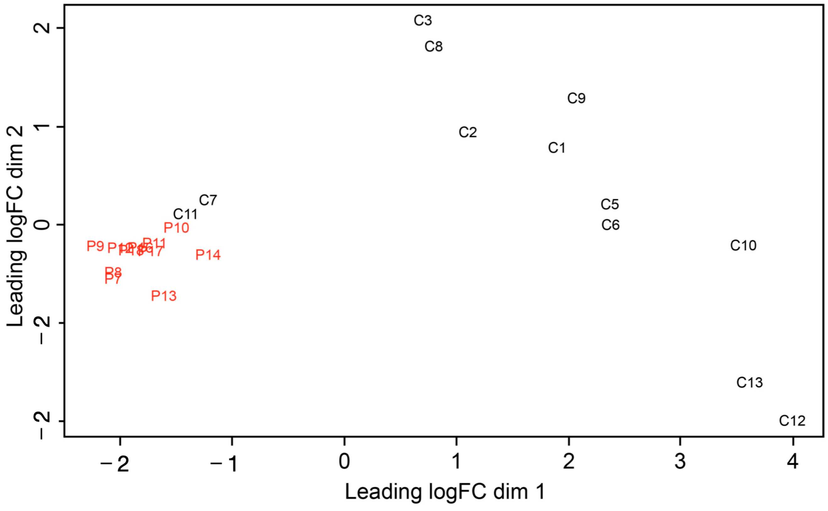
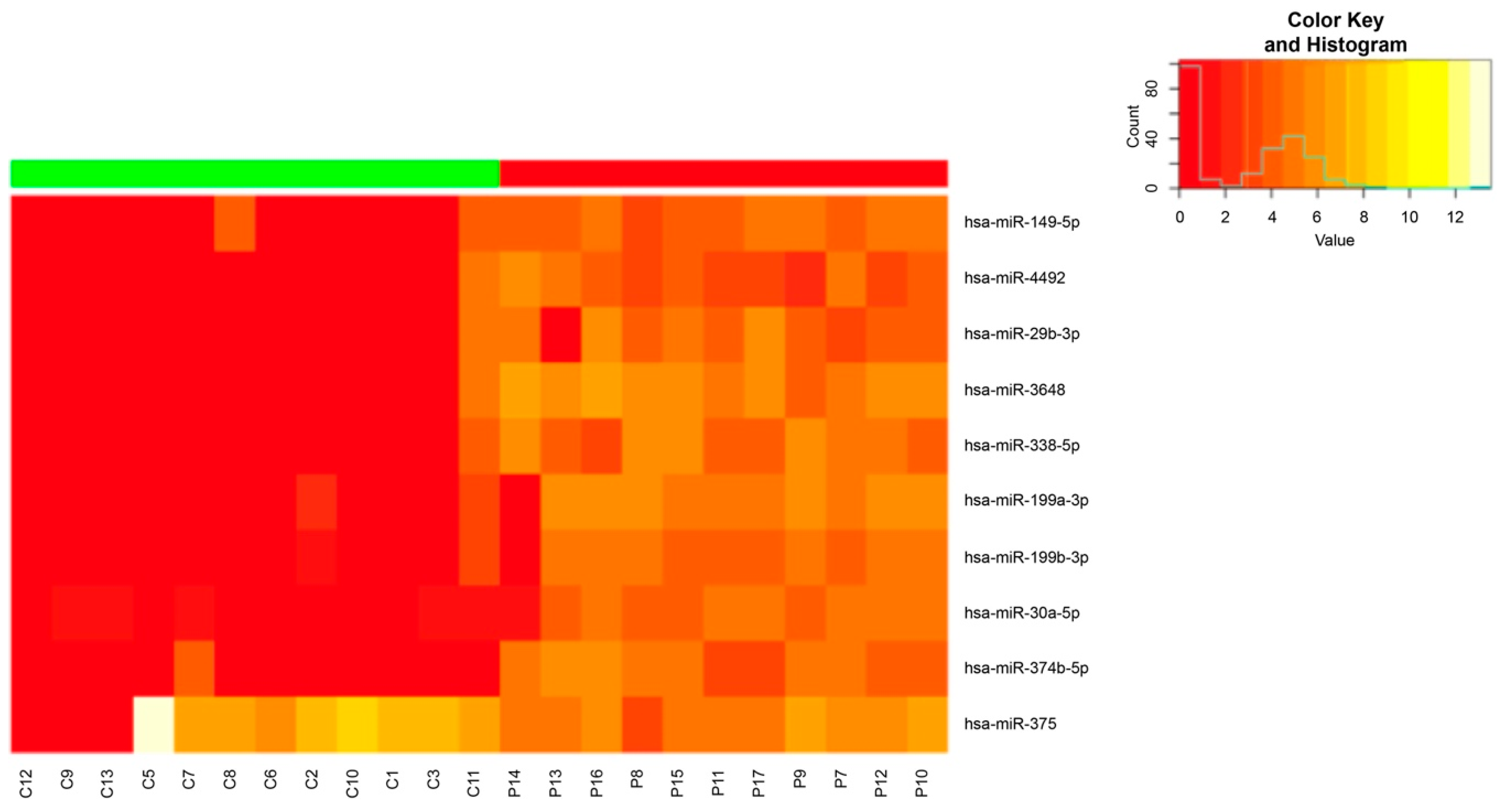
2.2. miRNA Expression Analysis in Patients with Advanced Chronic Periodontitis

The first exploratory analysis of the small RNA-seq data was performed using a multidimensional principal component analysis evaluating the miRNA expression levels in all the samples (Figure 1).
The miRNA expression profiles revealed two clearly differentiated groups. In the test group, miRNA expression presented a homogeneous profile that was distinctly grouped in the left region of the graph, while it was more heterogeneous in healthy subjects, yet clearly different from that of the patients with periodontitis. Notably, controls 7 and 11 were positioned closer to the test group.
A bioinformatics analysis identified 148 miRNAs showing significant differential expression between the two groups. These were selected for further analysis. The heatmap (Figure 2) revealed miRNAs whose expression clearly defined subjects in the test group. These 10 miRNAs defined a signature clearly that differentiated between CP patients and healthy subjects; therefore, they were selected as candidates for periodontitis biomarkers.
The HS and advanced CP patients had different miRNA expression levels in the GCF. The following 10 miRNAs defined the subjects with periodontitis: hsa-miR-199a-3p, hsa-miR-199b-3p, hsa-miR-338-5p, hsa-miR-374b-5p, hsa-miR-30a-5p, hsa-miR-375, hsa-miR-3648, hsa-miR-29b-3p, hsa-miR-4492, and hsa-miR-149-5p.

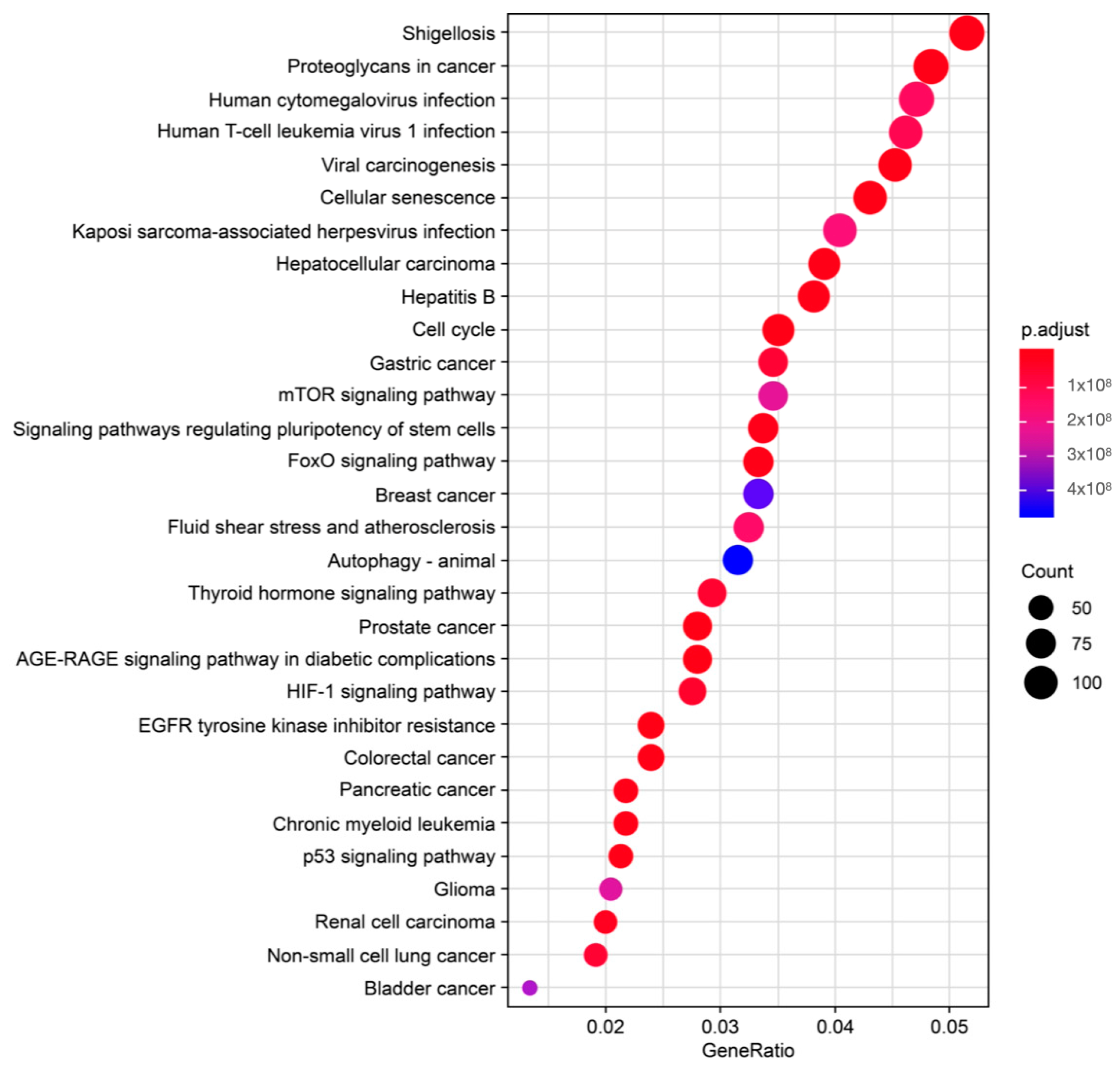
2.3. Selection of Reliable miRNAs to Define Periodontitis and Its Etiopathogenesis

The 10 miRNAs were analyzed using the DIANA-miRPath web tool, which allowed us to identify the target genes of each miRNA and analyze their involvement in biological KEGG pathways. These biological pathways were then manually searched in PubMed to pinpoint their direct and indirect relationships with periodontitis. Using this process, we were able to select up to 19 potential pathways that were closely related to periodontitis. The pathways were particularly linked to bone metabolism, inflammatory response, rupture of epithelial junctions, and bacterial invasion of epithelial cells. In addition, we performed an overrepresentation analysis (ORA) using the list of significant miRNAs (false discovery rate < 0.05), a relatively less strict analysis which did not take into account the differential expression analysis statistics. The ORA demonstrated that the miRNAs with differential expression were related to pathways such as proteoglycans in cancer, viral carcinogenesis, cellular senescence, cell cycle, and forkhead box class O signaling pathways. These are pathways with genes that have been proven to be involved in periodontitis (Figure 3).
Together with the power of miRNA expression, logistic regression, and the involvement in the KEGG biological pathways related to periodontal pathogenesis, a bioinformatics analysis identified miRNA signatures comprising miR-30a-5p, miR-199b-3p, miR-338-5p, and miR146a-5p as potential biomarkers of periodontitis. For miR-146a-5p, a tendency to overexpression was observed in the patients with periodontitis. Although the differences were not significant, we included this miRNA for analysis, since previous studies have highlighted its role in periodontitis [36,37].

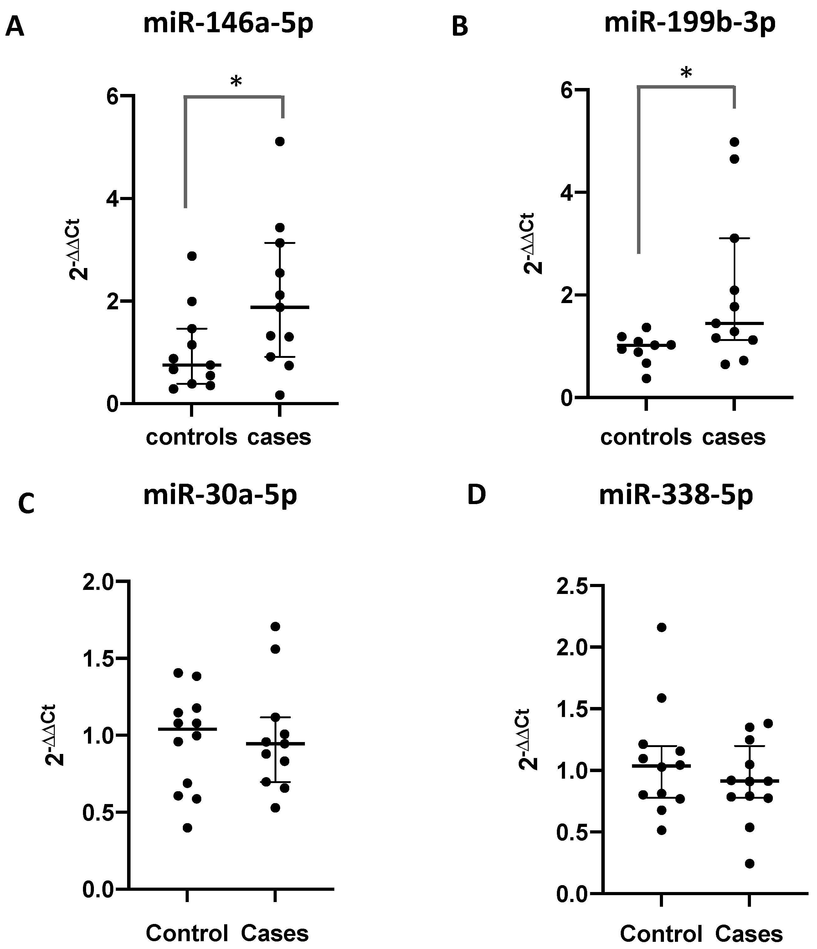
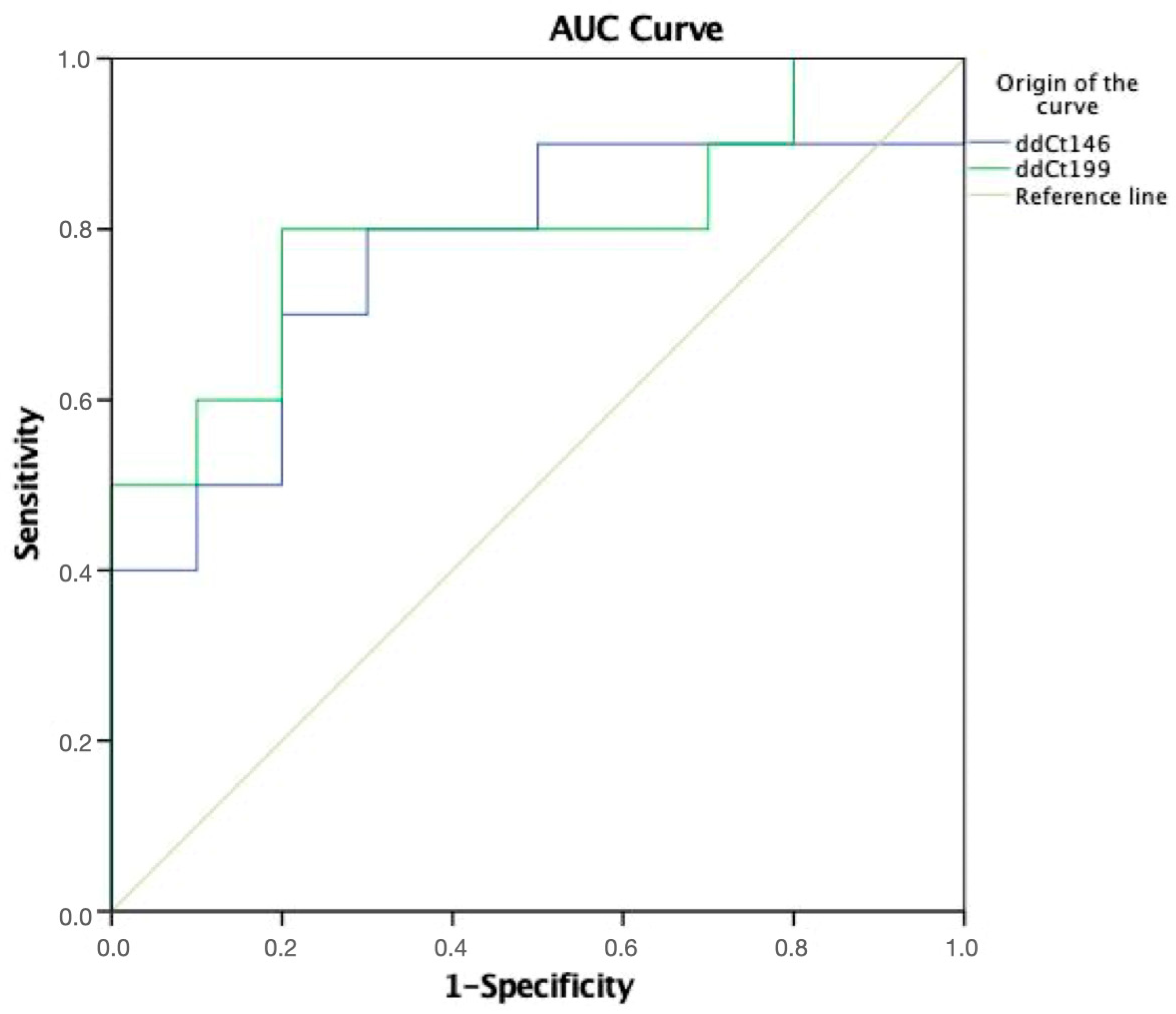
2.4. Validation of Differentially Expressed miRNAs via RT-qPCR

The miRNA signature proposed as a biomarker for periodontitis was validated via RT-PCR using CSF derived from different teeth obtained from the same subjects used in the small RNA-seq analysis. miR-146a-5p and miR-199b-3p were overexpressed in the GCF of the patients with periodontitis compared to their levels in the healthy subjects (p = 0.05 and p = 0.03, Mann–Whitney tests, respectively). Conversely, miR-30-5pa and miR-338-5p showed similar levels in both groups (Figure 4).
To evaluate the potential of the two validated miRNAs (miR-146a-5p and miR-199b-3) as diagnostic biomarkers, corresponding ROC curves were generated and analyzed (Figure 5). miR-199b was identified as the most powerful diagnostic biomarker of periodontitis. Improved AUC, sensitivity, and specificity values were obtained for the assay based on the use of miRNAs in GCF for the diagnosis of periodontitis. After repeating the experiment using different samples, miR146a-5p and miR-199b-3p were validated as reliable biomarkers to define advanced chronic periodontitis in GCF.
Figure 5. ROC curve analysis for miR-146a-5p and miR-199b-3p as diagnosis biomarkers of periodontitis. A detailed summary of the AUC, 95% CI lower and upper limit, sensitivity, and specificity of the two miRNAs are shown in the inserted Table 2. The AUCs for circulating miR-146a-5p and miR-199b-3p in the GCF were 0.74 and 0.78, respectively, indicating a high diagnostic value.
Ncrna 09 00073 g005
Table 2. Receiver operating characteristic curve analysis.
miRNAAUC95% CIp ValueCut-Off ValueSensitivitySpecificity
146a-5p0.740.53–0.960.051.2272.772.7
199b-3p0.780.57–0.990.091.1180.072.7
AUC, area under the curve; CI, confidence interval.

2.5. Analysis of the Correlation between GCF Volume and Expression Levels of the Validated miRNAs

A Spearman’s correlation analysis was performed between the miRNA expression levels and GCF volume, which has been previously proposed as a biomarker of periodontitis. A positive correlation was found between the GCF volume and the miR-199b and miR-146a expression levels (p < 0.004 and p < 0.027), in which the expression levels of the two miRNAs increased as the GCF volume increased (Table 3).

3. Discussion

Biomarkers represent a useful tool to help address the challenges of implementing preventive, predictive, and personalized medicine in all healthcare disciplines. In periodontal disease, the most important challenges are to diagnose the condition before it becomes clinically detectable and to avoid progression, improve patient management, and provide treatment. In this context, miRNAs have demonstrated an extraordinary performance as biomarkers for the early diagnosis and prognosis of several diseases [15]. To the best of our knowledge, our is the first study to analyze all the miRNAs present in the GCF of patients with chronic periodontitis, performed using small RNA-seq analysis. The cohort of subjects participating in this study was homogeneous in terms of demographic variables, and the clinical parameters clearly differentiated the two groups. The GCF volume increased and changed qualitatively with inflammation; however, although GCF reflects subclinical inflammation, it is not possible to determine the cellular origin of the markers studied [38].
In this study, we analyzed the differential expression profiles of miRNAs in GCF obtained from patients with chronic periodontitis using a strategy based on small RNA-seq. The miRNA expression profile clearly differentiated CP patients with chronic periodontitis from HS, providing a potential biomarker signature to identify patients with this pathology.
Since the sample size could not be calculated, we considered previous studies using a similar methodology for the sample size selection. In total, 23 subjects participated in our study, including 11 with advanced periodontitis and 12 healthy subjects. The power of the study was calculated via a post hoc analysis, showing a power of 76%. For future studies, 48 subjects are needed to reach 90% power. Nonetheless, our sample size can be considered sufficient, since it yielded significant differences. In our analysis, we found a signature of 10 miRNAs that could differentiate the patients from the healthy subjects. This miRNA signature was used to identify target genes which can alter the function of biological pathways related to periodontitis. Using the DIANA-miRPath web tool, we found that the selected miRNAs participated in processes related to bone metabolism, inflammatory response, rupture of epithelial junctions, and bacterial invasion of epithelial cells. The study of these molecular pathways, together with ORA, facilitated the selection of a series of miRNAs with a plausible role in periodontal pathogenesis, including miRNAs miR-30a-5p, miR-199b-3p, miR-338-5p, and miR-146a-5p. In addition, we performed a validation analysis of these miRNAs via RT-qPCR, finding that miR-146a-5p and miR-199b-3p were upregulated in the CP patients compared to the HS. Notably, an ROC curve analysis revealed miR-199b-3p to be the most powerful biomarker in periodontitis in the CP patient cohort. Moreover, among the miRNAs evaluated, miR-199b-3p and miR-146-5p showed a positive correlation with GCF volume (p < 0.004 and p < 0.027), confirming the potential use of both miRNAs as biomarkers of chronic periodontitis.
MiR-199b-3p and miR-146a-5p have been previously associated with inflammatory diseases, cardiovascular diseases, and cancer [39]. Both miRNAs target genes such as tumor necrosis factor alpha, prostaglandin, interleukin-1 beta, interleukin-6, bone morphogenetic protein 2, epidermal growth factor, and transforming growth factor beta [37,40,41]. Moreover, they are both involved in activating nuclear factor-kappa beta and altering osteoclastogenesis (RANK/RANKL/OPG axis). The activator of the nuclear factor-κB (RANK) receptor is a transmembrane protein that is expressed in both mature osteoclasts and their progenitors, and binding to its ligand (RANKL) determines osteoclast differentiation and activation. The levels of these markers correlate with periodontitis progression [37,41,42].
Of the few studies analyzing miRNAs in GCF, none have used high-throughput screening via small RNA-seq of all miRNAs. Saito et al. employed a ready-to-use PCR with 720 miRNAs, while Radovic et al. analyzed the expression levels of two miRNAs before and after periodontal treatment in patients with periodontitis and diabetes, reporting that the expression levels of miR-146a-5p and miR-155 decreased after treatment [36,43]. Some authors have analyzed the expression level of several miRNAs in GCF, but none have carried out a complete screening of all the miRNAs [44,45,46,47,48]. The results of this study agree with the results of Rovas et al., 2022 and Zhu and Zhong, 2022, reporting high expression level of miRNAs miR-30a and miR-146a in periodontitis [45,47].
Regarding the two healthy subjects with overexpression of the selected periodontitis-defining miRNAs, it is feasible that these miRNAs potentially indicate an increased risk or a subclinical state of periodontitis in subjects without clinical signs of disease. Further studies and patient follow-ups are needed to confirm this hypothesis.
The study of miRNAs in a non-invasive medium like GCF has the potential to improve the use of these biomarkers, and this study demonstrates the importance of research with these miRNAs in the field of periodontology. Nonetheless, our results should be interpreted in the context of certain limitations concerning the methodology and the source used to purify miRNAs, as no previous literature exists outlining optimized procedures for smallRNA-seq in microRNAs purified from GCF. Indeed, as far as we know, this is the first study using a smallRNA-seq approach to identify potential miRNAs in periodontitis-related GCF; a significant fact, given that epigenetic biomarkers could be useful in diagnosing stage I of periodontitis, as suggested by Tonetti et al. in their new classification of periodontal diseases [49]. Another limitation of our study was the selection process for the appropriate miRNA to be used as a reference to perform the qPCR experiments. Selecting appropriate reference genes to normalize the relative miRNA expression in biofluids is a current challenge in other biospecimens such as blood and saliva, and this was also a challenge in this study using GCF. An interesting future line of research would be to analyze the value of these epigenetic biomarkers for studying the bidirectional relationship of periodontal diseases with several systemic and non-communicable diseases.

4. Materials and Methods

This study was conducted in compliance with the revised Declaration of Helsinki and was approved by the Experimental Research Ethics Committee of the University of Valencia, Valencia, Spain (procedure number: H1477063568528). All individuals participating in the study were informed about the objective and methods of the study and provided written informed consent. At the time of this study, no previous reports allowed the calculation of the sample size, therefore studies with a similar methodology were consulted [43,50]. We selected a group of subjects with advanced chronic periodontitis (CP) with probing depths ≥ 6 mm and dental bone loss ≥ 50% and a group of healthy subjects (HS) without periodontal pockets, dental bone loss, or gingival inflammation. Both groups had an age range of 30–60 years, comprising individuals who smoked ≤10 cigarettes/d. The exclusion criteria included patients with systemic diseases, patients under treatment or medication that could affect bone metabolism or inflammation response, and patients taking antibiotics, smoking >10 cigarettes a day, or using antiseptic mouthwashes.

4.1. Clinical Records

A complete periodontal examination was performed in the periodontics clinic of the University of Valencia, which determined probing depths using a pressure-controlled probe, plaque index (plaque scoring according to Quigley and Hein, modified by Turesky et al., 1970) [51], gingival index (Ainamo and Bay, modified by Lindhe 1983) [52], clinical attachment level, bleeding on probing, furcation involvement, and mobility. In addition, complete periodontal X-rays were performed.

4.2. GCF Sample Collection and Small RNA Extraction and Quantification

For GCF sampling, single-rooted teeth with probing depths of ≥6 mm were selected. Three samples were taken from three different teeth in each subject. A single sample of one tooth was used for RNA sequencing, and a sample of a different tooth was used for the validation test. The remaining samples were stored at −80 °C.
Before the sample collection, partial isolation was performed using cotton wool. The supragingival plaque was removed, and the teeth were gently air-dried in an apico-coronal direction. Next, an absorbing paper strip (Periopaper®; Oraflow, New York, NY, USA) was placed on a Periotron® to define the zero value in the device. The same Periopaper® was carefully introduced into the gingival sulcus/periodontal pocket until resistance was felt, and it was then held in place for 30 s. Finally, the Periopaper® was reintroduced into the Periotron® to record the periotron units, which were later converted to microliter volume units of GCF. The Periopapers® were stored in 1.5 mL tubes at −80 °C for subsequent analysis in the laboratory [38]. In the control group, sample areas were selected in the same way as the test group, but the probing depth was always ≤3 mm and with no bleeding.
Next, we proceeded to purify the miRNAs from the crevicular fluid (CSF) samples, as previously described [50]. The Periopapers® were incubated in phosphate-buffered saline (pH 7) for 30 min at room temperature (22–26 °C) and centrifuged at 500 rpm (16,000× g) for 10 min. Subsequently, the total RNA was extracted from 200 µL of phosphate-buffered saline using an miRNeasy Serum/Plasma kit (Qiagen GmbH, Hilden, Germany) following the manufacturer’s instructions. Finally, the RNA (including miRNAs) was eluted from purification columns using 25 µL of RNAse-free water. The purified RNA (with miRNA) concentration was quantified using a NanoDrop ND 2000 UV-spectrophotometer (Thermo Fisher Scientific, Wilmington, DE, USA), and the sample quality was measured using a Small RNA Assay (NETFLEX® Small RNA-Seq kit v3 for Illumina platforms; Bioo Scientific® Corporation, Austin, TX, USA) and an Agilent 2100 Bioanalyzer (Agilent Technologies, Santa Clara, CA, USA).

4.3. Small RNA-Seq and Analysis

4.3.1. Library Preparation and Next-Generation Sequencing

Small RNA libraries were generated and indexed using NEXTFLEX® Small RNA-Seq kit v3 for Illumina Platforms (Bio Scientific® Corporation, Perkin Elmer, Inc., Waltham, MA, USA EEUU). In this modified protocol, the libraries were selected based on size (range, 90–170 bp) using a Blue Pippin instrument (Sage Science, Beverly, MA, USA). A positive RNA control was included (Human Brain Total RNA catalog #AM7962, ThermoFisher Scientific, Waltham, MA, USA). Single-end sequencing was performed using an Illumina NextSeq platform on High-Output 1 × 50 bp Run (NextSeq 500/550 High-Output v2 75 cycles kit, FC-404-2005) (Illumina, San Diego, CA, USA). Positive RNA from the brain is often used as control in smallRNA-seq analysis because it is known to contain a mixture of high-quality smallRNAs, including miRNAs, which can help ensure that the library construction, smallRNA-seq, and bioinformatic pipelines processes are properly executed to identify the miRNAs in biological samples.

4.3.2. Differential Expression Analysis between Advanced Chronic Periodontitis Patients and Healthy Subjects

The quality of the Illumina raw sequences was assessed using FastQC software V 0.11.9 (https://www.bioinformatics.babraham.ac.uk/projects/fastqc/, accessed on 3 March 2020). Based on the results obtained, the sequence reads were trimmed to remove sequencing adapters and low-quality bases using Cutadapt software V 2.9 (http://cutadapt.readthedocs.org/en/stable/, accessed on 19 March 2020). Data of sufficient quality were mapped against the human GRCh38 reference sequence from UCSC (University of California, Santa Cruz), obtained from Ensembl. Subsequently, an intersection was performed between the aligned position of reads and the miRNA coordinates taken from miRBase v21. The alignment and quantification steps were performed using the Subread and Rsubread packages [53,54].
miRNAs with low counts were filtered out prior to further analysis, after which the trimmed mean of M-values normalization method [55] was performed to eliminate composition biases between libraries. We also estimated the specific dispersions per gene with a negative binomial distribution [56,57]. A differential expression analysis was performed by comparing miRNA expression levels between CP patients and HS using scripts DESeq [58]. Additionally, the differential expression was re-analyzed, adjusting for sex using a quasi-likelihood F test [59]. The counts obtained from sequencing were transformed via a variance stabilization method using the R DESeq package [58]. The transformed data were used to fit a logistic regression model with Lasso penalty [60] using the 10 differentially expressed miRNAs obtained by adjusting for sex and evaluating the 23 study samples as observations.
The most important miRNAs in the model were selected by performing a cross-validation, choosing the miRNAs with coefficients different from 0 for the lambda value, with a minimum average cross-validation error. The data from the bioinformatics analysis were used to redraw a new heatmap using the 10 miRNAs finally selected to define advanced CP patients in the study.

4.4. Prediction of miRNA Targets and Overrepresentation Analysis (ORA)

To identify the most important pathways and genes regulated by differentially expressed miRNAs, we analyzed the overrepresented gene ontology (GO) terms and pathways using cluster Profiler. Ontology and pathway GO terms with an adjusted p-value < 0.05 were considered significantly overrepresented. We also used the DIANA-miRPath v3.0 functional analysis online suite to identify miRNAs controlling significant molecular pathways annotated in the Kyoto Encyclopedia of Genes and Genomes (KEGG) using the following default parameters: base v.7.0, a p value threshold of 0.001, and a microT threshold of 0.8. To reduce the number of false-positive miRNA targets, we applied a false discovery rate correction to the selected KEGG pathways. The algorithm used in this analysis was a one-tailed Fisher exact test [61]. We analyzed the pathways that have been previously associated with periodontal diseases. Finally, we chose the pathways and genes that were shared between GO and KEGG.

4.5. miRNA Validation Using Quantitative Reverse Transcription Polymerase Chain Reaction (RT-qPCR) in CSF Samples

RT-qPCR was performed to evaluate the eight miRNAs identified via the small RNA-seq analysis. A total of 23 GCF samples (11 from CP patients and 12 from HS) were analyzed. These 23 samples comprised different samples from a different tooth of each subject used in the small RNA sequencing analysis. RT-PCR was performed using the TaqMan® miRNA Reverse Transcription kit (Part No. 4366596; Applied Biosystems, Carlsbad, CA, USA), miRNA-specific stem-loop primers (Part No. 4366596; Applied Biosystems) (hsa-miR-338-5p (assay ID 002658, ThermoFisher Scientific), hsa-miR-199a-3p (assay ID 002304, ThermoFisher Scientific), hsa-miR-30a-5p (assay ID 000417, ThermoFisher Scientific), and hsa-miR-146a-5p (assay ID 000468; ThermoFisher Scientific)), and 100 ng of input cell-free RNA in a 20 µL RT reaction. A differential expression analysis showed that miR-223-3p had the most stable and homogeneous counts among the samples; therefore, this miRNA was used as an endogenous reference control (assay ID 002295, ThermoFisher Scientific). The miRNA expression levels were normalized to the level of has-miR-233-3p, which was used as a control.
The RT-PCR conditions were as follows: 16 °C for 30 min, followed by 42 °C for 30 min and a final inactivation step at 85 °C for 5 min. The real-time PCR reactions were performed in triplicate in scaled-down 10 µL reaction volumes using 5 µL of TaqMan® 2× Universal PCR Master Mix with No UNG (Applied Biosystems), 0.5 µL of TaqMan® Small RNA assay (20×) (Applied Biosystems), 3.5 µL of nuclease-free water, and 1 µL of RT product. The PCR conditions were as follows: 95 °C for 10 min, followed by 45 cycles at 95 °C for 15 s and 60 °C for 1 min. As above, hsa-miR-223-3p miRNA served as an endogenous control for normalization. The relative quantification of miRNAs was calculated using the 2(−ΔΔCT) method [62].

4.6. Statistical Analysis

The mean was taken as the best measure of tendency, and the standard deviation (SD) was taken as the measure of dispersion. In a descriptive analysis of the sample, the corresponding frequency tables were constructed for the qualitative variables, while descriptive statistics [mean, median, confidence interval (CI) for the mean, and standard deviation (SD)] were calculated for the quantitative variables. For comparison of means, normality tests were always applied with the objective of evaluating the appropriate comparison test (Student’s t test or Mann–Whitney U test). For comparisons of categorical variables such as sex and smoking habits, Chi-squared tests and Fisher’s exact tests were performed.
For validation analysis, the Mann–Whitney test was used to change the miRNA values to continuous values to allow between-group comparisons. For multiple comparisons, Kruskal–Wallis and Dunns post hoc tests were applied. The diagnostic capacity of each miRNA was validated using ROC (Receiver Operating Characteristic) curve analysis, calculating the area under the curve, the sensitivity and specificity, the positive predictive value (PPV), and the negative predictive value (NPV). The optimal cut-off point was determined by high values of sensitivity, specificity, and efficiency using the Youden index. Statistically significant differences were assumed when the p-value was less than 0.005(*). SPSS (IBM® SPSS® statistics version 24.0) was used for the calculations.

5. Conclusions

Notwithstanding the limitations of our study, our findings provide an miRNA signature comprising miR-199b-3p and miR-146a-5p that defines periodontitis, thus warranting future studies on this biomarker signature to evaluate its performance for identifying patients with a subclinical phenotype. Furthermore, it would be useful to identify biomarkers to distinguish different patient subsets and prevent the appearance of sequelae in periodontitis. These multiple applications of miRNA-based biomarkers in GCF could improve our knowledge of the relationship between periodontitis and systemic diseases and may help to implement precision periodontics.

Supplementary Materials

The following supporting information can be downloaded at: https://www.mdpi.com/article/10.3390/ncrna9060073/s1, Table S1: Descriptive analysis of the sample: tobacco and age; Table S2: Descriptive analysis of the sample: sex; Table S3: Descriptive analysis of the sample: cigarrets/day; Table S4: Descriptive analysis of the sample: Age; Table S5. Independent sample test: Age.

Author Contributions

All authors participated in the design and development of the study, performed experiments in the laboratory, performed statistical treatment of the data, and read and approved the final version of the manuscript. All authors have read and agreed to the published version of the manuscript.

Funding

The study did not receive any funding.

Institutional Review Board Statement

The study was conducted in accordance with the Declaration of Helsinki, and approved by the Ethics Committee of University of Valencia (H1477064568528, 7 November 2016)) for studies involving humans.

Informed Consent Statement

Informed consent was obtained from all subjects involved in the study.

Data Availability Statement

The data that support the findings of this study are available from the corresponding author upon reasonable request.

Acknowledgments

The authors wish to thank Epidisease S. L. (spin-off from the CIBER-ISCIII, INCLIVA Biomedical Research Center and University of Valencia) for its collaboration in the small RNA-seq data analysis and processing.

Conflicts of Interest

The authors have no financial or personal relationships with other individuals or organizations to declare.

References

  1. Pihlstrom, B.L.; Michalowicz, B.S.; Johnson, N.W. Periodontal diseases. Lancet 2005, 366, 1809–1820. [Google Scholar] [CrossRef] [PubMed]
  2. Hajishengallis, G. Periodontitis: From microbial immune subversion to systemic inflammation. Nat. Rev. Immunol. 2016, 15, 30–44. [Google Scholar] [CrossRef]
  3. Meyle, J.; Chapple, I. Molecular aspects of the pathogenesis of periodontitis. Periodontology 2000 2015, 69, 7–17. [Google Scholar] [CrossRef] [PubMed]
  4. Greenstein, G.; Hart, T.C. A Critical Assessment of Interleukin-1 (IL-1) Genotyping When Used in a Genetic Susceptibility Test for Severe Chronic Periodontitis. J. Periodontol. 2002, 73, 231–247. [Google Scholar] [CrossRef]
  5. Santonocito, S.; Polizzi, A.; Palazzo, G.; Indelicato, F.; Isola, G. Dietary factors affecting the prevalence and impact of periodontal disease. Clin. Cosm. Investig. Dent. 2021, 13, 283–292. [Google Scholar] [CrossRef] [PubMed]
  6. Santonocito, S.; Giudice, A.; Polizzi, A.; Troiano, G.; Merlo, E.M.; Sclafani, R.; Grosso, G.; Isola, G. A Cross-Talk between Diet and the Oral Microbiome: Balance of Nutrition on Inflammation and Immune System’s Response during Periodontitis. Nutrients 2022, 14, 2426. [Google Scholar] [CrossRef]
  7. Chapple, I.L.C.; Genco, R. Diabetes and periodontal diseases: Consensus report of the Joint EFP/AAP Workshop on Periodontitis and Systemic Diseases. J. Periodontol. 2013, 84, S106–S112. [Google Scholar] [CrossRef]
  8. Linden, G.J.; Lyons, A.; Scannapieco, F.A.; Sharma, P.; Dietrich, T.; Ferro, C.J.; Cockwell, P.; Chapple, I.L.C.C.; Chapple, I.L.C.C.; Genco, R.; et al. Periodontal systemic associations: Review of the evidence. J. Clin. Periodontol. 2013, 40, 8–19. [Google Scholar] [CrossRef]
  9. Sharma, P.; Dietrich, T.; Ferro, C.J.; Cockwell, P.; Chapple, I.L.C. Association between periodontitis and mortality in stages 3–5 chronic kidney disease: NHANES III and linked mortality study. J. Clin. Periodontol. 2016, 43, 104–113. [Google Scholar] [CrossRef]
  10. Tonetti, M.S.; Van Dyke, T.E. Periodontitis and atherosclerotic cardiovascular disease: Consensus report of the Joint EFP/AAPWorkshop on Periodontitis and Systemic Diseases. J. Periodontol. 2013, 84, S24–S29. [Google Scholar] [CrossRef]
  11. Stoecklin-Wasmer, C.; Guarnieri, P.; Celenti, R.; Demmer, R.T.; Kebschull, M.; Papapanou, P.N. MicroRNAs and Their Target Genes in Gingival Tissues. J. Dent. Res. 2012, 91, 934–940. [Google Scholar] [CrossRef]
  12. Thomson, D.W.; Bracken, C.P.; Goodall, G.J. Experimental strategies for microRNA target identification. Nucleic Acids Res. 2011, 39, 6845–6853. [Google Scholar] [CrossRef] [PubMed]
  13. Baek, D.; Villén, J.; Shin, C.; Camargo, F.D.; Gygi, S.P.; Bartel, D.P. The impact of microRNAs on protein output. Nature 2008, 455, 64–71. [Google Scholar] [CrossRef] [PubMed]
  14. Xie, Y.; Shu, R.; Jiang, S.; Liu, D.; Zhang, X. Comparison of microRNA profiles of human periodontal diseased and healthy gingival tissues. Int. J. Oral. Sci. 2011, 3, 125–134. [Google Scholar] [CrossRef]
  15. García-Giménez, J.L.; Seco-Cervera, M.; Tollefsbol, T.O.; Romá-Mateo, C.; Peiró-Chova, L.; Lapunzina, P.; Pallardó, F.V. Epigenetic biomarkers: Current strategies and future challenges for their use in the clinical laboratory. Crit. Rev. Clin. Lab. Sci. 2017, 54, 529–550. [Google Scholar] [CrossRef] [PubMed]
  16. O’Brien, J.; Hayder, H.; Zayed, Y.; Peng, C. Overview of microRNA biogenesis, mechanisms of actions, and circulation. Front. Endocrinol. 2018, 9, 402. [Google Scholar] [CrossRef]
  17. Stenvinkel, P.; Karimi, M.; Johansson, S.; Axelsson, J.; Suliman, M.; Lindholm, B.; Heimb?rger, O.; Barany, P.; Alvestrand, A.; Nordfors, L.; et al. Impact of inflammation on epigenetic DNA methylation ? a novel risk factor for cardiovascular disease? J. Intern. Med. 2007, 261, 488–499. [Google Scholar] [CrossRef]
  18. Ogata, Y.; Matsui, S.; Kato, A.; Zhou, L.; Nakayama, Y.; Takai, H. MicroRNA expression in inflamed and noninflamed gingival tissues from Japanese patients. J. Oral Sci. 2014, 56, 253–260. [Google Scholar] [CrossRef] [PubMed]
  19. Volinia, S.; Calin, G.A.; Liu, C.-G.; Ambs, S.; Cimmino, A.; Petrocca, F.; Visone, R.; Iorio, M.; Roldo, C.; Ferracin, M.; et al. A microRNA expression signature of human solid tumors defines cancer gene targets. Proc. Natl. Acad. Sci. USA 2006, 103, 2257–2261. [Google Scholar] [CrossRef]
  20. De Oliveira, N.F.P.; Andia, D.C.; Planello, A.C.; Pasetto, S.; Marques, M.R.; Nociti, F.H.; Line, S.R.P.; De Souza, A.P. TLR2 and TLR4 gene promoter methylation status during chronic periodontitis. J. Clin. Periodontol. 2011, 38, 975–983. [Google Scholar] [CrossRef]
  21. Loo, W.T.Y.; Jin, L.; Cheung, M.N.B.; Wang, M.; Chow, L.W.C. Epigenetic change in E-cadherin and COX-2 to predict chronic periodontitis. J. Transl. Med. 2010, 8, 110. [Google Scholar] [CrossRef]
  22. Schaefer, A.S.; Richter, G.M.; Nothnagel, M.; Laine, M.L.; Rühling, A.; Schäfer, C.; Cordes, N.; Noack, B.; Folwaczny, M.; Glas, J.; et al. A 3′UTR transition within DEFB1 is associated with chronic and aggressive periodontitis. Genes Immun. 2010, 11, 45–54. [Google Scholar] [CrossRef] [PubMed]
  23. Wang, Y.J.; He, L.; Yuan, M.; Tsang, W.W.N.; Hao, L.; Wang, M.; Chow, L.W.C.; Cheung, M.N.B.; Liu, Q.; Ng, E.L.Y.; et al. Epigenetic changes of TIMP-3, GSTP-1 and 14-3-3 sigma genes as indication of status of chronic inflammation and cancer. Int. J. Biol. Markers 2014, 29, e208–e214. [Google Scholar] [CrossRef]
  24. Zhang, S.; Barros, S.P.; Moretti, A.J.; Yu, N.; Zhou, J.; Preisser, J.S.; Niculescu, M.D.; Offenbacher, S. Epigenetic regulation of TNFA expression in periodontal disease. J. Periodontol. 2013, 84, 1606–1616. [Google Scholar] [CrossRef]
  25. Almiñana-Pastor, P.J.; Boronat-Catalá, M.; Micó-Martinez, P.; Bellot-Arcís, C.; Lopez-Roldan, A.; Alpiste-Illueca, F.M. Epigenetics and periodontics: A systematic review. Med. Oral Patol. Oral Cirugía Bucal 2019, 24, e659. [Google Scholar] [CrossRef] [PubMed]
  26. de Camargo Pereira, G.; Guimarães, G.N.; Planello, A.C.; Santamaria, M.P.; de Souza, A.P.; Line, S.R.; Marques, M.R. Porphyromonas gingivalis LPS stimulation downregulates DNMT1, DNMT3a, and JMJD3 gene expression levels in human HaCaT keratinocytes. Clin. Oral Investig. 2013, 17, 1279–1285. [Google Scholar] [CrossRef]
  27. Li, H.; Deng, Y.; Sun, K.; Yang, H.; Liu, J.; Wang, M.; Zhang, Z.; Lin, J. Structural basis of kindlin-mediated integrin recognition and activation. Proc. Natl. Acad. Sci. USA 2017, 114, 9349–9354. [Google Scholar] [CrossRef] [PubMed]
  28. Keller, A.; Leidinger, P.; Bauer, A.; ElSharawy, A.; Haas, J.; Backes, C.; Wendschlag, A.; Giese, N.; Tjaden, C.; Ott, K.; et al. Toward the blood-borne miRNome of human diseases. Nat. Methods 2011, 8, 841–843. [Google Scholar] [CrossRef]
  29. Xie, Y.; Shu, R.; Jiang, S.; Song, Z.; Guo, Q.; Dong, J.; Lin, Z. miRNA-146 negatively regulates the production of pro-inflammatory cytokines via NF-κB signalling in human gingival fibroblasts. J. Inflamm. 2014, 11, 38. [Google Scholar] [CrossRef] [PubMed]
  30. De Souza, A.P.; Planello, A.C.; Marques, M.R.; De Carvalho, D.D.; Line, S.R.P. High-throughput DNA analysis shows the importance of methylation in the control of immune inflammatory gene transcription in chronic periodontitis. Clin. Epigenetics 2014, 6, 15. [Google Scholar] [CrossRef] [PubMed]
  31. Uitto, V.-J. Gingival crevice fluid—An introduction. Periodontology 2000 2003, 31, 9–11. [Google Scholar] [CrossRef] [PubMed]
  32. Preshaw, P.M.; Alba, A.L.; Herrera, D.; Jepsen, S.; Konstantinidis, A.; Makrilakis, K.; Taylor, R. Periodontitis and diabetes: A two-way relationship. Diabetologia 2012, 55, 21–31. [Google Scholar] [CrossRef]
  33. Păunică, I.; Giurgiu, M.; Dumitriu, A.S.; Păunică, S.; Pantea Stoian, A.M.; Martu, M.A.; Serafinceanu, C. The Bidirectional Relationship between Periodontal Disease and Diabetes Mellitus—A Review. Diagnostics 2023, 13, 681. [Google Scholar] [CrossRef]
  34. Xu, S.; Song, M.; Xiong, Y.; Liu, X.; He, Y.; Qin, Z. The association between periodontal disease and the risk of myocardial infarction: A pooled analysis of observational studies. BMC Cardiovasc. Disord. 2017, 17, 50. [Google Scholar] [CrossRef]
  35. Blaizot, A.; Vergnes, J.N.; Nuwwareh, S.; Amar, J.; Sixou, M. Periodontal diseases and cardiovascular events:meta-analysis of observational studies. Int. Dent. J. 2008, 58, 342–348. [Google Scholar] [CrossRef]
  36. Radović, N.; Nikolić Jakoba, N.; Petrović, N.; Milosavljević, A.; Brković, B.; Roganović, J. MicroRNA-146a and microRNA-155 as novel crevicular fluid biomarkers for periodontitis in non-diabetic and type 2 diabetic patients. J. Clin. Periodontol. 2018, 45, 663–671. [Google Scholar] [CrossRef] [PubMed]
  37. Motedayyen, H.; Ghotloo, S.; Saffari, M.; Sattari, M.; Amid, R. Evaluation of MicroRNA-146a and Its Targets in Gingival Tissues of Patients With Chronic Periodontitis. J. Periodontol. 2015, 86, 1380–1385. [Google Scholar] [CrossRef]
  38. Griffiths, G.S. Formation, collection and significance of gingival crevice fluid. Periodontology 2000 2003, 31, 32–42. [Google Scholar] [CrossRef]
  39. Dolz, S.; Górriz, D.; Tembl, J.I.; Sánchez, D.; Fortea, G.; Parkhutik, V.; Lago, A. Circulating MicroRNAs as novel biomarkers of stenosis progression in asymptomatic carotid stenosis. Stroke 2017, 48, 10–16. [Google Scholar] [CrossRef]
  40. Zhu, L.; Xu, H.; Lv, W.; He, Z.; Ye, P.; Wang, Y.; Hu, J. miR-199b-5p Regulates Immune-Mediated Allograft Rejection after Lung Transplantation Through the GSK3β and NF-κB Pathways. Inflammation 2018, 41, 1524–1535. [Google Scholar] [CrossRef]
  41. Kadkhodazadeh, M.; Jafari, A.R.; Amid, R.; Ebadian, A.R.; Alipour, M.M.; Mollaverdi, F.; Lafzi, A. MiR146a and MiR499 gene polymorphisms in Iranian periodontitis and peri-implantitis patients. J. Long Term Eff. Med. Implant. 2013, 23, 9–16. [Google Scholar] [CrossRef] [PubMed]
  42. Roldán, A.L.; Giménez, J.L.G.; Illueca, F.A. Impact of periodontal treatment on the RANKL/OPG ratio in crevicular fluid. PLoS ONE 2020, 15, e0227757. [Google Scholar] [CrossRef]
  43. Saito, A.; Horie, M.; Ejiri, K.; Aoki, A.; Katagiri, S.; Maekawa, S.; Suzuki, S.; Kong, S.; Yamauchi, T.; Yamaguchi, Y.; et al. MicroRNA profiling in gingival crevicular fluid of periodontitis—A pilot study. FEBS Open Bio 2017, 7, 981–994. [Google Scholar] [CrossRef] [PubMed]
  44. Rovas, A.; Puriene, A.; Snipaitiene, K.; Punceviciene, E.; Buragaite-Staponkiene, B.; Matuleviciute, R.; Butrimiene, I.; Jarmalaite, S. Analysis of periodontitis-associated miRNAs in gingival tissue, gingival crevicular fluid, saliva and blood plasma. Arch. Oral Biol. 2021, 126, 105125. [Google Scholar] [CrossRef]
  45. Rovas, A.; Puriene, A.; Snipaitiene, K.; Punceviciene, E.; Buragaite-Staponkiene, B.; Matuleviciute, R.; Butrimiene, I.; Jarmalaite, S. Gingival crevicular fluid microRNA associations with periodontitis. J. Oral Sci. 2022, 64, 11–16. [Google Scholar] [CrossRef] [PubMed]
  46. Costantini, E.; Sinjari, B.; Di Giovanni, P.; Aielli, L.; Caputi, S.; Muraro, R.; Murmura, G.; Reale, M. TNFα, IL-6, miR-103a-3p, miR-423-5p, miR-23a-3p, miR-15a-5p and miR-223-3p in the crevicular fluid of periodontopathic patients correlate with each other and at different stages of the disease. Sci. Rep. 2023, 13, 126. [Google Scholar] [CrossRef]
  47. Zhu, J.; Zhong, Z. The expression and clinical significance of miR-30b-3p and miR-125b-1-3p in patients with periodontitis. BMC Oral Health 2022, 22, 325. [Google Scholar] [CrossRef] [PubMed]
  48. Isola, G.; Santonocito, S.; Distefano, A.; Polizzi, A.; Vaccaro, M.; Raciti, G.; Alibrandi, A.; Li Volti, G. Impact of periodontitis on gingival crevicular fluid miRNAs profiles associated with cardiovascular disease risk. J. Periodontal Res. 2023, 58, 165–174. [Google Scholar] [CrossRef] [PubMed]
  49. Tonetti, M.S.; Greenwell, H.; Kornman, K.S. Staging and grading of periodontitis: Framework and proposal of a new classification and case definition. J. Clin. Periodontol. 2018, 45, S149–S161. [Google Scholar] [CrossRef] [PubMed]
  50. Micó-Martínez, P.; García-Giménez, J.L.; Seco-Cervera, M.; López-Roldán, A.; Almiñana-Pastor, P.J.; Alpiste-Illueca, F.; Pallardó, F.V. MiR-1226 detection in GCF as potential biomarker of chronic periodontitis: A pilot study. Med. Oral Patol. Oral Cir. Bucal 2018, 23, e308–e314. [Google Scholar] [CrossRef]
  51. Turesky, S.; Gilmore, N.D.; Glickman, I. Reduced plaque formation by the chloromethyl analogue of victamine C. J. Periodontol. 1970, 41, 41–43. [Google Scholar] [CrossRef] [PubMed]
  52. Ainamo, J.; Bay, I. Problems and proposals for recording gingivitis and plaque. Int. Dent. J. 1975, 25, 229–235. [Google Scholar] [PubMed]
  53. Liao, Y.; Smyth, G.K.; Shi, W. The Subread aligner: Fast, accurate and scalable read mapping by seed-and-vote. Nucleic Acids Res. 2013, 41, e108. [Google Scholar] [CrossRef] [PubMed]
  54. Liao, Y.; Smyth, G.K.; Shi, W. FeatureCounts: An efficient general purpose program for assigning sequence reads to genomic features. Bioinformatics 2014, 30, 923–930. [Google Scholar] [CrossRef] [PubMed]
  55. Robinson, M.D.; Oshlack, A. A scaling normalization method for differential expresion analysis of RNA-seq data. Genome Biol. 2010, 11, R25. [Google Scholar] [CrossRef]
  56. Robinson, M.D.; Smyth, G.K. Small-sample estimation of negative binomial dispersion, with applications to SAGE data. Biostatistics 2008, 9, 321–332. [Google Scholar] [CrossRef]
  57. Robinson, M.D.; Smyth, G.K. Moderated statistical tests for assessing differences in tag abundance. Bioinformatics 2007, 23, 2881–2887. [Google Scholar] [CrossRef]
  58. Anders, S.; Huber, W. Differential expression analysis for sequence count data. Genome Biol. 2010, 11, R106. [Google Scholar] [CrossRef]
  59. Lun, A.T.L.; Mccarthy, D.J.; Marioni, J.C. A step-by-step workflow for low-level analysis of single-cell RNA-seq data with Bioconductor. F1000Research 2016, 5, 2122. [Google Scholar] [CrossRef]
  60. Friedman, J.; Hastie, T.; Tibshirani, R. Regularization paths for generalized linear models via coordinate descent. J. Stat. Softw. 2010, 33, 1–22. [Google Scholar] [CrossRef]
  61. Vlachos, I.S.; Zagganas, K.; Paraskevopoulou, M.D.; Georgakilas, G.; Karagkouni, D.; Vergoulis, T.; Dalamagas, T.; Hatzigeorgiou, A.G. DIANA-miRPath v3.0: Deciphering microRNA function with experimental support. Nucleic Acids Res. 2015, 43, W460–W466. [Google Scholar] [CrossRef] [PubMed]
  62. Livak, K.J.; Schmittgen, T.D. Analysis of relative gene expression data using real-time quantitative PCR and the 2−ΔΔCT method. Methods 2001, 25, 402–408. [Google Scholar] [CrossRef] [PubMed]
Figure 1. Principal component analysis of the samples. The plot shows the microRNA expression level trends between the patients and controls.
Ncrna 09 00073 g001
Figure 2. Heatmap representing microRNA expression levels. The red shades indicate low expression and predominate the control group, and the orange shades indicate greater expression of miRNAs in the test group than the control group.
Ncrna 09 00073 g002
Figure 3. Overrepresentation analysis. The plot shows the importance of the pathways in genes with a demonstrated involvement in periodontitis.
Ncrna 09 00073 g003
Figure 4. Relative expression levels of miRNAs found in the gingival crevicular fluid of patients with chronic periodontitis compared with those in healthy control participants. Box plot showing the relative expression levels of (A) miR-30a-5p, (B) miR-199a-3p, (C) miR-338-5p, and (D) miR-146a-5p in the healthy participants (controls, n = 12) and CP patients (cases, n = 11). The miRNA expression levels were normalized to those of miR-223-3p. miR-146a-5p and miR-199b-3p were overexpressed in the GCF of the periodontitis patients compared with the levels in the healthy subjects (p = 0.05 and p = 0.03, Mann–Whitney tests, respectively). The lines within the dispersion of points indicate the median. The bottom and top edges of the graph indicate the interquartile range. The symbol * indicates significant differences.
Ncrna 09 00073 g004
Table 1. Variables evaluated in the patient and control groups.
VariableHealthy SubjectsCP Patientsp Value
CAL2.92 (CI 2.66–3.17) (SD ± 0.40)5.06 (CI 4.60–5.52) (SD ± 0.68)p < 0.0001
Mean PD2.29 (CI 2.14–2.44) (SD ± 0.23)4.04 (CI 3.76–4.31) (SD ± 0.41)p < 0.0001
Mean PI2.03 (CI 1.70–2.36) (SD ± 0.52)3.32 (CI 2.85–3.61) (SD ± 0.56)p < 0.0001
BOP percentage13.90% (CI 5.09–22.72) (SD ± 13.87)80.19% (CI 67.5–92.8) (SD ± 18.83)p < 0.0001
CAL, clinical attachment level; PD, probing depth; PI, plaque index; BOP, bleeding on probing; CI, confidence interval; CP, chronic periodontitis; SD, standard deviation. t tests were used to the CAL, PD, PI, and BOP.
Table 3. Analysis of the correlation between the GCF volume and the expression levels of the validated miRNAs. Rho Sperman’s test was used.
GCF
Volume
Tooth 1
GCF
Volume
Tooth 2
GCF
Volume
Tooth 3
Rho Spearman2-ΔΔCt
miR-146a-5p
Correlation coefficient0.483 *0.514 *0.561 **
Sig. (bilateral)0.0270.0170.008
n212121
2-ΔΔCt
miR-199b-3p
Correlation coefficient0.615 **0.686 **0.717 **
Sig. (bilateral)0.0040.0010.000
n202020
2-ΔΔCt
miR-30a-5p
Correlation coefficient0.1140.1160.166
Sig. (bilateral)0.6240.6160.471
n212121
2-ΔΔCt
miR-338-5p
Correlation coefficient−0.0010.024−0.072
Sig. (bilateral)0.9960.9140.744
n232323
* and ** indicates significant differences. *: Significant correlation at level 0.05 (bilateral). **: Significant correlation at level 0.01 (bilateral).
Disclaimer/Publisher’s Note: The statements, opinions and data contained in all publications are solely those of the individual author(s) and contributor(s) and not of MDPI and/or the editor(s). MDPI and/or the editor(s) disclaim responsibility for any injury to people or property resulting from any ideas, methods, instructions or products referred to in the content.

