LEDs Combined with CHO Sources and CCC Priming PLB Regeneration of Phalaenopsis


1
The United Graduate School of Agricultural Sciences, Ehime University, Ehime 790-8577, Japan
2
Faculty of Agriculture, Kochi University, Kochi 783-8502, Japan
*
Author to whom correspondence should be addressed.
Horticulturae 2019, 5(2), 34; https://doi.org/10.3390/horticulturae5020034
Submission received: 19 February 2019
/
Revised: 15 April 2019
/
Accepted: 17 April 2019
/
Published: 6 May 2019
(This article belongs to the Special Issue Innovation in Propagation of Fruit, Vegetable and Ornamental Plants)
Abstract
:Throughout this study, the objective was to determine the most effective carbohydrate (CHO) sources under different light-emitting diodes (LEDs), and the impact of chlorocholine chloride (CCC), for the in vitro regeneration of the protocom-like bodies (PLBs) in Phalaenopsis ‘Fmk02010’. We applied 15 LEDs combined with three CHO sources and five CCC concentrations in the study. Organogenesis of PLBs was very poor in maltose both for the number of PLBs and their fresh weight (FW) compared to media containing sucrose and trehalose. Sucrose was the best CHO source under the red-white (RW) LED for the in vitro organogenesis of PLBs (PLBs: 54.13; FW: 0.109 g), while trehalose was best under the blue-white (BW) LED (PLBs: 36.33, FW: 0.129 g). The red-blue-white (RBW)-trehalose combination generated a suitable number of PLBs (35.13) with the highest FW (0.167 g). CCC at 0.01, 0.1, and 1 mgL−1CCC had no effect on PLB formation or FW, but 10 mg L−1 reduced both. RW-sucrose, BW-trehalose, and RBW-trehalose were the best combinations for PLB organogenesis. The addition of low concentrations of CCC in the plant culture medium are unnecessary.
Keywords:
protocorm-like bodies; light-emitting diode; trehalose; maltose; CCC; correlation; growth retardants1. Introduction
Phalaenopsis is the most important and valuable commercial orchid in the Orchidaceae family. It is widely accepted both as cut and pot flowers. Unlike most flowering plants, orchids have a very unique reproductive system. Propagation of Phalaenopsis, either vegetatively or by seed, is quite difficult. Tissue culture is the common method due to its successful and rapid propagation. PLB regeneration is the best and most efficient technique for orchid micropropagation [1], because it has a rapid proliferation capacity for producing a large number of protocorm-like bodies PLBs within a short period [2]. They can be induced directly from explants, such as shoot tips [3], flower stalk buds [4], root tips [5], and leaf segments [6]. The indirect regeneration of PLBs can be done by embryogenic callus culture using solid [7] or liquid [8] suspension cultures. Proper media compositions with optimum culture conditions are among the significant factors for fast and high quality plantlet regeneration through PLBs [9,10]. PLBs are the sole form of somatic embryo that imitates the zygotic embryo in natural seed, but unlike the zygotic embryo, it can grow continuously without any dormancy [11].
Media ingredients are the key factor for successful PLB regeneration in vitro. Plant tissue culture media generally have mineral salts, vitamins, growth regulators, and water [12]; another important component is the carbon source to supply energy [13]. There are many carbon sources like sucrose, fructose, glucose, trehalose, maltose, and sorbitol [8,14] used for plant tissue culture that might be in simple or complex forms [15]. It is well known that plants are sensitive to light. Light also affects PLB regeneration through photosynthetic and phototropic responses and may depend on light quality and photoperiod [16]. Fluorescent lights are commonly used. LED lights are currently used for in vitro cultivation. Power consumption of fluorescent lights is greater, and they produce a wide range of wavelengths (350–750 nm) that are unnecessary for plant development. Monochromatic LEDs emit light at specific wavelengths. LEDs are used commercially in plant tissue culture due to their numerous advantages compared to conventional light systems; such as wavelength specificity, durability, small size, long operating time, relatively cool emitting surface, and the ability to control spectral composition [17,18,19]. Concerning economic viability, the use of LEDs is increasing rapidly in agriculture due to their huge capacity to save electrical energy. LEDs are more efficient in in vitro culture than white fluorescent light. It is stipulated that they have the specific wavelengths that fits plants exact need for morphogenic responses [20]. The wavelength a plant needs varies according to the species.
LEDs are a unique type of semiconductor diode that have several technical benefits over usual light sources for photosynthesis [21]. LEDs allow wavelengths to be matched to the plant photoreceptors to influence plant morphology and metabolic composition [22,23,24]. Plant pigments absorb red wavelengths (600–700 nm) efficiently, with the most efficient being660 nm, which is close to the chlorophyll absorption peak, whereas the blue region includes the visible spectrum (400–500 nm) [25]. It has also been reported that red light has a significant role in starch accumulation through photosynthesis [26] and blue light in chloroplast development, chlorophyll formation, and stomatal opening [27]. Red and blue light are the best to drive photosynthetic metabolism. Green wavelength effects are opposite those of red and blue wavelengths [28].
A number of in vitro studies have reported vigorous plant growth under LEDs. LED lights have been previously used for PLB organogenesis in Cymbidium finlaysonianum [29], Dendrobiumkingianum [30,31], hybrid Cymbidium [32,33], and plantlet regeneration in gerbera [34]. A series of studies have already been conducted to improve the tissue culture of Phalaenopsis using a number of factors. We previously used growth regulators and elicitors for the PLB regeneration of Phalaenopsis [35,36]. The effects of light spectral quality on the photosynthetic ability varied by plant species [37]. Many plant species do not respond well under a sole LED color, and this limitation can be overcome by combining different colors. On the other hand, many researchers have reported the long-term effect of growth retardants on in vitro growth and development [38,39]. Chlorocholine chloride (CCC: (2-chloroethyl) trimethyl-ammonium chloride) can be used to manipulate plant growth [40,41,42]; it inhibits gibberellic acid biosynthesis [43,44]. Gibberellic acid reduces adenosine diphosphate-glucose pyrophosphorylase (AGPase) activity, which is responsible for the reduction of starch synthesis [45]. Application of CCC can counteract this starch synthesis reduction by blocking gibberellic acid synthesis. CCC may have an impact on plant growth by altering the hormone content.
The purpose of this study was to determine the best CHO source and LED light combination for successful PLB regeneration of Phalaenopsis ‘Fmk02010’. In addition, our goals was also to assess the impact of CCC priming in in vitro PLB propagation of Phalaenopsis.
2. Materials and Methods
2.1. Plant Materials and Culture Conditions
PLBs of Phalaenopsis ‘Fmk02010’ were multiplied in 2.2 gL−1 of PhytagelTM (Sigma-Aldrich®, Tokyo, Japan) solidified MS medium (modified) [46] at the Lab of Vegetable and Floricultural Science, Faculty of Agriculture and Marine Science, Kochi University, Japan. We added two major salts, ammonium nitrate (412.5 mgL−1) and potassium nitrate (950.0 mgL−1), to the MS medium for the modification. We excised single PLBs to use as explants. The pH was adjusted to 5.5–5.8 using 1 mM 2-(N-morpholino) ethanesulfonic acid sodium salt (MES-Na) prior to autoclaving. We used 30 mL of culture media in each 250-mL culture bottle (UM culture bottle: AsOne, Japan) and autoclaved at 121 °C for 15 min at 117.1 KPa.
2.2. CHO Sources and LED Lights
Sucrose, trehalose, and maltose (20 g/L) (Sigma-Aldrich®, Tokyo, Japan) were used as CHO sources before autoclaving. The PLBs for organogenesis were placed under fourteen different LED light sources with a control. These were: (1) control (C: white fluorescent light); (2) R (red LED); (3) G (green LED); (4) B (blue LED); (5) W (white LED); (6) RG (red → green LED); (7) RB (red → blue LED); (8) RW (red → white LED); (9) GB (green → blue LED); (10) GW (green → white LED); (11) BW (blue → white LED); (12) RGB (red → green → blue LED); (13) RBW (red → blue → white LED); (14) RGW (red → green → white LED); and (15) GBW (green → blue → white LED). All LED lamps were monochromic. We did not use two different monochromic LEDs together. A monochromic light supplemented with ≥1 monochromic light had a dissimilar light effect. For example, red LEDs supplemented with blue fluorescent were equivalent to cool-white fluorescent plus incandescent lamps [47]. The technique for the sole, double, and triple LED light combinations used in this study is shown in Scheme 1.
2.3. CCC Concentrations
Four different CCC concentrations with the control (BioReagent, Sigma-Aldrich®, Tokyo, Japan) were used. They were 0 (control), 0.01, 0.1, 1, and 10 mgL−1. Sucrose was used in the culture media, while other culture conditions were similar as described in Section 2.1.
2.4. PLB Culture, Data Collection, and Data Analysis
Experiments were organized in a randomized complete block design. Each bottle contained five PLBs (with three replications). PLBs were cultured 60 days for the LED-carbon source experiments and 42 days for the CCC-treated PLBs. The explants were cultured at 25 ± 2 °C with a 16-h photoperiod with 54 µmol/m−2 s−1 of irradiance. The number of PLBs (including budding PLBs), shoots, and roots were counted (Figure 1a). The length of shoots and the fresh weight (FW) of PLBs were measured. The average numbers and percentages were calculated as follows.
- ■
- Average number = Number of cultured explants with new PLBs/Total number of cultured explants
- ■
- Percentage of PLB (%) = (Number of cultured explants with new PLBs/Total number of cultured explants) × 100
Data are presented as the mean ± the standard error (SE). One-way ANOVA was analyzed by Minitab®17 (Minitab Inc., Pennsylvania 16801-3008, USA, 2017) using Tukey’s multiple comparisons test method with the 95% confidence interval.
3. Results
3.1. CHO Sources and LED Lights
RW-sucrose and the control did not significantly differ (Table 1). However, all other LED-sucrose combinations produced a significantly lower number of PLBs than the RW-sucrose, some significantly lower than the control as well. Within trehalose treatments, BW-trehalose performed well for the mean number of PLBs (36.33), which was closely followed by RBW-trehalose (35.13) (Table 1). Maltose showed the worst overall performance for PLB regeneration with all LED combinations (Table 1). PLBs under different LEDs showed statistically identical fresh weights for mediums with sucrose and most with trehalose. However, the medium with maltose showed significant differences among the different LEDs. Maximum mean fresh weight was found for RBW-trehalose (0.167 g), RBW-maltose (0.112 g), and RW-sucrose (0.109 g) (Table 1). The RBW-trehalose combination also produced a satisfactory number of PLBs (35.13) (Table 1). The CHO source-LED combinations with the first, second, and third highest values for the number of PLBs within each CHO source group (Figure 2) and fresh weight (Figure 3) are shown. Sucrose produced the highest and second highest numbers of PLBs, and trehalose was best for the fresh weight as the CHO source in the culture medium (Figure 2 and Figure 3).
After 60 days of culture, some treatments tended to produce shoots. Trehalose had a greater tendency for shoot growth under LED lights except GW, RGB, RBW, RGW, and GBW (Table 2). There were no shoots for trehalose under white fluorescent light (control). Shoots were produced with W-sucrose and RGW-sucrose, as well as with RG-maltose. Root formation was not observed in any of the treatment combinations except trehalose-RG (number: 0.03; length: 0.03 cm; data are not shown).
3.2. CCC Concentrations
The number of PLBs, PLB formation rate, and fresh weight of Phalaenopsis ‘Fmk02010’ with different concentrations of CCC in the culture medium are shown in Table 3. The number of PLBs and fresh weight were significantly lower at 10 mgL−1CCC. The maximum number of PLBs were produced in the culture medium treated with 0.01 mgL−1 of CCC. In this treatment, there was a 100% PLB formation rate. The PLB formation rates were 93.33%, 93.33%, 80.00%, and 33.33% at 0, 0.01, 1, and 10 mgL−1 of CCC, respectively. The maximum fresh weight was from the culture media having 0.01 mgL−1 of CCC, whereas the minimum fresh weight was found at 10 mgL−1 of CCC (Table 3). CCC at 0.01, 0.1, and 1 mg L−1 did not differ from the control values for number of PLBs or fresh weight. In the scatter plot (Figure 4), the relationship between PLB organogenesis and CCC concentration are shown. The R2 of the correlation was high for both number of PLBs (R2 = 0.915) and fresh weight (R2 = 0.747) to the different CCC concentrations. There was a negative relationship in both cases, suggesting that the number of PLBs and fresh weight would decrease with increasing CCC concentration in the culture medium.
4. Discussion
Among the combinations, RW-sucrose produced the maximum number of PLBs, but the fresh weight not the highest (Table 1). On the other hand, the BW-trehalose combination produced comparatively fewer PLBs than that of RW-sucrose, but the fresh weight was higher. Sucrose was the best CHO source for number of PLBs (Figure 2), whereas trehalose was the best regarding the fresh weight (Figure 3). Both number of PLBs and fresh weight are very important for successful and healthy PLB regeneration. Using trehalose in culture media was more effective than sucrose for friable callus formation in Phalaenopsis [48]. RW-sucrose, BW-trehalose, and RBW-trehalose combinations may be better for PLB organogenesis of Phalaenopsis considering both the number of PLBs and fresh weight. These three combinations were cultured in the white LED on the last 20 or 30 days; the first 20 or 30 days they were cultured under red, or blue, or red and blue LEDs. Results suggested that a white LED was important for rapid and healthy PLB growth, because the plant may have the ability to produce more chlorophyll under white light [49]. PLBs cultured under red LEDs for the early period showed a tendency to generate more PLBs. Plant growth was fragile under red light [50,51], and it stimulated endogenous gibberellins that cause cell proliferation and mitosis [52]. Red light increased multiplication rate [53], and our study also confirmed the increased multiplication of PLBs under red light. The red wavelengths (between 600 and 700 nm) were absorbed by plant pigments [23]. Hormones responsible for inflorescence formation, inflorescence elongation, and bud breakage were stimulated by red light [54]. PLBs cultured under blue LEDs in the early period produced a higher fresh weight. Trehalose-BW produced the maximum fresh weight of PLBs, and blue LEDs robustly encouraged PLB growth. Tanaka et al. [55] found blue LEDs to be effective for PLB formation in Phalaenopsis.
LEDs are an effective light source for plants [52,56], and light quality plays apart in the vital function of photosynthesis. The mechanism is that in which light is absorbed by chlorophyll [57]. Blue light plays an important role in chlorophyll biosynthesis [58,59,60] that may affect both the number of PLBs and fresh weight with a white LED. Chlorophyll contents are correlated with plant species or cultivar when grown under different light qualities [61]. Anuchai and Hsieh [62] found significantly higher chlorophyll (both a and b) and carotenoid content under blue light in Phalaenopsis. They also found a higher number of stems, fresh weight, and leaf length under red LEDs and higher RuBisCO enzyme activity. PLBs cultured under red LEDs in the early period, and then shifted to blue LEDs and white LEDs, showed significantly better results both for number of PLBs and fresh weight. The results suggested that red, or blue, or red and blue LEDs should be used initially, and then shifted to white LEDs for successful and healthier PLB regeneration of Phalaenopsis ‘Fmk02010’, but their effects also depend on the CHO sources in the culture medium. In our study, trehalose was better for available CHO for PLB organogenesis with BW LEDs (i.e., 30 days under a blue LED → 30 days under a white LED). Similarly, sucrose was better for available CHO under RW LEDs (i.e., 30 days under a red LED → 30 days under a white LED). Conversely, BW-trehalose produced the second largest number of shoots (Table 2). LEDs have been successfully applied in vitro in various plant species [20,29,30,31,32,33]. The ideal light stipulation for each plant species is unique. The response to the spectral composition of one plant species in vitro may not be similar for another plant species [63].
In our previous study, we used a number of growth regulators for PLB regeneration [35,36], so included the growth retardant CCC in the current study. The concentration of CCC played an important role in PLB organogenesis of Phalaenopsis “Fmk02010” ‘Fmk02010’. Growth retardants were extensively used in vivo to improve floricultural characteristics, especially to control plant height. Application of CCC seemed to be effective with a very low concentration. PLB formation was very sensitive to a high concentration (>0.01 mgL−1). Plant growth retardants like CCC could improve carbohydrate accumulation by increasing photosynthetic capacity and altering endogenous hormones [64,65]. CCC treatment can promote nutrient uptake, water balance, and protein synthesis in growing organs [66]. An increasing concentration of CCC resulted in a decreased number and percentage of PLB formation. The addition of CCC to the in vitro medium enhanced tuberization [67,68,69]. We found an effect of a higher concentration CCC on PLB organogenesis in culture media through the investigation of the relationship between CCC with PLB organogenesis. Similar relationships have also been studied previously [70,71,72]. CCC is an anti-gibberellin growth regulator that inhibits an early step in gibberellic acid (GA) biosynthesis [43]. Treatments with CCC counteract the reduction in starch synthesis by blocking GA synthesis. Plant growth retardants like CCC and paclobutrazol are able to inhibit gibberellin biosynthesis or action [73,74], and can control excessive vegetative growth [75,76] which ultimately increases quality attributes such as dry matter content [77]. CCC treatment is mostly effective for tuberous and bulbous pants. PLBs are tuber-like bodies. We observed that the addition of the growth retardant CCC had no effect on PLB formation or fresh weight except for a reduction at the highest concentration (Table 3).
5. Conclusions
Sucrose and trehalose can be used as excellent CHO sources in the culture media for PLB regeneration of Phalaenopsis. RW-sucrose was the best combination to produce the maximum number of PLBs. However, the combination of BW-trehalose also produced a large number with healthier PLBs; it also had a tendency to produce a greater number of shoots that would need immediate subculture for future preservation. RBW-trehalose generated a satisfactory number of PLBs with a higher fresh weight and did not generate any shoots. An excessive concentration of CCC (10 mgL−1) caused enormous reduction in the number of PLBs, the percentage of PLB formation, as well as fresh weight.
Author Contributions
H.M. (Hasan Mehraj) conceptualized and executed the study, as well as prepared the original draft of the manuscript; M.M.A. and H.M. (Hasan Mehbub) were responsible for data collection, compilation, and formal analysis; the manuscript was edited by S.U.H.
Funding
This research received no external funding.
Acknowledgments
We are especially thankful to the lab members who helped in the PLB subculture. We are grateful to Kazuhiko Shimasaki (Kochi University, Japan) for his technical guidance and lab support.
Conflicts of Interest
The authors declare that there is no conflict of interest.
References
- Arditti, J.; Ernst, R. Micropropagation of Orchids; Wiley: New York, NY, USA, 1993; pp. 1–682. [Google Scholar]
- Sheelavanthmath, S.S.; Murthy, H.N.; Hema, B.P.; Hahn, E.J.; Paek, K.Y. High frequency of protocorm like bodies (PLBs) induction and plant regeneration from protocorm and leaf sections of Aerides Crispum. Sci. Hortic. 2005, 106, 395–401. [Google Scholar] [CrossRef]
- Tokuhara, K.; Mii, M. Micropropagation of Phalaenopsis and Doritaenopsis by shoot tips of flower stalk buds. Plant Cell Rep. 1993, 13, 7–11. [Google Scholar] [CrossRef] [PubMed]
- Ichihashi, S. Micropropagation of Phalaenopsis through the culture of lateral buds from young flower stalks. Lindleyana 1992, 7, 208–215. [Google Scholar]
- Tanaka, M.; Senda, Y.; Hasegawa, A. Plantlet formation in root-tip culture in Phalaenopsis. Am. Orchid Soc. Bull. 1976, 45, 1022–1024. [Google Scholar]
- Park, S.Y.; Murthy, H.N.; Paek, K.Y. Rapid propagation of Phalaenopsis from floral stalk-derived leaves. In Vitro Cell. Dev. Biol. Plant 2002, 38, 168–172. [Google Scholar] [CrossRef] [Green Version]
- Tokuhara, K.; Mii, M. Induction of embryogenic callus and cell suspension culture from shoot tips excised from flower stalk buds in Phalaenopsis (Orchidaceae). In Vitro Cell. Dev. Biol. Plant 2001, 37, 457–461. [Google Scholar] [CrossRef]
- Tokuhara, K.; Mii, M. Highly-efficient somatic embryogenesis from cell suspension cultures of Phalaenopsis orchids by adjusting carbohydrate sources. In Vitro Cell. Dev. Biol. Plant 2003, 39, 635–639. [Google Scholar] [CrossRef]
- Chen, Y.C.; Chang, C.; Chang, W.C. A reliable protocol for plant regeneration from callus culture of Phalaenopsis. In Vitro Cell. Dev. Biol. Plant 2000, 36, 420–423. [Google Scholar] [CrossRef]
- Park, S.Y.; Yeung, E.C.; Chakrabarty, D.C.; Paek, K.Y. An efficient direct induction of protocorm-like bodies from leaf sub epidermal cells of Doritaenopsis hybrid using thin-section culture. Plant Cell Rep. 2002, 21, 46–51. [Google Scholar] [CrossRef]
- Bustam, S.; Sinniah, U.R.; Swamy, M.K. Simple and efficient in vitro method of storing Dendrobium sw Shavin White protocorm like bodies (PLBs). Bangladesh J. Bot. 2017, 46, 439–449. [Google Scholar]
- Murdad, R.; Latip, M.A.; Aziz, Z.A.; Ripin, R. Effects of carbon source and potato homogenate on in vitro growth and development of Sabah’s endangered orchid: Phalaenopsis gigantea. AsPac J. Mol. Biol. Biotechnol. 2010, 18, 199–202. [Google Scholar]
- Al-Khateeb, A.A. Regulation of in vitro bud formation of date palm (Phoenix dactylifera L.) cv. Khanezi by different carbon sources. Bioresour. Technol. 2008, 99, 6550–6555. [Google Scholar] [CrossRef] [PubMed]
- Liu, T.H.A.; Lin, J.J.; Wu, R.Y. The effects of using trehalose as a carbon source on the proliferation of Phalaenopsis and Doritaenopsis protocorm-like-bodies. Plant Cell Tissue Org. Cult. 2006, 86, 125–129. [Google Scholar] [CrossRef]
- Akter, S.; Nasiruddin, K.M.; Khaldun, A.B.M. Organogenesis of Dendrobium orchid using traditional media and organic extracts. J. Agric. Rural Dev. 2007, 5, 30–35. [Google Scholar] [CrossRef]
- Taiz, L.; Zeiger, E. Plant Physiology, 1st ed.; Benjamin-Cumings Publishing Co.: New York, NY, USA, 1991. [Google Scholar]
- Agarwal, A.; Gupta, S.D. Impact of light-emitting diodes (LEDs) and its potential on plant growth and development in controlled environment plant production system. Curr. Biotechnol. 2016, 5, 28–43. [Google Scholar] [CrossRef]
- Bello-Bello, J.J.; Pérez-Sato, J.A.; Cruz-Cruz, C.A.; Martínez-Estrada, E. Light-emitting diodes: Progress in plant micropropagation. In Chlorophyll; Jacob-Lopes, E., Ed.; InTech: London, UK, 2017; pp. 747–1216. [Google Scholar]
- Shukla, M.R.; Singh, A.S.; Piunno, K.; Saxena, P.K.; Jones, A.M.P. Application of 3D printing to prototype and develop novel plant tissue culture systems. Plant Methods 2017, 13, 6–15. [Google Scholar] [CrossRef] [PubMed]
- Gupta, S.D.; Jatothu, B. Fundamentals and applications of light-emitting diodes (LEDs) in in vitro plant growth and morphogenesis. Plant Biotechnol. Rep. 2013, 7, 211–220. [Google Scholar] [CrossRef]
- Mitchell, C.A.; Both, A.J.; Bourget, C.M.; Burr, J.F.; Kubota, C.; Lopez, R.G.; Morrow, R.C.; Runkle, E.S. LEDs: The future of greenhouse lighting. Chron. Hortic. 2012, 52, 6–10. [Google Scholar]
- Bourget, C.M. An introduction to light-emitting diodes. HortScience 2008, 43, 1944–1946. [Google Scholar] [CrossRef]
- Massa, G.D.; Kim, H.H.; Wheeler, R.M.; Mitchell, C.A. Plant productivity in response to LED lighting. HortScience 2008, 43, 1951–1956. [Google Scholar] [CrossRef]
- Morrow, R.C. LED lighting in horticulture. HortScience 2008, 43, 1947–1950. [Google Scholar] [CrossRef]
- Matioc-Precup, M.M.; Cachiţă-Cosma, D. The germination and growth of Brassica oleracea L. var. capitata f. rubra plantlets under the influence of colored light of different provenance. Studia Univ. Vasile Goldis Arad Ser. Stiintele Vietii 2012, 22, 193–202. [Google Scholar]
- Saebo, A.; Krekling, T.; Appelgren, M. Light quality affects photosynthesis and leaf anatomy of birch plantlets in vitro. Plant Cell Tissue Org. Cult. 1995, 41, 177–185. [Google Scholar] [CrossRef]
- Senger, H. The effect of blue light on plants and microorganisms. Phytochem. Photobiol. 1982, 35, 911–920. [Google Scholar] [CrossRef]
- Folta, K.M.; Maruhnich, A.S. Green light: A signal to slow down or stop. J. Expt. Bot. 2007, 58, 3099–3111. [Google Scholar] [CrossRef] [PubMed]
- Nahar, S.J.; Haque, S.M.; Shimasaki, K. Organogenesis of Cymbidium finlaysonianum under different sources of lights. Am. Eurasian J. Agric. Environ. Sci. 2015, 15, 2095–2101. [Google Scholar]
- Habiba, S.U.; Shimasaki, K.; Ahasan, M.M.; Alam, M.M. Effects of different light quality on growth and development of protocorm-like bodies (PLBs) in Dendrobium kingianum cultured in vitro. Bangladesh Res. Public J. 2014, 10, 223–227. [Google Scholar]
- Habiba, S.U.; Shimasaki, K.; Ahasan, M.M.; Alam, M.M. Effect of 6-Benzylaminopurine (BA) and Hyaluronic Acid (HA) under white light emitting diode (LED) on organogenesis in protocorm-like bodies (PLBs) of Dendrobium kingianum. Am. Eurasian J. Agric. Environ. Sci. 2014, 14, 605–609. [Google Scholar]
- Kamal, M.M.; Shimasaki, K.; Akhter, N. Effect of light emitting diode (LED) and N-Acetyleglucoseamine (NAG) on organogenesis in protocorm-like-bodies (PLBs) of Cymbidium hybrid cultured In Vitro. Plant Tissue Cult. Biotech. 2014, 24, 273–277. [Google Scholar] [CrossRef]
- Teixeira da Silva, J.A. The response of protocorm-like bodies of nine hybrid Cymbidium cultivars to light-emitting diodes. Environ. Exp. Biol. 2014, 12, 155–159. [Google Scholar]
- Wang, Z.; Li, G.; He, S.; Teixeira da Silva, J.A.; Tanaka, M. Effect of cold cathode fluorescent lamps on growth of Gerbera jamesonii plantlets in vitro. Sci. Hortic. 2011, 130, 482–484. [Google Scholar] [CrossRef]
- Sultana, K.S.; Mustafa, K.H.; Mehedi, K.H.; Sultana, S.; Mehraj, H.; Shimasaki, K.; Habiba, S.U. Effect of Hyaluronic Acid (HA) on organogenesis in protocorm-like-bodies (PLBs) of Phalaenopsis ‘Fmk02010’ cultured in vitro. Am. Eurasian J. Agric. Environ. Sci. 2015, 15, 1721–1724. [Google Scholar]
- Sultana, K.S.; Mustafa, K.H.; Mehedi, K.H.; Sultana, S.; Mehraj, H.; Shimasaki, K.; Habiba, S.U. Effect of two elicitors on organogenesis in protocorm-like bodies (PLBs) of Phalaenopsis ‘Fmk02010’ cultured in vitro. World Appl. Sci. J. 2015, 33, 1528–1532. [Google Scholar]
- Silva Batista, D.; Felipe, S.; Heitor, S.; Dulcineia Silva, T.; Motta de Castro, K.; Mamedes-Rodrigues, T.C.; Amaral Miranda, N.; Ríos-Ríos, A.M.; Vidal Faria, D.; Alexandre Fortini, E.; et al. Light quality in plant tissue culture: Does it matter? In Vitro Cell. Dev. Biol. Plant 2018, 54, 195–215. [Google Scholar] [CrossRef]
- Ruter, J.M. Growth and landscape establishment of Pyracantha and Juniperus after application of paclobutrazol. HortScience 1994, 29, 1318–1320. [Google Scholar] [CrossRef]
- Kozak, D.; Grodek, J. The consequent effect of growth retardants on the growth and development of Tibouchina urvilleana Cogn. shoots in vitro. Acta Sci. Pol. Hortorum Cultus 2005, 4, 123–128. [Google Scholar]
- Sharma, N.; Kaur, N.; Gupta, A.K. Effects of gibberellic acid and chlorocholine chloride on tuberization and growth of potato (Solanumtuberosum L.). J. Sci. Food Agric. 1998, 78, 466–470. [Google Scholar] [CrossRef]
- Sharma, N.; Kaur, N.; Gupta, A.K. Effect of chlorocholine chloride sprays on the carbohydrate composition and activities of sucrose metabolising enzymes in potato (Solanum tuberosum L.). Plant Growth Regul. 1998, 26, 97–103. [Google Scholar] [CrossRef]
- Berova, M.; Zlatev, Z. Physiological response and yield of paclobutrazol treated tomato plants (Lycopersicon esculentum Mill.). Plant Growth Regul. 2000, 30, 117–123. [Google Scholar] [CrossRef]
- Wang, H.; Li, H.; Liu, F.; Xiao, L. Chlorocholine chloride application effects on photosynthetic capacity and photoassimilates partitioning in potato (Solanum tuberosum L.). Sci. Hortic. 2009, 119, 113–116. [Google Scholar] [CrossRef]
- Wang, H.Q.; Xiao, L.T. Effects of chlorocholine chloride on phytohormones and photosynthetic characteristics in potato (Solanum tuberosum L.). J. Plant Growth Regul. 2009, 28, 21–27. [Google Scholar] [CrossRef]
- Mares, D.J.; Marschner, H.; Krauss, A. Effect of gibberellic acid on growth and carbohydrate metabolism of developing tubers of potato (Solanum tuberosum L.). Physiol. Plant. 1981, 52, 267–274. [Google Scholar] [CrossRef]
- Shimasaki, K.; Uemoto, S. Micropropagation of a terrestrial Cymbidium species using rhizomes developed from seeds and pseudobulbs. Plant Cell Tissue Org. Cult. 1990, 22, 237–244. [Google Scholar] [CrossRef]
- Bula, R.J.; Morrow, R.C.; Tibbitts, T.W.; Barta, D.J.; Ignatius, R.W.; Martin, T.S. Light emitting diodes as a radiation source for plants. HortScience 1991, 26, 203–205. [Google Scholar] [CrossRef] [PubMed]
- Duran, R.E.; Coskun, Y. In vitro culture response of Phalaenopsis orchid by different carbon sources. J. Biotechnol. 2015, 208, S107. [Google Scholar] [CrossRef]
- Bello-Bello, J.J.; Martínez-Estrada, E.; Caamal-Velázquez, J.H.; Morales-Ramos, V. Effect of LED light quality on in vitro shoot proliferation and growth of vanilla (Vanilla planifolia Andrews). Afr. J. Biotechnol. 2016, 15, 272–277. [Google Scholar] [CrossRef]
- Heo, J.W.; Shin, K.S.; Kim, S.K.; Paek, K.Y. Light quality affects in vitro growth of grape ‘Teleki 5BB7’. J. Plant Biol. 2006, 49, 276–280. [Google Scholar] [CrossRef]
- Moon, H.K.; Park, S.Y.; Kim, Y.W.; Kim, C.S. Growth of Tsuru-rindo (Tripterospermum japonicum) cultured in vitro under various sources of light-emitting diode (LED) irradiation. J. Plant Biol. 2006, 49, 174–179. [Google Scholar] [CrossRef]
- Manivannan, A.; Soundararajan, P.; Halimah, N.; Ko, C.H. Blue LED light enhances growth, phytochemical contents, and antioxidant enzyme activities of Rehmannia glutinosa cultured in vitro. Hortic. Environ. Biotechnol. 2015, 56, 105–113. [Google Scholar] [CrossRef]
- Mengxi, L.; Zhigang, X.; Yang, Y.; Yijie, F. Effects of different spectral lights on Oncidium PLBs induction, proliferation, and plant regeneration. Plant Cell Tissue Org. Cult. 2011, 106, 1–10. [Google Scholar] [CrossRef]
- Dueck, T.A.; Trouwborst, G.; Hogewoning, S.; Meinen, E. Can a high red: Far red ratio replace temperature induced inflorescence development in Phalaenopsis? Environ. Exp. Bot. 2016, 121, 139–144. [Google Scholar] [CrossRef]
- Tanaka, M.; Watanabe, T.; Giang, D.T.; Tanaka, M.; Takamura, T.; Watanabe, H. Morphogenesis in the PLB segments of Phalaenopsis cultured under LED irradiation system (Abstract only). J. Jpn. Soc. Hortic. Sci. 2001, 70 (Suppl. 1), 306. [Google Scholar]
- Lin, Y.; Li, J.; Li, B.; He, T.; Chun, Z. Effects of light quality on growth and development of protocorm-like bodies of Dendrobium officinale in vitro. Plant Cell Tissue Org. Cult. 2011, 105, 329–335. [Google Scholar] [CrossRef]
- Topchiy, N.M.; Sytnik, S.K.; Syvash, O.O.; Zolotareva, O.K. The effect of additional red irradiation on the photosynthetic apparatus of Pisumsativum. Photosynthetica 2005, 43, 451–456. [Google Scholar] [CrossRef]
- Li, H.M.; Tang, C.M.; Xu, Z.G.; Liu, X.Y.; Han, X.L. Effects of different light sources on the growth of non-heading Chinese cabbage (Brassica campestris L.). J. Agric. Sci. 2012, 4, 262–273. [Google Scholar] [CrossRef]
- Li, H.M.; Xu, Z.G.; Tang, C.M. Effect of light emitting diodes on growth and morphogenesis of upland cotton (Gossypium hirsutum L.) plantlets in vitro. Plant Cell Tissue Org. Cult. 2010, 103, 155–163. [Google Scholar] [CrossRef]
- Kurilcik, A.; Miklusyte-Canova, R.; Dapkuniene, S.; Zilinskaite, S.; Kurilcik, G.; Tamulaitis, G.; Duchovskis, P.; Zukauskas, A. In vitro culture of chrysanthemum plantlets using light-emitting diodes. Cent. Eur. J. Biol. 2008, 3, 161–167. [Google Scholar] [CrossRef]
- Li, H.M.; Tang, C.; Xu, Z. The effects of different light qualities on rapeseed (Brassica napus L.) plantlet growth and morphogenesis in vitro. Sci. Hortic. 2013, 150, 117–124. [Google Scholar] [CrossRef]
- Anuchai, J.; Hsieh, C.H. Effect of change in light quality on physiological transformation of in vitro Phalaenopsis ‘Fortune Saltzman’ seedlings during the growth period. Hortic. J. 2017, 86, 395–402. [Google Scholar] [CrossRef]
- Gupta, S.D.; Agarwal, A. Influence of LED lighting on in vitro plant regeneration and associated cellular redox balance. In Light Emitting Diodes for Agriculture; Dutta Gupta, S., Ed.; Springer: Singapore, 2017. [Google Scholar]
- Zheng, R.; Wu, Y.; Xia, Y. Chlorocholine chloride and paclobutrazol treatments promote carbohydrate accumulation in bulbs of Lilium Oriental hybrids ‘Sorbonne’. J. Zhejiang Univ. Sci. B 2012, 13, 136–144. [Google Scholar] [CrossRef]
- Tezuka, T.; Takahara, C.; Yamamoto, Y. Aspects regarding the action of CCC in hollyhock plants. J. Exp. Bot. 1989, 40, 689–692. [Google Scholar] [CrossRef]
- Grossmann, K. Plant growth retardants as tools in physiological research. Physiol. Plant. 1990, 78, 640–648. [Google Scholar] [CrossRef]
- Hussain, I.; Chaudhry, Z.; Muhammad, A.; Aasghar, R.; Naqvi, S.M.S.; Rashid, H. Effect of chlorocholine chloride, sucrose and BAP on in vitro tuberization in potato (Solanum tuberosum L. cv. Cardinal). Pak. J. Bot. 2006, 38, 275–282. [Google Scholar]
- Ray, A.; Bhattacharya, S. An improved micropropagation of Ecliptaalba by in vitro priming with chlorocholine chloride. Plant Cell Tissue Org. Cult. 2007, 92, 315–319. [Google Scholar] [CrossRef]
- Zakaria, M.; Hossain, M.M.; KhalequeMian, M.A.; Hossain, T.; Uddin, M.Z. In vitro tuberization of potato influenced by benzyl adenine and chloro choline chloride. Bangladesh J. Agric. Res. 2008, 33, 419–425. [Google Scholar] [CrossRef]
- Coleman, W.K.; Donnelly, D.J.; Coleman, S.E. Potato microtubers as research tools: A review. Am. J. Potato Res. 2001, 78, 47–55. [Google Scholar] [CrossRef]
- El-Sawy, A.; Bekheet, S.; Aly, U.I. Morphological and molecular characterization of potato microtubers production on coumarin inducing medium. J. Agric. Biol. 2007, 9, 675–680. [Google Scholar]
- Gopal, J.; Chamail, A.; Sarkar, D. In vitro production of microtubers for conservation of potato germplasm: Effect of genotype, abscisic acid, and sucrose. In Vitro Cell. Dev. Biol. Plant 2004, 40, 485–490. [Google Scholar] [CrossRef]
- Fletcher, A.; Gilley, A.; Sankhla, N.; Davies, T. Triazoles as plant growth regulators and stress protectants. Hortic. Rev. 1999, 24, 55–138. [Google Scholar] [CrossRef]
- Mansuroglu, S.; Karaguzel, O.; Ortacesme, V.; Sayan, M. Effect of paclobutrazol on flowering, leaf and flower colour of Consolida orientalis. Pak. J. Bot. 2009, 41, 2323–2332. [Google Scholar]
- Rademacher, W. Growth retardants: Effects on gibberellin biosynthesis and other metabolic pathways. Annu. Rev. Plant Physiol. Plant Mol. Biol. 2000, 51, 501–531. [Google Scholar] [CrossRef] [PubMed]
- Ghosh, A.; Chikara, J.; Chaudhary, D.; Prakash, A.; Boricha, G.; Zala, A. Paclobutrazol arrests vegetative growth and unveils unexpressed yield potential of Jatropha curcas. J. Plant Growth Regul. 2010, 29, 307–315. [Google Scholar] [CrossRef]
- Tsegaw, T.; Hammes, P. Growth responses of potato (Solanum tuberosum) grown in a hot tropical lowland to applied paclobutrazol: 2. Tuber attributes. New Z. J. Crop Hortic. Sci. 2005, 33, 43–51. [Google Scholar] [CrossRef]
Scheme 1.
Visualization of the experimental layout. C, control.

Figure 1.
PLB organogenesis of Phalaenopsis: (a) PLBs, shoots, and roots, (b) RW-sucrose; and (c) BW-trehalose.
Figure 1.
PLB organogenesis of Phalaenopsis: (a) PLBs, shoots, and roots, (b) RW-sucrose; and (c) BW-trehalose.
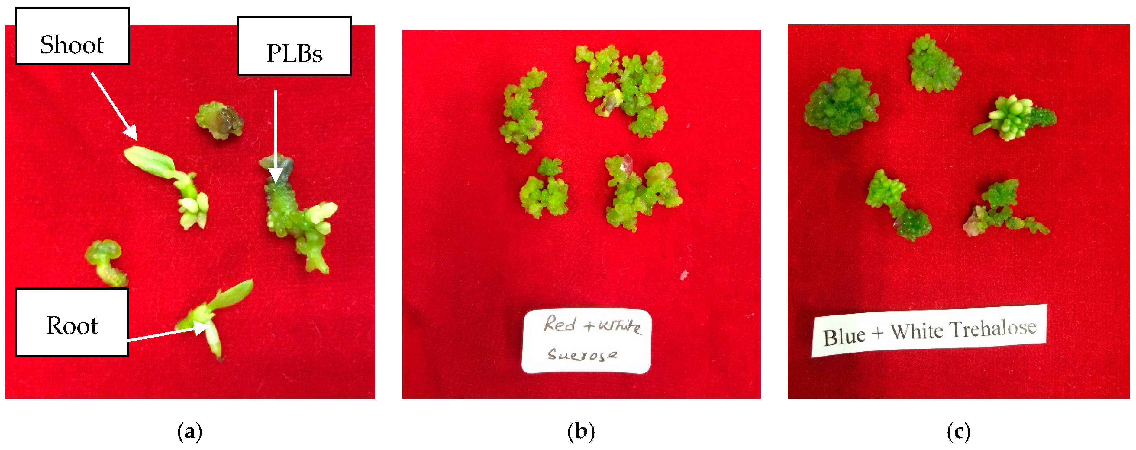
Figure 2.
Comparison of PLB production for the (a) highest (b) second highest and (c) third highest CHO source –LED combination within each CHO source. The X-axis the treatment combination, the Y-axis represents the number of PLBs, and the error bar represents the 95% confidence intervals. See text for meanings of abbreviations.

Figure 3.
Comparison of fresh weight for the (a) highest (b) second highest and (c) third highest within CHO source –LED combination each CHO source. The X-axis the treatment combination, the Y-axis represents the number of PLBs, and the error bar represents the 95% confidence intervals. See text for meanings of abbreviations.

Figure 4.
Correlation between the number of PLBs (left) and fresh weight (right) with CCC concentrations of the culture medium. Mean data were used for these analyses.
Figure 4.
Correlation between the number of PLBs (left) and fresh weight (right) with CCC concentrations of the culture medium. Mean data were used for these analyses.


Table 1.
Mean number of PLBs and fresh weight of Phalaenopsis ‘Fmk02010’withdifferent CHO sources and LED lights.
Table 1.
Mean number of PLBs and fresh weight of Phalaenopsis ‘Fmk02010’withdifferent CHO sources and LED lights.
| Light z | Mean Number of PLBs | Fresh Weight (g) | ||||
|---|---|---|---|---|---|---|
| Sucrose | Trehalose | Maltose | Sucrose | Trehalose | Maltose | |
| Control | 37.73 ± 4.40 y ab | 28.13 ± 4.87 abcd | 1.60 ± 0.53 b | 0.098 ± 0.057 a | 0.059 ± 0.033 ab | 0.043 ± 0.023 ab |
| R | 26.20 ± 3.38 bc | 15.87 ± 2.89 bcde | 2.73 ± 0.97 b | 0.062 ± 0.042 a | 0.059 ± 0.032 ab | 0.033 ± 0.019 b |
| G | 21.93 ± 3.76 bcd | 8.87 ± 1.55 de | 6.00 ± 1.35 b | 0.066 ± 0.045 a | 0.041 ± 0.022 ab | 0.033 ± 0.018 b |
| B | 20.47 ± 3.92 bcd | 27.67 ± 3.21 abcd | 2.27 ± 0.76 b | 0.043 ± 0.024 a | 0.137 ± 0.074 ab | 0.017 ± 0.009 b |
| W | 24.80 ± 4.49 bcd | 22.80 ± 2.92 abcde | 3.60 ± 0.67 b | 0.071 ± 0.039 a | 0.079 ± 0.042 ab | 0.028 ± 0.017 b |
| RG | 11.07 ± 3.97 cd | 19.33 ± 3.44 abcde | 4.47 ± 1.04 b | 0.029 ± 0.022 a | 0.063 ± 0.034 ab | 0.043 ± 0.023 ab |
| RB | 11.60 ± 3.11 cd | 15.33 ± 5.55 bcde | 1.47 ± 0.45 b | 0.026 ± 0.020 a | 0.091 ± 0.065 ab | 0.024 ± 0.013 b |
| RW | 54.13 ± 8.85 a | 12.13 ± 2.82 cde | 14.47 ± 3.39 a | 0.109 ± 0.063 a | 0.027 ± 0.020 b | 0.066 ± 0.041 ab |
| GB | 19.93 ± 4.83 bcd | 22.73 ± 3.67 abcde | 2.27 ± 1.03 b | 0.061 ± 0.035 a | 0.117 ± 0.062 ab | 0.020 ± 0.011 b |
| GW | 26.40 ± 3.60 bc | 8.73 ± 2.19 de | 4.80 ± 1.11 b | 0.080 ± 00.047 a | 0.047 ± 0.026 ab | 0.051 ± 0.027 ab |
| BW | 10.67 ± 4.11 cd | 36.33 ± 5.08 a | 3.80 ± 1.76 b | 0.020 ± 0.020 a | 0.129 ± 0.071 ab | 0.045 ± 0.024 ab |
| RGB | 5.47 ± 1.98 d | 5.00 ± 1.70 e | 8.40 ± 1.85 a | 0.015 ± 0.015 a | 0.066 ± 0.048 ab | 0.079 ± 0.047 ab |
| RBW | 13.53 ± 2.88 cd | 35.13 ± 4.36 ab | 13.73 ± 2.09 a | 0.034 ± 0.024 a | 0.167 ± 0.098 a | 0.112 ± 0.068 a |
| RGW | 19.07 ± 2.60 bcd | 29.40 ± 4.45 abc | 1.47 ± 0.35 b | 0.030 ± 0.021 a | 0.090 ± 0.048 ab | 0.022 ± 0.013 b |
| GBW | 12.20 ± 2.79 cd | 32.00 ± 7.77 ab | 4.73 ± 1.27 b | 0.056 ± 0.038 a | 0.088 ± 0.063 ab | 0.030 ± 0.017 b |
z Control (C: white fluorescent light); R (red LED); G (green LED); B (blue LED); W (white LED); RG (red → green LED); RB (red → blue LED); RW (red → white LED); GB (green → blue LED); GW (green → white LED); BW (blue → white LED); RGB (red → green → blue LED); RBW (red → blue → white LED); RGW (red → green → white LED); and GBW (green → blue → white LED). y Mean± SE values that do not share a letter are significantly different within each column, and those sharing a letter are statistically similar at P ≤ 0.05.
Table 2.
Shoot growth withdifferent CHO sources and LED lights during PLB organogenesis of Phalaenopsis ‘Fmk02010’.
Table 2.
Shoot growth withdifferent CHO sources and LED lights during PLB organogenesis of Phalaenopsis ‘Fmk02010’.
| Light z | Number of Shoots | Mean Shoot Length | ||||
|---|---|---|---|---|---|---|
| Sucrose | Trehalose | Maltose | Sucrose | Trehalose | Maltose | |
| Control | 0 | 0 | 0 | 0 | 0 | 0 |
| R | 0 | 0.38 ± 0.14 | 0 | 0 | 0.26 ± 0.10 | 0 |
| G | 0 | 0.38 ± 0.14 | 0 | 0 | 0.04 ± 0.02 | 0 |
| B | 0 | 0.75 ± 0.29 | 0 | 0 | 0.11 ± 0.05 | 0 |
| W | 0.13 ± 0.07 | 0.25 ± 0.09 | 0 | 0.05 ± 0.05 | 0.08 ± 0.03 | 0 |
| RG | 0 | 0.13 ± 0.07 | 0.13 ± 0.13 | 0 | 0.08 ± 0.04 | 0.03 ± 0.03 |
| RB | 0 | 0.25 ± 0.13 | 0 | 0 | 0.05 ± 0.03 | 0 |
| RW | 0 | 0.13 ± 0.07 | 0 | 0 | 0.03 ± 0.01 | 0 |
| GB | 0 | 0.13 ± 0.07 | 0 | 0 | 0.05 ± 0.03 | 0 |
| GW | 0 | 0 | 0 | 0 | 0 | 0 |
| BW | 0 | 0.38 ± 0.14 | 0 | 0 | 0.12 ± 0.04 | 0 |
| RGB | 0 | 0 | 0 | 0 | 0 | 0 |
| RBW | 0 | 0 | 0 | 0 | 0 | 0 |
| RGW | 0.25 ± 0.13 | 0 | 0 | 0.03 ± 0.03 | 0 | 0 |
| GBW | 0 | 0 | 0 | 0 | 0 | 0 |
z Control (C: white fluorescent light); R (red LED); G (green LED); B (blue LED); W (white LED); RG (red → green LED); RB (red → blue LED); RW (red → white LED); GB (green → blue LED); GW (green → white LED); BW (blue → white LED); RGB (red → green → blue LED); RBW (red → blue → white LED); RGW (red → green → white LED); and GBW (green → blue → white LED).
Table 3.
Role of CCC concentrations forthe in vitro PLB production of Phalaenopsis ‘Fmk02010’.
| CCC (mgL−1) | Number of PLBs | PLB Formation (%) | Fresh Weight (g) |
|---|---|---|---|
| 0 | 12.53 ± 1.71 z a | 93.33 | 0.175 ± 0.028 a |
| 0.01 | 15.67 ± 1.01 a | 100.00 | 0.211 ± 0.018 a |
| 0.1 | 13.73 ± 1.62 a | 93.33 | 0.191 ± 0.022 a |
| 1 | 11.07 ± 2.08 ab | 80.00 | 0.182 ± 0.027 a |
| 10 | 4.40 ± 1.74 b | 33.33 | 0.049 ± 0.019 b |
z Mean ± SE values that do not share a letter within each column are significantly different at P ≤ 0.05.
© 2019 by the authors. Licensee MDPI, Basel, Switzerland. This article is an open access article distributed under the terms and conditions of the Creative Commons Attribution (CC BY) license (http://creativecommons.org/licenses/by/4.0/).
Share and Cite
MDPI and ACS Style
Mehraj, H.; Alam, M.M.; Habiba, S.U.; Mehbub, H. LEDs Combined with CHO Sources and CCC Priming PLB Regeneration of Phalaenopsis. Horticulturae 2019, 5, 34. https://doi.org/10.3390/horticulturae5020034
AMA Style
Mehraj H, Alam MM, Habiba SU, Mehbub H. LEDs Combined with CHO Sources and CCC Priming PLB Regeneration of Phalaenopsis. Horticulturae. 2019; 5(2):34. https://doi.org/10.3390/horticulturae5020034
Chicago/Turabian StyleMehraj, Hasan, Md. Meskatul Alam, Sultana Umma Habiba, and Hasan Mehbub. 2019. "LEDs Combined with CHO Sources and CCC Priming PLB Regeneration of Phalaenopsis" Horticulturae 5, no. 2: 34. https://doi.org/10.3390/horticulturae5020034
Note that from the first issue of 2016, this journal uses article numbers instead of page numbers. See further details here.