Article

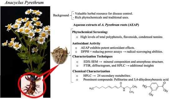
Phytochemistry, Antioxidant Potential, and Antibacterial Activities of Anacyclus pyrethrum: Promising Bioactive Compounds

by
Abdelmounaim Baslam
1,
Abdelfatah Aitbaba
1,
Rachida Aboufatima
2,
Fatimazahra Agouram
1,
Samia Boussaa
3,
Abderrahman Chait
1 and
Marouane Baslam
4,5,6,*
1
Laboratory of Pharmacology, Neurobiology, Anthropobiology and Environment, Department of Biology, Faculty of Sciences Semlalia, Cadi Ayyad University, Marrakech 40000, Morocco
2
Laboratory of Biological Engineering, Faculty of Sciences and Technology, Sultan Moulay Slimane University, Beni Mellal 23000, Morocco
3
Higher Institute of Nursing and Health Techniques, Ministry of Health and Social Protection, Rabat 10000, Morocco
4
Laboratory of Biochemistry, Department of Applied Biological Chemistry, Faculty of Agriculture, University of Niigata, Niigata 950-2181, Japan
5
Center of Agrobiotechnology and Bioengineering, Research Unit Labelled CNRST (Centre AgroBiotech-URL-7 CNRST- 05), Cadi Ayyad University, Marrakech 40000, Morocco
6
Laboratory of Agro-Food, Biotechnologies and Valorization of Plant Bioresources (AGROBIOVAL), Department of Biology, Faculty of Science Semlalia, Cadi Ayyad University, Marrakesh 40000, Morocco
*
Author to whom correspondence should be addressed.
Horticulturae 2023, 9(11), 1196; https://doi.org/10.3390/horticulturae9111196
Submission received: 29 September 2023 / Revised: 24 October 2023 / Accepted: 1 November 2023 / Published: 2 November 2023

Abstract

:
Secondary metabolites derived from plants have demonstrated significant biological activity and hold both edible and medicinal significance. These compounds play a crucial role in the development of pharmaceuticals, particularly in the context of inflammatory disorders and chronic diseases associated with oxidative stress. The objective of this study was to investigate the chemical characterization, antioxidant potential, and antibacterial properties of the aqueous extract of Anacyclus pyrethrum (AEAP). To achieve this, we employed various analytical techniques including HPLC–ESI–MS/MS, Fourier transform infrared, X-ray diffraction, scanning electron microscopy, and energy-dispersive X-ray spectroscopy. The antioxidant activity of the AEAP was assessed using DPPH (2,2-Diphenyl-1-picrylhydrazyl) and reducing power assays, while antibacterial activity was evaluated against both Gram-negative (Escherichia coli and Pseudomonas aeruginosa) and Gram-positive bacteria (Staphylococcus aureus and Enterococcus faecium) using the broth microdilution assay. Our findings demonstrated significant antioxidant activity of the AEAP, as well as broad-spectrum antibacterial activity against both Gram-positive and Gram-negative bacteria. HPLC analysis identified important bioactive compounds, notably pellitorine and 3,4-dihydroxybenzoic acid, known for their antioxidant and antibacterial properties. Overall, the AEAP demonstrated potent antioxidant and antibacterial activities, suggesting its potential as a valuable natural source of bioactive compounds with various therapeutic applications.

Graphical Abstract

1. Introduction

In recent years, there has been a growing interest in the use of herbal medicines for disease control due to their effectiveness, affordability, and limited side effects [1]. Natural products, particularly those derived from plants, have been a valuable source of raw materials for the discovery of bioactive compounds used in various industries, such as nutrition, cosmetics, and pharmaceuticals [2].
Anacyclus pyrethrum (L.) is one of wild species of flowering plant in the Asteraceae family and the Anacyclus genus, commonly known as pellitory, Aqar Qarha, Oud El Attas, and tigandizt. According to the Global Biodiversity Information Facility (GBIF) this plant is native to Morocco, Algeria, and Spain [3,4]. A. pyrethrum is a low-growing plant with finely divided, feathery leaves and produces daisy-like flowers with white petals and a yellow center. It is typically found in arid and rocky areas, particularly in mountainous regions, where it demonstrates remarkable adaptability to challenging and dry environments [5]. In Morocco, A. pyrethrum is commonly found in a variety of habitats, including clear-cut forests, scrublands, meadows, grasslands, lowlands, and mountainous regions, encompassing cold semi-arid, semi-humid, and humid climates [6]. This versatile species can be found at altitudes ranging from 1000 to 2500 m above sea level [6]. A. pyrethrum tends to flourish in well-drained soil and prefers open, unshaded areas with sparse tree and shrub cover within Mediterranean vegetation zones [6]. Reproduction in A. pyrethrum is primarily achieved through sexual means, mainly via seeds. However, asexual reproduction through plant parts or rhizomes is also feasible [3].
Extensive phytochemical screening of A. pyrethrum has unveiled a plethora of secondary metabolites, including alkaloids, tannins, coumarins, and flavonoids [7]. The root of A. pyrethrum is particularly rich in N-isobutyldienediylamide and polysaccharides [8]. Additionally, numerous experimental studies have demonstrated the wide range of biological effects associated with A. pyrethrum, including antioxidant and anti-inflammatory properties [9], antidepressant effects [10], and immunostimulatory capabilities [11]. Apart from its medicinal properties, A. pyrethrum has been traditionally used as a stimulant, cordial, and rubefacient [12]. It is recommended for gargling to alleviate symptoms of conditions such as rhinitis, neuralgia, rheumatic discomfort, and musculoskeletal pain. In Ayurvedic medicine, root of pellitory is combined with Withania somnifera and Vitis vinifera roots to treat epilepsy [13]. Furthermore, it has shown promising results in the remedy of conditions such as sciatica, paralysis, hemiplegia, and amenorrhea [14]. Anacycline, isobutylamide, inulin, and trace amounts of essential oil have been identified in the pellitory root [15]. Remarkably, there have been observations indicating that A. pyrethrum may lead to a reduction in insulin needs for those with insulin-dependent diabetes mellitus, along with the potential to decrease plasma glucose and serum cholesterol levels following oral administration for a duration of 3-6 weeks [16]. In Morocco, the roots of A. pyrethrum are known for their beneficial effects in traditional medicine, for various purposes, including the treatment of stomach diseases, stomatitis, cysts, articular rheumatism, and dental pain [17]. These attributes arise from a diverse array of phytochemical compounds, with over a hundred distinct compounds identified thus far. These compounds include phenolic compounds, flavonoids, and alkaloids [3,7,8].
Molecular studies on A. pyrethrum have provided valuable insights into its genetic makeup and the underlying mechanisms of its pharmacological properties. Manzanilla et al. (2022) showed the effectiveness of utilizing target capture genomic barcoding to identify and confirm the geographical source of specimens traded as A. pyrethrum, listed as a globally vulnerable medicinal plant in the International Union for Conservation of Nature (IUCN) Red List of Threatened Species [18]. Usmani et al. [3], by using gas chromatography–mass spectrometry (GC–MS), identified various potential bioactive compounds in A. pyrethrum, including pellitorine, germacrene D, trans caryophyllene, and naphthalene isomers. An additional molecular investigation into the essential oil of A. pyrethrum revealed the presence of several compounds (ca. 32 compounds), constituting approximately 93% of the overall chemical makeup of the essential oil.
These findings offer valuable insights into the chemical composition of compounds within A. pyrethrum, shedding light on their potential as bioactive substances and their associated pharmacological activities. Pellitorine, isolated in 1895 by Dunstan and Garnett from the roots of A. pyrethrum [19], stands out as a key compound. It has been documented to exhibit a range of biological effects, including antimicrobial, antiviral, diuretic, antioxidant, and analgesic properties [5,20]. These studies have also revealed the biosynthetic pathways and regulatory mechanisms that contribute to the plant medicinal properties [3]. Furthermore, molecular studies have also facilitated the identification and selection of high-yielding and disease-resistant cultivars of A. pyrethrum [21], thereby aiding in its cultivation and commercialization.
Expanding the therapeutic applications for A. pyrethrum could enhance treatment options, particularly in regions where traditional medicine plays a pivotal role in healthcare. In this context, this study aims to contribute to the valorization of A. pyrethrum from the Moroccan Middle Atlas. Our objective is to uncover novel therapeutic prospects while substantiating earlier research. We carried out a comprehensive investigation into the aqueous extract of A. pyrethrum (AEAP), delving into its phytochemical composition, antioxidant potential, antibacterial activities, and chemical profile.

2. Materials and Methods

2.1. Plant Samples and Extraction

2.1.1. Plant Samples

The roots of A. pyrethrum were gathered from the wild in its natural habitat at Bin El Ouidan region, Morocco (32°7′48″ latitude N/6°27′36″ Longitude W). The plants were initially authenticated by botanist Professor A. Ouhammou and pharmacologist Professor A. Chait from the Faculty of Sciences at Semlalia, Cadi Ayyad University. The plants were then stored under voucher specimen MARK-1003 in the herbarium of the Department of Biology, Faculty of Sciences Semlalia, Cadi Ayyad University, Marrakech, Morocco.

2.1.2. Plant Extraction

The powder was obtained by crushing the washed and dried roots, which were then extracted with distillated water (1 g/10 mL) under agitation for 12 h. The aqueous macerate was centrifuged (1200 rpm), filtered, and lyophilized using a Christ instrument. The extract underwent a microbial assessment, and no signs of microbial contamination (Escherichia coli, Salmonella, Staphylococcus aureus or Pseudomonas aeruginosa) were detected. The lyophilized dry powder was then aseptically stored in amber bottles at 4 °C until it was ready for utilization.

2.2. Phytochemical Study

A phytochemical analysis was conducted to assess the presence of secondary metabolites, including total phenols, flavonoids, and tannins in the extract. The results were reported as the average ± standard deviation (SD) of three independent experiments.

2.2.1. Determination of Total Phenolic Content (TPC)

The extract TPC was assessed using an adapted Folin-Ciocalteu method [22]. A 0.4 mL aliquot of the diluted extract was combined with 1.5 mL of the Folin–Ciocalteu reagent. Subsequently, 1.6 mL of a 7.5% sodium carbonate solution was introduced. The resultant mixture was then left to incubate in darkness at room temperature for 2 h. The absorbance was read at a wavelength of 765 nm. Gallic acid served as the reference standard, and the outcomes were expressed as milligrams of gallic acid equivalent per gram of dry weight (mg GAE/g DW).

2.2.2. Determination of Total Flavonoid Content (TFC)

The extract TFC was assessed using a method described in a previous study [23]. A 200 μL extract was diluted with 1 mL of distilled water. Subsequently 60 μL of 5% NaNO2 and 60 μL of 10% AlCl3 were introduced into the solution. After 5 min, 400 μL of 1 M NaOH was added to the mixture. The absorbance was read at 510 nm, and the TFC was expressed as milligrams of catechin equivalent per gram of dry matter (mg CE/g DM).

2.2.3. Quantification of Condensed Tannins

The total condensed tannin content was assessed following a method previously outlined [24]. Briefly, diluted samples (400 µL) were blended with 3 mL of a 4% methanol vanillin solution and 1.5 mL of concentrated HCL. The mixture was allowed to incubate for 15 min, and the absorbance was gauged at 500 nm. The total condensed tannin content was quantified and expressed as milligrams of catechin equivalent per gram of dry matter (mg CE/g DM).

2.3. Antioxidant Activity

2.3.1. Determination of DPPH Radical Scavenging Activity

The antioxidant activity of the extract was determined using the 2,2-diphenyl-1-picrylhydrazyl radical (DPPH) assay, as previously outlined [25] with certain modifications [26]. Specifically, 1.5 mL of a methanolic DPPH solution (6 × 10⁻⁵ M) was mixed with 60 μL of the extract at varying concentrations (1, 2, 4, 6, and 8 mg/mL) of AEAP. The mixture was then shielded from light and allowed to stand at room temperature for 30 min. Following this incubation, the absorbance was recorded at 515 nm, using a negative control consisting of 1.5 mL of the DPPH solution and 60 μL of methanol. The study also included positive controls, such as butylated hydroxytoluene (BHT) and quercetin. The percentage of inhibition was calculated using the formula:
% I n h i b i t i o n ( m g / m L ) = [ ( A _ c o n t r o l A _ s a m p l e ) / A _ c o n t r o l ] × 100
where A_control is the absorbance of the control and A_sample is the absorbance of the test compound.
The sample concentration that induces 50% inhibition (IC50) was determined from a graph plotting the percentages of inhibition against the sample concentrations.

2.3.2. Determination of Ferric Reducing Ability Power (FRAP) Activity

The FRAP test was carried out in accordance with a previously established method [27]. The assay involves the prevention of the formation of Fe(II)–ferrazine complexes when samples interact with ferrous iron. In the procedure, a mixture of distilled water (1 mL), phosphate buffer (2.5 mL; 0.2 M; pH = 6.6), and potassium ferricyanide (K3[Fe(CN)6]) (2.5 mL; 1%) was combined with 0.5 mL of solutions of varying concentrations. After incubating for 30 min, the mixture was supplemented with 2.5 mL of distilled water, 2.5 mL of 10% trichloroacetic acid, and 0.5 mL of FeCl3. The absorbance was read at 700 nm. Quercetin and BHT were used as positive controls.

2.4. Antibacterial Activity

2.4.1. Microbial Strains and Culture Conditions

The antibacterial activity of AEAP was assessed against a panel of Gram-positive bacteria, including Staphylococcus aureus (ATCC 25923) and Enterococcus faecium (ATCC 4602), as well as Gram-negative bacteria, such as Pseudomonas aeruginosa (ATCC 27653) and Escherichia coli (ATCC 25922). The bacterial cultures were incubated in Mueller–Hinton broth (MHB) for 24 h, and the final inoculum sizes were standardized to 1 × 10⁸ CFU/mL (0.5 McFarland standard).
To assess the antibacterial activity, three essential in vitro assays were employed. Firstly, the agar well diffusion method was employed to measure the inhibition zones (ZOI) induced by the plant extract. Secondly, the microdilution assay was performed to determine the minimal inhibitory concentration (MIC), which represents the lowest concentration of the extract that inhibits visible bacterial growth. Finally, the minimum bactericidal concentration (MBC) was established, which represents the minimal concentration of the extract lethal to the bacteria. All tests were conducted in triplicate for each bacterial strain to ensure accuracy and reproducibility of the results.

2.4.2. Agar Well Diffusion Inhibition Zones Test

For the antibacterial assessment, Petri dishes containing approximately 25 mL of Mueller–Hinton agar (MHA) were prepared and appropriately labeled. The bacterial inoculum was evenly spread across the agar surface using sterile cotton swabs, with the plate rotated 90° after each pass to ensure uniform distribution. Wells, each with a 4 mm diameter, were created in a sterile manner using a cork-borer. Subsequently, 100 µL of the plant extract, prepared at a concentration of 100 mg/mL, was dispensed into each well. As a positive control, gentamicin (10 µg/disc) was employed, while 100 µL of a 4% DMSO was used as a negative control to confirm that any observed antibacterial activity was not attributed to the solvent. The plates were left at room temperature for 30 min to allow for the proper diffusion of the plant extract. After incubation at 37 °C, the zones of bacterial inhibition surrounding each well were measured in mm, and the mean value was calculated to assess the extent of antibacterial activity [28].

2.4.3. MIC and MBC/MFC Determination

The antibacterial activity of AEAP was assessed using a broth dilution micro-method carried out in 96-well microtiter plates. This followed the protocols established by the Clinical and Laboratory Standards Institute (CLSI), with few modifications [29]. The AEAP and their polyphenolic fractions were first dissolved in distilled water to create stock solutions at a concentration of 100 mg/mL. From these stock solutions, serial twofold dilutions were prepared in Mueller–Hinton broth (MHB), resulting in a range of final concentrations from 50 to 0.19 mg/mL. Microbial cultures were then inoculated into the wells to reach a final concentration of 5 × 10⁵ CFU. Each determination was carried out in duplicate, with control wells containing only MHB and MHB + distilled water. Gentamicin served as a positive reference standard drug. After incubation for 24 h, the MIC was established as the lowest concentration of the extract that entirely inhibited visible bacterial growth. To determine the MBC, 20 µL from all clear MIC wells were plated onto agar plates. The MBC was defined as the lowest concentration of the plant extracts that resulted in the death of 99.9% of the bacterial inoculum. Each determination was performed in three independent experiments, and the results were calculated based on the mode.

2.5. Chemical Characterization

2.5.1. High-Performance Liquid Chromatography with Photodiode Array Detection-Tandem Mass Spectrometer (HPLC–PDA–MS/MS)

The LC system employed in this study was a Thermo Finnigan instrument (Thermo Electron Corporation, Waltham, MA, USA), featuring a reversed-phase column (Zorbax Eclipse XDB-C18, 4.6 × 150 mm, 3.5 µm, Agilent, Santa Clara, CA, USA). The LC instrument was connected to a mass spectrometer (LCQ-Duo ion trap) equipped with an electrospray ionization source. The mobile phase consisted of water and acetonitrile (ACN), both infused with 0.1% formic acid. A gradient flow of the mobile phase was initiated, starting with 5% ACN and gradually increasing to 30% over a span of 60 min. In the next 30 min, the ACN concentration was elevated to 90%. The flow rate was set at 1 mL/min with a split ratio of 1:1. Employing the full scan acquisition method, spectra were scanned within the range of m/z 50–2000 in the negative mode. Data evaluation was carried out using the X-calibur software (X-calibur™ 2.0.7, Thermo Fisher Scientific, Waltham, MA, USA).

2.5.2. Fourier Transform Infrared (FTIR)

Finely ground potassium bromide particles were mixed with A.pyrethrum powder at a ratio of 1:100. The resulting mixture were analyzed using an infrared spectrometer to investigate molecular vibrations within the sample particles. Infrared spectra were collected in the range of 400–4000 cm−1, enabling the identification and characterization of chemical bonds and functional groups present in the silver nanoparticles.

2.5.3. X-ray Diffraction (XRD)

The XRD patterns was acquired using a PAN analytical X’pert PRO X-ray diffractometer from JEOL (Tokyo, Japan). Measurements were conducted within the 2θ range of 20–80°, employing a fixed current of 40 mA and a voltage of 45 kV. Analysis involved depositing a suspension of myogenic AgNPs into a glass substrate. XRD data enabled scrutiny of the crystal structure and phase composition of the silver nanoparticles. The average crystal size was determined using Scherrer’s equation, which relates size (D) to wavelength (λ), the full width at half maximum of the diffraction peak (β), Scherrer’s constant (K), and Bragg’s angle (θ).

2.5.4. Scanning Electron Microscope (SEM)

Using a SEM, we conducted an analysis of the morphology, size, and shape of A.pyrethrum powder particles across magnification levels ranging from ×1,000 to ×1,000,000, following the methodology previously described [30]. Particle size determination was accomplished by processing acquired SEM images using the Image J program 1.53a.

2.5.5. Energy-Dispersive X-ray Spectroscopy (EDX)

Elemental composition of samples was ascertained through EDX spectrometry. The EDX analysis provided information about element presence and distribution within the samples. A transmission electron microscope (TEM) of the JEM-2100 model (JEOL, Tokyo, Japan) was used to evaluate the particle structure and size distribution. TEM offers detailed insights into the particles morphology, structure, and size distribution.

2.6. Statistical Analyses

The data were presented in the form of mean values along with the standard error of the mean (SEM) by utilizing Graphpad Prism 09. A one-way analysis of variance (ANOVA) was conducted and post hoc Tukey’s tests were performed to evaluate variations among the groups. Statistical significance was acknowledged at a p < 0.05.

3. Results

3.1. Phytochemical Screening

Our findings revealed the presence of total polyphenols (21.98 ± 0.05 mg GAE/g), flavonoid (8.1 ± 0.16 mg QE/g), and condensed tannins (5.49 ± 0.08 mg CE/g) within the AEAP (Table 1).

3.2. Antioxidant Activity

AEAP exhibited antioxidant activity using the DPPH and reducing power methods. The IC50 value was 1.6 ± 0.04 mg/mL, indicating its capability to convert the stable purple-colored DPPH radical into the yellow-colored DPPH-H form (Table 2). Additionally, AEAP showed reducing power with an IC50 value of 1.4 ± 0.04 mg/mL.

3.3. Antibacterial Activity

The AEAP displayed significant antibacterial activity as demonstrated by the inhibition zone diameter against the tested bacteria. When tested against S. aureus (ATCC 25923) at a concentration of 100 mg/mL, AEAP exhibited a mean inhibition zone of 17.65 ± 1.14 mm. Similarly, comparing AEAP to 10 µg/disc gentamicin against E. faecium (ATCC 4602), both agents showed comparable inhibition zones of 10.9 mm and 12.0 mm, respectively. Moreover, the antibacterial activity of AEAP was determined to be equally effective against Gram-negative as it was against Gram-positive bacteria. The inhibition zone diameter against E. coli (ATCC 25922) and P. aeruginosa (ATCC 27653) was measured at 17.7 mm and 15.5 mm, respectively. Comparatively, the positive control gentamicin exhibited inhibition zones of 23 mm and 22 mm, respectively (Table 3).
In order to determine the effective concentration of AEAP, broth microdilution tests were conducted, along with determining MIC and MBC. As illustrated in Table 4, AEAP exhibited bactericidal effect against the tested bacteria, including S. aureus (1.95 mg/mL), E. faecium (3.91 mg/mL), E. coli (8.51 mg/mL), and P. aeruginosa (2.5 mg/mL). These MIC values denoted the lowest extract concentration at which no visible bacterial growth occurred, indicating thereby its inhibitory potential. However, the MBC was found to be greater than 50 mg/mL.

3.4. Characterization of the Extract

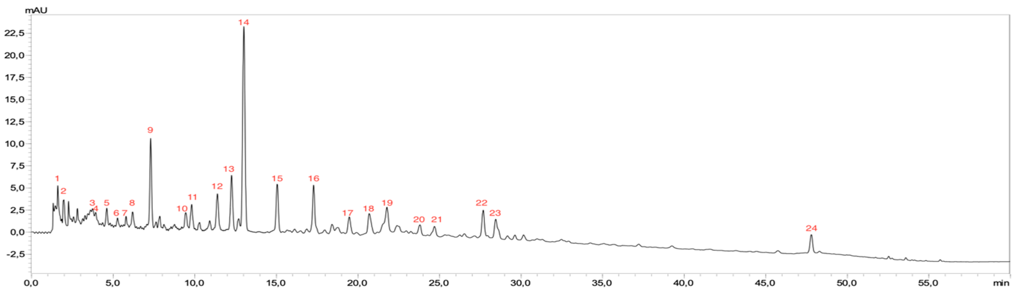
3.4.1. HPLC Analysis

The AEAP underwent analysis using HPLC–PDA–MS/MS, where the phytoconstituents were identified based on their MS/MS fragments and retention times, revealing a total of 24 secondary metabolites encompassing various phenolic compounds and organic acids (Figure 1 and Figure 2 and Table 5).
HPLC analysis of AEAP unveiled several compounds of interest. Predominantly, pellitorine emerged as the most abundant compound, featuring a retention time of 13.102 min and a molecular weight of 223. Pellitorine exhibited MS/MS fragmentation peaks at m/z 133 and 81. Another substantial compound, 3,4-dihydroxybenzoic acid, eluted at 7.124 min with an MW = 153, and an MS/MS fragmentation peak at m/z 108. Dihydroxybenzoic acid glucuronide, with a retention time of 12.289 min and an MW = 329, displayed an MS/MS fragmentation peak at m/z 153. Similarly, dihydrocaffeic acid, detected at 15.075 min with an MW of 181, demonstrated MS/MS fragmentation peaks at m/z 107 and 135. Feruloylquinic acid was observed at 17.482 min, possessing an MW of 367, and exhibited an MS/MS fragmentation peak at m/z 191. A recurrent appearance of dihydroxybenzoic acid glucuronide was noted at 12.289 min with an identical MW and MS/MS fragmentation pattern. Finally, hydroxybenzoic acid quinyl ester was found at 11.676 min, with an MW of 311, alongside an MS/MS fragmentation peak at m/z 137 (Table 5).
The HPLC–PDA–MS/MS analysis provided insights into the specific phytoconstituents present in AEAP. Notably, we identified 24 secondary metabolites, with pellitorine and 3,4-dihydroxybenzoic acid emerging as the most abundant components. Pellitorine, characterized by a molecular formula of C14H25NO and a molecular weight of 223.35 g/mol, and 3,4-dihydroxybenzoic acid, known as protocatechuic acid, with a molecular formula of C7H6O4 and a molecular weight of 154.12 g/mol, stood out as significant constituents within the extract (Figure 2).

3.4.2. EDX–SEM Analysis

The FTIR analysis of AEAP is depicted in Figure 3A, revealing a robust band within 3350 and 3460 cm−1, indicative of the stretching of the -NH2 and -OH group bonds. Notably, a band emerged at 2869–2938 cm−1, attributed to C-H vibration of -CH3. Additionally, a peak at 1641.07 cm−1 was credited to stretching and vibration of the carbonyl group, C=O, present in the extract. Furthermore, an absorption peak at ca. 1068.87 cm−1 indicated the stretching vibration of the -C-O-C bridge of the ring, suggesting that presence of cyclic molecules in the extract. Distinct bands associated with the phosphate group were observed within the range of 862.66 and 919.70 cm−1, indicating the presence of minerals in the extract.
Figure 3B showed the diffractogram of the AEAP, revealing a halo lies between 5° and 12° at 2θ, corresponding to a reflection plane of (020). Additional peaks were identified at 2θ around 28°; 41.5°; 50.9°, and 59.6°, aligning with (130), (211), (132) and (311) crystal reflection planes, respectively. These reflections could be attributed to a range of molecules present in the extract, including the specific active ingredient pellitorine, which is one of the most abundant molecules in the extract.
The coupling of EDX with SEM enabled microanalysis of AEAP, offering insight into its chemical composition in terms of atomic and weight percentages. The SEM image and EDX analysis (Figure 3C) illustrated the amorphous organization of molecules within the extract, visually represented by globular fibers stabilized by certain mineral elements as per EDX analysis. Moreover, elemental analysis results (Figure 3D) highlighted a substantial mineralogical composition.
Elements including K, Cl, P and Ca predominated in both weight and atomic percentages, as detailed in Table 6. Additionally, other elements were detected at lower levels, including S, Si, Al, Mg, Na and Fe.

4. Discussion

The purpose of this study was to present valuable insights into the phytochemical composition, antioxidant activity, and potential health benefits of the aqueous extract of A. pyrethrum. We aim to discuss the implications of our findings, their significance in the context of medicinal plants, and their potential applications in various fields. To provide insights into the chemical composition and mineralogical properties, the AEAP has been characterized using various techniques, including EDX–SEM, FTIR, diffractogram and HPLC analysis.
Polyphenols play a crucial role in shaping the biological characteristics of natural substances. In the current study, phytochemical screening revealed the presence of 21.98 ± 0.05 mg GAE/g of total polyphenols. This finding aligns with a previous investigation utilizing a hydroethanolic extract of A. pyrethrum, which reported a similar content (25.96 ± 1.93 mg GAE/g) obtained from Timehdite region, Morocco [31]. A study conducted in India on the hydromethanolic extract of A. pyrethrum reported a notably higher total phenolic content (62.89 ± 0.43 mg GAE/g) [12]. The concentrations of these polyphenols are significantly influenced by various factors, including the growing region, harvest time, extraction solvent, and storage conditions [32]. A. pyrethrum is known for its rich content of flavonoids, which are well-regarded for their antioxidant properties. In our research, we determined that the roots contain 8.1 ± 0.16 mg QE/g of flavonoids. In contrast, a previous study reported a lower level of flavonoids at 0.88 ± 0.02 mg QE/g [31]. Strikingly, the leaf examination revealed a significantly higher flavonoid content of 13.53 ± 0.05 mg QE/g [31]. Moreover, Kalim and colleagues found an even more substantial flavonoid content, measuring 38.89 ± 0.52 mg QE/g [12]. It is worth noting that recent research has shed light on the variability in assessing antioxidant activity due to the distinct mechanisms of action exhibited by different antioxidants [33]. Herein, we demonstrated remarkable antioxidant effects in A. pyrethrum roots, with an IC50 of 1.6 ± 0.04 mg/mL in the DPPH and 1.4 ± 0.04 mg/mL in the FRAP test. Notably, a study by Manouze et al. (2017) reported varying values in the DPPH test: 12.38 ± 0.28 μg/mL for the aqueous extract and 13.41 ± 0.67 μg/mL for the methanolic extract of A. pyrethrum sourced Oukaimed region, Morocco [9]. The high antioxidant activity observed in A. pyrethrum can be attributed to its rich phytochemical composition, particularly the presence of phenolic compounds. These phenolic compounds are believed to directly contribute to the plant antioxidative properties by playing a crucial role in mitigating the harmful effects of free radicals in the body, effectively safeguarding cells and tissues against oxidative damage [34]. Additionally, the ability of AEAP to effectively neutralize free radicals, as indicated by its low IC50 values in the DPPH and reducing power assays, highlights its potential in preventing cellular damage and mitigating the risk of oxidative stress-related diseases [35]. These findings align with the growing interest in plant-based antioxidants as alternatives to synthetic antioxidants [36,37].
The presence of polyphenols in the extract, such as hydroxybenzoic acid quinyl ester, dihydrocaffeic acid, feruloylquinic acid, caffeic acid, p-coumaric acid, p-coumaroylquinic acid, hydroxycoumarin, along with the alkylamide pellitorine, is of significance. These compounds have been previously confirmed to possess a wide range of beneficial activities, including, immunomodulatory, antithrombotic, antibacterial, antiviral, antioxidant, anti-inflammatory, analgesic, anticancer, antidiabetic, and antiprotozoal [20,38,39]. Additionally, the compounds in the extract have been reported to exhibit various pharmacological activities, such as antioxidant, antibacterial, neuroprotective, and nematicidal properties [40,41,42]. The identification of these compounds may pave the way for further research into their bioactivity and potential medicinal applications [14,43].
The HPLC analysis has successfully identified the presence of various bioactive compounds in the extract. Phenolic compounds may directly contribute to the antioxidative effects [44,45]. Flavonoids have the capability to effectively scavenge various reactive oxygen species (ROS), including superoxide and nitric oxide radicals [46,47]. The existence of these compounds and organic acids also plays a role in the overall bioactivity of A. pyrethrum.
The EDX–SEM analysis provided further insight into the characterization of AEAP, shedding light on its mineral composition and amorphous structure. The analysis revealed the presence of various mineral elements in A. pyrethrum extract, including K, Cl, P, Ca, S, Si, Al, Mg, Na, and Fe. These mineral elements exhibited varying weight % and atomic %, with K, Cl, P, and Ca being the predominant ones. The SEM microscopic image vividly depicted the amorphous arrangement of all the molecules within the extract, stabilized by select mineral elements as per EDX analysis. The data confirmed the presence of these mineral elements, while FTIR analysis revealed distinct bands in the spectrum of the aqueous extract. Notably, the FTIR analysis showcased the existence of specific functional groups, including -NH2, -OH, -CH3, and C=O, characteristic of flavonoids, tannins, and other secondary metabolites. The diffractogram analysis further revealed the presence of various crystal reflection planes, likely attributed to a range of molecules within the extract, including the active ingredient. The high content of elements and the amorphous structure of AEAP suggest potential benefits for its solubility and bioavailability, positioning it as a promising candidate for a range of applications [48].
Our results demonstrated significant antibacterial activity of A. pyrethrum against both Gram-positive and Gram-negative bacteria. Notably, we observed a substantial inhibition zone measuring 17.65 mm ± 1.14 against S. aureus, notably surpassing a previous er finding of a 17 mm inhibition zone. For P. aeruginosa, our study recorded an inhibition zone of 15.49 mm ± 2.01, closely aligning with the value of 15.65 mm observed in the same study [31]. This underscores the broad-spectrum antibacterial potential of A. pyrethrum. Furthermore, the observed bactericidal effect at relatively low concentrations underscores its efficacy in inhibiting bacterial growth. These findings align with previous studies [3,49,50] that have highlighted the antibacterial effects of A. pyrethrum against other bacterial strains (Listeria monocytogenes and Candida albicans), reinforcing its effectiveness in combating bacterial infections [51]. According to Elazzouzi et al. [7], A. pyrethrum aqueous extract exhibited relatively high antibacterial activity compared to ethanol and alkylamide extract. Jawhari et al. reported bactericidal effects across all components of A. pyrethrum, including the roots, capitula, seeds, and leaves, with variations noted across different bacterial strains, plant extracts, and plant parts employed in the study [31]. These findings attribute the observed antibacterial activity to the presence of secondary metabolites within the extract, particularly phenols and flavonoids, which contribute to the antibacterial effects [52].
Altogether, these findings suggest that A. pyrethrum holds promise for the development of natural antimicrobial agents, a crucial consideration in light of increasing antibiotic resistance. It is worth noting that, although this study did not include a toxicity assessment, prior research has consistently reaffirmed the non-toxic nature of A. pyrethrum extracts [14,53,54]. With an LD50 value exceeding 5000 mg/kg, indicating low acute toxicity and aligning with the Organization for Economic Co-operation and Development (OECD) guidelines for acute systemic toxicity classification, AEAP exhibits a robust safety profile [54].

5. Conclusions

This study highlights the remarkable potential of AEAP as a rich reservoir of bioactive compounds, boasting substantial antioxidant and antibacterial properties. AEAP exhibited robust antioxidant capabilities, effectively scavenging free radicals, and demonstrated a broad-spectrum antibacterial effectiveness against both Gram-positive and Gram-negative bacteria. The phytochemical profiling identified key bioactive compounds, with pellitorine and 3,4-dihydroxybenzoic acid standing out for their antioxidant and antibacterial effects. Furthermore, the comprehensive composition analysis of AEAP revealed its intricate composition, replete with diverse functional groups and molecules. These findings support further exploration of A. pyrethrum as a natural source of bioactive compounds with potential applications in various pharmacological interventions. Nevertheless, future studies should prioritize the isolation and characterization of individual bioactive compounds within AEAP and elucidate their mechanisms of action. Additionally, conducting clinical and preclinical trials to validate the health benefits of AEAP in humans is imperative. These investigations hold the promise of advancing our understanding of AEAP’s therapeutic potential and its prospective contributions to healthcare.

Author Contributions

Conceptualization, A.B. and M.B.; methodology, A.B. and F.A.; software, A.B. and R.A.; validation, M.B., S.B. and A.C.; formal analysis, A.B. and M.B.; investigation, A.B. and A.A.; resources, R.A. and S.B.; data curation, A.B. and A.C.; writing—original draft preparation, A.B. and A.A.; writing—review and editing, A.B., F.A., S.B. and M.B.; visualization, R.A and A.C.; supervision, M.B., S.B. and A.C.; project administration, S.B. and A.C. All authors have read and agreed to the published version of the manuscript.

Funding

The study received no external funding.

Data Availability Statement

Data are contained within the article.

Acknowledgments

M.B. acknowledges the technical and administrative support of the Faculty of Sciences Semlalia (Marrakech, Morocco).

Conflicts of Interest

The authors declare no conflict of interest.

References

  1. Ekor, M. The growing use of herbal medicines: Issues relating to adverse reactions and challenges in monitoring safety. Front Pharmacol. 2014, 4, 177. [Google Scholar] [CrossRef] [PubMed]
  2. Nasim, N.; Sandeep, I.S.; Mohanty, S. Plant-derived natural products for drug discovery: Current approaches and prospects. Nucleus 2022, 65, 399–411. [Google Scholar] [CrossRef] [PubMed]
  3. Elazzouzi, H.; Fadili, K.; Cherrat, A.; Amalich, S.; Zekri, N.; Zerkani, H.; Zair, T. Phytochemistry, Biological and Pharmacological Activities of the Anacyclus pyrethrum (L.) Lag: A Systematic Review. Plants 2022, 11, 2578. [Google Scholar] [CrossRef] [PubMed]
  4. The Global Biodiversity Information Facility. Anacyclus pyrethrum (L.). Available online: https://www.gbif.org/fr/species/3148578 (accessed on 20 October 2023).
  5. Usmani, A.; Khushtar, M.; Arif, M.; Siddiqui, M.A.; Sing, S.P.; Mujahid, M. Pharmacognostic and phytopharmacology study of Anacyclus pyrethrum: An insight. J. Appl. Pharm. Sci. 2016, 6, 144–150. [Google Scholar] [CrossRef]
  6. Fennane, M.; bn Tattou, M.; Mathez, J.; Ouyahya, A.; El Oualidi, J. Flore Pratique du Maroc. Trav. Inst. Sci. Sér. Bot. 1999, 1, 36. [Google Scholar]
  7. Elazzouzi, H.; Soro, A.; Elhilali, F.; Bentayeb, A.; El Belghiti, M.A.; Zair, T. Phytochemical study of Anacyclus pyrethrum (L.) of Middle Atlas (Morocco), and in vitro study of antibacterial activity of pyrethrum. Adv. Nat. Appl. Sci. 2014, 8, 131–141. [Google Scholar]
  8. Boonen, J.; Sharma, V.; Dixit, V.K.; Burvenich, C.; Spiegeleer, B.D. LC-MS N-alkylamide Profiling of an Ethanolic Anacyclus pyrethrum Root Extract. Planta Med. 2012, 80, 1787–1795. [Google Scholar] [CrossRef]
  9. Manouze, H.; Bouchatta, O.; Gadhi, A.C.; Bennis, M.; Sokar, Z.; Ba-M’hamed, S. Anti-inflammatory, Antinociceptive, and Antiox-idant Activities of Methanol and Aqueous Extracts of Anacyclus pyrethrum Roots. Front. Pharmacol. 2017, 8, 598. [Google Scholar] [CrossRef]
  10. Badhe, S.R.; Badhe, R.V.; Ghaisas, M.M.; Chopade, V.V.; Deshpande, A.D. Evaluations of antidepressant activity of Anacyclus pyrethrum root extract. Int. J. Green Pharm. IJGP. 2010, 4, 2. [Google Scholar] [CrossRef]
  11. Bendjeddou, D.; Lalaoui, K.; Satta, D. Immunostimulating activity of the hot water-soluble polysaccharide extracts of Anacyclus pyrethrum, Alpinia galanga and Citrullus colocynthis. J. Ethnopharmacol. 2003, 88, 155–160. [Google Scholar] [CrossRef]
  12. Kalim, M.D.; Bhattacharyya, D.; Banerjee, A.; Chattopadhyay, S. Oxidative DNA damage preventive activity and antioxidant potential of plants used in Unani system of medicine. BMC Complement Altern Med. 2010, 10, 77. [Google Scholar] [CrossRef] [PubMed]
  13. Sharma, V.; Thakur, M.; Chauhan, N.S.; Dixit, V.K. Immunomodulatory activity of petroleum ether extract of Anacyclus pyrethrum. Pharm Biol. 2010, 48, 1247–1254. [Google Scholar] [CrossRef] [PubMed]
  14. Manouze, H.; Bouchatta, O.; Bennis, M.; Sokar, Z.; Ba-M’hamed, S. Anticonvulsive and neuroprotective effects of aqueous and methanolic extracts of Anacyclus pyrethrum root in kainic acid-induced-status epilepticus in mice. Epilepsy Res. 2019, 158, 106225. [Google Scholar] [CrossRef]
  15. Khare, C.P. Anacyclus pyrethrum DC. In Indian Medicinal Plants: An Illustrated Dictionary; Khare, C.P., Ed.; Springer: New York, NY, USA, 2007; p. 1. [Google Scholar]
  16. Khare, C.P. Indian Medicinal Plants: An Illustrated Dictionary; Springer Science & Business Media: Berlin, Germany, 2008. [Google Scholar]
  17. Kumar, V.K.; Lalitha, K.G. Pharmacognostical Studies on the Root of Anacyclus pyrethrum DC; NISCAIR-CSIR: Delhi, India, 2012. [Google Scholar]
  18. Manzanilla, V.; Teixidor-Toneu, I.; Martin, G.J.; Hollingsworth, P.M.; de Boer, H.J.; Kool, A. Using target capture to address con-servation challenges: Population-level tracking of a globally-traded herbal medicine. Mol. Ecol. Resour. 2022, 22, 212–224. [Google Scholar] [CrossRef]
  19. Gulland, J.M.; Hopton, G.U., II. —Pellitorine, the pungent principle of Anacyclus pyrethrum. J. Chem. Soc. Resumed. 1930, 15, 6–11. [Google Scholar] [CrossRef]
  20. Ee, G.C.L.; Lim, C.M.; Rahmani, M.; Shaari, K.; Bong, C.F.J. Pellitorine, a Potential Anti-Cancer Lead Compound against HL60 and MCT-7 Cell Lines and Microbial Transformation of Piperine from Piper Nigrum. Molecules 2010, 15, 2398–2404. [Google Scholar] [CrossRef]
  21. Li, J.; Xu, Z.; Zeng, T.; Zhou, L.; Li, J.; Hu, H.; Wang, C. Overexpression of TcCHS Increases Pyrethrin Content When Using a Genotype-Independent Transformation System in Pyrethrum (Tanacetum cinerariifolium). Plants 2022, 11, 1575. [Google Scholar] [CrossRef]
  22. Singleton, V.L.; Orthofer, R.; Lamuela-Raventós, R.M. Analysis of total phenols and other oxidation substrates and anti-oxidants by means of folinciocalteu reagent. In Methods in Enzymology; Academic Press: Cambridge, MA, USA, 1999; Volume 299, pp. 152–178. [Google Scholar]
  23. Zhishen, J.; Mengcheng, T.; Jianming, W. The determination of flavonoid contents in mulberry and their scavenging effects on superoxide radicals. Food Chem. 1999, 64, 555–559. [Google Scholar] [CrossRef]
  24. Heimler, D.; Vignolini, P.; Dini, M.G.; Romani, A. Rapid Tests to Assess the Antioxidant Activity of Phaseolus vulgaris L. Dry Beans. J. Agric Food Chem. 2005, 53, 3053–3056. [Google Scholar] [CrossRef]
  25. Mansouri, A.; Embarek, G.; Kokkalou, E.; Kefalas, P. Phenolic profile and antioxidant activity of the Algerian ripe date palm fruit (Phoenix dactylifera). Food Chem. 2005, 89, 411–420. [Google Scholar] [CrossRef]
  26. Aitbaba, A.; Sokar, Z.; Chait, A. Analgesic and anti-inflammatory effects of hydroalcoholic extract of Astragalus ibrahimianus. Bangladesh J. Pharmacol. 2023, 18, 41–48. [Google Scholar]
  27. Oyaizu, M. Studies on Products of Browning Reaction Antioxidative Activities of Products of Browning Reaction Prepared from Glucosamine. Jpn. J. Nutr. Diet. 1986, 44, 307–315. [Google Scholar] [CrossRef]
  28. Alayed, H.S.; Devanesan, S.; AlSalhi, M.S.; Alkindi, M.G.; Alghamdi, O.G.; Alqhtani, N.R. Investigation of Antibacterial Activity of Carob-Mediated Calcium Hydroxide Nanoparticles against Different Aerobic and Anaerobic Bacteria. Appl. Sci. 2022, 12, 12624. [Google Scholar] [CrossRef]
  29. Ma, W. Methods for dilution antimicrobial susceptibility tests for bacteria that grow aerobically: Approved standard. CLSI NCCLS 2006, 26, M7-A7. [Google Scholar]
  30. Al-Dulimi, A.G.; Al-Saffar, A.Z.; Sulaiman, G.M.; Khalil, K.A.; Khashan, K.S.; Al-Shmgani, H.S.; Ahmed, E.M. Immobilization of L-asparaginase on Gold Nanoparticles for Novel Drug Delivery Approach as Anti-Cancer Agent Against Human Breast Carcinoma Cells. J. Mater Res. Technol. 2020, 25, 15394–15411. [Google Scholar] [CrossRef]
  31. Jawhari, F.Z.; Moussaoui, A.E.L.; Bourhia, M.; Imtara, H.; Saghrouchni, H.; Ammor, K.; Bari, A. Anacyclus pyrethrum var. pyrethrum (L.) and Anacyclus pyrethrum var. depressus (Ball) Maire: Correlation between Total Phenolic and Flavonoid Contents with Antioxidant and Antimicrobial Activities of Chemically Characterized Extracts. Plants 2021, 10, 149. [Google Scholar] [CrossRef] [PubMed]
  32. Cherrat, A.; Amalich, S.; Regragui, M.; Bouzoubae, A.; Elamrani, M.; Mahjoubi, M.; Zair, T. Polyphenols content and evaluation of antioxidant activity of Anacyclus pyrethrum (L.) lag. from timahdite a moroccan middle atlas region. Int. J. Adv. Res. 2017, 5, 569–577. [Google Scholar] [CrossRef]
  33. Munteanu, I.G.; Apetrei, C. Analytical Methods Used in Determining Antioxidant Activity: A Review. Int. J. Mol. Sci. 2021, 22, 3380. [Google Scholar] [CrossRef]
  34. Nagendrappa, G. An appreciation of free radical chemistry 3. Free radicals in diseases and health. Resonance 2005, 10, 65–74. [Google Scholar] [CrossRef]
  35. Mota, J.C.; Almeida, P.P.; Freitas, M.Q.; Stockler-Pinto, M.B.; Guimarães, J.T. Far from being a simple question: The complexity between in vitro and in vivo responses from nutrients and bioactive compounds with antioxidant potential. Food Chem. 2023, 402, 134351. [Google Scholar] [CrossRef]
  36. Arunachalam, K.; Sreeja, P.S.; Yang, X. The Antioxidant Properties of Mushroom Polysaccharides can Potentially Mitigate Oxidative Stress, Beta-Cell Dysfunction and Insulin Resistance. Front Pharmacol 2022, 13, 874474. [Google Scholar] [CrossRef] [PubMed]
  37. Das, K.; Roychoudhury, A. Reactive oxygen species (ROS) and response of antioxidants as ROS-scavengers during environ-mental stress in plants. Front Environ Sci. 2014, 2, 53. [Google Scholar] [CrossRef]
  38. Elufioye, T.O.; Habtemariam, S.; Adejare, A. Chemistry and Pharmacology of Alkylamides from Natural Origin. Front. Environ. Sci. 2020, 30, 622–640. [Google Scholar] [CrossRef] [PubMed]
  39. Mijangos-Ramos, I.F.; Zapata-Estrella, H.E.; Ruiz-Vargas, J.A.; Escalante-Erosa, F.; Gómez-Ojeda, N.; García-Sosa, K.; Cechinel-Filho, V.; Meira-Quintão, N.L.; Peña-Rodríguez, L.M. Bioactive dicaffeoylquinic acid derivatives from the root extract of Calea urticifolia. Rev. Bras Farmacogn. 2018, 28, 339–343. [Google Scholar] [CrossRef]
  40. Kaliaperumal, J.; Dulari, S.; Pandurangan, D.; Rathore, M.; Mittal, A.; Alam, M. Neuroprotective Activity of Protocatechuic Acid (3,4-Dihydroxybenzoic Acid) to Treat Parkinson’s Disease. J. Evol. Med. Dent Sci. 2020, 9, 2466–2471. [Google Scholar] [CrossRef]
  41. Nguyen, D.-M.; Seo, D.-J.; Kim, K.-Y.; Park, R.-D.; Kim, D.-H.; Han, Y.-S.; Kim, T.-H.; Jung, W.-J. Nematicidal activity of 3,4-dihydroxybenzoic acid purified from Terminalia nigrovenulosa bark against Meloidogyne incognita. Microb. Pathog. 2013, 59–60, 52–59. [Google Scholar] [CrossRef]
  42. Guglielmi, F.; Luceri, C.; Giovannelli, L.; Dolara, P.; Lodovici, M. Effect of 4-coumaric and 3,4-dihydroxybenzoic acid on oxidative DNA damage in rat colonic mucosa. Br. J. Nutr. 2003, 89, 581–587. [Google Scholar] [CrossRef]
  43. Ji, R.; Quan, Q.; Guo, X.; Zhang, J.; Song, Y.; Zhu, M.; Tan, P.; Han, J.; Liu, Y. Simultaneous determination of five N-alkylamides in the root of Anacyclus pyrethrum by HPLC and profiling of components in its methanolic root extract by UPLC/Q-TOF-MS. Rev. Bras. Farmacogn. 2019, 29, 152–161. [Google Scholar] [CrossRef]
  44. Biluca, F.C.; da Silva, B.; Caon, T.; Mohr, E.T.B.; Vieira, G.N.; Gonzaga, L.V.; Vitali, L.; Micke, G.; Fett, R.; Dalmarco, E.M.; et al. Investigation of phenolic compounds, antioxidant and anti-inflammatory activities in stingless bee honey (Meliponinae). Food Res. Int. 2020, 129, 108756. [Google Scholar] [CrossRef]
  45. Jawhari, F.Z.; Imtara, H.; El Moussaoui, A.; Khalis, H.; Es-Safi, I.; Al Kamaly, O.; Bari, A. Reproductive Biology of the Two Varieties of Anacyclus pyrethrum L.—Anacyclus pyrethrum var. pyrethrum (L.) Link and Anacyclus pyrethrum var. depressus (Ball.) Maire—An Endemic Endangered Species. Plants 2022, 11, 2299. [Google Scholar] [CrossRef]
  46. Kim, T.Y.; Leem, E.; Lee, J.M.; Kim, S.R. Control of Reactive Oxygen Species for the Prevention of Parkinson’s Disease: The Possible Application of Flavonoids. Antioxidants 2020, 9, 583. [Google Scholar] [CrossRef] [PubMed]
  47. Zandi, P.; Schnug, E. Reactive Oxygen Species, Antioxidant Responses and Implications from a Microbial Modulation Perspective. Biology 2022, 11, 155. [Google Scholar] [CrossRef]
  48. Kerboua, K.A.; Benosmane, L.; Namoune, S.; Ouled-Diaf, K.; Ghaliaoui, N.; Bendjeddou, D. Anti-inflammatory and anti-oxidant activity of the hot water-soluble polysaccharides fromzAnacyclus pyrethrum (L.). Lag. roots. J. Ethnopharmacol. 2021, 281, 114491. [Google Scholar] [CrossRef] [PubMed]
  49. Doudach, L.; Meddah, B.; Alnamer, R.; Chibani, F.; Cherrah, Y. In vitro antibacterial activity of the methanolic and aqueous ex-tracts of Anacyclus pyrethrum used in Moroccan traditional medicine. Int. J. Pharm. Pharm. Sci. 2012, 4, 4. [Google Scholar]
  50. Elazzouzi, H.; Zekri, N.; Zair, T.; El Belghiti, M.A. Volatiles profiling and antioxidant activity of Moroccan Artemisia ifranensis J. Didier and Anacyclus pyrethrum Link essential oils. Egypt J. Chem. 2020, 63, 3937–3947. [Google Scholar] [CrossRef]
  51. Daoudi, A.; Mohamed, B.; Jamal, I.; Laila, N. Antibacterial activity of aqueous extracts of Anacyclus pyrethrum (L) link and Corrigiola telephiifolia Pourr. from the middle atlas Region-Morocco. Eur. Sci. J. 2017, 13, 116. [Google Scholar]
  52. Jawhari, F.Z.; El Moussaoui, A.; Bourhia, M.; Imtara, H.; Mechchate, H.; Es-Safi, I.; Bari, A. Anacyclus pyrethrum (L): Chemical Com-position, Analgesic, Anti-Inflammatory, and Wound Healing Properties. Molecules 2020, 25, 5469. [Google Scholar] [CrossRef]
  53. Bezza, K.; Gabbas, Z.E.; Laadraoui, J.; Laaradia, M.A.; Oufquir, S.; Chait, A. Ameliorative potential of Anacyclus pyrethrum extract in generalized seizures in rat: Possible cholinergic mediated mechanism. Bangladesh J. Pharmacol. 2019, 14, 188–195. [Google Scholar] [CrossRef]
  54. Baslam, A.; Aitbaba, A.; Hanchi, A.L.; Tazart, Z.; Aboufatima, R.; Soraa, N.; Chait, A. Modulation of Gut Microbiome in Ec-stasy/MDMA-Induced Behavioral and Biochemical Impairment in Rats and Potential of Post-Treatment with Anacyclus pyrethrum L. Aqueous Extract to Mitigate Adverse Effects. Int. J. Mol. Sci. 2023, 24, 9086. [Google Scholar] [CrossRef]
Figure 1. HPLC-MS/MS profile of AEAP.
Horticulturae 09 01196 g001
Figure 2. Chemical structure of the most frequent secondary metabolites in AEAP.
Horticulturae 09 01196 g002
Figure 3. Physicochemical characterization of the AEAP: (A) Fourier Transform Infrared Spectroscopy, (B) X-ray Diffraction, (C) Scanning Electron Microscopy, and (D) Energy-Dispersive X-ray Spectroscopy.
Horticulturae 09 01196 g003
Table 1. Mean values of total polyphenol, flavonoid. and tannin contents in the AEAP.
Total Polyphenol Content
mg GAE/g DM
Flavonoids
mg CE/gDM
Tanins
mg CE/g DM
Crude extracts (mg/g)21.98 ± 0.058.1 ± 0.165.49 ± 0.08
GAE: gallic acid equivalent, CE: catechin equivalent, DM: dry matter.
Table 2. The antioxidant activity of the AEAP by 2,2-diphenyl-1-pierylhydrazyl (DPPH) and ferric reducing antioxidant power (FRAP).
Antioxidant AssayPlant Extract
(IC50 = mg/mL)
Standard Antioxidant
(IC50 = mg/mL)
QuercetinBHT
DPPH1.6 ± 0.040.1 ± 0.00.2 ± 0.0
FRAP1.4 ± 0.040.1 ± 0.00.1 ± 0.0
Table 3. Inhibition zones of the AEAP (100 mg/mL) against microbial strains.
Bacteria GroupBacterial StrainsReferenceDiameter Inhibition Zone (mm)
AEAPGentamicin
Gram-positive bacteria Staphylococcus aureusATCC 2592317.65 ± 1.1423
Enterococcus faeciumATCC 460210.87 ± 0.6912
Gram-negative bacteriaEscherichia coliATCC2592217.73 ± 0.6123
Pseudomonas aeruginosaATCC 2765315.49 ± 2.0122
Table 4. Minimal inhibitory concentration (MIC) and minimum bactericidal concentration (MBC) of the AEAP (50 mg/mL) against the microbial strains.
Bacteria GroupBacterial StrainsReferenceMIC (mg/mL)MBC (mg/mL)
Gram-positive bacteria Staphylococcus aureusATCC 259231.95>50
Enterococcus faeciumATCC 46023.91>50
Gram-negative bacteriaEscherichia coliATCC259228.51>50
Pseudomonas aeruginosaATCC 276532.5050
Table 5. Tentatively identified compounds in the AEAP using HPLC-ESI-MS/MS analysis.
Rt (min)MW MS/MSCompound Name
11.516191111Citric acid
21.935335169Gallic acid derivative
33.523267108, 153Dihydroxybenzoic acid derivative
43.642299153Dihydroxybenzoic acid rhamnoside
54. 42315108, 153Dihydroxybenzoic acid glucoside
65.243371135, 191Caffeoylglucaric acid
75.862169125Gallic acid
86.338341108, 167Vanillyl quinic acid
97.1241531083,4-dihydroxybenzoic acid
109.452225137Hydroxybenzoic acid glycerol
1110.921353135, 191Chlorogenic acid
1211.676311137Hydroxybenzoic acid quinyl ester
1312.289329153Dihydroxybenzoic acid glucuronide
1413.102223133,81Pellitorine
1515.075181107, 135Dihydrocaffeic acid
1617.482367191Feruloylquinic acid
1719.437353179, 191Cryptochlorogenic acid
1820.607179135Caffeic acid
1921.88163119p-Coumaric acid
2023.919337191p-Coumaroylquinic acid
2224.853161133Hydroxycoumarin
2227.796515179, 191Isochlorogenic acid b
2328.521447301Quercetin rhamnoside
2448.907515179, 191Isochlorogenic acid C
Rt: retention time; MW: molecular weight; MS/MS: Tandem Mass spectrometry.
Table 6. Elemental analysis of the AEAP.
Sample/ElementK CaClP S SiAlMgNa
AEAPWeight %33.6212.4210.608.024.003.262.861.860.52
Atomic %21.877.887.606.593.172.952.701.950.57
Disclaimer/Publisher’s Note: The statements, opinions and data contained in all publications are solely those of the individual author(s) and contributor(s) and not of MDPI and/or the editor(s). MDPI and/or the editor(s) disclaim responsibility for any injury to people or property resulting from any ideas, methods, instructions or products referred to in the content.

