The Potential Biomedical Application of NiCu Magnetic Nanoparticles


1
Faculty of Chemistry and Chemical Engineering, University of Maribor, Smetanova ulica 17, SI-2000 Maribor, Slovenia
2
Faculty of Medicine, Institute of Biomedical Sciences, University of Maribor, Taborska ulica 8, SI-2000 Maribor, Slovenia
3
Department of Pharmacology, Faculty of Medicine, University of Maribor, Taborska ulica 8, SI-2000 Maribor, Slovenia
*
Authors to whom correspondence should be addressed.
Magnetochemistry 2019, 5(4), 66; https://doi.org/10.3390/magnetochemistry5040066
Submission received: 8 October 2019
/
Revised: 15 November 2019
/
Accepted: 15 November 2019
/
Published: 6 December 2019
(This article belongs to the Special Issue Magnetic Nanoparticles)
Abstract
:Magnetic nanoparticles became increasingly interesting in recent years as a result of their tailorable size-dependent properties, which enable their use in a wide range of applications. One of their emerging applications is biomedicine; in particular, bimetallic nickel/copper magnetic nanoparticles (NiCu MNPs) are gaining momentum as a consequence of their unique properties that are suitable for biomedicine. These characteristics include stability in various chemical environments, proven biocompatibility with various cell types, and tunable magnetic properties that can be adjusted by changing synthesis parameters. Despite the obvious potential of NiCu MNPs for biomedical applications, the general interest in their use for this purpose is rather low. Nevertheless, the steadily increasing annual number of related papers shows that increasingly more researchers in the biomedical field are studying this interesting formulation. As with other MNPs, NiCu-based formulations were examined for their application in magnetic hyperthermia (MH) as one of their main potential uses in clinics. MH is a treatment method in which cancer tissue is selectively heated through the localization of MNPs at the target site in an alternating magnetic field (AMF). This heating destroys cancer cells only since they are less equipped to withstand temperatures above 43 °C, whereas this temperature is not critical for healthy tissue. Superparamagnetic particles (e.g., NiCu MNPs) generate heat by relaxation losses under an AMF. In addition to MH in cancer treatment, which might be their most beneficial potential use in biomedicine, the properties of NiCu MNPs can be leveraged for several other applications, such as controlled drug delivery and prolonged localization at a desired target site in the body. After a short introduction that covers the general properties of NiCu MNPs, this review explores different synthesis methods, along with their main advantages and disadvantages, potential surface modification approaches, and their potential in biomedical applications, such as MH, multimodal cancer therapy, MH implants, antibacterial activity, and dentistry.
1. Introduction
Magnetic nanoparticles (MNPs) attracted much interest in the last two decades, especially in the field of biomedicine. Their appealing potential biomedical applications rely on the strategic exploitation of their (extremely) small sizes, resulting in large surface areas, which distinguish them from bulk materials [1,2]. Additionally, MNPs have various unique magnetic properties, including their superparamagnetic nature, a high magnetic susceptibility, a low Curie temperature (TC), coercivity, and an inducible magnetic moment that allows them to be directed to a defined location or heated with an external alternating magnetic field (AMF) [3,4]. Among the properties that make MNPs ideal for biomedical applications are their biocompatibility, non-toxicity, and extensive aggregation in the desired tissue [5,6]. For their in vivo application, they must be coated by or encapsulated in a biocompatible polymer to prevent the formation of large aggregates, prevent the manipulation of their original structure, and potentially enable targeted biodegradation at the desired site in the body [7,8]. The important factors that determine the biocompatibility and toxicity of MNPs are the nature of the magnetically responsive component and the particles’ final size, core composition, and coatings [9,10,11]. Moreover, MNPs with sizes below 100 nm are known to possess lower sedimentation rates and improved tissular diffusion [12,13].
The potential biomedical applications of MNPs require further consideration in regard to some specific properties. In addition to their composition of non-toxic and non-immunogenic materials, their sizes (and size distributions) have to be even more carefully controlled to prolong their circulation after injection in the body, as well as to allow them to pass through the capillary systems of organs and tissues to avoid vessel embolism. High magnetization is crucial for their movement in the blood so that they can be controlled using an AMF and immobilized close to the targeted pathologic tissue [2,14].
The abovementioned properties of MNPs and their ability to work at both cellular and molecular levels enabled their investigation and, in some cases, application in vitro and in vivo as part of drug delivery systems [15,16,17], in the targeted delivery of cytotoxic drugs [16,18,19], as active components in hyperthermia treatment [17,20,21], as contrast agents in magnetic resonance imaging (MRI) [1,22,23], as radiotherapeutics [24,25], for advanced gene delivery applications [26,27], for separation and selection [24,28], in magnetorelaxometry [29,30], as active antibacterial materials [31,32], in tissue engineering [8,33], and as part of biotherapeutics [34,35]. Figure 1 summarizes the potential applications of MNPs in biomedicine.
In the last decade, the authors of this paper developed several novel NiCu-based MNPs that not only exhibit all the above mentioned characteristics but also hold great promise for many different applications, which include multi-layer coatings, solar energy, and regenerative medicine, as well as in various formulations used in biomedicine [36,37,38,39]. The versatile range of potential applications, especially their applicability in biomedicine, is related to their chemical stability, proven biocompatibility with different human and animal cells, and tailorable magnetic characteristics. Their magnetic properties can be controlled using different preparation methods, ranging from mechanical milling, hydrothermal reduction reactions, and emulsion techniques, through various electrochemistry-based and sol–gel methods, to plasma evaporation [36,40,41,42].
Despite the obvious potential of NiCu MNPs for biomedical applications, the general interest in their use for this purpose is rather low. Nevertheless, the steadily increasing annual number of related papers shows that increasingly more researchers in the field of biomedicine are studying this interesting formulation (Figure 2).
As is true for many other MNPs, NiCu MNPs were examined for their use in magnetic hyperthermia (MH) for the treatment of various cancers as their most interesting application type in biomedicine. For this purpose, biocompatible MNPs must be prepared. They are then applied to tumor tissues using different methods (e.g., controlled drug delivery) and subsequently heated in an AMF [38,43,44]. The heat produced through an AMF is influenced by various NP characteristics (size, shape, anisotropy, concentration in situ, and MH parameters, such as the frequency and field), which are all related (directly or indirectly) to their composition. The temperature to which the NPs are heated is defined by their Curie temperature (TC) [39], which must be between 42 and 46 °C to maximize the selective damage to cancer cells without harming the surrounding healthy tissue [39]. This sometimes requires high frequencies, which represents an important limitation to the use of MNPs in MH [45]. For many years, most of the related research was performed on the two most well-known iron oxides (Fe3O4, γ-Fe2O3), although several other materials (e.g., Fe-doped Au NPs and NiFe2O4) were also developed and successfully employed for this purpose [39]. Furthermore, as shown in several studies, iron oxides are not without limitations, especially if adjustments to TC play an essential role in therapeutic efficiency. Iron oxides tend to cause overheating because they have comparably higher Tc values, which can induce damage to healthy tissues in the proximity of the application site [46]. This effect does not occur with NiCu MNPs, which have a tunable TC that is much closer to optimal therapeutic temperatures [16,36,37]. In addition to the abovementioned investigations, research studies from the last couple of years showed that NiCu MNPs have great potential in several other types of biomedical applications, such as their use in bimodal cancer treatments that combine MH and controlled drug delivery [47,48,49]. Nevertheless, another important factor to be considered when applying a novel formulation for MH is the overall concentration of the respective NPs at the tumor site [50,51]. Specifically, NPs with a low specific loss power (SLP) need to be more concentrated in the tumor, with the consequent disadvantages of bioaccumulation and long-term toxicity [52,53].
In Section 2, this review explores different synthesis methods, along with their main advantages and disadvantages, potential surface modification approaches, and potential in biomedical applications, such as MH, multimodal cancer therapy, MH implants, antibacterial activity, and dentistry.
2. Synthesis of Magnetic NiCu NPs
Various kinds of synthesis methods, ranging from different chemical methods to physical methods, can be used for the preparation of NiCu MNPs. Figure 3 shows a graph that reveals the most commonly used methods, along with percentages of their application for the synthesis of NiCu MNPs according to the literature (the review was performed for the years 2004–2019).
There are many differences between methods, especially in regard to the ability to control the particle size and the composition of the resulting particles. Furthermore, not all methods are environmentally friendly since they often rely on high temperatures and toxic chemicals (solvents and reducing compounds) and require multiple synthesis steps with low yields [54].
2.1. Mechanical Milling
Mechanical milling is a relatively simple non-equilibrium processing method in which various powdered components are mixed and milled in an inert atmosphere, resulting in the formation of NPs [55]. High-energy ball milling is widely utilized for the synthesis of various nanomaterials, nanograins, nanoalloys, nanocomposites, and nano-quasicrystalline materials. The whole process for this technique is based on the collision phenomena between the “balls” used and the powdered sample. During this process, the powdered particles in the sample are stuck between the colliding balls, which leads to particle deformation/fractionation and, hence, the formation of ultimately smaller particles in the resulting powder. The nature of these processes depends upon the mechanical behavior of the powder components, their phase equilibria, and the stress state during milling [55,56]. Figure 4 shows some examples of the mechanical milling method for the preparation of NiCu MNPs.
2.2. Microemulsion Technique
In water-in-oil (W/O) microemulsion systems, fine microdroplets of the aqueous phase are trapped within assemblies of surfactant molecules (frequently in combination with a cosurfactant) dispersed in a continuous oil phase. The size of the reverse micelle is determined by the molar ratio of the water to the surfactant [3]. W/O microemulsions were shown to be an adequate, versatile, simple, and very fast procedure for preparing nanosized particles with a uniform size distribution. These are also the characteristics that could make this method useful for both in vivo and in vitro applications [2]. Figure 5 shows some examples of the microemulsion technique for the preparation of NiCu MNPs.
2.3. Sol–Gel Method
The sol–gel method provides a very versatile approach to the preparation of new materials [62]. This method enables the potential control of the textural and surface properties of the resulting materials. In the sol–gel process, the final metal-oxide products are delivered after a few chemical steps: hydrolysis, condensation, and the drying process. The sol–gel method can be classified into two routes, namely, the aqueous sol–gel method and nonaqueous sol–gel method, depending on the nature of the solvent utilized [63]. Figure 6 shows some examples of the sol–gel method for the preparation of NiCu MNPs.
2.4. Polyol Method
The polyol method involves the suspension of a metal precursor in a glycol solvent and the subsequent heating of the solution to a refluxing temperature [65]. This technique is used to synthesize metallic, oxide, and semiconductor NPs. A polyol is often used as the solvent and is combined with a reducing agent and a ligand to prevent NP agglomeration. The polyol method was shown to be very promising for the preparation of uniform NPs with a narrow size distribution for potential use in biomedical applications [65,66]. Figure 7 shows some examples of the polyol method for the preparation of NiCu MNPs.
2.5. Electrochemical Deposition
Electrodeposition of metals and alloys is of wide interest and finds application in a number of fields. The electrodeposition technique involves the use of a conducting surface, onto which metal or alloy sample coatings are deposited using electrolysis. This process is performed using an electrolyte with a strictly defined composition (bath), which can be an aqueous solution of a simple salt, complex salt, or mixture [69]. Figure 8 shows some examples of the electrochemical deposition method for the preparation of NiCu MNPs.
2.6. Hydrothermal Reduction
Hydrothermal synthesis refers to the synthesis of substances via chemical reactions in a sealed and heated solution above ambient temperature and pressure. The crystal growth is normally performed in an apparatus consisting of a steel pressure vessel called an autoclave. Hydrothermal synthesis involves water acting both as a catalyst and occasionally as a component of solid phases during synthesis at elevated temperature and pressure [71]. Figure 9 shows some examples of hydrothermal reduction for the preparation of NiCu MNPs.
2.7. Other Methods
MNPs can also be synthesized by other methods, such as hydrogen reduction [21,38], a combination of melting and ball milling [48], hot compressed water [54], electrical explosions [73], pulsed-spray evaporation [74], the solution combustion method [75], the sonoelectrochemical technique [76], solvothermal synthesis from metal precursors [77], and the polymeric precursor method [78]. Figure 10 summarizes some examples of other methods that were reported for the preparation of NiCu MNPs.
Figure 11 shows a schematic that depicts the general outlines of the most commonly employed methods to synthesize NiCu MNPs. Furthermore, Figure 12 shows TEM or SEM micrographs of the NPs resulting from most of the mentioned synthesis methods.
In Table 1, we summarize the available preparation methods of different NiCu formulations and their main properties (type, size, structure) and their general usability [80]. Table 1 also compares the different NiCu MNP preparation procedures, with a focus on the general properties of the synthesized NiCu formations, their advantages and disadvantages, and their possible biomedical applications.
3. NiCu MNP Functionalization Methods
Most MNPs require modifications to their surface for improved biocompatibility to increase the possibility of their use in biomedicine [81]. The following diverse groups of coating materials were used to modify MNP surface chemistry:
Some of the abovementioned surface functionalization methods were employed for NiCu MNPs.
Chatterjee et al. [47] prepared PEG-functionalized NiCu MNPs using two different preparation techniques. Ultrasonication was used to prepare a stable and homogenous dispersion of NiCu MNPs in PEG as part of the first method, and an oil (a mixture of n-hexane, mineral oil, and sodium sesquioleate)/water (suspension of NiCu particles in a PEG solution) microemulsion was prepared in the second method. In the second method, ultrasonication was used to homogenize the dispersion, followed by cross-linking. TEM analysis revealed that the first approach formed more composites, while, in the second approach, the encapsulation of spherical NiCu MNPs predominated. The average sizes of the prepared NPs (and composites) were between 200 and 500 nm, and the main factor that influenced their size was the initial NP size. The encapsulated particles had a TC in the desired range of 320–330 K, which is in the therapeutic range for MH applications. The saturation magnetization for the Ni70Cu30 composition was ~6–8 emu/g [47].
Biswas et al. [98] synthesized a NiCu nanoalloy with chain-like particles. Their preparation method was performed using two polymers: PEG and poly(4-vinylphenol) (PVPh). These materials were used to stabilize the structure of the forming nanoalloy and to control the structural features of the forming particles, respectively. Using this method, the authors obtained two types of final products, namely, NiCu–PEG and NiCu–PVPh alloy nanostructures. Simultaneously, they prepared a control NiCu alloy sample without the use of these polymers. The formed products differed in their dispersibility in organic and water-based solvents. For example, the NiCu–PEG particles could be dispersed in both water and (some) organic solvents (dioxan, xylene, acetonitrile, and ethanol). On the contrary, the NiCu–PVPh particles could be dissolved in water and in dimethylformamide (DMF). The authors used TEM analysis to assess the shapes and sizes of the prepared alloy nanostructures. For the NiCu–PEG sample, the images revealed chain-like structures that were formed from apparently spherical NPs with an average diameter of 8 ± 1.1 nm. For the other sample, NiCu–PVPh, similar nanochains were formed with a comparable average size of 7.5 ± 1.8 nm. The saturation magnetization for NiCu–PEG was 38 emu/g; it was 34 emu/g for NiCu–PVPh and 11.5 emu/g for bare CuNi. The presence of copper oxide over bare NiCu (X-ray diffraction (XRD) analysis) was probably the reason that the saturation magnetization value of bare NiCu was lower than that of the other two polymer alloy samples [98].
Araújo-Barbosa et al. [99] produced non-aggregated and monodispersed NiCu alloy NPs embedded in a chitosan matrix. A modified sol–gel method that included the use of the chitosan polymer prevented the particles from aggregating and favored the formation of particles with a narrow size distribution. The mixing step of the synthesis, which involved mixing Ni(NO3)2 × 6H2O and CuCl2 × 2H2O with a chitosan solution, was performed at room temperature. After 20 min of stirring, they added 1 mL of glutaraldehyde and stirred for an additional 20 min. They let the dispersion rest for 1 h to allow polymerization to occur. After polymerization, they treated the precursor in vacuum at 450 °C for 1.5 h. The second heat treatment was performed in a H2 gas atmosphere (6 mL/s) for 1.5 h at 450 °C. They prepared four samples with different compositions: Ni70Cu30, Ni75Cu25, Ni81Cu19, and Ni95Cu5 (all wt.%). The TEM images showed well-dispersed, non-aggregated NPs with specific morphologies. The sizes of the NPs ranged between 11 and 20 nm. Magnetic moments increased (per unit cell) with increasing Ni concentration, and the samples had a specific loss power of up to 5.86 W/g at a low field and frequency, which indicated a promising efficiency for MH [99].
Wen et al. [61] prepared NiCu MNPs by the reduction of CuSO4 × 5H2O and NiCl2 × 6H2O with KBH4 in a positive (hexane/sodium oleate/water) system and then functionalized their surface using oleic acid directly in the formed microemulsion. The as-synthesized alloy NPs exhibited an amorphous structure and were further annealed at 923 K for 0.5 h in argon to obtain the desired crystal structure. TEM analysis of the as-synthesized NPs showed an average particle size of 4–7 nm, and the particles did not agglomerate. To prevent their agglomeration during annealing, the authors of the study coated them with oleic acid, and they used thermogravimetric analysis to show that oleic acid efficiently covered the particle surface. A weight loss between 572.2 and 651.9 K was reported as a confirmation of the presence of oleic acid in the prepared samples. According to the authors, this weight loss can be attributed to the evaporation and decomposition of oleic acid. Analysis of the magnetic properties of the synthesized MNPs revealed special soft magnetic characteristics [61].
Pramanik et al. [100] prepared NiCu NPs composed of tunable ratios of the respective metals in SiO2 films. Initially, they formed undoped inorganic-organic hybrid solutions based on tetraethyl orthosilicate (TEOS), 3-(glycidoxypropyl) trimethoxysilane (GLYMO), n-butanol, water, methanol, HNO3, and aluminum acetylacetonate (Al(acac)3) [100]. To perform metal ion doping, the authors prepared five different sols to achieve various Ni/Cu ratios (Ni1−xCux; x = 0, 0.333, 0.5, 0.666, 1). The ratio of Ni1−xCux to SiO2 = 20:80 was kept constant, regardless of the sol composition. The final (control) sol was undoped, and its SiO2 content was the same as that of the other compositions. The next preparation step was the formation of silica coatings. Using the Ni1−xCux samples, they prepared five different thin film samples and dried them in a two-step process (drying at 60 °C for 1 h, followed by drying at 90 °C for another 1 h). The as-prepared thin film samples were further heat-treated in another two-step procedure (first in air at 450 °C for 1 h and then in a 10 wt.% H2/90 wt.% Ar atmosphere at 750 °C for 1 h). The first heat treatment step was to remove organics, and the second step was to complete the alloy formation. With the same steps, the control (undoped) NiCu coating was prepared on a polished silicon wafer. The authors then assessed the thicknesses of the prepared thin films. The average thickness was measured to be 310 ± 10 nm. Furthermore, the authors reported that the prepared film sample showed excellent adherence, as well as very good abrasion resistance [100]. From XRD diffractograms, the authors determined that a NiCu face-centered cubic (fcc) alloy was formed with an average size of 6 nm. Finally, the particle size was determined using TEM. The determined values were in agreement with those from the XRD analysis (~6.35 nm). TEM analysis further confirmed that the NiCu NPs were embedded in silica [100].
Stergar and Ferk et al. reported the preparation of NiCu MNPs using an emulsion-based approach [60] and the sol–gel process [37,40]. In both cases, the authors reported the use of silica as the stabilizing component for the prevention of MNP agglomeration. In the emulsion-based approach, they mixed the metal chlorides with hydrazine hydrate and NaOH to form a cationic water-in-oil microemulsion [101]. The silica surface protection layers (10 nm thick) were prepared from a solution of TEOS in ethanol with polyvinylpyrrolidone (PVP) as a surfactant and ultrasonication for 24 h at 60 °C. Apparently, the TC value was not affected by the coating procedure. The magnetization of the uncoated sample was 22 emu/g, and no hysteresis or superparamagnetic behavior was observed for this sample. On the other hand, the coated particles exhibited 7 emu/g magnetization, as well as marked hysteresis. According to the obtained results, the silica-based coating effectively diminished potential agglomeration during the heat treatment while simultaneously improving the biocompatibility of the MNPs [60]. In the sol–gel process-based method, Ferk et al. also prepared NiCu MNPs, which were stabilized using silica-based functionalization [37,40]. Firstly, the authors prepared a solution of Ni and Cu salts, citric acid, and other components for the synthesis (i.e., deionized water, ethanol, and TEOS). As in previous studies, the authors followed a multi-step drying/heating regime until the final NiCu MNP product was prepared. The process included a drying step for 72 h at room temperature and two-step calcination of the formed gel in air at 500 °C for 24 h [40] and at 800 °C for 6 h [37]. The final product was a powder mixture of Ni and Cu oxides in a silica matrix. To homogenize the product and form NiCu MNPs in silica, the authors used a H2/Ar atmosphere. The particles exhibited a narrow particle size distribution with an average diameter of 16.6 nm. The TC was 63 °C for Ni67.5Cu32.5, 54 °C for Ni62.5Cu37.5, and 51 °C for Ni60Cu40. In this case, the authors showed that, although silica prevented the agglomeration of the formed NiCu MNPs, it also had some negative influences on the samples. In particular, their magnetic and calorimetric properties were decreased. The as-prepared samples (in a silica matrix) were exposed to an etching solution (a mixture of NaOH/hydrazine hydrate) to obtain NiCu MNPs only. For 24 h, the samples were continuously stirred in an Ar atmosphere, after which the NiCu MNPs were collected by centrifugation [37,40].
4. Application of NiCu MNPs in Biomedicine
NiCu MNPs are still considered novel materials that can be produced through environmentally friendly methods and exhibit properties that are appropriate for biomedical use. Despite their relatively recent development, they were the subject of an increasing number of studies over the last few years (Figure 2).
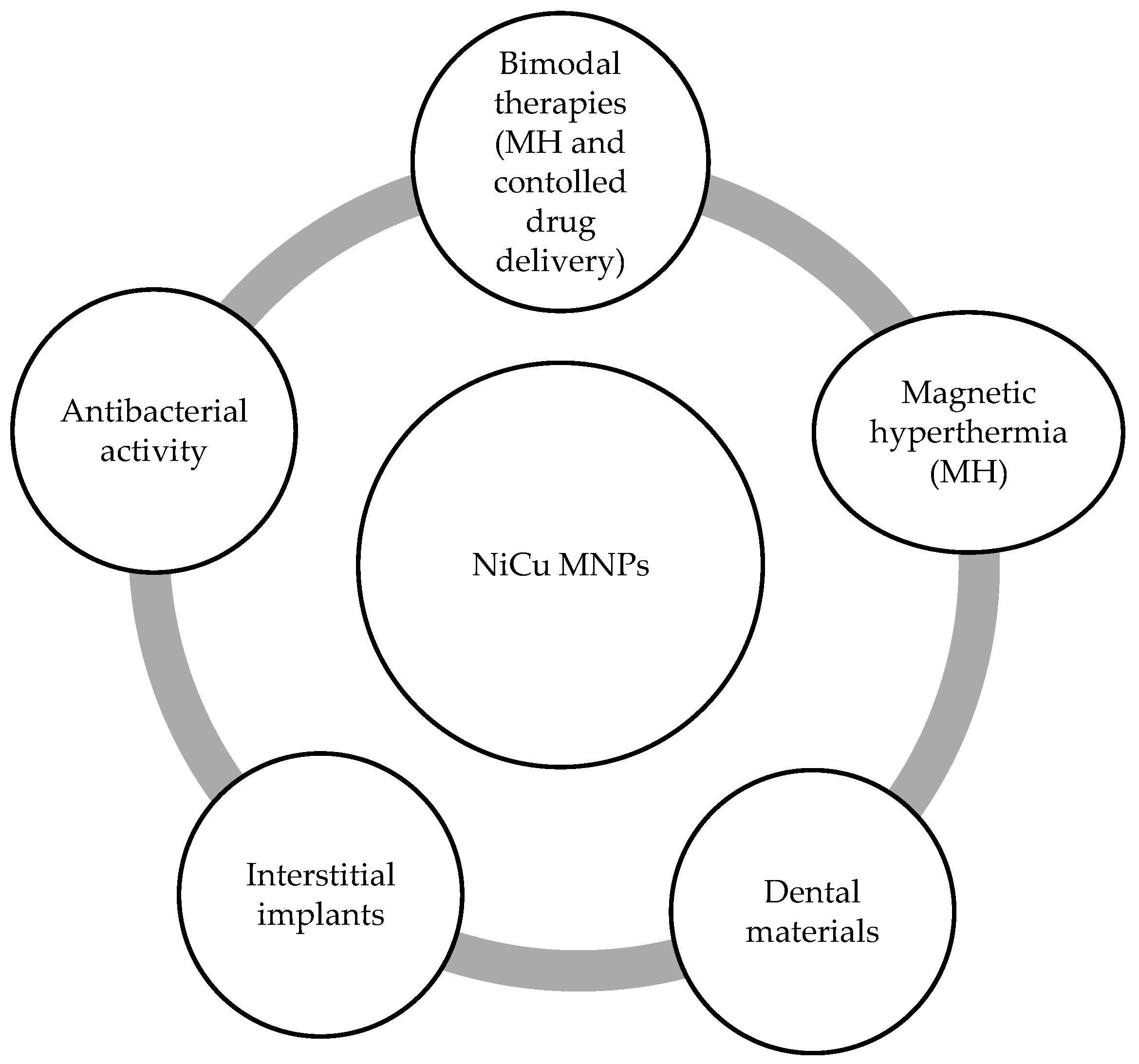
As in the case of other superparamagnetic MNP investigations, most of the NiCu MNP research studies focused on MH applications [36,37,38,39,40,47,48,60,79,99]. In addition to the studies mentioned above, newer studies reported their potential use in bimodal cancer treatment formulations (e.g., a combination of MH and controlled drug delivery) [16], as part of medical implants [49,101], in formulations with antibacterial activity [102], and as part of dental materials [103]. The potential uses of NiCu MNPs in biomedicine are summarized in Figure 13.
Because of the steadily increasing number of research studies on NiCu-based materials for these and other types of applications, this section reviews their different uses, with an explicit focus on synthesis, functionalization, and other aspects that make them appropriate for biomedical purposes.
4.1. Magnetic Hyperthermia
MH is a promising non-invasive type of cancer therapy that makes use of MNPs that generate heat under controllable external stimuli. It uses a combination of an AMF and biocompatible MNPs with a TC value as close as possible to 42–46 °C as heating agents. This temperature interval was proven to be ideal for destroying cancer cells with minimal harm to the healthy surrounding tissues [17]. Therefore, it is important to understand the underlying physical mechanisms via which heat is generated in small MNPs by AMFs [104]. Among the various available MNPs, NiCu MNPs with a tailorable TC in the temperature interval 42–46 °C seem ideally suited for future effective MH formulations [36,38,39,47,48].
Bettge et al. [48] developed a new, relatively simple procedure for NiCu alloy particle synthesis that was based on the combination of ball milling and the melting of bulk materials. The resulting formulation consisted of particles with sizes below a micron that were shown to be good candidate materials for self-controlled MH treatment. The milled particles were found to be slightly above 400 nm. However, their most important finding was that the prepared formulation exhibited a TC in the desired therapeutic range (46–47 °C), making them suitable for self-regulating MH. Using TEM analysis, the authors further determined that the particles were not uniform in shape, with some of them being spherical and the others having a flake-like appearance [48].
Chatterjee et al. [47] prepared novel NiCu NPs with a PEG surface layer using the polyol reduction technique. In addition to the other advantages of the PEG coating (e.g., decreased agglomeration, improved biocompatibility), a beneficial effect was observed on the TC, which fell from ~77 °C to the desired ~46 °C. Furthermore, the coated NPs showed a suitable saturation magnetization of 6–8 emu/g. These findings all suggest that the prepared PEG-functionalized NPs are promising materials for use in MH [47].
Kuznetsov et al. [38] produced alloy NiCu MNPs using various methods. The authors reported that the best results were obtained by the co-precipitation of Cu and Ni salts in the presence of Na2CO3 at room temperature, with a consecutive reduction in a H2 atmosphere (at 300–1000 °C). This initial particle preparation step was followed by various grinding/separation methods to achieve more uniform NP sizes, as well as a more homogenous composition. This step also enabled the control of the formulation’s TC and, thus, increased their potential for use in MH. The potential applicability of the prepared materials to cancer treatment was shown by testing this formulation on a rat liver tumor model. The results showed their effective potential use as part of magnetic fluid hyperthermia [38].
Ban et al. [36] showed the potential of a simple milling-based method to produce nanosized NiCu alloy NPs with potential application in MH. By optimizing the milling process, they were able to produce nanocrystalline Ni72.5Cu27.5 alloy NPs with an average size of 10 nm. Further, the particles exhibited a TC in the MH suitable therapeutic range (~45 °C). The authors proved the superparamagnetic nature of the prepared NiCu NPs by showing that the hysteresis loops lacked remanent magnetization. Significant heat induction of the powdered samples indicated that they are highly promising for use in self-regulating MH [36].
Stergar et al. [101] reported another interesting synthesis approach to preparing NixCu1−x NPs in a water/oil (W/O) microemulsion. Although the particles were partially agglomerated (confirmed using TEM analysis), they exhibited an average size of ~30 nm. The most likely explanation for the determined TC being very close to that of pure Ni was that the particles showed a core/shell structure. To homogenize the sample, the authors performed a heating step in a reducing atmosphere in a tube furnace at 900 °C. This led to the formation of a solid solution of Ni/Cu, which had an expected Tc value of ~65 °C according to the formulation’s nominal composition. Again, a silica coating proved to be the right choice to prevent particle agglomeration without affecting the TC value [60]. Although the developed synthesis method is very promising for the preparation of a versatile range of NiCu MNPs, the authors noted that further optimizations of the formulation are necessary to achieve a Tc in a range that is suitable for use in MH.
Ferk et al. [40] developed a sol–gel-based NiCu MNP preparation method, which yielded NPs with a narrow size distribution. The method included the reduction of Ni and Cu oxides in silica, leading to the formation of Ni1−x Cux MNPs in SiO2. To obtain pure particles, the authors etched the silica matrix using NaOH/hydrazine hydrate. Through this processing, only Ni67.5Cu32.5 nanospheres with a narrow size distribution were retained in the sample. Thermogravimetric analysis (TGA)/single differential thermal analysis (SDTA) was used to determine a TC value of ~65 °C for the samples [40]. This was the first study to report the successful production of superparamagnetic NiCu nanospheres with a narrow size distribution and the capability of controlling the TC for potential application in MH [40].
Amrollahi et al. [39] used mechanical milling to produce Ni0.5Cu0.5 NPs for potential use in MH applications. The ball milling-based initial synthesis stage (30 h) was concluded with a heating step for 1 h at 500 °C. Using XRD analysis, the authors showed that only single-phase NPs were formed. The particle size and rate of agglomeration were established using TEM. The authors reported a narrow size distribution of particles with an average size of ~20 nm, although these were mostly part of larger agglomerates. Nevertheless, the potential of the prepared NPs for use in MH was proven by their measured TC of 44 °C. To further demonstrate the suitability of these NPs for MH, the authors performed biocompatibility/cytotoxicity testing using human bone marrow stem cells (hMSCs) and proved that the formulation did not harm them [39]. A summary of various other related research studies that reported the use of NiCu MNPs in MH (including some of their most important properties) is shown in Table 2.
4.2. Bimodal Cancer Therapy (A Combination of MH and Controlled Drug Delivery)
Controlled drug delivery systems have several advantages compared with traditional pharmaceutical formulations. For example, they enable drug transportation to the desired site of action in the body with minimal damage to healthy surrounding tissues. Delivery systems with these characteristics are key for drugs that exhibit a narrow therapeutic index or drugs that are directly cytotoxic to healthy cells (e.g., most antitumor drugs) [17]. Recent findings suggest that NiCu MNPs are very promising for the combination of MH and controlled drug delivery [17].
Stergar et al. [16] prepared a novel controlled drug delivery formulation composed of Ni67.5Cu32.5 MNPs in a silica matrix synthesized using the sol–gel procedure. They used the fluorescent dye rhodamine 6G to evaluate the ability of MNPs to deliver the drug to various human cells (a healthy cell line of human skin-derived fibroblasts and the cancerous cell lines HeLa and Caco-2). The progressive Ni67.5Cu32.5 MNPs were shown to be a dependable (confirmed through biocompatibility and cytotoxicity assays) and efficient delivery system (in vitro cell delivery model) to various human cell types. The obtained results indicate that the proposed formulation has a very high potential for the future development of drug delivery systems that specifically target these cells. The process explored the protection of particles during drug delivery and MH, and the results confirmed the successful application of Ni67.5Cu32.5 particles with a TC of about 43 °C and drug accumulation in a silica protecting layer.
4.3. MH Implants
Interstitial hyperthermia using magnetic materials is known as magnetic induction hyperthermia. Soft heating or implant heating systems are excellent systems for this purpose. In these systems, the ferromagnetic material (implant) is firstly implanted in the cancer tissue, followed by the utilization of an eddy current or magnetic hysteresis loss under a high-frequency AMF to generate highly localized heating. The temperature of the cancer tissue is raised and regulated at the TC of the implant material [105].
Paulus et al. [49] explored novel, low-Tc ferromagnetic NiCu (28 wt.% Ni) and PdCo (6.15 wt.% Pd) formulations as part of interstitial implants in prostate cancer therapy. Initially, they performed a melting step, in which both alloys were put into a carbon arc furnace. This step was followed by a heat treatment step to induce sample recrystallization (at 1000 °C). The annealed NiCu and PdCo samples were spot-welded to Cu wire connections, and the junctions were closed using a water-resistant epoxy adhesive. Potential contaminants on the sample surface were removed by polishing using a 1-μm diamond paste. The as-prepared samples proved to be more corrosion-resistant, even after longer immersion times. The authors explained this phenomenon by the formation of passivation layers on the sample surface. It was further established that the PdCo alloy outperformed the NiCu sample with regard to the corrosion stabilization time in solution. This finding was additionally backed by proving the excellent corrosion resistance of this sample in vitro. On the basis of all the findings, the authors concluded that the developed formulations are promising for the intended use, even in long-term hyperthermia applications [49].
El-Sayed et al. [101] developed a manufacturing procedure of PdNi, PdCo, and NiCu ferromagnetic thermoseeds to increase the sharpness of TC for interstitial hyperthermia in cancer treatment. They characterized the prepared seeds by magnetic measurements as a function of temperature and magnetic field strength. The ferromagnetic thermoseeds showed a sharp decrease in ferromagnetic to paramagnetic transition temperatures at 52, 57, and 49 °C and the heat production of TC. These seeds provided a power output at 20 °C of about 150, 200, and 75 mW/cm, as measured from the hysteresis loops. The temperature dependence of the seed power was computed from a combination of Curie and induction laws. Their computations showed a good agreement with the values calculated from the area under the hysteresis loops at an AMF with a frequency of 100 kHz and field strength of 4 kA/m. The power automatically stopped when TC was reached, indicating that the seeds had self-limiting temperature control. This self-limitation indicates the high potential of these seeds for potential exploitation in clinics, especially for treating localized tumors [102].
4.4. Antibacterial Activity
Antibacterial activity is related to compounds that locally kill bacteria or slow their growth without being toxic to the surrounding tissue. In general, the agents can be classified as either bactericidal (agents that kill bacteria) or bacteriostatic (agents that slow bacterial growth) [106]. In the area of antibacterial agents, metal nanoparticles are of particular interest because of their high surface areas and, hence, their large number of potential active (antibacterial) sites per unit area. A distinct class of metal oxides with distinctive magnetic properties and superior biocompatibility is iron-oxide NPs [107]. Indeed, various metal NPs demonstrated broad-spectrum antibacterial properties against both Gram-positive and Gram-negative bacteria. For example, ZnO NPs were found to inhibit Staphylococcus aureus, and Ag NPs exhibited concentration-dependent antimicrobial activity against Escherichia coli and Pseudomonas aeruginosa [106,107,108,109]. NiCu MNPs were also previously studied for the same purpose. The example below shows their potential uses for this purpose.
Parimaladevi et al. [102] synthesized Cu, Ni, and CuNi bimetallic NPs through the solvothermal method. They added CuSO4 and NaOH, NiNO3 and NaOH, and CuSO4 and NiNO3 (1:1) to hydrazine (reducing agent) and ethylene glycol (solvent). The mixture was stirred for 15 min and transferred to an autoclave (150 °C, 5 h). The obtained precipitate was separated by centrifugation and washed repeatedly with water and ethanol. The antibacterial activity of Cu, Ni, and CuNi NPs was evaluated against Gram-positive Staphylococcus aureus and Gram-negative Escherichia coli using a standard agar well diffusion method. A standard antibacterial drug, cefixime, was used in each plate. The test results against the Gram-positive bacterium S. aureus showed a higher inhibition compared with the results against Gram-negative E. coli for all NPs. Larger zones of inhibition were recorded for CuNi NPs than the monometallic NPs; this observation is most likely the result of the unique distribution of the two different metals, enabling the simultaneous release (and activity) of two different metal ions, namely, Cu2+ and Ni2+. The observed toxicity was similar to that of cefixime, suggesting that the CuNi NPs were utilized in abundance at sites of possible infections, such as pneumonia and strep throat. The cost-effectiveness of CuNi NPs and their lethality toward both Gram-positive and Gram-negative bacteria may lead to numerous applications, such as antibacterial coatings, paints, surfaces, and films [102].
4.5. Dental Materials
There are various applications of NPs (carbon-based nanomaterials, hydroxyapatite, iron oxide, zirconia, silica, silver, and titania) in dentistry [110]. Biocomposite materials (e.g., tiny calcium phosphate crystallites) that bear structural and chemical similarity to hydroxyapatite were shown to effectively integrate into growing human bone. This is related to the fact that hydroxyapatite is a natural nanocomposite in the body that is formed naturally by bone-derived cells. Typically, these materials are used as part of implant coatings to improve the implant’s overall biocompatibility and often contribute to better wear resistance; both of these functions are important in the preparation of bone grafts. Recently, novel methods in dentistry were developed in which organically modified ceramic and its fillers are used to enhance dispersion and biocompatibility, accompanied by a simultaneous increase in the toughness of the implant. Iron-oxide nanoparticles are very useful for eradicating biofilms on dental implants. Alumina/zirconia nanocomposites are new implant materials that show better efficacy compared with ceramic materials. Zirconia-oxide nanoparticles were found to have anti-biofilm activity against certain bacteria (such as Enterococcus faecalis); therefore, they can be used effectively as polishing agents in dental practices [110,111]. NiCu MNPs are also emerging as potential materials for future use in dentistry. Some examples are described below.
Argueta-Figueroa et al. [103] developed various single-metal and bimetallic alloy materials based on Ni and Cu and tested their antibacterial activity. The main purpose of their study was to evaluate the formulation for use in dentistry. The bimetallic NiCu MNP formulation was prepared by chemical reduction with NaBH4. Antibacterial activity was assessed against three different bacteria, namely, Gram-positive Staphylococcus aureus, Gram-negative Escherichia coli, and the more dentistry-specific pathogen Streptococcus mutans (Gram-positive). Using the developed preparation method for the NiCu MNPs, the authors obtained a combination of three crystal structures, namely, both metal oxides and the alloy (determined through XRD analysis). Of all the synthesis parameters, pH had one of the strongest influences on the final ratio between the metals in the samples. A narrow size distribution and an average size of around 25 nm were found. The as-prepared Cu NPs showed a bactericidal effect on all three tested bacteria, whereas the Ni NPs and the bimetallic NiCu NPs were only bacteriostatic. From the obtained results, the authors concluded that the prepared materials have great potential for future use in dentistry [103].
5. Biosafety Considerations
As is the case with all other formulations for human use, NiCu MNPs must be appropriately evaluated for their safety. This is the most important evaluation since both metal elements can have toxic effects on the human body [112]. Nevertheless, as we showed in one of our previous (in vitro) studies [16], the biocompatibility of Ni67.5Cu32.5 MNPs in a silica matrix should not pose a problem. For the time span of the performed study, we are also positive that no leaching of metal ions occurred, which might have negatively affected cell growth [16]. As an integral part of this review, we also present various strategies of NiCu MNP functionalization. The purpose of functionalization is often to improve the biocompatibility of the core materials with the target (human body) environment. Of the most frequently used “shells”, silica is by far the most common [90,92]. Therefore, the developed MNPs have very high potential for the future development of drug delivery systems for specific applications targeting cells (and, hence, tissues). Since the core nanoalloy compositions also vary, it is also important to consider Ni/Cu ratios. For example, MNPs with the composition Cu0.5Ni0.5 were demonstrated to be safe for cancer treatment applications [39].
Although we understand that further studies are necessary to prove the “full” safety of NiCu MNPs for potential patients, we also acknowledge that there are already available strategies that can diminish even potential toxic effects (the bar for potential risks posed by chemotherapy agents is set relatively low) that might occur with Ni or Cu degradation products [113]. One strategy could be to take advantage of their superior magnetic properties to achieve MH with low MNP concentrations, which are non-toxic. Alternatively, as shown in Section 3, a core/shell strategy to mitigate toxicity can significantly increase the safety of the core material.
6. Conclusions
The presented review summarizes the most important aspects of the preparation of NiCu MNPs with regard to their potential use in biomedicine. This includes different possible synthesis procedures, as well as the necessary functionalization/modification steps that need to be taken to either boost their biocompatibility or prevent agglomeration, which are both factors that contribute to their improved properties for application. As can be concluded from the currently available literature, which grew for the last couple of years, NiCu MNPs could be among future formulations for use as MH formulations, drug delivery systems, or other applications. With an already proven track record of successful application (currently only in in vitro and animal models), the potential of NiCu MNPs will only grow; many research groups are now testing their suitability using various additional tissue models to determine their application as part of the abovementioned uses or even in regenerative medicine, which was not addressed above [114].
Nevertheless, NiCu MNPs do not come without hurdles. Future challenges in their development certainly include the optimization of their synthesis and functionalization, which need to be green, to yield uniform small-sized nanoparticles with a narrow size distribution that are less prone to agglomeration. Additionally, careful composition control is necessary to prepare NiCu MNPs with a TC in the therapeutic range to minimize potential damage to healthy tissues. By meeting all of these criteria, their potential in biomedical applications will almost be without limitations, either as part of formulations for “traditional” applications of magnetic materials (e.g., MH) or as part of novel multi-modal drug delivery systems or dentistry implants that exhibit antibacterial activity.
Funding
This research was funded by Slovenian National Agency, grant numbers P3-0036, J1-9169, and J3-1762.
Conflicts of Interest
The authors declare no conflicts of interest.
References
- Liu, Z.; Kiessling, F.; Gätjens, J. Advanced Nanomaterials in Multimodal Imaging: Design, Functionalization, and Biomedical Applications. J. Nanomater. 2010, 2010, 15. [Google Scholar] [CrossRef] [Green Version]
- Tartaj, P.; del Puerto Morales, M.; Veintemillas-Verdaguer, S.; González-Carreño, T.; Serna, C.J. The preparation of magnetic nanoparticles for applications in biomedicine. J. Phys. D Appl. Phys. 2003, 36, R182. [Google Scholar] [CrossRef]
- Lu, A.-H.; Salabas, E.L.; Schüth, F. Magnetic Nanoparticles: Synthesis, Protection, Functionalization, and Application. Angew. Chem. Int. Ed. 2007, 46, 1222–1244. [Google Scholar] [CrossRef] [PubMed]
- Mornet, S.; Vasseur, S.; Grasset, F.; Veverka, P.; Goglio, G.; Demourgues, A.; Portier, J.; Pollert, E.; Duguet, E. Magnetic nanoparticle design for medical applications. Progr. Solid State Chem. 2006, 34, 237–247. [Google Scholar] [CrossRef]
- Dürr, S.; Janko, C.; Lyer, S.; Tripal, P.; Schwarz, M.; Zaloga, J.; Tietze, R.; Alexiou, C. Magnetic nanoparticles for cancer therapy. Nanotechnol. Rev. 2013, 2, 395–409. [Google Scholar] [CrossRef] [Green Version]
- Ahola, S.; Salmi, J.; Johansson, L.S.; Laine, J.; Osterberg, M. Model films from native cellulose nanofibrils. Preparation, swelling, and surface interactions. Biomacromolecules 2008, 9, 1273–1282. [Google Scholar] [CrossRef]
- Park, J.W.; Lee, G.U. Properties of mixed lipid monolayers assembled on hydrophobic surfaces through vesicle adsorption. Langmuir 2006, 22, 5057–5063. [Google Scholar] [CrossRef]
- Gao, Y.; Liu, Y.; Xu, C. Magnetic Nanoparticles for Biomedical Applications: From Diagnosis to Treatment to Regeneration. In Engineering in Translational Medicine; Cai, W., Ed.; Springer: London, UK, 2014; pp. 567–583. [Google Scholar] [CrossRef]
- Tweedle, M.F. The Chemistry of Contrast Agents in Medical Magnetic Resonance Imaging Edited by André E. Merbach and Éva Tóth (University of Lausanne). J. Wiley & Sons: Chichester, New York, Weinheim, Brisbane, Singapore, Toronto. 2001. xii + 472 pp. $160.00. ISBN: 0-471-60778-9. J. Am. Chem. Soc. 2002, 124, 884–885. [Google Scholar] [CrossRef]
- Catherine, C.B.; Curtis, A.S.G. Functionalisation of magnetic nanoparticles for applications in biomedicine. J. Phys. D Appl. Phys. 2003, 36, R198. [Google Scholar]
- Le Trequesser, Q.; Seznec, H.; Delville, M.-H. Functionalized nanomaterials: Their use as contrast agents in bioimaging: Mono- and multimodal approaches. Nanotechnol. Rev. 2013, 2, 125. [Google Scholar] [CrossRef] [Green Version]
- Portet, D.; Denizot, B.; Rump, E.; Lejeune, J.-J.; Jallet, P. Nonpolymeric Coatings of Iron Oxide Colloids for Biological Use as Magnetic Resonance Imaging Contrast Agents. J. Colloid Interface Sci. 2001, 238, 37–42. [Google Scholar] [CrossRef] [PubMed]
- Issa, B.; Obaidat, I.M.; Albiss, B.A.; Haik, Y. Magnetic nanoparticles: Surface effects and properties related to biomedicine applications. Int. J. Mol. Sci. 2013, 14, 21266–21305. [Google Scholar] [CrossRef] [PubMed] [Green Version]
- Jordan, A.; Scholz, R.; Maier-Hauff, K.; Johannsen, M.; Wust, P.; Nadobny, J.; Schirra, H.; Schmidt, H.; Deger, S.; Loening, S.; et al. Presentation of a new magnetic field therapy system for the treatment of human solid tumors with magnetic fluid hyperthermia. J. Magn. Magn. Mater. 2001, 225, 118–126. [Google Scholar] [CrossRef] [Green Version]
- Wong, J.; Prout, J.; Seifalian, A. Magnetic Nanoparticles: New Perspectives in Drug Delivery. Curr. Pharm. Des. 2017, 23, 2908–2917. [Google Scholar] [CrossRef]
- Stergar, J.; Ban, I.; Gradišnik, L.; Maver, U. Novel drug delivery system based on NiCu nanoparticles for targeting various cells. J. Sol-Gel Sci. Technol. 2018, 88, 57–65. [Google Scholar] [CrossRef]
- Kumar, C.S.S.R.; Mohammad, F. Magnetic nanomaterials for hyperthermia-based therapy and controlled drug delivery. Adv. Drug Deliv. Rev. 2011, 63, 789–808. [Google Scholar] [CrossRef] [Green Version]
- Bernkop-Schnurch, A.; Walker, G. Multifunctional matrices for oral peptide delivery. Crit. Rev. Ther. Drug Carr. Syst. 2001, 18, 459–501. [Google Scholar] [CrossRef]
- Chomoucka, J.; Drbohlavova, J.; Huska, D.; Adam, V.; Kizek, R.; Hubalek, J. Magnetic nanoparticles and targeted drug delivering. Pharmacol. Res. 2010, 62, 144–149. [Google Scholar] [CrossRef]
- Salunkhe, A.B.; Khot, V.M.; Pawar, S.H. Magnetic hyperthermia with magnetic nanoparticles: A status review. Curr. Top. Med. Chem. 2014, 14, 572–594. [Google Scholar] [CrossRef]
- Laurent, S.; Dutz, S.; Häfeli, U.O.; Mahmoudi, M. Magnetic fluid hyperthermia: Focus on superparamagnetic iron oxide nanoparticles. Adv. Colloid Interface Sci. 2011, 166, 8–23. [Google Scholar] [CrossRef]
- Sun, C.; Lee, J.S.; Zhang, M. Magnetic nanoparticles in MR imaging and drug delivery. Adv. Drug Deliv. Rev. 2008, 60, 1252–1265. [Google Scholar] [CrossRef] [PubMed] [Green Version]
- Stephen, Z.R.; Kievit, F.M.; Zhang, M. Magnetite Nanoparticles for Medical MR Imaging. Mater. Today 2011, 14, 330–338. [Google Scholar] [CrossRef]
- Mohammed, L.; Gomaa, H.G.; Ragab, D.; Zhu, J. Magnetic nanoparticles for environmental and biomedical applications: A review. Particuology 2017, 30, 1–14. [Google Scholar] [CrossRef]
- Hauser, A.K.; Wydra, R.J.; Stocke, N.A.; Anderson, K.W.; Hilt, J.Z. Magnetic nanoparticles and nanocomposites for remote controlled therapies. J. Control. Release Off. J. Control. Release Soc. 2015, 219, 76–94. [Google Scholar] [CrossRef] [Green Version]
- McBain, S.C.; Yiu, H.H.P.; Dobson, J. Magnetic nanoparticles for gene and drug delivery. Int. J. Nanomed. 2008, 3, 169–180. [Google Scholar]
- Majidi, S.; Sehrig, F.Z.; Samiei, M.; Milani, M.; Abbasi, E.; Dadashzadeh, K.; Akbarzadeh, A. Magnetic nanoparticles: Applications in gene delivery and gene therapy. Artif. Cells Nanomed. Biotechnol. 2016, 44, 1186–1193. [Google Scholar] [CrossRef]
- Šafařík, I.; Šafaříková, M. Magnetic Nanoparticles and Biosciences. Monatshefte Chem. Chem. Mon. 2002, 133, 737–759. [Google Scholar] [CrossRef]
- Ludwig, F.; Heim, E.; Mäuselein, S.; Eberbeck, D.; Schilling, M. Magnetorelaxometry of magnetic nanoparticles with fluxgate magnetometers for the analysis of biological targets. J. Magn. Magn. Mater. 2005, 293, 690–695. [Google Scholar] [CrossRef]
- Wiekhorst, F.; Steinhoff, U.; Eberbeck, D.; Trahms, L. Magnetorelaxometry assisting biomedical applications of magnetic nanoparticles. Pharm. Res. 2012, 29, 1189–1202. [Google Scholar] [CrossRef] [Green Version]
- Ismail, R.A.; Sulaiman, G.M.; Abdulrahman, S.A.; Marzoog, T.R. Antibacterial activity of magnetic iron oxide nanoparticles synthesized by laser ablation in liquid. Mater. Sci. Eng. C 2015, 53, 286–297. [Google Scholar] [CrossRef]
- Thukkaram, M.; Sitaram, S.; Kannaiyan, S.K.; Subbiahdoss, G. Antibacterial Efficacy of Iron-Oxide Nanoparticles against Biofilms on Different Biomaterial Surfaces. Int. J. Biomater. 2014, 2014, 716080. [Google Scholar] [CrossRef] [PubMed] [Green Version]
- Ito, A.; Shinkai, M.; Honda, H.; Kobayashi, T. Medical application of functionalized magnetic nanoparticles. J. Biosci. Bioeng. 2005, 100, 1–11. [Google Scholar] [CrossRef] [PubMed] [Green Version]
- Mok, H.; Zhang, M. Superparamagnetic iron oxide nanoparticle-based delivery systems for biotherapeutics. Expert Opin. Drug Deliv. 2013, 10, 73–87. [Google Scholar] [CrossRef] [PubMed] [Green Version]
- Williams, H.M. The application of magnetic nanoparticles in the treatment and monitoring of cancer and infectious diseases. Biosci. Horiz. Int. J. Stud. Res. 2017, 10, hzx009. [Google Scholar] [CrossRef] [Green Version]
- Ban, I.; Stergar, J.; Drofenik, M.; Ferk, G.; Makovec, D. Synthesis of copper–nickel nanoparticles prepared by mechanical milling for use in magnetic hyperthermia. J. Magn. Magn. Mater. 2011, 323, 2254–2258. [Google Scholar] [CrossRef]
- Ferk, G.; Stergar, J.; Makovec, D.; Hamler, A.; Jagličić, Z.; Drofenik, M.; Ban, I. Synthesis and characterization of Ni–Cu alloy nanoparticles with a tunable Curie temperature. J. Alloys Compd. 2015, 648, 53–58. [Google Scholar] [CrossRef]
- Kuznetsov, A.A.; Leontiev, V.G.; Brukvin, V.A.; Vorozhtsov, G.N.; Kogan, B.Y.; Shlyakhtin, O.A.; Yunin, A.M.; Tsybin, O.I.; Kuznetsov, O.A. Local radiofrequency-induced hyperthermia using CuNi nanoparticles with therapeutically suitable Curie temperature. J. Magn. Magn. Mater. 2007, 311, 197–203. [Google Scholar] [CrossRef]
- Amrollahi, P.; Ataie, A.; Nozari, A.; Seyedjafari, E.; Shafiee, A. Cytotoxicity Evaluation and Magnetic Characteristics of Mechano-thermally Synthesized CuNi Nanoparticles for Hyperthermia. J. Mater. Eng. Perform. 2015, 24, 1220–1225. [Google Scholar] [CrossRef]
- Ferk, G.; Stergar, J.; Drofenik, M.; Makovec, D.; Hamler, A.; Jagličić, Z.; Ban, I. The synthesis and characterization of nickel–copper alloy nanoparticles with a narrow size distribution using sol–gel synthesis. Mater. Lett. 2014, 124, 39–42. [Google Scholar] [CrossRef]
- Songping, W.; Jing, N.; Li, J.; Zhenou, Z. Preparation of ultra-fine copper–nickel bimetallic powders with hydrothermal–reduction method. Mater. Chem. Phys. 2007, 105, 71–75. [Google Scholar] [CrossRef]
- Feng, J.; Zhang, C.-P. Preparation of Cu–Ni alloy nanocrystallites in water-in-oil microemulsions. J. Colloid Interface Sci. 2006, 293, 414–420. [Google Scholar] [CrossRef] [PubMed]
- Hergt, R.; Andra, W.; d’Ambly, C.G.; Hilger, I.; Kaiser, W.A.; Richter, U.; Schmidt, H.G. Physical limits of hyperthermia using magnetite fine particles. IEEE Trans. Magn. 1998, 34, 3745–3754. [Google Scholar] [CrossRef]
- Jordan, A.; Scholz, R.; Wust, P.; Fähling, H.; Roland, F. Magnetic fluid hyperthermia (MFH): Cancer treatment with AC magnetic field induced excitation of biocompatible superparamagnetic nanoparticles. J. Magn. Magn. Mater. 1999, 201, 413–419. [Google Scholar] [CrossRef]
- Chang, D.; Lim, M.; Goos, J.; Qiao, R.; Ng, Y.Y.; Mansfeld, F.M.; Jackson, M.; Davis, T.P.; Kavallaris, M. Biologically Targeted Magnetic Hyperthermia: Potential and Limitations. Front. Pharmacol. 2018, 9, 831. [Google Scholar] [CrossRef] [Green Version]
- Andra, W.; Nowak, H. Magnetism in Medicine: A Handbook, 2nd ed.; Wiley-VCH: Weinheim, Germany, 2007; 629p. [Google Scholar]
- Chatterjee, J.; Bettge, M.; Haik, Y.; Jen Chen, C. Synthesis and characterization of polymer encapsulated Cu–Ni magnetic nanoparticles for hyperthermia applications. J. Magn. Magn. Mater. 2005, 293, 303–309. [Google Scholar] [CrossRef]
- Bettge, M.; Chatterjee, J.; Haik, Y. Physically synthesized Ni-Cu nanoparticles for magnetic hyperthermia. Biomagn. Res. Technol. 2004, 2, 4. [Google Scholar] [CrossRef] [Green Version]
- Paulus, J.A.; Parida, G.R.; Tucker, R.D.; Park, J.B. Corrosion analysis of NiCu and PdCo thermal seed alloys used as interstitial hyperthermia implants. Biomaterials 1997, 18, 1609–1614. [Google Scholar] [CrossRef]
- Abenojar, E.C.; Wickramasinghe, S.; Bas-Concepcion, J.; Samia, A.C.S. Structural effects on the magnetic hyperthermia properties of iron oxide nanoparticles. Prog. Nat. Sci. Mater. 2016, 26, 440–448. [Google Scholar] [CrossRef] [Green Version]
- Engelmann, U.M.; Roeth, A.A.; Eberbeck, D.; Buhl, E.M.; Neumann, U.P.; Schmitz-Rode, T.; Slabu, I. Combining Bulk Temperature and Nanoheating Enables Advanced Magnetic Fluid Hyperthermia Efficacy on Pancreatic Tumor Cells. Sci. Rep. 2018, 8, 13210. [Google Scholar] [CrossRef]
- Soetaert, F.; Kandala, S.K.; Bakuzis, A.; Ivkov, R. Experimental estimation and analysis of variance of the measured loss power of magnetic nanoparticles. Sci. Rep. 2017, 7, 6661. [Google Scholar] [CrossRef] [Green Version]
- Allia, P.; Barrera, G.; Tiberto, P. Nonharmonic Driving Fields for Enhancement of Nanoparticle Heating Efficiency in Magnetic Hyperthermia. Phys. Rev. Appl. 2019, 12, 034041. [Google Scholar] [CrossRef]
- Sue, K.; Tanaka, S.; Hiaki, T. Synthesis of Ni–Cu Particles by Hydrogen Reduction in Hot-compressed Water. Chem. Lett. 2006, 35, 50–51. [Google Scholar] [CrossRef]
- Wang, L.-L.; Jiang, J.-S. Preparation of α-Fe2O3 nanoparticles by high-energy ball milling. Phys. B Condens. Matter 2007, 390, 23–27. [Google Scholar] [CrossRef]
- Ali, M.E.; Ullah, M.; Maamor, A.; Hamid, S.B.A. Surfactant Assisted Ball Milling: A Simple Top down Approach for the Synthesis of Controlled Structure Nanoparticle. Adv. Mater. Res. 2014, 832, 356–361. [Google Scholar] [CrossRef]
- Durivault, L.; Brylev, O.; Reyter, D.; Sarrazin, M.; Bélanger, D.; Roué, L. Cu–Ni materials prepared by mechanical milling: Their properties and electrocatalytic activity towards nitrate reduction in alkaline medium. J. Alloys Compd. 2007, 432, 323–332. [Google Scholar] [CrossRef]
- Ge, Y.; Gao, T.; Wang, C.; Shah, Z.H.; Lu, R.; Zhang, S. Highly efficient silica coated CuNi bimetallic nanocatalyst from reverse microemulsion. J. Colloid Interface Sci. 2017, 491, 123–132. [Google Scholar] [CrossRef]
- Ahmed, J.; Ramanujachary, K.V.; Lofland, S.E.; Furiato, A.; Gupta, G.; Shivaprasad, S.M.; Ganguli, A.K. Bimetallic Cu–Ni nanoparticles of varying composition (CuNi3, CuNi, Cu3Ni). Colloids Surf. A Physicochem. Eng. Asp. 2008, 331, 206–212. [Google Scholar] [CrossRef]
- Stergar, J.; Ban, I.; Drofenik, M.; Ferk, G.; Makovec, D. Synthesis and Characterization of Silica-Coated Cu1−xNix Nanoparticles; Institute of Electrical and Electronics Engineers: New York, NY, USA, 2012; p. 4. [Google Scholar]
- Wen, M.; Liu, Q.-Y.; Wang, Y.-F.; Zhu, Y.-Z.; Wu, Q.-S. Positive microemulsion synthesis and magnetic property of amorphous multicomponent Co-, Ni- and Cu-based alloy nanoparticles. Colloids Surf. A Physicochem. Eng. Asp. 2008, 318, 238–244. [Google Scholar] [CrossRef]
- Wang, X.L.; Ben Ahmed, N.; Alvarez, G.S.; Tuttolomondo, M.V.; Helary, C.; Desimone, M.F.; Coradin, T. Sol-gel Encapsulation of Biomolecules and Cells for Medicinal Applications. Curr. Top. Med. Chem. 2015, 15, 223–244. [Google Scholar] [CrossRef]
- Brinker, C.J.; Scherer, G.W. Sol-Gel Science: The Physics and Chemistry of Sol-Gel Processing; Academic Press: Boston, MA, USA, 1990. [Google Scholar]
- Leontyev, V. Magnetic properties of Ni and Ni–Cu nanoparticles. Phys. Status Solidi (b) 2013, 250, 103–107. [Google Scholar] [CrossRef]
- Hachani, R.; Lowdell, M.; Birchall, M.; Hervault, A.; Mertz, D.; Begin-Colin, S.; Thanh, N.T. Polyol synthesis, functionalisation, and biocompatibility studies of superparamagnetic iron oxide nanoparticles as potential MRI contrast agents. Nanoscale 2016, 8, 3278–3287. [Google Scholar] [CrossRef] [PubMed] [Green Version]
- Songvorawit, N.; Tuitemwong, K.; Tuitemwong, P. Single Step Synthesis of Amino-Functionalized Magnetic Nanoparticles with Polyol Technique at Low Temperature. ISRN Nanotechnol. 2011, 2011, 6. [Google Scholar] [CrossRef] [Green Version]
- Carroll, K.J.; Calvin, S.; Ekiert, T.F.; Unruh, K.M.; Carpenter, E.E. Selective Nucleation and Growth of Cu and Ni Core/Shell Nanoparticles. Chem. Mater. 2010, 22, 2175–2177. [Google Scholar] [CrossRef]
- Bonet, F.; Grugeon, S.; Dupont, L.; Urbina, R.H.; Guéry, C.; Tarascon, J.M. Synthesis and characterization of bimetallic Ni–Cu particles. J. Solid State Chem. 2003, 172, 111–115. [Google Scholar] [CrossRef]
- Jayakrishnan, D.S. 5—Electrodeposition: The versatile technique for nanomaterials. In Corrosion Protection and Control Using Nanomaterials; Saji, V.S., Cook, R., Eds.; Woodhead Publishing: Sawston, UK, 2012; pp. 86–125. [Google Scholar] [CrossRef]
- Foyet, A.; Hauser, A.; Schäfer, W. Double template electrochemical deposition and characterization of NiCo and NiCu alloys nanoparticles and nanofilms. J. Solid State Electrochem. 2007, 12, 47–55. [Google Scholar] [CrossRef]
- Feng, S.H.; Li, G.H. Chapter 4—Hydrothermal and Solvothermal Syntheses. In Modern Inorganic Synthetic Chemistry, 2nd ed.; Xu, R., Xu, Y., Eds.; Elsevier: Amsterdam, The Netherlands, 2017; pp. 73–104. [Google Scholar] [CrossRef]
- Songping, W.; Li, J.; Jing, N.; Zhenou, Z.; Song, L. Preparation of ultra fine copper–nickel bimetallic powders for conductive thick film. Intermetallics 2007, 15, 1316–1321. [Google Scholar] [CrossRef]
- Kwon, Y.S.; An, V.V.; Ilyin, A.P.; Tikhonov, D.V. Properties of powders produced by electrical explosions of copper–nickel alloy wires. Mater. Lett. 2007, 61, 3247–3250. [Google Scholar] [CrossRef]
- Bahlawane, N.; Premkumar, P.A.; Tian, Z.; Hong, X.; Qi, F.; Kohse-Höinghaus, K. Nickel and Nickel-Based Nanoalloy Thin Films from Alcohol-Assisted Chemical Vapor Deposition. Chem. Mater. 2010, 22, 92–100. [Google Scholar] [CrossRef]
- Pál, E.; Kun, R.; Schulze, C.; Zöllmer, V.; Lehmhus, D.; Bäumer, M.; Busse, M. Composition-dependent sintering behaviour of chemically synthesised CuNi nanoparticles and their application in aerosol printing for preparation of conductive microstructures. Colloid Polym. Sci. 2012, 290, 941–952. [Google Scholar] [CrossRef]
- Zin, V.; Brunelli, K.; Dabalà, M. Characterization of Cu–Ni alloy electrodeposition and synthesis of nanoparticles by pulsed sonoelectrochemistry. Mater. Chem. Phys. 2014, 144, 272–279. [Google Scholar] [CrossRef]
- Sopousek, J.; Vrestal, J.; Pinkas, J.; Broz, P.; Bursik, J.; Styskalik, A.; Skoda, D.; Zobac, O.; Lee, J. Cu–Ni nanoalloy phase diagram—Prediction and experiment. Calphad 2014, 45, 33–39. [Google Scholar] [CrossRef] [Green Version]
- de León-Quiroz, E.L.; Puente-Urbina, B.A.; Vázquez-Obregón, D.; García-Cerda, L.A. Preparation and structural characterization of CuNi nanoalloys obtained by polymeric precursor method. Mater. Lett. 2013, 91, 67–70. [Google Scholar] [CrossRef]
- Stergar, J.; Ferk, G.; Ban, I.; Drofenik, M.; Hamler, A.; Jagodič, M.; Makovec, D. The synthesis and characterization of copper–nickel alloy nanoparticles with a therapeutic Curie point using the microemulsion method. J. Alloys Compd. 2013, 576, 220–226. [Google Scholar] [CrossRef]
- Ban, I.; Stergar, J.; Maver, U. NiCu magnetic nanoparticles: Review of synthesis methods, surface functionalization approaches, and biomedical applications. Nanotechnol. Rev. 2018, 7, 187. [Google Scholar] [CrossRef]
- Hao, R.; Xing, R.; Xu, Z.; Hou, Y.; Gao, S.; Sun, S. Synthesis, Functionalization, and Biomedical Applications of Multifunctional Magnetic Nanoparticles. Adv. Mater. 2010, 22, 2729–2742. [Google Scholar] [CrossRef]
- An, L.; Li, Z.; Wang, Y.; Yang, B. Synthesis of Fe3O4/PMMA Nanocomposite Particles by Surface-Initiated ATRP and Characterization. Chem. J. Chin. Univ. 2006, 27, 1372–1375. [Google Scholar]
- Morales, M.A.; Finotelli, P.; Coaquira, J.A.H.; Rocha-Leão, M.H.; Diaz-Aguila, C.; Baggio-Saitovitch, E.; Rossi, A.M. In situ synthesis and magnetic studies of iron oxide nanoparticles in calcium-alginate matrix for biomedical applications. Mater. Sci. Eng. C 2008, 28, 253–257. [Google Scholar] [CrossRef]
- Yallapu, M.M.; Foy, S.P.; Jain, T.K.; Labhasetwar, V. PEG-functionalized magnetic nanoparticles for drug delivery and magnetic resonance imaging applications. Pharmaceut. Res. 2010, 27, 2283–2295. [Google Scholar] [CrossRef] [Green Version]
- Kim, D.K.; Mikhaylova, M.; Wang, F.H.; Kehr, J.; Bjelke, B.; Zhang, Y.; Tsakalakos, T.; Muhammed, M. Starch-Coated Superparamagnetic Nanoparticles as MR Contrast Agents. Chem. Mater. 2003, 15, 4343–4351. [Google Scholar] [CrossRef]
- Li, G.-Y.; Jiang, Y.-R.; Huang, K.-L.; Ding, P.; Yao, L.-L. Kinetics of adsorption of Saccharomyces cerevisiae mandelated dehydrogenase on magnetic Fe3O4–chitosan nanoparticles. Colloids Surf. A Physicochem. Eng. Asp. 2008, 320, 11–18. [Google Scholar] [CrossRef]
- Saranya, D.; Rajan, R.; Suganthan, V.; Murugeswari, A.; Raj, N.A.N. Synthesis and Characterization of Pullulan Acetate Coated Magnetic Nanoparticle for Hyperthermic Therapy. Procedia Mater. Sci. 2015, 10, 2–9. [Google Scholar] [CrossRef] [Green Version]
- Berry, C.C.; Wells, S.; Charles, S.; Curtis, A.S. Dextran and albumin derivatised iron oxide nanoparticles: Influence on fibroblasts in vitro. Biomaterials 2003, 24, 4551–4557. [Google Scholar] [CrossRef]
- Gaihre, B.; Aryal, S.; Khil, M.S.; Kim, H.Y. Encapsulation of Fe3O4 in gelatin nanoparticles: Effect of different parameters on size and stability of the colloidal dispersion. J. Microencapsul. 2008, 25, 21–30. [Google Scholar] [CrossRef] [PubMed]
- Gupta, A.K.; Gupta, M. Synthesis and surface engineering of iron oxide nanoparticles for biomedical applications. Biomaterials 2005, 26, 3995–4021. [Google Scholar] [CrossRef] [PubMed]
- Shete, P.B.; Patil, R.M.; Tiwale, B.M.; Pawar, S.H. Water dispersible oleic acid-coated Fe3O4 nanoparticles for biomedical applications. J. Magn. Magn. Mater. 2015, 377, 406–410. [Google Scholar] [CrossRef]
- Silva, S.M.; Tavallaie, R.; Sandiford, L.; Tilley, R.D.; Gooding, J.J. Gold coated magnetic nanoparticles: From preparation to surface modification for analytical and biomedical applications. Chem. Commun. 2016, 52, 7528–7540. [Google Scholar] [CrossRef] [Green Version]
- Martínez-González, R.; Estelrich, J.; Busquets, M.A. Liposomes Loaded with Hydrophobic Iron Oxide Nanoparticles: Suitable T2 Contrast Agents for MRI. Int. J. Mol. Sci. 2016, 17, 1209. [Google Scholar] [CrossRef] [Green Version]
- Liang, J.; Zhang, X.; Miao, Y.; Li, J.; Gan, Y. Lipid-coated iron oxide nanoparticles for dual-modal imaging of hepatocellular carcinoma. Int. J. Nanomed. 2017, 12, 2033–2044. [Google Scholar] [CrossRef] [Green Version]
- Huang, H.-C.; Chang, P.-Y.; Chang, K.; Chen, C.-Y.; Lin, C.-W.; Chen, J.-H.; Mou, C.-Y.; Chang, Z.-F.; Chang, F.-H. Formulation of novel lipid-coated magnetic nanoparticles as the probe for in vivo imaging. J. Biomed. Sci. 2009, 16, 86. [Google Scholar] [CrossRef] [Green Version]
- Hauser, A.K.; Anderson, K.W.; Hilt, J.Z. Peptide conjugated magnetic nanoparticles for magnetically mediated energy delivery to lung cancer cells. Nanomedicine 2016, 11, 1769–1785. [Google Scholar] [CrossRef] [Green Version]
- Scarberry, K.E.; Dickerson, E.B.; McDonald, J.F.; Zhang, Z.J. Magnetic Nanoparticle−Peptide Conjugates for in Vitro and in Vivo Targeting and Extraction of Cancer Cells. J. Am. Chem. Soc. 2008, 130, 10258–10262. [Google Scholar] [CrossRef] [PubMed]
- Biswas, M.; Saha, A.; Dule, M.; Mandal, T.K. Polymer-Assisted Chain-like Organization of CuNi Alloy Nanoparticles: Solvent-Adoptable Pseudohomogeneous Catalysts for Alkyne–Azide Click Reactions with Magnetic Recyclability. J. Phys. Chem. C 2014, 118, 22156–22165. [Google Scholar] [CrossRef]
- Araújo-Barbosa, S.; Morales, M.A. Nanoparticles of Ni1−xCux alloys for enhanced heating in magnetic hyperthermia. J. Alloys Compd. 2019, 787, 935–943. [Google Scholar] [CrossRef]
- Pramanik, S.; Pal, S.; Bysakh, S.; De, G. CuxNi1−x alloy nanoparticles embedded SiO2 films: Synthesis and structure. J. Nanopart. Res. 2010, 13, 321–329. [Google Scholar] [CrossRef]
- El-Sayed, A.H.; Aly, A.A.; Ei-Sayed, N.I.; Mekawy, M.M.; Ei-Gendy, A.A. Calculation of heating power generated from ferromagnetic thermal seed (PdCo-PdNi-CuNi) alloys used as interstitial hyperthermia implants. J. Mater. Sci. Mater. Med. 2007, 18, 523–528. [Google Scholar] [CrossRef]
- Parimaladevi, R.; Parvathi, V.P.; Lakshmi, S.S.; Umadevi, M. Synergistic effects of copper and nickel bimetallic nanoparticles for enhanced bacterial inhibition. Mater. Lett. 2018, 211, 82–86. [Google Scholar] [CrossRef]
- Argueta-Figueroa, L.; Morales-Luckie, R.A.; Scougall-Vilchis, R.J.; Olea-Mejía, O.F. Synthesis, characterization and antibacterial activity of copper, nickel and bimetallic Cu–Ni nanoparticles for potential use in dental materials. Progr. Nat. Sci. Mater. Int. 2014, 24, 321–328. [Google Scholar] [CrossRef] [Green Version]
- Pankhurst, Q.A.; Connolly, J.; Jones, S.K.; Dobson, J. Applications of magnetic nanoparticles in biomedicine. J. Phys. D Appl. Phys. 2003, 36, R167. [Google Scholar] [CrossRef] [Green Version]
- Shimizu, T.; Matsui, M. New magnetic implant material for interstitial hyperthermia. Sci. Technol. Adv. Mat. 2003, 4, 469–473. [Google Scholar] [CrossRef]
- Arokiyaraj, S.; Saravanan, M.; Udaya Prakash, N.K.; Valan Arasu, M.; Vijayakumar, B.; Vincent, S. Enhanced antibacterial activity of iron oxide magnetic nanoparticles treated with Argemone mexicana L. leaf extract: An in vitro study. Mater. Res. Bull. 2013, 48, 3323–3327. [Google Scholar] [CrossRef]
- Prabhu, Y.T.; Rao, K.V.; Kumari, B.S.; Kumar, V.S.S.; Pavani, T. Synthesis of Fe3O4 nanoparticles and its antibacterial application. Int. Nano Lett. 2015, 5, 85–92. [Google Scholar] [CrossRef]
- Wang, L.; Hu, C.; Shao, L. The antimicrobial activity of nanoparticles: Present situation and prospects for the future. Int. J. Nanomed. 2017, 12, 1227–1249. [Google Scholar] [CrossRef] [PubMed] [Green Version]
- Ramalingam, B.; Parandhaman, T.; Das, S.K. Antibacterial Effects of Biosynthesized Silver Nanoparticles on Surface Ultrastructure and Nanomechanical Properties of Gram-Negative Bacteria viz. Escherichia coli and Pseudomonas aeruginosa. ACS Appl. Mater. Interfaces 2016, 8, 4963–4976. [Google Scholar] [CrossRef] [PubMed]
- Priyadarsini, S.; Mukherjee, S.; Mishra, M. Nanoparticles used in dentistry: A review. J. Oral Biol. Craniofac. Res. 2018, 8, 58–67. [Google Scholar] [CrossRef] [Green Version]
- Besinis, A.; De Peralta, T.; Tredwin, C.J.; Handy, R.D. Review of Nanomaterials in Dentistry: Interactions with the Oral Microenvironment, Clinical Applications, Hazards, and Benefits. ACS Nano 2015, 9, 2255–2289. [Google Scholar] [CrossRef] [Green Version]
- Minocha, S.; Mumper, R.J. Effect of carbon coating on the physico-chemical properties and toxicity of copper and nickel nanoparticles. Small 2012, 8, 3289–3299. [Google Scholar] [CrossRef]
- Shih, K.; Tang, Y. Prolonged toxicity characteristic leaching procedure for nickel and copper aluminates. J. Environ. Monit. JEM 2011, 13, 829–835. [Google Scholar] [CrossRef]
- Milojević, M.; Gradišnik, L.; Stergar, J.; Klemen, M.S.; Stožer, A.; Vesenjak, M.; Dobnik Dubrovski, P.; Maver, T.; Mohan, T.; Kleinschek, K.S.; et al. Development of multifunctional 3D printed bioscaffolds from polysaccharides and NiCu nanoparticles and their application. Appl. Surf. Sci. 2019, 488, 836–852. [Google Scholar] [CrossRef]
Figure 1.
A schematic that illustrates the biomedical applications of magnetic nanoparticles (MNPs).
Figure 1.
A schematic that illustrates the biomedical applications of magnetic nanoparticles (MNPs).

Figure 2.
Research papers found using the keywords “MNPs AND biomedical applications” (left part of the figure) and “NiCu NPs AND biomedical applications” (right part of the figure). The number of papers is shown according to the year of publication. The data were obtained by searching the ScienceDirect search engine on 20 September 2019.

Figure 3.
A comparative chart that depicts different synthesis methods of NiCu MNPs.
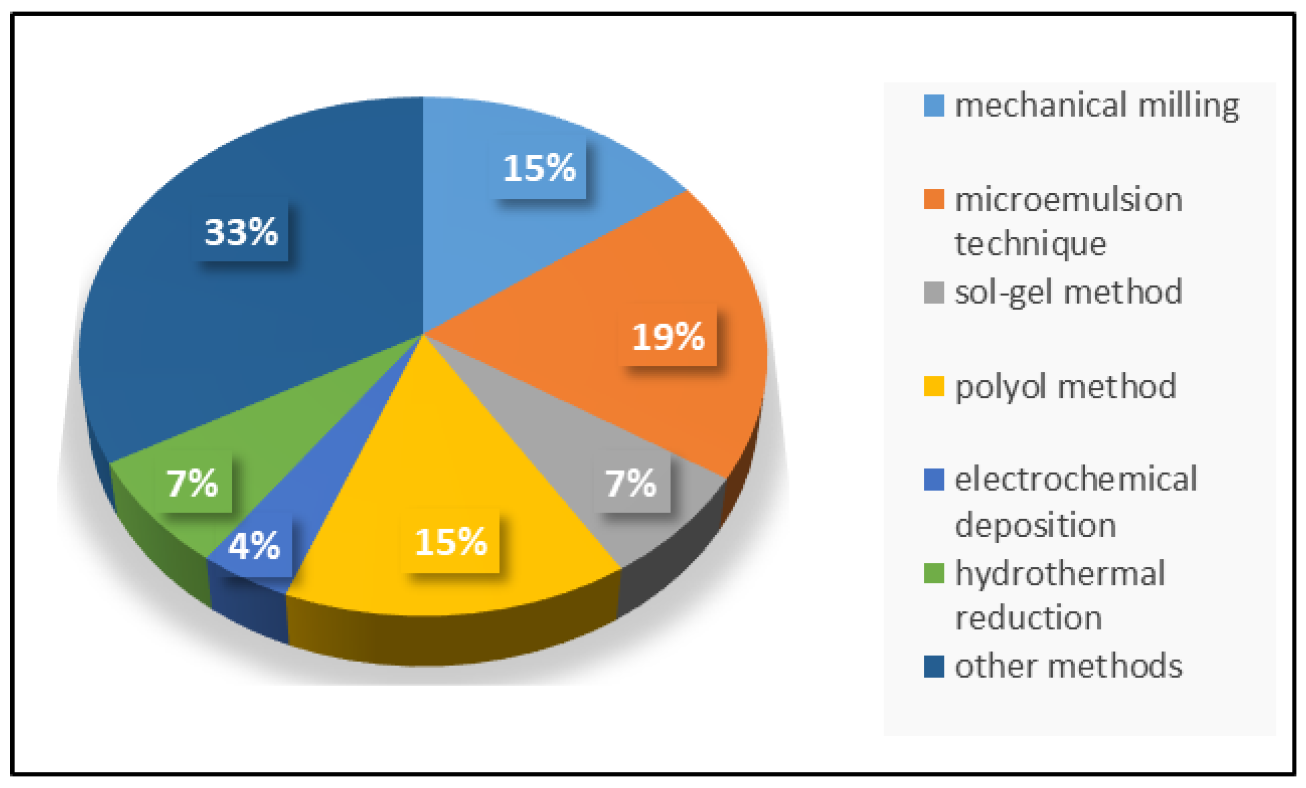
Figure 4.
Mechanical milling as a method for the synthesis of NiCu MNPs MH—magnetic hyperthermia. The references to respective studies mentioned in the figure are as follows: Bettge et al. [48], Ban et al. [36], Amrollahi et al. [39], and Durivalt et al. [57].

Figure 5.
The microemulsion technique as a method for the synthesis of NiCu MNPs. The references to respective studies mentioned in the figure are as follows: Ge et al. [58], Feng and Zhang [42], Ahmed et al. [59], Stergar et al. [60], and Wen et al. [61].

Figure 6.
The sol–gel process as a method for the synthesis of NiCu MNPs. The references to respective studies mentioned in the figure are as follows: Leontyev et al. [64], and Ferk et al. [40].
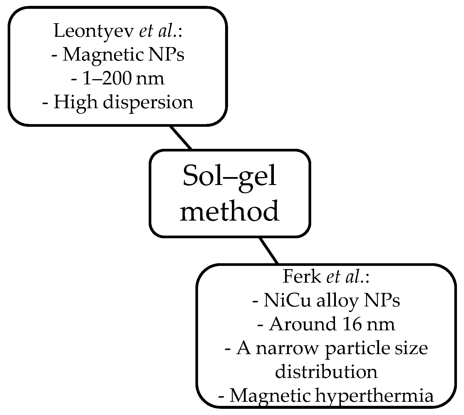
Figure 7.
The polyol method as a method for the synthesis of NiCu MNPs. PEG—polyethylene glycol. The references to respective studies mentioned in the figure are as follows: Caroll et al. [67], Bonet et al. [68], and Chatterjee et al. [47].
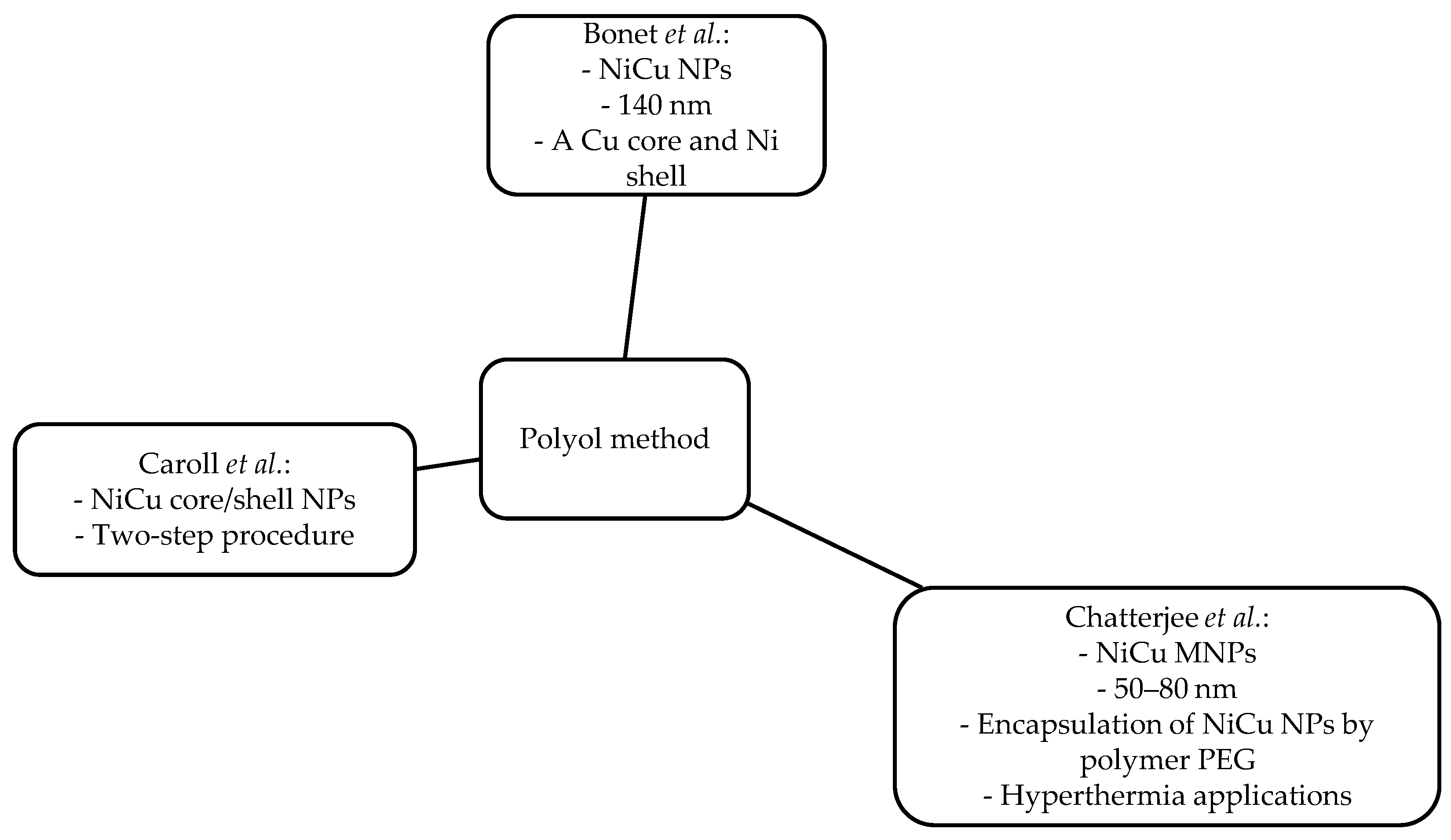
Figure 8.
Electrochemical deposition as a method for the synthesis of NiCu MNPs. The reference to the mentioned study is Foyet et al. [70].
Figure 8.
Electrochemical deposition as a method for the synthesis of NiCu MNPs. The reference to the mentioned study is Foyet et al. [70].

Figure 9.
The hydrothermal method as a method for the synthesis of NiCu MNPs. The references to respective studies mentioned in the figure are as follows: Songping et al. [41], and Songping et al. [72].

Figure 10.
Other methods for the synthesis of NiCu MNPs. Fcc—face-centered cubic. The references to respective studies mentioned in the figure are as follows: León-Quiroz et al. [78], Sopousek et al. [77], Zin et al. [76], Sue et al. [54], Kuznetsov et al. [38], Bahlawane et al. [74], Pál et al. [75], Kwon et al. [73], and Bettge et al. [48].

Figure 11.
Schematic representation of different synthesis methods of NiCu MNPs: (a) mechanical milling, (b) microemulsion, (c) sol–gel, (d) the polyol method, (e) electrochemical deposition, and (f) hydrothermal reduction.
Figure 11.
Schematic representation of different synthesis methods of NiCu MNPs: (a) mechanical milling, (b) microemulsion, (c) sol–gel, (d) the polyol method, (e) electrochemical deposition, and (f) hydrothermal reduction.
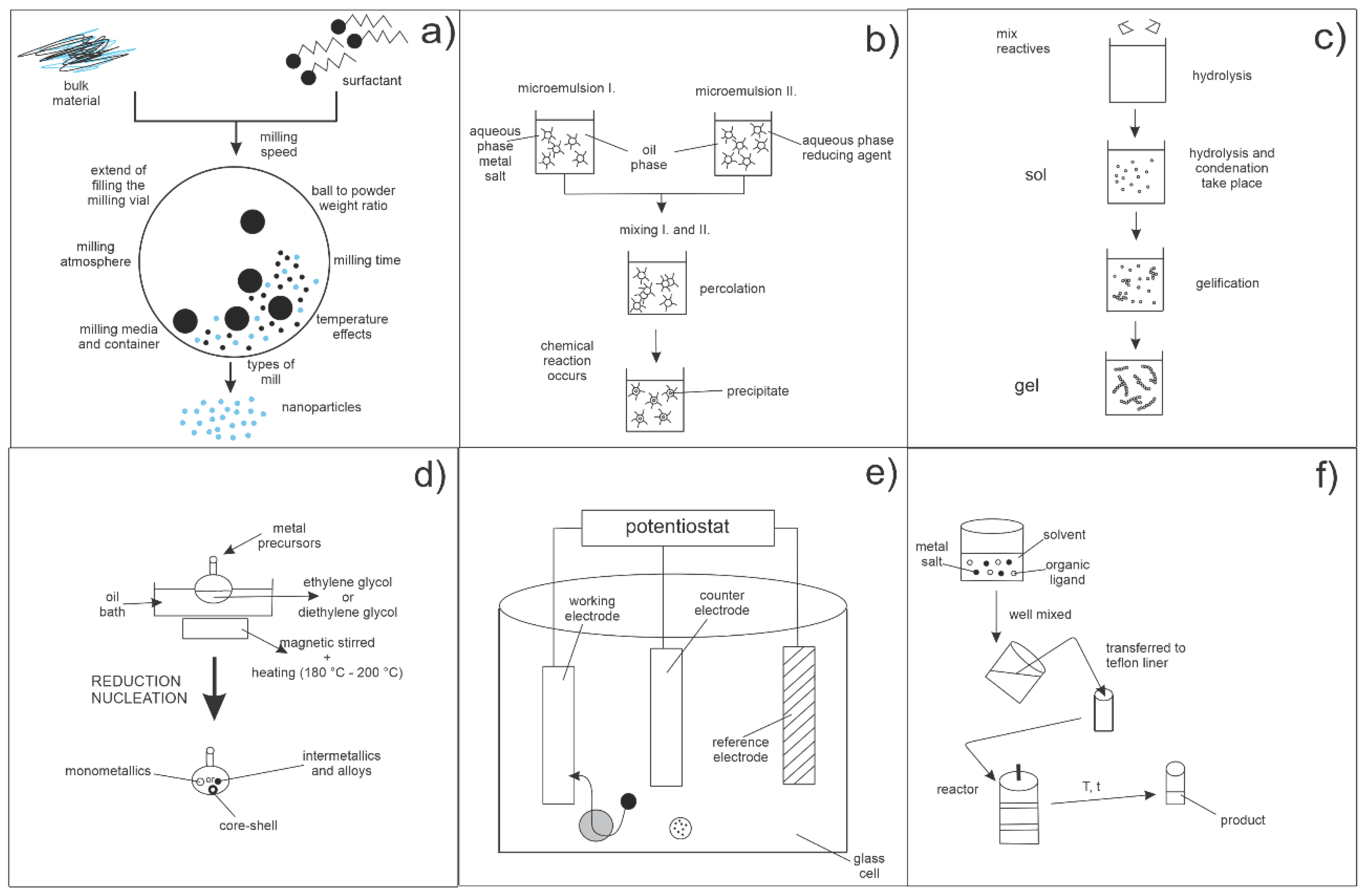
Figure 12.
TEM or SEM micrographs of NiCu MNPs prepared using different synthesis methods: (a) mechanical milling [36], (b) microemulsion [79], (c) sol–gel [40], (d) the polyol method [47], (e) electrochemical deposition [77], and (f) hydrothermal reduction [41]. Permission for micrograph reuse was granted by the respective publishers.
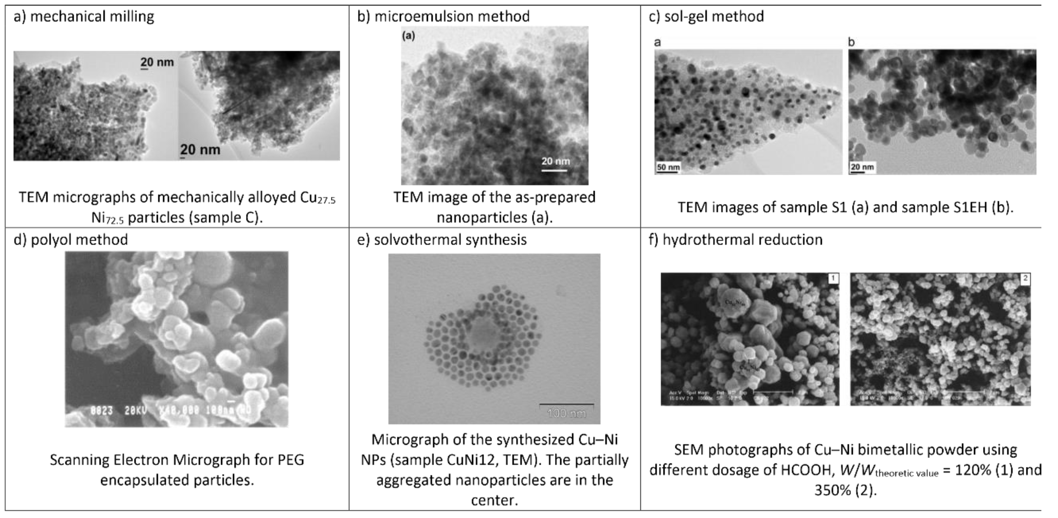
Figure 13.
Potential applications of NiCu MNPs in biomedicine. The references to respective studies mentioned in the figure are as follows: Antibacterial activity [60], Bimodal therapies [17], MH [37,38,39,40,41,48,49,96,98,99], Interstitial implants [50,100], Dental materials [101].

Table 1.
Preparation methods, types of material, magnetic nanoparticle (MNP) size, advantages and disadvantages, and biomedical applications of different NiCu structures. MH—magnetic hyperthermia.
Table 1.
Preparation methods, types of material, magnetic nanoparticle (MNP) size, advantages and disadvantages, and biomedical applications of different NiCu structures. MH—magnetic hyperthermia.
| Method | Types of Material | MNP Size | Advantages | Disadvantages | Biomedical Applications | References |
|---|---|---|---|---|---|---|
| Mechanical milling | - Nanocrystalline NixCu1−x alloys - NiCu nanoparticles | 10–436 nm | - A good milling yield (>95%) - Broad NP size distribution - Biocompatibility - TC = 46–47 °C | - Fe contamination - TC of 45 °C - Agglomeration - Agglomeration without a liquid medium | - Controlled MH applications | [36,39,48,57] |
| Microemulsion technique | - NiCu alloy nanoparticles | 4–30 nm | - Spherical, uniform NPs - Monodispersed, spherical NPs - Crystallization behavior - Fast synthesis procedure | - A core/shell structure - Additional step (reduction) | - MH | [42,58,59,60,61] |
| Sol–gel method | - NiCu alloy nanoparticles | 1–200 nm | - Spherical shape - Narrow NP size distribution, spherical shape | - High dispersion - The silica matrix influences magnetic behavior | - MH | [40,64] |
| Polyol method | - NiCu magnetic nanoparticles - NiCu core/shell nanoparticles | 40–140 nm | - A uniform shape - TC = ~77 °C - Simple procedure | - A Cu core and a Ni shell - Two-step procedure | - MH | [47,67,68] |
| Electrochemical deposition | - Magnetic alloy nanofilms | / | - Nanostructured films | - Two peaks in the polarization curve | / | [70] |
| Hydrothermal technique | - Ultra-fine NiCu bimetallic powders - Ultra-fine NiCu bimetallic powders with a core/shell structure | 0.5 ± 0.2 μm | - Excellent dispersibility, a uniform size - Finest dispersibility | - Agglomeration | / | [41,72] |
| Other methods | - NiCu alloy nanoparticles - NiCu alloy thin films - NiCu alloy NPs | 5–1000 nm | - The composition of the alloy could be controlled by changing the Ni/Cu molar ratio in the starting solution - Applying a magnetic field (44 kHz) on the liver resulted in temperature stabilization (42 °C) - Superparamagnetic behavior - Irregular shapes of NPs | - Polydispersed particles - Additional thermal treatment - Heterogeneously distributed particle size - Broad size distribution - Oxidized surface in air | - MH | [38,48,54,73,74,75,76,77,78] |
Table 2.
Type, synthesis method, composition (w), average particle size (dx), Curie temperature (TC), magnetization (Ms), and specific absorption rate (SAR) of NiCu materials for MH. PEG—polyethylene glycol.
Table 2.
Type, synthesis method, composition (w), average particle size (dx), Curie temperature (TC), magnetization (Ms), and specific absorption rate (SAR) of NiCu materials for MH. PEG—polyethylene glycol.
| Types of Materials | Synthesis Methods | Surface Modification | w (atm. %) | dx (nm) | TC (°C) | MS (emu/g) | SAR (W/g) | References |
|---|---|---|---|---|---|---|---|---|
| CuNi alloy NPs | Polyol reduction method -------and---------- physical melting process | PEG | Cu30Ni70 | 50–80 ----------- 300–400 | 43–46 | 45 ---------------- 6–8 | / | Chatterjee et al. [47] |
| NiCu alloy NPs | Sol–gel method | Silica matrix | Ni67.5Cu32.5 Ni62.5Cu37.5 Ni60Cu40 | 15–20 | 51–63 | 3–9 | 0.12–0.60 | Ferk et al. [37] |
| NiCu alloy NPs | Sol–gel method | Silica matrix | Ni67.5Cu32.5 | 16 | 65 | 8 | / | Ferk et al. [40] |
| Cu1−xNix NPs | Cationic water-in-oil (W/O) microemulsions | Silica | Ni70Cu30 Ni72.5Cu27.5 Ni60Cu40 | / | 45–88 | 22 (uncoated) 7 (coated) | / | Stergar et al. [60] |
| Ni1−xCux alloys | Modified sol–gel method | Chitosan matrix | Ni70Cu30 Ni75Cu25 Ni81Cu19 Ni95Cu5 | 11–20 | 77–277 | 21–50 | 0.289–0.576 | Araújo-Barbosa et al. [99] |
| CuNi NPs | Mechano-thermal method | / | Cu50Ni50 | 20 | 44 | 18 | 260.44 | Amrollahi et al. [39] |
| CuNi alloy NPs | Co-precipitation of salts from solution followed by reduction in hydrogen and thermal treatment (sol–gel method) | NaCl matrix | 25–30 atm.% of Cu | 30 | 40–60 | / | / | Kuznetsov et al. [38] |
| NiCu NPs | Melting and ball milling | / | Ni70Cu30 | 436 | 46–47 | / | / | Bettge et al. [48] |
| CuxNi1−x alloys | Mechanical milling | / | Cu40Ni60 Cu30Ni70 Cu27.5Ni72.5 Cu27Ni73 Cu25Ni75 Cu20Ni80 | 10–12 | 24–174 | 4.4–32.9 | / | Ban et al. [36] |
| CuNi alloy NPs | Microemulsion method | / | Cu27.5Ni72.5 | 28 | 43 | 2.5–20 | 4.3–41.6 | Stergar et al. [79] |
© 2019 by the authors. Licensee MDPI, Basel, Switzerland. This article is an open access article distributed under the terms and conditions of the Creative Commons Attribution (CC BY) license (http://creativecommons.org/licenses/by/4.0/).
Share and Cite
MDPI and ACS Style
Stergar, J.; Ban, I.; Maver, U. The Potential Biomedical Application of NiCu Magnetic Nanoparticles. Magnetochemistry 2019, 5, 66. https://doi.org/10.3390/magnetochemistry5040066
AMA Style
Stergar J, Ban I, Maver U. The Potential Biomedical Application of NiCu Magnetic Nanoparticles. Magnetochemistry. 2019; 5(4):66. https://doi.org/10.3390/magnetochemistry5040066
Chicago/Turabian StyleStergar, Janja, Irena Ban, and Uroš Maver. 2019. "The Potential Biomedical Application of NiCu Magnetic Nanoparticles" Magnetochemistry 5, no. 4: 66. https://doi.org/10.3390/magnetochemistry5040066
Note that from the first issue of 2016, this journal uses article numbers instead of page numbers. See further details here.