Article

Separation and Concentration of Astaxanthin and Lutein from Microalgae Liquid Extracts Using Magnetic Nanoparticles

by
Maria G. Savvidou
1,*,†,
Thalia Tsiaka
2,
Panagiotis Zoumpoulakis
2,‡,
Emanouella Maggiorou
1,
Konstantinos Tyrovolas
1,
Antonio Molino
3,
Evangelos Hristoforou
1,* and
Angelo Ferraro
1,*
1
Laboratory of Electronic Sensors, National Technical University of Athens, Zografou Campus 9, Iroon Polytechnique Street, 15780 Zografou, Greece
2
Institute of Chemical Biology, National Hellenic Research Foundation, 48, Vas. Constantinou Avenue, 11635 Athens, Greece
3
ENEA, Italian National Agency for New Technologies, Energy and sustainable Economic Development, Department of Sustainability—CR Portici. P. Enrico Fermi, 1, Portici, 80055 Naples, Italy
*
Authors to whom correspondence should be addressed.
Current address: Department of Biomedical Engineering, Tufts University, Medford, MA 02155, USA.
Current address: Department of Food Science and Technology, University of West Attica, Ag. Spyridonos, 12243 Egaleo, Greece.
Magnetochemistry 2022, 8(8), 80; https://doi.org/10.3390/magnetochemistry8080080
Submission received: 22 June 2022 / Revised: 12 July 2022 / Accepted: 23 July 2022 / Published: 27 July 2022

Abstract

:
The downstream processing of natural active molecules remains the most significant cost in the production pipeline. This considerable cost is largely attributed to rigorous chromatographic purification protocols. In an ongoing effort to abate the dependence on chromatography in downstream processing, alternative affinity matrices in the form of magnetic particles (e.g., iron oxide) have emerged as viable candidates. Nevertheless, biotechnological applications of iron oxide particles are still confined to the research level or for low-throughput clinical applications. Herein, we describe an efficient, quick, and environmentally friendly method for the isolation of astaxanthin and lutein, two carotenoids with very similar chemical structure, from extracts of the microalga Haematococcus pluvialis. The technology proposed, named Selective Magnetic Separation (SMS), is based on the use of magnetic materials carrying affinity ligands that bind carotenoids and is applied as second step of purification. The method, thanks to functionalized magnetic nanoparticles, reduces the use of organic or toxic solvents. In the present work, we examined the most efficient binding conditions such as temperature, magnetic nanoparticles concentration, and elution time, as well as their effects on carotenoids recovery, with the aim to improve the non-covalent binding between the ligand (amines) and astaxanthin/lutein. Our initial results clearly showed that it is possible to use magnetic separation as an alternative to chromatography to isolate important and valuable compounds.

1. Introduction

Magnetic separation is a process based on magnetically responsive carriers of micro- or nano-meter size covalently bound to a ligand, which demonstrates an exclusive affinity with the molecule of interest [1]. Usually, the functionalized magnetic particles are dissolved in the solution containing the target molecule [2] or spread on top of a flat electromagnetic surface in order to enhance the active trapping area. Afterwards, the separation of the magnetic complexes from the rest of the mixture is performed using magnetic gradients [3,4]. In order to successfully separate the highest quantity of a given molecule, a proper reaction condition of the magnetic material with the compound source represents a crucial step [5]. The detachment of the compound of interest from the magnetic material is another key step both for the reusability of the magnetic particles and for the recovery yield of the product [5]. Since target molecules are trapped by the ligand through relatively weak bonds, the separation from magnetic particles is performed using mild chemical treatments such as pH and ionic strength variations, solvents, or by changing physical parameters as pressure or temperature [3,6]. After ligand-target bonds disruption, magnetic particles are magnetically removed from the target compound solution with a permanent magnet or, alternatively, an electromagnet. Magnetic separation techniques have been used already in various isolation efforts of biologically active compounds, as well as in genomic isolation from algae and cyanobacteria [7,8,9,10]. Furthermore, fatty acid isolation from olive and sunflower oils based on magnetic nanoparticles achieved an 85% efficiency [11]. Concerning human health safety, several studies confirmed that iron oxide nanoparticles (Fe3O4) when properly functionalized, present low toxicity towards human lung cell line (A549), as well as mesenchymal stem cells [12,13]. Nevertheless, the magnetic separation remains a technique mostly used for research purposes, with little or none used in high-throughput separation of industrially important molecules.
Microalgae are cell factories which produce numerous biologically active compounds such as proteins, polysaccharides, peptides, lipids, oligonucleotides, as well as pigments such as carotenoids, chlorophyll, and others. These high-added value products have a tremendous use in various biological and biotechnological applications, in cosmetics, animal feed, human food, energy, and, recently, in biomedical and pharmaceutical applications [5,14]. The simplicity of the growth conditions of microalgae cells, as well as the ability to grow at a high rate, result in the accumulation and high yield of the above-mentioned secondary metabolites. However, the isolation, separation, and purification of such compounds still represent the most crucial processes for an economical and sustainable production [14,15]. The techniques commonly used require large volumes of reagents and specific equipment leading to increased cost, making the isolation of the above-referred compounds non-profitable for the industries.
Carotenoids (astaxanthin, lutein, zeaxanthin, fucoxanthin, and β-carotene) are organic pigments belonging to the tetraterpenes family, consisting of isoprene units and being a member of the photosynthesis process [16]. Astaxanthin, for example, is very attractive for important industrial markets, both as a food-grade coloring as well as an antioxidant agent [17]. Following the microalgae cell wall disruption by mechanical or chemical methods, the isolation of carotenoids and their separation or isolation from other constituents can be achieved by solid-liquid extraction [18], supercritical solid extraction [19], ultrasound, pulse field, or even enzymatic assisted extraction [20], via the use of Generally Recognized As Safe (GRAS) solvents [21] and CO2 supercritical fluid extraction [22]. Chromatography is extensively used for carotenoid isolation, which bears advantages but also limits [23,24]. Specifically, for the extraction of carotenoids from microalgae, polar solvents are needed [25], which may represent an environmental threat.
In this study, we established an efficient, quick, and environmentally friendly method for the isolation of astaxanthin and lutein (two carotenoids with very similar chemical structure) from extracts of Haematococcus pluvialis (H. pluvialis), avoiding organic or toxic solvents. We used affinity interaction between amine groups, immobilized on magnetic nanoparticles, and astaxanthin/lutein chemical structures. We examined the most efficient binding conditions such as temperature, magnetic nanoparticles concentration, and elution time on carotenoids recovery with the aim to improve the non-covalent binding of ligand (amines) and astaxanthin, as well as lutein. Our results, as proof-of-concept, support the possibility for scaling-up isolation processes based on magnetic separation, which can overcome purification drawbacks linked to the standard methods such as chromatography or solvent extraction.

2. Materials and Methods

2.1. Microalgae Extracts

Carotenoids extracts from H. pluvialis organism were prepared using GRAS solvents and CO2 supercritical fluid extraction [21,22]. A brief component description of H. pluvialis biomass and extracts is provided in Table 1, and more information is provided in References [21,22].

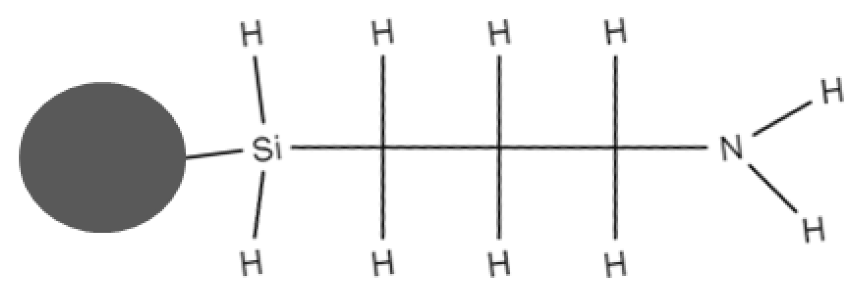
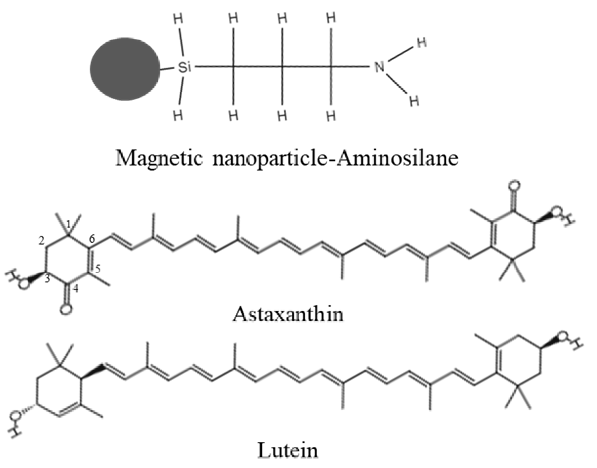
2.2. Magnetic Nanoparticles

Aqueous dispersion of magnetic nanoparticles FluidMAG-Amine with hydrodynamic diameter of 100 nm was purchased from Chemicell (Chemicell GmbH, Berlin, Germany). The concentration of magnetic nanoparticles was 25 μg/µL. The functional group covalently bound to the nanoparticles surface was aminosilane (see Figure 1).

2.3. Reagents and Standards for Liquid Chromatography-Mass Spectrometry

Trans-Astaxanthin were purchased from Sigma-Aldrich (St. Louis, MO, USA) and trans-lutein were obtained from Extrasynthese (Genay, France). No carotenoids internal standard was used for the analysis. All solvents used were of LC-MS grade. Methanol, acetonitrile, and methyl-tert-butyl ether (MTBE) were acquired from Fluka (Darmstadt, Germany) and Fischer Chemical (Pittsburgh, PA, USA).

2.4. Liquid Chromatography-Tandem Mass Spectrometry (LC-MS/MS) Analysis

The LC-MS instrumentation used for the analysis included (i) a quaternary pump, (ii) an autosampler with a thermostat tray oven (Accela, Thermo Scientific, Waltham, MA, USA), (iii) an Acclaim C30 reversed-phase column (3 μm particle size, 150 × 2.1 mm i.d), (iv) a guard column, and (v) LTQ Orbitrap Velos mass spectrometer (Thermo Scientific, USA). The temperature of the tray oven was set at 10 °C and the temperature of the C30 column was adjusted at 25 °C. The applied LC-MS method is based on the method developed by Tsiaka and colleagues [26].
The mobile phase consisted of three different solvents, acetonitrile (ACN) (A), methanol (B), and MTBE (C). The flow rate of the mobile phase was set at 350 μL/min. As described by Tsiaka and colleagues [26], the gradient elution program included the following steps: 0–5 min (30% A, 70% B), 5.1–13 min (22.9% A, 65.8% B, and 11.3% C), 13.1–14 min (5% A, 75% B, and 20% C), 14–14.1 min (30% A, 70% B), and 14.10–20 min (30% A, 70% B). All samples for LC-MS analysis were dissolved in MeOH-MTBE 1:1 v/v (injection solvent) and the injection volume was set at 5 μL. In particular, (a) 100 μL of control sample were diluted in 500 μL of injection solvent, (b) 50 μL of sample S1 were diluted in 500 μL of injection solvent, and (c) 50 μL of sample S2 were diluted in 450 μL of injection solvent.
Optimal MS source conditions are presented in our previous works [26]. Atmospheric pressure chemical ionization (APCI) in positive mode was used for the identification and content estimation of astaxanthin and lutein, which were conducted by selected-reaction monitoring (SRM) mode. The retention times (RTs) and the collision energies required for carotenoid fragmentation, as well as the MS/MS mass transitions of parent to product ions are shown in Table 2. All spectra were processed using Xcalibur software (version 2.1, Thermo Scientific, Waltham, MA, USA).
The loss of water from hydroxy carotenoid astaxanthin produced the ion with m/z = 579.3, which was used for the estimation of astaxanthin content. In the case of lutein, the intensity of product ion with m/z = 551.4 was higher than that of parent ion of m/z = 569.4. Thus, the mass transition used for lutein content estimation n was the fragmentation of m/z = 551.4 to the product ion with m/z = 533.3. In addition, the fragment ion with m/z = 495.3 was observed only during the fragmentation of lutein and it can be used for the elucidation of lutein from other isomer carotenoids, such as zeaxanthin [27,28].

2.5. Saturation of FluidMAG-Amine with Astaxanthin

A commercial solution of astaxanthin with a concentration of 1 µg/mL was dissolved in ethanol and used to optimize the conditions for the most efficient recovery of the carotenoids. For each test, 50 ng of astaxanthin was used with variable volumes of magnetic nanoparticles and buffer for a total volume of 1 mL. The reaction time between magnetic nanoparticles and the solution containing astaxanthin was 1 h in continuous shaking. Following the binding of astaxanthin to the amine group, the carotenoid was eluted using 1 mL of acetone and the absorbance was measured at 530 nm. For spectrophotometer measurements, we used the X-ma 1000 machine (Human Corporation, Seoul, South Korea) with a fixed wavelength of 530 nm. The elution time was studied by using variable concentrations of magnetic nanoparticles (250, 750 and 1250 µg) in low (4 °C), high (40 °C), or room (25 °C) temperature.
After optimizing the best extraction conditions, we tested several raw microalgae extracts containing carotenoids [21,22]. In total, 0.1 mL of the raw extracts (in hexane or ethanol) were mixed with 0.9 mL of PBS buffer. The PBS buffer was made by 0.01 M sodium phosphate, 0.0027 M potassium chloride and 0.137 M sodium chloride (Phosphate buffered saline tablets, Merk, Germany). The mixture remained on a shaker for variable time points and temperatures (1 h and 2 h; low (4 °C), high (40 °C), or room (25 °C) temperature). After testing raw extract in hexane, it was noticed that such solvent was immiscible with the water-based PBS buffer, therefore only raw extracts in ethanol were used for all the tests.
After each binding or elution step, tubes containing suspensions were placed in a special magnetic rack (MagnetoPURE BIG SIZE, Chemicell GmbH, Germany) for 10 min, the supernatants were removed, eventually its absorbance measured, and then the nanoparticles were washed three times with 500 μL PBS. Following this, 200 μL of acetone was mixed with the nanoparticles and the solution was left from 10 to 60 min at room temperature. Then, the supernatants were used for spectrophotometer and LCMS analysis.

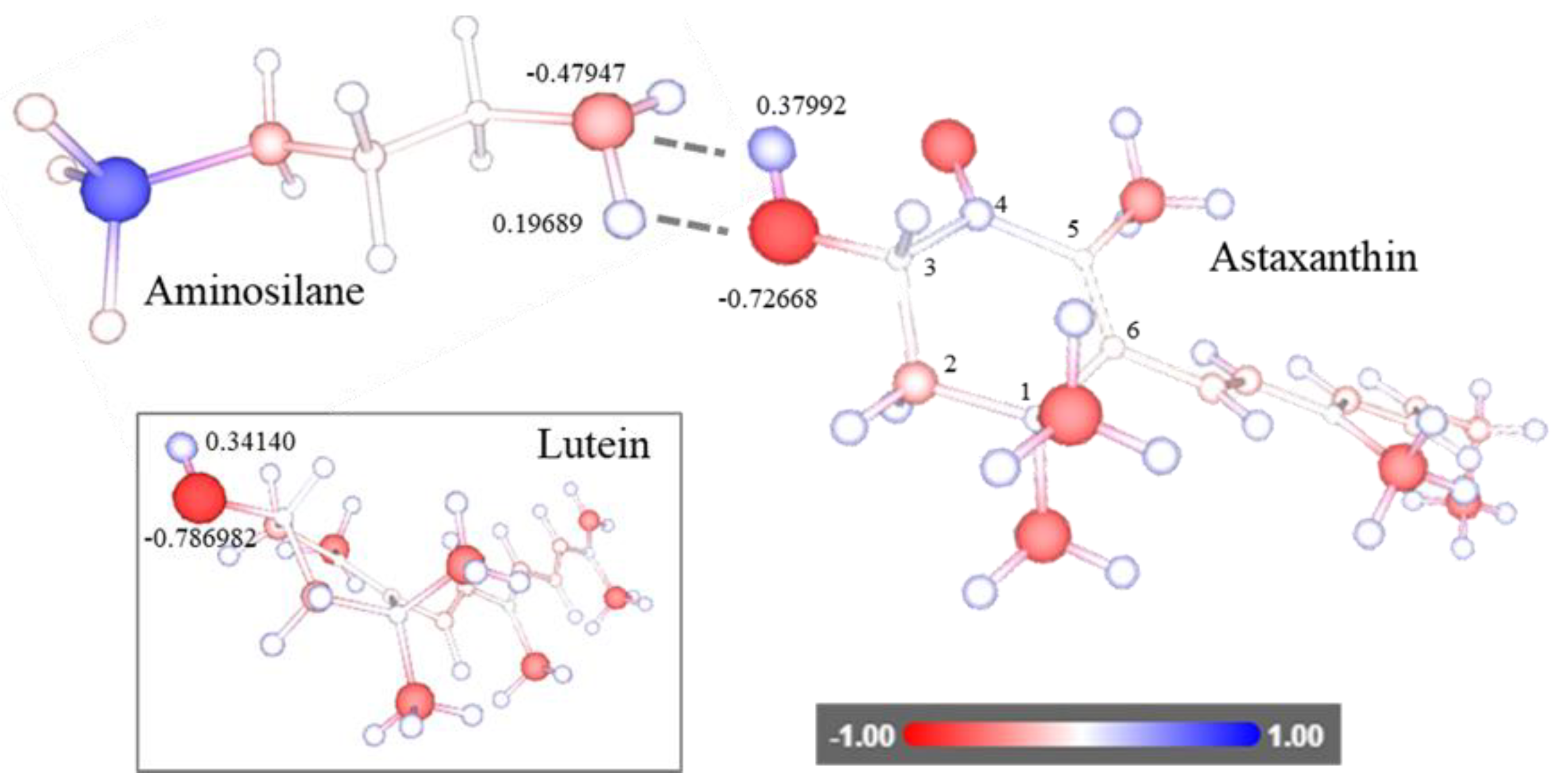
2.6. Software for Structural Analysis

Structural visualization of carotenoids and aminosilane were performed using MolView platform, an open-source web application which allows one to download or draw chemical structures (https://molview.org/, accessed on 1 December 2021). To calculate the atomic charges of carotenoids and aminosilane groups, we used the web application AtomicChargeCalculator [29], owned by Masaryk University, which allows one to use the software free of charge. MOL files were generated or retrieved from MolView website and uploaded on the AtomicChargeCalculator web application (https://webchem.ncbr.muni.cz/Platform/ChargeCalculator, accessed on 1 December 2021).

3. Results

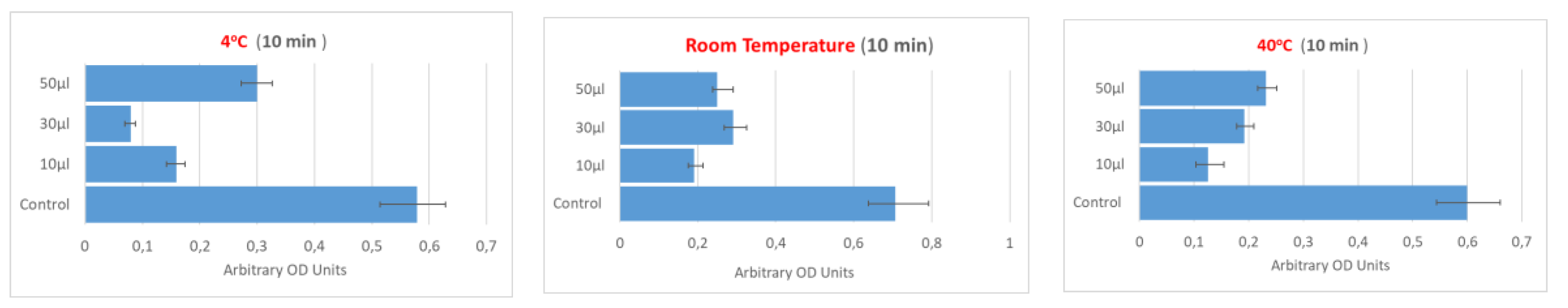
3.1. Effect of Temperature, Magnetic Nanoparticles Concentration and Elution Time on Commercial Astaxanthin Recovery from Solution

We proceeded to study specific parameters optimization affecting the binding using magnetic nanoparticles functionalized with amine groups and astaxanthin solutions, which were prepared with highly pure commercial astaxanthin standards.
Temperature, magnetic particle concentration, as well as elution time have been examined in quadruplicates with a concentration of astaxanthin of 1 µg/mL in ethanol as described in Materials and Methods. Following the binding of astaxanthin to magnetic nanoparticles bearing amine groups, the carotenoid was eluted using 1 mL of acetone and the absorbance was measured (Figure 2). After 10 min of elution, it was found that the binding reaction was successful with all nanoparticles concentrations and temperatures tested (Figure 2). However, since at room temperature the recovery yield of astaxanthin was nearly 50%, using 30 µL and 50 µL (750 and 1250 µg) of magnetic nanoparticles, we concluded that the upcoming reaction can be conducted at room temperature without dramatically affecting the reaction yields (Figure 2).
Increasing the elution time at 60 min (Figure 3) resulted in an almost duplication of astaxanthin recovery yield when compared to 10 min, for each temperature and each nanoparticle concentration tested, indicating that, for the elution, at least 1 h incubation in acetone is necessary. Furthermore, we observed that magnetic nanoparticle concentrations, especially after 1 h of elution, showed no critical difference in their ability to bind astaxanthin, as well as the three different temperatures tested.
Therefore, from the above optimized conditions we concluded that the best parameters to recover astaxanthin from a liquid solution were: (i) 10 µL (250 μg) of magnetic nanoparticles functionalized with aminosilane groups, (ii) final reaction volume of 1 mL, (iii) incubation/mixing of 1 h at room temperate, and (iv) elution time of at least 1 h with acetone at room temperature.

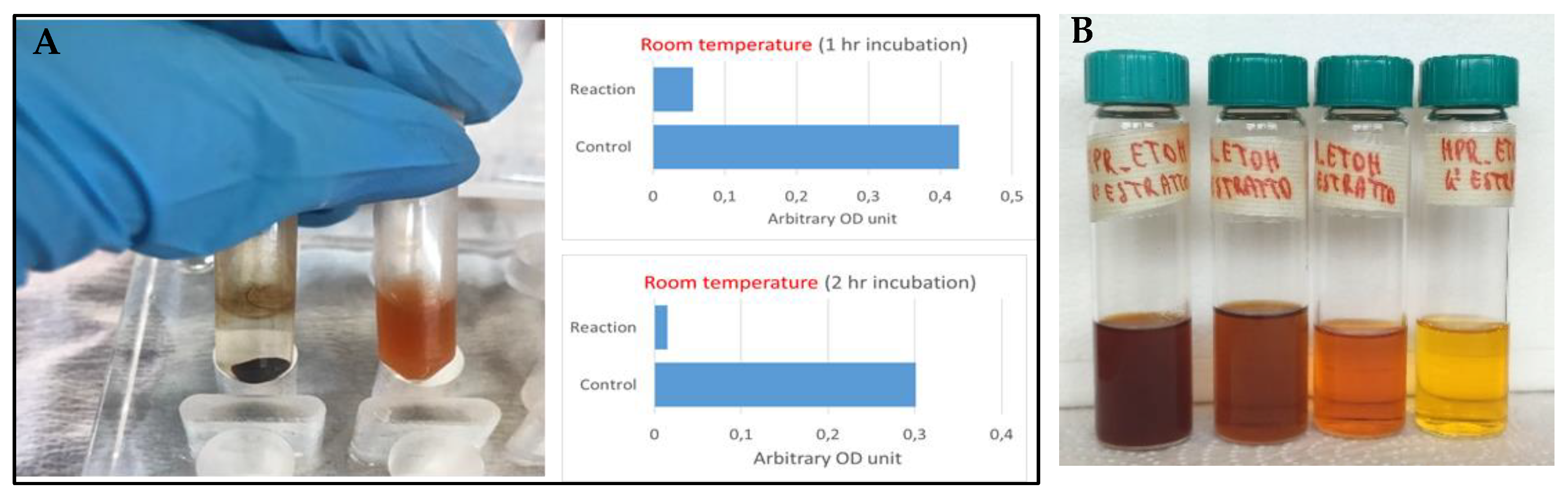
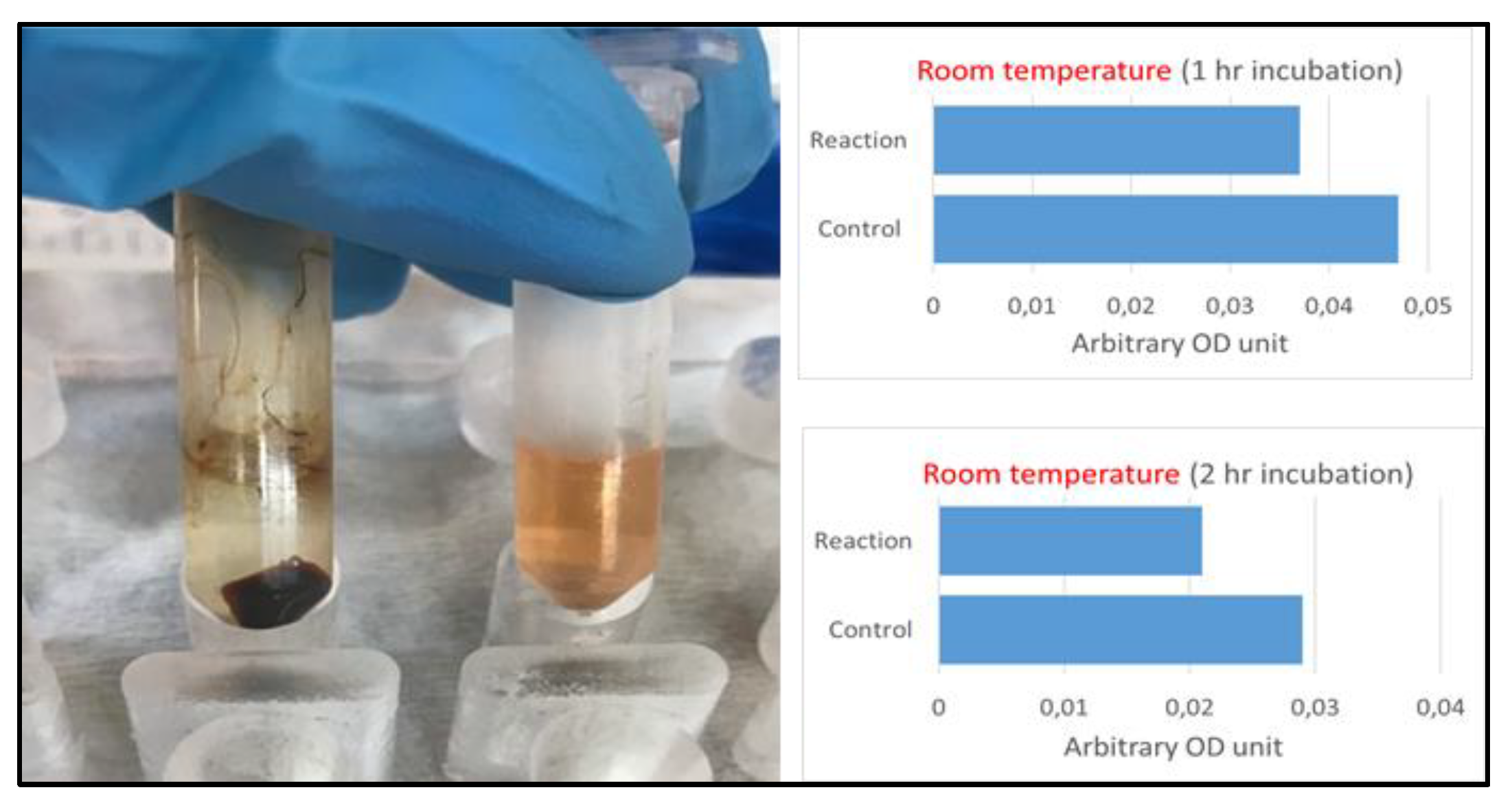
3.2. Magnetic Separation of Astaxanthin from H. pluvialis Extracts

The water-based solution of the magnetic nanoparticles guided us to prepare the extracts from H. pluvialis by using CO2 supercritical fluid extraction and ethanol [22] instead of other organic solvents [21], which might not form a homogenous solution with water. Figure 4A shows that after 1 h of incubation at room temperature of magnetic nanoparticles with microalgae extracts (first preparation, Figure 4B) containing carotenoids, almost all carotenoid molecules were trapped on the nanoparticles pellet. The red/orange color, typical of carotenoids alcoholic solutions, vanished due to the addition of magnetic nanoparticles. Spectrophotometric analysis of the alcoholic solution where carotenoids were initially suspended (without eluting the trapped biomolecules with acetone), showed that >90% of the color was withdrawn from the mixture after 2 h incubation (Figure 4A).
The same results were obtained with a second preparation of carotenoids (Figure 4B) from H. pluvialis. In this case, the spectrophotometric measurement was performed on the carotenoids eluted from the magnetic nanoparticles pellet using acetone. When magnetic nanoparticles were added to the H. pluvialis extracts, after 1 h of incubation almost all of the compound was retained on the magnetic nanoparticles surface as the solution appeared clean and clear (Figure 5).

3.3. Carotenoids Purity Analysis after Magnetic Separation

The H. Pluvialis extracts on which the magnetic separation was tested did not only contain astaxanthin and lutein, but also other carotenoids and other metabolites as well. Therefore, to evaluate how selective the aminosilane groups are on trapping only astaxanthin and lutein, we performed LC-MS tests. From the nanoparticles pellets (Figure 4A and Figure 5), astaxanthin and lutein were eluted using 0.2 mL of acetone. After 1 h of incubation with acetone, sample tubes were placed on a magnetic rack for 10 min in order to pellet magnetic nanoparticles (free of carotenoids) and the supernatant was transferred to fresh tubes for further LC-MS analysis. The astaxanthin and lutein content of control (H. pluvialis raw extracts) and two magnetically isolated samples, was determined by the calibration curves using astaxanthin and lutein commercial standards. The concentrations of standard solutions used to construct the calibration curves ranged between 0.025–15 μg/mL (for astaxanthin) (n = 11) and 0.5–15 μg/mL (for lutein) (n = 8). The regression coefficients (R2) were 0.996 for astaxanthin and 0.997 for lutein, respectively, verifying the linearity of the method. Intra-day repeatability was performed by running three replicates of the three quality control (QC) samples on the same day, confirming that the developed LC-MS/MS method was precise since %RSD of QC samples was lower than 15% [30]. All samples (control, S1, and S2) were measured in triplicate at confidence level 95% (p-value ≤ 0.05). Intra-day repeatability was performed by running quality control (QC), and the quality parameters were the following: Astaxanthin Low = 0.05 μg/mL, Medium = 1 μg/mL, High = 15 μg/mL; Lutein: 0.5 μg/mL, 5 ug/mL, 15 μg/mL. The concentrations of the two analyses in each sample are presented in Table 3.
Based on the results of ANOVA test, astaxanthin concentration differed significantly in all three samples (p-value ≤ 0.05). The control sample, acquired from CO2 supercritical fluid extraction, contained the lowest concentration of trans-astaxanthin, whereas samples S1 (first carotenoids preparation) and S2 (second carotenoids preparation) presented up to two times higher astaxanthin content when compared to control sample. The same trend was also observed in the case of lutein content, where control sample contained two times lower lutein concentration than samples S1 and S2 (p-value ≤ 0.05). The ratio of the trans-form of lutein to trans-astaxanthin was between to roughly 2.0 to 2.5 in favor of lutein.
This optimized method constitutes a meticulous and high yield (>90%) method for selective separation of carotenoids from microalgae extracts, partial for astaxanthin and lutein with astaxanthin being the sovereign carotenoid. Selective magnetic separation of carotenoids with amine groups is a profitable method for large scale processes leading to high concentrated carotenoid levels.

4. Discussion

In this study, we examined the ability of aminosilane functionalized magnetic nanoparticles to trap two chemically related carotenoids from commercial preparation as well as from microalgae extracts, providing a proof-of-concept about the ability of SMS method to isolate carotenoids faster, more simply, and possibly at a lower cost when compared to traditional methods such as chromatography. The aminosilane functionalized nanoparticles should not have intrinsic specificity for carotenoids, however, based on our experiments, by mixing them with commercial astaxanthin as well as with carotenoids from H. pluvialis microalgae extracts we observed a clear separation of carotenoids from the solution since such solution discoloured (Figure 4A and Figure 5). Therefore, further improvement of SMS is necessary, especially regarding selectivity as well as other parameters affecting the process, such as the pH and pKa of both carotenoids; indeed, it is possible that by changing the reaction pH (in our case, roughly 7 due to the PBS buffer), the protonation of aminosilane group changes and, in turn, a more favourable interaction is possible. Nevertheless, our results indicate that affinity magnetic matrices are a valid alternative that can be explored for downstream processing of natural active molecules.
Astaxanthin and lutein consist of two terminal β-ionone-type rings joined by a polyene chain (Figure 6). They have two asymmetric carbons located at the 3,3′-position of the β-ionone ring, with a hydroxyl group (-OH) on either end of the molecule. Oxygen is present in the ring system as both a hydroxyl and a keto (C=O) group (Figure 6). Astaxanthin exists as cis or trans geometrical isomers [31]. Lutein is naturally present with a variety of isomers characterized by cis/trans (geometry, also indicated as E/Z [32].
Our hypothesis for a possible mechanism underneath the successful binding of these two carotenoids to the magnetic nanoparticles aminosilane group (Figure 6) implies that the formation of hydrogen bonds between radical groups present on the β-ionone-type rings and the amine group present on the nanoparticles surface. Several combinations of hydrogen bonds are possible and most probably there is no favorite one; however, we noticed that two hydrogen bonds can be established between the amine group and the hydroxylic groups at the 3 position of the β-ionone ring (Figure 7). As reported in Figure 7, both oxygen and nitrogen possess a relatively high negative charge, which in turn provoke a considerable positive charge on the hydrogen atoms bond to them. Such highly charged atoms provide an ideal environment to form hydrogen bonds. Since hydroxylic groups are present in both carotenoids and at the same position on the β-ionone rings, we believe that both molecules may form strong binding to nanoparticles through their hydroxylic groups.
Even though hydroxylic groups are very common in a variety of synthetic and natural compounds, and thus the selectivity of target compound is affected, we believe that in certain circumstances, a decent degree of selectivity can be obtained. Indeed, we developed this method to concentrate and extract carotenoids from raw microalgae extracts. A multistep process based on CO2 supercritical extraction was previously utilized to increase the yield of carotenoids extraction from H. pluvialis [22]. This process, though very effective, results in multiple vials with decreasing carotenoids concentration (Figure 4B).
The magnetic separation method offers a valid alternative to other techniques, since the low-concentrated solution can be mixed with magnetic nanoparticles and carotenoids can be easily concentrated.
Traditionally, HPLC and reversed phase HPLC are extensively used for carotenoid isolation and remain the best method to purify such molecules [23,24]. Concerning identification, gas chromatography coupled with mass spectroscopy are useful methods for the identification of carotenoids and retinoids [23]. Despite the high success rate of carotenoid isolation by the above methods, there are a few parameters that need to be taken in account. Lipid content in the microalgae extract may affect the HPLC isolation. High lipid content must be diluted in organic solvent, which can be miscible in HPLC mobile phase. Moreover, high lipid content in a simultaneously low carotenoid content needs saponification for the proper separation of the lipids from the carotenoids, as well as the carotenoids from chlorophylls. Furthermore, the form of carotenoid is important, e.g., carotenes as well as xanthophylls form ester linkages and so can be directly extracted by lipophilic solvents [33,34,35]. Therefore, based on the source, extracts undergo different protocols each time for carotenoid isolation by chromatographic methods, leading to time consumption and high costs [36].
The SMS technique, due to its nature, supports a highly efficient separation for the bioactive compounds, existing in low concentration, from extracts and other mixtures of liquid media, while also not disrupting sensitive biological compounds such as proteins and peptides in comparison to the traditional column chromatography methods. Furthermore, similarly to chromatography, SMS is applied after a first step of broad chemical separation, but unlike chromatography, SMS presents relatively low cost (no pressurization, no cartridges, reduces amount of solvent for the mobile phase) and, most importantly, it can be applied on a large scale since functionalized nanoparticles can be mixed in relatively large solution volume. In addition, nanoparticles can be reused. Even though we did not perform recycling experiments, we believe that magnetic nanoparticles can be reused several times. In fact, assuming that aminosilane groups are damaged during the elution process, the nanoparticle core remains intact and can undergo to a second round of functionalization, providing that the same binding performance is used again. On the other hand, if aminosilane groups are not damaged during the elution process, nanoparticles can be reused directly in a second round of isolation without any additional step to regenerate them.
Isolation of astaxanthin from H. pluvialis extracts with organic solvents, breakdown pretreatment process of cells, enzyme lysis, mechanical disruption, and spray drying achieved lower efficiencies from 78% up to 87% [37,38]. In the above processes, temperature was mostly kept in higher degrees [38] when compared to this study, while incubation time was similar [39], indicating that the SMS is an affordable process.

5. Conclusions

We demonstrated a successful binding of astaxanthin/lutein to the functionalized magnetic nanoparticles without the necessity of any costly ligand, but rather just an amine group supporting a less costly and time consuming method. Specifically, in commercial astaxanthin solution, the optimized conditions for the best recovery were 250 µg of magnetic nanoparticles in a final volume of 1 mL and an elution time of at least 2 h. These conditions resulted in a successful isolation (>90%) of astaxanthin and lutein from H. pluvialis extracts after two hours of incubation. The SMS efficiency in this study was higher than the one achieved from an HPLC-DAD-MS/MS carotenoid isolation process [24].
The efficient isolation of carotenoids from microalgae extracts in our study supports the necessity of further studies for the SMS on the recovery of high value added products. Presently, magnetic strategies are also used for the harvesting (de-watering) of microalgae cells [40], as well as cultivation [41], and have gained advantages when compared to other competitive techniques. Therefore, it is now possible to design biotechnological processes based on the use of magnetic nanoparticles that allow cultivation, harvesting, and extraction of natural compound from microalgae cells.

Author Contributions

Conceptualization, A.F. and E.H.; methodology, M.G.S., A.M., T.T. and P.Z.; formal analysis, M.G.S., E.M., K.T. and A.F.; investigation, M.G.S. and T.T.; resources, A.F., A.M. and E.H.; data curation, A.F.; writing—original draft preparation, M.G.S. and A.F.; writing—review and editing E.H. and A.F.; visualization, A.F.; supervision, E.H.; project administration, E.H. and A.F.; funding acquisition, A.F., E.H. and A.M. All authors have read and agreed to the published version of the manuscript.

Funding

This paper has received funding from the Bio Based Industries Joint Undertaking under the European Union’s Horizon 2020 research and innovation program under grant agreement No 745695 (VALUEMAG).

Institutional Review Board Statement

Not applicable.

Informed Consent Statement

Not applicable.

Data Availability Statement

All raw data are available under request.

Conflicts of Interest

The authors declare no conflict of interest.

References

  1. Safarik, I.; Safarikova, M. Magnetic techniques for the isolation and purification of proteins and peptides. Biomagn. Res. Technol. 2004, 2, 7. [Google Scholar] [CrossRef] [Green Version]
  2. Kim, S.-E.; Van Tieu, M.; Hwang, S.Y.; Lee, M.-H. Magnetic Particles: Their Applications from Sample Preparations to Biosensing Platforms. Micromachines 2020, 11, 302. [Google Scholar] [CrossRef] [Green Version]
  3. Plouffe, B.; Murthy, S.K.; Lewis, L.H. Fundamentals and application of magnetic particles in cell isolation and enrichment: A review. Rep. Prog. Phys. 2014, 78, 016601. [Google Scholar] [CrossRef]
  4. Almomani, F. Algal cells harvesting using cost-effective magnetic nano-particles. Sci. Total Environ. 2020, 720, 137621. [Google Scholar] [CrossRef]
  5. Safarik, I.; Pospiskova, K.; Baldikova, E.; Safarikova, M. Magnetic particles for microalgae separation and biotechnology. In Food Bioactives; Springer: Cham, Switzerland, 2017; pp. 153–169. [Google Scholar]
  6. Ge, S.; Agbakpe, M.; Zhang, W.; Kuang, L.; Wu, Z.; Wang, X. Recovering magnetic Fe3O4–ZnO nanocomposites from algal biomass based on hydrophobicity shift under UV irradiation. ACS Appl. Mater. Interfaces 2015, 7, 11677–11682. [Google Scholar] [CrossRef]
  7. Bruce, I.J.; Davies, M.J.; Howard, K.; Smethurst, D.E.; Todd, M. Magnetizable Solid-phase Supports for Purification of Nucleic Acids. J. Pharm. Pharmacol. 1996, 48, 147–149. [Google Scholar] [CrossRef]
  8. I Taylor, J.; Hurst, C.D.; Davies, M.J.; Sachsinger, N.; Bruce, I.J. Application of magnetite and silica–magnetite composites to the isolation of genomic DNA. J. Chromatogr. A 2000, 890, 159–166. [Google Scholar] [CrossRef]
  9. Matsunaga, T.; Nakayama, H.; Okochi, M.; Takeyama, H. Fluorescent detection of cyanobacterial DNA using bacterial magnetic particles on a MAG-microarray. Biotechnol. Bioeng. 2001, 73, 400–405. [Google Scholar] [CrossRef]
  10. Rudi, K.; Kroken, M.; Dahlberg, O.; Deggerdal, A.; Jakobsen, K.; Larsen, F. Rapid, Universal Method to Isolate PCR-Ready DNA Using Magnetic Beads. BioTechniques 1997, 22, 506–511. [Google Scholar] [CrossRef] [PubMed]
  11. Cano, M.; Sbargoud, K.; Allard, E.; Larpent, C. Magnetic separation of fatty acids with iron oxide nanoparticles and application to extractive deacidification of vegetable oils. Green Chem. 2012, 14, 1786–1795. [Google Scholar] [CrossRef]
  12. Rozhina, E.; Danilushkina, A.; Akhatova, F.; Fakhrullin, R.; Rozhin, A.; Batasheva, S. Biocompatibility of magnetic nanoparticles coating with polycations using A549 cells. J. Biotechnol. 2020, 325, 25–34. [Google Scholar] [CrossRef] [PubMed]
  13. Ishmukhametov, I.; Batasheva, S.; Rozhina, E.; Akhatova, F.; Mingaleeva, R.; Rozhin, A.; Fakhrullin, R. DNA/Magnetic Nanoparticles Composite to Attenuate Glass Surface Nanotopography for Enhanced Mesenchymal Stem Cell Differentiation. Polymers 2022, 14, 344. [Google Scholar] [CrossRef] [PubMed]
  14. Ventura, S.; Nobre, B.; Ertekin, F.; Hayes, M.; Garciá-Vaquero, M.; Vieira, F.; Koc, M.; Gouveia, L.; Aires-Barros, M.; Palavra, A. Extraction of value-added compounds from microalgae. In Microalgae-Based Biofuels and Bioproducts; Elsevier: Amsterdam, The Netherlands, 2017; pp. 461–483. [Google Scholar]
  15. Levasseur, W.; Perré, P.; Pozzobon, V. A review of high value-added molecules production by microalgae in light of the classification. Biotechnol. Adv. 2020, 41, 107545. [Google Scholar] [CrossRef] [PubMed]
  16. Dhan Prakash, D.P.; Charu Gupta, C.G. Carotenoids: Chemistry and health benefits. In Phytochemicals of Nutraceutical Importance; CABI: Wallingford, UK, 2014; pp. 181–195. [Google Scholar] [CrossRef]
  17. Jaime, L.-C.; Sánchez-Machado, D.I. Astaxanthin, lutein, and zeaxanthin. In Nonvitamin and Nonmineral Nutritional Supplements; Academic Press: Cambridge, MA, USA, 2019; pp. 19–25. [Google Scholar]
  18. Hosokawa, M.; Wanezaki, S.; Miyauchi, K.; Kurihara, H.; Kohno, H.; Kawabata, J.; Odashima, S.; Takahashi, K. Apoptosis-Inducing Effect of Fucoxanthin on Human Leukemia Cell Line HL-60. Food Sci. Technol. Res. 1999, 5, 243–246. [Google Scholar] [CrossRef] [Green Version]
  19. Guedes, A.C.; Gião, M.S.; Matias, A.A.; Nunes, A.V.; Pintado, M.E.; Duarte, C.M.; Malcata, F.X. Supercritical fluid extraction of carotenoids and chlorophylls a, b and c, from a wild strain of Scenedesmus obliquus for use in food processing. J. Food Eng. 2013, 116, 478–482. [Google Scholar] [CrossRef] [Green Version]
  20. Wijesinghe, W.; Jeon, Y.-J. Enzyme-assistant extraction (EAE) of bioactive components: A useful approach for recovery of industrially important metabolites from seaweeds: A review. Fitoterapia 2012, 83, 6–12. [Google Scholar] [CrossRef]
  21. Molino, A.; Rimauro, J.; Casella, P.; Cerbone, A.; Larocca, V.; Chianese, S.; Karatza, D.; Mehariya, S.; Ferraro, A.; Hristoforou, E.; et al. Extraction of astaxanthin from microalga Haematococcus pluvialis in red phase by using generally recognized as safe solvents and accelerated extraction. J. Biotechnol. 2018, 283, 51–61. [Google Scholar] [CrossRef]
  22. Molino, A.; Mehariya, S.; Iovine, A.; Larocca, V.; Di Sanzo, G.; Martino, M.; Casella, P.; Chianese, S.; Musmarra, D. Extraction of Astaxanthin and Lutein from Microalga Haematococcus pluvialis in the Red Phase Using CO2 Supercritical Fluid Extraction Technology with Ethanol as Co-Solvent. Mar. Drugs 2018, 16, 432. [Google Scholar] [CrossRef] [Green Version]
  23. Furr, H.C. Analysis of Retinoids and Carotenoids: Problems Resolved and Unsolved. J. Nutr. 2004, 134, 281S–285S. [Google Scholar] [CrossRef]
  24. Petry, F.C.; Mercadante, A.Z. New method for carotenoid extraction and analysis by HPLC-DAD-MS/MS in freeze-dried citrus and mango pulps. J. Braz. Chem. Soc. 2018, 29, 205–215. [Google Scholar] [CrossRef]
  25. Butnariu, M. Methods of Analysis (Extraction, Separation, Identification and Quantification) of Carotenoids from Natural Products. J. Ecosyst. Ecography 2016, 6, 193. [Google Scholar] [CrossRef]
  26. Tsiaka, T.; Fotakis, C.; Lantzouraki, D.; Tsiantas, K.; Siapi, E.; Sinanoglou, V.; Zoumpoulakis, P. Expanding the Role of Sub-Exploited DOE-High Energy Extraction and Metabolomic Profiling towards Agro-Byproduct Valorization: The Case of Carotenoid-Rich Apricot Pulp. Molecules 2020, 25, 2702. [Google Scholar] [CrossRef] [PubMed]
  27. Rivera, S.M.; Vilaró, F.; Zhu, C.; Bai, C.; Farré, G.; Christou, P.; Canela-Garayoa, R. Fast Quantitative Method for the Analysis of Carotenoids in Transgenic Maize. J. Agric. Food Chem. 2013, 61, 5279–5285. [Google Scholar] [CrossRef]
  28. Van Breemen, R.B.; Dong, L.; Pajkovic, N.D. Atmospheric pressure chemical ionization tandem mass spectrometry of carotenoids. Int. J. Mass Spectrom. 2012, 312, 163–172. [Google Scholar] [CrossRef] [PubMed] [Green Version]
  29. Ionescu, C.-M.; Sehnal, D.; Falginella, F.L.; Pant, P.; Pravda, L.; Bouchal, T.; Vařeková, R.S.; Geidl, S.; Koča, J. AtomicChargeCalculator: Interactive web-based calculation of atomic charges in large biomolecular complexes and drug-like molecules. J. Cheminform. 2015, 7, 50. [Google Scholar] [CrossRef] [PubMed] [Green Version]
  30. Abraham, J. International conference on harmonisation of technical requirements for registration of pharmaceuticals for human use. In Handbook of Transnational Economic Governance Regimes; Brill Nijhoff: Leiden, The Netherlands, 2010; pp. 1041–1053. [Google Scholar]
  31. Brotosudarmo, T.H.P.; Limantara, L.; Setiyono, E. Structures of Astaxanthin and Their Consequences for Therapeutic Application. Int. J. Food Sci. 2020, 2020, 2156582. [Google Scholar] [CrossRef]
  32. Humphries, J.M.; Khachik, F. Distribution of Lutein, Zeaxanthin, and Related Geometrical Isomers in Fruit, Vegetables, Wheat, and Pasta Products. J. Agric. Food Chem. 2003, 51, 1322–1327. [Google Scholar] [CrossRef]
  33. Todorović, B.; Grujić, V.J.; Krajnc, A.U.; Kranvogl, R.; Ambrožič-Dolinšek, J. Identification and Content of Astaxanthin and Its Esters from Microalgae Haematococcus pluvialis by HPLC-DAD and LC-QTOF-MS after Extraction with Various Solvents. Plants 2021, 10, 2413. [Google Scholar] [CrossRef]
  34. Ullah, Z.; Ali, S.; Hussain, A.; Öztürk, M.; Ertaş, A.; Alamzeb, M.; Rashid, M.U.; Ullah, H.; Zaman, R.; Imtiaz, M. In vitro antioxidant, anticholinesterase, tyrosinase activity studies, and LC-MS/MS simultaneous determination of 37 bioactive compounds in Indigofera heterantha. S. Afr. J. Bot. 2022, 148, 537–545. [Google Scholar] [CrossRef]
  35. Zhang, L.; Wang, S.; Yang, R.; Mao, J.; Jiang, J.; Wang, X.; Zhang, W.; Zhang, Q.; Li, P. Simultaneous determination of tocopherols, carotenoids and phytosterols in edible vegetable oil by ultrasound-assisted saponification, LLE and LC-MS/MS. Food Chem. 2019, 289, 313–319. [Google Scholar] [CrossRef]
  36. Craft, N.E. Chromatographic Techniques for Carotenoid Separation. Curr. Protoc. Food Anal. Chem. 2001, F2.3.1–F2.3.15. [Google Scholar] [CrossRef]
  37. Aman, R.; Carle, R.; Conrad, J.; Beifuss, U.; Schieber, A. Isolation of carotenoids from plant materials and dietary supplements by high-speed counter-current chromatography. J. Chromatogr. A 2005, 1074, 99–105. [Google Scholar] [CrossRef]
  38. Cuellar-Bermudez, S.P.; Aguilar-Hernandez, I.; Cardenas-Chavez, D.L.; Ornelas-Soto, N.; Romero-Ogawa, M.A.; Parra-Saldivar, R. Extraction and purification of high-value metabolites from microalgae: Essential lipids, astaxanthin and phycobiliproteins. Microb. Biotechnol. 2015, 8, 190–209. [Google Scholar] [CrossRef] [PubMed]
  39. Fujii, K. Process integration of supercritical carbon dioxide extraction and acid treatment for astaxanthin extraction from a vegetative microalga. Food Bioprod. Process. 2012, 90, 762–766. [Google Scholar] [CrossRef]
  40. Savvidou, M.; Dardavila, M.; Georgiopoulou, I.; Louli, V.; Stamatis, H.; Kekos, D.; Voutsas, E. Optimization of Microalga Chlorella vulgaris Magnetic Harvesting. Nanomaterials 2021, 11, 1614. [Google Scholar] [CrossRef] [PubMed]
  41. Savvidou, M.G.; Ferraro, A.; Schinas, P.; Mamma, D.; Kekos, D.; Hristoforou, E.; Kolisis, F.N. Magnetic Immobilization and Growth of Nannochloropsis oceanica and Scenedasmus almeriensis. Plants 2021, 11, 72. [Google Scholar] [CrossRef] [PubMed]
Figure 1. Chemical structure of FluidMAG-Amine nanoparticles.
Magnetochemistry 08 00080 g001
Figure 2. Effect of temperature (4 °C, 25 °C RT, 40 °C), magnetic nanoparticles concentration (10 μL, 30 μL, 50 μL) and elution time (10 min) on the recovering of commercial astaxanthin. Histograms represent solutions absorbance, for each sample a spectrophotometer measure was performed at 530 nm. Units are presented as OD (Optical Density).
Magnetochemistry 08 00080 g002
Figure 3. Effect of temperature (4 °C, 25 °C RT, 40 °C), magnetic nanoparticles concentration (10 μL, 30 μL, 50 μL), and elution time (60 min) on the recovering of commercial astaxanthin. Histograms represent solutions absorbance, for each sample a spectrophotometer measure was performed at 530 nm. Units are presented as OD (Optical Density).
Magnetochemistry 08 00080 g003
Figure 4. (A) Effect of incubation time (1 & 2 h) at room temperature and 250 µg of magnetic nanoparticles on recovery of carotenoids from H. pluvialis extracts prepared using CO2 supercritical fluid extraction and ethanol as solvent. Spectrophotometric data are presented using arbitrary units to indicate solution absorbance (also known as Optical Density, OD). Charts on the left report the OD of control and reaction samples (supernatant). (B) Carotenoid solutions resulting from multistep CO2 supercritical fluid extraction of H. pluvialis biomass.
Magnetochemistry 08 00080 g004
Figure 5. Effect of incubation time (1 & 2 h) at room temperature and 250 µg of magnetic nanoparticles on the recovery of carotenoids from H. pluvialis extracts prepared using CO2 supercritical fluid extraction and ethanol as solvent. Spectrophotometric data are presented using arbitrary units to indicate solution absorbance (also known as Optical Density, OD). Charts on the left report the OD of control and reaction samples (pellet acetone eluted).
Magnetochemistry 08 00080 g005
Figure 6. Chemical structure of FluidMAG-Amine nanoparticles, astaxanthin, and lutein.
Magnetochemistry 08 00080 g006
Figure 7. Proposed mechanism for carotenoids binding on aminosilane functionalized magnetic nanoparticles. Dash lines represent hydrogen bonds. Numbers represent the electrical charges of atoms. Electrical charge scale has been normalized with an arbitrary range between -1 (max negative charge) and 1 (max positive charge) using AtomicChargeCalculator software [29]. Red and blue color intensity indicate the charge magnitude.
Magnetochemistry 08 00080 g007
Table 1. H. pluvialis biomass and extracts composition.
mg/g H. Pluvialis Biomassmg/g ExtractsRecovery %
Moisture2.790
Ash40.21.583.93
β-carotene0.992.550.0
Astaxanthin2045.8548.20
Lutein7.710.0526.00
Proteins256.7122.6147.76
Carbohydrates630.360.57
Total Dietary Fibers585.239.86.80
Lipids2617.3966.88
Table 2. Retention times and MS/MS transitions of the examined carotenoids.
CarotenoidsRetention Time, RT (Minutes)Collision Energy (eV)Parent Ion (m/z)
[M + H]+
Product Ion Used for Carotenoid Determination (m/z)
Astaxanthin3.2440597.4579.3
Lutein3.8235551.4533.3
Table 3. Astaxanthin and lutein concentration (mg/mL) for control, S1 and S2 samples.
CarotenoidsControl SampleS1 SampleS2 Sample
Astaxanthin (mg/mL) (±stdev)0.06944 (±0.00039) c0.1617 (±0.0028) a0.1424 (±0.0015) b
Lutein (mg/mL) (±stdev)0.17123 (±0.00035) c0.36852 (±0.00018) a0.3326 (±0.0011) b
a, b, c: Different letters at the same row indicate statistically significant differences (p-value ≤ 0.05). Control sample is the raw microalgae extract (first bottle on the left of Figure 4B); S1 and S2 represent the control sample that undergo magnetic separation and the reaction is repeated twice.
Publisher’s Note: MDPI stays neutral with regard to jurisdictional claims in published maps and institutional affiliations.

