
Magnetic Nylon 6 Nanocomposites for the Microextraction of Nucleic Acids from Biological Samples

Institute of Chemical Biology and Fundamental Medicine SB RAS, 630090 Novosibirsk, Russia
*
Authors to whom correspondence should be addressed.
Magnetochemistry 2022, 8(8), 85; https://doi.org/10.3390/magnetochemistry8080085
Submission received: 29 June 2022
/
Revised: 21 July 2022
/
Accepted: 31 July 2022
/
Published: 3 August 2022
(This article belongs to the Special Issue Magnetic Nanospecies: Synthesis, Properties, Physical and Biomedical Applications)
Abstract
:Magnetic Fe3O4 nanoparticles (MNPs) have great potential for nucleic acid separation, detection, and delivery. MNPs are considered a valuable tool in biomedicine due to their cost-effectiveness, stability, easy surface functionalization, and the possibility of the manipulations under a magnetic field. Herein, the synthesis of magnetic nylon 6 nanocomposites (MNPs@Ny6) was investigated. Transmission electron microscopy (TEM) was used for morphology and size analysis. A new method of UV-induced immobilization of oligonucleotides on MNPs@Ny6 for nucleic acid magnetic separation was proposed. MNPs@Ny6 shows a high oligonucleotide binding capacity of 2.2 nmol/mg with 73.3% loading efficiency. The proposed system has been applied to analyze model mixtures of target RNA on the total yeast RNA background. The RNA target isolation efficiency was 60% with high specificity. The bind RNA release was 88.8% in a quantity of 0.16 nmol/mg. The total RNA capture efficiency was 53%. Considering this, the MNPs@Ny6 is an attractive candidate for nucleic acids-specific magnetic isolation.
1. Introduction
Magnetic nanoparticles (MNPs) have broad application prospects in various molecule loading and separation applications due to their unique properties [1,2,3,4,5,6,7,8,9,10,11,12,13,14,15,16,17,18,19,20]. MNPs represent a significant doorway for biomolecules such as proteins, nucleic acids (NA), and metabolites separation and detection [2,14,15,16,17,18,20,21,22,23,24,25,26,27,28,29,30]. The possibility of selectively sensing and analyzing single nucleic acid molecules provides rich information about the organism. Traditional extraction of NA is a laborious method, which requires several steps, organic solvents, and many chromatographic systems and can lead to DNA or RNA degradation. Moreover, the NA concentration is extremely low. If the DNA can withstand various procedures, the RNA has much lower concentration and stability and may degrade by other biomolecules, enzymes, and basic pH solutions. Nonetheless, various functional RNA control intracellular signaling and metabolism are involved in numerous mechanisms in health and disease [28].
Solid-phase protocols allow more efficient NA isolation than other methods [16,18,28]. Magnetic solid-phase extraction (MSPE) is a fast and easy procedure with high NA yield and purity [18,26,31,32,33,34,35,36,37,38,39,40,41]. MSPE may be an automated method for NA microextraction without specific equipment [17,18]. Usually, only a magnetic test tube rack is required for the 5–10 min effective NA capture, washing, and elution. Nevertheless, the list of commercially available MNPs for DNA extraction is not comprehensive [18]. RNA is an unstable molecule. In this way, the magnetic RNA isolation kits are extremely rare and ineffective. Although many MNPs are commercially available, the NA kit cost is extremely high (€200–300). The companies rarely present information about the specificity, capacity, and release efficiency. For many NA kits, binding capacity and release are low, which hinders the routine application of this facile technology. Finally, the existing magnetic DNA and RNA kits are not allowed specific NA isolation.
Magnetite nanoparticles (Fe3O4) are the most perspectives for the nucleic acids’ isolation relying on their ferrimagnetic properties. However, for specific NA capture, the surface coating is required [17,18,28,33]. Nylon 6 (Ny6) is a frequently used polyamide polymer for various composites synthesis [13,42,43,44,45,46]. Ny6 is a high-toughness, chemically stable, low-density, easily prepared by typical materials, and biocompatible polymer [47]. Due to Ny6’s biocompatible nature, Ny6 is used in sutures, catheters, dentures, and rarely for bioinspired materials or nanocomposites [47]. The Ny6 chain has hydrogen bonds between amide groups. It makes Ny6 aggregating possible, performing porous and high surface-to-volume ratio structures. Moreover, amide group and non-polar linker possess amphiphilic interaction, possible with various types of target molecules.
Herein, we investigate the surface modification of iron oxide magnetic nanoparticles with Ny6 (MNP@Ny6) as a promising system for NA capture, washing, and elution from biological samples. The efficiency of UV-induced immobilization of the oligonucleotide probe on the MNP@Ny6 surface was studied. The MNP@Ny6-Olig may efficiently isolate target RNA on the total yeast RNA background. The MNP@Ny6-Olig nanoprobe shows high specificity, high NA binding capacity, and excellent release. The proposed system may be a perspective for NA microextraction from biological samples for various diagnostics or fundamental applications.
2. Results and Discussion
2.1. Synthesis and Characterization of MNP- and MNP@Ny6-Based Oligonucleotide Nanoprobes
Magnetic mixed iron oxide nanoparticles (MNP) were synthesized by coprecipitation preliminarily deoxygenated aqueous solutions of Fe2+/Fe3+ salts in a ratio of 1:2 without any stabilized agents [48,49]. The MNPs precipitation was done by aqueous ammonia solution under stirring (500 rpm), argon atmosphere at 20 °C. The resulting MNPs have an average size of 11.0 ± 2.5 nm (Figure 1A).
The synthesis of oligonucleotide-modified MNP was done by the widely used tetraethyl orthosilicate/3-aminopropyltriethoxysilane (TEOS/APTES) method [25,50,51,52,53,54,55] with subsequent 2,4,6-trichloro-1,3,5-triazine and amino-derivative of oligonucleotide two-steps reaction (Figure 2A). TEOS increases MNPs’ stability in solution, followed by the APTES amino group’s introduction for subsequent chemical functionalization. Finally, after the oligonucleotide conjugation, MNP-Olig nanoconjugates were washed with water and isolated from the supernatant by magnetic separation. According to the amount of the oligonucleotide in the wastes, the binding capacity of MNP-Olig is 1.5 ± 0.3 nmol/mg (Olig/MNP). The efficiency of oligonucleotide binding under the presented condition is 50%. Due to the impossibility of native oligonucleotide concentration measuring in such a complex solution, the oligonucleotide with fluorescein (FAM) residue on the 5′-end was used for the capacity calculations. The oligonucleotide sequences are presented in Table 1.
The MNP and Ny6 nanocomposites were synthesized by co-precipitation of Ny6 solution in 2,2,2-trifluoroethanol and water suspension of MNPs (Figure 2). The resulted mixture was left overnight for complete particle precipitation. The MNP@Ny6 nanocomposites were isolated by magnetic decantation, washed with ethanol and water, and dried in vacuo. Two ratios of MNP/Ny6 were used to obtain 1% and 5% (by weight) magnetic core content in the nanocomposites (MNP1%@Ny6, MNP5%@Ny6). MNPs are precipitated on nylon surfaces, as shown in Figure 1 and Figure 2, forming MNP@Ny6 nanocomposites. For MNP5%@Ny6, a more uniform coating of MNP on the nylon surface is observed.
Near-infrared (NIR) spectra (4000–12,500 cm−1) of Ny6, MNP, and MNP@Ny6 nanocomposites easily show Ny6 content in the samples (Figure 3). MNP@Ny6 was very quickly and easily isolated by magnetic decantation. The hydrodynamic diameter by dynamic light scattering (DLS) of MNP@Ny6 nanocomposites was 598 ± 47 nm. TEM images of MNP@Ny6 nanocomposites are presented in Figure 1B–E. The MNPs precipitated on Ny6 petals forming a high surface-to-volume structure. It is essential to have low precipitation time for biomedical applications.
MNP@Ny6 nanocomposites were used for UV-induced immobilization of oligonucleotides on the Ny6 surface. In our previous work [56], oligonucleotides were bound on the nylon surface using UV irradiation and oligonucleotides with a short terminal oligothymidylate tract. The presence of ten thymidine fragment allows easy immobilization of NA with a good yield (oligonucleotide sequence Table 1). The capacity characteristic was measured using FAM-containing oligonucleotides. The MNP1%@Ny6 and MNP5%@Ny6 capacity were 2.2 ± 0.2 and 2.0 ± 0.3 nmol/mg, respectively. The efficiency of labeling per oligonucleotide was 73.3% for MNP1%@Ny6 and 66.7% for MNP5%@Ny6. The oligonucleotide with a biotin label on the 5′-end was synthesized to show the stability and no destruction of the functional part of the sequence (Table 1, Figure 4). The biotin label is introduced into the oligonucleotide and, thereby, fixed on the Ny6 surface. The biotin residue interacts with streptavidin, alkaline phosphatase conjugate, and 5-bromo-4-chloro-3-indolyl-1-phosphate (BCIP) and nitroblue tetrazolium (NBT) chromogenic substrates, yielding intensive blue color (Figure 4 right) [56,57,58]. It indicates the oligonucleotide immobilization on the MNP@Ny6 surface and no oligonucleotide chain destruction.
2.2. Oligonucleotides Microextraction from Model Samples by MNP-Olig and MNP@Ny6-Olig Nanocomposites
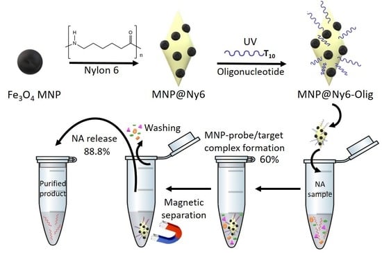
The principle of the specific NA magnetic solid-phase extraction is presented in Figure 5. For this method, the MNP with a probe for the NA sequence (MNP-Olig) is required. MNP-Olig adsorbs specific NA from the biological sample. The next stage is magnetic separation, washing, and NA release.
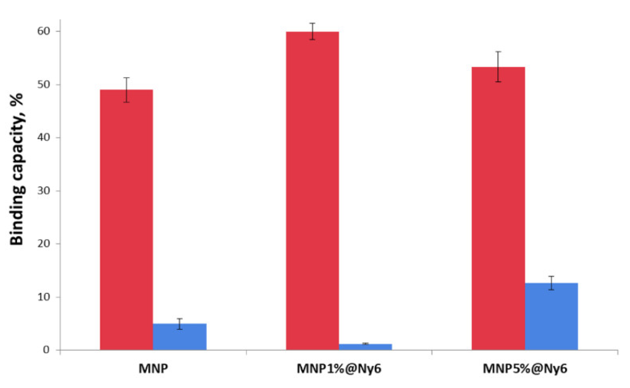
The model target RNA with FAM dye was synthesized (Table 2) for the NA microextraction experiments. A short RNA sequence corresponding to mRNA NELFA (WHSC2, NM_005663) was used as the target RNA analyte. The mRNA encodes a member of the negative elongation factor (NELF) protein complex, which participates in the regulation of RNA polymerase II transcription elongation. It is essential for cancer treatment [59]. For the RNA capture on the MNPs’ surface, more stable than RNA DNA-capture, oligonucleotides were immobilized (Table 2). Oligonucleotide No. 1 sequence is complementary to the RNA, and oligonucleotide No. 2, no. Therefore, oligonucleotide No. 1 will form a stable DNA/RNA complex with target RNA. Oligonucleotide No. 1 and No. 2 sequences were immobilized on MNP and MNP@Ny6 nanoparticles. For the 5′-FAM-RNA isolation, MNP-Olig 1 or Olig 2 nanocomposites were added under stirring to the target RNA solution with interfering components (total yeast RNA). The total yeast RNA concentration was one hundred higher than the target RNA concentrations. After 1 h, MNPs were magnetically decanted, and supernatant fluorescence intensity was measured. The capacity data for MNP nanocomposites are presented in Figure 6. The most specific for the target RNA and having the highest capacity is MNP1%@Ny6. The MNP1%@Ny6 capacity is 60%, which corresponds to 0.18 nmol/mg.
The 50% formamide aqueous solution was added to the MNPs for the target RNA release. The mixture was stirred (500 rpm) under heating at 90 °C for 10 min. After the magnetic decantation, the supernatant solution was analyzed by fluorescence. The most efficient RNA release was obtained for MNP1%@Ny6 nanocomposites, which correspond to 88.8% and 0.16 nmol/mg. Low release for MNP may be explained by NA nonspecific binding on the MNPs surface [20]. The resulted data for oligonucleotide immobilization on the MNP surface, target RNA capacity, and RNA release is presented in Table 3. The most perspective for the further experiment is MNP1%@Ny6. The resulting MNP1%@Ny6 nanocomposites could be used for specific RNA absorption because of their easy synthesis, high affinity and specificity, and efficient NA release. Further assay optimization and extensive biological studies may improve the experiments’ time, NA yield, and specificity. Moreover, the experiments with various biological samples such as blood, plasma, cell lysis, etc., are required to estimate MNP1%@Ny6 for commercial and scientific applications. In particular, the resultant NA-bound MNP1%@Ny6 nanocomposites may be used directly for polymerase chain reaction (PCR) experiments to simplify gene expression detection.
3. Materials and Methods
3.1. Materials
The FeCl3∙6H2O was purchased from PanReac AppliChem (MW = 270.32, 97–102%, Darmstadt, Germany). The FeCl2∙4H2O was obtained from Acros organics (MW = 198.81, 99+%, Geel, Belgium). Nylon 6 (Product No. 181110), tetraethyl orthosilicate, (3-aminopropyl)triethoxysilane, 2,2,2-trifluoroethanol, N,N-dimethylformamide, Tween 20, and all solvents, reagents were purchased from Sigma (St. Louis, MO, USA) at the highest available grade and used without purification. 2,4,6-Trichloro-1,3,5-triazine was obtained from Merck (Darmstadt, Germany). Fluoresceine (6-FAM) amidite was purchased from Lumiprobe (Moscow, Russia). FAM CPG for oligonucleotide synthesis was obtained from Primitech (Product No. 008c-500.2, Minsk, Belarus). Deionized water (Milli-Q) was used for the synthesis procedures. The following buffer solutions were prepared: 0.1 M sodium borate buffer (pH 9.0), alkaline phosphatase (AP) buffer (10 mM Tris-HCl, 50 mM KCl, 0.5% Tween-20, 1.8 mM MgCl2) which was adjusted to the pH 7.5 or 9.5 by sodium hydroxide, buffer for NA binding (pH 7.5, 9.5, 0.02 M Tris-HCl, 0.02 M MgCl2, 0.1 M NaCl).
3.2. Magnetic Nanocomposites Characterization
The nanoparticles were characterized by transmission electron microscopy (TEM), dynamic light scattering (DLS), and near-infrared (NIR) spectroscopy. The DLS was carried out on a Malvern Zetasizer Nano device (Malvern Instruments, Worcestershire, UK) in deionized water (~300 μg/mL concentration). For TEM, a drop of a sample was allowed to adsorb for 1 min on a copper grid covered with formvar film. The liquid excess was removed, and the grids were examined under a Jem-1400 device (Jeol, Tokyo, Japan) at an accelerating voltage of 80 kV. The NIR spectra were carried out using a MPA NIR-spectrometer (Bruker, Ettlingen, Germany) in the range 4000–12,500 cm−1. The reflection spectra of samples pre-dried from suspension in acetone were recorded as a thin layer on a glass slide (Corning, Glendale, Arizona, USA, size 75 × 25 mm, thickness 0.96–1.06 mm) using the Sphere Macrosample mode. The fluorescence intensity was determined on a Clariostar fluorimeter (BMG LABTECH, Ortenberg, Germany) in the emission wavelength range of FAM fluorescent dye. The λex range of FAM was 414–483 nm, and λem was 517 nm.
3.3. Synthesis and Isolation of Oligonucleotides
Oligonucleotides were synthesized in an ASM-800 synthesizer (Biosset, Novosibirsk, Russia) according to the standard protocol of the 2-cyanoethyl phosphoramidite method. Oligonucleotides were purified by RP-HPLC performed in the Agilent 1200 series chromatograph (Agilent, Santa Clara, CA, USA) on a column (4.6 × 150 mm) containing the Eclipse XDB-C18 sorbent (5 µm) with a 0–90% linear gradient of acetonitrile concentration in 0.02 M triethylammonium acetate solution for 30 min at a flow rate of 1.5 mL/min. The detection of the intensity of optical absorption was carried out at wavelengths 260, 280, 300, and 360 nm. The target product fraction was evaporated in vacuo. Coevaporations removed the bulk of triethylammonium acetate with ethanol. To remove the protecting dimethoxytrityl group, oligonucleotides were treated with 80% acetic acid (25 °C, 5 min). Purified oligonucleotides were concentrated, followed by precipitation with 2% LiClO4 in acetone, washing with pure acetone, and desiccation under a vacuum. As needed, the oligonucleotides were dissolved in deionized water and stored at −20 °C. The concentration of oligonucleotides was determined by UV spectroscopy [60]. UV spectra were recorded on a UV-1800 spectrometer (Shimadzu, Kyoto, Japan).
3.4. Magnetic Nanoparticles (MNP) Synthesis
Magnetite particles were synthesized by our colleagues’ previously published co-precipitating Fe2+ and Fe3+ salts ([Fe3+]/[Fe2+] = 2) method [48]. The total concentration of Fe ions was 0.15 mol/L. The MNPs precipitation was done by aqueous ammonia solution under intensive mechanical stirring (500 rpm) and argon atmosphere at 20 °C in preliminarily deoxygenated water. Magnetite nanoparticles were aged for 3 days at 20 °C before characterization. The MNPs were separated by magnetic decantation and washed with water three times. The resulting MNPs have an average size of 11.0 ± 2.5 nm by TEM. Properties of the MNP have no differences between published data (data not shown) [48]. MNP retains colloidal stability during 4–5 months in an aqueous solution under 4 °C without significant changes in magnetic precipitation properties, color, and size evaluated by DLS and TEM (data not shown).
3.5. Synthesis of Oligonucleotide-Modified MNPs (MNP-Olig)
MNPs aqueous suspension of 100 μL (11.3 mg/mL) was dispersed into a mixture of 0.833 mL ethanol and 67 μL of deionized water and was sonicated for 15 min. The 25 μL of ammonia solution and 3.5 μL of tetraethyl orthosilicate (TEOS) were added to the mixture under sonicating. The resulting solution was stirred for 18 h at 20 °C. The nanoparticles were collected by magnetic separation and washed with ethanol (3 × 1.5 mL). Then, 50 μL of 5% ethanol solution of (3-aminopropyl) triethoxysilane (APTES) were then added to the MNPs under vigorous stirring (900 rpm) for 2 h at 20 °C. The product was magnetically separated and washed with ethanol (3 × 1.5 mL) and acetonitrile (1 × 1.5 mL). Finally, 1 mL of a 2,4,6-Trichloro-1,3,5-triazine (TCT) solution (10 mg/mL) in acetonitrile was added to the precipitate under stirring (500 rpm) for 2 h at 20 °C. The final product was collected by magnetic separation, washed with acetonitrile (3 × 3 mL), and dried in vacuo.
Oligonucleotide-NH2 (Table 1) 300 μL 10−5 M in 0.05 M sodium borate buffer, pH 9.0 were added to the 1.13 mg of obtained MNPs under stirring (500 rpm) for 15 h at 20 °C. The MNP-Olig conjugate was magnetically isolated and washed with deionized water (3 × 1 mL). The water fractions were analyzed spectrophotometrically and/or by fluorescence (FAM residue) to calculate the MNPs capacity.
3.6. Synthesis of MNP and Nylon 6 Nanocomposites (MNP@Ny6)
Nylon 6 was dissolved in 2,2,2-trifluoroethanol under stirring for 30 min at 60 °C to obtain a 5% solution. To the 0.125 mg (for MNP1%@Ny6) or 0.625 mg (for MNP5%@Ny6) MNPs suspension in 125 μL of water, 250 μL of 2,2,2-trifluoroethanol and 250 μL of Ny6 solution were added under stirring for 5 min. Deionized water was gradually added to the reaction mixture until the precipitate began to form. The resulting suspension was left overnight for complete precipitation of MNP@Ny6 nanocomposites. MNPs were collected by magnetic separation, washed with ethanol (3 × 1.5 mL), deionized water, and dried in vacuo.
MNP1%@Ny6 precipitates in aqueous solution in the 1.5 mL tube for 3–5 s on the magnetic separation stands.
3.7. Synthesis of Oligonucleotide-Modified MNP@Ny6 (MNP@Ny6-Olig)
T10-oligonucleotide (Table 1) 300 μL 10−5 M in 0.05 M sodium borate buffer, pH 9.0 were added to the 1.13 mg of obtained MNP@Ny6 under stirring (500 rpm) for 15 h at 20 °C. The resulting mixture was placed under a source of UV irradiation (two mercury lamps of low-pressure PI-15, λ = 253.7 nm) at a distance of 10 cm for 10 min [56]. MNP@Ny6-Olig was collected by magnetic separation, washed with deionized water (3 × 1 mL), and dried in vacuo. The water fractions were analyzed spectrophotometrically and/or by fluorescence (FAM residue) to calculate the MNPs capacity.
MNP1%@Ny6-Olig precipitates in aqueous solution in the 1.5 mL tube for 3–5 s on the magnetic separation stands.
3.8. Colorimetric Biotin Detection
The revealing of the resulting biotin-containing products was carried out by the method [56]. Briefly, a solution of 0.1% egg albumin in AP buffer (pH 7.5) was added to 2 mg of the MNP@Ny6 containing the biotinylated probe and incubated under stirring (500 rpm) for 15 min at 20 °C. Streptavidin-alkaline phosphatase solution 50 μL (1 μg/mL) in AP buffer (pH 7.5) containing 0.05% Triton and 0.05% SDS were added to the MNPs and incubated under stirring for 30 min at 20 °C. MNPs were magnetically decanted and washed with 20% ethanol in AP buffer (pH 7.5) solution (4 × 200 μL); AP buffer (pH 9.5, 200 μL). Next, 100 μL of 0.495 mg NBT and 0.6 mg BCIP chromogenic substrate solution in AP buffer (pH 9.5) were added. The result of the colorimetric analysis was visualized for 30 min by recording the change in the solution color to blue.
3.9. RNA Binding on MNP-Olig or MNP@Ny6-Olig Nanocomposites
To the 1.13 mg MNP-Olig or MNP@Ny6-Olig, 300 μL 0.5% of yeast total RNA solution in PBS (pH 7.4) were added. The mixture was incubated for 20 min under stirring (500 rpm) at 4 °C. To the latter solution, 1 μL 0.3 mM target FAM-RNA (Table 2) in PBS (pH 7.4) was added and incubated under stirring (500 rpm) for 1 h at 4 °C. The resulted MNP complexes with RNA were magnetically decanted. The supernatant solution was analyzed by fluorescence (FAM residue) to calculate the RNA binding efficiency.
3.10. RNA Release
To the 1.13 mg MNP-Olig or MNP@Ny6-Olig, 300 μL of 50% formamide solution were added and incubated under stirring (500 rpm) for 10 min at 95 °C. The MNPs were magnetically decanted. The supernatant solution was analyzed by fluorescence (FAM residue) to calculate the RNA release efficiency.
4. Conclusions
In summary, we reported the synthesis of magnetic nanoparticles and nylon 6 nanocomposites (MNP@Ny6) for RNA microextraction and purification that could be implemented in the detection of a wide range of gene expression or pathogens. MNP@Ny6 may serve as an almost universal platform for various sequence NA attachments. The UV-immobilization of oligonucleotide on the nylon 6 surface requires only 10 min to produce a vital MNP@Ny6-Olig target NA-specific probe. MNP1%@Ny6 shows a high oligonucleotide binding capacity of 2.2 nmol/mg with 73.3% loading efficiency. Compared with the other well-known recognition mechanisms, the interaction of NA is universal and easily modified. MNP@Ny6-Olig possesses high specificity and capacity and efficient NA release. For MNP1%@Ny6-Olig, the RNA target isolation efficiency was 60% with high specificity. The bind RNA release was 88.8% in a quantity of 0.16 nmol/mg. The total RNA capture efficiency was 53%. In this regard, MNP1%@Ny6-Olig nanocomposites have great potential in the DNA extraction area for various applications.
Author Contributions
Data curation and investigation, A.B., A.C. and E.D.; conceptualization, E.D. and A.C.; writing—original paper, A.C., A.B. and E.D.; funding acquisition, A.C. and E.D.; project administration, A.C. All authors have read and agreed to the published version of the manuscript.
Funding
This study was funded by the Russian Science Foundation (grant No. 22-24-00996), and the nanoparticles synthesis was supported by the Ministry of Science and Higher Education of the Russian Federation (state registration No. 121031300042-1).
Institutional Review Board Statement
Not applicable.
Informed Consent Statement
Not applicable.
Data Availability Statement
Not applicable.
Conflicts of Interest
The authors declare no conflict of interest.
References
- Reyes-Gallardo, E.M.; Lucena, R.; Cárdenas, S.; Valcárcel, M. Polymer-nanoparticles composites in bioanalytical sample preparation. Bioanalysis 2015, 7, 1723–1730. [Google Scholar] [CrossRef] [PubMed]
- Bowers, A.N.; Trujillo-Rodríguez, M.J.; Farooq, M.Q.; Anderson, J.L. Extraction of DNA with magnetic ionic liquids using in situ dispersive liquid–liquid microextraction. Anal. Bioanal. Chem. 2019, 411, 7375–7385. [Google Scholar] [CrossRef] [PubMed]
- Kovrigina, E.; Chubarov, A.; Dmitrienko, E. High Drug Capacity Doxorubicin-Loaded Iron Oxide Nanocomposites for Cancer Therapy. Magnetochemistry 2022, 8, 54. [Google Scholar] [CrossRef]
- Anderson, S.D.; Gwenin, V.V.; Gwenin, C.D. Magnetic Functionalized Nanoparticles for Biomedical, Drug Delivery and Imaging Applications. Nanoscale Res. Lett. 2019, 14, 1–16. [Google Scholar] [CrossRef] [Green Version]
- Lamichhane, N.; Sharma, S.; Verma, A.K.; Roy, I.; Sen, T. Iron oxide-based magneto-optical nanocomposites for in vivo biomedical applications. Biomedicines 2021, 9, 288. [Google Scholar] [CrossRef]
- Chouhan, R.S.; Horvat, M.; Ahmed, J.; Alhokbany, N.; Alshehri, S.M.; Gandhi, S. Magnetic nanoparticles—a multifunctional potential agent for diagnosis and therapy. Cancers 2021, 13, 2213. [Google Scholar] [CrossRef] [PubMed]
- Shabatina, T.I.; Vernaya, O.I.; Shabatin, V.P.; Melnikov, M.Y. Magnetic nanoparticles for biomedical purposes: Modern trends and prospects. Magnetochemistry 2020, 6, 30. [Google Scholar] [CrossRef]
- Ganapathe, L.S.; Mohamed, M.A.; Yunus, R.M.; Berhanuddin, D.D. Magnetite (Fe3O4) nanoparticles in biomedical application: From synthesis to surface functionalisation. Magnetochemistry 2020, 6, 68. [Google Scholar] [CrossRef]
- Hepel, M. Magnetic nanoparticles for nanomedicine. Magnetochemistry 2020, 6, 3. [Google Scholar] [CrossRef] [Green Version]
- Dulińska-Litewka, J.; Łazarczyk, A.; Hałubiec, P.; Szafrański, O.; Karnas, K.; Karewicz, A. Superparamagnetic iron oxide nanoparticles-current and prospective medical applications. Materials 2019, 12, 617. [Google Scholar] [CrossRef] [Green Version]
- Chen, W.; Lu, F.; Chen, C.C.V.; Mo, K.C.; Hung, Y.; Guo, Z.X.; Lin, C.H.; Lin, M.H.; Lin, Y.H.; Chang, C.; et al. Manganese-enhanced MRI of rat brain based on slow cerebral delivery of manganese (II) with silica-encapsulated MnxFe1-xO nanoparticles. NMR Biomed. 2013, 26, 1176–1185. [Google Scholar] [CrossRef]
- Ray, S.; Cheng, C.A.; Chen, W.; Li, Z.; Zink, J.I.; Lin, Y.Y. Magnetic heating stimulated cargo release with dose control using multifunctional MR and thermosensitive liposome. Nanotheranostics 2019, 3, 166–178. [Google Scholar] [CrossRef]
- Ghambari, H.; Reyes-Gallardo, E.M.; Lucena, R.; Saraji, M.; Cárdenas, S. Magnetic polyamide nanocomposites for the microextraction of benzophenones from water samples. Molecules 2019, 24, 953. [Google Scholar] [CrossRef] [Green Version]
- Ma, Y.; Chen, T.; Iqbal, M.Z.; Yang, F.; Hampp, N.; Wu, A.; Luo, L. Applications of magnetic materials separation in biological nanomedicine. Electrophoresis 2019, 40, 2011–2028. [Google Scholar] [CrossRef]
- Marengo, A.; Cagliero, C.; Sgorbini, B.; Anderson, J.L.; Emaus, M.N.; Bicchi, C.; Bertea, C.M.; Rubiolo, P. Development of an innovative and sustainable one-step method for rapid plant DNA isolation for targeted PCR using magnetic ionic liquids. Plant Methods 2019, 15, 23. [Google Scholar] [CrossRef]
- Wang, L.; He, K.; Sadak, O.; Wang, X.; Wang, Q.; Xu, X. Visual detection of in vitro nucleic acid replication by submicro- and nano-sized materials. Biosens. Bioelectron. 2020, 169, 112602. [Google Scholar] [CrossRef]
- Sosa-Acosta, J.R.; Iriarte-Mesa, C.; Ortega, G.A.; Díaz-García, A.M. DNA–Iron Oxide Nanoparticles Conjugates: Functional Magnetic Nanoplatforms in Biomedical Applications. Top. Curr. Chem. 2020, 378, 1–29. [Google Scholar] [CrossRef]
- Li, P.; Li, M.; Yue, D.; Chen, H. Solid-phase extraction methods for nucleic acid separation. A review. J. Sep. Sci. 2022, 45, 172–184. [Google Scholar] [CrossRef]
- Chubarov, A.S. Serum Albumin for Magnetic Nanoparticles Coating. Magnetochemistry 2022, 8, 13. [Google Scholar]
- Bobrikova, E.; Chubarov, A.; Dmitrienko, E. The Effect of pH and Buffer on Oligonucleotide Affinity for Iron Oxide Nanoparticles. Magnetochemistry 2021, 7, 128. [Google Scholar] [CrossRef]
- Vanyorek, L.; Ilosvai, Á.M.; Szőri-Dorogházi, E.; Váradi, C.; Kristály, F.; Prekob, Á.; Fiser, B.; Varga, T.; Kónya, Z.; Viskolcz, B. Synthesis of iron oxide nanoparticles for DNA purification. J. Dispers. Sci. Technol. 2021, 42, 693–700. [Google Scholar] [CrossRef]
- Wang, J.; Ali, Z.; Si, J.; Wang, N.; He, N.; Li, Z. Simultaneous extraction of DNA and RNA from hepatocellular carcinoma (Hep G2) based on silica-coated magnetic nanoparticles. J. Nanosci. Nanotechnol. 2017, 17, 802–806. [Google Scholar] [CrossRef]
- Danthanarayana, A.N.; Manatunga, D.C.; De Silva, R.M.; Chandrasekharan, N.V.; De Silva, K.M.N. Magnetofection and isolation of DNA using polyethyleneimine functionalized magnetic iron oxide nanoparticles. R. Soc. Open Sci. 2018, 5, 181369. [Google Scholar] [CrossRef] [Green Version]
- Tang, C.; He, Z.; Liu, H.; Xu, Y.; Huang, H.; Yang, G.; Xiao, Z.; Li, S.; Liu, H.; Deng, Y.; et al. Application of magnetic nanoparticles in nucleic acid detection. J. Nanobiotechnol. 2020, 18, 1–19. [Google Scholar] [CrossRef] [Green Version]
- Chacón-Torres, J.C.; Reinoso, C.; Navas-León, D.G.; Briceño, S.; González, G. Optimized and scalable synthesis of magnetic nanoparticles for RNA extraction in response to developing countries’ needs in the detection and control of SARS-CoV-2. Sci. Rep. 2020, 10, 19004. [Google Scholar] [CrossRef]
- Ali, T.H.; Mandal, A.M.; Heidelberg, T.; Hussen, R.S.D. Sugar based cationic magnetic core–shell silica nanoparticles for nucleic acid extraction. RSC Adv. 2022, 12, 13566–13579. [Google Scholar] [CrossRef]
- Frenea-Robin, M.; Marchalot, J. Basic Principles and Recent Advances in Magnetic Cell Separation. Magnetochemistry 2022, 8, 11. [Google Scholar] [CrossRef]
- Gessner, I.; Fries, J.W.U.; Brune, V.; Mathur, S. Magnetic nanoparticle-based amplification of microRNA detection in body fluids for early disease diagnosis. J. Mater. Chem. B 2021, 9, 9–22. [Google Scholar] [CrossRef] [PubMed]
- Min, J.H.; Woo, M.K.; Yoon, H.Y.; Jang, J.W.; Wu, J.H.; Lim, C.S.; Kim, Y.K. Isolation of DNA using magnetic nanoparticles coated with dimercaptosuccinic acid. Anal. Biochem. 2014, 447, 114–118. [Google Scholar] [CrossRef] [PubMed]
- Sosa-Acosta, J.R.; Silva, J.A.; Fernández-Izquierdo, L.; Díaz-Castañón, S.; Ortiz, M.; Zuaznabar-Gardona, J.C.; Díaz-García, A.M. Iron Oxide Nanoparticles (IONPs) with potential applications in plasmid DNA isolation. Colloids Surf. A Physicochem. Eng. Asp. 2018, 545, 167–178. [Google Scholar] [CrossRef]
- Berensmeier, S. Magnetic particles for the separation and purification of nucleic acids. Appl. Microbiol. Biotechnol. 2006, 73, 495–504. [Google Scholar] [CrossRef]
- Hossain, S.; Rahman, M.; Nahar, Y.; Rahman, A.; Sharafat, M.K.; Hossain, M.; Ochiai, B.; Elaissari, A.; Ahmad, H. A simple in situ synthesis of iron oxide magnetic nanoparticles embedded in thermosensitive polymer for DNA capture. J. Mater. Res. 2020, 35, 2441–2450. [Google Scholar] [CrossRef]
- Damavandi, F.; Wang, W.; Shen, W.Z.; Cetinel, S.; Jordan, T.; Jovel, J.; Montemagno, C.; Wong, G.K.S. Enrichment of low abundance DNA/RNA by oligonucleotide-clicked iron oxide nanoparticles. Sci. Rep. 2021, 11, 13053. [Google Scholar] [CrossRef]
- Lim, S.H.; Bestvater, F.; Buchy, P.; Mardy, S.; Yu, A.D.C. Quantitative analysis of nucleic acid hybridization on magnetic particles and quantum dot-based probes. Sensors 2009, 9, 5590–5599. [Google Scholar] [CrossRef] [Green Version]
- Pang, Y.; Wang, C.; Wang, J.; Sun, Z.; Xiao, R.; Wang, S. Fe3O4@Ag magnetic nanoparticles for microRNA capture and duplex-specific nuclease signal amplification based SERS detection in cancer cells. Biosens. Bioelectron. 2016, 79, 574–580. [Google Scholar] [CrossRef]
- Li, B.; Mou, X.; Chen, Z.; Chen, H.; Deng, Y.; Li, S.; Su, E.; He, L.; He, N. The development of a rapid high-quality universal nucleic acid extraction kit based on magnetic separation. Sci. China Chem. 2017, 60, 1602–1608. [Google Scholar] [CrossRef]
- Pinchon, E.; Leon, F.; Temurok, N.; Morvan, F.; Vasseur, J.J.; Clot, M.; Foulongne, V.; Cantaloube, J.F.; Perre, P.V.; Daynès, A.; et al. Rapid and specific DNA detection by magnetic field-enhanced agglutination assay. Talanta 2020, 219, 121344. [Google Scholar] [CrossRef]
- Yang, S.Y.; Chieh, J.J.; Wang, W.C.; Yu, C.Y.; Hing, N.S.; Horng, H.E.; Hong, C.Y.; Yang, H.C.; Chang, C.F.; Lin, H.Y. Magnetic nanoparticles for high-sensitivity detection on nucleic acids via superconducting-quantum-interference-device-based immunomagnetic reduction assay. J. Magn. Magn. Mater. 2011, 323, 681–685. [Google Scholar] [CrossRef]
- Camacho-fernández, J.C.; Jin, M.; Liu, X.; Berg, A.V.D.; Zhou, G. Ultrasensitive DNA detection based on two-step quantitative amplification on magnetic nanoparticles. Nanotechnology 2016, 27, 335102. [Google Scholar]
- Pivetal, J.; Frénéa-Robin, M.; Haddour, N.; Vézy, C.; Zanini, L.F.; Ciuta, G.; Dempsey, N.M.; Dumas-Bouchiat, F.; Reyne, G.; Bégin-Colin, S.; et al. Development and applications of a DNA labeling method with magnetic nanoparticles to study the role of horizontal gene transfer events between bacteria in soil pollutant bioremediation processes. Environ. Sci. Pollut. Res. 2015, 22, 20322–20327. [Google Scholar] [CrossRef] [Green Version]
- Bordelon, H.; Russ, P.K.; Wright, D.W.; Haselton, F.R. A Magnetic Bead-Based Method for Concentrating DNA from Human Urine for Downstream Detection. PLoS ONE 2013, 8, e68369. [Google Scholar] [CrossRef] [PubMed] [Green Version]
- Zhang, C.; Tjiu, W.W.; Liu, T.; Lui, W.Y.; Phang, I.Y.; Zhang, W. De Dramatically enhanced mechanical performance of nylon-6 magnetic composites with nanostructured hybrid one-dimensional carbon nanotube—Two-dimensional clay nanoplatelet heterostructures. J. Phys. Chem. B 2011, 115, 3392–3399. [Google Scholar] [CrossRef] [PubMed]
- Mohammadi, S.Z.; Safari, Z.; Madady, N. Synthesis of Co3O4@SiO2 Core/Shell–Nylon 6 Magnetic Nanocomposite as an Adsorbent for Removal of Congo Red from Wastewater. J. Inorg. Organomet. Polym. Mater. 2020, 30, 3199–3212. [Google Scholar] [CrossRef]
- Choi, E.Y.; Kim, K.; Kim, C.K.; Kang, E. Reinforcement of nylon 6,6/nylon 6,6 grafted nanodiamond composites by in situ reactive extrusion. Sci. Rep. 2016, 6, 37010. [Google Scholar] [CrossRef] [PubMed] [Green Version]
- Saeed, K.; Khan, I.; Ahad, M.; Shah, T.; Sadiq, M.; Zada, A.; Zada, N. Preparation of ZnO/Nylon 6/6 nanocomposites, their characterization and application in dye decolorization. Appl. Water Sci. 2021, 11, 1–10. [Google Scholar] [CrossRef]
- Dmitrienko, E.V.; Bulushev, R.D.; Haupt, K.; Kosolobov, S.S.; Latyshev, A.V.; Pyshnaya, I.A.; Pyshnyi, D.V. A simple approach to prepare molecularly imprinted polymers from nylon-6. J. Mol. Recognit. 2013, 26, 368–375. [Google Scholar] [CrossRef]
- Shakiba, M.; Rezvani Ghomi, E.; Khosravi, F.; Jouybar, S.; Bigham, A.; Zare, M.; Abdouss, M.; Moaref, R.; Ramakrishna, S. Nylon—A material introduction and overview for biomedical applications. Polym. Adv. Technol. 2021, 32, 3368–3383. [Google Scholar] [CrossRef]
- Kirillov, V.L.; Balaev, D.A.; Semenov, S.V.; Shaikhutdinov, K.A.; Martyanov, O.N. Size control in the formation of magnetite nanoparticles in the presence of citrate ions. Mater. Chem. Phys. 2014, 145, 75–81. [Google Scholar] [CrossRef]
- Kirillov, V.L.; Yakushkin, S.S.; Balaev, D.A.; Dubrovskiy, A.A.; Semenov, S.V.; Knyazev, Y.V.; Bayukov, O.A.; Velikanov, D.A.; Yatsenko, D.A.; Martyanov, O.N. Dimethylsulfoxide as a media for one-stage synthesis of the Fe3O4-Based ferro fluids with a controllable size distribution. Mater. Chem. Phys. 2019, 225, 292–297. [Google Scholar] [CrossRef]
- Clavijo, C.; Osma, J.F. Functionalized leather: A novel and effective hazardous solid waste adsorbent for the removal of the diazo dye congo red from aqueous solution. Water 2019, 11, 1906. [Google Scholar] [CrossRef] [Green Version]
- Wang, Y.; Sun, Y.; Wang, J.; Yang, Y.; Li, Y.; Yuan, Y.; Liu, C. Charge-Reversal APTES-Modified Mesoporous Silica Nanoparticles with High Drug Loading and Release Controllability. Appl. Mater. Interfaces 2016, 8, 17166–17175. [Google Scholar] [CrossRef]
- Digigow, R.G.; Dechézelles, J.; Dietsch, H.; Geissbühler, I.; Vanhecke, D.; Geers, C.; Hirt, A.M.; Rothen-rutishauser, B. Preparation and characterization of functional silica hybrid magnetic nanoparticles. J. Magn. Magn. Mater. 2014, 362, 72–79. [Google Scholar] [CrossRef] [Green Version]
- Chandra, S.; Beaune, G.; Shirahata, N.; Winnik, F.M. A one-pot synthesis of water soluble highly fluorescent silica nanoparticles. J. Mater. Chem. B 2017, 5, 1363. [Google Scholar] [CrossRef] [Green Version]
- Ismail, A.F.; Goh, P.; Rezaei, M.; Arzhandi, D.; Ismail, N. Aptes and teos modified binary recyclable hybrid Fe3O4@GO nanocomposite for photocatalytic dye removal. J. Teknol. 2018, 80, 157–164. [Google Scholar] [CrossRef] [Green Version]
- Hao, N.; Jayawardana, K.W.; Chen, X.; Zoysa, T.D.; Yan, M. One-step synthesis of amine-functionalized hollow mesoporous silica nanoparticles as efficient antibacterial and anticancer materials. ACS Appl. Mater. Interfaces 2015, 7, 1040–1045. [Google Scholar] [CrossRef] [Green Version]
- Dmitrienko, E.V.; Pyshnaya, I.A.; Pyshnyi, D.V. Oligonucleotide Derivatives in the Hybridization Analysis of Nucleic Acids. I. Covalent Immobilization of Oligonucleotide Probes on Nylon. Russ. J. Bioorgan. Chem. 2010, 36, 645–656. [Google Scholar] [CrossRef]
- Gebeyehu, G.; Rao, P.Y.; Soochan, P.; Simms, D.A.; Klevan, L. Novel biotinylated nucleotide-analogs for labeling and colorimetric detection of DNA. Nucleic Acids Res. 1987, 15, 4513–4534. [Google Scholar] [CrossRef] [Green Version]
- Juthani, N.; Doyle, P.S. A Platform for Multiplexed Colorimetric microRNA Detection using Shape-encoded Hydrogel Particles. Analyst 2020, 145, 5134. [Google Scholar] [CrossRef]
- Dmitrienlo, E.; Naumova, O.; Fomin, B.; Kupryushkin, M.; Volkova, A.; Amirkhanov, N.; Semenov, D.; Pyshnaya, I.; Pyshnyi, D. Surface modification of SOI-FET sensors for label-free and specific detection of short RNA analyte. Nanomedicine 2016, 11, 2073–2082. [Google Scholar] [CrossRef]
- Lomzov, A.A.; Kupryushkin, M.S.; Shernyukov, A.V.; Nekrasov, M.D.; Dovydenko, I.S.; Stetsenko, D.A.; Pyshnyi, D.V. Diastereomers of a mono-substituted phosphoryl guanidine trideoxyribonucleotide: Isolation and properties. Biochem. Biophys. Res. Commun. 2019, 513, 807–811. [Google Scholar] [CrossRef]
Figure 1.
TEM images of magnetic nanoparticles (MNPs). (A) High-magnification TEM image initial Fe3O4 MNPs, the bar indicates 100 nm. (B) Low-magnification TEM image MNP1%@Ny6, the bar indicates 2 μm. (C) High-magnification TEM image MNP1%@Ny6, the bar indicates 400 nm. (D) Low-magnification TEM image MNP5%@Ny6, the bar indicates 1 μm. (E) High-magnification TEM image MNP5%@Ny6, the bar indicates 200 nm.

Figure 2.
Two synthetic routes of oligonucleotide surface modification on MNPs (A) and MNP@Ny6 nanocomposites (B). TEOS―tetraethyl orthosilicate, APTES—(3-aminopropyl) triethoxysilane, TCT—2,4,6-trichloro-1,3,5-triazine. For the procedures, oligonucleotides with the amino group or T10 oligothymidylate tract were used (for structures, see Table 1).

Figure 3.
Fourier transform near-infrared spectra of MNP, Ny6, MNP1%@Ny6, and MNP5%@Ny6. The spectra were carried out using MPA NIR-spectrometer (Bruker, Billerica, MA, USA) in the range 4000–12,500 cm−1. The reflection spectra of samples pre-dried from suspension in acetone were recorded as a thin layer on a glass slide (Corning, USA, size 75 × 25 mm, thickness 0.96–1.06 mm) using the Sphere Macrosample mode.
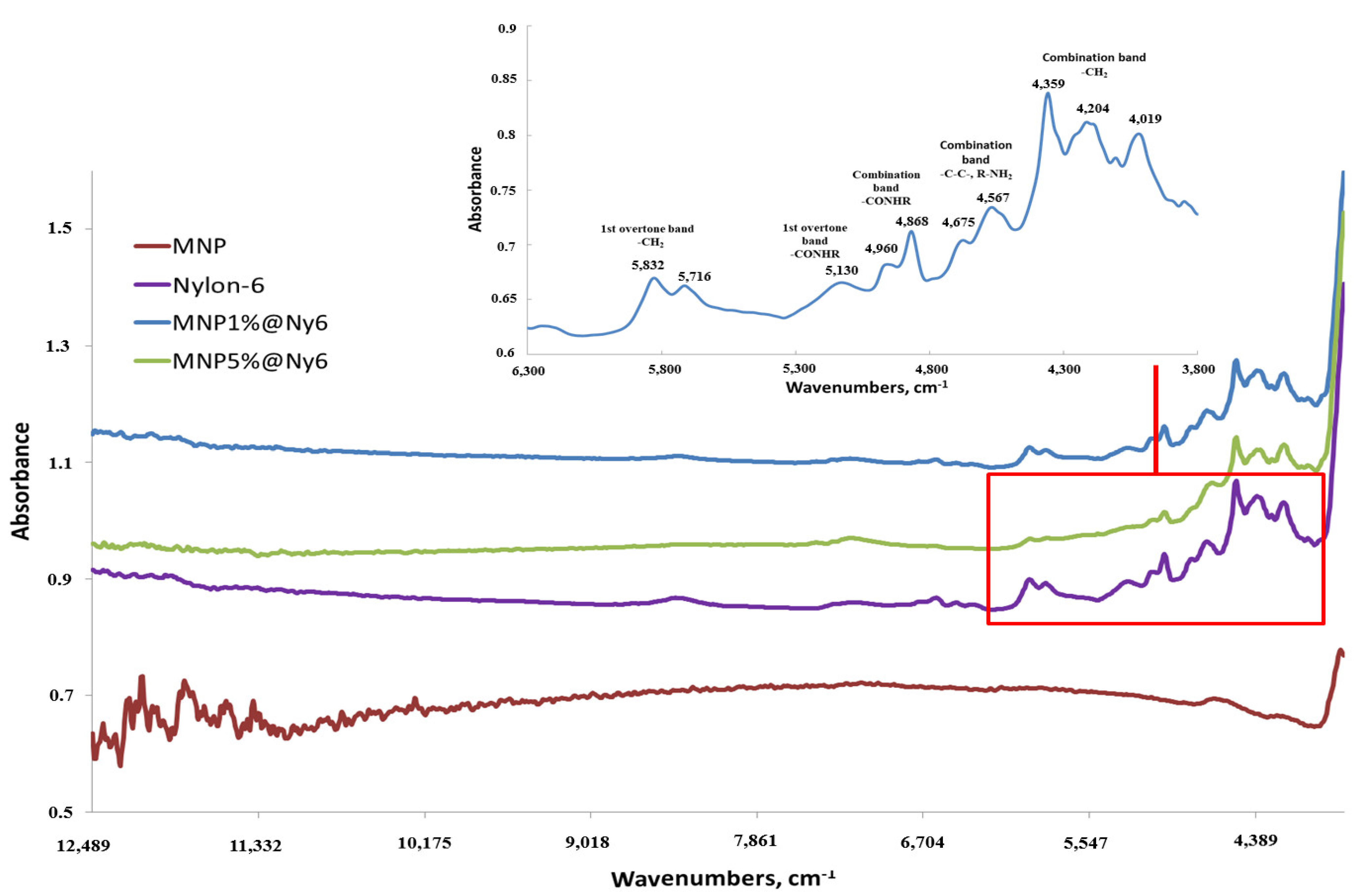
Figure 4.
The oligonucleotide UV-immobilization efficiency studies use colorimetric assay for biotin label detection. Biotin label interacts with streptavidin, alkaline phosphatase conjugate, and BCIP/NBT chromogenic substrates, yielding intensive dark blue color. BCIP—5-bromo-4-chloro-3-indolyl phosphate, NBT—nitro blue tetrazolium.
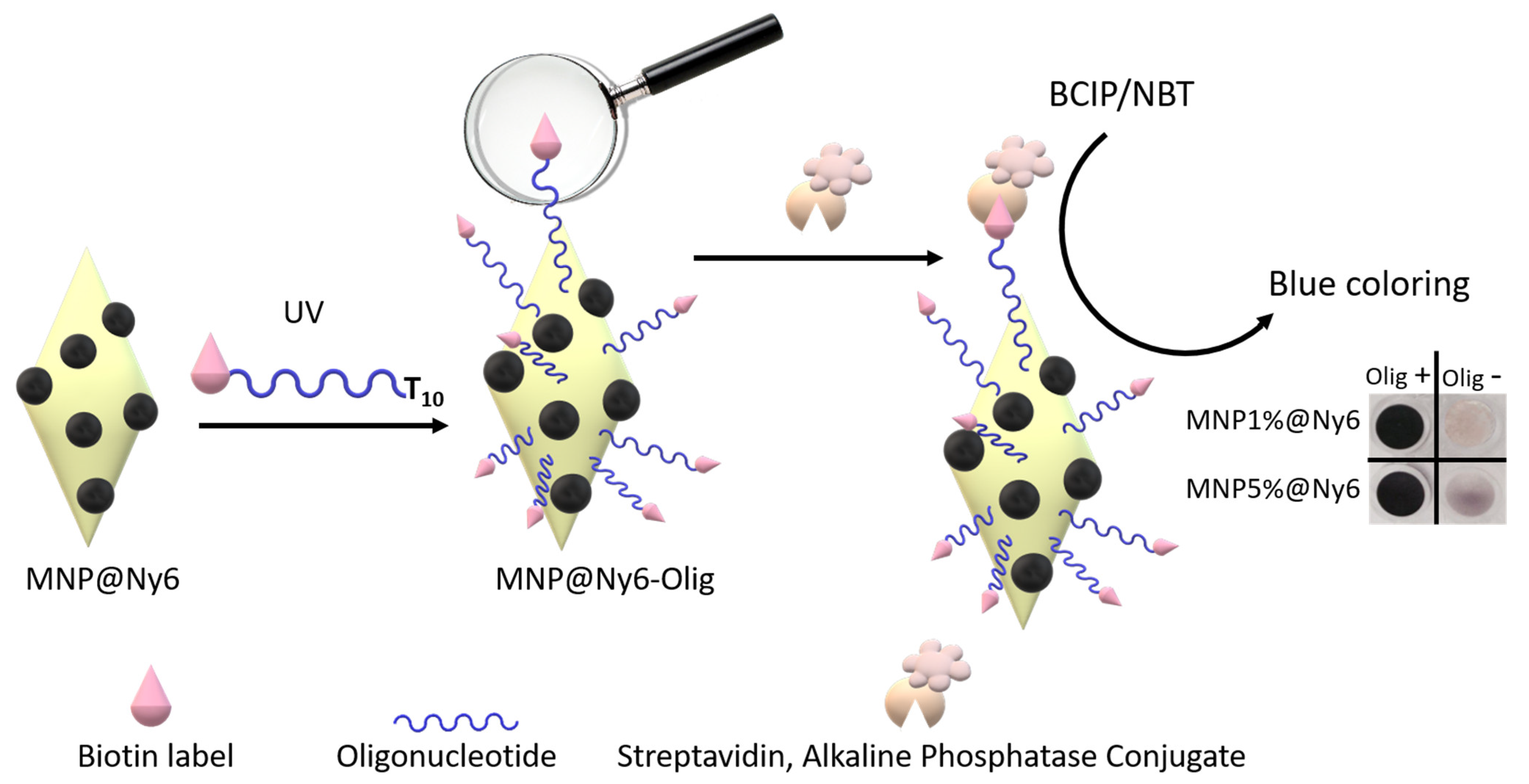
Figure 5.
Basic principles of NA magnetic solid-phase extraction.

Figure 6.
Binding capacity on MNP and MNP@Ny6 in percent for FAM-RNA isolation. Red—nanocomposites with oligonucleotide No. 1 (specific for RNA target), Blue—nanocomposites with oligonucleotide No. 2 (nonspecific). Initial amount of FAM-RNA is 0.30 nmol (see Table 3).
Figure 6.
Binding capacity on MNP and MNP@Ny6 in percent for FAM-RNA isolation. Red—nanocomposites with oligonucleotide No. 1 (specific for RNA target), Blue—nanocomposites with oligonucleotide No. 2 (nonspecific). Initial amount of FAM-RNA is 0.30 nmol (see Table 3).

Table 1.
Oligonucleotide sequences.
| Description | 5′-Sequence-3′ | NA Type, Function |
|---|---|---|
| oligonucleotide-NH2 | GTCTTCCTTCTCCGCTT-Sp1-NH2 | DNA, capture probe |
| FAM-oligonucleotide-NH2 | FAM-Sp2-GTCTTCCTTCTCCGCTT-Sp1-NH2 | |
| T10-oligonucleotide | (T)10-GTCTTCCTTCTCCGCTT | |
| T10-oligonucleotide-FAM * | (T)10-GTCTTCCTTCTCCGCTT-Sp2-FAM | |
| T10-oligonucleotide-Biotin | (T)10-GTCTTCCTTCTCCGCTT-Sp3-Biotin | |
Sp1 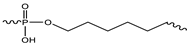 Sp2 Sp2 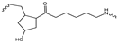 Sp3 Sp3 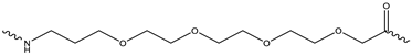 | ||
* FAM—fluoresceine residue. FAM-labeled oligonucleotides were used for capacity calculation experiments.
Table 2.
Oligonucleotide sequences for RNA microextraction experiments.
| Description | 5′-Sequence-3′ | NA Type, Function |
|---|---|---|
| oligonucleotide-NH2 No. 1 specific for RNA | GTCTTCCTTCTCCGCTT-Sp1-NH2 * | DNA, capture probe for MNP |
| oligonucleotide-NH2 No. 2 non-specific for RNA | TAACCGATTTCAGATTT-Sp1-NH2 | |
| T10-oligonucleotide No. 1 specific for RNA | (T)10-GTCTTCCTTCTCCGCTT | DNA, capture probe for MNP@Ny6 |
| T10-oligonucleotide No. 2 non-specific for RNA | (T)10-TAACCGATTTCAGATTT | |
| Target | Fam- GAGGCGAAGCGGAGAAGGAAGAC | RNA, target |
* Sp1 structure see in Table 1.
Table 3.
MNP nanocomposites characteristics.
| MNP Type | Capture DNA Probe Immobilization | Initial Target RNA Amount | Target RNA Binding | Target RNA Release | RNA Capture Efficiency |
|---|---|---|---|---|---|
| MNP | 50.0% | 0.30 nmol | 0.147 ± 0.015 nmol 49.0% a | 0.033 ± 0.010 nmol 22.4% b | 11.0% c |
| MNP1%Ny6 | 73.3% | 0.180 ± 0.010 nmol 60.0% | 0.160 ± 0.013 nmol 88.9% | 53.3% | |
| MNP5%Ny6 | 66.7% | 0.160 ± 0.018 nmol 53.3% | 0.087 ± 0.013 nmol 54.4% | 29.0% |
a binding efficiency, % = bound target RNA/initial RNA × 100%; b release efficiency, % = released target RNA/bound target RNA × 100%; c RNA capture efficiency, % = released target RNA/initial target RNA × 100%.
Publisher’s Note: MDPI stays neutral with regard to jurisdictional claims in published maps and institutional affiliations. |
© 2022 by the authors. Licensee MDPI, Basel, Switzerland. This article is an open access article distributed under the terms and conditions of the Creative Commons Attribution (CC BY) license (https://creativecommons.org/licenses/by/4.0/).
Share and Cite
MDPI and ACS Style
Bulgakova, A.; Chubarov, A.; Dmitrienko, E. Magnetic Nylon 6 Nanocomposites for the Microextraction of Nucleic Acids from Biological Samples. Magnetochemistry 2022, 8, 85. https://doi.org/10.3390/magnetochemistry8080085
AMA Style
Bulgakova A, Chubarov A, Dmitrienko E. Magnetic Nylon 6 Nanocomposites for the Microextraction of Nucleic Acids from Biological Samples. Magnetochemistry. 2022; 8(8):85. https://doi.org/10.3390/magnetochemistry8080085
Chicago/Turabian StyleBulgakova, Anastasia, Alexey Chubarov, and Elena Dmitrienko. 2022. "Magnetic Nylon 6 Nanocomposites for the Microextraction of Nucleic Acids from Biological Samples" Magnetochemistry 8, no. 8: 85. https://doi.org/10.3390/magnetochemistry8080085
Note that from the first issue of 2016, this journal uses article numbers instead of page numbers. See further details here.