
Magnetic Nanoparticles: An Overview for Biomedical Applications
1
Department of Chemistry, University of Delhi, Delhi 110007, India
2
Department of Chemistry, Galgotias University, Greater Noida 203201, India
*
Authors to whom correspondence should be addressed.
Magnetochemistry 2022, 8(9), 107; https://doi.org/10.3390/magnetochemistry8090107
Submission received: 18 August 2022
/
Revised: 6 September 2022
/
Accepted: 14 September 2022
/
Published: 17 September 2022
(This article belongs to the Special Issue New Trends in Magneto-Optical Ceramics)
Abstract
:The use of magnetic nanoparticles has greatly expanded for numerous biomedical applications over the past two decades due to their high surface area, size-dependent superparamagnetic properties, precision tunability, and easy surface modification. Magnetic nanoparticles can be engineered and manipulated with other nanoparticles and functional compounds to form multi-modal systems useful in theragnosis. However, superior biocompatibility, high loading efficacy, regulated drug release, and in vitro and in vivo stability are necessary for the efficient incorporation of these nanoparticles into physiological systems. In recent years, considerable advancements have been made and reported both in synthesis and application, given the broad range of biomedical-related prospective uses of magnetic nanoparticles. Here, in this review, we have highlighted some essential works, specifically related to the application of magnetic nanoparticles in drug delivery, magnetic hyperthermia, magnetic resonance imaging, magnetic particle imaging, biosensors, and tissue engineering.
1. Introduction
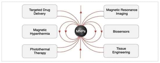
Nano-sized materials or particles and their formulations, due to their exclusive physical and chemical properties, are being extensively explored by scientists and researchers around the world for various industrial (material fillers, solar cells, superconductors, cosmetics, catalysis, etc.) and biomedical applications [1,2,3]. Among various organic, inorganic, and hybrid nanomaterials, magnetic nanoparticles have drawn special attention due to their interesting properties, such as large surface area, small size, ease of surface functionalization, and superparamagnetism. Apart from their applications in energy storage [4], magnetic fluids [5], environmental remediation [6], gyroscopic devices [7], catalysis [8], and magnetic inks [9], magnetic nanoparticles have been proven to exhibit noteworthy applications in the biomedicine field. These magnetic particles have been used for targeted delivery of drug/therapeutic agents [10], transfection of various viral/non-viral vectors and nucleic acids (referred to as magnetofection) [11], magnetic separation [12], magnetic field/light-induced hyperthermia [13], diagnostic imaging as contrast agents [14], tissue engineering [15], and various biosensing/immunosensing applications [16]. A number of formulations of these magnetic particles have been approved by the U.S. Food and Drug Administration for imaging and therapeutic applications, and some are under early clinical trials [17].
Magnetic hyperthermia and photothermal therapy have emerged as promising candidates for cancer therapy as they provide minimally invasive and site-specific treatment that ensures the minimization of the damage caused to normal tissues [13]. Preclinical models have shown the immense potential of these therapies in preventing metastasis [18]. Photothermal therapy is mostly used in combination with other forms of treatments and therapies, most commonly chemotherapy. The combination of different modes of therapy has often shown synergism which would enhance efficiency and reduce dosage [19]. Magnetic resonance imaging is a reliable diagnostic technique that is non-invasive and gives detailed anatomical images. It utilizes contrast agents to enhance the contrast between the target site and the surrounding tissues [14]. Ferrite nanoparticles have been shown to be efficient contrast agents as they can influence the relaxation process and enhance the sensitivity and detection ability [20]. Among various magnetic nanoparticles, owing to their high biocompatibility and chemical stability, iron oxide nanoparticles have been exploited the most for various biomedical applications. Pure-metal-based magnetic nanoparticles, despite their high saturation magnetization values, are not suitable for in vivo applications due to their high toxicity and need to be properly surface-functionalized or protected [21,22]. In addition to stabilizing the magnetic nanoparticles, these protective materials can be further surface modified with other ligands or nanoparticles for desired applications. This review aims to give the readers an overview of the promising use of magnetic nanoparticles and their formulations for various biomedical applications. In the following sections, the composition, properties, and synthetic routes commonly adopted, followed by characterization techniques available for magnetic nanoparticles, are discussed briefly. Finally, we have concisely highlighted and discussed the applications and research undertaken over the years of these magnetic particles, particularly for magnetically-guided drug delivery, magnetic hyperthermia, photothermal therapy (PTT), magnetic resonance imaging (MRI), magnetic particle imaging (MPI), biosensors, and tissue engineering.
2. Composition and Properties of Magnetic Nanoparticles
Multiple types of magnetic nanoparticles have been fabricated. These nanoparticles are generally made up of pure metals (Fe, Co, Ni), metal alloys (CoPt, FePt), metal oxides, or ferrites [23,24]. Iron oxide nanoparticles (IONPs) are the most studied and explored. They are composed of crystalline magnetite (Fe3O4) or maghemite (γ-Fe2O3). Magnetite corresponds to an inverse spinel structure, where the anions (oxygen) form the cubic close-packed lattice, with Fe2+ occupying the octahedral holes, half the Fe3+ occupying the octahedral holes, and the other half occupying tetrahedral holes [25,26]. Maghemite is considered as Fe2+ deficient magnetite [27]. Ferrites are basically metal oxides with a general formula MFe2O4 and have a spinel structure. M is a divalent cation such as Fe2+, Co2+, Ni2+, Mn2+, Cu2+, Zn2+, etc. There must be Fe3+ in the chemical formula of ferrites. These transition metal-doped ferrite nanoparticles find a lot of applications in materials chemistry [28,29]. Co, Cu, Ni, and Zn doped ferrite nanoparticles have been readily used for the purpose of catalysis of reactions involving organic compounds [30].
The size, surface charge, intrinsic magnetic properties, stability in an aqueous medium, and toxicity must be taken into consideration in these nanosystems for biomedical applications. These physicochemical properties ultimately decide the in vivo fate of the particles. Generally, small-sized nanoparticles, typically less than 20 nm, are desirable. At such small sizes, ferri- and ferromagnetic nanoparticles attain a single magnetic domain, having only one large magnetic moment value. The particles exhibit superparamagnetic properties with high magnetic moments, fast response in the presence of an external magnetic field, and show negligible remnant magnetization and coercivity in the absence of an external magnetic field [17,31]. The superparamagnetic material loses its magnetization as soon as the magnetic field is withdrawn. This ensures a safe removal from the body as there is no risk of unwanted aggregation of the nanoparticles. However, these particles in a small size range with large values of surface-to-volume ratio tend to aggregate due to their high surface energy. Furthermore, the overall size of the nanoparticles must be small enough to avoid splenic filtration but sufficiently large to prevent renal clearance [32]. Naked or uncoated iron oxide particles are unstable and tend to lose their magnetism due to their oxidation in the air. Thus, to retain and enhance their stability in a physiological environment, magnetic nanoparticles must be properly surface-modified or coated. The coating material also alters the final surface charge of the magnetic nanoparticles. The nanoparticles must be able to evade capture by RES (reticuloendothelial system) and have prolonged blood circulation time for their sufficient accumulation at the target site. Positively charged particles non-specifically bind to cells and have a small blood half-life value, while negatively charged particles are readily eliminated by the liver. Therefore, particles with a neutral surface or covered with neutral coating agents are preferred [33,34].
The stability (both compositional and colloidal) and biocompatibility of magnetic nanoparticles are critical determinants of their biomedical applications. For the stability of nanoparticles, their composition, size, shape, and surface-coating play major roles. It is generally known that smaller nanoparticles (below 10 nm in diameter) with adequate surface coating using inert polymers such as dextran or polyethylene glycol (PEG) remain both compositionally (does not erode) and colloidally (does not agglomerate) stable. Moreover, spherical nanoparticles are more stable than their anisotropic counterparts owing to relatively small surface area and uniform surface coating. Hot colloidal methods that lead to nanoparticle synthesis in a non-aqueous medium, followed by their aqueous dispersion using surface passivation with inert hydrophilic polymers, lead to better surface coating as compared to those synthesized directly in an aqueous media [35,36].
Superparamagnetic nanoparticles (temporary magnets) are better suited for biomedical applications than their ferromagnetic counterparts (permanent magnets), as in the former, the magnetism can be externally controlled. For most biomedical applications, it is desired that the particles remain mostly non-magnetic and become magnetic only within a specified spatio–temporal window during imaging and/or therapy [17]. Moreover, as superparamagnetic nanoparticles are usually smaller than ferromagnetic ones, they have better pharmacodynamics, biodistribution, and avenues of excretion.
Magnetic nanoparticles composed of transition metals such as Co, Ni, Mn, etc., are generally not favored for biomedical applications due to their high toxicity. In a free state, they induce the generation of reactive oxygen species (ROS), which results in oxidative stress and ultimately damages the cells. [37] Therefore, they need to be functionalized with biocompatible inorganic/organic materials. Iron oxide nanoparticles are generally considered biocompatible as iron ions obtained upon their metabolism can be added to the iron storage system present in the body [38]. The toxicity of magnetic nanoparticles is also dose-dependent. Magnetic nanoparticles are generally non-toxic at small dosages. However, at higher dosages, they can show toxic effects (e.g., iron ion-induced cell death via ferroptosis). Therefore, the required dosage of magnetic nanoparticles to suit a particular biomedical application is a key factor that determines their biocompatibility. Required dosage, in turn, is dictated by the targeting as well as the functional efficiency of the nanoparticles. Therefore, pure-phase (e.g., magnetite only) magnetic nanoparticles with higher saturation magnetization and/or MR relaxation time are better suited than mixed phase (e.g., magnetite and maghemite mixture) nanoparticles with lower functional parameters. Efficiently targeting diseased sites using externally applied magnetic fields or surface-attached biorecognition agents also lowers the required dosage of the nanoparticle, thus enhancing their biocompatibility [39].
3. Synthesis Methods
Magnetic particles that have dimensions in the nano range are desirable for various biomedical applications. Their properties, namely structural, magnetic, crystalline, and catalytic, depend upon the synthetic procedure. Further, functional magnetic nanoparticles made up of a magnetic core with appropriate protective coating or surface functionality are generally synthesized. Various single and multi-step synthetic procedures have been reported for the synthesis of the magnetic core of the required nanoparticle (Figure 1).
Co-precipitation or wet-precipitation is the oldest and most commonly used method to synthesize magnetic nanoparticles that have an average diameter below 50 nm. Simple as well as mixed metal oxide nanoparticles are obtained by using a stoichiometric mixture of aqueous salt solutions of two metal ions, and the particles are precipitated out upon the addition of an appropriate base (such as NaOH or NH4OH). However, particles with irregular morphologies and broader size distribution are obtained by this method [40,41]. Highly crystalline monodisperse particles smaller than 30 nm can be obtained by the thermal decomposition method at high temperatures. In this method, an organometallic precursor in a non-polar boiling solvent is decomposed in the presence of a stabilizing agent such as 1-octadecene, hexadecyl amine, oleic acid, etc., at high temperatures. The commonly used organometallic precursors are metal carbonyls (such as Fe(CO)5), Fe(Cup)3 (Cup = N-nitroso-N-phenylhydroxylamine) or metal acetyl acetonates [Mn+(acetylacetonate)n] (where M can be Fe, Co, Mn, Ni, or Cr; n = 2, 3) [42,43,44]. The only limitation associated with this method is that hydrophobic particles coated with organic molecules are obtained, which need to be further functionalized with hydrophilic materials. Metallic nanoparticles can be obtained by heating volatile metal compounds in an inert atmosphere by the chemical vapor condensation (CVC) technique. Metal oxide nanoparticles can be obtained from these metal particles by their further oxidation. However, specialized instrumentation facilities are required, and the commonly used precursors, such as Fe(CO)5, are toxic [45]. Hydrothermal synthesis at high temperatures (>100 °C) and high pressures (>1 atm), generally carried out in an autoclave, is another method to synthesize magnetic nanoparticles. This is a versatile one-pot synthesis method and can also be used to obtain surface-functionalized particles [46]. Nanoreactors or reverse-microemulsion systems (water-in-oil systems) stabilized by surfactants can be used to synthesize magnetic nanoparticles, and by varying the water: surfactant ratio (wo value), the size of the particles can be controlled [47,48]. Coating materials can also be added during synthesis using this method. In addition to these synthetic routes, electrochemical synthesis, combustion, laser-pyrolysis techniques, sol–gel, microwave-assisted and microbial synthesis, among others, have been reported [14,49,50,51]. Table 1 summarizes some of the synthesis methods along with their merits and demerits.
Magnetic core nanoparticles owing to their large surface area and energy, are sensitive to agglomeration and oxidation. The surface of the particles is rapidly oxidized in ambient conditions, which has a dramatic effect on their magnetic properties. In order to protect the particles from oxidation and forming clusters, they are generally encapsulated or surface functionalized by a biocompatible material. These materials also help in altering the surface charge of the nanoparticle and provide chemical functionalities for bioconjugation, affecting their biodistribution and pharmacokinetics. Several natural (dextran, albumin) and synthetic polymers (PEG, PAA) are mostly used to coat the surface of magnetic nanoparticles, either during the synthesis or after the synthesis [53,54,55]. However, the thickness of the coating can have a negative effect on the magnetic response of the particle [56]. Other coating materials include inorganic molecules such as silica, gold, and carbon [57,58,59]. Liposomes, which are phospholipid bilayer membrane vesicles, can encapsulate a large number of magnetic cores and deliver them simultaneously [60].
4. Characterization
Post-synthesis-prepared magnetic nanoparticles must be thoroughly characterized by using a variety of characterization techniques before employing them for various biomedical applications. The surface morphology and size of the magnetic nanoparticles can alter their physicochemical properties. Transmission electron microscopy (TEM) and scanning electron microscopy (SEM) are most commonly used for the determination of particle size and morphology of nanoparticles/nanomaterials [61]. TEM can also be used to visualize and measure the thickness of the coating material on the magnetic core particles, which is not possible using SEM [62]. The atomic force microscopy (AFM) technique is often employed for determining the size, morphology, and surface roughness of the nanoparticles [63]. Dynamic light scattering (DLS) or photon correlation spectroscopy (PCS) is used to find the hydrodynamic diameter and size distribution (polydispersity) of the particles [64]. Various analytical techniques such as energy-dispersive X-ray spectroscopy (EDS), X-ray fluorescence (XRF) spectroscopy, X-ray photoelectron spectroscopy (XPS), atomic absorption spectroscopy (AAS), and inductively coupled plasma mass spectroscopy (ICP-MS) are employed to determine the elemental composition of the magnetic particles [65]. Mössbauer spectroscopy can be used to differentiate between two types of iron oxide nanoparticles (magnetite and maghemite) and can also determine their relative concentrations in a sample [66]. The spatial distribution (elemental mapping) of elements in the nanoparticles can also be obtained using EDS analysis. The crystalline structure and phase purity of magnetic core nanoparticles can be ascertained using X-ray diffraction (XRD) spectroscopy. The sharp peaks obtained in the XRD pattern can be used to determine the size of particles using the “Scherrer equation” [65]. The selected area electron diffraction (SAED) patterns can be obtained using a TEM instrument and are often used to evaluate the crystal structure and other lattice parameters. Techniques such as XPS, zeta potential measurement, and Fourier-transform infrared (FT-IR) spectroscopy are useful in the surface characterization of nanoparticles [40]. XPS can identify as well as quantify every element (except H and He) and determine its exact electronic state present on the surface of the particle. The surface charge of the particles is determined by measuring the zeta potential. FT-IR spectroscopy is employed for identifying surface functionalities (mainly organic). These surface characterization techniques are useful in confirming the presence of coating or attachment of functionalities on the surface of the magnetic core [67]. In addition to determining thermal stability, thermal gravimetric analysis (TGA) can also determine the binding efficiency and coating formation on the particle’s surface [68].
The magnetic properties are generally evaluated using a vibrating sample magnetometer (VSM) and superconducting quantum interference device (SQUID). VSM produces a hysteresis curve depicting changes in magnetization/magnetic moment of nanoparticles on varying the external magnetic field. Superparamagnetic nanoparticles have high saturation magnetization values with coercivity and remnant magnetization values equal to or very close to zero. Thus, a VSM curve passing through the origin with no open loop is obtained for superparamagnetic particles. The saturation magnetization value can reduce upon coating or surface functionalizing the particle [69,70]. SQUID monitors change in magnetization as a function of temperature and applied magnetic field. SQUID (up to 10−10 emu) is more sensitive as compared to VSM (up to 10−6 emu) [71]. Other less commonly used techniques for magnetic characterization are electron paramagnetic resonance (EPR) spectroscopy, the physical-property measurement system (PPMS), and Kerr spectroscopy based on the magneto-optic Kerr effect (MOKE) [72,73,74]. Table 2 provides a summary of the above-mentioned characterization techniques.
5. Biomedical Applications
Magnetic nanoparticles have been widely used in industrial and analytical purposes for some time. Lately, they have also been explored for applications in biomedicine owing to their biocompatibility and low toxicity. There has been a tremendous advancement in the biomedical use of magnetic nanoparticles, some of which have even made it to clinical trials [75]. As already mentioned, the properties of these nanoparticles can be easily controlled by the synthesis process, so these can be designed according to a particular biomedical requirement as these properties decide the pharmacokinetics and the distribution. The ease of controllability allows them to be very versatile when it comes to their biomedical applications. They can act as a delivery agent, providing magnetically-guided delivery, or can themselves act as theragnostic agents. They can be used as a contrast agent in magnetic resonance imaging and can function as a therapeutic agent for magnetic field or/and light-induced hyperthermia. In this review, we have described some of the important biomedical applications of magnetic nanoparticles in the following sections.
5.1. Magnetically-Guided Drug Delivery
The idea of magnetically-guided delivery simply means that the movement of magnetic nanoparticles in the body can be controlled by the influence of an external magnetic field. This concept came into existence over three decades ago. These nanoparticles loaded with a drug can be internalized at the target site and can be easily removed using an external magnetic field once the treatment is complete [76]. This reduces the dosage of the drug, which in turn reduces the side effects and toxicity to normal cells. This whole idea was employed for the very first time in the 1940s for the treatment of wastewater [77]. The metallic nanoparticle core is usually coated with another material in order to disperse them in an aqueous medium and prevent their agglomeration [78]. The most commonly used coating materials are: (a) Organic polymers, (b) Surfactants, (c) Metals (such as gold or silver), (d) Inorganic materials (such as silica or carbon), (e) Bioactive structures (such as proteins, ligands, or liposomes), etc. [79]. The first step in the process is the loading of the drug into these magnetic particles, which is performed on the coated layer on the magnetic nanoparticle. Then, these drug-loaded particles are injected into the bloodstream, and they are then carried to the target site using an external magnetic field. The particles easily accumulate in tumors and other inflammatory sites owing to the enhanced permeability and retention (EPR) effect [80].
5.1.1. In Vitro Studies
Kettering et al. synthesized IONPs coated with starch. The uptake of NPs was studied in the presence and absence of magnets in the breast carcinoma cell line. The number of nanoparticles taken up by the cells was quantified by measuring the iron content. It was observed that appreciably more nanoparticles accumulated in the tumor cells under the influence of a magnet [81]. Similarly, Chorny et al. synthesized magnetic nanocarriers by precipitating iron oxide-based ferrofluid using calcium oleate. These carriers were loaded with antioxidant enzymes (SOD and CAT). The carriers were analyzed for their uptake in the presence and absence of a magnetic field, and the cells were studied for the amount of iron accumulated in them. It was observed that while there was not any substantial uptake in the absence of a magnetic field, a 10-min magnetic field exposure effectively internalized particles in the cells. These nanocarriers also promoted stability to the loaded enzymes by protecting them from proteolytic degradation [82].
Carbon nanotubes (CNTs) were synthesized by Chen et al., which were co-loaded with iron oxide nanoparticles and CdTe quantum dots. These carriers effectively transferred doxorubicin (DOX, an anticancer drug) under the influence of an external magnet into the cells (HeLa cell line) (Figure 2) [10]. Hybrid hollow nanogel comprising iron oxide nanoparticles was synthesized by Chiang et al. by assembling the citric acid-coated particles with a graft polymer. This system was loaded with doxorubicin, and it was effectively transferred to HeLa cells with the help of an external magnet [83].
An attempt was made to overcome the blood–brain barrier (BBB) using magnetic targeting by Thomsen et al. Iron oxide nanoparticles were chosen as the carrier, and it was observed that there was an appreciable increase in the amount of drug that crossed the BBB in an in vitro model with the help of an external magnet [84]. In a recent study by Mushtaq et al., cobalt ferrite nanoparticles were coated with polymers, and the polymer layer was loaded with doxorubicin. It was observed that the release was considerably higher in the presence of a magnetic field. Moreover, the nanoparticles were proven safe after the in vitro studies [85].
The in vitro studies mentioned so far only employed a two-dimensional cell culture. Child et al. studied the effect of the magnetic targeting potential of nanoparticles in the cells that were grown in a three-dimensional model. Their findings confirmed the application of magnetically-targeted delivery in the three-dimensional model, which is more relatable to the in vivo system [86].
5.1.2. In Vivo Studies
One initial study by Widder et al. involved magnetic nanoparticles loaded in albumin microspheres along with the drug Adriamycin. This carrier allowed a very high accumulation of the drug at the target site in the mice model with the help of a magnet. The accumulated amount was 100 times higher than the free drug [87]. In a study by Lübbe et al., iron oxide nanoparticles were coated with starch, and 4’-epidoxorubicin was chemically linked to it. The system was injected into nude mice through I.V. (intravenous) injection and was effectively directed towards the target site using a magnet with a strength of 0.5–0.8 T [88].
Mitoxantrone-loaded magnetic nanoparticles were used by Jurgons et al. to study magnetically-guided delivery in the rabbit model. The particles were attracted to an experimental tumor, which was completely abrogated [89]. A similar system was used by Tietze et al. to compare the biodistribution and efficacy of mitoxantrone when taken up magnetically as compared to when injected free in the rabbit. There was a significant improvement in both when magnetic targeting was employed [90].
Liposomes have been used to deliver paclitaxel, but these are prone to opsonization by the phagocytes in circulation and by macrophages in the liver and the spleen. This problem was addressed using magneto-liposomes by Zhang et al. The mice were first injected with breast tumor cells, and the magneto–liposome formulation was administered through an intravenous injection. The particles were guided to the site using a circular permanent magnet. The results confirmed that the particles effectively accumulated in the tumor site with appreciable efficiency and reduced side effects [91]. In another study by Béalle et al., magneto–liposomes with a high content of magnetic nanoparticles (as high as 30% of the volume fraction) were synthesized; they were named ultra-magnetic liposomes. They effectively portrayed magnetically-guided drug delivery in vitro as well as in vivo models [92].
IONPs coated with a polymer were used to target brain tumors by Chertok et al. The polymer was starch or gum Arabic, which was modified using polyethyleneimine (PEI). This formulation was injected into rats bearing orthotopic gliosarcoma through the intracarotid route. The particles effectively accumulated in the tumor owing to magnetic targeting [93]. Aryan et al. employed superparamagnetic iron oxide nanoparticles to successfully deliver therapeutic agents to the desired branch in a 3D carotid bifurcation using an external magnetic field. They concluded that the particle size and the magnet configuration were the main parameters that need to be fine-tuned to achieve optimum efficiency for magnetic drug delivery [94].
Mah et al. developed a new method for delivery of recombinant adeno-associated virus 2 (rAAV) using magnetic microspheres reversibly conjugated with rAAV vectors for targeted gene therapy. Magnetic microsphere-bound rAAV and free rAAV were injected intramuscularly in the right gastrocnemius of 129/SvJ mice. rAAV vector delivery via magnetic microspheres showed greater transduction efficiencies than delivery via a free vector alone [95].
In another work by Shen et al., a very complex nanoformulation was made comprising folic acid (FA), iron oxide, CdTe quantum dots, a tetrapeptide, chitosan, and camptothecin (CPT). CPT is an anticancer drug, quantum dots are a fluorescent probe, and folic acid is a targeting ligand (as folate receptor is overexpressed in several cancers). Chitosan acted as a gelation material that combined iron oxide and quantum dots to form a hybrid nanogel. The tetrapeptide (GFFG: Gly-Phe-Phe-Gly, or LGPV: Leu-Gly-Pro-Val) acted as a linker to connect folic acid to the core. This hybrid formulation was administered in sarcoma-bearing mice. The particles were effectively trapped inside the tumor using a magnetic field and were selective toward cancer cells (Figure 3) [96].
The accumulation of magnetic nanoparticles with the help of a magnetic field is easier for targets that are closer to the surface. As the distance from the surface increases, it becomes more difficult to maneuver the nanoparticles. Yellen et al. addressed this problem by inserting magnetic implants in the body of an organism near the target site [97]. Another attempt was made to improve magnetic targeting in deep tissues by Pouponneau et al. Micro-carriers loaded with iron-cobalt nanoparticles and doxorubicin were administered to rabbits, and then they were kept inside an MRI scanner. The path towards the target site was pre-programmed, and the imaging gradient coils moved the nanoparticles around in the body. It was inferred that by employing the scanner, magnetic targeting was effective at any depth [98].
5.2. Magnetic Hyperthermia
Hyperthermia is heat generation that is used to heat the nearby cells and kill them. It can be used as a localized form of therapy which is gaining a lot of attention lately. Heat therapy is a highly effective treatment of cancer as tumor cells have a higher metabolic rate, and as a result, they are more sensitive to heat than normal cells [99]. When magnetic nanoparticles are exposed to an alternating magnetic field, energy is absorbed and then released in the form of heat by either hysteresis losses or relaxation mechanisms (Neel or Brownian) [100]. The heat generated depends upon the nanoparticle (size and composition) and the magnetic field (frequency and amplitude) [101]. For ferromagnetic materials, heat is generated through hysteresis losses, but these are almost negligible for superparamagnetic materials. For superparamagnetic material, the heat generation is due to Neel and Brownian relaxation mechanisms. If the particle size of a superparamagnetic nanoparticle is less than 20 nm, then heat is produced due to Neel relaxation, while for bigger-sized particles, Brownian relaxation and hysteresis losses produce heat. The optimum size range that could be used for treatment should be around 10–100 nm, and the product of frequency and the amplitude of the external magnetic field should not exceed the value 5 × 10⁹ Am−1s−1 to ensure a safe treatment in the patients. IONPs, owing to their biocompatibility, non-toxicity, and high saturation magnetization, have been well explored for hyperthermia applications employing core–shell formulations [102].
In one study by Huong et al., IONPs were coated with a polymer, O-Carboxyl methyl chitosan, and loaded with (a) curcumin or (b) curcumin and folate. The heat generation capacity of the system was evaluated using an external magnetic field (Amplitude = 60 Oe, and Frequency = 236 kHz). Both the formulations were injected into the mice harboring sarcoma-180 tumors, and the dosage was kept at 20 μg/kg. Both systems effectively generated heat, and a better biodistribution was observed when the folate was attached to the particles [103].
Citric-acid-coated iron oxide nanoparticles were synthesized by Hardiansyah et al. and were loaded in liposomes, along with the anticancer drug doxorubicin. The MTT assay established that the magnetic nanocarriers do not cause any cytotoxicity in the cell line L-929. The particles showed a synergistic effect between chemotherapy and magnetic hyperthermia when studied in the cell line CT-26 (colorectal cell line) [104].
Cobalt ferrite nanoparticles (CFNPs) have also been explored from the perspective of hyperthermia and are known to display efficient heat generation even better than Fe3O4. They have high saturation magnetization and magnetic anisotropy [105]. Silica-coated CFNPs of two different sizes (14 nm and 40 nm) were synthesized. The heat generation was found to be size-dependent, and it was better for the smaller size [13]. Citrate-coated CFNPs were synthesized by Kahil et al., and the heating efficiency was evaluated in three different media (water, saline solution, and a cell suspension). There was a temperature increase of 47 °C in the saline medium within about 13 min at a particle concentration of 8 mg/mL (Frequency = 198 kHz, Field Intensity = 9.4 kAm−1). The cell viability dropped to zero when the exposure time was 30 min [106].
In another study by Oh et al., DMSA (meso-2,3-dimercaptosuccinic acid) was used as a coating on CFNPs. These particles were loaded with doxorubicin, and their hyperthermia, as well as magnetically-targeted delivery potential, was explored. The release of the drug was higher when an external magnetic field was involved. The temperature increased to about 42–45 °C when the cells (MDA-MB-231) were exposed to the magnetic field (Frequency = 307 kHz, Field Intensity = 30 kAm−1) for 20 min. The combination therapy of doxorubicin and CFNPs provided significant cytotoxicity [107].
A comparison was made between cube-shaped iron oxide (IONCs) and cobalt ferrite (Co-Fe NCs) nanoparticles with respect to in vivo tumor regression and heat production efficacy by Balakrishnan et al. Co-Fe NCs showed enhanced cytotoxic effects due to intrinsic cobalt toxicity, mild hyperthermia, and their unique ability to form growing chains at tumor sites under the influence of AMF (alternating magnetic field) (Figure 4) [108].
Ferrite composites have also been studied and employed for hyperthermia applications. A mixture of cobalt-doped and manganese-doped ferrite nanoparticles were coated with a graft copolymer by Iatridi et al. These particles were exposed to a magnetic field with a strength of 250 Oe and a frequency of 765 kHz, and a temperature of 44 °C was attained with an exposure time of about 10 min. The potential of these composite nanoparticles for magnetic resonance imaging was also evaluated, and they showed promising results [109].
A multimodal nanoformulation was made with the core of CFNPs, and coating of polydopamine and zeolitic imidazole framework by Yang et al. The system was loaded with two anticancer drugs; doxorubicin was loaded in the mesoporous cobalt ferrite core, and camptothecin was loaded in the shell framework. The delivery of drugs was stimulated by exposure to a laser light of 808 nm, and complete tumor regression was observed in HepG2 tumor-harboring mice after 9 h post-injection upon laser and magnetic-field exposure. These particles showed promising results in magnetic resonance imaging contrast enhancement as well [110].
Manganese ferrite nanoparticles (MFNPs) have also been used for various biomedical applications, including magnetic hyperthermia. They have a large magnetic spin magnitude which in turn gives larger relaxivity; this improves heat generation on exposure to the alternating magnetic field. In a study by Iqbal et al., silica-coated MFNPs were synthesized and coated with silica, and their usability in hyperthermia was evaluated. The nanoparticles were taken at a concentration of 2.6 mg/mL, and the temperature rose to a maximum value of 42 °C when the field of strength of 5.5 kAm−1 was employed [111].
In another recent study by Ghutepatil et al., MFNPs were coated with amino-functionalized silica. The particles were very small, about 13 nm, and superparamagnetic in nature. The magnetic field used had a frequency of 265 kHz, and the strength varied in the range 167.6–335.2 Oe. The best results were obtained when the field strength was highest, and a specific absorption rate (SAR) of 261.53 Wg−1 was obtained. The biocompatibility study of these particles was performed using the cell line L929. The results established that the particles were safe to use as they displayed no cytotoxicity [112].
5.3. Photothermal Therapy (PTT)
Photothermal therapy is another localized and non-invasive therapy for cancer in which the heat is generated upon exposure to light (near-infrared, NIR) of suitable wavelength and power density. The common photothermal agents that have been studied and explored are gold nanoparticles, graphene, and carbon nanotubes. One shortcoming all these agents suffer is that their distribution is not specific, and they are not selective in accumulating inside the tumor. These need to be made selective towards the target, and in addition, an imaging moiety also needs to be attached to follow their trajectory in the body. Iron oxide has been used to give targeting potential to these photothermal agents and effectively accumulate them inside the tumor with the help of an external magnetic field. Several studies reported that these magnetic nanoparticles are made as the core, and the photothermal agents are attached to it.
The heating effects of Fe3O4 have been studied upon light exposure without the incorporation of any of the above-mentioned photothermal agents. Shen et al. observed that just after a 5 min exposure to the NIR laser, the temperature rose from 25 °C to 80 °C. Carboxymethyl chitosan-coated Fe3O4 was intravenously administered to mice. The particles could easily accumulate in the tumor with the help of a magnet. The tumor reached a temperature of 52 °C when irradiated with a laser of 1.5 Wcm−2 for 5 min and was destroyed after two days. These particles also have the additional advantage of magnetic resonance imaging [113].
Different morphologies of iron oxide were synthesized by Chu et al.: spherical, wire-like, and hexagonal. All these were coated with a polymer (carboxy-terminated polyethylene glycol-phospholipid) to enable good water dispersion and evaluated as photothermal agents. Three different lasers were employed, and the temperature rise was studied as a function of concentration and laser wavelength. The different morphologies were compared for their heat-generating efficiency. These nanoparticles were employed for in vitro studies using the Eca-109 cell line (esophageal cancer cells) and employed for in vivo studies using a mice model [114].
In another study by Chen et al., a thermal decomposition method was utilized to synthesize highly crystallized iron oxide nanoparticles, which were coated with the polysiloxane-based copolymer. When this formulation was irradiated with an 885 nm laser with a power density of 2.5 Wcm−2, a temperature increase of 33 °C was observed. The formulation was administered in a mice model bearing SUM-159 tumors. The same conditions were employed to irradiate the tumor, and complete tumor regression was achieved in a period of three weeks. There was not any relapse of cancer when observed for three months post-treatment. The effect of lattice arrangement on heating efficiency was also established [115]. The potential of other ferrite nanoparticles should also be explored for photothermal therapy.
In another study by Espinosa et al., the bimodal nature of iron oxide nanoparticles was exploited by employing a magnet as well as light-induced hyperthermia (Figure 5). Iron oxide nanocubes were chosen for this study as they are known to be very efficient magnetic hyperthermia agents. The aqueous dispersion of particles was simultaneously exposed to both an 808 nm laser and an alternating magnetic field. The heating efficiency was five times that obtained from magnetic hyperthermia (MHT) alone. The magnetic hyperthermia efficiency in the cells is known to be inhibited by the phenomenon of intracellular confinement during in vitro studies. This was overcome by simultaneous laser excitation, and the efficiency was as high as 15 times when both magnet and light were employed at the same time. This dual treatment ensured complete apoptosis-mediated cell death. For in vivo studies of the solid tumor in mice model, individual therapy only provided a partial reduction in tumor growth while the dual-mode resulted in full tumor regression [116]. This sort of synergism has also been observed in other forms of combination therapy employing various modes of treatment, such as chemotherapy, photothermal therapy, magnetothermal therapy, and photodynamic therapy. Magnetic nanoparticles already have many multiple applications in targeted delivery as well as in therapy. This efficiency and usability of these particles can further be increased by loading a suitable theranostic agent on them and observing the enhancement in treatment and reduction in side effects and toxicity.
5.4. Magnetic Resonance Imaging (MRI)
In radiology, magnetic resonance imaging is a valuable imaging technique utilized for the clinical diagnosis of diseases or anomalies. Hydrogen atoms (protons), which are ubiquitously present, are affected differently by the applied magnetic field based on their position and the atoms by which they are surrounded in the body. These protons are made to align and precess around a strong magnetic field followed by perturbation using a radiofrequency pulse. The perturbed protons come back to their equilibrium state by two independent relaxation pathways, T1 (longitudinal or spin–lattice) and T2 (transverse or spin–spin) relaxation. The variation in these processes is monitored and used to generate images showing a contrast between tissues of different anatomy/pathology. Exogeneous contrast agents, which generally shorten (sometimes increase) relaxation time, are generally employed to improve visibility and obtain clearer images. Traditional gadolinium (Gd)-based contrast agents are paramagnetic and show relatively low relaxivities values. At low levels, Gd-chelates can shorten T1 relaxation time, while high levels are required for T2 shortening, which can be toxic [117,118]. Superparamagnetic nanoparticles are a relatively new class of MRI contrast agents and can shorten both T1 and T2 relaxation time at low concentration/dose in comparison to paramagnetic agents. They can be size-tailored, appropriately functionalized, and adjusted according to the target tissue. These nanoparticles show more pronounced T2 relaxation due to the large difference in susceptibility of the surrounding medium and particles. The protons diffuse through this magnetic field gradient, resulting in the dephasing of magnetic moments, leading to shortened T2 relaxation time. Thus, they can be used to generate negative/hypointense contrast using T2 pulse sequences. However, the thickness of the coating material on nanoparticles can hinder the T1 shortening as it requires close interaction between the contrast agent and protons [32]. Superparamagnetic iron oxide nanoparticles (SPIONs) have been extensively examined as MRI contrast agents for the diagnosis and management of tumors. Kim et al. synthesized chitosan-coated SPIONs, which showed enhanced MRI contrasts in vitro in comparison to Resovist, a commercially available MRI contrast agent [14].
In a study by Chee et al., they designed a library of 86 short peptides and ligands to functionalize iron oxide nanoparticles to form ultra-small superparamagnetic iron oxide nanoparticles (USPIONs). They chose the ligand whose long-term stability in physiological conditions, general binding to living cells, and lack of cytotoxicity at high doses provided IONPs with the best properties for application in vivo (Figure 6). When compared to commercial MRI contrast agents (Magnevist and Resovist), IONPs functionalized with this ligand demonstrated a significant improvement in the contrast between the liver tumor and healthy liver tissue [119].
Lymphotropic SPIONs were shown to correctly detect and identify otherwise occult lymph-node metastases by Harisinghani et al. The SPIONs were able to enter the lymph nodes by interstitial-lymphatic fluid and, in association with high-resolution MRI, revealed small nodal metastases, which cannot be diagnosed by conventional MRI. This non-invasive method has important implications for staging and treatment of prostate cancer in comparison to the dissection of lymph nodes by laparoscopic surgery, which is the standard procedure for diagnosing prostate cancer [120].
Iron oxide nanoparticles have been shown to cross the BBB. Two SPIONs, Ferumoxide (incompletely dextran-coated iron oxide nanoparticles), which is approved by the U.S. FDA for hepatic imaging, and Ferumoxtran-10 (completely dextran-coated iron oxide nanoparticles), were evaluated by Varallyay et al. for the delineation of intracranial tumors and compared with conventional Gd-chelates for MRI in humans. Ferumoxides did not show any significant T1/T2 signal intensity changes due to incomplete coating resulting in rapid opsonization and short blood half-life. However, Ferumoxtran-10 showed imaging enhancement comparable to Gd-chelate [121].
Magnetic nanoparticles have also been explored for their potential as MRI contrast agents for cardiovascular disease imaging. VCAM-1 (vascular cell adhesion molecule 1) or CD106 (cluster of differentiation 106), an immunoglobin superfamily protein expressed on endothelial cells stimulated by cytokines, actively participates in the development of atherosclerosis and thus can act as a biomarker. Second-generation VCAM-1 targeting fluorescent magnetic nanoparticles, VINP-28 (VCAM-1 internalizing nanoparticles) made up of iron oxide core were synthesized by Nahrendorf et al., which were able to selectively target macrophages and endothelial cells that expressed VCAM-1 [122].
Magnetic nanoparticles can also serve as molecular MR imaging agents, which involves the characterization as well as the quantification of various biological processes at the molecular and cellular levels. Cell migration, enzyme activities, and apoptosis have been investigated using these particles. Feridex (ferumoxide), which is used for hepatic/liver imaging, is rapidly taken up by healthy liver cells (Kupffer cells), which appear to be hypointense in MR images. Whereas, due to the lack of Kupffer cells in the presence of liver tumor, the signal intensity is not altered in affected areas [123].
Magnetic nanoparticles, in conjunction with other imaging agents such as radioisotopes and fluorescent molecules, can serve as multimodal imaging systems. These multimodal systems can improve diagnostic quality by combining the strengths of individual imaging techniques. In a study by Xie et al., dopamine iron oxide nanoparticles embedded in the HSA matrix and labeled with 64Cu-DOTA (radioactive diagnostic agent) and Cy5.5 (fluorescent dye) were tested in the U87MG xenograft mouse model for in vivo triple-functional MRI/PET/NIRF multimodal imaging (Figure 7) [124].
Magnetic nanoparticles have also been explored for applications in theranostics. Hayashi et al. employed a core–shell iron oxide nanoparticle cluster containing DOX for MRI-guided magnetic thermochemotherapy. The anticancer drug DOX was released from the core–shell nanosystem in response to AMF. The heat generated upon the application of AMF softened the shells, thereby facilitating drug release. The release of DOX continued even after the removal of AMF for continuous chemotherapy. The core–shell nanosystem accumulated in the abdominal tumors of the mice, facilitating visualization by MRI. This nanosystem exhibited higher therapeutic efficacy than chemotherapy and magnetic hyperthermia alone [125].
In another study by Gao et al., polypeptide nanoparticles loaded with the anticancer drug cisplatin and SPIONs were developed for stimulus-responsive MRI-guided chemo-ferroptosis combination therapy. In vivo investigation showed preferential accumulation in the tumor location by T2-weighted MRI, and this system released cisplatin and Fe2+/3+ (for ferroptosis) in response to the acidic tumor microenvironment [126]. Chan et al. synthesized multifunctional nanocomposites made up of CTAB-functionalized kaolinite carrying FePt NPs and DOX. The nanocomposites served as magnetic hyperthermia agents as well as simultaneously visualizing and treating hepatocellular carcinoma cells in a mice model [127].
5.5. Magnetic Particle Imaging (MPI)
Currently available imaging techniques, such as X-ray, ultrasound, X-ray computed tomography (CT), and MRI, are very helpful for detecting tissue architectural changes that typically accompany cancer. However, the native contrast of tumors, especially metastatic and diffuse tumors, generally does not differ from the healthy tissues for effective diagnosis. An exciting non-invasive (radiation-free) new imaging modality, magnetic particle imaging (MPI), was introduced in 2005 by Gleich and Weizenecker. [128] MPI is a highly sensitive tracer imaging technique that can directly measure the precise location and concentration of SPIONs in vivo. The SPION electronic moment, which is 22 million times stronger than nuclear MRI moments, is imaged using MPI. It takes advantage of the non-linear magnetization of SPIONs. When a time-varying excitation field is applied during signal production, SPIONs instantly “flip” producing a signal in the receiver coil. A high magnetic gradient is used to establish a field-free point (FFP), a single point in space having a magnetic field zero, in order to localize this signal. When a time-varying excitation field is added to the imaging field of view, SPIONs outside the FFP become saturated, and only the SPIONs at the FFP retain their ability to flip. The observed signal is assigned to a spot on the image that corresponds to the precise location of FFP in space. Due to the absence of background signal from the tissues, as only the signal from the particles is received, MPI provides high-resolution images with excellent contrast and a high signal-to-noise ratio. The iron-oxide tracers employed for MPI are safer compared to the tracers used in MRI and CT [129,130].
Song et al. explored MPI by using Janus semiconductor polymer-coated Fe3O4 nanoparticles. These NPs demonstrated seven-fold intensity compared to commercial MRI tracers and were able to image as few as 250 cancerous cells in vivo. As compared to 109 cells, which is the current clinical imaging capability, this system showed significantly higher resolution and sensitivity [131].
Szwargluski et al. employed MPI for the detection of intracranial hemorrhage (cerebral bleeding) in C57BL/6 mice. They were able to identify intracranial hemorrhage within 3 min following the intravenous infusion of SPIONs (tracer) and were also able to track hematoma expansion in real-time. Based on their physical features, core size, viscosity, and temperature, the tracers can be distinguished using multi-contrast MPI. By using in vivo multi-contrast MPI, they could differentiate between coagulated and non-coagulated blood in the hematoma, which is a valuable source of information during surgical procedures [132].
A key challenge associated with magnetic hyperthermia is the unwanted accumulation of SPIONs at off-target organs, such as the liver and spleen, resulting in collateral heat damage. Tay et al. developed an image-guided theranostic platform by combining MPI with magnetic hyperthermia (Figure 8). They demonstrated the MPI-guided spatial localization of magnetic hyperthermia to tumors and minimized collateral damage to the liver at a distance of 1–2 cm [133].
5.6. Biosensors
Magnetic nanoparticle-based biosensors are receiving increased interest in the field of biomedicine. In comparison to traditional biosensors, they are known to show much higher sensitivity, high signal-to-noise ratio, low detection limit, and shorter analysis time. Magnetic nanoparticles are generally functionalized with biomolecules for detecting viruses, cells, proteins, nucleic acids, enzymes, etc. [16].
In a study by Wang et al., gold-coated iron oxide (Fe3O4@Au) magnetic SERS (surface-enhanced Raman scattering) nanotags were synthesized for simultaneously detecting two respiratory viruses (HAdV and FluA H1N1). The tags comprised three parts: a superparamagnetic Fe3O4@Au core for magnetic and SERS activity, a double layer of DTNB (5,5′-dithio-bis-(2-nitrobenzoic acid)) dye for strong Raman signal, and specific virus-capturing antibodies on the surface. Based on the remarkable performance of the nanotags, a new SERS-LFIA (lateral flow immunoassay) strip was further developed, which showed 2000 times more sensitivity in comparison to the standard gold strip and, therefore, can be beneficial for the early detection of respiratory viruses (Figure 9) [134].
The enzyme-mimicking activity (nanozyme) of iron oxide nanoparticles has also been exploited for the development of biosensors for detecting L-cysteine, glucose, hydrogen peroxide, etc. The peroxidase-mimicking ability of these particles was used to detect circulating autoantibodies in serum/plasma samples. These autoantibodies are important biomarkers for early cancer detection. Nanoporous gold-loaded ferric oxide nanocubes (Au-NPFe2O3NC) were synthesized by Masud et al., which were used as colorimetric and electrochemical molecular sensors for detecting p53 autoantibody by the naked eye in serum/plasma samples [135].
In another study by Kim et al., the enzyme-mimicking catalytic activity of iron oxide nanoparticles was further enhanced by decorating platinum on the surface of the particles (Fe3O4-Pt/core–shell NPs or MPt/CS NPs). Human chorionic gonadotropin (hCG) is an important biomarker for pregnancy. The anti-hCG antibodies modified nanoparticles (hCG/Ab-MPt CS NPs) were integrated into LFIA strips, which showed twice the sensitivity as compared to standard gold strips [136]. Thus, LFIA strips, in combination with magnetic nanoparticles, can act as better point-of-care testing devices.
5.7. Tissue Engineering
Tissue engineering (TE), an interdisciplinary field, has been able to translate and combine fundamental principles of engineering, biology, and chemistry for the development of effective and functional materials/devices for restoring, maintaining, and replacing biological tissues [137]. One of the interesting applications of magnetic nanoparticles is tissue engineering. Magnetic nanoparticles can bind to the cell membrane or can be internalized in the cell, making it possible to regulate and control cellular functions using an external magnetic field. They can also be integrated with three-dimensional scaffolds, which are biomaterials (hydrogels, films, fibers) that are known to mimic the extra-cellular matrix of the tissue and act as a support for cells to attach and proliferate. This approach of combining magnetic particles with scaffolds has resulted in their potential for the treatment of diseases and tissue repair, including muscle, bone, cardiac, and nerve tissue regeneration [15,138]. Cells can convert physical/mechanical stimuli into biochemical reactions by a pathway called mechanotransduction. These mechanical stimuli help promote cell proliferation.
In a study by Cartmell et al., RGD (tripeptide Arg-Gly-Asp)-coated CrO2 magnetic nanoparticles adhered to human osteoblasts, and the cells were exposed daily to a magnetic field for 21 days. After 21 days, the osteoblasts were viable and proliferated, and the bone matrix mineralized as compared to the control (without applied magnetic field) [139].
Magnetic force-based TE (Mag-TE), a novel strategy was introduced by Ishii et al. in which mesenchymal stem cells (MSC) were labeled with magnetic cationic liposomes (MCL), and with the help of magnetic force, they were able to create an MSC sheet (∼300 μm thickness) comprising of 10–15 layers of MSC cells. The MSC sheet was engrafted into nude mice having ischemic tissue and successfully stimulated revascularization [140].
Another study by Kito et al. reported the formation of iPS (induced pluripotent stem) cell sheets by the Mag-TE technique for reparative angiogenesis. Magnetized fetal liver kinase-1+ (Flk-1+) cells were obtained by incubating mouse iPS cell-derived Flk-1+ cells with MCLs and were subsequently mixed with extra-cellular matrix (ECM), which formed iPS cell sheets on applying magnetic force. The iPS sheets were implanted in nude mice and were able to accelerate the revascularization of ischemic tissues (Figure 10) [141]. Thus, magnetic nanoparticles are “functional” materials and can serve as important tools for a wide range of applications in regenerative medicine and tissue engineering in the near future.
Table 3 summarizes some other formulations of magnetic nanoparticles that have been reported for biomedical applications.
6. Conclusions and Future Prospect
In this review, we have presented a systematic overview of magnetic nanoparticles and their corresponding biomedical applications. The advent of magnetic nanoparticles has made considerable advancements, particularly in the last two decades. They exhibit the possibility of tunable composition, morphology, and size, which makes them truly diverse in terms of properties and applications. The fabrication of tailor-made complex magnetic nanoparticle formulations in conjugation with active moieties, such as drugs, enzymes, and targeting ligands, has proven to exhibit a multitude of applications. Certain peculiar properties of these particles make them very conducive for use in the field of biomedical applications: the ability to act as magnetic-targeting agents, diagnostic and therapeutic abilities, good drug load capacity, and facile surface-functionalization. One aspect that makes them particularly unique and valuable is the possibility of multimodality; the same nanoparticle formulation can be used as a drug carrier, a targeting agent, and as a theranostic agent. This reduces the overall toxicity and enhances efficacy by a great deal.
Iron oxide nanoparticles have been shown to be safe and non-toxic, but the toxicity of other magnetic nanoparticles still remains a concern, and it requires extensive research to overcome it. From a synthesis perspective, there are various worthwhile study areas to pursue. Reproducible synthetic procedures for creating biocompatible formulations in a single step that are chemically stable, consistent in size, and well dispersed in aqueous media with efficient surface coatings are required for the best results in biological applications. The translation of the use of magnetic particles for humans requires special attention and effort; substantial research has been carried out regarding magnetically-induced hyperthermia and drug delivery applications in animal models, but taking it a step further to clinical trials still remains a challenging area for future research. The long-term stability and toxicity need to be evaluated in both in vitro and in vivo studies. Another challenge that should not go unnoticed is the persistent existence and noticeable ecotoxicity of certain nanoparticles. With further developments in comprehensive research on magnetic nanoparticles, their efficient clinical usage may be practically feasible in the near future.
Author Contributions
Conceptualization, I.R. and S.G.; writing—original draft preparation, I.R., S.G. and A.M.; writing—review and editing, I.R. and S.G.; supervision, I.R. and S.G.; project administration, I.R. and S.G.; funding acquisition, I.R. All authors have read and agreed to the published version of the manuscript.
Funding
This research received no external funding.
Institutional Review Board Statement
Not applicable.
Informed Consent Statement
Not applicable.
Data Availability Statement
Not applicable.
Acknowledgments
A.M. acknowledges fellowship support from University Grants Commission (UGC), Government of India.
Conflicts of Interest
The authors declare no conflict of interest.
References
- Stark, W.J.; Stoessel, P.R.; Wohlleben, W.; Hafner, A. Industrial applications of nanoparticles. Chem. Soc. Rev. 2015, 44, 5793–5805. [Google Scholar] [CrossRef] [PubMed]
- McNamara, K.; Tofail, S.A.M. Nanoparticles in biomedical applications. Adv. Phys. X 2017, 2, 54–88. [Google Scholar] [CrossRef]
- Gandhi, S.; Roy, I. Drug delivery applications of casein nanostructures: A minireview. J. Drug Deliv. Sci. Technol. 2021, 66, 102843. [Google Scholar] [CrossRef]
- Zhang, H.W.; Liu, Y.; Sun, S.H. Synthesis and assembly of magnetic nanoparticles for information and energy storage appli-cations. Front. Phys. China 2010, 5, 347–356. [Google Scholar] [CrossRef]
- Zahn, M. Magnetic fluid and nanoparticle applications to nanotechnology. J. Nanopart. Res. 2001, 3, 73–78. [Google Scholar] [CrossRef]
- Zhu, J.; Wei, S.; Chen, M.; Gu, H.; Rapole, S.B.; Pallavkar, S.; Ho, T.C.; Hopper, J.; Guo, Z. Magnetic nanocomposites for envi-ronmental remediation. Adv. Powder Technol. 2013, 24, 459–467. [Google Scholar] [CrossRef]
- Krug, B.; Asumadu, J. Magnetic nanoparticle-based gyroscopic detection device: A review. In Proceedings of the IEEE Interna-tional Conference on Industrial Technology, Taipei, Taiwan, 14–17 March 2016. [Google Scholar]
- Zhang, Q.; Yang, X.; Guan, J. Applications of magnetic nanomaterials in heterogeneous catalysis. ACS Appl. Nano Mater. 2019, 2, 4681–4697. [Google Scholar] [CrossRef]
- Vaseem, M.; Ghaffar, F.A.; Farroqui, M.F.; Shamim, A. Iron Oxide Nanoparticle-Based Magnetic Ink Development for Fully Printed Tunable Radio-Frequency Devices. Adv. Mater. Technol. 2018, 3, 1700242. [Google Scholar] [CrossRef]
- Chen, M.L.; He, Y.J.; Chen, X.W.; Wang, J.H. Quantum dots conjugated with Fe3O4-filled carbon nanotubes for cancer-targeted imaging and magnetically guided drug delivery. Langmuir 2012, 28, 16469–16477. [Google Scholar] [CrossRef]
- Bi, Q.; Song, X.; Hu, A.; Luo, T.; Jin, R.; Ai, H.; Nie, Y. Magnetofection: Magic magnetic nanoparticles for efficient gene deliv-ery. Chin. Chem. Lett. 2020, 31, 3041–4046. [Google Scholar] [CrossRef]
- Leong, S.S.; Ahmad, Z.; Low, S.C.; Camacho, J.; Faraudo, J.; Lim, J. Unified View of Magnetic Nanoparticle Separation under Magnetophoresis. Langmuir 2020, 36, 8033–8055. [Google Scholar] [CrossRef] [PubMed]
- Cano, M.E.; Medina, R.H.; Fernandez, V.V.A.; Garcia-Casillas, P.E. Magnetic heating ability of silica-cobalt ferrite nanopar-ticles. Rev. Mex. Ing. Quim. 2014, 13, 555–561. [Google Scholar]
- Kim, E.H.; Lee, H.S.; Kwak, B.K.; Kim, B.K. Synthesis of ferrofluid with magnetic nanoparticles by sonochemical method for MRI contrast agent. J. Magn. Magn. Mater. 2005, 289, 328–330. [Google Scholar] [CrossRef]
- Gil, S.; Mano, J.F. Magnetic composite biomaterials for tissue engineering. Biomater. Sci. 2014, 2, 812–818. [Google Scholar] [CrossRef] [PubMed]
- Rocha-Santos, T.A.P. Sensors and biosensors based on magnetic nanoparticles. TrAC Trends Anal. Chem. 2014, 62, 28–36. [Google Scholar] [CrossRef]
- Cardoso, V.F.; Francesko, A.; Ribeiro, C.; Bañobre-López, M.; Martins, P.; Laneros-Mendez, S. Advances in magnetic nanoparticles for biomedical applications. Adv. Healthc. Mater. 2018, 7, 1700845. [Google Scholar] [CrossRef]
- Zhu, X.; Li, L.; Tang, J.; Yang, C.; Yu, H.; Liu, K.; Zheng, Z.; Gu, X.; Yu, Q.; Xu, F.J.; et al. Cascade-responsive nano-assembly for efficient photothermal-chemo synergistic inhibition of tumor metastasis by targeting cancer stem cells. Biomaterials 2022, 280, 121305. [Google Scholar] [CrossRef]
- Chang, T.; Qiu, Q.; Ji, A.; Qu, C.; Chen, H.; Cheng, Z. Organic single molecule based nano-platform for NIR-II imaging and chemo-photothermal synergistic treatment of tumor. Biomaterials 2022, 287, 121670. [Google Scholar] [CrossRef]
- Zuo, X.; Xu, H.; Zhang, J.; Sui, Y.; Fang, T.; Zhang, D. Carbothermal treated ferrite nanoparticles with improved magnetic heating efficiency and T1-MRI performance. J. Magn. Magn. Mater. 2022, 548, 168999. [Google Scholar] [CrossRef]
- Wu, K.; Su, D.; Liu, J.; Saha, R.; Wang, J.P. Magnetic nanoparticles in nanomedicine: A review of recent advances. Nanotechnology 2019, 30, 502003. [Google Scholar] [CrossRef]
- Martins, P.M.; Lima, A.C.; Ribeiro, S.; Lanceros-Mendez, S.; Martins, P. Magnetic Nanoparticles for Biomedical Applications: From the Soul of the Earth to the Deep History of Ourselves. ACS Appl. Bio Mater. 2021, 4, 5839–5870. [Google Scholar] [CrossRef] [PubMed]
- Kianfar, E. Magnetic nanoparticles in targeted drug delivery: A review. J. Supercond. Nov. Magn. 2021, 34, 1709–1735. [Google Scholar] [CrossRef]
- Gandhi, S.; Roy, I. Synthesis and characterization of manganese ferrite nanoparticles, and its interaction with bovine serum albumin: A spectroscopic and molecular docking approach. J. Mol. Liq. 2019, 296, 111871. [Google Scholar] [CrossRef]
- Dehsari, H.S.; Ksenofontov, V.; Möller, A.; Jakob, G.; Asadi, K. Determing magnetite/maghemite composition and core-shell nanostructure from magnetization curve for iron oxide nanoparticles. J. Phys. Chem. C 2018, 122, 28292–28301. [Google Scholar] [CrossRef]
- Kharisov, B.I.; Dias, H.V.R.; Kharissova, O.V.; Jiménez-Pérez, V.M.; Pérez, B.O.; Flores, B.M. Iron-containing nanomaterials: Synthesis, properties, and environmental applications. RSC Adv. 2012, 2, 9325–9358. [Google Scholar] [CrossRef]
- Cornell, R.M.; Schwertmann, U. The Iron Oxides: Structure, Properties, Reactions, Occurrences and Uses, 2nd ed.; Wiley-VCH: Weinheim, Germany, 2003; pp. 15–38. [Google Scholar]
- Gaikwad, R.S.; Chae, S.Y.; Mane, R.S.; Han, S.H.; Joo, O.S. Cobalt ferrite nanocrystallites for sustainable hydrogen production application. Int. J. Electrochem. 2011, 6, 729141. [Google Scholar] [CrossRef]
- Lu, H.C.; Chang, J.E.; Vong, W.W.; Chen, H.T.; Chen, Y.L. Porous ferrite synthesis and catalytic effect on benzene degradation. Int. J. Phys. Sci. 2011, 6, 855–865. [Google Scholar]
- Braga, T.P.; Sales, B.M.C.; Pinheiro, A.N.; Herrera, W.T.; Saitovitch, E.B.; Valentini, A. Catalytic properties of cobalt and nickel ferrites dispersed in mesoporous silicon oxide for ethylbenzenedehydrogenation with CO2. Catal. Sci. Technol. 2011, 1, 1383–1392. [Google Scholar] [CrossRef]
- Hao, R.; Xing, R.; Xu, Z.; Hou, Y.; Gao, S.; Sun, S. Synthesis, functionalization, and biomedical applications of multifunctional magnetic nanoparticles. Adv. Mater. 2010, 22, 2729–2742. [Google Scholar] [CrossRef]
- Sun, C.; Lee, J.S.H.; Zhang, M. Magnetic nanoparticles in MR imaging and drug delivery. Adv. Drug Deliv. Rev. 2008, 60, 1252–1265. [Google Scholar] [CrossRef]
- Chouly, C.; Pouliquen, D.; Lucet, I.; Jeune, J.J.; Jallet, P. Development of superparamagnetic nanoparticles for MRI: Effect of particle size, charge, and surface nature on biodistribution. J. Microencapsul. 1996, 13, 245–255. [Google Scholar] [CrossRef] [PubMed]
- Fujita, T.; Nishikawa, M.; Ohtsubo, Y.; Ohno, J.; Takakura, Y.; Sezaki, H.; Hashida, M. Control of in vivo ate of albumin derivatives utilizing combined chemical modification. J. Drug Target. 1994, 2, 157–165. [Google Scholar] [CrossRef] [PubMed]
- Kush, P.; Kumar, P.; Singh, R.; Kaushik, A. Aspects of high-performance and bio-acceptable magnetic nanoparticles for biomedical application. Asian J. Pharm. Sci. 2021, 16, 704–737. [Google Scholar] [CrossRef] [PubMed]
- Markides, H.; Rotherham, M.; El Haj, A.J. Biocompatibility and Toxicity of Magnetic Nanoparticles in Regenerative Medicine. J. Nanomater. 2012, 2012, 614094. [Google Scholar] [CrossRef]
- Kudr, J.; Haddad, Y.; Richtera, L.; Heger, Z.; Cernak, M.; Adam, V.; Zitka, O. Magnetic nanoparticles: From design and synthesis to real world applications. Nanomaterials 2017, 7, 243. [Google Scholar] [CrossRef]
- Weissleder, R.; Stark, D.D.; Engelstad, B.L.; Bacon, B.R.; Compton, C.C.; White, D.L.; Jacobs, P.; Lewis, J. Superparamagnetic iron oxide: Pharmacokinetics and toxicity. AJR Am. J. Roentgenol. 1989, 152, 167–173. [Google Scholar] [CrossRef]
- Gokduman, K.; Bestepe, F.; Li, L.; Yarmush, M.L.; Usta, O.B. Dose-, treatment- and time-dependent toxicity of superparamagnetic iron oxide nanoparticles on primary rat hepatocytes. Nanomedicine 2018, 13, 11. [Google Scholar] [CrossRef]
- Faraji, M.; Yamini, Y.; Rezaee, M. Magnetic nanoparticles: Synthesis, stabilization, functionalization, characterization, and applications. J. Iran. Chem. Soc. 2010, 7, 1–37. [Google Scholar] [CrossRef]
- Gandhi, S.; Issar, S.; Mahapatro, A.K.; Roy, I. Cobalt ferrite nanoparticles for bimodal hyperthermia and their mechanistic interactions with lysozyme. J. Mol. Liq. 2020, 310, 113194. [Google Scholar] [CrossRef]
- Wu, W.; Wu, Z.; Yu, T.; Jiang, C.; Kim, W.S. Recent progress on magnetic iron oxide nanoparticles: Synthesis, surface functional strategies and biomedical applications. Sci. Technol. Adv. Mater. 2015, 16, 023501. [Google Scholar] [CrossRef]
- Soundararajan, D.; Kim, K.H. Synthesis of CoFe2O4 magnetic nanoparticles by thermal decomposition. J. Magn. 2014, 19, 5–9. [Google Scholar] [CrossRef]
- Wu, X.; Zhou, K.; Wu, W.; Cui, X.; Li, Y. Magnetic properties of nanocrystalline CuFe2O4 and kinetics of thermal decomposition of precursor. J. Therm. Anal. Calorim. 2011, 111, 9–16. [Google Scholar] [CrossRef]
- Choi, C.J.; Dong, X.L.; Kim, B.K. Microstructure and magnetic properties of Fe nanoparticles synthesized by chemical vapor condensation. Mater. Trans. 2001, 42, 2046–2049. [Google Scholar] [CrossRef]
- Li, J.; Shi, X.; Shen, M. Hydrothermal synthesis and functionalization of iron oxide nanoparticles for MR imaging applications. Part. Part. Syst. Charact. 2014, 31, 1223–1237. [Google Scholar] [CrossRef]
- Frey, N.A.; Peng, S.; Cheng, K.; Sun, S. Magnetic nanoparticles: Synthesis, functionalization, and applications in bioimaging and magnetic energy storage. Chem. Soc. Rev. 2009, 38, 2532–2542. [Google Scholar] [CrossRef] [PubMed]
- Foroughi, F.; Hassanzadeh-Tabrizi, S.A.; Bigham, A. In situ microemulsion synthesis of hydroxyapatite-MgFe2O4 nanocomposites as a magnetic drug delivery system. Mater. Sci. Eng. C 2016, 68, 774–779. [Google Scholar] [CrossRef] [PubMed]
- Wang, W.W. Microwave-induced polyol-process synthesis of MIIFe2O4 (M = Mn, Co) nanoparticles and magnetic property. Mater. Chem. Phys. 2008, 108, 227–231. [Google Scholar] [CrossRef]
- Ang, K.H.; Alexandrou, I.; Mathur, N.D.; Amaratunga, G.A.J.; Haq, S. The effect of carbon encapsulation on the magnetic properties of Ni nanoparticles produced by arc discharge in de-ionized water. Nanotechnology 2004, 15, 520–524. [Google Scholar] [CrossRef]
- Khan, A.A.; Khan, S.; Khan, S.; Rentschler, S.; Laufer, S.; Deigner, H.P. Biosynthesis of iron oxide magnetic nanoparticles using clinically isolated Pseudomonas aeruginosa. Sci. Rep. 2021, 11, 20503. [Google Scholar] [CrossRef]
- Cabrera, L.; Gutierrez, S.; Menendez, N.; Morales, M.P.; Herrasti, P. Magnetite nanoparticles: Electrochemical synthesis and characterization. Electrochim. Acta 2008, 53, 3436–3441. [Google Scholar] [CrossRef]
- Weissleder, R.; Bogdanov, A.; Neuwelt, E.A.; Papisov, M. Long-circulating iron oxides for MR imaging. Adv. Drug Dev. Rev. 1995, 16, 321–334. [Google Scholar] [CrossRef]
- Kievit, F.M.; Veiseh, O.; Bhattarai, N.; Fang, C.; Gunn, J.W.; Lee, D.; Ellenbogen, R.G.; Olson, J.M.; Zhang, M. PEI-PEG-Chitosan copolymer coated iron oxide nanoparticles for safe gene delivery: Synthesis, complexation, and transfection. Adv. Funct. Mater. 2009, 19, 2244–2251. [Google Scholar] [CrossRef] [PubMed] [Green Version]
- Xie, J.; Wang, J.; Niu, G.; Huang, J.; Chen, K.; Li, X.; Chen, X. Human serum albumin coated iron oxide nanoparticles for efficient celllabeling. Chem. Commun. 2010, 46, 433–435. [Google Scholar] [CrossRef] [PubMed]
- Duan, X.; Li, Y. Physicochemical characteristics of nanoparticles affect circulation, biodistribution, cellular internalization, and trafficking. Small 2013, 9, 1521–1532. [Google Scholar] [CrossRef]
- Lu, Y.; Yin, Y.; Mayers, B.T.; Xia, Y. Modifying the surface properties of superparamagnetic iron oxide nanoparticles through a sol-gel approach. Nano Lett. 2002, 2, 183–186. [Google Scholar] [CrossRef]
- Wu, W.; He, Q.; Chen, H.; Tang, J.; Nie, L. Sonochemical synthesis, structure and magnetic properties of air-stable Fe3O4/Au nanoparticles. Nanotechnology 2007, 18, 145609. [Google Scholar] [CrossRef]
- Park, J.B.; Jeong, S.H.; Jeong, M.S.; Kim, J.Y.; Cho, B.K. Synthesis of carbon-encapsulated magnetic nanoparticles by pulsed laser irradiation of solution. Carbon 2008, 46, 1369–1377. [Google Scholar] [CrossRef]
- Martina, M.S.; Fortin, J.P.; Ménager, C.; Clément, O.; Barratt, G.; Grabielle-Madelmont, C.; Gazeau, F.; Cabuil, V.; Lesieur, S. Generation of superparamagnetic liposomes revealed as highly efficient MRI contrast agents for in vivo imaging. J. Am. Chem. Soc. 2005, 127, 10676–10685. [Google Scholar] [CrossRef]
- Li, Q.; Kartikowati, C.W.; Horie, S.; Ogi, T.; Iwaki, T.; Okuyama, K. Correlation between particle size/domain structure and magnetic properties of highly crystalline Fe3O4 nanoparticles. Sci. Rep. 2017, 7, 9894. [Google Scholar] [CrossRef]
- Liu, J.; Kitamoto, Y. Influence of silica coating process on fine structure and magnetic properties of iron oxide nanoparticles. Electrochim. Acta 2015, 183, 148–152. [Google Scholar] [CrossRef]
- Zhang, Y.; Yang, M.; Ozkan, M.; Ozkan, C.S. Magnetic force microscopy of iron oxide nanoparticles and their cellular uptake. Biotechnol. Prog. 2009, 25, 923–928. [Google Scholar] [CrossRef] [PubMed]
- De Jaeger, N.; Demeyere, H.; Finsy, R.; Sneyers, R.; Vanderdeelan, J.; van der Meeren, P.; van Laethem, M. Particle sizing by photon correlation spectroscopy part I: Monodisperse lattices: Influence of scattering angle and concentration of dispersed material. Part. Part. Syst. Charact. 1991, 8, 179–186. [Google Scholar] [CrossRef]
- Ali, A.; Shah, T.; Ullah, R.; Zhou, P.; Guo, M.; Ovais, M.; Tan, Z.; Rui, Y. Review on recent progress in magnetic nanoparticles: Synthesis, characterization, and diverse applications. Front. Chem. 2021, 9, 629054. [Google Scholar] [CrossRef]
- Joos, A.; Rümenapp, C.; Wagner, F.E.; Gleich, B. Characterization of iron oxide nanoparticles by Mössbauer spectroscopy at ambient temperature. J. Magn. Magn. Mater. 2016, 339, 123–129. [Google Scholar] [CrossRef]
- Grass, R.N.; Athanassiou, E.K.; Stark, W.J. Covalently functionalized cobalt nanoparticles as a platform for magnetic separations in organic synthesis. Angew. Chem. Int. Ed. 2007, 46, 4909–4912. [Google Scholar] [CrossRef]
- Shen, L.; Laibinis, P.E.; Hatton, T.A. Bilayer surfactant stabilized magnetic fluids: Synthesis and interactions at interfaces. Langmuir 1999, 15, 447–453. [Google Scholar] [CrossRef]
- Zhao, X.; Shi, Y.; Wang, T.; Cai, Y.; Jiang, G. Preparation of silica-magnetite nanoparticle mixed hemimicelle sorbents for extraction of several typical phenolic compounds from environmental water samples. J. Chromatogr. A 2008, 1188, 140–147. [Google Scholar] [CrossRef]
- Hu, H.; Yuan, Y.; Lim, S.; Wang, C.H. Phase structure dependence of magnetic behaviour in iron oxide nanorods. Mater. Des. 2020, 185, 108241. [Google Scholar] [CrossRef]
- Zahid, M.; Nadeem, N.; Hanif, M.A.; Bhatti, I.A.; Bhatti, H.N.; Mustafa, G. Metal ferrites and their graphene-based nanocomposites: Synthesis, characterization, and applications in wastewater treatment. In Magnetic Nanostructures; Abd-Elsalam, K.A., Mohamed, M.A., Prasad, R., Eds.; Springer: Berlin/Heidelgerg, Germany, 2019; pp. 181–212. [Google Scholar]
- Díaz-Pardo, R.; Valenzuela, R. Characterization of Magnetic Phases in Nanostructured Ferrites by Electron Spin Resonance. In Advanced Electromagnetic Waves; Bashir, S.O., Ed.; IntechOpen: London, UK, 2015. [Google Scholar]
- Singh, A.K.; Srivastava, O.N.; Singh, K. Shape and size-dependent magnetic properties of Fe3O4 nanoparticles synthesized using piperidine. Nanoscale Res. Lett. 2017, 12, 298. [Google Scholar] [CrossRef]
- Balasubramanian, S.; Panmand, R.; Kumar, G.; Mahajan, S.M.; Kale, B.B. Magneto-optic evaluation of antiferromagnetic α-Fe2O3 nanoparticles coated on a quartz substrate. Int. Soc. Opt. Photonics 2016, 12, 97580O. [Google Scholar]
- Pang, C.L.K.; Lee, K. Hyperthermia in Oncology, 1st ed.; CRC Press: Boca Raton, FL, USA, 2015; pp. 18–53. [Google Scholar]
- Neuberger, T.; Schöpf, B.; Hofmann, H.; Hofmann, M.; von Rechenberg, B. Superparamagnetic nanoparticles for biomedical applications: Possibilities and limitations of a new drug delivery system. J. Magn. Magn. Mater. 2005, 293, 483–496. [Google Scholar] [CrossRef]
- Arias, J.L.; Gallardo, V.; Ruiz, M.A.; Delgado, A.V. Magnetite/poly(alkylcyanoacrylate) (core/shell) nanoparticles as 5-Fluorouracil delivery systems for active targeting. Eur. J. Pharm. Biopharm. 2008, 69, 54–63. [Google Scholar] [CrossRef]
- Lu, A.H.; Salabas, E.L.; Schüth, F. Magnetic Nanoparticles: Synthesis, Protection, Functionalization, and Application. Angew. Chem. Int. Ed. Engl. 2007, 46, 1222–1244. [Google Scholar] [CrossRef] [PubMed]
- Shubayev, V.I.; Pisanic II, T.R.; Jin, S. Magnetic nanoparticles for theragnostics. Adv. Drug Del. Rev. 2009, 61, 467–477. [Google Scholar] [CrossRef] [PubMed]
- Maeda, H.; Wu, J.; Sawa, T.; Matsumura, Y.; Jori, K. Tumor vascular permeability and the EPR effect in macromolecular therapeutics: A review. J. Control. Release 2000, 65, 271–284. [Google Scholar] [CrossRef]
- Kettering, M.; Winter, J.; Zeisberger, M.; Bremer-Streck, S.; Oehring, H.; Bergemann, C.; Alexiou, C.; Hergt, R.; Halbhuber, K.J.; Kaiser, W.A.; et al. Magnetic nanoparticles as bimodal tools in magnetically induced labelling and magnetic heating of tumour cells: An in vitro study. Nanotechnology 2007, 18, 175101. [Google Scholar] [CrossRef]
- Chorny, M.; Hood, E.; Levy, R.J.; Muzykantov, V.R. Endothelial delivery of antioxidant enzymes loaded into non-polymeric magnetic nanoparticles. J. Control. Release 2010, 146, 144–151. [Google Scholar] [CrossRef]
- Chiang, W.H.; Ho, V.T.; Chen, H.H.; Huang, W.C.; Huang, Y.F.; Lin, S.C.; Chern, C.S.; Chiu, H.C. Superparamagnetic Hollow Hybrid Nanogels as a Potential Guidable Vehicle System of Stimuli-Mediated MR Imaging and Multiple Cancer Therapeutics. Langmuir 2013, 29, 6434–6443. [Google Scholar] [CrossRef]
- Thomsen, L.B.; Linemann, T.; Pondman, K.M.; Lichota, J.; Kim, K.S.; Pieters, R.J.; Visser, G.M.; Moos, T. Uptake and Transport of Superparamagnetic Iron Oxide Nanoparticles through Human Brain Capillary Endothelial Cells. ACS Chem. Neurosci. 2013, 4, 1352–1360. [Google Scholar] [CrossRef] [PubMed]
- Mushtaq, M.W.; Kanwal, F.; Batool, A.; Jamil, T.; Zia-ul-Haq, M.; Ijaz, B.; Huang, Q.; Ullah, Z. Polymer-coated CoFe2O4 nanoassemblies as biocompatible magnetic nanocarriers for anticancer drug delivery. J. Mater. Sci. 2017, 52, 9282–9293. [Google Scholar] [CrossRef]
- Child, H.W.; del Pino, P.A.; de la Fuente, J.M.; Hursthouse, A.S.; Stirling, D.; Mullen, M.; McPhee, G.M.; Nixon, C.; Jayawarna, V.; Berry, C.C. Working Together: The Combined Application of a Magnetic Field and Penetratin for the Delivery of Magnetic Nanoparticles to Cells in 3D. ACS Nano 2011, 5, 7910–7919. [Google Scholar] [CrossRef] [PubMed]
- Widder, K.J.; Senyei, A.E.; Scarpelli, D.G. Magnetic Microspheres: A Model System for Site Specific Drug Delivery in Vivo. Proc. Soc. Exp. Biol. Med. 1978, 158, 141–146. [Google Scholar] [CrossRef] [PubMed]
- Lübbe, A.S.; Bergemann, C.; Huhnt, W.; Fricke, T.; Riess, H.; Brock, J.W.; Huhn, D. Preclinical experiences with magnetic drug targeting: Tolerance and efficacy. Cancer Res. 1996, 56, 4694–4701. [Google Scholar]
- Jurgons, R.; Seliger, C.; Hilpert, A.; Trahms, L.; Odenbach, S.; Alexiou, C. Drug loaded magnetic nanoparticles for cancer therapy. J. Phys. Condens. Matter 2006, 18, S2893–S2902. [Google Scholar] [CrossRef]
- Tietze, R.; Lyer, S.; Dürr, S.; Struffert, T.; Engelhorn, T.; Schwarz, M.; Eckert, E.; Göen, T.; Vasylyev, S.; Peukert, W.; et al. Efficient drug-delivery using magnetic nanoparticles—Biodistribution and therapeutic effects in tumour bearing rabbits. Nanomedicine 2013, 9, 961–971. [Google Scholar] [CrossRef]
- Zhang, J.Q.; Zhang, Z.R.; Yang, H.; Tan, Q.Y.; Qin, S.R.; Qiu, X.L. Lyophilized Paclitaxel Magnetoliposomes as a Potential Drug Delivery System for Breast Carcinoma via Parenteral Administration: In Vitro and in Vivo Studies. Pharm. Res. 2005, 22, 573–583. [Google Scholar] [CrossRef]
- Béalle, G.; Corato, R.D.; Kolosnjaj-Tabi, J.; Dupuis, V.; Clément, O.; Gazeau, F.; Wilhelm, C.; Ménager, C. Ultra Magnetic Liposomes for MR Imaging, Targeting, and Hyperthermia. Langmuir 2012, 28, 11834–11842. [Google Scholar] [CrossRef]
- Chertok, B.; David, A.E.; Yang, V.C. Polyethyleneimine-modified iron oxide nanoparticles for brain tumor drug delivery using magnetic targeting and intra-carotid administration. Biomaterials 2010, 31, 6317–6324. [Google Scholar] [CrossRef]
- Aryan, H.; Beigzadeh, B.; Siavashi, M. Euler-Lagrange numerical simulation of improved magnetic drug delivery in a three-dimensional CT-based carotid artery bifurcation. Comput. Methods Programs Biomed. 2022, 219, 106778. [Google Scholar] [CrossRef]
- Mah, C.; Fraites, T.J.; Zolotukhin, I.; Song, S.; Flotte, T.R.; Dobson, J.; Batich, C.; Byrne, B.J. Improved Method of Recombinant AAV2 Delivery for Systemic Targeted Gene Therapy. Mol. Ther. 2002, 6, 106–112. [Google Scholar] [CrossRef]
- Shen, J.M.; Guan, X.M.; Liu, X.Y.; Lan, J.F.; Cheng, T.; Zhang, H.X. Luminescent/magnetic hybrid nanoparticles with folate-conjugated peptide composites for tumor-targeted drug delivery. Bioconj. Chem. 2012, 23, 1010–1021. [Google Scholar] [CrossRef] [PubMed]
- Yellen, B.B.; Forbes, Z.G.; Halverson, D.S.; Fridman, G.; Barbee, K.A.; Chorny, M.; Levy, R.; Friedman, G. Targeted drug delivery to magnetic implants for therapeutic applications. J. Magn. Magn. Mater. 2005, 293, 647–654. [Google Scholar]
- Pouponneau, P.; Leroux, J.P.; Soulez, G.; Gaboury, L.; Martel, S. Co-encapsulation of magnetic nanoparticles and doxorubicin into biodegradable microcarriers for deep tissue targeting by vascular MRI navigation. Biomaterials 2011, 32, 3481–3486. [Google Scholar] [CrossRef] [PubMed]
- Tran, N.; Webster, T.J. Magnetic nanoparticles: Biomedical applications and challenges. J. Mater. Chem. 2010, 20, 8760–8767. [Google Scholar] [CrossRef]
- Laurent, S.; Dutz, S.; Häfeli, U.O.; Mahmoudi, M. Magnetic fluid hyperthermia: Focus on superparamagnetic iron oxide nanoparticles. Adv. Colloid Interface Sci. 2011, 166, 8–23. [Google Scholar] [PubMed]
- Psimadas, D.; Baldi, G.; Ravagli, C.; Comes, F.M.; Locatelli, C.; Innocenti, C.; Sangregorio, C.; Loudos, G. Comparison of the magnetic, radiolabeling, hyperthermic and biodistribution properties of hybrid nanoparticles bearing CoFe2O4 and Fe3O4 metal cores. Nanotechnology 2014, 25, 25101. [Google Scholar] [CrossRef]
- Kefeni, K.K.; Msagati, T.A.M.; Nkambule, T.T.I.; Mamba, B.B. Spinel ferrite nanoparticles and nanocomposites for biomedical applications and their toxicity. Mater. Sci. Eng. C 2020, 107, 110314. [Google Scholar]
- Huong, L.; Nam, N.H.; Doan, D.H.; My Nhung, H.T.; Quang, B.T.; Nam, P.H.; Thong, P.Q.; Phuc, N.X.; Thu, H.P. Folate attached, curcumin loaded Fe3O4 nanoparticles: A novel multifunctional drug delivery system for cancer treatment. Mater. Chem. Phys. 2016, 172, 98–104. [Google Scholar] [CrossRef]
- Hardiansyah, A.; Huang, L.Y.; Yang, M.C.; Liu, T.Y.; Tsai, S.C.; Yang, C.Y.; Kuo, C.Y.; Chan, T.Y.; Zou, H.M.; Lian, W.N.; et al. Magnetic liposomes for colorectal cancer cells therapy by high-frequency magnetic field treatment. Nanoscale Res. Lett. 2014, 9, 497. [Google Scholar] [CrossRef]
- Gandhi, S.; Roy, I. Methylene blue loaded, silica coated cobalt ferrite nanoparticles with potential for combination therapy. Mater. Res. Express 2019, 6, 7. [Google Scholar] [CrossRef]
- Kahil, H.; El Sayed, H.M.; Elsayed, E.M.; Sallam, A.M.; Talaat, M.; Sattar, A.A. Effect of in vitro magnetic fluid hyperthermia using citrate coated cobalt ferrite nanoparticles on tumor cell death. Rom. J. Biophys. 2015, 25, 209–224. [Google Scholar]
- Oh, Y.; Moorthy, M.S.; Manivasagan, P.; Bharathiraja, S.; Oh, J. Magnetic hyperthermia and pH-responsive effective drug delivery to the sub-cellular level of human breast cancer cells by modified CoFe2O4 nanoparticles. Biochimie 2017, 133, 7–19. [Google Scholar] [CrossRef] [PubMed]
- Balakrishnan, P.B.; Silvestri, N.; Fernandez-Cabada, T.; Marinaro, F.; Fernandes, S.; Fiorito, S.; Miscuglio, M.; Serantes, D.; Ruta, S.; Livesey, K.; et al. Exploiting Unique Alignment of Cobalt Ferrite Nanoparticles, Mild Hyperthermia, and Controlled Intrinsic Cobalt Toxicity for Cancer Therapy. Adv. Mater. 2020, 32, 2003712. [Google Scholar] [CrossRef]
- Iatridi, Z.; Vamvakidis, K.; Tsougos, I.; Vassiou, K.; Dendrinou-Samara, C.; Bokias, G. Multifunctional Polymeric Platform of Magnetic Ferrite Colloidal Superparticles for Luminescence, Imaging, and Hyperthermia Applications. ACS Appl. Mater. Interfaces 2016, 8, 35059–35070. [Google Scholar] [CrossRef]
- Yang, J.C.; Chen, Y.; Li, Y.H.; Yin, X.B. Magnetic Resonance Imaging-Guided Multi-Drug Chemotherapy and Photothermal Synergistic Therapy with pH and NIR-Stimulation Release. ACS Appl. Mater. Interfaces 2017, 9, 22278–22288. [Google Scholar] [CrossRef]
- Iqbal, Y.; Bae, H.; Rhee, I.; Hong, S. Magnetic heating of silica-coated manganese ferrite nanoparticles. J. Magn. Magn. Mater. 2016, 409, 80–86. [Google Scholar] [CrossRef]
- Ghutepatil, P.R.; Salunkhe, A.B.; Khot, V.M.; Pawar, S.H. APTES (3-aminopropyltriethoxy silane) functionalized MnFe2O4 nanoparticles: A potential material for magnetic fluid hyperthermia. Chem. Pap. 2019, 73, 2189–2197. [Google Scholar] [CrossRef]
- Shen, S.; Kong, F.; Guo, X.; Wu, L.; Shen, H.; Xie, M.; Wang, X.; Jin, Y.; Ge, Y. CMCTS stabilized Fe3O4 particles with extremely low toxicity as highly efficient near-infrared photothermal agents for in vivo tumor ablation. Nanoscale 2013, 5, 8056–8066. [Google Scholar] [CrossRef] [PubMed]
- Chu, M.; Shao, Y.; Peng, J.; Dai, X.; Li, H.; Wu, Q.; Shi, D. Near-infrared laser light mediated cancer therapy by photothermal effect of Fe3O4 magnetic nanoparticles. Biomaterials 2013, 34, 4078–4088. [Google Scholar] [CrossRef]
- Chen, H.; Burnett, J.; Zhang, F.; Zhang, J.; Paholak, H.; Sun, D. Highly crystallized iron oxide nanoparticles as effective and biodegradable mediators for photothermal cancer therapy. J. Mater. Chem. B 2014, 2, 757–765. [Google Scholar] [CrossRef]
- Espinosa, A.; Corato, R.D.; Kolosnjaj-Tabi, J.; Flaud, P.; Pellegrino, T.; Wilhelm, C. Duality of Iron Oxide Nanoparticles in Cancer Therapy: Amplification of Heating Efficiency by Magnetic Hyperthermia and Photothermal Bimodal Treatment. ACS Nano 2016, 10, 2436–2446. [Google Scholar] [PubMed]
- Terreno, E.; Castelli, D.D.; Viale, A.; Aime, S. Challenges for Molecular Magnetic Resonance Imaging. Chem. Rev. 2010, 110, 3019–3042. [Google Scholar] [CrossRef] [PubMed]
- Rümenapp, C.; Gleich, B.; Haase, A. Magnetic Nanoparticles in Magnetic Resonance Imaging and Diagnostics. Pharm. Res. 2012, 29, 1165–1179. [Google Scholar] [PubMed]
- Chee, H.L.; Gan, C.R.R.; Ng, M.; Low, L.; Fernig, D.G.; Bhakoo, K.K.; Paramelle, D. Biocompatible Peptide-Coated Ultrasmall Superparamagnetic Iron Oxide Nanoparticles for In Vivo Contrast-Enhanced Magnetic Resonance Imaging. ACS Nano 2018, 12, 6480–6491. [Google Scholar]
- Harisinghani, M.G.; Barentsz, J.; Hahn, P.F.; Deserno, W.M.; Tabatabaei, S.; van de Kaa, C.H.; de la Rosette, J.; Weissleder, R. Noninvasive detection of clinically occult lymph-node metastases in prostate cancer. N. Engl. J. Med. 2003, 348, 2491–2499. [Google Scholar]
- Varallyay, P.; Nesbit, G.; Muldoon, L.L.; Nixon, R.R.; Delashaw, J.; Cohen, J.I.; Petrillo, A.; Rink, D.; Neuwelt, E.A. Comparison of two superparamagnetic viral-sized iron oxide particles ferumoxides and ferumoxtran-10 with a gadolinium chelate in imaging intracranial tumors. AJNR Am. J. Neuroradiol. 2002, 23, 510–519. [Google Scholar]
- Nahrendorf, M.; Jaffer, F.A.; Kelly, K.A.; Sosnovik, D.E.; Aikawa, E.; Libby, P.; Weissleder, R. Noninvasive vascular cell adhesion molecule-1 imaging identifies inflammatory activation of cells in atherosclerosis. Circulation 2006, 114, 1504–1511. [Google Scholar]
- Bulte, J.W.M.; Kraitchman, D.L. Iron oxide MR contrast agents for molecular and cellular imaging. NMR Biomed. 2004, 17, 484–499. [Google Scholar]
- Xie, J.; Chen, K.; Huang, J.; Lee, S.; Wang, J.; Gao, J.; Li, X.; Chen, X. PET/NIRF/MRI triple functional iron oxide nanoparticles. Biomaterials 2010, 31, 3016–3022. [Google Scholar]
- Hayashi, K.; Sato, Y.; Sakamoto, W.; Yogo, T. Theranostic Nanoparticles for MRI-Guided Thermochemotherapy: “Tight” Clustering of Magnetic Nanoparticles Boosts Relaxivity and Heat-Generation Power. ACS Biomater. Sci. Eng. 2017, 3, 95–105. [Google Scholar]
- Gao, Z.; He, T.; Zhang, P.; Li, X.; Zhang, Y.; Lin, J.; Hao, J.; Huang, P.; Cui, J. Polypeptide-Based Theranostics with Tumor-Microenvironment-Activatable Cascade Reaction for Chemo-ferroptosis Combination Therapy. ACS Appl. Mater. Interfaces 2020, 12, 20271–20280. [Google Scholar] [CrossRef] [PubMed]
- Chan, M.S.; Hsieh, M.R.; Liu, R.S.; Wei, D.H.; Hsiao, M. Magnetically Guided Theranostics: Optimizing Magnetic Resonance Imaging with Sandwich-Like Kaolinite-Based Iron/Platinum Nanoparticles for Magnetic Fluid Hyperthermia and Chemotherapy. Chem. Mater. 2020, 32, 697–708. [Google Scholar] [CrossRef]
- Gleich, B.; Weizenecker, J. Tomographic imaging using the nonlinear response of magnetic particles. Nature 2005, 435, 1214–1217. [Google Scholar] [CrossRef] [PubMed]
- Yu, E.Y.; Bishop, M.; Zheng, B.; Ferguson, R.M.; Khandhar, A.P.; Kemp, S.J.; Krishnan, K.M.; Goodwill, P.W.; Conolly, S.M. Magnetic Particle Imaging: A Novel in Vivo Imaging Platform for Cancer Detection. Nano Lett. 2017, 17, 1648–1654. [Google Scholar] [PubMed] [Green Version]
- Bauer, L.M.; Situ, S.F.; Griswold, M.A.; Samia, A.C.S. Magnetic Particle Imaging Tracers: State-of-the-Art and Future Directions. J. Phys. Chem. Lett. 2015, 6, 2509–2517. [Google Scholar] [CrossRef]
- Song, G.; Chen, M.; Zhang, Y.; Cui, L.; Qu, H.; Zheng, X.; Wintermark, M.; Liu, Z.; Rao, J. Janus Iron Oxides @ Semiconducting Polymer Nanoparticle Tracer for Cell Tracking by Magnetic Particle Imaging. Nano Lett. 2018, 18, 182–189. [Google Scholar]
- Szwargulski, P.; Wilmes, M.; Javidi, E.; Thieben, F.; Graeser, M.; Koch, M.; Gruettner, C.; Adam, G.; Gerloff, C.; Magnus, T.; et al. Monitoring Intracranial Cerebral Hemorrhage Using Multicontrast Real-Time Magnetic Particle Imaging. ACS Nano 2020, 14, 13913–13923. [Google Scholar] [CrossRef]
- Tay, Z.W.; Chandrasekharan, P.; Chiu-Lam, A.; Hensley, D.W.; Dhavalikar, R.; Zhou, X.Y.; Yu, E.Y.; Goodwill, P.W.; Zheng, B.; Rinaldi, C.; et al. Magnetic Particle Imaging-Guided Heating in Vivo Using Gradient Fields for Arbitrary Localization of Magnetic Hyperthermia Therapy. ACS Nano 2018, 12, 3699–3713. [Google Scholar]
- Wang, C.; Wang, C.; Wang, X.; Wang, K.; Zhu, Y.; Rong, Z.; Wang, W.; Xiao, R.; Wang, S. Magnetic SERS Strip for Sensitive and Simultaneous Detection of Respiratory Viruses. ACS Appl. Mater. Interfaces 2019, 11, 19495–19505. [Google Scholar]
- Masud, M.K.; Yadav, S.; Islam, M.N.; Nguyen, N.T.; Salomon, C.; Kline, R.; Alamri, H.R.; Alothman, Z.A.; Yamauchi, Y.; Hossain, M.S.A.; et al. Gold-Loaded Nanoporous Ferric Oxide Nanocubes with Peroxidase-Mimicking Activity for Electrocatalytic and Colorimetric Detection of Autoantibody. Anal. Chem. 2017, 89, 11005–11013. [Google Scholar] [CrossRef]
- Kim, M.S.; Kweon, S.H.; Cho, S.; An, S.S.A.; Kim, M.I.; Doh, J.; Lee, J. Pt-Decorated Magnetic Nanozymes for Facile and Sensitive Point-of-Care Bioassay. ACS Appl. Mater. Interfaces 2017, 9, 35133–35140. [Google Scholar] [CrossRef] [PubMed]
- Howard, D.; Buttery, L.D.; Shakesheff, K.M.; Roberts, S.J. Tissue engineering: Strategies, stem cells and scaffolds. J. Anat. 2008, 213, 66–72. [Google Scholar] [CrossRef] [PubMed]
- Ito, A.; Kamihira, M. Tissue Engineering Using Magnetite Nanoparticles. In Progress in Molecular Biology and Translational Science, 1st ed.; Elsevier: Amsterdam, The Netherlands, 2011; pp. 355–395. [Google Scholar]
- Cartmell, S.H.; Dobson, J.; Verschueren, S.B.; El Haj, A.J. Development of magnetic particle techniques for long-term culture of bone cells with intermittent mechanical activation. IEEE Trans. Nanobiosci. 2002, 1, 92–97. [Google Scholar] [CrossRef] [PubMed]
- Ishii, M.; Shibata, R.; Numaguchi, Y.; Kito, T.; Suzuki, H.; Shimizu, K.; Ito, A.; Honda, H.; Murohara, T. Enhanced angiogenesis by transplantation of mesenchymal stem cell sheet created by a novel magnetic tissue engineering method. Arterioscler. Thromb. Vasc. Biol. 2011, 31, 2210–2215. [Google Scholar] [PubMed]
- Kito, T.; Shibata, R.; Ishii, M.; Suzuki, H.; Himeno, T.; Kataoka, Y.; Yamamura, Y.; Yamamoto, T.; Nishio, N.; Ito, S.; et al. iPS cell sheets created by a novel magnetite tissue engineering method for reparative angiogenesis. Sci. Rep. 2013, 3, 1418. [Google Scholar]
- Yang, H.Y.; Jang, M.S.; Gao, G.H.; Lee, J.H.; Lee, D.S. pH-Responsive biodegradable polymeric micelles with anchors to interface magnetic nanoparticles for MR imaging in detection of cerebral ischemic area. Nanoscale 2016, 8, 12588–12598. [Google Scholar] [PubMed]
- Carvalho, S.M.; Leonel, A.G.; Mansur, A.A.P.; Carvalho, I.C.; Krambrock, K.; Mansur, H.S. Bifunctional magnetopolymersomes of iron oxide nanoparticles and carboxymethylcellulose conjugated with doxorubicin for hyperthermo-chemotherapy of brain cancer cells. Biomater. Sci. 2019, 7, 2102–2122. [Google Scholar]
- Price, D.N.; Stromberg, L.R.; Kunda, N.K.; Muttil, P. In Vivo Pulmonary Delivery and Magnetic-Targeting of Dry Powder Nano-in-Microparticles. Mol. Pharm. 2017, 14, 4741–4750. [Google Scholar] [CrossRef]
- Cho, M.H.; Lee, E.J.; Son, M.; Lee, J.H.; Yoo, D.; Kim, J.W.; Park, S.W.; Shin, J.S.; Cheon, J. A magnetic switch for the control of cell death signalling in in vitro and in vivo systems. Nat. Mater. 2012, 11, 1038–1043. [Google Scholar]
- Feng, Q.; Zhang, Y.; Zhang, W.; Hao, Y.; Wang, Y.; Zhang, H.; Hou, L.; Zhang, Z. Programmed near-infrared light-responsive drug delivery system for combined magnetic tumor-targeting magnetic resonance imaging and chemo-phototherapy. Acta Biomater. 2017, 49, 402–413. [Google Scholar]
- Wang, Y.; Zou, L.; Qiang, Z.; Jiang, J.; Zhu, Z.; Ren, J. Enhancing Targeted Cancer Treatment by Combining Hyperthermia and Radiotherapy Using Mn–Zn Ferrite Magnetic Nanoparticles. ACS Biomater. Sci. Eng. 2020, 6, 3550–3562. [Google Scholar] [CrossRef] [PubMed]
- Li, J.; Wang, X.; Zheng, D.; Lin, X.; Wei, Z.; Zhang, D.; Li, Z.; Zhang, Y.; Wu, M.; Liu, X. Cancer cell membrane-coated magnetic nanoparticles for MR/NIR fluorescence dual-modal imaging and photodynamic therapy. Biomater. Sci. 2018, 6, 1834–1845. [Google Scholar] [CrossRef] [PubMed]
- Lee, M.S.; Su, C.M.; Yeh, J.C.; Wu, P.R.; Tsai, T.Y.; Lou, S.L. Synthesis of composite magnetic nanoparticles Fe3O4 with alendronate for osteoporosis treatment. Int. J. Nanomed. 2016, 11, 4583–4594. [Google Scholar] [CrossRef] [PubMed]
- Geilich, B.M.; Gelfat, I.; Sridhar, S.; van de Ven, A.L.; Webster, T.J. Superparamagnetic iron oxide-encapsulating polymersome nanocarriers for biofilm eradication. Biomaterials 2017, 119, 78–85. [Google Scholar] [CrossRef]
- Wang, C.; Gu, B.; Liu, Q.; Pang, Y.; Xiao, R.; Wang, S. Combined use of vancomycin-modified Ag-coated magnetic nanoparticles and secondary enhanced nanoparticles for rapid surface-enhanced Raman scattering detection of bacteria. Int. J. Nanomed. 2018, 13, 1159–1178. [Google Scholar] [CrossRef]
- Lee, C.N.; Wang, Y.M.; Lai, W.F.; Chen, T.J.; Yu, M.C.; Fang, C.L.; Yu, F.L.; Tsai, Y.H.; Chang, W.H.S.; Zuo, C.S.; et al. Super-paramagnetic iron oxide nanoparticles for use in extrapulmonary tuberculosis diagnosis. Clin. Microbiol. Infect. 2012, 18, E149–E157. [Google Scholar]
- He, Y.; Wang, Y.; Yang, X.; Xie, S.; Yuan, R.; Chai, Y. Metal Organic Frameworks Combining CoFe2O4 Magnetic Nanoparticles as Highly Efficient SERS Sensing Platform for Ultrasensitive Detection of N-Terminal Pro-Brain Natriuretic Peptide. ACS Appl. Mater. Interfaces 2016, 8, 7683–7690. [Google Scholar] [CrossRef]
- Materia, M.E.; Leal, M.P.; Scotto, M.; Balakrishnan, P.B.; Avugadda, S.K.; García-Martín, M.L.; Cohen, B.E.; Chan, E.M.; Pellegrino, T. Multifunctional Magnetic and Upconverting Nanobeads as Dual Modal Imaging Tools. Bioconj. Chem. 2017, 28, 2707–2714. [Google Scholar] [CrossRef]
- Chowdhury, A.D.; Sharmin, S.; Nasrin, F.; Yamazaki, M.; Abe, F.; Suzuki, T.; Park, E.Y. Use of Target-Specific Liposome and Magnetic Nanoparticle Conjugation for the Amplified Detection of Norovirus. ACS Appl. Bio Mater. 2020, 3, 3560–3568. [Google Scholar] [CrossRef]
- Jiang, X.; Zhang, S.; Ren, F.; Chen, L.; Zeng, J.; Zhu, M.; Cheng, Z.; Gao, M.; Li, Z. Ultrasmall Magnetic CuFeSe2 Ternary Nanocrystals for Multimodal Imaging Guided Photothermal Therapy of Cancer. ACS Nano 2017, 11, 5633–5645. [Google Scholar]
- Cha, B.G.; Jeong, H.G.; Kang, D.W.; Nam, M.J.; Kim, C.K.; Kim, D.Y.; Choi, I.Y.; Ki, S.K.; Kim, S.I.; Han, J.H.; et al. Customized lipid-coated magnetic mesoporous silica nanoparticle doped with ceria nanoparticles for theragnosis of intracerebral hemorrhage. Nano Res. 2018, 11, 3582–3592. [Google Scholar] [CrossRef]
- Zhao, Z.; Cui, H.; Song, W.; Ru, X.; Zhou, W.; Yu, X. A simple magnetic nanoparticles-based viral RNA extraction method for efficient detection of SARS-CoV-2. bioRxiv 2020. [Google Scholar] [CrossRef]
- Liu, B.; Li, C.; Chen, G.; Liu, B.; Deng, X.; Wei, Y.; Xia, J.; Xing, B.; Ma, P.; Lin, J. Synthesis and Optimization of MoS2@Fe3O4-ICG/Pt(IV) Nanoflowers for MR/IR/PA Bioimaging and Combined PTT/PDT/Chemotherapy Triggered by 808 nm Laser. Adv. Sci. 2017, 4, 1600540. [Google Scholar] [CrossRef] [PubMed]
- Tao, C.; Lina, X.; Changxuan, W.; Cong, L.; Xiaolan, Y.; Tao, H.; Hong, A. Orthogonal test design for the optimization of superparamagnetic chitosan plasmid gelatin microspheres that promote vascularization of artificial bone. J. Biomed. Mater. Res. Part B Appl. Biomater. 2020, 108, 1439–1449. [Google Scholar] [CrossRef]
- Najafipour, A.; Gharieh, A.; Fassihi, A.; Sadeghi-Aliabadi, H.; Mahdavian, A.R. MTX-Loaded Dual Thermoresponsive and pH-Responsive Magnetic Hydrogel Nanocomposite Particles for Combined Controlled Drug Delivery and Hyperthermia Therapy of Cancer. Mol. Pharm. 2021, 18, 275–284. [Google Scholar] [CrossRef] [PubMed]
Figure 1.
Synthesis methods commonly employed for the preparation of magnetic nanoparticles.

Figure 2.
(a) Scheme for preparation of DOX-Fe3O4@CNT-HQDs-Trf Conjugates. (A) Hollow cavity of CNTs filled with Fe3O4 NPs (Fe3O4@CNT); (B) Coated with poly (sodium 4-styrenesulfonate) (PSS); (C) Exterior decorated with poly(allylamine) (PAH) capped SiO2-coated quantum dots (HQDs) to give Fe3O4@CNT-HQDs; (D) Surface further decorated with transferrin (Trf) to give Fe3O4@CNT-HQDs-Trf; (E) Further loaded with DOX giving DOX-Fe3O4@CNT-HQDs-Trf. (b) Illustration of magnetically-guided delivery of prepared nanosystem. (c) Fluorescence microscopy images of HeLa cells incubated with (A) Fe3O4@CNT-HQDs, (B) Fe3O4@CNT-HQDs-Trf, and (C) DOX-Fe3O4@CNT-HQDs-Trf under applied magnetic field; (D) HEK293 cells directly labeled with DOX-Fe3O4@CNT-HQDs-Trf. (i) bright-field images, (ii) fluorescent images, (iii) merged images. Reproduced with permission from Ref. [10]. Copyright 2012 American Chemical Society.

Figure 3.
(A) Schematic diagram of chitosan-based luminescent and magnetic nanoparticles (CLMNPs) with tetrapeptide (GFFG/LGPV) conjugated to folate; CLMNPs-tetrapeptide-FA composites. (B) VSM curves for Fe3O4 NPs (MNPs), CLMNPs, CLMNPs-GFFG-FA, and CLMNPs-LGPV-FA at 305 K in an applied magnetic field. (C) TEM image of CLMNPs showing Fe3O4 NPs and CdTe quantum dots in chitosan matrix. (D) Sarcoma-bearing Male S-180 Kunming mouse guided by a magnet after being injected with CLMNPs-GFFG-FA (200 μg/mL in PBS) through the tail vein. (E,F) Fluorescence microscopy images (λexc = 488 nm, 10
) of tumor slices influenced without ((E), control) and with (F) magnet. Reproduced with permission from Ref. [96]. Copyright 2012 American Chemical Society.

Figure 4.
Comparison of in vivo potency of IONC and Co-Fe NC. (A) Timeline depiction of treatment strategy adopted. The particles were injected on day 0 (0.7 mg IONCs or 0.7 mg Co-Fe NCs) followed by hyperthermia treatment (HT) using clinically approved AMF (frequency = 110 kHz; field strength = 20 kAm−1). (B,C) TEM images of IONCs (18 ± 3 nm) and Co-Fe NCs (17 ± 2 nm), respectively. (D) Infrared images of treated mice on days 1, 2, and 3 (HT1, HT2, HT3). Black and white arrows indicate the temperature of the skin (Tskin) and tumor (Ttumor), respectively. (E) ΔT plot (ΔT = Ttumor–Tskin). (F) The Tumor growth curve indicates a greater reduction in tumor volume for animals treated with Co-Fe NCs. Reproduced with permission from Ref. [108]. Copyright 2020 John Wiley & Sons.

Figure 5.
(A) TEM image of iron oxide nanocubes. (B) Schematic illustration of experimental device consisting of a magnetic coil and 808 nm laser for combined magnetic hyperthermia and photothermal therapy. (C) Thermal images acquired of cells by infrared camera after in vitro treatment. (D) Thermal images of mouse (in vivo study) acquired by infrared camera after treatment. (E) Tumor growth curve for a period of 8 days. Reproduced with permission from Ref. [116]. Copyright 2016 American Chemical Society.

Figure 6.
(A) Schematic illustration of preparation of peptide-coated USPIONs as MRI contrast agents. (a) Phase transfer of oleic acid coated USPIONs from chloroform to aqueous medium by ligand exchange with TMAOH; (b) Peptide coating by ligand exchange; (c) Determining best-performing peptide-coated USPION (i.e., 2PG-S*VVVT-PEG4-ol coated USPION) by funnel selection process for in vivo MRI application. (B) In vivo MR images of NCr nude mice in upper (coronal) and lower (transverse) plane after 0, 0.5, and 1 h of intravenously injecting 2PG-S*VVVT-PEG4-ol coated USPIONs. (C) Quantitative analysis of liver contrast in NCr nude mice after accumulation of peptide-coated USPIONs (dose: 1.0 mg Fe per kg). (D) In vivo MR images in upper and lower plane of orthotopic xenograph liver tumor model after 0, 0.5, and 1 h of intravenously injecting 2PG-S*VVVT-PEG4-ol coated USPIONs. (E) Quantitative analysis of contrast-to-noise ratio (CNR) after accumulation of peptide-coated USPIONs (dose: 2.0 mg Fe per kg). (TMAOH: Tetramethylammonium hydroxide; 2PG = (PO3H2)-O-CH2-CO-; S* = Ser(PO3H2); PEG4-ol = −NH-(CH2-CH2-O)3-CH2-CH2-OH; T: Threonine; V: Valine). Reproduced with permission from Ref. [119]. Copyright 2018 American Chemical Society.

Figure 7.
(A) Triple-functional 64Cu-DOTA and Cy5.5-labeled iron oxide nanoparticles. (B) NIRF (near-infrared fluorescence) in vivo images. (C) In vivo PET (positron emission tomography) images. (D) MRI images pre-and post-injection (after 18 h). Reproduced with permission from Ref. [124]. Copyright 2010 Elsevier.

Figure 8.
Workflow of treatment with SPIONs accumulated in liver and tumor of a U87MG xenograft mouse. Step 1: MPI image scan showing high contrast visualization of biodistribution of SPIONs in healthy organs (liver) and region of concern (tumor). Imaging parameters are set such that SPIONs do not generate heat; Step 2: Tumor region is selected to localize hyperthermia; Step 3: The magnetic field gradients are set such that FFR (field-free region) is centered at tumor; Step 4: Heat scan performed such that heating is localized at tumor (FFR) and sparing the nearby liver. Reproduced with permission from Ref. [133]. Copyright 2018 American Chemical Society.

Figure 9.
(A) Synthesis scheme for Fe3O4@Au SERS nanotags modified with antibodies. (B) Operating procedure of prepared SERS-LFIA strip for simultaneous detection of HAdV and FluA H1N1 viruses. Reprinted with permission from Ref. [134]. Copyright 2019 American Chemical Society.
Figure 9.
(A) Synthesis scheme for Fe3O4@Au SERS nanotags modified with antibodies. (B) Operating procedure of prepared SERS-LFIA strip for simultaneous detection of HAdV and FluA H1N1 viruses. Reprinted with permission from Ref. [134]. Copyright 2019 American Chemical Society.

Figure 10.
(A) Scheme for preparing iPS cell sheets. After fluorescence-activated cell sorting, iPS cells with (Flk-1+) and without (Flk-1−) Flk-1 receptor were incubated with MCLs for 2 h. Labeled cells were then mixed with ECM in ultra-low attachment plates followed by placement on a cylindrical magnet to obtain cell sheets. (B) The iPS cell sheet floated on the surface of culture media with the help of a magnet. (C) The sheet was transferred to the magnet covered with a polyvinylidene difluoride (PVDF) membrane. (D) Using the same magnet, the sheet was placed on the adductor muscles of mice. Reprinted with permission from Ref. [141]. Copyright 2013 Springer Nature.


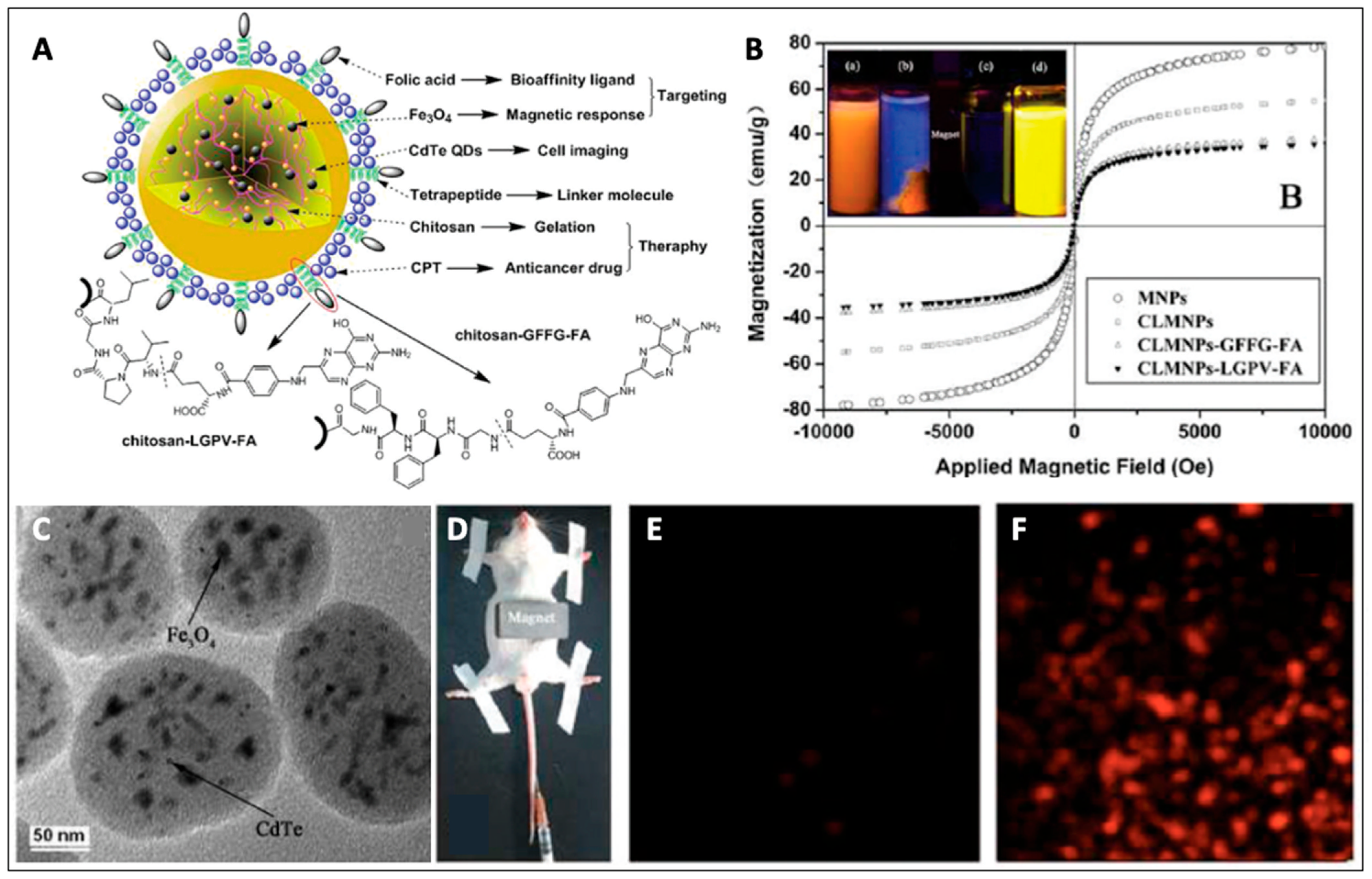
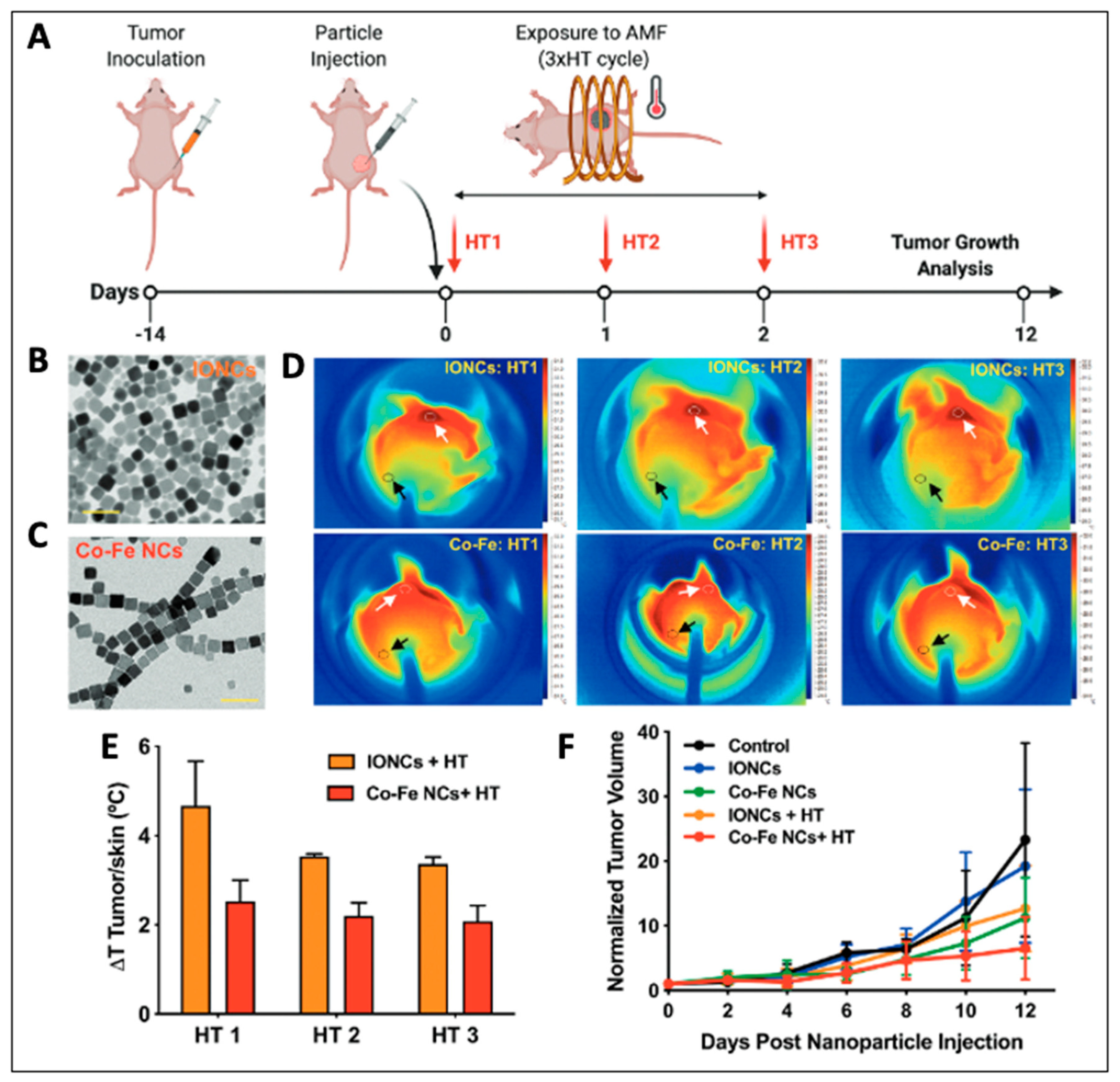
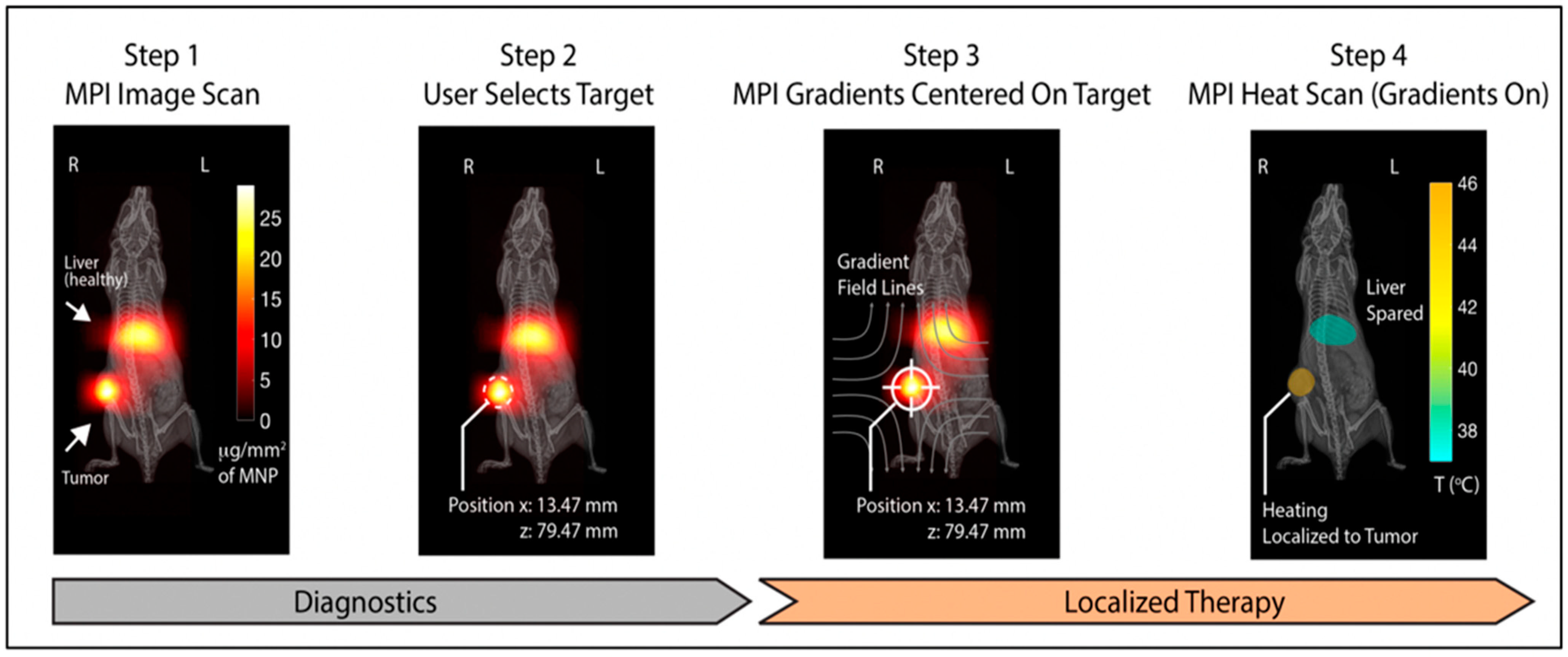
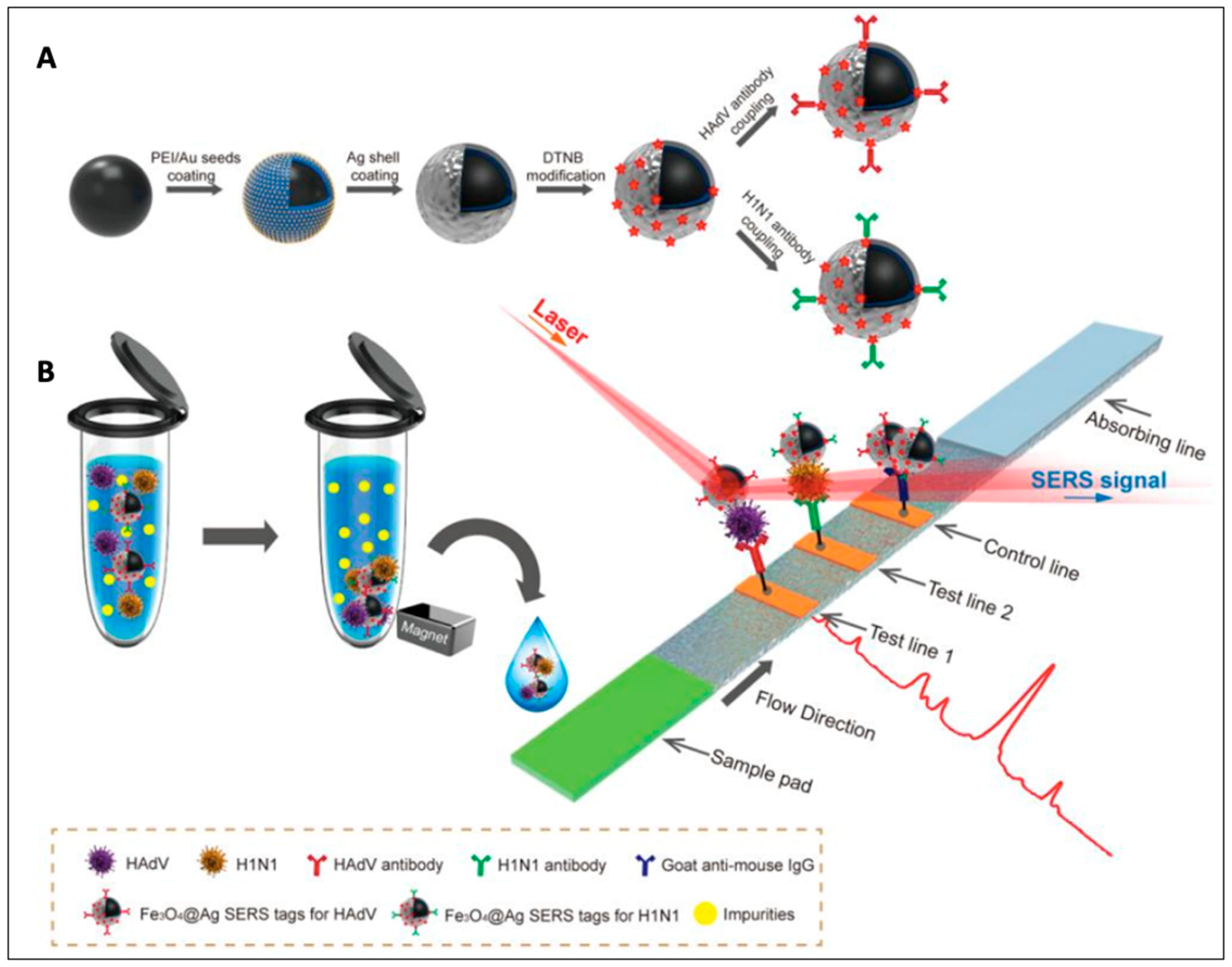
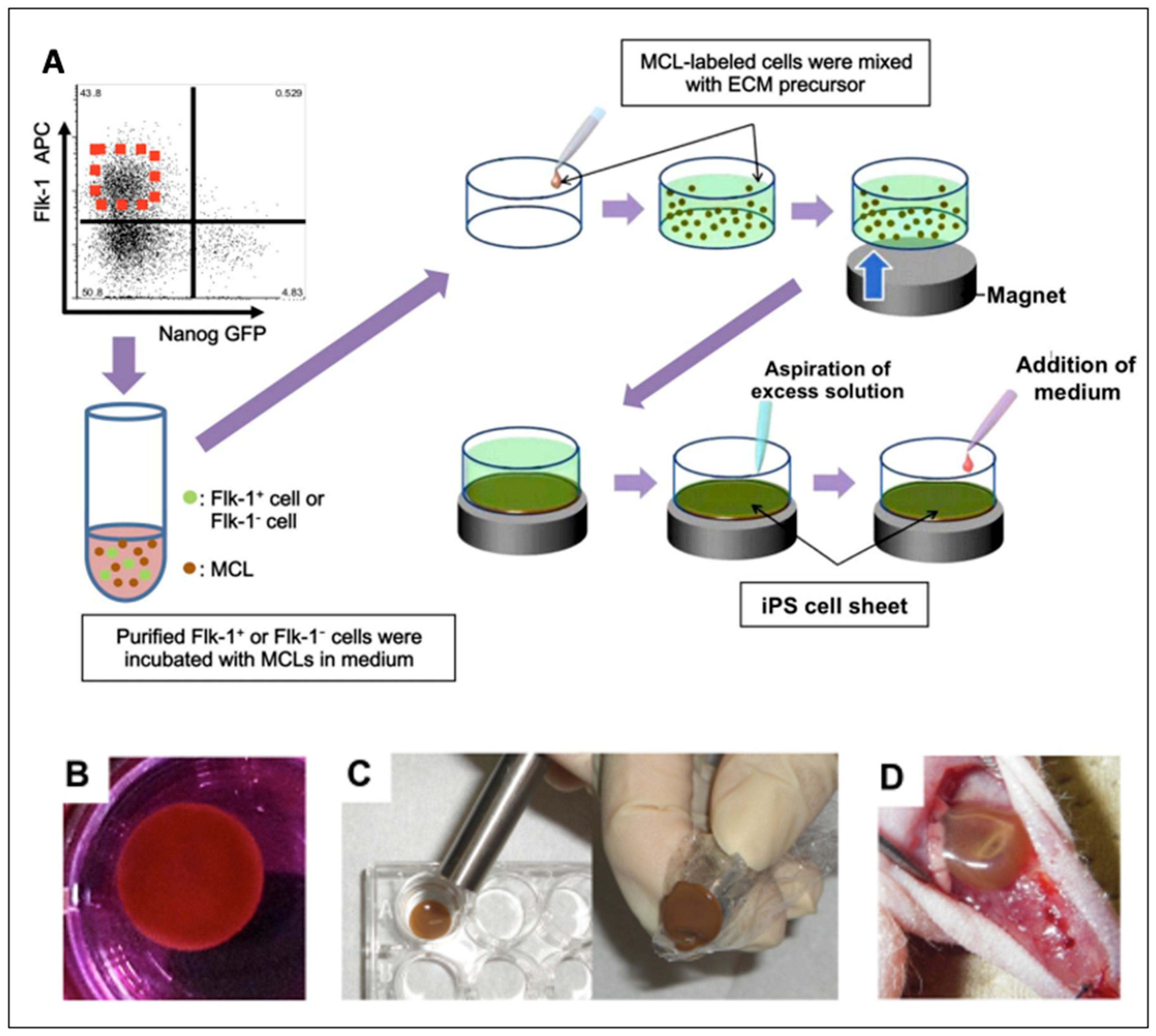
Table 1.
Summary of methods employed for synthesis of magnetic nanoparticles.
| Synthesis Method | Procedure | Merits | Demerits | Ref. |
|---|---|---|---|---|
| Co-precipitation | magnetic particles precipitated out from stoichiometric mixture of metal ions aqueous solution by addition of base | hydrophilic NPs; simple and scalable | broader size distribution | [40] |
| Thermal decomposition | organometallic precursor decomposed in non-polar boiling solvent at high temperatures in presence of stabilizing agent | monodisperse and size-controlled NPs; high yield | hydrophobic non-biocompatible NPs; toxic solvents | [42] |
| Hydrothermal | magnetic nanoparticles synthesized from aqueous metal solutions at high temperatures (>100 °C) and high pressure (>1 atm) in presence of an oxidizing agent | crystalline and water dispersible NPs of high purity | slow; large reaction time | [46] |
| Chemical vapor condensation | heating volatile metal precursors in an inert atmosphere | easy to prepare; small size NPs | precursors are toxic; specialized instruments required | [45] |
| Reverse microemulsion | particles formed inside hydrophilic core of micelles formed from surfactant molecules | simple room temperature synthesis; size controllability | low yield | [48] |
| Microwave | microwave-assisted heating of metal salt solutions in presence of oxidizing agent/base | homogenous heating and fast process | small scale production; expensive equipment | [49] |
| Electrochemical | magnetic particles synthesized from dissolution of sacrificial metal anode | size controllability by varying current density and potential | small scale production; broad size distribution | [52] |
Table 2.
Commonly used techniques for characterization of magnetic nanoparticles.
| Characterization Technique | Application |
|---|---|
| Transmission electron microscopy (TEM) | morphology, size distribution, thickness of coating |
| Scanning electron microscopy (SEM) | surface morphology, size distribution |
| Atomic force microscopy (AFM) | size, morphology, surface roughness |
| Dynamic light scattering (DLS) | hydrodynamic size, polydispersity |
| Energy dispersive X-ray spectroscopy (EDS) | elemental composition, elemental mapping |
| X-ray fluorescence spectroscopy (XFS) | qualitative and quantitative elemental analysis |
| Atomic absorption spectroscopy (AAS) | qualitative and quantitative elemental analysis |
| Inductively coupled plasma mass spectroscopy (ICP-MS) | elemental composition |
| Mössbauer spectroscopy | differentiating between magnetite and maghemite |
| X-ray diffraction (XRD) spectroscopy | crystalline structure |
| Selected area electron diffraction (SAED) | crystal structure, lattice parameters |
| Zeta potential | surface charge |
| Fourier-transform infrared (FT-IR) spectroscopy | surface functionality, presence of coating |
| Thermal gravimetric analysis (TGA) | thermal stability, presence of coating |
| Vibrating sample magnetometer (VSM) | change in magnetization as function of external magnetic field |
| Superconducting quantum interference device (SQUID) | change in magnetization as function of temperature and external magnetic field |
| Electron paramagnetic resonance (EPR) spectroscopy | characterization of magnetic phases |
| Physical-property measurement system (PPMS) | direct or alternating current (DC/AC) magnetization measurement as a function of temperature and/or applied magnetic field |
| Kerr spectroscopy | imaging differences in magnetization on the magnetic material’s surface |
Table 3.
Some of the important magnetic nanoparticle formulations employed for various biomedical applications (NA: not available).
Table 3.
Some of the important magnetic nanoparticle formulations employed for various biomedical applications (NA: not available).
| Magnetic Nanoparticles Formulation | Composition | Size (nm) | Biomedical Application | In Vitro/In Vivo Application | Ref. |
|---|---|---|---|---|---|
| Fe3O4-loaded mPEG-b-P(DPA-DE)LG micelles | hydrophobic SPIONs encapsulated in hydrophobic core of mPEG-b-P(DPA-DE)LG micelles; Dopamine (DPA) acted as anchor for SPIONs for facilitating self-assembly of polymer | DLS: 147 ± 3 TEM: ~120 | pH-responsive nanocarrier for MRI | Rat model with cerebral ischemia | [142] |
| MION@CMC-DOX | MIONs stabilized and coated by biocompatible CMC layer and bioconjugated with DOX via amide bonds | DLS: 38 ± 2 TEM: 7 ± 2 | combined chemotherapy and magnetic hyperthermia | HEK 293T and U87 cells | [143] |
| Nano-in-microparticles (NIMs) | dry powder containing SPIONs and DOX in lactose matrix | DLS: NA TEM: 1000–5000 | magnetically-targeted pulmonary delivery | A549 cells; Male Balb/c mice | [144] |
| Zinc doped iron oxide NPs conjugated to DR4 Ab | thiolated Zn0.4Fe2.6O4 NPs conjugated with protein A through sulpho-SMCC crosslinker; protein A binds with the Fc region of DR4 Ab | DLS: 32 TEM: NA | non-invasive approach to turn “on” apoptosis cell signalling using magnetic field | DLD-1 cells; zebrafish | [145] |
| HMCuS/DOX@IONP-PEG | DOX loaded positively charged hollow mesoporous CuS NPs capped with negatively charged IONPs and further surface conjugated with PEG | DLS: 124.5 ± 3.8 TEM: NA | multimodal system for combined chemotherapy, MRI, and NIR-light activated PTT and PDT | MCF-7 cells; tumor bearing mice | [146] |
| Hyaluronic acid-modified Mn-Zn ferrite (HA-MZF) nanoparticles | Mn0.6Zn0.4Fe2O4 (MZF) NP encapsulated in micelles using NH2-PEG2000-PCL3400 to form MZF-NH2 micelles and further surface modified with HA using EDC/NHS crosslinking reaction | DLS: 195 TEM: NA | receptor (CD44) mediated targeted delivery with combined hyperthermia and radiotherapy | A549 cells; Male Balb/c mice | [147] |
| Photosensitizer (Ce6) and cancer cell membrane (CCM) coated SSAP (SSAP-Ce6@CCM) | oleic acid coated SPIONs embedded in crosslinked matrix of styrene and AA, and further modified with PEI using carboxyl groups of AA; Ce6 adsorbed onto the surface and finally cloaked with CCM | DLS: 192 TEM: NA | PDT and dual-modal MR/NIR fluorescence imaging | SMMC-7721 tumor-bearing mice | [148] |
| Bisphosphonate conjugated dextran-coated Fe3O4 (Bis/Dex/Fe3O4) NPs | bisphosphonate (alendronate) covalently attached by EDC/NHS crosslinking to carboxyl surface modified dextran-coated Fe3O4 NPs | DLS: NA TEM: 20 | radiofrequency-induced thermolysis of osteoclasts for treatment of osteoporosis | male Wistar rats | [149] |
| Methicillin and SPIONs containing nano polymerosome | hydrophobic SPIONs and hydrophilic methicillin containing multi-compartment nano polymerosome made up of amphiphilic dieblock co-polymer (mPEG-PDLLA) | DLS: 83 ± 6 TEM: NA | penetration of Staphylococcus epidermidis biofilm with the aid of magnetic field | male Wistar rats | [150] |
| Fe3O4@Ag-Van MNPs | cationic PEI modified Fe3O4 NPs were coated with Ag colloidal NPs to form Fe3O4@Ag NPs; vancomycin (Van) was conjugated to carboxyl modified Fe3O4@Ag NPs | DLS: NA TEM: 290 | SERS-based biosensor for detecting and capturing bacteria | S. aureus 04018, E. coli BL21 strains | [151] |
| Mtb (Mycobacterium tuberculosis) antibody (MTBsAb) conjugated SPIONs | dextran-coated on SPIONs crosslinked by epichlorohydrin; treated with EDBE to generate amino groups at dextran ends followed by addition of SA to form SPIO-EDBE-SA; finally conjugated with MtbsAb | DLS: NA TEM: 3.8 ± 0.4 | ultrasensitive MR imaging for diagnosis of extrapulmonary tuberculosis | C57BL/6 mice bearing Mtb granulomas | [152] |
| Antibody (Ab1) conjugated Au functionalized CoFe2O4 (CoFe2O4@AuNPs@Ab1) NPs | primary antibodies (Ab1) immobilized by Au NPs functionalized CoFe2O4 NPs | DLS: NA TEM: 80 | SERS-based immunosensor for detecting NT-proBNP (biomarker for heart failure) | - | [153] |
| Magnetic (IONP) and upconverting (UCNP) nanobeads (MUCNBs) | IONPs and UCNPs simultaneously incorporated by wrapping in amphiphilic poly(maleic anhydride-alt-1-octadecene) polymer | DLS: 120 ± 10 TEM: NA | NIR fluorescence imaging and T2 contrast agents | HeLa-WT and A431 cells | [154] |
| Calcein−liposome/virus (NoV)/Fe3O4 nanoconjugates | APTES functionalized Fe3O4 NPs and calcein liposome conjugated separately with NoV-specific antibody form a sandwich-like structure in presence of NoV | DLS: 1120 TEM: NA | fluorometric detection of Norovirus (NoV) | - | [155] |
| CuFeSe2 −99mTc nanocrystals | CuFeSe2 nanocrystals fabricated in presence of trithiol-terminated poly-(methacrylic acid) and further labeled with radioactive 99mTc | DLS: NA TEM: 4.1 ± 0.4 | Multimodal imaging (photoacoustic, magnetic resonance and computed tomography imaging) PTT | 4T1 cells; 4T1 tumor-bearing BALB/c mice | [156] |
| Lipid-coated Fe3O4 mesoporous silica NPs doped with ceria NPs | ceria NPs loaded on surface of mesoporous silica NPs embedded with Fe3O4 NPs and further encapsulated by a lipid bilayer | DLS: ~250 TEM: ~100 | MRI and anti-inflammatory effect by scavenging ROS for theragnosis of intracerebral hemorrhage | RAW 264.7 cells; male Sprague-Dawley rats | [157] |
| Poly (amino ester) coated Fe3O4 NPs | poly (amino ester) having carboxyl groups coated on Fe3O4 NPs | DLS: NA TEM: 10.22 ± 2.8 | viral-RNA extraction method for detection of SARS-CoV-2 | - | [158] |
| MoS2@Fe3O4-ICG/Pt(IV) nanoflowers | PEI functionalized MoS2 nanoflowers covalently grafted with Fe3O4 NPs and further loaded with ICG and Pt(IV) prodrugs | DLS: NA TEM: 80 | multimodal infrared thermal/photoacoustic/magnetic resonance imaging with combined PDT, PTT, and chemotherapy | L929 cells; H22 tumor-bearing Balb/c mice | [159] |
| Superparamagnetic (Fe3O4) chitosan plasmid gelatin microspheres | superparamagnetic chitosan-coated Fe3O4 NPs attached with (pReceiver-M29-VEGF165/DH5a) plasmids embedded in gelatin microspheres | DLS: NA TEM: NA | neovascularization in artificial bone scaffolds on application of magnetic field | white rabbits | [160] |
| Core–shell Fe3O4 poly(NIPAM-co-DEAEMA) magnetic hydrogel composites loaded with methotrexate | Fe3O4 NPs (core) surface modified with TMSPMA encapsulated in poly(NIPAM-co-DEAEMA) shell and loaded with methotrexate | DLS: NA TEM: 50–70 | dual pH- and thermo- responsive drug delivery and hyperthermia | MCF-7 cells | [161] |
(mPEG-b-P(DPA-DE)LG = methyloxy-poly(ethylene glycol)-blockpoly[dopamine-2-(dibutylamino) ethylamine-L-glutamate]; MIONs = magnetic iron oxide nanoparticles; CMC = carboxymethylcellulose; DOX = doxorubicin; DR4 = death receptor 4; Ab = antibody; sulpho-SMCC = sulphosuccinimidyl-4-[N-maleimidomethyl]cyclohexane-1-carboxylate; Fc region = fragment crystallizable region; PDT = photodynamic therapy; EDC = 1-ethyl-3-(3-dimethylaminopropyl) carbodiimide; NHS = N-hydroxysuccinimide; SSAP = styrene and acrylic acid crosslinked SPION modified with polyethyleneimine; AA = acrylic acid; PEI = polyethyleneimine; mPEG-PDLLA = diblock co-polymer of methoxypoly(ethelyne glycol)5000 and poly(D)-(L)-lactic acid50,000; SERS = surface enhanced Raman scattering; EDBE = 2,2′-(ethylenedioxy)bis(ethylamine); SA = succinic anhydride; NT-pro = N-terminal pro-hormone; BNP = B-type natriuretic peptide; APTES = (3-aminopropyl)-triethoxysilane; ICG = indocyanine green; TMSPMA = 3-(Trimethoxysilyl)propyl methacrylate; NIPAM = N-isopropylacrylamide; DEAEMA = 2-(N,N-diethylaminoethyl) methacrylate.
Publisher’s Note: MDPI stays neutral with regard to jurisdictional claims in published maps and institutional affiliations. |
© 2022 by the authors. Licensee MDPI, Basel, Switzerland. This article is an open access article distributed under the terms and conditions of the Creative Commons Attribution (CC BY) license (https://creativecommons.org/licenses/by/4.0/).
Share and Cite
MDPI and ACS Style
Mittal, A.; Roy, I.; Gandhi, S. Magnetic Nanoparticles: An Overview for Biomedical Applications. Magnetochemistry 2022, 8, 107. https://doi.org/10.3390/magnetochemistry8090107
AMA Style
Mittal A, Roy I, Gandhi S. Magnetic Nanoparticles: An Overview for Biomedical Applications. Magnetochemistry. 2022; 8(9):107. https://doi.org/10.3390/magnetochemistry8090107
Chicago/Turabian StyleMittal, Ashi, Indrajit Roy, and Sona Gandhi. 2022. "Magnetic Nanoparticles: An Overview for Biomedical Applications" Magnetochemistry 8, no. 9: 107. https://doi.org/10.3390/magnetochemistry8090107
Note that from the first issue of 2016, this journal uses article numbers instead of page numbers. See further details here.