1. Introduction
The variety of research methods of modern paleontological science is great. Information on the internal structure of paleontological objects is important for their studying. Traditionally, this information is obtained by the methods of mechanical polishing, preparation and partial destruction of the specimens. With the advent of the X-ray equipment, it becomes possible to study fossil remains without destroying them. Significant progress is associated with the development of tomography facilities on synchrotron radiation sources and thermal neutron sources. Using both types of radiation for investigation of the paleontological objects is of great importance because they consist of different mineral components, and some components cannot produce a sufficient contrast in the image taken only with one type of radiation [
1,
2,
3].
In 2011, the cooperation of the Russian National Research Centre “Kurchatov Institute” and the Borissiak Paleontological Institute of Russian Academy of Sciences in the field of fossil studies using synchrotron and neutron tomography has begun. In addition to the early X-ray scale of contrast of minerals and rocks [
4] an analogous neutron scale has been constructed [
5]. Comparison of these two scales shows four possible cases for any pair of minerals and rocks which can compose a fossil: the pair is distinguishable only in X-ray radiation, the pair is distinguishable only in neutrons, the pair is distinguishable in both types of radiation, and the pair is indistinguishable in both types of radiation. So, the scales [
4,
5] can be useful for the purpose of optimal planning of tomographic study. Brachiopod shells, skeletal remains of extinct echinoderms, fossil plants have been also studied [
5,
6]. The tomography facilities on the Kurchatov synchrotron radiation source and the IR-8 research reactor are used [
7,
8]. This article briefly presents the results of the joint research carried out in recent years.
2. Methods
Experiments on synchrotron and thermal-neutron tomography are performed at the dedicated stations of the Kurchatov source of synchrotron radiation and the IR -8 research reactor of National Research Centre “Kurchatov Institute. Synchrotron radiation is generated by a bending magnet and transmits through a copper filter of 1.5 mm thickness and thus the beam with the spectral maximum of about 56 keV is formed. This beam is incident on the object under study. Object rotates around the vertical axis with an angular step of 0.5°. Projections of the object are recorded by an area detector consisting of a scintillation screen (based on CsI(Tl) or Gd2O2S(Tb)), objective lens, and charge coupled device matrix. The time of exposure of one image is about 300 ms. Since the height of the beam of synchrotron radiation is small, projections are composed from a series of images taken at the sample vertical displacement with the 1 mm step. The spatial resolution in synchrotron tomography varies from 80 to 130 μm depending on the screen. All the slices presented below are obtained with the 130 μm resolution except the one where otherwise indicated.
Neutron beam with a wavelength of 1.526 Å, formed by reflection from a copper (111) crystal is incident on the object under study. Object rotates around the vertical axis with an angular step of 0.5°. The object projections are recorded by an area detector consisting of a ZnS(Ag) +
6LiF scintillator, an objective lens and a CCD matrix. The exposure time of one projection at a reactor power of 5.8 MW is about 100 s. The spatial resolution in neutron tomography is 160 μm. Tomographic reconstruction is done using the filtered back-projection method [
9].
3. Study of the Internal Structure and Preservation of Brachiopod Shells of the Genus Kaninospirifer Kulikov et Stepanov, 1975
The size of the shells does not allow studying the material on a laboratory microtomograph, and therefore, they have been studied using synchrotron and neutron tomography. In the study, several species of this genus are used. The preservation of the shells becomes the determining factor for the effectiveness of the research. This is expressed in the number of details of the intracavitary structures that could be seen. Mineral composition is determined by visual examination, by contrast of structures and rocks in tomographic slices, and by the X-ray phase analysis. For all specimens, it can be noted that their shells, like in vivo, consist of calcite, which was not replaced by other minerals. All shells can be divided into three groups according to the preservation.
The images of the brachiopod species
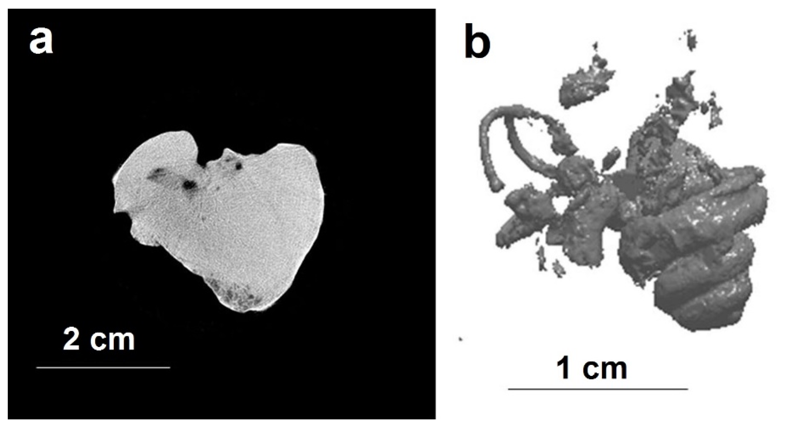
Kaninospirifer kaninensis (Licharew, 1943) from the Permian deposits of the Kanin Peninsula are characterized by a good contrast between shell and matrix. This contrast arises due to several circumstances. Shells of
K. kaninensis are thick-walled (
Figure 1a). The shell wall consists of calcium carbonate. The matrix is a fine-grained sandstone or siltstone. In sandstone, there are small inclusions of debris of shells. Secondarily, in the shell wall, on its outer surface, small pyrite crystals have grown, they are found singly and do not affect the contrast of the shell and internal rock. Due to the good contrast between thick-walled carbonatic shells and fine grained sandstone, the completely preserved fossil shells can be well distinguished from the matrix. In the shell wall, damage is also observed in the form of meandering channels that extend to the outer surface of the shell. These are burrows of boring organisms, perhaps annelids, which settled in the brachiopod shells, probably both in vivo and post-mortem.
The examples of brachiopods of other kind of preservation occur also from Kanin Peninsula. The brachiopods
K. kaninensis,
K. borealis Kulikov and Stepanov, 1975,
K. stepanovi Grunt, 2006 are also represented by thick-walled calcite shells, but the matrix differs from the previously described one. It is a sandstone, but the carbonate component can be easily seen. The rock is filled with fragments of shells and large fragments of brachiopod valves, whole shells of gastropods, colonies of bryozoans. Small carbonate grains are present in it. Sometimes cementation with high-contrast minerals is observed (
Figure 1b). Borings are also noted in the brachiopod valves. With such preservation, the contrast of the images of the shells in matrix is high, only the images of shell detritus can interfere with the images of the internal structures and so some of them are lost.
The brachiopods of
K. adpressum (Liu Fa et Waterhouse, 1985) and
K. incertiplicatus Pavlova, 1991 from the Permian deposits of South and Inner Mongolia are of special preservation. Shells are deformed to varying degrees. The walls of the shells are thick and consist of calcite. The matrix is different from that from Kanin Peninsula. Judging from the contrast, it is somewhat silicificated, but there is also a carbonate component in it, which reduces the contrast of internal structures (
Figure 1c). There are large carbonate inclusions in the rock, which further reduces the quality of the visualization of structures. In some sections of the shell there are small inclusions of high-contrast mineral. Despite the described preservation, most of the internal structures have been studied.
Thus, the preservation of the shells of the brachiopods of the genus
Kaninospirifer enables their studying using synchrotron and neutron tomography. Nondestructive methods completely replaced the mechanical polishing and preparation, and so shells and valves are kept intact. The revealed structures completely represent the internal structure of the bulk parts of the fossil shells. Based on the results of the study of the brachiopod of the genus
Kaninospirifer, the species differences in the internal structure can be revealed. It is possible to clarify the details of the structure of the shells important for a systematic description of new structures. For this kind of composition of the specimens, the images obtained by synchrotron and the neutron tomography are similar in accordance to [
5].
4. Study of the Endocranial Cavity of a Fossil Mammal
An incomplete skull of a fossil carnivore of an unclear taxonomic affiliation has been studied by the synchrotron tomography. The specimen belongs to the collection of the Vernadsky State Geological Museum (Moscow, Russia) at number GGM-400-27/PV-00799. The previous collection number is Sa-301. In the old catalogue of the museum it is listed under this number as “
Pterodon dasyuroides, occipital part of skull” and marked “Purchased in 1891, Quercy” [
10]. The specimen is of about 10 × 6 × 6 cm size. It includes the occipital part of the skull, broken off along the line of the postorbital constriction, without zygomatic arches, occipital ridge and muzzle. The elements of the auditory region are well preserved (region of the petrosal bone, the mastoid process and ectotympanic bone on the left side of the skull).
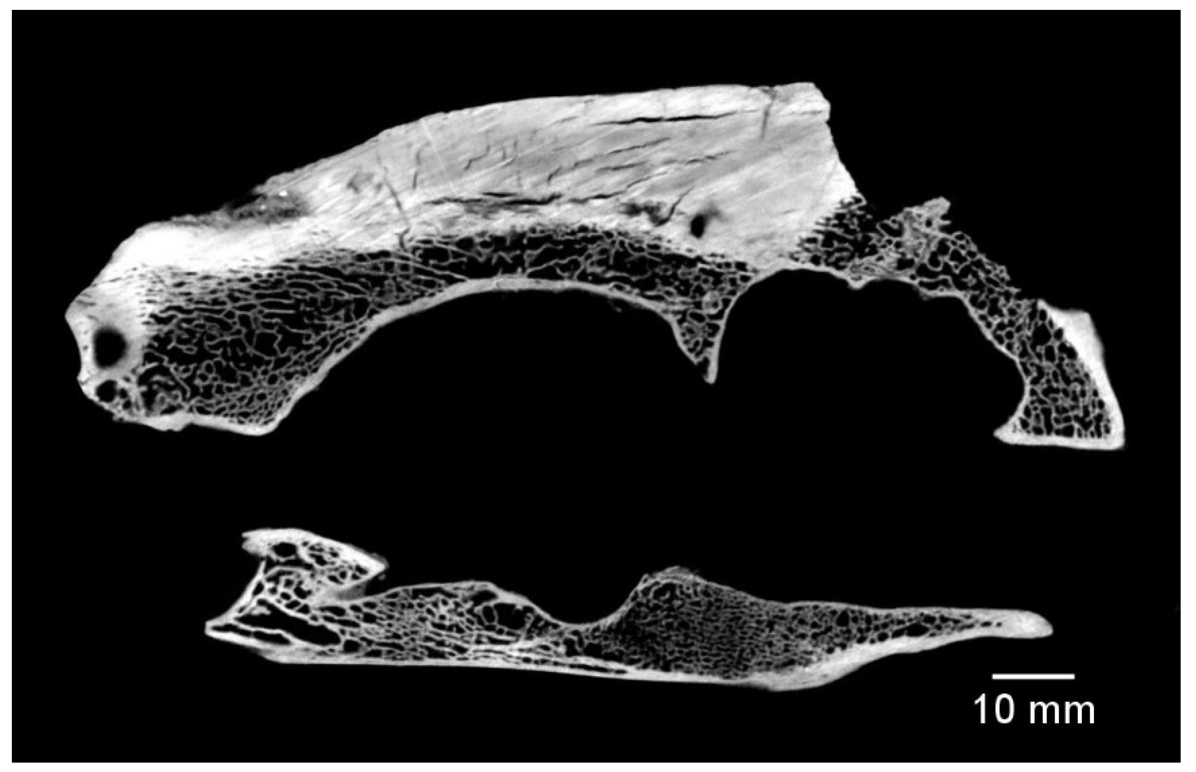
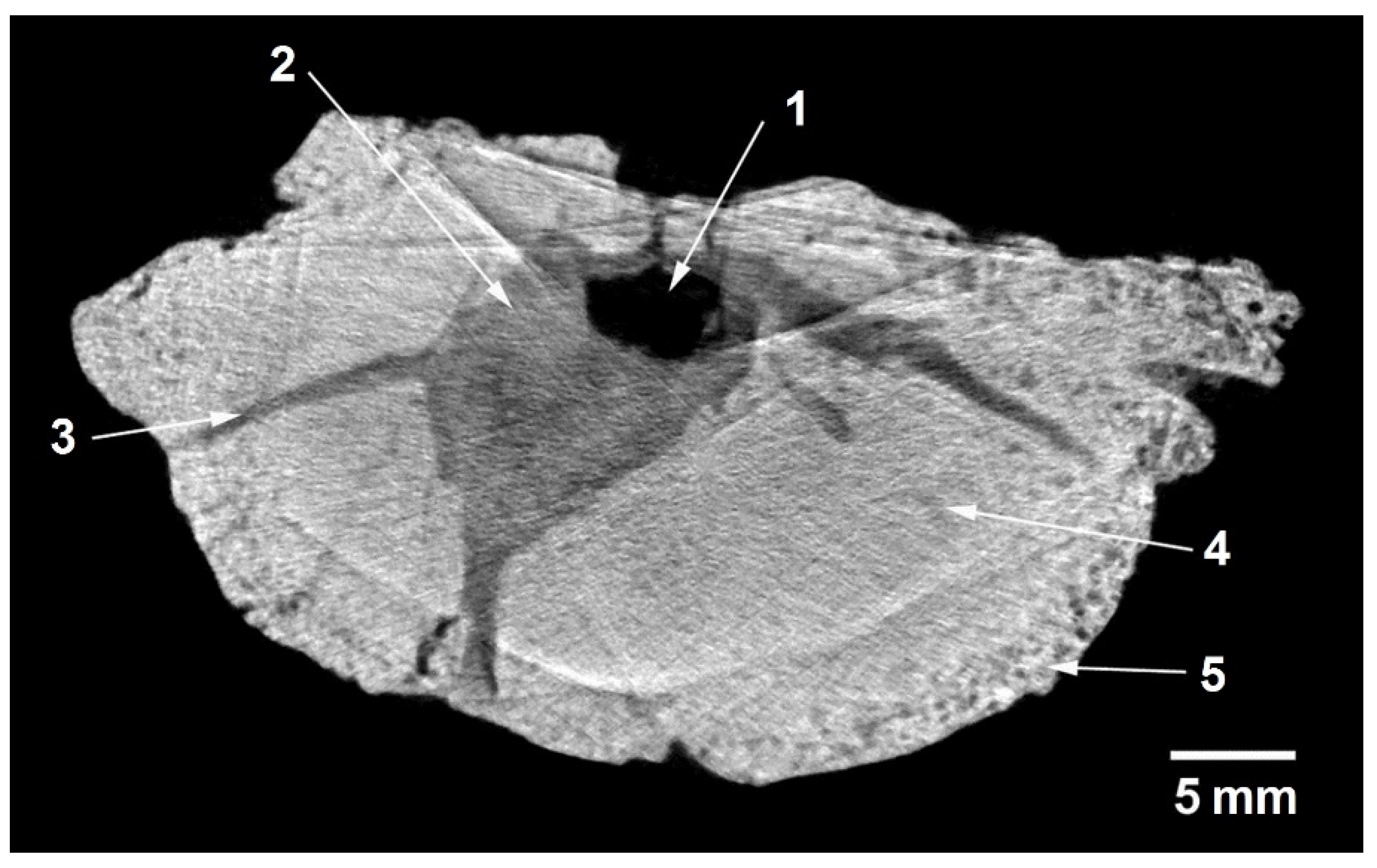
The synchrotron tomography shows that the specimen is well cleaned of the matrix, and only a small amount of weakly cemented fine-grained sand is found in the folds of the surface of the cerebral cavity (
Figure 2). The bones consist of calcium carbonate, hydroxyapatite and a small admixture of accessory minerals (data on the mineral composition obtained on the basis of X-ray fluorescence analysis). The thickness of the bone was measured, it varies from 4 to 12 mm, its density is volatile—in the region of nasal bulbs and tympanal bulla its density is reduced. The thickness of the bone in the dorsal part is minimal, and at the base of the skull reaches its maximum values.
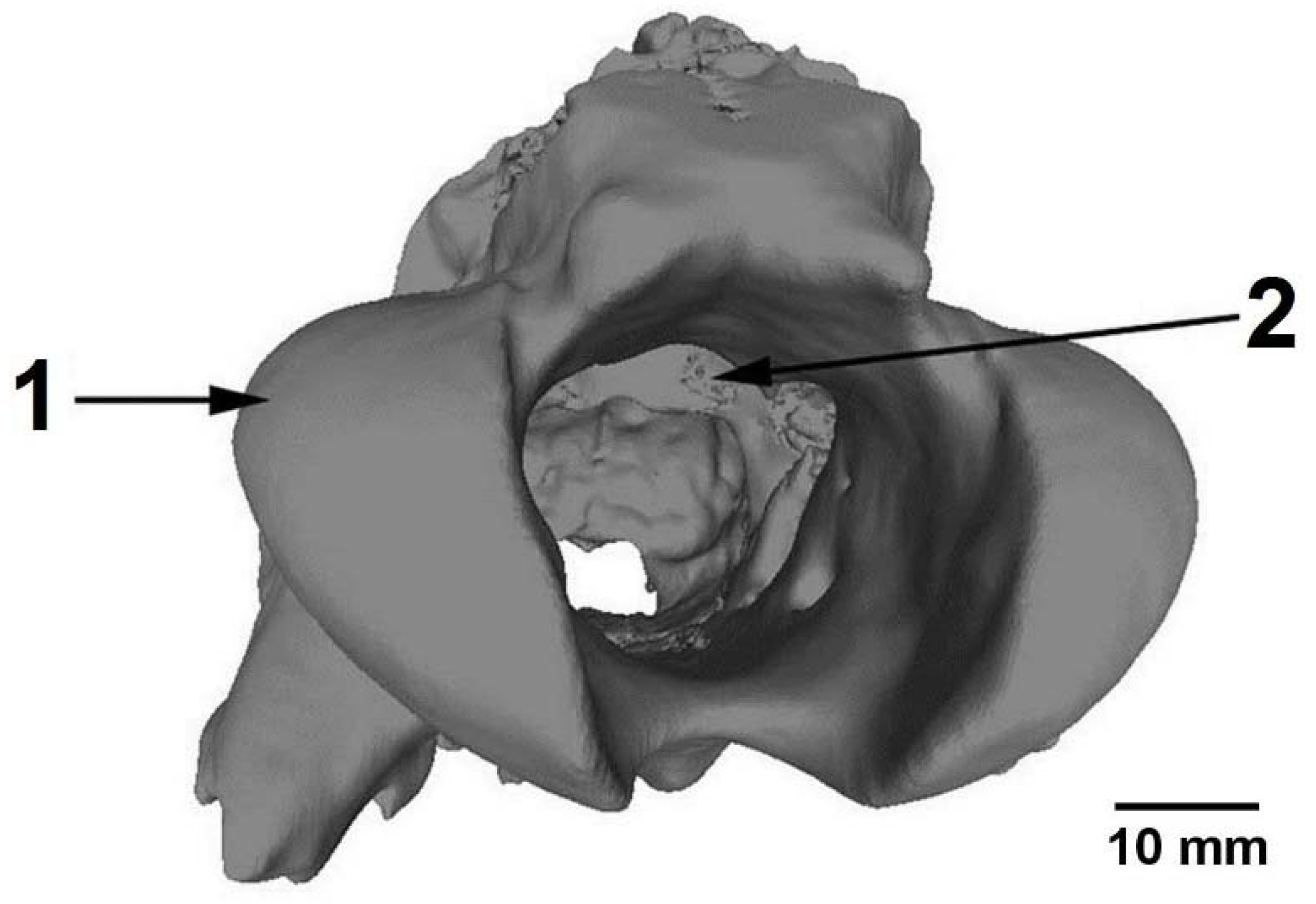
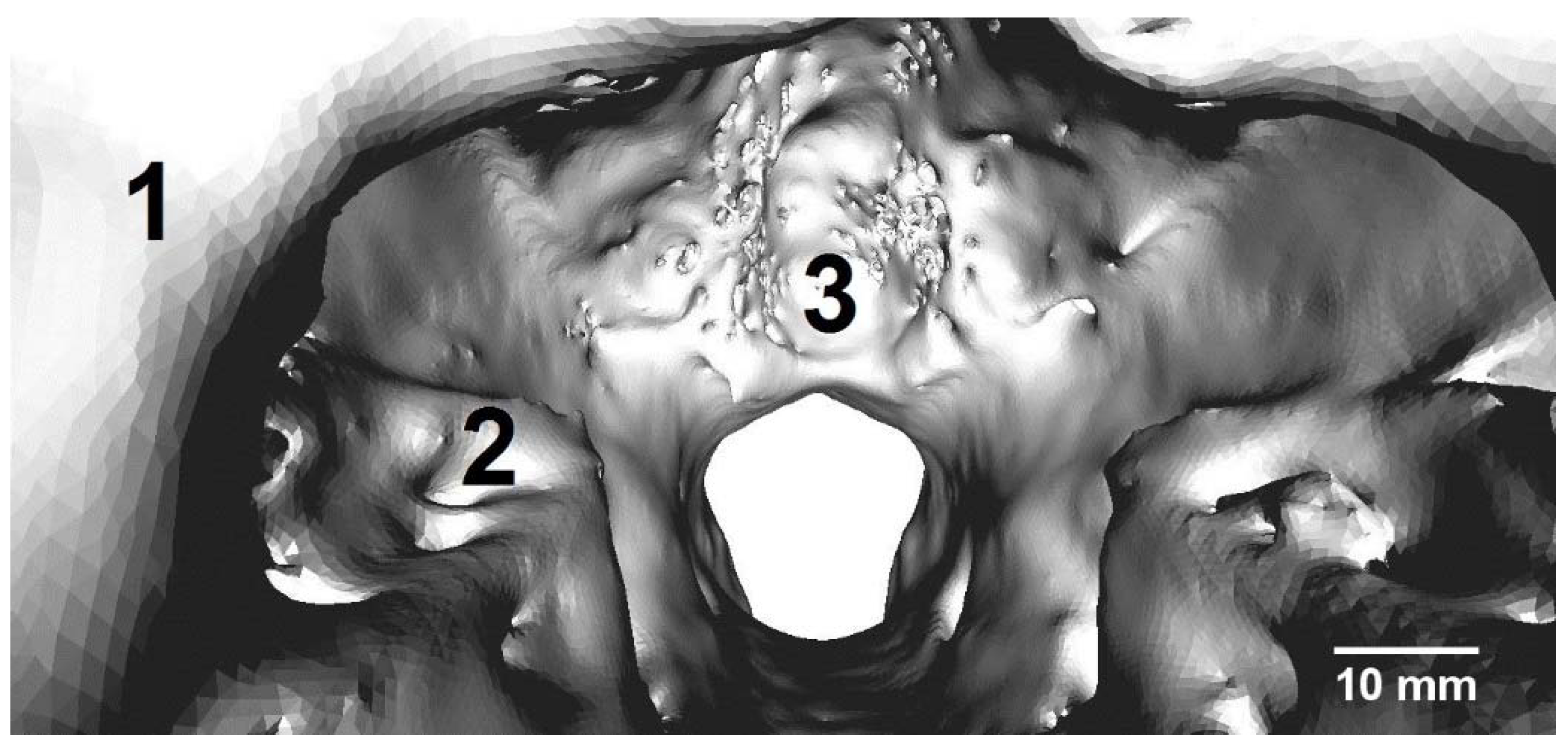
A three-dimensional model of the inner surface of the cerebral cavity has been created based on the tomography data. The model makes possible to refine the taxonomic assignment of this specimen. Also it makes possible to study the structure of the surface of the endocranial cavity, which hosted the brain of this fossil predator (
Figure 3 and
Figure 4). It clearly shows the structure and the degree of development of the sulci and girusis, the ratio of the sizes of the various parts of the hemispheres of the brain and the cerebellum. The model obtained makes it possible to carry out a morphofunctional analysis of the brain of the fossil animal and to gain insight into the development of its afferentation organs, locomotion, and ecology. Similar studies were conducted earlier on the representatives of fossil predatory mammals, e.g., [
11,
12].
5. Study of the Petrosal Bones of Fossil Cetaceans
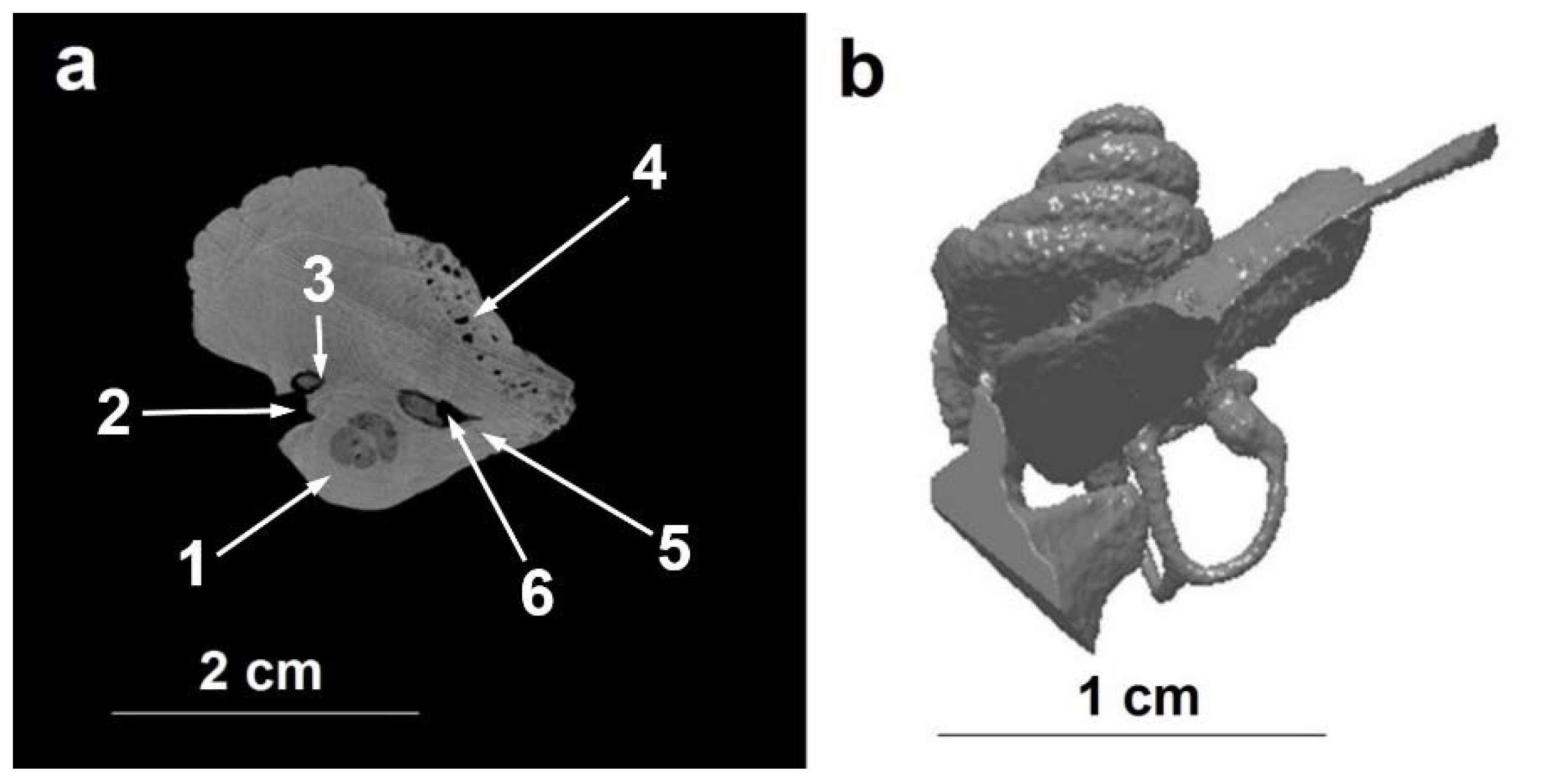
Synchrotron tomography of the petrous part of the temporal bone (bone of the ear capsule of the inner ear) has been performed for a representative of the genus
Kurdalagonus Tarasenko and Lopatin, 2012. The maximum thickness of the object in the direction of the incident X-ray beam reaches to 35 mm. The results of the tomography are shown in
Figure 5. Since the voids of the bony labyrinth of the petrosal bone are filled with a slightly absorbing substance, a rather sharp contrast of these structures is obtained in comparison with the dense structure of the fossilized bone (
Figure 5a). The object itself consists of sufficiently homogeneous components and, obviously, is replaced by hydroxylapatite. The basic details of the structure of the voids of the bony labyrinth of the petrosal bone are well distinguished: semicircular canals, endo- and perelymphatic ducts, the bony spiral canal of the cochlea, and the facial canal (Canalis nervi facialis). Based on the results of tomography, it is possible to construct a three-dimensional model of the bony labyrinth of the petrosal bone for
Kurdalagonus sp. (
Figure 5b).
To study the petrosal bone of the Miocene baleen Vampalus sayasanicus Tarasenko and Lopatin, 2012, both synchrotron and neutron tomography have been used. In the sections of this specimen obtained by the synchrotron tomography, there is a strong contrast from the X-ray absorbing rock that filled the semicircular canals and partially filled the channels in the region of the round window. This gives rise to strong reconstruction artifacts that make it difficult to interpret some sections.
The results of the neutron and X-ray tomography are complementary for this specimen. The three-dimensional model is constructed on the basis of the synchrotron tomography data because of its better spatial resolution (
Figure 6b). Semicircular canals are clearly discernible here, but they are partly filled with rock. The bony spiral channel of the cochlea is not reconstructed for some places due to the strong contrast from the X-ray absorbing rock, which partially fills the channel.
6. Study of the Lower Jaw of Dicynodont
A specimen of fossil dicynodont (Order Anomodontia, Family Dicynodontidae, Dicynodontidae gen. indet.) from the collection of the Paleontological Institute of the Russian Academy of Sciences, PIN number 4822/238 is represented by a fragment of the left branch of the lower jaw in the symphysis region. A fragment of the jaw was found in the sediments of the left bank of the Chashchenka river (the location of “Chashchenka”, Vologda region, Babushkinsky district). Fossilized remains of vertebrates in this location are being washed out by a stream of river from the low-thickness sand lentils in the bulk of clay sediments. Bones themselves have a color from dark brown to almost black, which may indicate a significant degree of phosphatization of residues. Some specimens of fossil bones from the site bear traces of weathering and cracking, to which they were subjected even before their burial in the bulk of sedimentary rocks. In these cases, the cracks in the bones are filled with fine sedimentary materials, mainly sandstone with a clay component, which are subsequently cemented with limestones. The specimen, however, does not show any signs of cracking, but the pores on the surface of the bone and the openings from the vessels that emerge at its surface are also filled with cemented sedimentary material.
Study of the described sample by synchrotron tomography has been undertaken with the aim of studying the structure of unusual large channels in the bulk of the bone, the outlet of which is observed at the distal end of the jaw symphysis. Due to the peculiarities of fossilization, these channels could be prepared by mechanical means for no more than 1–1.5 cm, but their possible extend in the bulk of the bone remained unknown. The used tomographic method makes it possible to clearly distinguish not only the zones of internal cavities free from rock, but also to trace the course of channels filled with cemented rock clearly (
Figure 7), due to the fact that the density of the rock on the tomography slices is significantly lower than the surrounding dense fossilized bone tissue. The spatial resolution of the method makes it possible to trace not only the large channels but also the small ones which contained the blood vessels with a diameter of no more than 1–2 mm.
Earlier, the same morphological structures were examined on the specimen of the lower jaw of dicynodont (specimen PIN number 4549/23) of the same age from the location of Klimovo-1 (Vologda Region, Velikoustyugsky District). The study was carried out using the X-ray laboratory microtomograph. In contrast to the above-described specimen, the jaw from the location of Klimovo-1 has no traces of phosphatization and the rock that fills the internal cavities in the bone is much less cemented, not so dense, which makes it possible to use the mechanical methods of preparation to a greater extent. Despite this, the resolving power of the laboratory method turned out to be somewhat lower than the method of synchrotron tomography.
7. Conclusions
The synchrotron and neutron tomography methods have been used to study paleontological objects. All the objects have been never investigated before and the results will be used for future analysis. The internal structure and preservation of the brachiopod shells of the genus Kaninospirifer, the skull of a fossil carnivorous mammal, the petrosal bone of fossil cetaceans, and the lower jaw of dicynodont have been investigated. The initial data for the morphofunctional analysis of the brain of a fossil mammal have been obtained. All the objects are kept intact. The use of the synchrotron tomography is sufficient in the most cases with its usual advantage in spatial resolution. In special cases the use of the neutron tomography makes it possible distinguish the parts of an object, which produce low contrast or high noise with the X-rays. Generally, tomography makes it possible to investigate the unique objects without destruction and to clarify the details of the structure of objects, important for the systematics, and to describe new structures.
Author Contributions
A.P., A.K. (Andrey Kurkin), A.L. and K.T. supplied the objects and described the results, E.K., A.K. (Alexander Kaloyan) and K.P performed tomography, E.K. performed 3-D rendering.
Funding
The work was partially supported by RFBR, research project No. 18-35-00206. The work was partially supported by the Ministry of Education and Science of the Russian Federation FTP “Research and development in priority areas for the development of the scientific and technological complex of Russia for 2014-2020” (agreement No. 14.619.21.0007, project RFMEFI61917X0007) using Kurchatov Synchrotron Radiation Source in NRC “Kurchatov Institute”. The work was partially supported by the Fundamental Research Program of the Russian Academy of Sciences “Biodiversity of Natural Systems. Biological resources of Russia: assessment of the state and fundamental principles of monitoring” and of the Program of the Presidium of the RAS “Problems of the origin of life and the formation of the biosphere”.
Conflicts of Interest
The authors declare no conflict of interest. The founding sponsors had no role in the design of the study; in the collection, analyses, or interpretation of data; in the writing of the manuscript, and in the decision to publish the results.
References
- Sutton, M.; Rahman, I.; Garwood, R. Techniques for Virtual Palaeontology; John Wiley Sons: Hoboken, NJ, USA, 2013; 208p, ISBN 1118591259, 9781118591253. [Google Scholar]
- Tafforeau, P.; Boistel, R.; Boller, E.; Bravin, A.; Brunet, M.; Chaimanee, Y.; Cloetens, P.; Feist, M.; Hoszowska, J.; Jaeger, J.J.; et al. Applications of X-ray Synchrotron microtomography for non-destructive 3D studies of paleontological specimens. Appl. Phys. A 2006, 83, 195–202. [Google Scholar] [CrossRef]
- Laaß, M.; Schillinger, B.; Werneburg, I. Neutron tomography and X-ray tomography as tools for the morphological investigation of non-mammalian synapsids. Phys. Procedia 2017, 88, 100–108. [Google Scholar] [CrossRef]
- Pakhnevich, A.V. On effectiveness of micro-CT research of paleontological objects. In Modern Paleontology: Classical and New Methods; Paleontological Institute: Moscow, Russia, 2009; pp. 127–141. (In Russian) [Google Scholar]
- Kaloyan, A.A.; Kovalenko, E.S.; Pakhnevich, A.V.; Podurets, K.M. The contrast scale of minerals for neutron tomography of paleontologic and geologic objects. Russ. Geol. Geophys. 2017, 58, 1435–1440. [Google Scholar] [CrossRef]
- Kaloyan, A.A.; Kovalenko, E.S.; Pakhnevich, A.V.; Podurets, K.M.; Rozhnov, S.V.; Somenkov, V.A. Synchrotron and Neutron Tomography for the Investigation of Paleontological Objects. J. Surf. Investig. X-ray Synchrotron Neutron Tech. 2014, 8, 1093–1099. [Google Scholar] [CrossRef]
- Podurets, K.M.; Pogorelyi, D.K.; Kaloyan, A.A.; Kovalenko, E.S.; Kohn, V.G. Multimode X-ray Tomography at the Mediana Station of the Kurchatov Synchrotron Radiation Source. J. Surf. Investig. X-ray Synchrotron Neutron Tech. 2012, 6, 845–848. [Google Scholar] [CrossRef]
- Glazkov, V.P.; Kaloyan, A.A.; Kovalenko, E.S.; Podurets, K.M.; Somenkov, V.A.; Yakovenko, E.V. A Neutron Tomograph at the IR-8 Reactor of the National Research Centre Kurchatov Institute. Instrum. Exp. Tech. 2014, 57, 531–534. [Google Scholar] [CrossRef]
- Kak, A.C.; Slaney, M. Principles of Computerized Tomographic Imaging; IEEE Press: New York, USA, 1988; ISBN O-87942-198-3. [Google Scholar]
- Pavlova, M.V. Catalogue of Collections of Geological Department of Imperial Moscow University; Issue 1, Part II; Imperial Moscow Univeristy: Moscow, Russia, 1910; 184p. (In Russian) [Google Scholar]
- Saveliev, S.V.; Lavrov, A.V. A Morphofunctional reconstruction of the brain of Neohyaenodon horridus (Hyaenodontidae, Creodonta) based on the natural endocranial cast. Paleontol. J. 2001, 35, 75–83. [Google Scholar]
- Saveliev, S.V.; Lavrov, A.V. Morphofunctional reconstruction of the brain of Paroxyaena pavlovi (Hyaenodontidae, Creodonta) based on a natural endocranial cast. Paleontol. J. 2007, 41, 661–670. [Google Scholar] [CrossRef]
Figure 1.
Synchrotron tomography sections of brachiopod shells of the genus Kaninospirifer: (a)—specimen 4900/78 PIN, whole shell of K. kaninensis in matrix; between the mouth of the Bol. Krutaya River and Nadtejsala Cape, Kanin Peninsula; Permian, the Urzhum tier of the Biarmian Division; (b)—specimen 11111/91 TsNIGR museum, ventral valve of K. kaninensis in a heterogeneous matrix; Kanin Peninsula; Permian, the Urzhum tier of the Biarmian Division; (c)—specimen 3385/371 PIN, ventral valve of K. adpressum; Inner Mongolia; Upper Permian (80 μm spatial resolution); 1—boring, 2—internal structures, 3—fragments of brachiopod valves, 4—cementation with high contrast mineral, 5—valve, 6—large carbonate inclusion in the rock.
Figure 2.
Specimen number GGM-400-27/PV-00799 from the collection of the Vernadsky State Museum. Sagittal tomographic slice of the endocranial cavity.
Figure 2.
Specimen number GGM-400-27/PV-00799 from the collection of the Vernadsky State Museum. Sagittal tomographic slice of the endocranial cavity.
Figure 3.
3D rendering of the specimen number GGM-400-27/PV-00799 from the collection of the Vernadsky State Museum. A view into the cerebral cavity of the skull from the side of the occipital aperture in a three-dimensional rendering 1—occipital condyle, 2—cerebellar cavity.
Figure 3.
3D rendering of the specimen number GGM-400-27/PV-00799 from the collection of the Vernadsky State Museum. A view into the cerebral cavity of the skull from the side of the occipital aperture in a three-dimensional rendering 1—occipital condyle, 2—cerebellar cavity.
Figure 4.
3D rendering of the specimen number GGM-400-27/PV-00799 from the collection of the Vernadsky State Museum. A view in the caudal direction from the line of the tentorium osseum on the cerebellar cavity: the structures of the walls of the cerebellar cavity are visible 1—tentorium osseum, 2—petrosum, 3—vermis.
Figure 5.
Petrosum of Kurdalagonus sp. (5462/2 PIN, Republic of Adygea, Fortepianka 2 locality, Late Miocene): (a)—reconstructed X-ray tomography cross-section through the septum between the inner acoustic meatus (IAM) and the foramen for the facial nerve; (b)—3D rendering of the bony labyrinth of the petrosum (uneven channels due to the filling of voids with the dense rock); 1—cochlea, 2—vestibular fossa, 3—foramen of the anterior semicircular canal, 4—suprameatal fossa, 5—septum between the IAM and the foramen for the facial nerve, 6—IAM.
Figure 6.
Petrosum of Vampalus sayasanicus (5341/4 PIN, Republic of Chechnya, Sayasan locality, Late Miocene): (a)—reconstructed neutron tomography cross-section; (b)—3D rendering of the bony labyrinth of the petrosum (uneven channels due to the filling of voids with the dense rock).
Figure 6.
Petrosum of Vampalus sayasanicus (5341/4 PIN, Republic of Chechnya, Sayasan locality, Late Miocene): (a)—reconstructed neutron tomography cross-section; (b)—3D rendering of the bony labyrinth of the petrosum (uneven channels due to the filling of voids with the dense rock).
Figure 7.
Sample 4822/238 PIN, transverse section of the left branch of the jaw Dicynodontidae gen. indet.: 1—channel area, empty of rock, 2—channel area, filled with cemented rock, 3—thin channel from blood vessel emerging to the bone surface, 4—area of dense fossilized bone tissue, 5—area of porous fossilized bone tissue.
© 2018 by the authors. Licensee MDPI, Basel, Switzerland. This article is an open access article distributed under the terms and conditions of the Creative Commons Attribution (CC BY) license (http://creativecommons.org/licenses/by/4.0/).


