Review

The Further Adventures of Newborn Screening for Biotinidase Deficiency: Where It Is at and What We Still Need to Know

1
Department of Research Administration, Henry Ford Hospital, Detroit, MI 48202, USA
2
Center for Molecular Medicine and Genetics, Wayne State University School of Medicine, Detroit, MI 48201, USA
Int. J. Neonatal Screen. 2016, 2(4), 9; https://doi.org/10.3390/ijns2040009
Submission received: 6 August 2016 / Revised: 29 September 2016 / Accepted: 25 October 2016 / Published: 28 October 2016
(This article belongs to the Special Issue Newborn Screening-Past, Present and Future)

Abstract

:
Biotinidase deficiency is an inherited metabolic disorder that, if untreated, can result in neurological and cutaneous symptoms. If treated with the vitamin biotin, individuals with the disorder can markedly improve, but still may have some irreversible problems if therapy is delayed. If treated at birth, biotin therapy can prevent the development of symptoms as indicated by long-term outcomes. Therefore, the disorder readily meets the major criteria for newborn screening. Our laboratory has been instrumental in developing, piloting and establishing newborn screening for the disorder in the United States and in many countries. This review discusses some of the “behind-the-scenes” aspects of how we spread the word about the disorder and what we learned from over 30 years of newborn screening. We also discuss some of the controversies and issues about biotinidase deficiency that remain to be addressed. Based on the successful outcomes of older adolescents and adults with profound biotinidase deficiency identified by newborn screening, this is one of the best, if not the best, disorder for which to perform newborn screening. In summary, “If an individual has to have an inherited metabolic disorder, biotinidase deficiency is the one to have.”

Graphical Abstract

1. Introduction

I was invited by Dr. Harvey Levy, the editor of this Special Issue about the history of newborn screening, to write a personalized story about the discovery, implementation and outcomes of the newborn screening of biotinidase deficiency (OMIM #253260). I reminded Dr. Levy that I recently wrote an invited review article about the story of newborn screening of biotinidase deficiency entitled “The Story of Newborn Screening for Biotinidase Deficiency; The Role of Serendipity”, in the International Journal of Newborn Screening [1]. In fact, I later learned that this article was the first to be published in this journal. The article deals predominantly with the story of the piloting and mandating of newborn screening for biotinidase deficiency in the Commonwealth of Virginia, which has the first newborn screening program for biotinidase deficiency in the world. Moreover, this year, Consolidated Laboratories in Richmond, the laboratory that performs all the newborn screening in Virginia, celebrates its 50th anniversary of performing newborn screening in Virginia and will highlight its role in the development and implementation of the newborn screening of biotinidase deficiency.
Our laboratory, located at the Medical College of Virginia Commonwealth University, was fortunate to be instrumental in delineating and advancing our knowledge about multiple aspects of biotinidase deficiency. We discovered that biotinidase deficiency is the primary defect for most individuals with late-onset multiple carboxylase deficiency [2,3]. We delineated the major clinical features of the disorder [4]. We developed the first newborn screening test for biotinidase deficiency [5] and piloted the first newborn screening for biotinidase deficiency in Virginia [6]. We cloned and determined the cDNA that encodes biotinidase [7] and elucidated the genomic organization of the biotinidase gene [8]. We characterized over 150 mutations causing profound biotinidase deficiency among newborns identified by screening in the United States [9]. We have shown that most children with partial biotinidase deficiency have one specific mutation that reduces enzyme activity by about 50% [10]. We have characterized the putative three-dimensional structure of human biotinidase (EC 3.5.1.12) by computer modeling [11]. We developed the first biotinidase-deficient, transgenic mouse to study various neurological and physiological aspects of the disorder [12,13,14]. We have been involved with establishing the American College of Medical Genetics guidelines for the newborn screening of biotinidase deficiency [15]. We have recently demonstrated the successful long-term outcomes of older adolescents and adults with profound biotinidase deficiency identified by newborn screening [16].
Therefore, rather than being repetitious, I would like to refer the reader to the above-cited article before reading this review. In this article, I will present a series of vignettes describing our advances in our understanding of biotinidase deficiency through newborn screening, some of the controversies about newborn screening for the disorder, and what we can do to help resolve these issues.

2. Taking Care of Some Old Business

Before addressing these issues, I would first like to address a major omission from the above-cited paper [1]. When I began my fellowship in Human Genetics at Yale University, I had hoped to work on the hyperammonemias, one of the areas of research being pursued in Leon Rosenberg’s laboratory. However, my colleague, Stephanos Mantagos, who began his fellowship at the same time, had already written a review about hyperammonemias in Greece. Therefore, Leon thought it was best for him to work on the hyperammonemias. I subsequently was urged to choose a research project in one of three other areas being pursued in his laboratory: methylmalonic acidemia, homocystinuria and propionic acidemia. After considering the choices, I was most interested in propionic acidemia, headed by Hujen “Ted” Hsia. Therefore, I worked on propionic acidemia or deficiency of propionyl-CoA carboxylase, a biotin-dependent disorder. Over the next two years, this work led me to expand my interests to include the study of biotin metabolism in humans. I came across the Medical Chemistry doctoral thesis work of Jaskko Pispa in Helsinki, Finland, on the enzyme biotinidase [17]. His work, however, was very exclusive; he never really discussed the actual biochemical and/or physiological role of the enzyme. That fortunately was left for us. It was this twist of fate that led me to look for possible causes of late-onset multiple carboxylase deficiency and ultimately to consider biotinidase deficiency as a potential primary defect in this disorder. It was then, in a single paragraph of an National Institutes of Health grant application, that I proposed a mechanism of how biotinidase deficiency could be responsible for at least some of the cases of late-onset multiple carboxylase deficiencies. The description was prophetic and proved true for almost all cases of the disorder.

2.1. Late-Onset Multiple Carboxylase Deficiency Becomes Biotinidase Deficiency

Several groups in Europe initially had reported on children with late-onset multiple carboxylase deficiency. These investigators included Kim Bartlett at the University of Newcastle-Upon-Tyne [18,19,20,21,22,23], Regula Barmgartner and Terttu Suomala at University Children’s Hospital in Berne, Switzerland [24,25], and Jean-Marie Saudubray and Arnold Munnich at the Hôpital des Enfants-Malades, Paris, France [26,27,28,29,30]. Our laboratory in Virginia also studied aspects of multiple carboxylase deficiency [31,32,33].
The following is a story that I think exemplifies how scientific advances really occur, but are only rarely reported for posterity. To be certain that my recollection of the story was correct, I contacted Kim Bartlett. It was not only an excellent opportunity to make sure the facts were correct, but also a chance to reminisce about the great times we had studying inherited metabolic disease back then. The story follows: In 1981, Kim and Jean-Marie Saudubray were in communication about putting their heads together to determine the primary defect in late-onset multiple carboxylase deficiency. We all knew that carboxylase activities were low in leukocytes from an affected individual with multiple carboxylase deficiency prior to biotin therapy, but were normal or above in their leukocytes after biotin supplementation. Kim was invited to Paris to meet with Jean-Marie and members of his laboratory to develop joint strategies to answer the question. Kim relates that after they had met for about three hours, one of Jean-Marie’s fellows, Arnold Munnich, walked into the meeting with our paper that was just published in The New England Journal of Medicine [2] in hand and announced to the group, “d’une Bombe”, or the “bombshell”. Our paper demonstrated that biotinidase was markedly deficient in the sera of several individuals with late-onset multiple carboxylase deficiency, and their parents had activities of about 50% of the mean normal activity. These results suggested that biotinidase deficiency is the primary defect in some and maybe most individuals with late-onset multiple carboxylase deficiency. However, finding that the parents had intermediate activity between their affected children and controls, which were expected for an autosomal recessively inherited trait, essentially “nailed” that deficiency of biotinidase was the primary enzyme defect.
Soon after our paper appeared, I was invited to present our laboratory’s work at the international meeting of the Society for the Study of Inborn Errors of Metabolism in Newcastle-Upon-Tyne, Kim’s hometown. Besides getting my first taste of beef and kidney pie and Newcastle Brown, I finally met Kim Bartlett in person. When I introduced myself to him, I was greeted with a friendly handshake. I will never forget what happened next. He looked me in the eyes and, with a slightly sneering grin, uttered, “I would like to kick your behind.” Actually, he said something similar. I smiled and realized that I may never again receive such a wonderful and well-appreciated compliment from a research colleague!

2.2. Spreading the Word about Biotinidase Deficiency

The colorimetric enzymatic assay used in our laboratory to measure biotinidase using the artificial substrate N-biotinyl-ε-p-paraminobenzoate in serum and other tissues [3] was taken directly from the doctoral dissertation of Jaakko Pispa [17,34]. We developed a newborn screening assay to measure biotinidase activity using this artificial substrate in blood-soaked filter paper discs that are used in other newborn screening assays [5]. Not long after we identified biotinidase deficiency as the primary enzymatic defect using this substrate, Kim Bartlett published his assay for measuring biotinidase in serum using another artificial substrate, N-biotinyl-aminoquinoline [35,36]. In both cases, the artificial substrates were not commercially available. We proceeded to synthesize the aminquinoline substrate, but the assay did not work in our hands. To this day, I really do not know why it failed. Therefore, we continued to perform all of our clinical and research using the colorimetric assay, including the newborn screening assay. After requests from numerous laboratories, Sigma Chemicals made the colorimetric assay substrate commercially available. This allowed any laboratory to perform the simple, inexpensive enzymatic assay.
Because the enzymatic substrate for the biotinidase assay was not commercially available and had to be synthesized, our laboratory received samples for confirmatory testing of children suspected of having the disorder from around the world. With time, the European investigators above began to confirm that their patients with multiple carboxylase deficiency had biotinidase deficiency and proceeded to report their clinical and laboratory studies [37,38,39,40,41,42,43,44,45]. In fact, we collaborated and published with Regula and Terttu [46,47]. Almost all the individuals with late-onset multiple or combined carboxylase deficiency were shown to have biotinidase deficiency. Children with biotinidase deficiency were subsequently identified by their phenotype rather than the characteristic organic acid pattern, because about 20% of symptomatic individuals did not exhibit typical organic aciduria [4]. These reports presented the broader spectrum of the clinical phenotype of the disorder.

2.3. Newborn Screening for Biotinidase Deficiency Catches on

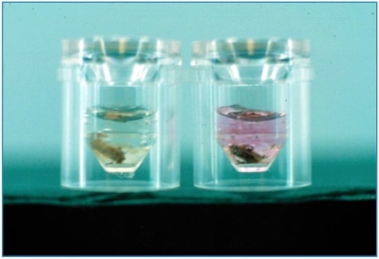
We designed and modified the quantitative serum colorimetric assay for newborn screening using blood-soaked filter paper discs (Figure 1) [5]. Our first attempt to perform the assay using blood spots was successful. We took several months to “tweak” the assay. We designed the assay so that the kinetics were not exactly linear with concentration and time, but rather would provide almost no color development with activities below 30% of mean normal activity. This worked well in our laboratory. However, with all these good intentions, most of the other laboratories that began to perform newborn screening did their own “tweaking” of our assay conditions, resulting in some laboratories having difficulty in ascertaining infants with partial deficiency. Although these manipulations of the screening assay had obvious ramifications for identifying individuals with partial deficiency, it is unlikely they would miss an infant with profound deficiency. Over the years, various methods have been published that measure biotinidase activity in filter-paper blood spots [36,48,49]. In addition, commercial companies began to sell kits for assaying enzyme activity. Like all kits for scientific studies, propriety often hampered the ability to alter the assay to meet a specific laboratory’s requirements. This has been an obvious bane to us old timers who understood the biochemistry behind our procedures and could readily modify them when necessary. Today’s laboratory personnel and even the supervisory staff are often restricted from modifying an assay by the commercial company’s “black box” methodologies.
We always knew that we were measuring biotinidase activity using an artificial substrate, which was not the natural substrate of biotinidase, biocytin or biotinyl-epsilon-peptides. However, we hoped that the ease and cost of using these artificial substrates would ascertain all or essentially all individuals with biotinidase deficiency. We always considered that we could potentially miss some enzyme-deficient individuals, especially those with partial biotinidase deficiency. To date, I am not aware of any false-positive results from confirmatory diagnosis of newborn screening.
The Medical Center in Virginia wanted us to patent the method for newborn screening, and we did submit a patent application. However, the U.S. Patent Office said that we had essentially already disclosed the method in an earlier abstract that described our method using blood spots in less detail, but it was still sufficient for someone to replicate it. Therefore, the method was not patentable. I learned that a patent of a method is only as good as the ability to have surveillance over those using the method. That would be relatively easy for newborn screening laboratories. In Virginia in 1984, we estimated that the cost of testing, including the test materials and the salary of a technician, who performed the tests and the follow-up of putative positives, was between 11 and 22 cents per individual screened! If the test was patented and licensed, we would have to charge a licensing fee of between a penny and five cents per test; this could potentially be too costly for a state to consider incorporating the testing in their state. I actually was only interested in finding enzymatically-deficient children and starting them on biotin. Therefore, I was not too upset, but rather relieved, that the patent was not granted. However, altruism obviously did not stop the significant steady increase in the cost of testing to rise over the years, especially with the entrance of commercial companies making newborn screening kits.
In general, it appears that whichever assay or kit was being used, the laboratories were identifying individuals with profound biotinidase deficiency and hopefully most individuals with partial biotinidase deficiency.
I was invited to present our work at national and international pediatric and newborn screening meetings. Rapidly, other states and countries began pilot newborn screening programs for biotinidase deficiency. Over the years, I would call all the locations that were performing screening around the world to learn about their results and to determine the incidence of the disorder in their locations [50,51]. I would arrive at the laboratory early in the morning, about 5 a.m. E.S.T, and call the European locations. Later in the morning, I would call the locations in the eastern states, and in the afternoon, I called laboratories in the midwestern and western states. Finally, in the evening, I would call the few Pacific locations, Australia and New Zealand. I was encouraged to see the number of individuals with biotinidase deficiency increase, thereby readily exceeding the threshold requirement demanded by Dr. Robert Guthrie to warrant newborn screening for a disorder!
Once enzymatic testing for biotinidase deficiency became readily available, many countries began identifying symptomatic children with profound biotinidase deficiency. The most striking numbers came, not unexpectedly, from those countries having a high degree of consanguinity, such as Turkey [52], Saudi Arabia [53] and the United Arab Emirates [54]. A large number of individuals with profound biotinidase deficiency were ascertained in a relatively short time period. I realize that this happens with many autosomal recessive disorders in these countries; it is almost as if the disorder was an epidemic occurring in these locations. Today, the incidence of biotinidase deficiency is likely to increase in various European countries because of the increased immigration of individuals from countries having a high degree of consanguineous matings, just as has been found in Sweden [55].
In addition, unlike the experience in many western countries where most of the affected individuals are compound heterozygotes, because of the consanguinity, most of the affected individuals in Turkey, Saudi Arabia and the United Arab Emirates were homozygous for specific mutations, allowing us to ascertain the actual effects of a single mutation on both enzyme activity and phenotype.

2.4. Newborn Screening for Biotinidase Deficiency Is Not for Everybody…Yet

There are still locations in the world that have not added biotinidase deficiency to their newborn screening programs [56]. One argument is that their clinicians would readily recognize the symptoms of the disorder, perform the appropriate diagnostic tests, confirm the diagnosis and begin treatment with biotin before irreversible damage has occurred. This has obviously not happened as exemplified in several reports [57,58]. Because there is no newborn screening in these locations, the authors of the reports admonish clinicians to think of biotinidase deficiency in their differential diagnosis. Unfortunately, older children, adolescents and adults do not usually exhibit the same symptoms as the young symptomatic children, with which most clinicians are familiar [46,59]. In addition, enzyme-deficient individuals may remain asymptomatic until adolescence or adulthood [60,61].
Newborn screening is meant to identify individuals with the disorder as early as possible so that an effective treatment can be initiated to prevent the development of symptoms. This is a major reason that newborn screening for biotinidase deficiency has been so widely accepted. I discussed the arguments for screening both profound and partial biotinidase deficiency in a recent review and commentary [56]. Although the arguments in favor of screening for profound biotinidase deficiency are well documented, the arguments for screening for partial biotinidase deficiency are still controversial. Individuals with partial biotinidase deficiency range from asymptomatic to minor symptoms and even major neurological issues. Even those who are symptomatic are milder than seen in those with profound deficiency; symptoms can occur from infancy to adulthood with most resolving with biotin treatment [56].
At this time, all states in the United States screen their newborns for both profound and partial biotinidase deficiency. These children are usually still referred to geneticists and metabolic specialists for definitive diagnosis. As with most newborn screening for rare inherited metabolic disease, the primary care physician of an individual with partial biotinidase deficiency also benefits from this knowledge. If a child is known to have partial biotinidase deficiency and, despite taking biotin, develops a clinical problem, the symptoms are most likely not due to the biotinidase deficiency, but to some other disorder or condition.
Another very important issue has been the cost-effectiveness of performing newborn screening for biotinidase deficiency. There have been several studies that have addressed this issue, and they have all agreed that newborn screening for biotinidase deficiency is cost-effective [62,63,64].

2.5. What about the Natural History of Biotinidase Deficiency?

There was a relatively short time from the discovery of the disorder to the implementation of newborn screening of the disorder in many locations around the world. This has resulted in many asymptomatic children with the enzyme deficiency being identified and treated with biotin before developing symptoms. Although this is obviously a major positive goal of newborn screening, it has rapidly narrowed our “window of opportunity” to learn about the natural history of the disorder. However, as discussed above, there are still locations in the world that do not screen for biotinidase deficiency. It is in these locations that we have found a resistance to incorporate screening for profound biotinidase deficiency, let alone partial deficiency, for a variety of reasons. A common reason is that the incidence of the disorder is not high enough. However, as I have stated, cost-effective analyses have all favored the screening for biotinidase deficiency. Nevertheless, a positive effect of the unfortunate circumstance of not screening in these locations is continuing information about the natural history of the disorder even at the sacrifice of delayed diagnosis in symptomatic individuals. These individuals run the risk of developing irreversible features even after the diagnosis is made and treatment is initiated. This has definitely occurred in multiple adolescents or adults who have presented with myelopathic and/or visual abnormalities [59,65]. Because these older individuals do not exhibit the “classical” features of young symptomatic children with biotinidase deficiency, these symptoms are rarely included in the differential diagnosis and, therefore, delay the correct diagnosis and treatment. In fact, unless a neurologist, ophthalmologist or geneticist thinks of this possibility or fortunately performs urinary organic acid analysis, some of these individuals may never be correctly diagnosed and/or appropriately treated.

2.6. We Are Correct to Screen for Biotinidase Deficiency

The outcome of individuals with profound biotinidase deficiency being treated early is an important issue. We quickly learned that essentially all symptomatic children with profound deficiency would improve with biotin supplementation; however, we also learned that some symptoms were irreversible once they occurred. These included optic atrophy, sensorineural hearing loss and severe developmental delays [65]. Our hope was that early initiation of biotin therapy, such as immediately after birth based on newborn screening, would offer the best chance to prevent the development of symptoms. Various reports appeared in the literature that described the children who were ascertained by newborn screening and have been on biotin supplementation [66,67,68]. However, most of these reports only described children and young adolescents. When we published the successful experience of 25 years of newborn screening in Michigan, it became obvious that only the younger individuals (mean age of eight years) continued to be followed in genetics/metabolic clinics [16]. Therefore, I proceeded to locate older adolescents, over 15 years old, and adults to determine how they have done over the years. I located 44 individuals with profound biotinidase deficiency identified by newborn screening with a mean age of almost 23 years and with an age range of 16 to 32 years. These individuals have all done exceedingly well [69]. In addition, we have demonstrated that the offspring of treated women with profound biotinidase deficiency have been healthy. Moreover, contacting and talking to these individuals was one of the most gratifying experiences and the perfect culmination of over 30 years of clinical and laboratory investigations!

2.7. What Questions about Biotinidase Deficiency and Its Newborn Screening Still Need Answering?

As discussed above, we must continue to add to our knowledge about the natural history of profound biotinidase deficiency, especially in those locations that are not screening or when individuals with profound biotinidase fail to comply with biotin treatment. We must determine the reversibility or irreversibility of symptoms in these individuals once the diagnosis is made.
We are only aware of a single example of genotype-phenotype correlation in biotinidase deficiency. We found that those symptomatic children in Turkey with null mutations appeared to be more likely to develop sensorineural hearing loss than those with missense mutations [70]. However, this does not mean that only untreated individuals with null mutations develop hearing loss, but this group may be more likely to develop it and they may develop it earlier. With the increasing use of mutation analysis for confirmatory testing, we must continue to evaluate symptomatic individuals for potential genotype-phenotype correlations.
Symptomatic individuals with profound biotinidase deficiency with identical genotypes in different families and in the same family often exhibit considerable clinical phenotypic variability. In addition, some untreated individuals with profound biotinidase deficiency present with symptoms as children, whereas others do no not exhibit symptoms until adolescence or adulthood. We and others have considered that these differences might be due to dietary or environmental factors, but we have failed to confirm this explanation. It is also possible that there are epigenetic differences in biotin metabolism that we do not understand at this time. This question warrants further evaluation.
As discussed above, one of the most controversial questions that needs answering is the necessity of treating individuals for partial biotinidase deficiency. If the decision is that they do not warrant treatment, then why should we screen for it? I have presented evidence for newborn screening for partial biotinidase deficiency and for its treatment [56]. In this paper I proposed that we must diligently report children and older individuals with partial deficiency who develop symptoms and those who are identified by newborn screening and are not treated with biotin. We should document the types, variation and severity of their clinical features, whether they exhibit abnormal urinary organic acids and whether their symptoms improve with biotin therapy.
Because of the controversy regarding the treatment of individuals with partial biotinidase deficiency, it is important to consider the small number of individuals with deficient enzymatic activity who may have a Km mutation. In one study of 201 individuals, 10 were found to have Km defects; of these, six had profound deficiency using the substrate concentration of 0.15 nmol/L N-biotinyl-p-aminobenzoate in the colorimetric biotinidase assay, but four had activity in the 18%–20% range of the mean normal activity [41] or within the partially deficient range. The authors suggested that individuals with residual activity, especially in the partial deficiency range, should have Km enzymatic studies. They suggested screening these individuals by assaying their activity at the routine substrate concentration and at a 10-fold higher substrate concentration. If they had an increase in their enzymatic activities at the higher concentration, they likely had a Km defect.
Most states and many countries treat all individuals with a partial biotinidase range (10%–30% of the mean normal activity). Therefore, those individuals with Km defects would not be missed. However, if a location or physician chooses not to treat a child with partial biotinidase deficiency, then it is possible to fail to treat the rare individual with a Km mutation who should be treated. Because the treatment of individuals with biotinidase deficiency is to supply them with sufficient free biotin (5–10 mg/day), biotin treatment circumvents the enzyme defect. Therefore, the dose of biotin necessary for an individual with a Km defect is not expected to be different from that used to treat other individuals with either profound or partial biotinidase deficiency. Since many individuals with biotinidase deficiency are now having mutational analysis, it would be helpful to know which mutations are due to Km defects; however, Km studies are not performed routinely in diagnostic laboratories. Until this information is available, this may be another reason for treating all individuals with partial biotinidase deficiency.
This Km study was published at about the same time that the biotinidase gene was elucidated and mutation analysis was being performed. We are aware that only the mutations of one of these individuals have been published [44,46], but not the others. Another problem is that the individuals are likely heterozygous for their mutations, and we cannot readily attribute which mutation corresponds to the enzyme with the Km defect. Furthermore, we are not aware that the authors have determined and published the mutations of the other individuals found to have Km defects.
As stated above, newborn screening for biotinidase deficiency uses either the colorimetric or the fluorimetric enzymatic assay. Recently, a study indicated that the fluorimetric assay was slightly more sensitive to the colorimetric assay in identifying the deficiency [71]. However, the disparity would likely be in identifying partially deficient versus those with normal activity. Although the difference in sensitivity was not statistically significant, further studies are warranted. Moreover, because neither method uses the natural substrate, biocytin, perhaps a method using the natural substrate, although more difficult, should also be compared with both of the methods that use the artificial substrate.
Does biotinidase have other functions besides recycling biotin from biotinylated compounds? We had previously shown that in vitro, biotinidase is capable of biotinylating histones [72]; however, other studies have suggested that this is not a physiological function of the enzyme [73,74]. If the enzyme has other functions, what are they and what does it mean for biotinidase deficiency, treated and untreated?
We developed a transgenic, knock-out mouse with profound biotinidase deficiency [12]. We hoped that the mouse fed a biotin-depleted diet would exhibit the same or similar clinical findings as the untreated human with the enzyme deficiency. In fact, we demonstrated that the animal does exhibit many of the behavioral, neurological and cutaneous abnormalities the symptomatic human does. We had expectations that if the clinical features were similar, then we could use these mice to better understand the role of biotinidase in various organ systems and in the disease state. Although we did evaluate the neurological and immunological aspects of the biotin-depleted mice, the results of our studies did not add greatly to our understanding of the biology or mechanism of the disease [13,14]. This may be due to our unsophisticated methodologies or to our failure to adequately understand the interactions of various pathways, especially in the brain. Such animal models are often used to evaluate gene therapy interventions; however, this is obviously not necessary in readily treatable biotinidase deficiency. The biotinidase-deficient mouse may still be a useful tool to compare biotinidase-deficient states in biotin-replete and biotin-depleted states or may answer questions using more advanced technologies, such as gene expression. Future studies using these animals will tell us whether such lines of research will prove fruitful.
When we first described partial biotinidase deficiency and began performing mutation analysis, we noted almost all individuals with enzymatic activities in the 10%–30% range of the mean normal activity had a mutation causing profound deficiency on one allele and the D444H mutation on the other allele, which causes an approximately 50% decrease in activity. However, as more individuals with activity in the partially deficient range have been described, other combinations of mutations, besides the D444H variant, were noted to result in activities in the partially deficient range. As others have recommended, perhaps it is time to discuss the degree of enzyme activity and not just a dichotomous distribution as profound and partial deficiency. Although profound and partial biotinidase deficiency may still be useful functional descriptions, we must consider that individuals with biotinidase deficiency have activities within the continuum of enzyme activities and because the D444H mutation is common, there is an increased frequency of individuals who are heterozygous for the D444H mutation and another profound mutation.

2.8. A Legacy Fulfilled

Biotinidase deficiency meets all the major criteria for inclusion in a newborn screening program. The successful outcomes of individuals with profound biotinidase deficiency identified by newborn screening have been extremely gratifying. It is my distinct pleasure to have had the opportunity to spend my career studying a single inherited metabolic disorder in depth. Obviously, this work has been a joint effort with all my students, fellows and physician and laboratory colleagues. In particular, I would like to thank my mentors and the other scientists around the world who have dedicated much or a portion of their careers to the study of this disorder. I have been so fortunate to have spent my life’s work on a disorder that has resulted in such positive results and outcomes. When all is said and done, “If you have to have an inherited metabolic disorder, biotinidase deficiency is the one to have!” [75].

Acknowledgments

The work described in this publication was supported in part by the Safra Research Fund at Henry Ford Hospital. I also thank Diane Verde for her expert review and suggestions in this manuscript.

Conflicts of Interest

The authors declare no conflict of interest.

References

  1. Wolf, B. The Story of Biotinidase Deficiency and Its Introduction into Newborn Screening: The Role of Serendipity. Int. J. Neonatal Screen. 2015, 1, 3–12. [Google Scholar] [CrossRef]
  2. Wolf, B.; Grier, R.E.; Parker, W.D.; Goodman, S.I.; Allen, R.J. Deficient biotinidase activity in late-onset multiple carboxylase deficiency. N. Engl. J. Med. 1983, 308, 161. [Google Scholar] [CrossRef] [PubMed]
  3. Wolf, B.; Grier, R.E.; Allen, R.J.; Goodman, S.I.; Kien, C.L. Biotinidase deficiency: The enzymatic defect in late-onset multiple carboxylase deficiency. Clin. Chim. Acta 1983, 131, 273–281. [Google Scholar] [CrossRef]
  4. Wolf, B.; Grier, R.E.; Allen, R.J.; Goodman, S.I.; Kien, C.L.; Parker, W.D.; Howell, D.M.; Hurst, D.L. Phenotypic variation in biotinidase deficiency. J. Pediatr. 1983, 103, 233–237. [Google Scholar] [CrossRef]
  5. Heard, G.S.; Secor McVoy, J.R.; Wolf, B. A screening method for biotinidase deficiency in newborns. Clin. Chem. 1984, 30, 125–127. [Google Scholar] [PubMed]
  6. Wolf, B.; Heard, G.S.; Jefferson, L.G.; Proud, V.K.; Nance, W.E.; Weissbecker, K.A. Clinical findings in four children with biotinidase deficiency detected through a statewide neonatal screening program. N. Engl. J. Med. 1985, 313, 16–19. [Google Scholar] [CrossRef] [PubMed]
  7. Cole, H.; Reynolds, T.R.; Buck, G.B.; Lockyer, J.M.; Denson, T.; Spence, J.E.; Hymes, J.; Wolf, B. Human serum biotinidase: cDNA cloning, sequence and characterization. J. Biol. Chem. 1994, 269, 6566–6570. [Google Scholar] [PubMed]
  8. Knight, H.C.; Reynolds, T.R.; Meyers, G.A.; Pomponio, R.J.; Buck, G.A.; Wolf, B. Structure of the human biotinidase gene. Mamm. Genome 1998, 9, 327–330. [Google Scholar] [CrossRef] [PubMed]
  9. Procter, M.; Wolf, B.; Crockett, D.K.; Mao, R. The biotinidase variants registry: A paradigm public database. Genes Genom. Genet. 2013. [Google Scholar] [CrossRef] [PubMed]
  10. Swango, K.L.; Demirkol, M.; Huner, G.; Pronicka, E.; Sykut-Cegielska, J.; Schulze, A.; Wolf, B. Partial biotinidase deficiency is usually due to the D444H mutation in the biotinidase gene. Hum. Genet. 1998, 102, 571–575. [Google Scholar] [CrossRef] [PubMed]
  11. Pindolia, K.; Jensen, K.; Wolf, B. Three dimensional structure of human biotinidase: Computer modeling and functional correlations. Mol. Genet. Metab. 2013, 92, 13–22. [Google Scholar] [CrossRef] [PubMed]
  12. Pindolia, K.; Jordan, M.; Guo, C.; Mathews, N.; Mock, D.; Strovel, E.; Blitzer, M.; Wolf, B. Development and characterization of a mouse with profound biotinidase deficiency: A biotin-responsive neurocutaneous disorder. Mol. Genet. Metab. 2010, 102, 161–169. [Google Scholar] [CrossRef] [PubMed]
  13. Pindolia, K.; Chen, J.; Cardwell, C.; Cui, X.; Chopp, M.; Wolf, B. Neurological deficits in mice with profound biotinidase deficiency are associated with demylination and axonal degeneration. Neurobiol. Dis. 2012, 47, 428–435. [Google Scholar] [CrossRef] [PubMed]
  14. Pindolia, K.; Li, H.; Cardwell, C.; Wolf, B. Characterization and functional analysis of cellular immunity in mice with biotinidase deficiency. Mol. Genet. Metab. 2014, 112, 49–56. [Google Scholar] [CrossRef] [PubMed]
  15. Cowan, T.M.; Blitzer, M.G.; Wolf, B. Technical standards and guidelines for the diagnosis of biotinidase deficiency. Genet. Med. 2010, 12, 464–470. [Google Scholar] [CrossRef] [PubMed]
  16. Jay, A.M.; Conway, R.L.; Feldman, G.L.; Nahhas, F.; Spencer, L.; Wolf, B. Outcomes of individuals with profound and partial biotinidase deficiency ascertained by newboorn screening in Michigan over 25 years. Genet. Med. 2015, 17, 205–209. [Google Scholar] [CrossRef] [PubMed]
  17. Pispa, J. Animal biotinidase. Ann. Med. Exp. Biol. Fenn. 1965, 43 (Suppl. 5), 1–39. [Google Scholar]
  18. Bartlett, K.; Ghniem, H.K.; Stirk, J.H.; Wastell, H.J.; Sherrah, H.S.A.; Leonard, J.V. Enzyme studies in combined carboxylase deficiency. Ann. N. Y. Acad. Sci. 1985, 447, 235–251. [Google Scholar] [CrossRef] [PubMed]
  19. Gompertz, D.; Bartlett, K.; Blair, D.; Stern, C.M.M. Child with a defect in leucine metabolism associated with beta hydroxyisovaleric aciduria and beta-methylcrotonylglycinuria. Arch. Dis. Child. 1973, 48, 975–977. [Google Scholar] [CrossRef] [PubMed]
  20. Gompertz, D.; Goodey, P.A.; Bartlett, K. Evidence for the enzymatic defect in beta-methylcrotonylglycinuria. FEBS Lett. 1973, 32, 13–14. [Google Scholar] [CrossRef]
  21. Bartlett, K.; Gompertz, D. Combined carboxylase defect: Biotin-responsiveness in cultured fibroblasts. Lancet 1976, 2, 804. [Google Scholar] [CrossRef]
  22. Bartlett, K.; Ng, H.; Dale, G.; Green, A.; Leonard, J.V. Studies on cultured fibroblasts from patients with defects of biotin-dependent carboxylation. J. Inherit. Metab. Dis. 1981, 4, 183–189. [Google Scholar] [CrossRef] [PubMed]
  23. Ghneim, H.K.; Bartlett, K. Mechanism of biotin-responsive combined carboxylase deficiency. Lancet 1982, 1, 1187–1188. [Google Scholar] [CrossRef]
  24. Baumgartner, R.; Suormala, T.; Wick, H.; Geisert, J.; Lehnert, W. Infantile multiple carboxylase deficiency: Evidence for normal intestinal absorption but renal loss of biotin. Helv. Pediatr. Acta 1982, 37, 499–502. [Google Scholar]
  25. Baumgartner, E.R.; Sourmala, T.; Wick, H.; Bonjour, J.P. Biotin-responsive multiple carboxylase deficiency (MCD): Deficient biotinidase activity associated with renal loss of biotin. J. Inherit. Metab. Dis. 1984, 7 (Suppl. 2), 123–125. [Google Scholar] [PubMed]
  26. Samboni, M.; Gaudry, M.; Marquet, A.; Munnich, A.; Saudubray, J.M.; Marsac, C. Search for the biochemical basis of biotin dependent multiple carboxylase deficiencies: Determination of biotin activation in cultured fibroblasts. Clin. Chim. Acta 1981, 122, 241. [Google Scholar] [CrossRef]
  27. Munnich, A.; Saudubray, J.M.; Ogier, H.; Coude, F.X.; Marsac, C.; Roccichioli, F.; Labarthe, J.C.; Cuzanave, C.; Laugier, J.; Harpentier, C.; et al. Deficit multiple des carboxylases. Arch. Fr. Pediatr. 1981, 38, 83. [Google Scholar] [PubMed]
  28. Munnich, A.; Fischer, A.; Saudubray, J.M.; Griscelli, C.; Coude, F.X.; Ogier, H.; Charpentier, C.; Frezal, J. Biotin-responsive immunoregulatory dysfunction in multiple carboxylase deficiency. J. Inherit. Metab. Dis. 1981, 4, 113–114. [Google Scholar] [CrossRef]
  29. Fischer, A.; Munnich, A.; Saudubray, J.M. Biotin-responsive immunoregulatory dysfunction in multiple carboyxlase deficiency. J. Clin. Immunol. 1982, 2, 35–38. [Google Scholar] [CrossRef] [PubMed]
  30. Munnich, A.; Saudubray, J.M.; Cotjsson, A.; Coude, F.X.; Ogier, H.; Charpentier, C.; Marsac, C.; Garre, G.; Bourgeay-Causse, M.; Frezal, J. Biotin dependent multiple carboxylase deficiency presenting as a congenital lactic acidosis. Eur. J. Pediatr. 1981, 137, 203–206. [Google Scholar] [PubMed]
  31. Feldman, G.L.; Wolf, B. Deficient acetyl-CoA carboxylase activity in multiple carboxylase deficiency. Clin. Chim. Acta 1981, 111, 147–151. [Google Scholar] [CrossRef]
  32. Feldman, G.L.; Hsia, Y.E.; Wolf, B. Biochemical characterization of biotin responsive multiple carboxylase deficiency: Heterogeneity within the bio genetic complementation group. Am. J. Hum. Genet. 1981, 33, 692–701. [Google Scholar] [PubMed]
  33. Wolf, B.; Hsia, Y.E.; Sweetman, L.; Feldman, G.; Boychuk, R.B.; Bart, R.D.; Crowell, D.H.; Di Mauro, R.M.; Nyhan, W.L. Multiple carboxylase deficiency: Cinical and biochemical improvement following neonatal biotin treatment. Pediatrics 1981, 68, 113–118. [Google Scholar] [PubMed]
  34. Koivusalo, M.; Pispa, J. Biotinidase activity in animal tissue. Acta Physiol. Scand. 1963, 58, 13–19. [Google Scholar] [CrossRef] [PubMed]
  35. Wastell, H.; Dale, G.; Bartlett, K. A sensitive fluorimetric rate assay for biotinidase using a new derivative of biotin, biotinyl-6-aminoquinoline. Anal. Biochem. 1984, 140, 69–73. [Google Scholar] [CrossRef]
  36. Chan, P.W.; Bartlett, K. A new solid-phase assay for biotin and biocytin and its application to the study of patients with biotinidase deficiency. Clin. Chim. Acta 1986, 159, 185–196. [Google Scholar] [CrossRef]
  37. Wastell, H.J.; Bartlett, K.; Dale, G.; Shein, A. Biotinidase deficiency: A survey of 10 cases. Arch. Dis. Child. 1988, 63, 1244–1249. [Google Scholar] [CrossRef] [PubMed]
  38. Schubiger, G.; Caflisch, U.; Baumgartner, R.; Suormala, T.; Bachmann, C. Biotinidase deficiency: Clinical course and biochemical findings. J. Inherit. Metab. Dis. 1984, 7, 129–130. [Google Scholar] [CrossRef] [PubMed]
  39. Suormala, T.; Wick, H.; Bonjour, J.P.; Baumgartner, E.R. Intestinal absorption and renal excretion of biotin in patients with biotinidase deficiency. Eur. J. Pediatr. 1985, 144, 21–26. [Google Scholar] [CrossRef] [PubMed]
  40. Suormala, T.M.; Baumgartner, E.R.; Wick, H.; Scheibenreiter, S.; Schweitzer, S. Comparison of patients with complete and partial biotinidase deficiency: Biochemical studies. J. Inherit. Metab. Dis. 1990, 13, 76–92. [Google Scholar] [CrossRef] [PubMed]
  41. Suormala, T.; Ramaekers, V.T.; Schweitzer, S.; Fowler, B.; Laub, M.C.; Schwermer, C.; Bachmann, J.; Baumgartner, E.R. Biotinidase Km-variants: Detection and detailed biochemical investigations. J. Inherit. Metab. Dis. 1995, 18, 689–700. [Google Scholar] [CrossRef] [PubMed]
  42. Baumgartner, E.R.; Suormala, T.M.; Wick, H.; Probst, A. Biotinidase deficiency: A cause of subacute necrotizing encephalomyelopathy (Leigh syndrome). Report of a case with lethal outcome. Pediatr. Res. 1989, 26, 260–266. [Google Scholar] [CrossRef] [PubMed]
  43. Baumgartner, E.R.; Suormala, T.; Wick, H.; Bausch, J.; Bonjour, J.-P. Biotinidase deficiency: Factors responsible for the increased biotin requirement. J. Inherit. Metab. Dis. 1985, 8 (Suppl. 1), 59–64. [Google Scholar] [CrossRef] [PubMed]
  44. Ramaekers, V.Th.; Suormala, T.M.; Brab, M.; Duran, R.; Heimann, G.; Baumgartner, E.R. A biotinidase Km variant causing late onset bilateral optic neuropathy. Arch. Dis. Child. 1992, 67, 115–119. [Google Scholar] [CrossRef] [PubMed]
  45. Suormala, T.M.; Baumgartner, E.R.; Bausch, J.; Holick, W.; Wick, H. Quantitative determination of biocytin in urine of patients with biotinidase deficiency using high-performance liquid chromatography. Clin. Chim. Acta 1988, 177, 253–269. [Google Scholar] [CrossRef]
  46. Wolf, B.; Pomponio, R.J.; Norrgard, K.J.; Lott, I.T.; Baumgartner, E.R.; Suormala, T.; Raemaekers, V.Th.; Coskun, T.; Tokatli, A.; Ozalp, I.; et al. Delayed-onset profound biotinidase deficiency. J. Pediatr. 1998, 132, 362–365. [Google Scholar] [CrossRef]
  47. Wolf, B.; Jensen, K.; Huner, G.; Demirkol, M.; Baykal, T.; Divry, P.; Rolland, M.O.; Perez-Cerda, C.; Ugarte, M.; Straussberg, R.; et al. Seventeen novel mutations that cause profound biotinidase deficiency. Mol. Genet. Metab. 2002, 77, 108–111. [Google Scholar] [CrossRef]
  48. Broda, E.; Baumgartner, E.R.; Scholl, S.; Stopsack, M.; Horn, A.; Rhode, H. Biotinidase determination in serum and dried blood spots-high sensitivity fluorometric ultramicro-assay. Clin. Chim. Acta 2001, 314, 175–185. [Google Scholar] [CrossRef]
  49. Dobrowolski, S.F.; Angeletti, J.; Banas, R.A.; Naylor, E.W. Real time PCR assays to detect common mutations in the biotinidase gene and application of mutational analysis to newborn screening for biotinidase deficiency. Mol. Genet. Metab. 2003, 78, 100–107. [Google Scholar] [CrossRef]
  50. Wolf, B. Worldwide survey of neonatal screening for biotinidase deficiency. J. Inherit. Metab. Dis. 1991, 14, 923–927. [Google Scholar] [CrossRef] [PubMed]
  51. Wolf, B.; Heard, G.S. Screening for biotinidase deficiency in newborns: Worldwide experience. Pediatrics 1990, 85, 512–517. [Google Scholar] [PubMed]
  52. Pomponio, R.J.; Coskun, T.; Demirkol, M.; Tokatli, A.; Ozalp, I.; Huner, G.; Baykal, T.; Wolf, B. Novel mutations cause biotinidase deficiency in Turkish children. J. Inherit. Metab. Dis. 1999, 23, 120–128. [Google Scholar] [CrossRef]
  53. Pomponio, R.J.; Ozand, P.I.; Al Essa, M.; Wolf, B. Novel mutations in children with profound biotinidase deficiency from Saudi Arabia. J. Inherit. Metab. Dis. 2000, 23, 185–187. [Google Scholar] [CrossRef] [PubMed]
  54. Al-Jasmi, F.A.; Al-Shamai, A.; Hertecant, J.L.; Al-Hamad, S.M.; Sould, A.K. Inborn errors of metabolism in the United Arab Emirates: Disorders detected by newborn screening. J. Inherit. Metab. Dis. Rep. 2016, 28, 127–135. [Google Scholar]
  55. Ohlsson, A.; Guthenberg, C.; Holme, E.; von Dobein, U. Profound biotinidase deficiency: A rare disease among native Swedes. J. Inherit. Metab. Dis. 2010, 33 (Suppl. 3), S175–S180. [Google Scholar] [CrossRef] [PubMed]
  56. Wolf, B. Why perform newborn screening for profound and partial biotinidase deficiency? Mol. Genet. Metab. 2015, 114, 382–387. [Google Scholar] [CrossRef] [PubMed]
  57. Mc Sweeney, N.; Grunewald, S.; Bhate, S.; Gunesan, V.; Chong, W.K.; Hemingway, C. Two unusual clinical and radiological presentations of biotinidase deficiency. J. Eur. Pediatr. Neurol. Soc. 2010, 14, 535–538. [Google Scholar] [CrossRef] [PubMed]
  58. Grunewald, S.; Champion, M.P.; Leonard, J.V.; Schaper, J.; Morris, A.A.M. Biotinidase defiicency: A treatable leukoencephalopathy. Neuropediatrics 2004, 35, 211–216. [Google Scholar] [CrossRef] [PubMed]
  59. Wolf, B. Biotinidase deficiency should be considered in individuals exhibiting myelopathy with or without vision loss. Mol. Genet. Metab. 2015, 116, 113–118. [Google Scholar] [CrossRef] [PubMed]
  60. Haine, S.R.; Langmuir, R.A. Optic neuropathy due to biotinidase deficiency in 19-year-old amn. JAMA Opthalmol. 2014, 132, 228–230. [Google Scholar] [CrossRef] [PubMed]
  61. Bottin, L.; Prud’hon, S.; Guey, S.; Giannesini, C.; Wolf, B.; Pindoliua, K.; Stankoff, B. Biotinidase deficiency mimicking neuromylitis optica: Initially exhibiting symptoms in adulthood. Mult. Scler. J. 2015, 21, 1604–1607. [Google Scholar] [CrossRef] [PubMed]
  62. Carroll, A.E.; Downs, S.M. Comprehensive cost-utility analysis of newborn screening strategies. Pediatrics 2006, 117, S287–S295. [Google Scholar] [PubMed]
  63. Schoos, R.; Verloes, A.; Bourguignon, J.P.; Koulischer, L. Programs of systematic screening in neonatolgy. Rev. Med. Liege 1998, 53, 311–315. [Google Scholar] [PubMed]
  64. Vallejo-Torres, L.; Rodreguez, I.V.; Perez-Cerda, C.; Plana, J.C.; Pineda, M.; Elgarresta, A.A.; Martin-Hernandez, E.; Crouce, M.L. Cost-effectiveness analysis of newborn screening for biotinidase deficiency. Pediatrics 2015, 136, e424–e432. [Google Scholar] [CrossRef] [PubMed]
  65. Wolf, B. The neurology of biotinidase deficiency. Mol. Genet. Metab. 2011, 104, 27–34. [Google Scholar] [CrossRef] [PubMed]
  66. Moslinger, D.; Stockler-Ipsiroglu, S.; Scheibenreiter, S.; Tiefenthaler, M.; Muhl, A.; Seidl, R.; Strobl, W.; Plecko, B.; Suormala, T.; Baumgartner, E.R. Clinical and neuropsychological outcome in 33 patients with biotinidase deficiency ascertained by nationwide newborn screening and family studies in Austria. Eur. J. Pediatr. 2002, 161, 167–168. [Google Scholar] [CrossRef]
  67. Möslinger, D.; Mühl, A.; Suormala, T.; Baumgartner, R.; Ströckler-Ipsiroglu, S. Molecular characterization and neuropsychological outcome of 21 patients with profound biotinidase deficiency detected by newborn screening and family studies. Eur. J. Pediatr. 2003, 162, S46–S49. [Google Scholar] [CrossRef] [PubMed]
  68. Weber, P.; Schoo, S.; Baumgartner, E.R. Outcome in patients with profound biotinidase deficiency: Relevance of newborn screening. Dev. Med. Child. Neurol. 2004, 46, 481–484. [Google Scholar] [CrossRef] [PubMed]
  69. Wolf, B. Successful outcomes of older adolescents and adults with profound biotinidase deficiency identified by newborn screening. Genet. Med. 2016. [Google Scholar] [CrossRef] [PubMed]
  70. Sivri, H.S.; Genc, G.A.; Tokatli, A.; Dursun, A.; Coskun, T.; Aydin, H.I.; Sennaroglu, L.; Belgin, E.; Jensen, K.; Wolf, B. Hearing loss in biotinidase deficiency: Genotype-phenotype correlation. J. Pediatr. 2007, 150, 439–442. [Google Scholar] [CrossRef] [PubMed]
  71. Iseeri-ERten, S.O.; Dikmen, Z.G.; Ulusu, N.N. Comparison of spectrophotometric and fluorimetric methods in evaluation of biotinidase deficiency. J. Med. Biochem. 2016, 35, 123–129. [Google Scholar]
  72. Hymes, J.; Fleischhauer, K.; Wolf, B. Biotinylation of histones by human serum biotinidase: Assessment of biotinyl-transferase activity in sera from normal individuals and children with biotinidase deficiency. Biochem. Mol. Med. 1995, 56, 76–83. [Google Scholar] [CrossRef] [PubMed]
  73. Narang, M.A.; Dumas, R.; Ayer, R.; Gravel, R.A. Localization of holocarboxylase synthetase in the nucleus of cells: A novel role for biotin in human biology. Am. J. Hum. Genet. 2002, 71, 174. [Google Scholar]
  74. Narong, M.A.; Dumas, R.; Ayers, L.M.; Gravel, R.A. Reduced histone biotinylation in multiple carboxylase deficiency patients: A nuclear role of holocarboxylase synthetase. Hum. Mol. Genet. 2004, 13, 15–23. [Google Scholar] [CrossRef] [PubMed]
  75. Wolf, B. Biotinidase deficiency: If you have to have an inherited metabolic disease, this is the one to have. Genet. Med. 2012, 14, 565–575. [Google Scholar] [CrossRef] [PubMed]
Figure 1. Colorimetric assay of biotinidase activity. On the left, no or straw-colored development of a one-eighth-inch-diameter blood-soaked filter paper spot from a child with profound bioitinidase deficiency compared to the mauve color development in a sample from a child with normal biotinidase activity. The colorimetric assay was performed in plastic, disposable cups that were inserted into a modified Guthrie template tray. Initially, individuals with putative bioitinidase deficiency were identified by observation comparing color development with a series of control samples with those having 25%, 50% 75% and 100% of normal activity.
Neonatalscreening 02 00009 g001