Article

Newborn Screening for Severe Combined Immunodeficiency in Taiwan

1
Department of Medical Genetics, National Taiwan University Hospital, Taipei 10041, Taiwan
2
Department of Pediatrics, National Taiwan University Hospital, Taipei 10041, Taiwan
3
Newborn Screening, Department of Clinical Pathology, Taipei Institute of Pathology, Taipei 10374, Taiwan
4
Neonatal Screening Center, Chinese Foundation of Health, Taipei 11070, Taiwan
5
Department of Pediatrics, Chang Gung University College of Medicine and Children’s Hospital, Taoyuan 33305, Taiwan
6
Department of Pediatrics, National Cheng Kung University Hospital, Tainan 70403, Taiwan
*
Author to whom correspondence should be addressed.
Int. J. Neonatal Screen. 2017, 3(3), 16; https://doi.org/10.3390/ijns3030016
Submission received: 30 March 2017 / Revised: 2 June 2017 / Accepted: 19 June 2017 / Published: 23 June 2017

Abstract

:
A study of newborn screening for severe combined immunodeficiency (SCID) by detecting the T-cell receptor excision circle (TRECs) copy number in dried blood spots (DBSs) collected from newborns 3 days of age began in 2010 in Taiwan, and SCID screening was subsequently implemented country-wide in 2012. A total of 920,398 newborns were screened during a period of 78 months. Of these, 175 newborns (0.02%) were requested to undergo an immune function survey, and 136 cases (1 in 6768 newborns) were ultimately diagnosed as having T cell lymphopenia. The screening detected seven cases of typical SCID, with an incidence of 1 in 131,485 newborns (95% confidence interval, 1/63,693~1/271,434). Hematopoietic stem cell transplantation was performed in six patients before overt infection occurred, and the survival rate was 100%. The screening also detected eight cases of SCID variants and 20 cases of 22q11.2 deletion syndrome. Other etiologies of T lymphopenia were identified, and those newborns were evaluated and managed according to their immunological status. Owing to the introduction of newborn screening by measuring the TREC copy number, early administration of treatments became possible for newborns with conditions that put them at risk of primary or secondary immunodeficiency.

1. Introduction

Severe combined immunodeficiency (SCID) is a group of rare inherited disorders with profound defects in T-cell and B-cell immunity. At least 20 genes have been associated with SCID [1]. Currently, curative treatments such as hematopoietic stem cell transplantation (HSCT) [2,3,4] and gene therapy [5,6,7] can improve a patient’s outcome if the treatment can be initiated before severe infections occur. Previously, a significant number of children with SCID would die before a diagnosis was made [8] or before a curative treatment could be administered [2,9,10]. Because of the burden of tuberculosis (TB) in Taiwan, all newborns receive Bacillus Calmette-Guérin (BCG) vaccination at birth to avoid TB meningitis [11]. However, this policy has also put newborns with primary immunodeficiencies, such as SCID, at a higher risk for disseminated BCGitis [12,13,14].
Since consanguinity is not allowed in Taiwan and there is no founder mutations described for SCID previously, it is difficult to screen patients before the onset of symptoms. In 2010, the National Taiwan University Hospital (NTUH) Newborn Screening Center initiated a pilot newborn screening program for SCID by measuring the T-cell receptor excision circle (TREC) copy number [15]. This program successfully identified patients with SCID and other conditions causing T-cell lymphopenia and therefore allowed patients to receive appropriate prophylaxis against infections and early institution of HSCT [15,16]. The Taiwan Advisory Committee on Immunization Practices (ACIP) therefore delayed the timing of BCG vaccination in 2012, when SCID screening became available nationwide in order to allow for the screening results to be obtained prior to immunization, while maintaining the protection of babies against TB meningitis. The surveillance data revealed that the percentage of infants receiving BCG vaccination at the ages of 1–5 months increased from 18% in 2010 to 46.9% in 2014, without an increase in the incidence of TB meningitis [17]. Because there was a decrease in the prevalence of tuberculosis, in 2016, the ACIP recommended BCG immunization at the age of 5–8 months to further decrease the risk of BCG vaccination-related complications. In this article, we summarize the performance of SCID screening and provide evidence that SCID screening improved the health of newborns.

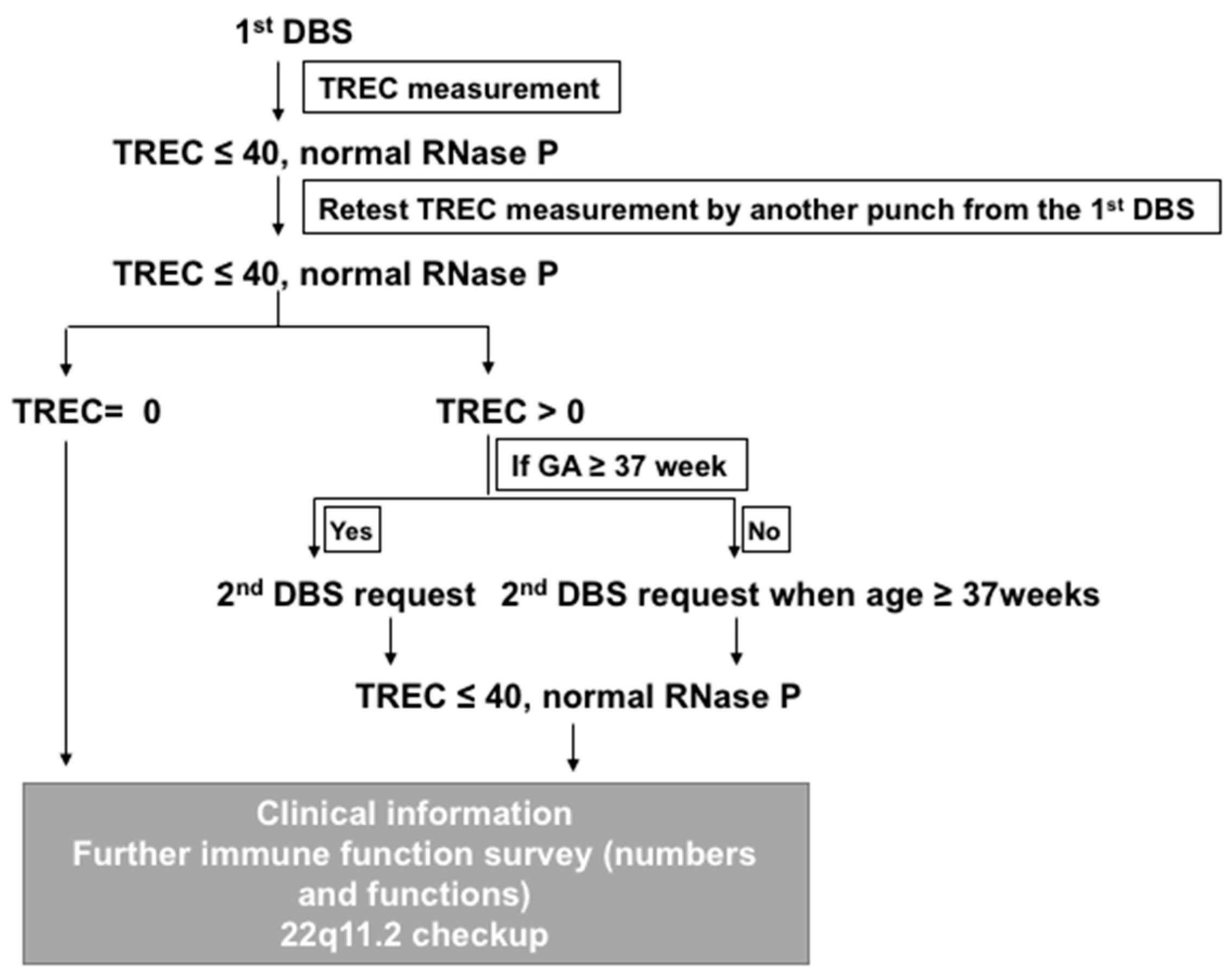
2. Method Development in NTUH

The T-cell receptor excision circles (TRECs) assay is a verified tool used for SCID screening in newborns [18,19]. However, there are many versions of primers and probes [19,20] and different combinations of reaction conditions used for the assay [1]. In addition, the DNA extracted from dried blood spots using routine filter collection cards may not be robust enough for product amplification. Therefore, we tested different combinations of reagents and conditions with the help of Francis Lee and Robert F. Vogt, Jr., at the Newborn Screening and Molecular Biology Branch, Centers for Disease Control and Prevention (CDC). The first step was to test different TREC primers. TREC2F/2R [20] provide more robust results with DBS DNA than the original primers used [18], which produced more nonspecific bands in our system. The reference gene we chose was RNase P, which we assayed using primers designed by the CDC. A plasmid (pcDNA™3.1/myc-His B MCS-TREC (Eco RI/Xba I)) was established so that a standard curve for copy number calculation could be used in each experiment.
Second, the efficacy and the quality of DNA extraction from different brands of filter paper cards differs a lot. We have adjusted the extraction method so that we are able to obtain adequate DNA and minimize the concentration of inhibitors in the elutes of DBSs. We found that the combination of 0.01% Triton X-100 in phosphate-buffered saline (PBS) and Qiagen Neutralization Solution performed the best in our system, along with an additional 0.4 μg/μL Bovine serum albumin (BSA) in the PCR mixture. Since the anticoagulants affect the PCR amplification efficiency [21,22], we tested parallel DBS samples using EDTA or heparin as the anticoagulants and compared the Ct (threshold cycle) of TREC (corresponding to the TREC copy numbers) and the Ct of RNase P. The median Ct values for TREC and RNase P without anticoagulant were 32.9 (range 32.4–33.8) and 26.71 (range 24.9–27.2), respectively. Compared to these medians, using EDTA in the blood slightly increased the Ct values of both TREC and RNase P (by 2% and 1%, respectively), while using heparin in the blood slightly decreased the values by 1% and 4%. The TREC number of heparinized blood actually increase from a median of 255 copies/μL whole blood (136–349) in no-anticoagulant blood to 289 copies/μL (199–358). Heparin is the most frequently used anticoagulant in Taiwan newborn screening. Since its presence did not result in any significant interference in PCR amplification, we did not modify the sample collection procedure for the SCID NBS.
Finally, we tested different brands of PCR mixtures to see whether better linearity could be obtained in the lower copy range. Otherwise, patients with SCID, who have no TREC, would be indistinguishable from patients with 22q11.2 deletion syndrome who have low TREC copy numbers. We also set qPCR assays to detect TUPLE1 gene copy number on DBS using A3M and M3M probes [23] so that we can identify infants with 22q11.2 deletion syndrome.
The distribution of TREC numbers in the newborn population were mentioned previously [7]. The average TREC copy number is 185 copies/μL (standard deviation (S.D.) 121, median 156). Our current cutoff for TREC is 40 copies/μL, which is equal to the lowest first percentile of the population.

3. Updated Results of Screening

Our pilot study detected two patients with SCID and five with 22q11.2 deletion syndrome from a total of 106,391 newborns screened [15]. These results led to a country-wide voluntary SCID newborn screening executed by all three newborn screening centers, including the NTUH. The annual birthrate is approximately 200,000 newborns in Taiwan, and the acceptance rate was approximately 85–88% for all births in Taiwan. All screening programs are still active and participate in TREC Proficiency Testing covered by the Newborn Screening Quality Assurance Program (NSQAP) and managed by the Newborn Screening and Molecular Biology Branch, CDC, USA. After testing, the DBS will be freeze and stored for at least 3 years.
The NTUH screening algorithm are described in Figure 1. The same cut-offs are used for term and preterm infants and the difference is the preterm infants are re-tested at 37 weeks of gestational age (GA). The other two NBS centers in Taiwan, Chinese Foundation of Health (CFOH) and Taipei Institute of Pathology (TIP), apply a similar algorithm, except for the cutoff values used due to different reagents among centers. CFOH uses LightMixTM Modular TREC (Roche Diagnostics, Basel, Switzerland), and the cutoffs is equal to the lowest 0.5 percentile of the population. TIP uses EnLiteTM Neonatal TREC Kit (PerkinElmer, Turku, Finland), and the cutoff is set per manufacturer. TIP has the lowest cutoff value at 25 copies/μL for both the 1st and the 2nd DBS samples, while CFOH uses different cutoffs for the 1st and the 2nd DBS samples (initially 35 and 30 copies/μL, respectively, revised to 50 and 40 copies/μL, respectively, in September 2015). The confirmatory algorithm includes the white cell counts and flow cytometry for lymphocyte subsets including T cells, B cells, NK cells, CD4 and CD8 cells (both naïve and memory cells). Mitogen proliferation is only performed when the flow quantitation is abnormal without recognizable etiologies, and for infants with 22q11.2 deletion syndrome. In addition, for all newborns with abnormal results, NTUH checks the original DBS by a TUPLE1 gene copy number analysis for chromosome 22q11.2 deletion [15]. If the sample is positive for the TUPLE1 deletion, the diagnosis would be confirmed in a second blood sample by multiplex ligation-dependent probe amplification (MLPA) [15]. MLPA is the preferred test for newborns suspecting 22q11.2 deletion due to low TREC numbers or due to the clinical presentations. Three immunologists who have diagnosed and cared for infants with SCID ascertain the final diagnosis and perform the follow-up of SCID cases. The classification of conditions with low TREC copy numbers identified by newborn screening follows the R4S laboratory performance database [24].
The data summarized here were collected until 30 June 2016 (Table 1). There were 175 infants (0.02%) referring for a flow cytometry. Different TREC cutoff values and systems resulted in significantly different referral rates for flow cytometry. The rates of referral for flow cytometry were 0.02% for the NTUH and CFOH (p by Chi-Square Test = 0.6868), but the rate was nearly 0 for the TIP (p < 0.0001). Among them, 136 infants (77.7%) confirmed as having T cell lymphopenia. Finally, over a period of 78 months, SCID screening detected seven cases of typical SCID, giving an incidence of 1 in 131,485 infants (95% CI 1/63,693~1/271,434). This finding was similar to the incidence in previous report by screening [25] (p = 0.1356), or by the clinical estimation (an incidence of 1/214,136 (1/119,575–383,479), p = 0.3083) [26].
Among the seven typical SCID cases, three males (43%) had an IL2RG mutation (Cases 1, 4, and 5), 1 (14%) had RAG1 mutations (Case 2), and another three patients had unidentified gene mutations (Cases 3, 6 and 7). Cases 1 (IL2RG gene: c.846 + 2deltgag), Case 2 (RAG1 gene: p.R474C/p.R776Q compound heterozygote), and Case 3 have been described (previous No. 1, 2, and 5) [7]. The mutations for Case 4 and 5 were c.982 C > T (p.Arg328 *) and c.865 C > T (p.Arg289 *), respectively, on IL2RG. Both Case 3 and 6 were subjected to whole exome sequence but the gene defects were still unknown. As for Case 7, she presented no mutations in the IL2RG, JAK3, IL7Ra, RAG1, RAG2, CD3 ε/δ/ζ chains, or Artemis genes. All samples were screened negative for 22q11.2 deletion (TUPLE1). All samples had normal ADA activity as described below. However, there are at least 20 genes associated with SCID, and it is possible that Case 3, 6, and 7 pose mutations on those unapproached genes. Two newborns (Case 1 and 5) with an IL2RG mutation had a positive family history of recurrent early male infant deaths, but neither of the families was aware of the diagnosis before birth. Case 6’s older sister had died at the age of 1 year when preparing for HSCT because of repeated infections. Case 1–6 underwent HSCT. Case 7 had one older brother who had died at the age of 2 years due to a brain tumor, therefore the parents refused further arrangements such as HSCT for her. Her first DBS also showed that the Kappa-deleting element recombination circle (KREC) value was 30 copies/μL, which was as low as that in Case 2. She was fine at last contact when age 1 year. All eight newborns with SCID variants underwent different extents of gene mutation scanning, but no mutations were found. There was no patient with leaky SCID or Omenn syndrome. All of the patients with SCID were still alive without frequent infections up to the last contact. During this period, there were no SCID cases who were initially missed by the TREC screening. However, four SCID cases were reported shortly before the initiation of the country-wide screening. All four samples had zero copies of TREC in their first NBS DBSs.

4. 22q11.2 Deletion Syndrome

The incidence of 22q11.2 deletion syndrome with low TREC numbers in NTUH program, was 1 in 24,405 newborns (95% CI 1/15,438~1/38,580), similar to the previous report [25] (p = 0.0729). Table 2 lists the immunological profiles of the patients with 22q11.2 deletion syndrome identified through the NTUH newborn screening program (Table 2). The median first TREC value was 15 (range 6–34) copies/μL. Only two patients (2/18, 11%) were reported to have associated congenital heart disease, including one case of hypoplastic left heart syndrome (HLHS). The median T lymphocyte count was 1314/μL (793–2846). When a confirmatory test was performed at a median age of 25 days (range, 15–79), sixty percent of the patients (9/15) still presented with T-cell lymphopenia (<1500/μL). Nine of the 16 newborns had abnormal lymphocyte subtyping (10/16, 63%), including five (31%) with a high proportion of B cells (>32%) and seven (44%) with a high proportion of NK cells (>18%). Seven of the nine (78%) newborns were associated with T lymphopenia. Due to having 22q11.2 deletion, those infants were followed regularly especially for immune, electrolytes, development and stature, and had instructions regarding to live vaccines. There were no reported adverse events associated with live vaccines when they were given later, after several careful evaluations, including a proliferation test.
The extreme phenotype of the 22q11.2 deletion syndrome appeared in the one patient identified with a TREC number of zero copies/μL at the first screening by TIP. She also presented with thymus aplasia, a ventricular septal defect, hypoparathyroidism and hypocalcemia. The initial workup revealed lymphopenia (1400/μL) and T lymphopenia (T cells 0.7%, B cells 50.8%, NK cells 38.9%). Because she developed repeated infections even with the early diagnosis and proper managements, she received HSCT for her profound combined immunodeficiency at the age of 5 months. Her lymphopenia resolved (lymphocytes 1718/μL, T 23%, B 1% and NK 50%) and there was no recurrent infection thereafter till the age of 2 years.

5. Secondary Etiologies for T-Lymphopenia

In addition to prematurity and 22q11.2 deletion syndrome, there were 38 cases of T-cell lymphopenia attributed to T cell loss (n = 24) include sampling after operations for congenital heart diseases, volvulus, and congenital diaphragmatic hernia and other medical conditions (n = 14). The most predominant associated condition was congenital heart disease (4 cases with hypoplastic left heart syndrome and 5 with complete transposition of the great vessels), vascular leakage and gastrointestinal anomalies. After the notification of abnormal SCID screen results and the following immune status evaluation, some of these patients received intravenous immunoglobulin (IVIG) due to associated hypogammaglobulinemia and repeated infections. We also detected two neonatal leukemias (1 with 46, XY, t(11:19)(q23;p13.3)), one case of trisomy 21 syndrome, one of 14q microdeletion, one of maternal HIV infection, and several of maternal systemic lupus erythematosus (SLE) or maternal antiphospholipid syndrome with or without treatment. The newborns with maternal drug- or antibody-related effects recovered their lymphocyte counts by six months after birth. The two newborns with leukemia were immediately referred to pediatric hematologists for further management. Therefore, identifying newborns with low TREC copy numbers also enabled better management of these infants, including environmental precautions, changes in the immunization reschedule, and medical treatment.

6. Rare Conditions

A newborn with abnormal TREC copy numbers (1st DBS, 30; 2nd DBS 33) was referred for a confirmatory immunological function evaluation. His older brother had presented with congenital cutis mamorata telangiectasia and died at the age of 1 month owing to Ralstonia pickettii (Burkholderia) septic shock. This newborn, similar to his older brother, also presented with sparse hair, eyebrows, and eyelashes at birth, then a generalized skin rash developed at the age of three days. The confirmatory lymphocyte subtyping and immunoglobulin levels at the age of 1 month revealed combined immunodeficiency (B cells 50.2%, T cells 31.87%, NK cells 5.1%, IgA 2.67 mg/dL, IgM < 4.47 mg/dL, IgG 286 mg/dL, IgE < 4.1 IU/mL). The proliferation response to phytohaemagglutinin (PHA) was normal (Stimulation Index (S.I.) 29; 50% of normal) but the response was suboptimal for anti-CD3/anti-CD28 antibodies stimulation (S.I. 3.5; <10% of normal). Combining this information with the deceased brother’s history, the linear hypopigmented skin pattern, and the fact that the grandmother had only given birth to females, the IKBKG gene was checked in the patient, his brother and their mother [28]. The results revealed a hemizygous IKBKG c.520_523dupCAGG mutation in the patient and his brother, with a heterozygous change in the mother. This mutation results in a splicing error with exon 5 skipping. Under a diagnosis of X-linked recessive anhidrotic ectodermal dysplasia-associated immunodeficiency (EDA-ID), HSCT was planned, coupled with IVIG supplemented q3week, Trimethoprim-sulfamethoxazole prophylaxis, and isonicotinylhydrazide (INH) plus Rifampin use for possible mycobacterium infection after BCG vaccination. However, HSCT was not performed due to recurrent infections and poor family support. The patient died at the age of 11 months due to pulmonary hemorrhage after repeated Klebsiella pneumoniae sepsis complicated with a brain abscess and status epilepticus, Burkholderia cepacia complex sepsis, a Mucormycosis skin infection, and Mycobacterium abscessus infection.
A female newborn had TREC copy numbers of 38, 28 and 0 copies/μL at three different time points. The first DBS also showed 0 copies of KREC. The mother had been on Ritodrine for tocolysis for six weeks. The baby was delivered at the gestational age of 39 weeks, three weeks after the tocolytic agents had been stopped. When flow cytometry was performed at the age of three weeks, B lymphocytes were barely detected (1% of cells). At the age of three months, she continued to have T lymphopenia (1267/μL), but her B lymphocytes increased and she had a normal proliferation response. She developed normally without a significant history of infection at the age of 18 months. She had no identified mutations in IL2RG, IL7Ra, RAG1, RAG2, or the CD3 ε/δ/ζ chains. Therefore we hypothesize that Ritodrine induced lymphocytopenia in this patient.

7. Differences in the Methods Used and Their Rates of Detection

The three contributing screening programs employ different methods, the EnLite™ Neonatal TREC Kit and real-time PCR. EnLite™ Neonatal TREC Kits were designed to screen only for SCID, not for other conditions such as DiGeorge syndrome or variant SCID. Not surprisingly, the EnLite™ Neonatal TREC Kit had the lowest referral rate at almost 0. Using real-time PCR, we identified several infants with less acute SCID syndromes (variant SCID), SCID-like syndrome (22q11.2 deletion), and many other cases of secondary T-cell lymphopenia. In addition, real-time PCR has the potential to be multiplexed immediately to detect spinal muscular atrophy (SMA) [29] or X-linked agammaglobulinemia (XLA) [30]. Therefore, more than two-thirds of Taiwan newborns were screened using the real-time PCR method. The overall refer rate (0.02%) is lower than the previous report [25] (p < 0.001), although the rate varies among programs. Our data suggests that the three methods used in Taiwan have similar specificity since there was no false negatives during this period.
Of note, the incidence of 22q11.2 deletion determined among the three programs was significantly different (relative risk at NTUH = 1, odds ratio (O.R.) at CFOH = 0.09 (95% CI 0.01–0.68) (p = 0.0034), and O.R. at TIP = 0.11 (95% CI 0.02–0.86) (p = 0.0109). Based on the NTUH previous data [15], NTUH added a DBS molecular test in addition to a flow cytometry analysis. Otherwise, a correct diagnosis may be missed in those newborns presenting with no heart problems and normal lymphocyte counts. We also noted that there is no apparent relationship between the TREC number in the DBS to chromosome microdeletion. In addition, we were aware of several 22q11.2 deletion patients, who presented clinical symptoms but with a normal or borderline normal TREC copy number in the 1st DBS. We believe that adding a molecular test for 22q11.2 deletion syndrome is warranted in cases with low TREC copies number to identify those newborns at risk of immunodeficiency.

8. BCG Policy and Impact

The national effort to provide BCG vaccination at birth to protect infants from tuberculosis meningitis and disseminated tuberculosis was launched in Taiwan in 1965. Currently, the coverage rate is above 97%. The importance of BCG vaccination in this region was shown in two different outcomes: the risk of tuberculosis meningitis in unvaccinated children was 16 times higher than in BCG vaccinated children, and the mortality rate of extrapulmonary tuberculosis declined almost 100-fold compared to the pre-vaccination era [11]. However, several adverse events related to or suspected to be related to BCG vaccination have been reported, especially among children who were vaccinated by the age of 1 month [17,31]. Most importantly, there have been at least three children with SCID complicated with disseminated BCG infection [9,12,32,33], and one child died of this complication. Although BCG vaccination is still recommended in Taiwan, the SCID NBS program was expected to decrease the risk of complicated disseminated BCG infection in SCID newborns. Therefore, in 2012, the Advisory Committee on Immunization Practices (ACIP), CDC, Taiwan, approved delaying BCG vaccination from 24 h of age to 1–4 weeks of age to allow SCID NBS to be performed. Although the rate of BCG vaccination completion by the age of 1 month dropped from 80% before 2010 to 53% in 2014, the incidence of TB meningitis remained 1–2 cases annually, which was also probably due to the decrease in the incidence of tuberculosis. As a result, the ACIP further revised the immunization program in 2016. BCG vaccination is now scheduled at the age of 5–8 months if there is no significant tuberculosis exposure risk. The new immunization schedule should be beneficial for decreasing both the risk of osteomyelitis/osteitis in normal infants and disseminated BCG infection due to SCID and other primary immunodeficiencies.

9. Under Evaluation

ADA Deficiency

Since the TREC assay may not reliably detect patients with ADA deficiency [34], we also tested the newborns for ADA deficiency by determining the concentrations of two purine metabolites, Ado and dAdo, using NeoBase2 Non-derivatized MSMS kit (PerkinElmer, Turku, Finland). After we had screened 21,591 newborns, there were no cases showing elevation of Ado and dAdo. The average concentrations of Ado and dAdo were 0.63 μM (S.D.0.23) and 0.01 μM (S.D. 0.02), respectively. We also tested retrospectively the 11 samples (SCID and variants SCID identified by NTUH) and no samples showed elevation of Ado and dAdo.

10. Kappa-Deleting Element Recombination Circle (KREC) Screening

Similar to T lymphocytes, newly developed B cells can be detected using the KREC [35]. A combined TREC/KREC approach has been able to identify patients with severe B cell disorders such as X-linked agammaglobulinemia [30] and delayed-onset ADA deficiency [36]. To identify other forms of primary immunodeficiency, such as X-linked agammaglobulinemia (XLA) [37,38,39], we have tested the feasibility of performing a KREC assay as previous described [40]. The KREC assay could accurately indicate SCID with a RAG1 mutation and other situations such as newborns with pancytopenia or B lymphopenia, but the correlation in newborns with a IL2RG mutation was low. In addition, the KREC assay was not able to identify X-linked hyper-IgM syndrome, the second most common antibody deficiency [37,41]. Therefore, a pilot TREC/KREC assay has not been initiated yet. A Swedish pilot program is in progress that involves the simultaneous detection of both TREC and KREC to identify T and B cell deficiencies, and the results will help in the decision [42].

11. Summary

In Taiwan, the T-cell receptor excision circle (TREC) assay has been adopted as part of the routine newborn screening program. Infants with SCID have been identified through this program and have received hematopoietic stem cell transplants before the development of infections. All of the patients identified by this assay were alive at the time of writing this report, which is in contrast to the results of previous reports, confirming that the prompt HSCT improved their outcomes. Without SCID screening, at least two SCID patients, although they had a positive family history, would have been missed. The molecular etiologies were unknown in 43% of the SCID infants and in other infants with persistent or transient lymphopenia. Further genomic approaches are warranted to formulate a better monitoring and follow-up plan.
Newborns with other etiologies of T-cell lymphopenia, such as patients with a 22q11.2 deletion and congenital heart disease, have also been identified through this screening program; therefore, a notification of a possible decrease in immunity and increase in the risk of infection could be delivered to the families. This led to better awareness about the need for environment protection, changes in the immunization schedule, and even medical intervention for these infants.

Acknowledgments

We would like to thank Francis Lee and Robert F. Vogt, Jr. and their colleagues at the Newborn Screening and Molecular Biology Branch, Centers for Disease Control and Prevention (CDC) for continued support and discussion about the TREC assay. PerkinElmer partially support the NeoBase™ Non-derivatized MSMS kit used for ADA deficiency screen.

Author Contributions

Yin-Hsiu Chien, Shu-Chuan Chiang and Wuh-Liang Hwu conceived and designed the experiments; Shu-Chuan Chiang and Chen-Chen Liu performed the experiments; Yin-Hsiu Chien, Hui-Chen Ho, and Shu-Min Kao contributed the screening data; Yin-Hsiu Chien, Ni-Chung Lee, Meng-Yao Lu, Hsin-Hui Yu, Tang-Her Jaing, Wen-I Lee, Kuei-Wen Chang, Chi-Chang Shieh, and Jiann-Shiuh Chen contributed patients’ data; Yin-Hsiu Chien wrote the paper.

Conflicts of Interest

The authors declare no conflicts of interest.

References

  1. Kwan, A.; Puck, J.M. History and current status of newborn screening for severe combined immunodeficiency. Semin. Perinatol. 2015, 39, 194–205. [Google Scholar] [CrossRef] [PubMed]
  2. Yang, Y.L.; Lu, M.Y.; Jou, S.T.; Lin, K.H.; Lin, D.T. Hematopoietic stem cell transplantation in Taiwanese children with primary immunodeficiency. J. Formos. Med. Assoc. 2005, 104, 101–106. [Google Scholar] [PubMed]
  3. Abd Hamid, I.J.; Slatter, M.A.; McKendrick, F.; Pearce, M.S.; Gennery, A.R. Long-term outcome of hematopoietic stem cell transplantation for IL2RG/JAK3 SCID: A cohort report. Blood 2017, 129, 2198–2201. [Google Scholar] [CrossRef] [PubMed]
  4. Myers, L.A.; Patel, D.D.; Puck, J.M.; Buckley, R.H. Hematopoietic stem cell transplantation for severe combined immunodeficiency in the neonatal period leads to superior thymic output and improved survival. Blood 2002, 99, 872–878. [Google Scholar] [CrossRef] [PubMed]
  5. Shaw, K.L.; Garabedian, E.; Mishra, S.; Barman, P.; Davila, A.; Carbonaro, D.; Shupien, S.; Silvin, C.; Geiger, S.; Nowicki, B.; et al. Clinical efficacy of gene-modified stem cells in adenosine deaminase-deficient immunodeficiency. J. Clin. Investig. 2017, 127, 1689–1699. [Google Scholar] [CrossRef] [PubMed]
  6. Hacein-Bey-Abina, S.; Pai, S.Y.; Gaspar, H.B.; Armant, M.; Berry, C.C.; Blanche, S.; Bleesing, J.; Blondeau, J.; de Boer, H.; Buckland, K.F.; et al. A modified gamma-retrovirus vector for X-linked severe combined immunodeficiency. N. Engl. J. Med. 2014, 371, 1407–1417. [Google Scholar] [CrossRef] [PubMed]
  7. De Ravin, S.S.; Wu, X.; Moir, S.; Anaya-O’Brien, S.; Kwatemaa, N.; Littel, P.; Theobald, N.; Choi, U.; Su, L.; Marquesen, M.; et al. Lentiviral hematopoietic stem cell gene therapy for X-linked severe combined immunodeficiency. Sci. Transl. Med. 2016, 8, 335ra357. [Google Scholar] [CrossRef] [PubMed]
  8. Lin, J.S.; Shyur, S.D.; Lin, H.Y. Severe combined immunodeficiency with B-lymphocytes (T-B+SCID): Report of two cases. Zhonghua Min Guo Xiao Er Ke Yi Xue Hui Za Zhi 1998, 39, 406–411. [Google Scholar] [PubMed]
  9. Lee, W.I.; Huang, J.L.; Jaing, T.H.; Shyur, S.D.; Yang, K.D.; Chien, Y.H.; Chiang, B.L.; Soong, W.J.; Chiou, S.S.; Shieh, C.C.; et al. Distribution, clinical features and treatment in Taiwanese patients with symptomatic primary immunodeficiency diseases (PIDs) in a nationwide population-based study during 1985–2010. Immunobiology 2011, 216, 1286–1294. [Google Scholar] [CrossRef] [PubMed]
  10. Morio, T.; Atsuta, Y.; Tomizawa, D.; Nagamura-Inoue, T.; Kato, K.; Ariga, T.; Kawa, K.; Koike, K.; Tauchi, H.; Kajiwara, M.; et al. Outcome of unrelated umbilical cord blood transplantation in 88 patients with primary immunodeficiency in Japan. Br. J. Haematol. 2011, 154, 363–372. [Google Scholar] [CrossRef] [PubMed]
  11. Chan, P.C.; Huang, L.M.; Kuo, H.S. Is neonatal bacillus calmette-guerin vaccination protective in Taiwan? J. Formos. Med. Assoc. 2008, 107, 195–197. [Google Scholar] [CrossRef]
  12. Huang, L.H.; Shyur, S.D.; Weng, J.D.; Shin, C.; Huang, F.Y.; Tzen, C.Y. Disseminated cutaneous bacille calmette-guerin infection identified by polymerase chain reaction in a patient with X-linked severe combined immunodeficiency. Pediatr. Dermatol. 2006, 23, 560–563. [Google Scholar] [CrossRef] [PubMed]
  13. Lee, W.I.; Huang, J.L.; Yeh, K.W.; Jaing, T.H.; Lin, T.Y.; Huang, Y.C.; Chiu, C.H. Immune defects in active mycobacterial diseases in patients with primary immunodeficiency diseases (PIDs). J. Formos. Med. Assoc. 2011, 110, 750–758. [Google Scholar] [CrossRef] [PubMed]
  14. Marciano, B.E.; Huang, C.Y.; Joshi, G.; Rezaei, N.; Carvalho, B.C.; Allwood, Z.; Ikinciogullari, A.; Reda, S.M.; Gennery, A.; Thon, V.; et al. BCG vaccination in patients with severe combined immunodeficiency: Complications, risks, and vaccination policies. J. Allergy Clin. Immunol. 2014, 133, 1134–1141. [Google Scholar] [CrossRef] [PubMed]
  15. Chien, Y.H.; Chiang, S.C.; Chang, K.L.; Yu, H.H.; Lee, W.I.; Tsai, L.P.; Hsu, L.W.; Hu, M.H.; Hwu, W.L. Incidence of severe combined immunodeficiency through newborn screening in a Chinese population. J. Formos. Med. Assoc. 2015, 114, 12–16. [Google Scholar] [CrossRef] [PubMed]
  16. Chang, T.Y.; Jaing, T.H.; Lee, W.I.; Chen, S.H.; Yang, C.P.; Hung, I.J. Single-institution experience of unrelated cord blood transplantation for primary immunodeficiency. J. Pediatr. Hematol. Oncol. 2015, 37, e191–e193. [Google Scholar] [CrossRef] [PubMed]
  17. Huang, H.Y.; Chang, Y.L. Adverse events induced by BCG immunization and immunization schedule in Taiwan and in Japan. Taiwan Epidemiol. Bull. 2015, 31, 631–633. [Google Scholar] [CrossRef]
  18. Baker, M.W.; Grossman, W.J.; Laessig, R.H.; Hoffman, G.L.; Brokopp, C.D.; Kurtycz, D.F.; Cogley, M.F.; Litsheim, T.J.; Katcher, M.L.; Routes, J.M. Development of a routine newborn screening protocol for severe combined immunodeficiency. J. Allergy Clin. Immunol. 2009, 124, 522–527. [Google Scholar] [CrossRef] [PubMed]
  19. Chan, K.; Puck, J.M. Development of population-based newborn screening for severe combined immunodeficiency. J. Allergy Clin. Immunol. 2005, 115, 391–398. [Google Scholar] [CrossRef] [PubMed]
  20. Hazenberg, M.D.; Otto, S.A.; Cohen Stuart, J.W.; Verschuren, M.C.; Borleffs, J.C.; Boucher, C.A.; Coutinho, R.A.; Lange, J.M.; Rinke de Wit, T.F.; Tsegaye, A.; et al. Increased cell division but not thymic dysfunction rapidly affects the T-cell receptor excision circle content of the naive T cell population in HIV-1 infection. Nat. Med. 2000, 6, 1036–1042. [Google Scholar] [CrossRef] [PubMed]
  21. Yokota, M.; Tatsumi, N.; Nathalang, O.; Yamada, T.; Tsuda, I. Effects of heparin on polymerase chain reaction for blood white cells. J. Clin. Lab. Anal. 1999, 13, 133–140. [Google Scholar] [CrossRef]
  22. Al-Soud, W.A.; Radstrom, P. Purification and characterization of PCR-inhibitory components in blood cells. J. Clin. Microbiol. 2001, 39, 485–493. [Google Scholar] [CrossRef] [PubMed]
  23. Yang, C.; Huang, C.H.; Cheong, M.L.; Hung, K.L.; Lin, L.H.; Yu, Y.S.; Chien, C.C.; Huang, H.C.; Chen, C.W.; Huang, C.J. Unambiguous molecular detections with multiple genetic approach for the complicated chromosome 22q11 deletion syndrome. BMC Med. Genet. 2009, 10, 16. [Google Scholar] [CrossRef] [PubMed]
  24. McHugh, D.; Cameron, C.A.; Abdenur, J.E.; Abdulrahman, M.; Adair, O.; Al Nuaimi, S.A.; Ahlman, H.; Allen, J.J.; Antonozzi, I.; Archer, S.; et al. Clinical validation of cutoff target ranges in newborn screening of metabolic disorders by tandem mass spectrometry: A worldwide collaborative project. Genet. Med. 2011, 13, 230–254. [Google Scholar] [CrossRef] [PubMed]
  25. Kwan, A.; Abraham, R.S.; Currier, R.; Brower, A.; Andruszewski, K.; Abbott, J.K.; Baker, M.; Ballow, M.; Bartoshesky, L.E.; Bonilla, F.A.; et al. Newborn screening for severe combined immunodeficiency in 11 screening programs in the United States. JAMA 2014, 312, 729–738. [Google Scholar] [CrossRef] [PubMed]
  26. Lee, W.I.; Jaing, T.H.; Hsieh, M.Y.; Kuo, M.L.; Lin, S.J.; Huang, J.L. Distribution, infections, treatments and molecular analysis in a large cohort of patients with primary immunodeficiency diseases (PIDs) in Taiwan. J. Clin. Immunol. 2006, 26, 274–283. [Google Scholar] [CrossRef] [PubMed]
  27. Shearer, W.T.; Rosenblatt, H.M.; Gelman, R.S.; Oyomopito, R.; Plaeger, S.; Stiehm, E.R.; Wara, D.W.; Douglas, S.D.; Luzuriaga, K.; McFarland, E.J.; et al. Lymphocyte subsets in healthy children from birth through 18 years of age: The pediatric AIDS clinical trials group P1009 study. J. Allergy Clin. Immunol. 2003, 112, 973–980. [Google Scholar] [CrossRef] [PubMed]
  28. Lee, N.C.; Huang, C.H.; Hwu, W.L.; Chien, Y.H.; Chang, Y.Y.; Chen, C.H.; Ko, T.M. Pseudogene-derived IKBKG gene mutations in incontinentia pigmenti. Clin. Genet. 2009, 76, 417–419. [Google Scholar] [CrossRef] [PubMed]
  29. Taylor, J.L.; Lee, F.K.; Yazdanpanah, G.K.; Staropoli, J.F.; Liu, M.; Carulli, J.P.; Sun, C.; Dobrowolski, S.F.; Hannon, W.H.; Vogt, R.F. Newborn blood spot screening test using multiplexed real-time PCR to simultaneously screen for spinal muscular atrophy and severe combined immunodeficiency. Clin. Chem 2015, 61, 412–419. [Google Scholar] [CrossRef] [PubMed]
  30. Borte, S.; Wang, N.; Oskarsdottir, S.; von Dobeln, U.; Hammarstrom, L. Newborn screening for primary immunodeficiencies: Beyond SCID and XLA. Ann. N. Y. Acad. Sci. 2011, 1246, 118–130. [Google Scholar] [CrossRef] [PubMed]
  31. Chiu, N.C.; Lin, M.C.; Lin, W.L.; Wang, S.Y.; Chi, H.; Huang, L.M.; Tang, R.B.; Huang, Y.C.; Liu, C.C.; Huang, F.Y.; et al. Mycobacterium bovis BCG-associated osteomyelitis/osteitis, Taiwan. Emerg. Infect. Dis. 2015, 21, 539–540. [Google Scholar] [CrossRef] [PubMed]
  32. Lee, W.I.; Liang, F.C.; Huang, J.L.; Jaing, T.H.; Wang, C.H.; Lin, T.Y.; Huang, Y.C.; Huang, W.L.; Jou, R.; Hsieh, M.Y.; et al. Immunologic analysis of HIV-uninfected Taiwanese children with BCG-induced disease. J. Clin. Immunol. 2009, 29, 319–329. [Google Scholar] [CrossRef] [PubMed]
  33. Huang, H.Y.; Chang, Y.L. Evolution and determinants of BCG immunization policy: Canada’s experience. Taiwan Epidemiol. Bull. 2016, 32, 351–362. [Google Scholar] [CrossRef]
  34. La Marca, G.; Canessa, C.; Giocaliere, E.; Romano, F.; Duse, M.; Malvagia, S.; Lippi, F.; Funghini, S.; Bianchi, L.; Della Bona, M.L.; et al. Tandem mass spectrometry, but not T-cell receptor excision circle analysis, identifies newborns with late-onset adenosine deaminase deficiency. J. Allergy Clin. Immunol. 2013, 131, 1604–1610. [Google Scholar] [CrossRef] [PubMed]
  35. Van Zelm, M.C.; Szczepanski, T.; van der Burg, M.; van Dongen, J.J. Replication history of B lymphocytes reveals homeostatic proliferation and extensive antigen-induced B cell expansion. J. Exp. Med. 2007, 204, 645–655. [Google Scholar] [CrossRef] [PubMed]
  36. Speckmann, C.; Neumann, C.; Borte, S.; la Marca, G.; Sass, J.O.; Wiech, E.; Fisch, P.; Schwarz, K.; Buchholz, B.; Schlesier, M.; et al. Delayed-onset adenosine deaminase deficiency: Strategies for an early diagnosis. J. Allergy Clin. Immunol. 2012, 130, 991–994. [Google Scholar] [CrossRef] [PubMed]
  37. Chan, H.Y.; Yang, Y.H.; Yu, H.H.; Chien, Y.H.; Chiang, L.L.; Chiang, B.L. Clinical characteristics and outcomes of primary antibody deficiency: A 20-year follow-up study. J. Formos Med. Assoc. 2014, 113, 340–348. [Google Scholar] [CrossRef] [PubMed]
  38. Lin, M.T.; Chien, Y.H.; Shyur, S.D.; Huang, L.H.; Chiang, Y.C.; Wen, D.C.; Liang, P.H.; Yang, H.C. De novo mutation in the BTK gene of atypical X-linked agammaglobulinemia in a patient with recurrent pyoderma. Ann. Allergy Asthma Immunol. 2006, 96, 744–748. [Google Scholar] [CrossRef]
  39. Jou, S.T.; Chien, Y.H.; Yang, Y.H.; Wang, T.C.; Shyur, S.D.; Chou, C.C.; Chang, M.L.; Lin, D.T.; Lin, K.H.; Chiang, B.L. Identification of variations in the human phosphoinositide 3-kinase p110delta gene in children with primary B-cell immunodeficiency of unknown aetiology. Int. J. Immunogenet. 2006, 33, 361–369. [Google Scholar] [CrossRef] [PubMed]
  40. Sottini, A.; Ghidini, C.; Zanotti, C.; Chiarini, M.; Caimi, L.; Lanfranchi, A.; Moratto, D.; Porta, F.; Imberti, L. Simultaneous quantification of recent thymic T-cell and bone marrow B-cell emigrants in patients with primary immunodeficiency undergone to stem cell transplantation. Clin. Immunol. 2010, 136, 217–227. [Google Scholar] [CrossRef] [PubMed]
  41. Tsai, H.Y.; Yu, H.H.; Chien, Y.H.; Chu, K.H.; Lau, Y.L.; Lee, J.H.; Wang, L.C.; Chiang, B.L.; Yang, Y.H. X-linked hyper-IgM syndrome with CD40LG mutation: Two case reports and literature review in Taiwanese patients. J. Microbiol. Immunol. Infect. 2015, 48, 113–118. [Google Scholar] [CrossRef] [PubMed]
  42. Barbaro, M.; Ohlsson, A.; Borte, S.; Jonsson, S.; Zetterstrom, R.H.; King, J.; Winiarski, J.; von Dobeln, U.; Hammarstrom, L. Newborn screening for severe primary immunodeficiency diseases in Sweden-a 2-year pilot TREC and KREC screening study. J. Clin. Immunol. 2017, 37, 51–60. [Google Scholar] [CrossRef] [PubMed]
Figure 1. The screening algorithm used by the NTUH. DBS: dried blood spots; GA: gestational age.
Ijns 03 00016 g001
Table 1. The number of infants screened and the incidence of SCID (and other conditions) in 3 contributing programs.
InstitutionNTUHCFOHTIPTotal
Duration of study78 m51 m47 m
No. newborns screened439,289267,945213,164920,398
No. referred for additional testing
(% of total screened)
105 (0.02%)60 (0.02%)10 (0.00%)175 (0.02%)
No. with T lymphopenia (incidence)84 (19.12)48 (17.91)4 (1.88)136 (14.78)
No. with SCID (incidence)5 (1.14)2 (0.75)07 (0.76)
No. with variant SCID (incidence)6 (1.37)2 (0.75)08 (0.87)
No. with 22q11.2 deletion (incidence)18 (4.10)1 (0.37)1 (0.47)20 (2.17)
No. of T cell loss211224
No. of premature infants2336059
No. with other conditions103114
Incidence: per 105. The numbers in the last three bottom are the absolute numbers for each condition. Conditions in T cell loss include sampling after operations for congenital heart diseases, volvulus, and congenital diaphragmatic hernia. Other conditions included maternal HIV, maternal systemic lupus erythematosus or other autoimmune disorders, Down syndrome, chromosome anomalies other than 22q11.2 deletion, leukemia, etc. NTUH: National Taiwan University Hospital, Taipei, Taiwan; CFOH: Chinese Foundation of Health, Taipei, Taiwan; TIP: Taipei Institute of Pathology, Taipei, Taiwan.
Table 2. Immunological profiles of chromosome 22q11.2 deletion syndrome identified by the Newborn Screening Program.
NoBBW (gm)GA (wk)Sex1st TREC (Copies/μL)Age of Confirm (Days)WBC (/μL)ALC (/μL)T Cells (%)B Cells (%)NK Cells (%)T Cells (/μL)B Cells (/μL)NK Cells (/μL)
1289038M1519--------
2300040M153912,7003556 338551173 2841956
3128631F343210,0503146 5725111806 783359
4300037M172375203309 751572478 506245
5285035M3124--------
6 *271038F136717,1703795 751252846 444187
7230039M164013,1705663 234629131425821658
8320038M231870003003 5025201515742601
9335040M212011,9703878 4927131903 1032491
10365038M281581402035 443713898 751264
11162033M154311,1105666 2737301519 20941710
12274536M61967801627 652761059 43898
13326038M722--462031---
14229439M154660802402 33568793 1345192
15342037M142564502838 4929151391 823426
16 *271038M177992302446 4917301199 416734
17287040F82965703002 324611961 1381330
18288038M112348502149 5316211139 344451
Reference median (10th–90th percentile) [27] 10,600 (7200–18,000)5400 (3400–7600)73 (53–84)15 (6–32)8 (4–18)3680 (2500–5500)730 (300–2000)420 (170–1100)
BBW: birth body weight; GA: gestational age; wk: weeks; WBC: white blood cells count; Lym: lymphocytes; NK: nature killer cells; ALC: absolute lymphocyte counts. *: with congenital heart diseases.

