Article

Epidemiology of Community-Acquired Respiratory Tract Infections in Patients Admitted at the Emergency Departments

1
School of Medicine, Lebanese American University Medical Center-Rizk Hospital, Lebanese American University, Chouran, Beirut P.O. Box 13-5053, Lebanon
2
Faculty of Medicine and Medical Sciences, University of Balamand, Tripoli, North P.O. Box 100, Lebanon
3
Middle East Hospital, Bsalim, Mount Lebanon P.O. Box 60-387, Lebanon
4
Centre Hospitalier du Nord, Zghorta, North P.O. Box 100, Lebanon
*
Author to whom correspondence should be addressed.
These authors contributed equally to this work.
Current Address: College of Medicine, Central Michigan University, Mt. Pleasant, MI 48859, USA.
Trop. Med. Infect. Dis. 2022, 7(9), 233; https://doi.org/10.3390/tropicalmed7090233
Submission received: 25 July 2022 / Revised: 23 August 2022 / Accepted: 1 September 2022 / Published: 8 September 2022
(This article belongs to the Special Issue Feature Papers in One Health)

Abstract

:
Objectives: Community-acquired respiratory infections (CARTIs) are responsible for serious morbidities worldwide. Identifying the aetiology can decrease the use of unnecessary antimicrobial therapy. In this study, we intend to determine the pathogenic agents responsible for respiratory infections in patients presenting to the emergency department of several Lebanese hospitals. Methods: A total of 100 patients presenting to the emergency departments of four Lebanese hospitals and identified as having CARTIs between September 2017 and September 2018 were recruited. Specimens of upper and lower respiratory tract samples were collected. Pathogens were detected by a multiplex polymerase chain reaction respiratory panel. Results: Of 100 specimens, 84 contained at least one pathogen. Many patients were detected with ≥2 pathogens. The total number of pathogens from these 84 patients was 163. Of these pathogens, 36 (22%) were human rhinovirus, 28 (17%) were Streptococcus pneumoniae, 16 (10%) were metapneumovirus, 16 (10%) were influenza A virus, and other pathogens were detected with lower percentages. As expected, the highest occurrence of pathogens was observed between December and March. Respiratory syncytial virus accounted for 2% of the cases and only correlated to paediatric patients. Conclusion: CARTI epidemiology is important and understudied in Lebanon. This study offers the first Lebanese data about CARTI pathogens. Viruses were the most common aetiologies of CARTIs. Thus, a different approach must be used for the empirical management of CARTI. Rapid testing might be useful in identifying patients who need antibiotic therapy.

1. Introduction

Community-acquired respiratory tract infections (CARTIs) are among the most common infections causing major morbidities and mortalities [1]. The World Health Organization recorded 1.6–2.2 million deaths caused by acute respiratory illness in children aged <5 years [2]. An accurate and immediate diagnosis of respiratory infections, which leads to timely and appropriate therapy, can improve patient prognosis [3]. Moreover, it prevents the use of unnecessary antimicrobial therapy [4,5] and decreases the spread of these infections [6]. The over-prescription of antimicrobial agents for respiratory infections is mainly caused by challenges in the clinical setting in identifying viral and/or bacterial infections [7]. The CDC reported that the number of deaths caused by multidrug-resistant infections in the United States of about 23,000 per year. However, an estimation for this was done in 2019 by Burnham et al., who estimated the number of deaths to be around 150,000 per year [8]. Antibiotics are commonly overprescribed to avoid missing a bacterial infection. In a previous study, 506 of 1000 individuals were prescribed antibiotics, and 70% of these prescriptions were inappropriate [9]. Even though most infections have viral aetiologies, 1287 antibiotic prescriptions are provided to 1000 children annually [10]. Due to the inappropriate use of antibiotics and travelling, there is a significant concern about managing the spread of emerging treatment-resistant bacteria [11]. The management of respiratory infections caused by gram-positive antibiotic-resistant bacteria (i.e., methicillin-resistant Staphylococcus aureus) is another challenge [12]. In addition, in the last decade, several epidemics of respiratory infections caused by novel viruses, such as coronavirus, influenza A H1N1, [11] and severe acute respiratory syndrome coronavirus 2, have emerged.
Although CARTIs have different aetiologies, they have similar clinical presentations [13]. Therefore, their diagnosis is based on appropriate laboratory testing. Conventional laboratory diagnostic methods for the routine detection of respiratory pathogens have several limitations [14]. In our country, routine cultures have low sensitivity, are time-consuming, and cannot rapidly provide a microbiological result during the early acute phase of presentation. Direct immunofluorescence assays and immune-chromatographic antigen tests can provide immediate results [14,15,16]. Molecular biology tests can be used to detect different viral and bacterial pathogens within hours [15,16]. Further, they have a high sensitivity and specificity and are, thus, reliable alternatives to other biological assays.
A literature review was performed to assess the aetiologies of CARTIs in various countries. Data were used to compare the causative agents of CARTIs between Lebanon and other countries.
Several studies worldwide identified the type of pathogens responsible for respiratory infections [17,18,19]. A study conducted at a tertiary care centre in Pakistan found that the respiratory syncytial virus (RSV) is the most common causative agent of respiratory infections in children aged <2 years during the winter season [17]. Another study analysed 1941 samples via polymerase chain reaction (PCR) for RSV infection. Results showed that 24% of the samples tested positive [18]. A surveillance study conducted in Pakistan between 2008 and 2011 showed that influenza A is the most common causative organism among people who are tested [19].
The microbiology of various causative agents is specific to each country and each region. Some similarities exist in the frequency of viral agents compared to bacterial agents. The microbiological aetiology in Asian patients with CAP differs from other regions for many reasons. This is why treatment guidelines of one country should not be used in other countries [20]. Community-acquired pneumonia (CAP) in North America is mainly caused by Streptococcus pneumoniae, Mycoplasma pneumoniae, Chlamydia pneumoniae, and Legionella [21]. In Asia, the distributions of 955 adult CAP cases according to causative agents are as follows: 29%, S. pneumoniae; 15%, Haemophilus influenza; 13%, C. pneumoniae; and 11%, M. pneumoniae [22]. A cross-sectional study conducted in Australia in 2019 showed that viral infections caused by viruses, such as influenza, parainfluenza, adenovirus, and RSV, account for 21% of hospitalised CAP cases [23]. However, in Asia, these viruses account for 41% of CAP cases [24]. Thus, a significant proportion of CARTIs have viral aetiologies, and clinicians still overuse antibiotics in patients with respiratory diseases. Therefore, knowing the aetiologies in our country is an important step in the One Health approach to maintaining the efficacy of available antimicrobials by decreasing inappropriate prescriptions.
Despite improvements in our knowledge of the aetiology and management of CARTIs, they remain a major cause of morbidity and mortality. Although different aetiological agents can cause these infections, a small number of agents are responsible in most cases [25]. In Lebanon, data about the type of respiratory viral and bacterial infections in the community are insufficient. Thus, this limitation can lead to the suboptimal treatment of these infections and can increase morbidity and mortality rates and the inappropriate use of antibiotics. As evident with the COVID-19 pandemic, transmission from animals and the environment has become a real threat. This is why epidemiological studies on respiratory infections are important as part of the one health approach. Hence, this study aimed to identify the causative agents of respiratory infections in patients presenting to the emergency rooms of four hospitals in Lebanon.

2. Methods

The current study was conducted between September 2017 and September 2018. In total, 100 respiratory specimens were collected from 100 patients presenting to the emergency departments of Center Hospitalier du Nord, Middle East Institute of Health University Hospital, Notre Dame University Hospital, and LAU Medical Center-Rizk Hospital in Lebanon. The study was approved by the Institutional Review Board of The Lebanese American University, IRB#: LAUMCRH.RH1.16/Jan/2018. All patients were diagnosed with CARTIs during the acute phase of presentation. The specimens comprised upper and lower respiratory tract samples such as nasal, nasopharyngeal, sputum, and bronchoalveolar lavage specimens. Sterile materials such as swabs and containers were used. Specimens were transported to the microbiology laboratory of the Faculty of Medicine and Medical Sciences of the University of Balamand, where the genetic material was extracted and kept at 80 °C until the multiplex PCR panel was performed for the qualitative detection of 32 respiratory pathogens.
Each specimen container was labelled with the patient’s respective code, and no personal information about the patient’s identity was included or revealed. Data on the demographic and clinical characteristics of the participants were collected and analysed using Excel sheets. A general assessment of the clinical presentation, including relevant information, such as the final clinical diagnosis, was performed.

Molecular Experiments

All samples were extracted using NucliSENS easyMag (BioMérieux Marcy-l’Étoile, France). Each extract was subjected to multiplex PCR using the FTD® Respiratory Pathogens 33 (FTD) method, which is an in vitro test with eight multiplex reverse-transcription (RT)-PCR reactions for the qualitative detection of the following viruses, bacteria, and fungi causing respiratory infections: influenza viruses A, B, and C; parainfluenza viruses 1, 2, 3, and 4; coronaviruses NL63, 229E, OC43, and HKU1; human metapneumoviruses A and B; rhinovirus; RSV A and B; adenovirus; enterovirus; parechovirus; bocavirus; cytomegalovirus; Pneumocystis jirovecii; M. pneumoniae; C. pneumoniae; S. pneumoniae; Haemophilus influenzae type B; S. aureus; Moraxella catarrhalis; Bordetella spp.; Klebsiella pneumoniae; Legionella spp.; Salmonella spp.; and H. influenzae. In total, 400 µL of each sample was extracted using easyMag®, based on the manufacturer’s instructions (BioMérieux, Marcy-l’Étoile, France). RT-PCR was run on Applied Biosystems ABI-7500 using the following conditions: 42 °C for 15 min, followed by 50 °C for 15 min, 94 °C for 3 min, and 95 °C for 10 min. Thereafter, the test was performed based on the following parameters: 40 cycles at 94 °C for 8 s and 40 cycles at 95 °C for 8 s, 60 °C for 34 s, and 60 °C for 34 s.

3. Results

In total, 100 specimens (including 69 sputum samples, 5 deep tracheal aspirates, and 24 upper respiratory tract specimens) were collected. Moreover, 84 specimens contained at least one pathogen. The numbers of samples collected from different hospitals were as follows: n = 49, Center Hospitalier du Nord; n = 26, Middle East Institute of Health University Hospital; n = 3, Notre Dame University Hospital; and n = 22, LAU Medical Center-Rizk Hospital. Of 84 positive cases, 59 involved more than two pathogens. The total number of pathogens was 163 in 84 cases. Of these, 36 (22%) involved human rhinovirus; 28 (17%), S. pneumoniae; 16 (10%), metapneumovirus; 16 (10%), influenza A virus; the remaining pathogens with lower percentages (Figure 1). Table 1 shows the number of detections (1, 2, 3, or 4) for each of the pathogens.
The distribution of infections was as follows: 47%, viral alone; 14%, bacterial alone; and 39%, both viral and bacterial. Viruses and bacteria were detected in 66% and 34% of all samples, respectively. These rates were comparable in the paediatric, adult, and geriatric groups.
The distribution of viral and bacterial infections was almost similar among the paediatric, adult, and geriatric groups (Figure 2).
The highest number of pathogens was observed between December and March. RSV accounted for 2% of the cases, and was only detected in paediatric patients.
The number of any pathogen detected was almost similar between the adult, geriatric, and paediatric groups (Figure 3).
Figure 4 shows the prevalence of the causative pathogens of community-acquired upper and lower respiratory tract infections (URTIs and LRTIs, respectively) in geriatric, adult, and paediatric patients. In the paediatric group, URTIs were caused most commonly by human rhinovirus (18.64%); LRTIs were caused mainly by human rhinovirus (25%), human metapneumoviruses A/B (25%) or human coronavirus (25%). In adults, URTIs were caused mainly by S. pneumoniae (21.62%) and LRTIs by human rhinovirus (26.47%). In the geriatric group, URTIs were caused mainly by human rhinovirus (33.33%), influenza A virus (33.33%) and LRTIs by S. pneumoniae (50%) and human adenovirus (50%).

4. Discussion

This study aimed to determine the causative pathogens of respiratory infections in community-dwelling patients presenting to the emergency room of four hospitals in Lebanon. CARTIs commonly had viral aetiologies. In this study, 66% of the respiratory samples contained different pathogens, including viruses. Human rhinovirus was the most commonly identified virus. It generally causes URTIs, and it can predispose patients to a superimposed LRTI. By contrast, S. pneumoniae was detected 28 times. Similar to the results of other studies, it is the most commonly identified bacterium (Figure 1).
Four different pathogens were detected in three samples. Although this finding is not common in other similar studies [26,27,28], all cases involved pediatric patients. Based on our analysis, two of three cases might have a superimposed bacterial infection caused by S. pneumonia and S. aureus. The pediatric group is more at risk of respiratory diseases than the adult and geriatric groups. The infections might have been caused by primary and secondary pathogens, which explains the higher frequency of pathogen detection. These pathogens could be associated with a previous respiratory infection or possible colonisation in cases of bacterial infections.
Globally, pneumonia is still associated with high mortality and morbidity rates and healthcare-related costs [1,2,8]. Despite advancements in CAP management, the identification and treatment of causative agents remain a challenge. Moreover, although the incidence of CAP decreased after the introduction of polysaccharide vaccines, pneumococcus is still the most frequent causative pathogen of pneumonia in the USA [26]. Moreover, intracellular pathogens such as M. pneumoniae, C. pneumonia, and L. pneumoniae commonly cause CAP. Contrastingly, viral aetiologies account for 7–36% of CAP cases [26]. A meta-analysis showed that 22% of adult CAP cases in Europe have viral aetiologies [27].
Based on a previous study, viruses are the most common cause of URTIs and LRTIs, followed by atypical bacteria and bacterial pathogens [28]. This finding is in accordance with ours. Via further investigation of viral pathogens, a systemic review examined the etiologic pathogens causing acute respiratory infections in older adults. Results showed that parainfluenza viruses, human metapneumoviruses, RSV, influenza viruses, adenoviruses, rhinoviruses, and coronaviruses are important causes of acute respiratory infections [29]. Another study reported that the prevalence of viral and combined viral–bacterial CAP is high in hospitalised school-age children. This result supports the notion that the presence of a virus, acting either as a direct or an indirect pathogen, may be the rule rather than the exception in CAP development in school-age children requiring hospitalisation [30]. Our study had similar findings. That is, viral infections were commonly observed in the paediatric, adult, and geriatric groups. In addition, coinfection is still the most common type of infection in these groups.
The most common causative agents of URTIs and LRTIs in paediatric, adult, and geriatric patients between Lebanon and other countries were compared (Figure 4). In France, older patients commonly experience URTIs caused by human rhinoviruses and influenza A virus. These results are similar to ours. However, S. pneumoniae was a common causative pathogen [31]. Moreover, it was a major causative bacterium of URTIs in adult patients in the USA and Jordan, as in Lebanon [32,33]. Meanwhile, rhinovirus was the main causative pathogen in the USA, as in Lebanon, for the specified category [32]. Rhinoviruses are the most common viral aetiologies of URTIs in paediatric patients in Lebanon. In contrast, the main causes of bacterial LRTIs in adults were S. pneumoniae in the USA, Europe, and Lebanon, H. influenzae in Turkey, M. pneumoniae in Canada, and M. pneumoniae in Malawi [31,32,34,35,36,37]. Meanwhile, influenza viruses were the main cause of LRTIs for this category in Malawi. However, it was human rhinovirus in our study [34]. C. pneumoniae was the main causative bacterium of URTIs in paediatric patients in Jordan. Meanwhile, it was S. pneumoniae in our study despite the low adult vaccination rates in both countries [33]. Another research showed that compared with other countries, Canada has a lower prevalence of S. pneumoniae causing LRTI in adults (Table 2) [36]. This finding might be attributed to the high vaccination rates against S. pneumoniae. That is, 88% of adults are up to date on their vaccination schedule [38].
Our study showed the common causative agents of URTIs and LRTIs in paediatric, adult, and older populations. The results were comparable to those of studies conducted in nearby and other countries [31,32,33,34,35,36,37]. Hence, they could be used as a guide for the empiric treatment of RTIs in Lebanon and its neighbouring countries. New studies are being conducted in some specific regions, and the pathogens detected might be different from the previous studies mentioned. A study conducted in rural areas of the Philippines showed that Haemophilus influenzae (12%) was the most commonly detected bacteria and influenza virus (5%) the most commonly detected virus [39]. Another study in adults having a CAP in Zambia found that Mycobacterium tuberculosis was the most common isolate (20%), followed by Candida species (18%), Klebsiella pneumoniae (12%), and Pseudomonas aeruginosa (7%) [40].
The current study had some limitations. That is, epidemiological data were not evaluated. Accordingly, the association between the characteristics of patients and disease course and aetiology was not examined. Finally, radiological findings were not assessed.

5. Conclusions

This study initially provided data about the causative pathogens of CARTIs among Lebanese. Viruses were the most common aetiologies of CARTIs in the paediatric, adult, and geriatric groups. Coinfection was the most frequent type of infection, and S. pneumoniae was also commonly detected. Thus, a different approach must be used for the empirical management of CARTI. It should be based on rapid testing, which can identify the patient group who will benefit from antibiotic therapy. It is a useful tool for antimicrobial stewardship. It can help improve and limit the misuse of antibiotics in this patient group, which is part of the One Health approach that aims to mitigate antimicrobial resistance. Further epidemiological studies during and after the COVID-19 pandemic are needed to better understand the dynamics of transmission between humans, animals, and the environment.

Author Contributions

A.M. and M.H. contributed equally as first authors. Conceptualization, M.H., A.M., Z.D., J.M., A.F., E.N., R.N., E.A., K.M. and R.H.; Data curation, M.H., A.M., Z.D., J.M., A.F., E.N., R.N., E.A., K.M. and R.H.; Formal analysis, Z.D.; Methodology, M.H., A.M., Z.D., J.M., A.F., E.N., R.N., E.A., K.M. and R.H.; Project administration, M.H., A.M., Z.D., J.M., A.F., E.N., R.N., E.A., K.M. and R.H.; Resources, Z.D.; Supervision, R.H.; Writing—original draft, M.H. and A.M.; Writing—review & editing, R.H. All authors have read and agreed to the published version of the manuscript.

Funding

This research received no external funding.

Institutional Review Board Statement

The study was conducted in accordance with the Declaration of Helsinki, and approved by the Institutional Review Board of The Lebanese American University, IRB #: LAUMCRH.RH1.16/Jan/2018.

Informed Consent Statement

Patient consent was waived cause data was collected anonymously.

Data Availability Statement

Data is available from authors.

Conflicts of Interest

The authors declare no conflict of interest.

References

  1. Verani, J.R.; McCracken, J.; Arvelo, W.; Estevez, A.; Lopez, M.R.; Reyes, L.; Moir, J.C.; Bernart, C.; Moscoso, F.; Gray, J.; et al. Surveillance for hospitalized acute respiratory infection in Guatemala. PLoS ONE 2013, 8, e83600. [Google Scholar] [CrossRef]
  2. Stein, R.T.; Marostica, P.J. Community-acquired pneumonia: A review and recent advances. Pediatr. Pulmonol. 2007, 42, 1095–1103. [Google Scholar] [CrossRef] [PubMed]
  3. Abeele, A.M.; Cartuyvels, R. Timely diagnosis of respiratory tract infections: Evaluation of the performance of the Respifinder assay compared to the xTAG respiratory viral panel assay. J. Clin. Virol. 2011, 52, 314–316. [Google Scholar] [CrossRef]
  4. Gonzales, R.; Malone, D.C.; Maselli, J.H.; Sande, M.A. Excessive antibiotic use for acute respiratory infections in the United States. Clin. Infect. Dis. 2001, 33, 757–762. [Google Scholar] [CrossRef]
  5. Fox, J.D. Respiratory virus surveillance and outbreak investigation. J. Clin. Virol. 2007, 40 (Suppl. S1), S24–S30. [Google Scholar] [CrossRef]
  6. Loeffelholz, M.J.; Pong, D.L.; Pyles, R.B.; Xiong, Y.; Miller, A.L.; Bufton, K.K.; Chonmaitree, T. Comparison of the FilmArray Respiratory Panel and Prodesse real-time PCR assays for detection of respiratory pathogens. J. Clin. Microbiol. 2011, 49, 4083–4088. [Google Scholar] [CrossRef] [PubMed]
  7. Ross, M.H.; Zick, B.L.; Tsalik, E.L. Host-Based Diagnostics for acute respiratory infections. Clin. Ther. 2019, 41, 1923–1938. [Google Scholar] [CrossRef] [PubMed]
  8. Burnham, J.P.; Olsen, M.A.; Kollef, M.H. Re-estimating annual deaths due to multidrug-resistant organism infections. Infect Control. Hosp. Epidemiol. 2019, 40, 112–113. [Google Scholar] [CrossRef]
  9. Fleming-Dutra, K.E.; Hersh, A.L.; Shapiro, D.J.; Bartoces, M.; Enns, E.A.; File, T.M., Jr.; Finkelstein, J.A.; Gerber, J.S.; Hyun, D.Y.; Linder, J.A.; et al. Prevalence of inappropriate antibiotic prescriptions among US ambulatory care visits, 2010–2011. JAMA 2016, 315, 1864–1873. [Google Scholar] [CrossRef]
  10. Donnelly, J.P.; Baddley, J.W.; Wang, H.E. Antibiotic utilization for acute respiratory tract infections in US emergency departments. Antimicrob. Agents Chemother. 2014, 58, 1451–1457. [Google Scholar] [CrossRef] [Green Version]
  11. Park, S.; Park, J.Y.; Song, Y.; How, S.H.; Jung, K.S. Respiratory Infections Assembly of the APSR. Emerging respiratory infections threatening public health in the Asia-Pacific region: A position paper of the Asian Pacific Society of Respirology. Respirology 2019, 24, 590–597. [Google Scholar] [CrossRef] [PubMed]
  12. Liapikou, A.; Cilloniz, C.; Mensa, J.; Torres, A. New antimicrobial approaches to gram positive respiratory infections. Pulm. Pharm. 2015, 32, 137–143. [Google Scholar] [CrossRef]
  13. Bulla, A.; Hitze, K.L. Acute respiratory infections: A review. Bull. World Health Organ. 1978, 56, 481–498. [Google Scholar] [PubMed]
  14. Pérez-Ruiz, M.; Pedrosa-Corral, I.; Sanbonmatsu-Gámez, S.; Navarro-Marí, M. Laboratory detection of respiratory viruses by automated techniques. Open Virol. J. 2012, 6, 151–159. [Google Scholar] [CrossRef] [PubMed]
  15. Renaud, C.; Crowley, J.; Jerome, K.R.; Kuypers, J. Comparison of FilmArray Respiratory Panel and laboratory-developed real-time reverse transcription-polymerase chain reaction assays for respiratory virus detection. Diagn. Microbiol. Infect Dis. 2012, 74, 379–383. [Google Scholar] [CrossRef]
  16. Fox, J.D. Nucleic acid amplification tests for detection of respiratory viruses. J. Clin. Virol. 2007, 40 (Suppl. S1), S15–S23. [Google Scholar] [CrossRef]
  17. Bashir, U.; Alam, M.M.; Sadia, H.; Zaidi, S.S.Z.; Kazi, B.M. Molecular characterization of circulating respiratory syncytial virus (RSV) genotypes in Gilgit Baltistan Province of Pakistan during 2011-2012 winter season. PLoS ONE 2013, 8, e74018. [Google Scholar] [CrossRef]
  18. Bashir, U.; Nisar, N.; Arshad, Y.; Alam, M.M.; Ashraf, A.; Sadia, H.; Kazi, B.M.; Zaidi, S.S.Z. Respiratory syncytial virus and influenza are the key viral pathogens in children <2 years hospitalized with bronchiolitis and pneumonia in Islamabad Pakistan. Arch. Virol. 2017, 162, 763–773. [Google Scholar] [CrossRef]
  19. Badar, N.; Bashir Aamir, U.; Mehmood, M.R.; Nisar, N.; Alam, M.M.; Kazi, B.M.; Zaidi, S.S.Z. Influenza virus surveillance in Pakistan during 2008-2011. PLoS ONE 2013, 8, e79959. [Google Scholar] [CrossRef]
  20. Tsang, K.W.; File, T.M., Jr. Respiratory infections unique to Asia. Respirology 2008, 13, 937–949. [Google Scholar] [CrossRef]
  21. Matsushima, T.; Miyashita, N.; File, T.M., Jr. Etiology and management of community-acquired pneumonia in Asia. Curr. Opin. Infect Dis. 2002, 15, 157–162. [Google Scholar] [CrossRef] [PubMed]
  22. Song, J.H.; Oh, W.S.; Kang, C.I.; Chung, D.R.; Peck, K.R.; Ko, K.S.; Yeom, J.S.; Kim, C.K.; Kim, S.W.; Chang, H.-H.; et al. Epidemiology and clinical outcomes of community-acquired pneumonia in adult patients in Asian countries: A prospective study by the Asian network for surveillance of resistant pathogens. Int. J. Antimicrob. Agents 2008, 31, 107–114. [Google Scholar] [CrossRef] [PubMed]
  23. Robert, L.; Mark, V.; Moayed, A.; Nivashen, A.; Vinod, R.; Sophie, P.; Mohamed, E.W.; Rusheng, C. Antimicrobial prescribing and outcomes of community-acquired pneumonia in Australian hospitalized patients: A cross-sectional study. J. Int. Med. Res. 2021, 49, 3000605211058366. [Google Scholar] [CrossRef] [PubMed]
  24. Yin, C.C.; Huah, L.W.; Lin, J.T.; Goh, A.; Ling, H.; Moh, C.O. Lower respiratory tract infection in hospitalized children. Respirology 2003, 8, 83–89. [Google Scholar] [CrossRef] [PubMed]
  25. Mandell, L.A.; Wunderink, R.G.; Anzueto, A.; Bartlett, J.G.; Campbell, G.D.; Dean, N.C.; Dowell, S.F.; File, T.M., Jr.; Musher, D.M.; Niederman, M.S.; et al. Infectious Diseases Society of America/American Thoracic Society consensus guidelines on the management of community-acquired pneumonia in adults. Clin. Infect. Dis. 2007, 44 (Suppl. S2), S27–S72. [Google Scholar] [CrossRef]
  26. Cillóniz, C.; Cardozo, C.; García-Vidal, C. Epidemiology, pathophysiology, and microbiology of community-acquired pneumonia. Ann. Res. Hosp. 2018, 2, 1. [Google Scholar] [CrossRef]
  27. Alimi, Y.; Lim, W.S.; Lansbury, L.; Leonardi-Bee, J.; Nguyen-Van-Tam, J.S. Systematic review of respiratory viral pathogens identified in adults with community-acquired pneumonia in Europe. J. Clin. Virol. 2017, 95, 26–35. [Google Scholar] [CrossRef]
  28. Lieberman, D.; Lieberman, D.; Korsonsky, I.; Ben-Yaakov, M.; Lazarovich, Z.; Friedman, M.G.; Dvoskin, B.; Leinonen, M.; Ohana, B.; Boldur, I. A comparative study of the etiology of adult upper and lower respiratory tract infections in the community. Diagn. Microbiol. Infect Dis. 2002, 42, 21–28. [Google Scholar] [CrossRef]
  29. Shi, T.; Arnott, A.; Semogas, I.; Falsey, A.R.; Openshaw, P.; Wedzicha, J.; Campbell, H.; Nair, H.; Zhang, S.; Li, Y.; et al. The etiological role of common respiratory viruses in acute respiratory infections in older adults: A systematic review and meta-analysis. J. Infect Dis. 2020, 222 (Suppl. S7), S563–S569. [Google Scholar] [CrossRef]
  30. Tsolia, M.N.; Psarras, S.; Bossios, A.; Audi, H.; Paldanius, M.; Gourgiotis, D.; Kallergi, K.; Kafetzis, D.A.; Constantopoulos, A.; Papadopoulos, N. Etiology of community-acquired pneumonia in hospitalized school-age children: Evidence for high prevalence of viral infections. Clin. Infect Dis. 2004, 39, 681–686. [Google Scholar] [CrossRef] [Green Version]
  31. Masse, S.; Capai, L.; Falchi, A. Epidemiology of respiratory pathogens among elderly nursing home residents with acute respiratory infections in Corsica, France, 2013–2017. Biomed Res. Int. 2017, 2017, 1423718. [Google Scholar] [CrossRef] [PubMed]
  32. Garibaldi, R.A. Epidemiology of community-acquired respiratory tract infections in adults. Incidence, etiology, and impact. Am. J. Med. 1985, 78, 32–37. [Google Scholar] [CrossRef]
  33. Al-Ali, M.K.; Batchoun, R.G.; Al-Nour, T.M. Etiology of community-acquired pneumonia in hospitalized patients in Jordan. Saudi. Med. J. 2006, 27, 813–816. [Google Scholar] [PubMed]
  34. Aston, S.J.; Ho, A.; Jary, H.; Huwa, J.; Mitchell, T.; Ibitoye, S.; Greenwood, S.; Joekes, E.; Daire, A.; Mallewa, J.; et al. Etiology and risk factors for mortality in an adult community-acquired pneumonia cohort in Malawi. Am. J. Respir. Crit. Care Med. 2019, 200, 359–369. [Google Scholar] [CrossRef]
  35. Ozyilmaz, E.; Akan, O.A.; Gulhan, M.; Ahmed, K.; Nagatake, T. Major bacteria of community-acquired respiratory tract infections in Turkey. Jpn. J. Infect Dis. 2005, 58, 50–52. [Google Scholar]
  36. Marrie, T.J.; Poulin-Costello, M.; Beecroft, M.D.; Herman-Gnjidic, Z. Etiology of community-acquired pneumonia treated in an ambulatory setting. Respir. Med. 2005, 99, 60–65. [Google Scholar] [CrossRef]
  37. Woodhead, M. Community-acquired pneumonia in Europe: Causative pathogens and resistance patterns. Eur. Respir. J. Suppl. 2002, 36, 20s–27s. [Google Scholar] [CrossRef]
  38. Vaccine Uptake in Canadian Adults: Highlights from the 2016 Adult National Immunization Coverage Survey (aNICS)-Canada.ca. Available online: https://www.canada.ca/en/services/health/publications/healthy-living/2016-vaccine-uptake-canadian-adults-survey.html (accessed on 12 April 2021).
  39. Lupisan, S.; Suzuki, A.; Macalalad, N.; Egos, R.; Sombrero, L.; Okamoto, M.; Dapat, C.; Mondoy, M.; Galang, H.; Zeta, V.F.F.; et al. Etiology and epidemiology of community-acquired pneumonia in adults requiring hospital admission: A prospective study in rural Central Philippines. Int. J. Infect Dis. 2019, 80, 46–53. [Google Scholar] [CrossRef]
  40. Ziko, L.M.; Hoffman, T.W.; Fwoloshi, S.; Chanda, D.; Nampungwe, Y.M.; Patel, D.; Bobat, H.; Moonga, A.; Chirwa, L.; Hachaambwa, L.; et al. Aetiology and prognosis of community-acquired pneumonia at the Adult University Teaching Hospital in Zambia. PLoS ONE 2022, 17, e0271449. [Google Scholar] [CrossRef]
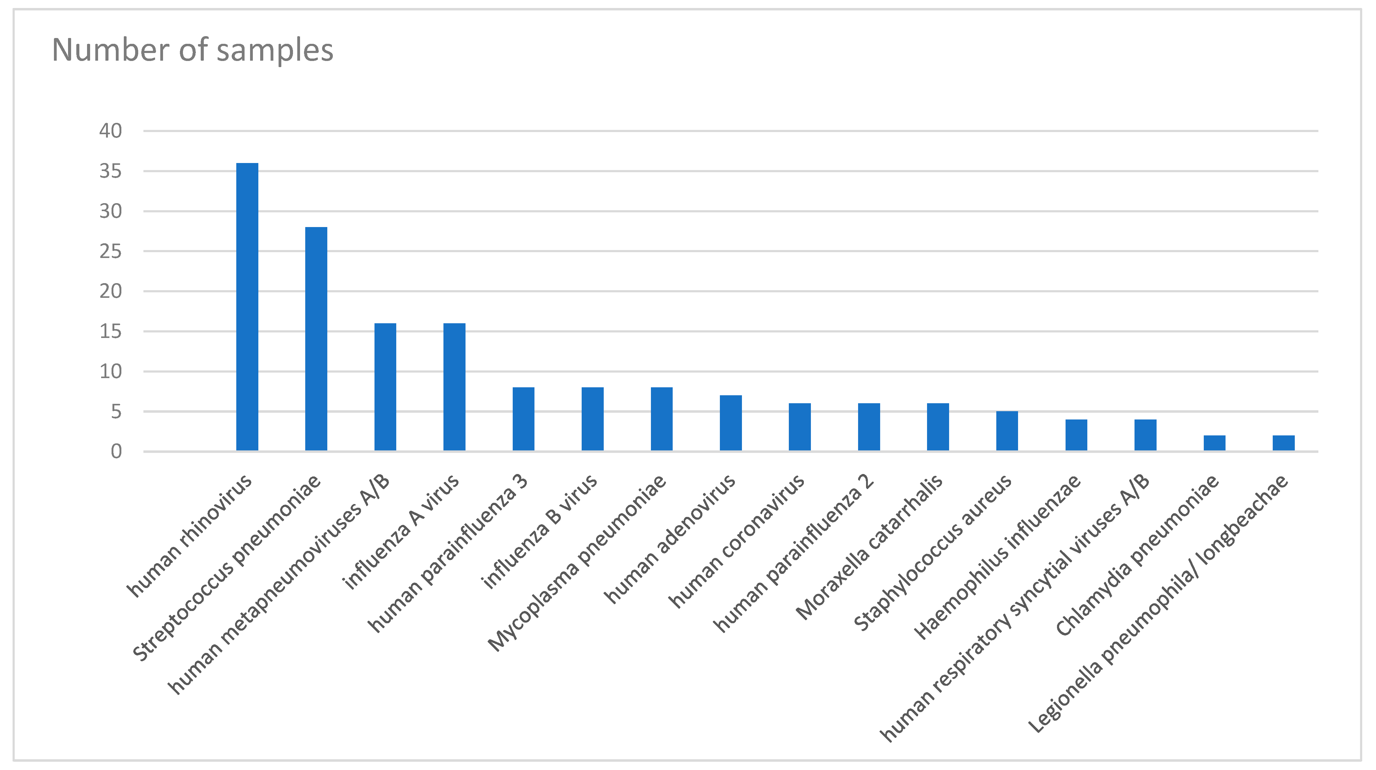
Figure 1. Frequency of isolated pathogens from patients recruited in this study.
Tropicalmed 07 00233 g001
Figure 2. Distribution of viral and bacterial infections in different age groups.
Tropicalmed 07 00233 g002
Figure 3. Number of pathogens detected in different age groups.
Tropicalmed 07 00233 g003
Figure 4. Prevalence of causative pathogens of upper respiratory tract infections and lower respiratory tract infections in paediatric, adult, and older patients.
Tropicalmed 07 00233 g004aTropicalmed 07 00233 g004b
Table 1. Number of detections for each pathogen.
PathogenTotal NumberNumber of Detections
1 detection2 detections3 detections4 detections
human rhinovirus3622383
Streptococcus pneumoniae2861642
human metapneumoviruses A/B165551
influenza A virus166730
human parainfluenza 381241
influenza B virus83410
Mycoplasma pneumoniae82330
human adenovirus70430
human coronavirus60321
human parainfluenza 260141
Moraxella catarrhalis60321
Staphylococcus aureus50311
Haemophilus influenzae40220
human respiratory syncytial viruses A/B40211
Chlamydia pneumoniae20110
Legionella pneumophila/longbeachae20200
human parainfluenza 410100
Table 2. Prevalence of causative pathogens of upper and lower respiratory tract infections in paediatric, adult, and older patients in the USA, Canada, Europe, France, Malawi, Turkey, and Jordan.
Aetiology of Respiratory Tract Infections
SourceCountrySampling TechniqueUpper Respiratory Tract InfectionsLower Respiratory Tract Infections
ElderlyADULTSPediatricAdults
Viral AetiologyBacterial AetiologyViral ArtiologyBacterial AetiologyBacterial AetiologyViral Aetiology
[32]USAnot mentioned Presumed virus or chlamydia 30–40% (not tested) Streptooccus pneumonia
Group A Streptococci 5–10%rhinovirus 25–30% Enteric gram-negative organisms
Mycoplasma 5–10%coronavirus > 10% Staphylococcus aureus
Influenza Virus, RSV, Adenovirus and Parainfluenza Virus 10–15% Hemophilus influenza
Other viruses Pseudomonas aeruginosa
[36]CanadaBlood culture Unknown 51.6%
sputum culture Mycoplasma pneumoniae 15%
acute and convalescent serum samples for serology Chlamydia pneumoniae 12%
Antibodies to Mycoplasma pneumoniae and Chlamydia pneumoniae determined using enzyme-linked immunosorbent assays Streptococcus pneumoniae 5.9%
Haemophilus influenzae 4.9%
Chlamydia pneumoniae and Mycoplasma pneumoniae 2.1%
Haemophilus parainfluenzae 1.9%
Staphylococcus aureus 1.1%
Moraxella catarrhalis 1.1%
Streptococcus species 0.9%
Other 2.8%
[37]Europe (UK, Spain and Sweden)not mentioned No pathogen identified 49.8%viruses 11.7%
Streptococcus pneumoniae 19.3%
Mycoplasma pneumoniae 11.1%
Chlamydia pneumoniae 8%
Haemophilus influenzae 3.3%
[31]FranceQiaAmp MinElute virus spin kitsInfluenza A (H3N2)
real-time Reverse Transcription quantitative PCR (RT-qPCR)Human rhinovirus 16%
Human coronavirus OC43 7%
Respiratory Syncytial Virus 5%
Human metapneumovirus 5%
Influenza B/Victoria 5%
[34]Malawiblood culture No pathogen detected 39.4%Influenza viruses 8.8%
Streptococcus pneumoniae urinary antigen detection Mycobacterium tuberculosis 23%Adenovirus 7.7%
sputum mycobacterial culture Streptococcus pneumoniae 21.4%Coronaviruses 6.8%
Xpert MTB/RIF Nontuberculous mycobacteria 2.9%Parainfluenza viruses 3.7%
nasopharyngeal aspirate multiplex PCR Salmonella enterica serovar Typhi 2.2%Rhinovirus 4.2%
Nontyphoidal Salmonella 1.6%Bocavirus 2.9%
Mycoplasma pneumoniae 1.3%Metapneumovirus 2.0%
Other gram-negative enteric bacilli 0.7%RSV 1.8%
Staphylococcus aureus 0.4%Enterovirus 1.1%
Chlamydia pneumoniae 0.4%Parechovirus 1.1%
[35]Turkeysputum cultures Haemophilus influenzae 44.9%
Streptococcus pneumoniae 25.5%
Moraxella catarrhalis 12.2%
Pseudomonas aeruginosa 3.1%
Klebsiella spp. 1%
Haemophilus parainfluenzae 1%
Staphylococcus aureus 1%
[33]Jordansputum cultures Streptococcus pneumoniae 26% Chlamydia pneumoniae 14%
Chlamydia pneumoniae 23% Mycoplasma pneumoniae 6%
Haemophilus influenzae 17% Streptococcus pneumoniae 3%
Mycoplasma pneumoniae 9% Haemophilus influenzae 3%
Legionella pneumophila 6% Pseudomonas aeruginosa 3%
Klebsiella pneumoniae 6%
Publisher’s Note: MDPI stays neutral with regard to jurisdictional claims in published maps and institutional affiliations.

