Article

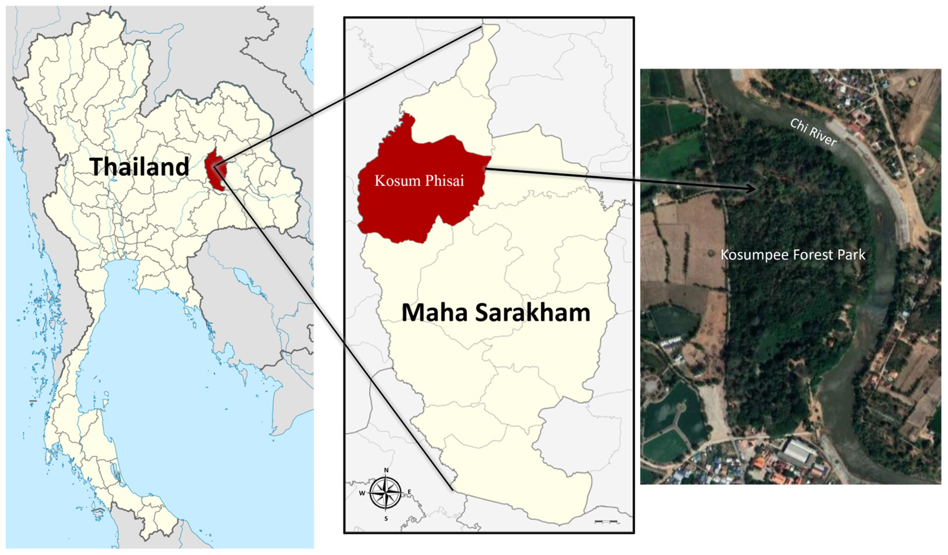
Molecular Characterization of Staphylococcus aureus Complex Isolated from Free-Ranging Long-Tailed Macaques at Kosumpee Forest Park, Maha Sarakham, Thailand

1
One Health Research Unit, Mahasarakham University, Maha Sarakham 44000, Thailand
2
Veterinary Infectious Disease Research Unit, Mahasarakham University, Maha Sarakham 44000, Thailand
3
Faculty of Veterinary Sciences, Mahasarakham University, Maha Sarakham 44000, Thailand
4
Faculty of Environment and Resource Studies, Mahasarakham University, Maha Sarakham 44150, Thailand
5
Department of Psychology, Center for Global Field Study and Washington National Primate Research Center, University of Washington, Seattle, WA 98195, USA
6
Department of National Parks, Wildlife and Plant Conservation, Bangkok 10900, Thailand
7
Departments of Psychology, Global Health, Anthropology and Center for Global Field Study, Washington National Primate Research Center, University of Washington, Seattle, WA 98195, USA
*
Author to whom correspondence should be addressed.
Trop. Med. Infect. Dis. 2023, 8(7), 374; https://doi.org/10.3390/tropicalmed8070374
Submission received: 21 June 2023 / Revised: 16 July 2023 / Accepted: 18 July 2023 / Published: 20 July 2023
(This article belongs to the Special Issue Feature Papers in One Health)

Abstract

:
The Staphylococcus (S.) aureus complex, including methicillin-resistant S. aureus (MRSA) and methicillin-susceptible S. aureus (MSSA), and S. argenteus are bacterial pathogens that are responsible for both human and animal infection. However, insights into the molecular characteristics of MRSA, MSSA, and S. argenteus carriages in wildlife, especially in long-tailed macaques, rarely have been reported in Thailand. The objective of this study was to assess molecular characterization of MRSA, MSSA, and S. argenteus strains isolated from free-ranging long-tailed macaques (Macaca fascicularis) at Kosumpee Forest Park, Maha Sarakham, Thailand. A total of 21 secondary bacterial isolates (including 14 MRSA, 5 MSSA, and 2 S. argenteus) obtained from the buccal mucosa of 17 macaques were analysed by a Polymerase chain reaction (PCR) to identify several virulence genes, including pvl, tst, hla, hlb clfA, spa (x-region), spa (IgG biding region), and coa. The most prevalent virulence genes were clfA, coa, and the spa IgG biding region which presented in all isolates. These data indicated that MRSA, MSSA, and S. argenteus isolates from the wild macaques at Kosumpee Forest Park possess a unique molecular profile, harbouring high numbers of virulence genes. These findings suggest that wild macaques may potentially serve as carriers for distribution of virulent staphylococcal bacteria in the study area.

1. Introduction

The Staphylococcus aureus complex is an opportunistic bacterial pathogen capable of colonizing a diverse range of animal species, including humans, causing severe infections [1]. It poses a zoonotic risk, as it can spread among environments, humans, and various animals, such as pets, livestock, and wildlife [2]. Methicillin-resistant Staphylococcus aureus (MRSA) is a drug-resistant strain of S. aureus, exhibiting resistance to methicillin and other antibiotics through the expression of a penicillin-binding protein that confers resistance to β-lactam antibiotics [3]. MRSA has emerged as a dangerous antimicrobial-resistant pathogen capable of colonizing humans, domestic animals, livestock, and wildlife, causing acute and chronic infections in both animal and human hosts [4]. It differs from methicillin-susceptible Staphylococcus aureus (MSSA) due to its resistance to β-lactam antibiotics, which is mediated by the mecA gene carried on the Staphylococcal Cassette Chromosome mec (SCCmec). This genetic element encodes an alternative penicillin-binding protein, namely, PBP-2a, which alters the target site for β-lactam antibiotics. The majority of clinical MRSA isolates (90%) carry the mecA gene on their SCCmec [5,6]. The main mechanism of drug resistance involves a significantly decreased affinity of β-lactam antibiotics for this altered substrate [7]. MRSA is classified into three groups: hospital/healthcare-associated MRSA (HA-MRSA), community-associated MRSA (CA-MRSA), and livestock-associated MRSA (LA-MRSA). HA-MRSA is associated with nosocomial and surgical wound infections, leading to treatment failure and increased morbidity and mortality. CA-MRSA typically causes various infections in humans, including sepsis, pneumonia, skin infections, and soft tissue infections. LA-MRSA is specifically associated with animal infections. MRSA has been shown to spread epidemiologically in both human communities and animal populations, including companion animals, livestock, and wildlife [8].
A number of studies have reported that S. aureus isolated from non-human primates had close similarity with human isolates, leading to the concern of amphixenoses or zoonotic diseases that can be transmitted from humans to other species (e.g., non-human primates) and vice versa. [2,9,10]. Among non-human primates, an increasing number of studies have reported the presence of MRSA, including in captive gorillas in the USA [11], chimpanzees in Africa [10,11], wild rhesus macaques in Nepal [12], and, most recently, in wild long-tailed macaques in Thailand [2]. Current research indicates that MRSA can be transmitted from animals to humans and vice versa [10,13].
The S. aureus complex possesses numerous virulence genes and produces a diverse range of virulence factors that contribute to its pathogenicity [14,15]. Key virulence factors include coagulase, protein A, hemolysin, clumping factors, toxic shock syndrome toxin-1, and Panton–Valentine leucocidin [14,15,16]. Coagulase promotes clotting by interacting with soluble fibrinogen, leading to abscess formation and bacterial persistence in host tissues [17]. Protein A and the IgG-binding protein facilitate immunoglobulin precipitation by binding the Fc region, masking surface antigens that inhibit opsonization and phagocytic killing by immune cells [18,19]. Hemolysins lyse red blood cells and host cells to acquire nutrients [20]. Clumping factors enable attachments to host epithelial cells [21]. Panton–Valentine leukocidin selectively kills phagocytic cells, resulting in tissue necrosis [22].
At present, there are very few studies examining the molecular characteristics of the S. aureus complex (both MRSA and MSSA) and S. argenteus in free-ranging wildlife (including macaques), especially in Thailand. Understanding of the virulence genes’ carriage is essential for accessing the severity of the pathogens in wildlife and developing appropriate strategies to mitigate and prevent this infectious disease transmission. The purpose of the present study was to identify the molecular characteristics of MRSA, MSSA, and S. argenteus by identifying and investigating the prevalence of virulence genes, including pvl (Panton–Valentin leukocydin), tst (toxic shock syndrome toxin-1), hla (alpha hemolysin), hlb (beta hemolysin), clfA (clumping factor A), spa (x-region of staphylococcal protein A), spa (IgG biding region of staphylococcal protein A), and coa (coagulase) in MRSA, MSSA, and S. argenteus isolated from buccal swab of free-ranging long-tailed macaques at Kosumpee Forest Park, Maha Sarakham, Thailand.

2. Materials and Methods

2.1. Sample Collection and Preparation

Bacterial isolates from the buccal cavities of wild long-tailed macaques were collected as part of our previous work focusing on the human–primate conflict and coexistence at Kosumpee Forest Park (KFP) [2,23]. Sampling procedures, the study site, and the KFP macaque population have been detailed in our previous studies [2,23,24,25]. Buccal swab samples were collected between 10–11 November 2018 from 30 randomly selected wild long-tailed macaques living around KFP (16°15′12.6″ N 103°04′02.0″ E) in Maha Sarakham province, Northeastern Thailand (Figure 1). The macaques were captured using a nylon mesh trapping cage, measuring 1.50 × 0.40 × 0.40 m. Tiletamine–zolazepam (Zoletil® 100 mg/mL, Virbac, Carros, France) was delivered intramuscularly (via blowpipe) to anesthetize the macaques in the cage. Once sedated, the macaques were retrieved from trapping cage for sampling. Their vital signs were monitored through the sampling protocol until recovery and subsequent release back to their groups. Buccal samples were collected following aseptic techniques involving swabbing of the macaques’ buccal cavity using sterile collection swabs composed with transport media (Deltalab, Bacelona, Spain). All trapping and sampling procedures were supervised and conducted by wildlife veterinary specialists from Mahasarakham University (MSU) according to the protocol and approval by the Institutional Animal Care and Use Committee at Mahasarakham University and the Thai Department of National Parks (see ethics statement at Institutional Review Board Statement part). Upon collection, all buccal swab samples were kept at 4 °C and transferred to the One Health Research Unit (OHRU), Veterinary Public Health laboratory of the Faculty of Veterinary Sciences at Mahasarakham University, for bacteriological analysis.
Thirty buccal swab samples were prepared for bacterial culture using Baird–Parker Agar supplemented with egg yolk and potassium tellurite (Oxoid, Hampshire, UK), a selective media for analysing S. aureus. Cultures were incubated at 37 °C for 24 h. Suspected bacterial colonies were identified for S. aureus using conventional methods such as Gram staining, the catalase enzyme test, mannitol salt agar selectivity (MSA), deoxyribonuclease (DNase) test, tube coagulase test (TCT), and confirmed by a rapid agglutination test, which was accomplished using the Staphaurex™ Plus latex agglutination test (Thermo Fisher Scientific, Waltham, MA, USA). S. aureus ATTC 25923 was used as a positive control strain for conventional methods of bacterial identification.
Genomic DNA of all bacterial isolates was extracted using a DNA extraction kit (Geneaid, Taiwan) following the protocol for Gram-positive bacteria. As described in our previous work [2], methicillin-resistant S. aureus (MRSA) and S. argenteus isolates were determined by PCR amplification method for the presence of the mecA gene to confirm MRSA and the NRPS gene to differentiate S. argenteus.

2.2. Detection of Staphylococcal Virulence Genes

Genomic DNA of all bacterial isolates was quantified and measured via the OD 260/280 nm ratio with the NanoDrop 1000 Spectrophotometer (Thermo Scientific, Branchburg, NJ, USA) and stored in microtubes at −20 °C until molecular identification. All Staphylococcus spp. bacterial DNA were amplified for eight virulence genes: tst, coa, spa (x-region), spa (IgG-binding region), hla, hlb, clfA, and pvl. Amplification was achieved with specific oligonucleotide primers, as shown in Table 1.
All PCR reaction mixtures contained a total volume of 25 μL and comprised 10 mM of each forward and reverse primer, 0.2 mM dNTPs, 2 mM MgCl2, 1 U of Taq DNA polymerase, 1× Taq reaction buffer, and 100 ng DNA template. PCR was performed using a thermocycler (Biometra GmbH, Jena, Germany), and the program began with an initial denaturation at 95 °C for 10 min. This was followed by 35 cycles of 95 °C for 30 s, 55 °C for 30 s and 72 °C for 30 s and a final extension at 72 °C for 10 min. PCR amplicons were subjected to 1.5% agarose gel electrophoresis and staining with ViSafe Red Gel Stain (Vivantis, Malaysia). The targeted PCR products were observed and visualized for their DNA bands under ultraviolet light by a Gel Doc XR+ Documentation System (Bio-Rad, Hercules, CA, USA). The control bacterial template for the PCR included the strains ATCC 19095 (tst) and ATCC13565 (coa, clfA, hla, hlb, spa x-region, IgG-binding region). For pvl, the PCR product was selected for nucleotide sequencing, and we analysed their sequence bases to confirm (Accession number: AB084255.1).

2.3. Statistical Analysis

Descriptive statistics were used to define the prevalence of virulence genes of MRSA, MSSA, and S. argenteus. The Chi-square test and Fisher’s exact test of independence were used to analyse the difference between virulence genes detected for each bacterial group in the SPSS statistics program (version 22). Values were considered statistically significance with a probability value (p-value) ≤ 0.05.

3. Results

A total of 21 bacterial isolates (14 MRSA, 5 MSSA, and 2 S. argenteus) from 17 macaques were analysed by a Polymerase chain reaction (PCR) to test for the presence of virulence genes, including pvl, tst, hla, hlb clfA, spa (x-region), spa (IgG biding region), and coa, as shown in Figure 2. The most prevalent staphylococcal virulence genes for all bacterial groups were clfA, coa, and spa (IgG biding region), which were present in all bacterial isolates (100%; 21/21), followed by spa (x-region) (95.24%; 20/21).
Staphylococcal virulence genes such as tst and pvl were highly prevalent at 57.14% (12/21) and 61.9% (13/21), respectively. For the hemolysin gene, 15 (85.71%) of the isolates carried hla, which was the most prevalent hemolysin gene in this study. Additionally, 12 (71.43%) of the isolates carried hlb. Interestingly, all bacterial isolates carried at least one type of haemolysis gene, and 11 isolates (52.38%) carried both hla and hlb genes, as shown in Table 2. In addition, a comparative analysis of the prevalence of virulence genes among MRSA, MSSA, and S. argenteus using a Chi-square test and Fisher’s exact test revealed no significant differences in the likelihoods of those bacterial pathogens harbouring each virulence gene (p-value > 0.05).

4. Discussion

The prevalence of the major virulence genes of MRSA, MSSA, and S. argenteus in buccal swabs of long-tailed macaques was examined in this study. The results indicated the presence of eight virulence genes that were major virulence indicators linked to the staphylococcal pathogenicity of the bacterial samples obtained from the long-tailed macaques. These findings also were similar to previous studies that isolated bacteria from animal origins [15,32]. Several genes in this study (hla, hlb, tst, and pvl) also have been detected in aggressive isolates of S. aureus [33,34]. These virulence genes spread through the isolates at a high frequency (57.14–100%) after amplification by PCR. As such, it can be inferred that the MRSA, MSSA, and S. argenteus isolated from the long-tailed macaques in this study may have had high pathogenicity. Furthermore, the majority of the isolates in our study demonstrated a variety of virulence gene combinations, demonstrating that these bacterial samples had considerable genetic diversity.
It is well known that S. aureus produces TSST-1 that has been reported to be associated with toxic shock syndrome [35] and produces systemic infections [36]. Surveillance for the prevalence of tst, which encodes the pyrogenic toxin superantigen TSST-1 in S. aureus isolates from the oral carriage of the wild macaques, is very important from a One Health standpoint since it could provide information regarding the status of staphylococcal virulence species circulating in potential wildlife reservoirs in the study area. The tst gene is located on the pathogenicity islands (PAIs) of staphylococcal bacteria, which assists the immunopathogenesis of S. aureus/S. argenteus via the anti-inflammatory chemokines of the host and also facilitates the induction of immunosuppression [37]. Consistent with the results of other studies, this gene has been reported to be detected in S. aureus (both MRSA and MSSA) isolates from wildlife reservoirs, including wild avians, red deer, wild rodents, boars, and also non-human primates [36]. Our results indicated that S. aureus carrying the toxic shock syndrome toxin gene is widely distributed throughout the macaque population at the forest park. These bacteria possess the ability to produce toxic proteins that may be significant in the pathogenicity of infectious disease for animals and can be harmful to humans who become infected from the animal reservoirs.
In this study, we found a moderate to high prevalence of the pvl gene, which encodes for Panton–Valentine leucocidin in MRSA, MSSA, and S. argenteus. It has not been reported in wildlife reservoirs in Thailand to date. Nonetheless, MRSA and MSSA isolates carrying the pvl gene have been reported in wild rodents and non-human primates from Germany, Portugal, Czech, and France [36]. Furthermore, some studies have reported that most of the S. aureus isolates carrying the pvl gene derived from wildlife carriages were frequently detected from non-human primates [36,38]. These reports indicate that S. aureus harbouring the pvl gene derived from wild monkeys may play a role as natural maintenance hosts for pathogenic S. aureus (both MRSA and MSSA), which can affect human health. Panton–Valentine leucocidin is a significant pore-forming cytotoxin that is regularly associated with abscesses in hosts and plays a key role in the induction of leukocyte destruction and severe necrotic lesions of soft tissues and skin [39]. This cytotoxin has a high potential virulence and could be responsible for clinical manifestations, as reported in a previous study [40].
Several addition genes encoding general virulence factors of S. aureus (MRSA and MSSA) and S. argenteus were identified, including coa, hla, hlb, clfA, spa (x region), and spa (IgG-binding region). All bacterial isolates in this study harboured coa, clfA, and spa (IgG-binding region). The spa is known to be the fundamental virulence gene for S. aureus, regarding disease development and severity by encoding staphylococcal protein A [15]. Protein A has an ability as a bacterial trait to precipitate immunoglobulins, mask underlying surface antigens, and inhibit opsonization and phagocytic killing of staphylococci by PMN cells [41]. For the Coagulase protein encoded from coa genes, it is secreted from S. aureus to promote coagulation, which leads to the formation of abscesses and bacterial persistence in host tissues [17]. Expression of the clfA gene that encodes clumping factors A of S. aureus is thought to enhance bacterial growth and promote infection in the face of host defence mechanisms, such as phagocytosis [42]. Furthermore, both alpha and beta hemolysins encoded from hla and hlb genes, respectively, can damage platelets and destroy lysosomes, in turn, causing necrosis and ischemia in the host [20].
Our study of the molecular characterization of S. aureus complex (both MRSA and MSSA) and S. argenteus indicate that these bacteria have the potential to be aggressive pathogens among wild long-tailed macaques in Kosumpee Forest Park, Thailand. This indicates a need for vigilance regarding wildlife reservoirs of these bacterial zoonoses. Given their pathogenic potential to cause severe disease in both animals and humans, a proactive approach to veterinary public health risk management is essential to protect the people and animals who may come into contact with the infected animals in this study area.

5. Conclusions

The wild long-tailed macaques at Kosumpee Forest Park may serve as a potential reservoir of S. aureus (MRSA and MSSA) and S. argenteus. Based on the molecular characterization of these Staphylococci bacteria in this study, the macaques were found to carry antimicrobial resistance pathogens and virulence strains that could potentially affect other animals and humans. Given the genetic diversity of MRSA, MSSA, and S. argenteus in wild monkey reservoirs and the close interactions between the people and macaques at Kosumpee Forest Park, regular monitoring of those pathogens is needed for effective risk prevention and intervention.

Author Contributions

Conceptualization, N.P.; project administration, R.C.K., A.K. and T.T.; sample collection, N.P., P.T., T.T. and A.K.; methodology, N.P., P.T. and T.T.; data analysis, N.P. and R.C.K.; Drafted and revised the manuscript, N.P., P.K. and R.C.K. All authors have read and agreed to the published version of the manuscript.

Funding

This research project was financially supported by Mahasarakham University. R.C.K. and P.K.’s effort was supported in part by the National Institutes of Health (NIH) Office of Research Infra-structure Programs (ORIP) under award number P51OD010425 to the Washington National Primate Research Center, USA.

Institutional Review Board Statement

All research procedures involving animal use were approved by the Institutional Animal Care and Use Committee of Mahasarakham University (IACUC Protocol No. IACUC-MSU-0009/2016). All animal sample collection was complied with the applicable laws of Thailand.

Informed Consent Statement

Not applicable.

Data Availability Statement

Not applicable.

Acknowledgments

The authors would like to thank all the staff at Kosumpee Forest Park, Pitchakorn Petcharat, Ratchanon Kusolsongkhrokul, Papichchaya Doemlim, Panitporn Damrongsukij, Thanyaphorn Chamnandee, Kittisak Saengthong, Suvit Pathomthanasarn from the One Health Research Unit VET MSU, and students from the Faculty of Environment and Resource Studies at MSU for their assistance with the sampling protocol. We are grateful to the Faculty of Veterinary Sciences at MSU for use of the diagnostic laboratory. Finally, we are grateful to the Thailand Department of National Parks—Wildlife and Plant Conservation (DNP) and the National Research Council of Thailand for their approval (NRCT project approval to RCK—Project ID: 2016/048; “Healthy Coexistence between Human and Non-human Primates: A One Health Approach”).

Conflicts of Interest

The authors declare no conflict of interest.

References

  1. Howden, B.P.; Giulieri, S.G.; Wong Fok Lung, T.; Baines, S.L.; Sharkey, L.K.; Lee, J.Y.H.; Hachani, A.; Monk, I.R.; Stinear, T.P. Staphylococcus aureus host interactions and adaptation. Nat. Rev. Microbiol. 2023, 21, 380–395. [Google Scholar] [CrossRef] [PubMed]
  2. Pumipuntu, N.; Chamnandee, T.; Saengthong, K.; Pathomthanasarn, S.; Tanee, T.; Kyes, P.; Thamsenanupap, P.; Karaket, A.; Roberts, M.C.; Kyes, R.C. Investigation of methicillin-resistant Staphylococcus aureus, methicillin-susceptible Staphylococcus aureus, and Staphylococcus argenteus from wild long-tailed macaques (Macaca fascicularis) at Kosumpee Forest Park, Maha Sarakham, Thailand. Vet. World. 2022, 15, 2693–2698. [Google Scholar] [CrossRef] [PubMed]
  3. Cihalova, K.; Chudobova, D.; Michalek, P.; Moulick, A.; Guran, R.; Kopel, P.; Adam, V.; Kizek, R. Staphylococcus aureus and MRSA Growth and Biofilm Formation after Treatment with Antibiotics and SeNPs. Int. J. Mol. Sci. 2015, 16, 24656–24672. [Google Scholar] [CrossRef] [PubMed] [Green Version]
  4. Heaton, C.J.; Gerbig, G.R.; Sensius, L.D.; Patel, V.; Smith, T.C. Staphylococcus aureus epidemiology in wildlife: A systematic review. Antibiotics 2020, 9, 89. [Google Scholar] [CrossRef] [Green Version]
  5. Wielders, C.L.; Fluit, A.C.; Brisse, S.; Verhoef, J.; Schmitz, F.J. mecA gene is widely disseminated in Staphylococcus aureus population. J. Clin. Microbiol. 2002, 40, 3970–3975. [Google Scholar] [CrossRef] [Green Version]
  6. Shukla, S.K.; Ramaswamy, S.V.; Conradt, J.; Stemper, M.E.; Reich, R.; Reed, K.D.; Graviss, E.A. Novel polymorphisms in mec genes and a new mec complex type in methicillin-resistant Staphylococcus aureus isolates obtained in rural Wisconsin. Antimicrob. Agents Chemother. 2004, 48, 3080–3085. [Google Scholar] [CrossRef] [Green Version]
  7. Rahman, M.M.; Amin, K.B.; Rahman, S.M.M.; Khair, A.; Rahman, M.; Hossain, A.; Rahman, A.K.M.A.; Parvez, M.S.; Miura, N.; Alam, M.M. Investigation of methicillin-resistant Staphylococcus aureus among clinical isolates from humans and animals by culture methods and multiplex PCR. BMC Vet. Res. 2018, 14, 300. [Google Scholar] [CrossRef] [PubMed] [Green Version]
  8. Turner, N.A.; Sharma-Kuinkel, B.K.; Maskarinec, S.A.; Eichenberger, E.M.; Shah, P.P.; Carugati, M.; Holland, T.L.; Fowler, V.G., Jr. Methicillin-resistant Staphylococcus aureus: An overview of basic and clinical research. Nat. Rev. Microbiol. 2019, 17, 203–218. [Google Scholar] [CrossRef]
  9. Schaumburg, F.; Alabi, A.S.; Köck, R.; Mellmann, A.; Kremsner, P.G.; Boesch, C.; Becker, K.; Leendertz, F.H.; Peters, G. Highly divergent Staphylococcus aureus isolates from African non-human primates. Environ. Microbiol. Rep. 2012, 4, 141–146. [Google Scholar] [CrossRef]
  10. Roberts, M.C.; Joshi, P.R.; Monecke, S.; Ehricht, R.; Müller, E.; Gawlik, D.; Diezel, C.; Braun, S.D.; Paudel, S.; Acharya, M.; et al. Staphylococcus aureus and methicillin resistant S. aureus in Nepalese primates: Resistance to antimicrobials, virulence, and genetic lineages. Antibiotics 2020, 9, 689. [Google Scholar] [CrossRef]
  11. Nagel, M.; Dischinger, J.; Türck, M.; Verrier, D.; Oedenkoven, M.; Ngoubangoye, B.; Le Flohic, G.; Drexler, J.F.; Bierbaum, G.; Gonzalez, J.P. Human-associated Staphylococcus aureus strains within great ape populations in Central Africa (Gabon). Clin. Microbiol. Infect. 2013, 19, 1072–1077. [Google Scholar] [CrossRef] [Green Version]
  12. Roberts, M.C.; Joshi, P.R.; Monecke, S.; Ehricht, R.; Müller, E.; Gawlik, D.; Paudel, S.; Acharya, M.; Bhattarai, S.; Pokharel, S.; et al. MRSA strains in Nepalese rhesus macaques (Macaca mulatta) and their environment. Front. Microbiol. 2019, 10, 2505. [Google Scholar] [CrossRef] [PubMed] [Green Version]
  13. Schaumburg, F.; Alabi, A.S.; Peters, G.; Becker, K. New epidemiology of Staphylococcus aureus infection in Africa. Clin. Microbiol. Infect. 2014, 20, 589–596. [Google Scholar] [CrossRef] [Green Version]
  14. Indrawattana, N.; Sungkhachat, O.; Sookrung, N.; Chongsa-nguan, M.; Tungtrongchitr, A.; Voravuthikunchai, S.; Kong-ngoen, T.; Kurazono, H.; Chaicumpa, W. Staphylococcus aureus clinical isolates: Antibiotic susceptibility, molecular characteristics, and ability to form biofilm. Biomed. Res. Int. 2013, 2013, 314654. [Google Scholar] [CrossRef] [PubMed] [Green Version]
  15. Pumipuntu, N.; Tunyong, W.; Chantratita, N.; Diraphat, P.; Pumirat, P.; Sookrung, N.; Chaicumpa, W.; Indrawattana, N. Staphylococcus spp. associated with subclinical bovine mastitis in central and northeast provinces of Thailand. PeerJ 2019, 7, e6587. [Google Scholar] [CrossRef] [PubMed] [Green Version]
  16. Xu, J.; Tan, X.; Zhang, X.; Xia, X.; Sun, H. The diversities of staphylococcal species, virulence, and antibiotic resistance genes in the subclinical mastitis milk from a single Chinese cow herd. Microb. Pathog. 2015, 88, 29–38. [Google Scholar] [CrossRef]
  17. Cheng, A.G.; McAdow, M.; Kim, H.K.; Bae, T.; Missiakas, D.M.; Schneewind, O. Contribution of coagulases towards Staphylococcus aureus disease and protective immunity. PLoS Pathog. 2010, 6, 1001036. [Google Scholar] [CrossRef] [Green Version]
  18. Koreen, L.S.; Ramaswamy, V.; Graviss, E.A.; Naidich, S.; Musser, J.M.; Kreiswirth, B.N. spa typing method for discriminating among Staphylococcus aureus isolates: Implications for use of a single marker to detect genetic micro- and macrovariation. J. Clin. Microbiol. 2004, 42, 792–799. [Google Scholar] [CrossRef] [Green Version]
  19. Viau, M.; Longo, N.S.; Lipsky, P.E.; Zouali, M. Staphylococcal protein deletes B-1a and marginal zone B lymphocytes expressing human immunoglobulins: An immune evasion mechanism. J. Immunol. 2005, 175, 7719–7727. [Google Scholar] [CrossRef] [PubMed]
  20. Inoshima, I.; Inoshima, N.; Wilke, G.A.; Powers, M.E.; Frank, K.M.; Wang, Y.; Bubeck Wardenburg, J.A. A Staphylococcus aureus pore-forming toxin subverts the activity of ADAM10 to cause lethal infection in mice. Nat. Med. 2011, 17, 1310–1314. [Google Scholar] [CrossRef] [PubMed] [Green Version]
  21. El-Sayed, A.; Alber, J.; Limmler, C.; Bonner, B.; Huhn, A.; Kaleta, E.F.; Zschech, M. PCR-based detection of genes encoding virulence determinants in Staphylococcus aureus from birds. J. Vet. Med. B. Infect. Dis. Vet. Public Health 2005, 52, 38–44. [Google Scholar] [CrossRef] [PubMed]
  22. Lina, G.; Piemont, Y.; Godail, G.F.; Bes, M.; Peter, M.O.; Gauduchon, V.; Vandenesch, F.; Etienne, J. Involvement of panton-valentine leucocidin producing Staphylococcus aureus in primary skin infections and pneumonia. Clin. Infect. Dis. 1999, 29, 1128–1132. [Google Scholar] [CrossRef] [PubMed] [Green Version]
  23. Kyes, R.C.; Tanee, T.; Thamsenanupap, P.; Karaket, A.; Iskandar, E.; Kyes, P. Population status of the long-tailed macaques (Macaca fascicularis) at Kosumpee Forest Park, Maha Sarakham, Thailand. Am. J. Primatol. 2018, 80, 22. [Google Scholar]
  24. Schurer, J.M.; Ramirez, V.; Kyes, P.; Tanee, T.; Patarapadungkit, N.; Thamsenanupap, P.; Trufan, S.; Grant, E.T.; Garland-Lewis, G.; Kelley, S.; et al. Long-tailed macaques (Macaca fascicularis) in urban landscapes: Investigating gastrointestinal parasitism and barriers for healthy co-existence in northeast Thailand. Am. J. Trop. Med. Hyg. 2019, 100, 357–364. [Google Scholar] [CrossRef] [PubMed] [Green Version]
  25. Pumipuntu, N.; Tanee, T.; Kyes, P.; Thamsenanupap, P.; Karaket, A.; Kyes, R.C. Leptospira Seroprevalence in free-ranging long-tailed macaques (Macaca fascicularis) at Kosumpee Forest Park, Maha Sarakham, Thailand. Infect. Dis. Rep. 2023, 15, 16–23. [Google Scholar] [CrossRef]
  26. Wu, D.; Li, X.; Yang, Y.; Zheng, Y.; Wang, C.; Deng, L.; Liu, L.; Li, C.; Shang, Y.; Zhao, C.; et al. Superantigen gene profiles and presence of exfoliative toxin genes in community-acquired meticillin-resistant Staphylococcus aureus isolated from Chinese children. J. Med. Microbiol. 2011, 60, 35–45. [Google Scholar] [CrossRef] [Green Version]
  27. Aslantas, O.; Demir, C.; Turutoglu, H.; Cantekin, Z.; Ergun, Y.; Dogruer, G. Coagulase gene polymorphism of Staphylococcus aureus isolated form subclinical mastitis. Turk. J. Vet. Anim. Sci. 2007, 31, 253–257. [Google Scholar]
  28. Tristan, A.; Ying, L.; Bes, M.; Etienne, J.; Vandenesch, F.; Lina, G. Use of multiplex PCR to identify Staphylococcus aureus adhesins involved in human hematogenous infections. J. Clin. Microbiol. 2003, 41, 4465–4467. [Google Scholar] [CrossRef] [Green Version]
  29. Jarraud, S.; Mougel, C.; Thioulouse, J.; Lina, G.; Meugnier, H.; Forey, F.; Nesme, X.; Etienne, J.; Vandenesch, F. Relationships between Staphylococcus aureus genetic background, virulence factors, agr groups (alleles), and human disease. Infect. Immun. 2002, 70, 631–641. [Google Scholar] [CrossRef] [PubMed] [Green Version]
  30. Frénay, H.M.; Bunschoten, A.E.; Schouls, L.M.; Van Leeuwen, W.J.; Vandenbrouke-Grauls, C.M.; Verhoef, J.; Mooi, F.R. Molecular typing of methicillin-resistant Staphylococcus aureus on the basis of protein a gene polymorphism. Eur. J. Clin. Microbiol. Infect. Dis. 1996, 15, 60–64. [Google Scholar] [CrossRef]
  31. Seki, K.; Sakurada, J.; Seong, H.K.; Murai, M.; Tachi, H.; Ishii, H.; Masuda, S. Occurrence of coagulase serotype among Staphylococcus aureus strains isolated from healthy individuals-special reference to correlation with size of protein-A gene. Microbiol. Immunol. 1998, 42, 407–409. [Google Scholar] [PubMed]
  32. Indrawattana, N.; Pumipuntu, N.; Suriyakhun, N.; Jangsangthong, A.; Kulpeanprasit, S.; Chantratita, N.; Sookrung, N.; Chaicumpa, W.; Buranasinsup, S. Staphylococcus argenteus from rabbits in Thailand. MicrobiologyOpen 2019, 8, e00665. [Google Scholar] [CrossRef] [Green Version]
  33. Rasmi, A.H.; Ahmed, E.F.; Darwish, A.M.A.; Gad, G.F.M. Virulence genes distributed among Staphylococcus aureus causing wound infections and their correlation to antibiotic resistance. BMC Infect. Dis. 2022, 22, 652. [Google Scholar] [CrossRef]
  34. Ballah, F.M.; Islam, M.S.; Rana, M.L.; Ullah, M.A.; Ferdous, F.B.; Neloy, F.H.; Ievy, S.; Sobur, M.A.; Rahman, A.T.; Khatun, M.M.; et al. Virulence determinants and methicillin resistance in biofilm-forming Staphylococcus aureus from various food sources in Bangladesh. Antibiotics 2022, 11, 1666. [Google Scholar] [CrossRef]
  35. Akineden, Ö.; Annemüller, C.; Hassan, A.A.; Lämmler, C.; Wolter, W.; Zschöck, M. Toxin genes and other characteristics of Staphylococcus aureus isolates from milk of cows with mastitis. Clin. Diagn. Lab. Immunol. 2001, 8, 959–964. [Google Scholar] [CrossRef] [PubMed] [Green Version]
  36. Abdullahi, I.N.; Fernández-Fernández, R.; Juárez-Fernández, G.; Martínez-Álvarez, S.; Eguizábal, P.; Zarazaga, M.; Lozano, C.; Torres, C. Wild Animals Are Reservoirs and Sentinels of Staphylococcus aureus and MRSA Clones: A Problem with “One Health” Concern. Antibiotics 2021, 10, 1556. [Google Scholar] [CrossRef]
  37. Zheng, Y.; Qin, C.; Zhang, X.; Zhu, Y.; Li, A.; Wang, M.; Tang, Y.; Kreiswirth, B.N.; Chen, L.; Zhang, H.; et al. The tst gene associated Staphylococcus aureus pathogenicity island facilitates its pathogenesis by promoting the secretion of inflammatory cytokines and inducing immune suppression. Microb. Pathog. 2019, 138, 103797. [Google Scholar] [CrossRef] [PubMed]
  38. Schaumburg, F.; Pauly, M.; Anoh, E.; Mossoun, A.; Wiersma, L.; Schubert, G.; Flammen, A.; Alabi, A.S.; Muyembe-Tamfum, J.J.; Grobusch, M.P.; et al. Staphylococcus aureus complex from animals and humans in three remote African regions. Clin. Microbiol. Infect. 2015, 21, 345.e1–345.e8. [Google Scholar] [CrossRef] [PubMed] [Green Version]
  39. Rankin, S.; Roberts, S.; O’Shea, K.; Maloney, D.; Lorenzo, M.; Benson, C.E. Panton, valentine leukocidin (PVL) toxin positive MRSA strains isolated from companion animals. Vet. Microbiol. 2005, 108, 145–148. [Google Scholar] [CrossRef]
  40. Prashanth, K.; Rao, K.R.; Reddy, P.V.; Saranathan, R.; Makki, A.R. Genotypic characterization of Staphylococcus aureus obtained from humans and bovine mastitis samples in India. J. Glob. Infect. Dis. 2011, 3, 115–122. [Google Scholar] [CrossRef]
  41. Falugi, F.; Kim, H.K.; Missiakas, D.M.; Schneewind, O. Role of protein A in the evasion of host adaptive immune responses by Staphylococcus aureus. mBio 2013, 4, e00575-13. [Google Scholar] [CrossRef] [PubMed] [Green Version]
  42. Herman-Bausier, P.; Labate, C.; Towell, A.M.; Derclaye, S.; Geoghegan, J.A.; Dufrêne, Y.F. Staphylococcus aureus clumping factor A is a force-sensitive molecular switch that activates bacterial adhesion. Proc. Natl. Acad. Sci. USA 2018, 115, 5564–5569. [Google Scholar] [CrossRef] [PubMed] [Green Version]
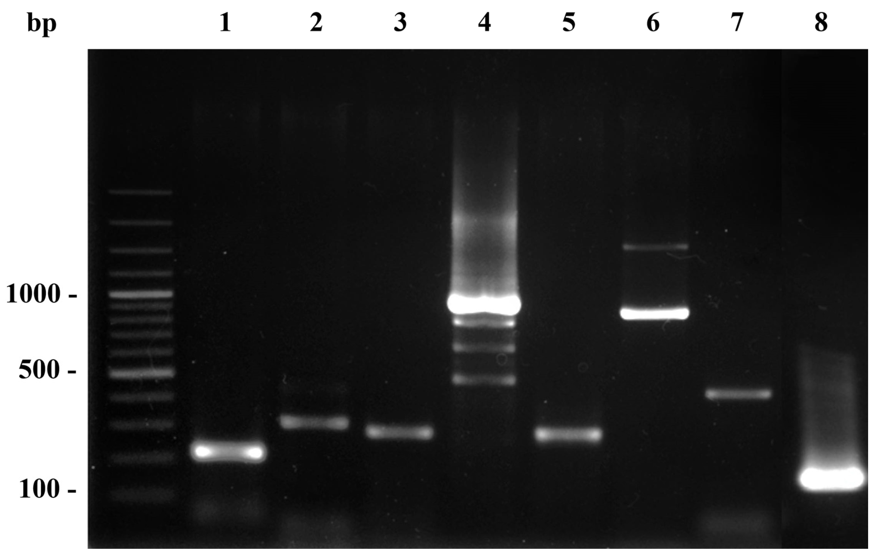
Figure 1. Location of the study area at Kosumpee Forest Park, located in Kosum Phisai district, Maha Sarakham province, in Northeast Thailand.
Tropicalmed 08 00374 g001
Figure 2. PCR amplicons of virulence genes of staphylococcal bacteria. Lane M; DNA molecular size ladder; lane 1–8, PCR amplicons of hla (209 bp), hlb (309 bp), clfA (229 bp), coa (pleomorphism 410, 740, 910, 970 bp), spa x-region (320 bp), spa IgG-biding region (920 bp), pvl (433 bp), and tst gene (180 bp) respectively.
Tropicalmed 08 00374 g002
Table 1. Specific oligonucleotide primers for amplification of Staphylococcus spp. virulence genes.
Target GeneSequence (5′–3′)Amplicon Size (bp)Ref.
tstF: TTCACTATTTGTAAAAGTGTCAGACCCACT
R: TACTAATGAATTTTTTTATCGTAAGCCCTT
180[26]
coaF: CGAGACCAAGATTCAACAAG
R: AAAGAAAACCACTCACATCA
410, 740, 910, 970[27]
clfAF: ATTGGCGTGGCTTCAGTGCT
R: CGTTTCTTCCGTAGTTGCATTTG
292[28]
hlaF: CTGATTACTATCCAAGAAATTCGATTG
R: CTTTCCAGCCTACTTTTTTATCAGT
209[29]
hlbF: GTGCACTTACTGACAATAGTGC
R: GTTGATGAGTAGCTACCTTCAGT
309[29]
spa (x-region)F: CAAGCACCA AAAGAGGAA
R: CACCAGGTTTAACGACAT
320[30]
spa (IgG-biding region)F: CACCTGCTGCAAATGCTGCG
R: GGCTTGTTGTTGTCTTCCTC
920[31]
pvlF: ATCATTAGGTAAAATGTCTGGACATGATCCA
R: GCATCAASTGTATTGGATAGCAAAAGC
433[32]
Table 2. Number of virulence genes detected in MRSA, MSSA, and S. argenteus isolated from macaque buccal swabs.
Macaque IDIsolate
No.
Bacterial IdentificationVirulence Genes Detection
M 11.1MRSAclfA, coa, spa (Ig), spa(X), hlb, tst, pvl
1.2MSSAclfA, coa, spa (Ig), spa(X), hla, hlb, pvl
M 22.2MRSAclfA, coa, spa (Ig), spa(X), hla, tst, pvl
M 44.1MRSAclfA, coa, spa (Ig), spa(X), hla, tst
M 77.3MRSAclfA, coa, spa (Ig), spa(X), hla, hlb, tst
M 99.1MRSAclfA, coa, spa (Ig), spa(X), hla, hlb, tst
M 1515.1MRSAclfA, coa, spa (Ig), spa(X), hla, hlb, tst
M 1616.1MRSAclfA, coa, spa (Ig), spa(X), hla, hlb
M 1717.1S. argenteusclfA, coa, spa (Ig), spa(X), hla, pvl
M 1919.2MSSAclfA, coa, spa (Ig), spa(X), hla, pvl
M 2121.3MSSAclfA, coa, spa (Ig), spa(X),
21.1MRSAclfA, coa, spa (Ig), spa(X), hla, hlb, tst, pvl
M 2222.1MSSAclfA, coa, spa (Ig), spa(X), hla, tst
22.4MRSAclfA, coa, spa (Ig), hla
M 2424.1MRSAclfA, coa, spa (Ig), spa(X), hla, hlb, pvl
M 2525.1MRSAclfA, coa, spa (Ig), spa(X), hla, hlb, tst, pvl
M 2626.5MSSAclfA, coa, spa (Ig), spa(X), hla, hlb, pvl
M 2727.3MRSAclfA, coa, spa (Ig), spa(X), hla, hlb, tst, pvl
M 2828.1MRSAclfA, coa, spa (Ig), spa(X), hla, hlb, tst, pvl
28.4S. argenteusclfA, coa, spa (Ig), spa(X), hlb, pvl
M 2929.5MRSAclfA, coa, spa (Ig), spa(X), hla, hlb, pvl
spa (X) = spa x-region; spa (Ig) = spa IgG biding region.
Disclaimer/Publisher’s Note: The statements, opinions and data contained in all publications are solely those of the individual author(s) and contributor(s) and not of MDPI and/or the editor(s). MDPI and/or the editor(s) disclaim responsibility for any injury to people or property resulting from any ideas, methods, instructions or products referred to in the content.

