Article

Protection of Testis against Lipopolysaccharide-Induced Toxicity: Mildronate-Induced L-Carnitine Depletion as a Modulator of Gut Microbiome Composition and Gastrointestinal Inflammation

by
Artem P. Gureev
1,2,*,
Polina I. Babenkova
1,
Veronika V. Nesterova
1,
Arina D. Tsvetkova
1,
Mariya V. Gryaznova
1,2 and
Ekaterina A. Shaforostova
1
1
Department of Genetics, Cytology and Bioengineering, Voronezh State University, 394018 Voronezh, Russia
2
Laboratory of Metagenomics and Food Biotechnology, Voronezh State University of Engineering Technology, 394036 Voronezh, Russia
*
Author to whom correspondence should be addressed.
Gastrointest. Disord. 2023, 5(4), 536-548; https://doi.org/10.3390/gidisord5040044
Submission received: 21 August 2023 / Revised: 22 September 2023 / Accepted: 27 November 2023 / Published: 5 December 2023

Abstract

:
L-carnitine plays a critical role in sperm functioning and maintaining male fertility. Mildronate is a widely used drug for treating cardiovascular diseases. Mildronate inhibits L-carnitine biosynthesis and transport into cells while increasing glucose supply. Therefore, it is speculated that mildronate may impair male fertility by depleting L-carnitine. On the other hand, mildronate is known to have anti-inflammatory effects, which can positively influence the male reproductive system in certain physiological conditions. In this study, we induced inflammation in mice through lipopolysaccharide (LPS) injections and examined some inflammation markers in the testes and intestine, which contribute significantly to the development of systemic inflammation. We demonstrated that mildronate reduces inflammation in mouse testes and preserves mitochondrial DNA integrity. Importantly, mildronate-induced L-carnitine depletion did not have a negative impact on testicular properties or sperm count. We propose that the anti-inflammatory effect of mildronate may be linked to its action on the bacterial composition of the gut microbiome. Mildronate increases the Firmicutes/Bacteroidetes ratio, which is reduced after LPS injections. In contrast to L-carnitine supplementation, mildronate does not decrease the level of Alloprevotella, a bacterial genus that is necessary for reducing inflammation. Additionally, mildronate decreased the expression of pro-inflammatory cytokines and inflammation markers in the intestine, which aligns with our hypothesis regarding its anti-inflammatory effect.

1. Introduction

Inflammation significantly affects the male reproductive system, and it can arise from various sources, such as bacterial and viral pathogens, as well as autoimmune processes [1]. The gastrointestinal tract, and primarily the microorganisms associated with it, known as the gut microbiome, play a crucial role in the regulation of inflammatory processes [2]. Disturbance in the bacterial composition of the gut microbiome can lead to thinning of the mucosal barrier layer, making it more susceptible to pathogen penetration. The accumulation of pathogenic bacteria leads to the production of various endotoxins, including lipopolysaccharides (LPS), which exacerbate inflammation [3]. Recent research has highlighted the “gut-brain axis,” demonstrating the link between the gut microbiome and inflammatory damage to brain structures, impacting cognitive and behavioral characteristics [4]. However, the impact of inflammatory processes on male reproductive health, particularly the relationship between the “gut-testis axis” and the bacterial composition of the gut microbiota, remains understudied [5].
Some metabolically active compounds may have anti-inflammatory effects. L-carnitine is a water-soluble compound synthesized from lysine and methionine, stored in skeletal muscles, heart, brain, and testis. The concentration of L-carnitine in epididymis and sperm is about 2000 times higher than in plasma [6]. L-carnitine plays a vital role in sperm maturation, enhancing sperm fertilization and motility, and regulating Sertoli cell functions [7]. It also suppresses proinflammatory and oxidative stress markers in the testes, offering protective effects against male infertility in severely infected or septic patients [8].
Conversely, certain drugs aim to reduce L-carnitine production. Mildronate, widely used as an anti-ischemic drug for the treatment of cardiovascular and neurological diseases in Eastern European and Asian countries, inhibits L-carnitine biosynthesis and its cellular transport, thereby preventing the accumulation of cytotoxic intermediate products of fatty acid β-oxidation [9]. Furthermore, mildronate enhances glucose supply to the heart, as glucose requires less oxygen for oxidation compared to fatty acids [10]. Surprisingly, mildronate also exhibits anti-inflammatory effects [11,12]. However, not all aspects of metabolic reorganization induced by mildronate have been studied in detail. This was probably one of the reasons why the World Anti-Doping Agency included mildronate in the prohibited list for athletes [13]. Mildronate treatment significantly decreases plasma L-carnitine concentrations [14], potentially leading to sperm disorders due to reduced concentration of L-carnitine in the testes. Consequently, long-term treatment with mildronate could result in significant dysfunctions of the male reproductive system. Also, it is known that L-carnitine possesses antioxidant properties [15]. Therefore, it is not entirely clear how depletion of L-carnitine in tissues will affect the antioxidant status.
A study reported that intramuscular injection of mildronate led to a significant increase in testes mass, reduced the spermatogenesis index, decreased the number of normal spermatids, and lowered testosterone levels [16]. There is data confirming that mildronate treatment induced a significant decrease in L-carnitine concentration in both plasma and testes extracts. However, mildronate did not change testosterone concentration, sexual motivation, densities and motility of spermatozoa in the cauda epididymis [17]. Conversely, under certain pathological conditions, mildronate might have a positive effect on the male reproductive system. It is known that mildronate decreased the plasma levels of trimethylamine N-oxide (TMAO), a metabolite associated with atherosclerosis [18]. TMAO induces expression of pro-inflammatory cytokines and adhesion molecules through activation of nuclear factor-κB, such as tumor necrosis factor-alpha (TNF-α) and interleukin 6 (IL-6) [19]. The gut microbiome plays a critical role in the metabolism of TMAO [20]. Both L-carnitine and mildronate supplementation may change the bacterial composition in the gut microbiome [21,22] and thereby impact the metabolism of TMAO and pro-inflammatory cytokines. The anti-inflammatory effect of mildronate is a potential way to the protection of the male reproductive system.
As such, conflicting findings exist regarding the impact of mildronate on the male reproductive system. The aims of this study are to investigate sperm quality, analyze gene expression in the testis and gut, assess mtDNA integrity as a marker of oxidative stress, and examine the bacterial composition of the gut microbiome in the context of mildronate and L-carnitine treatment during LPS-induced inflammation.

2. Results

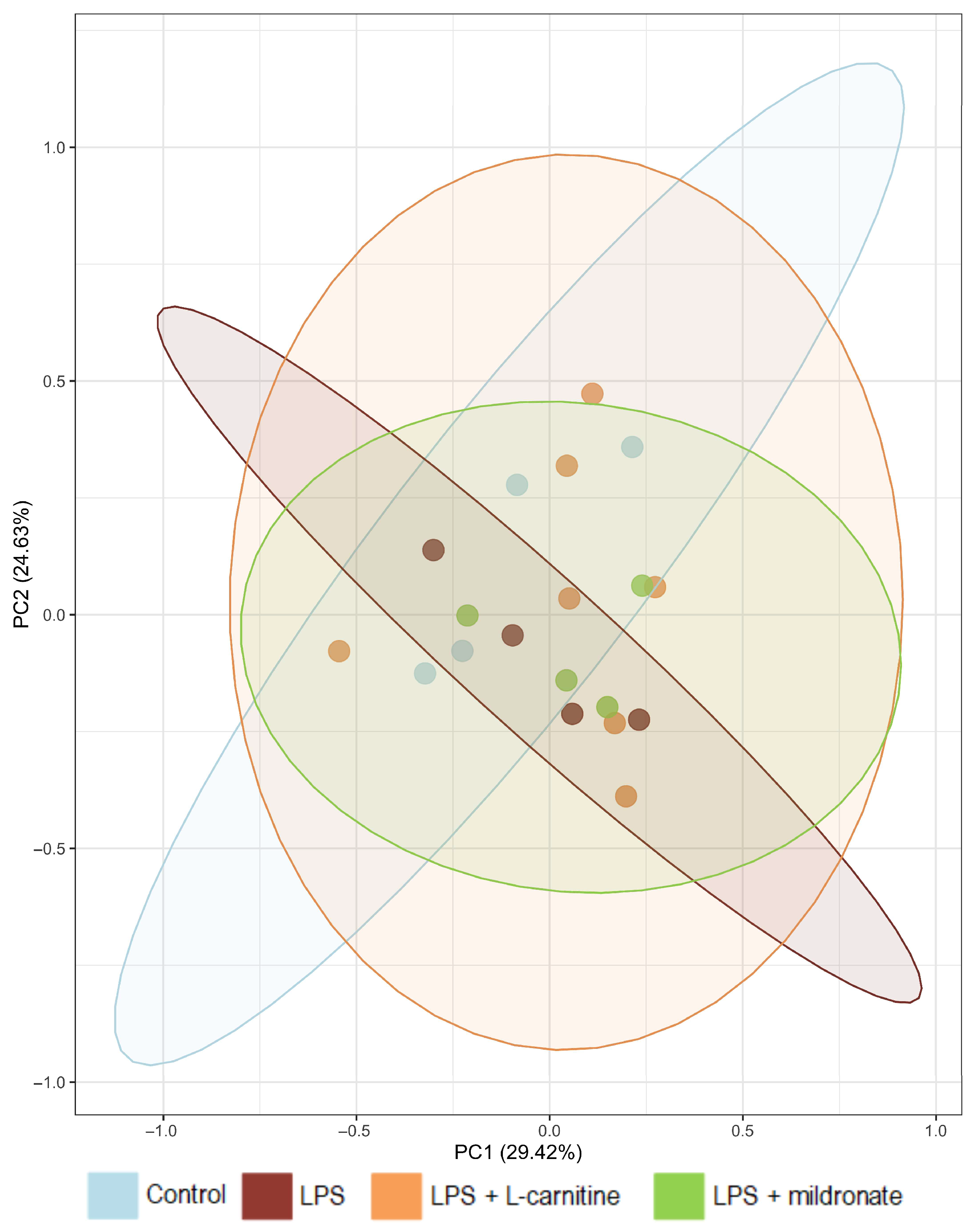
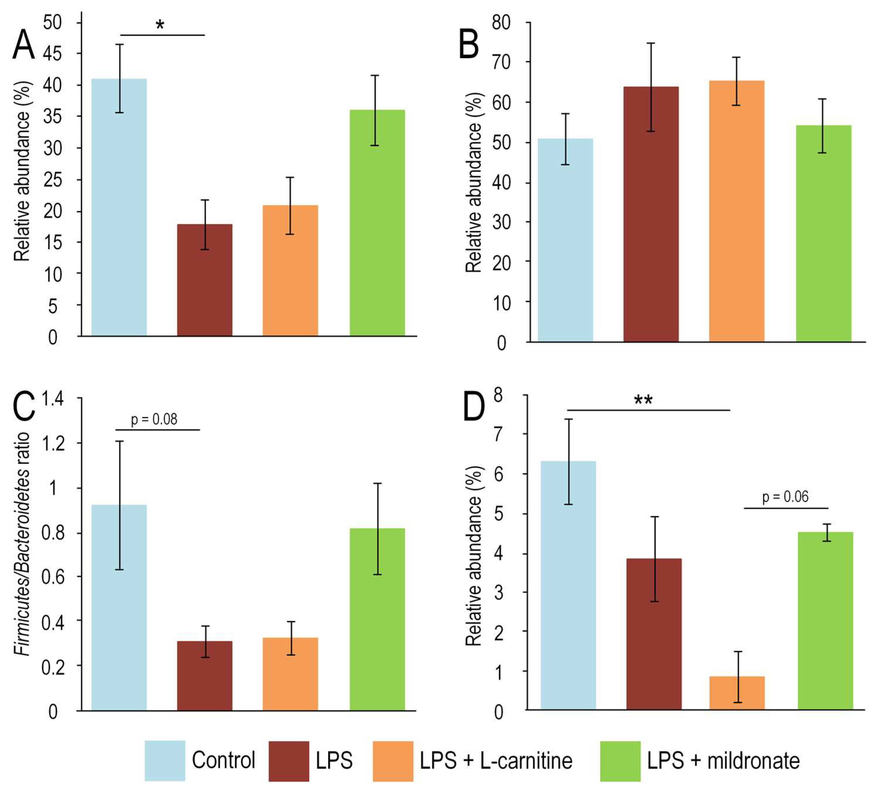
2.1. Bacterial Composition of the Gut Microbiome

We identified the microbial community structures for each experimental group of mice at the phylum, class, order, family, and genus level. The overall gut microbial community was mainly comprised of 5 phyla, 6 classes, 11 orders, 16 families and 40 genera. We have presented the profiles of intestinal microbial communities for each mouse at the genus. Bacteroidota phylum and Firmicutes prevailed in the gut microbiome for all groups of mice. Principal component analysis (PCA) did not reveal significant differences in the bacterial composition of the gut microbiota (Figure 1). However, it was found that mice receiving LPS injections had a more than two-fold decrease in the abundance of Firmicutes (p < 0.05). Mice receiving L-carnitine prior to injections also showed a decrease in Firmicutes levels, but there were no statistically significant differences compared to the control group. The level of Firmicutes bacteria was almost at the same level as the control group in mice receiving LPS injections along with mildronate (Figure 2A). The abundance of Bacteroidetes did not change depending on the modulation of L-carnitine metabolism and LPS injections (Figure 2B). However, due to the significant differences in the abundance of Firmicutes, differences in the Firmicutes/Bacteroidetes ratio were observed. The highest Firmicutes/Bacteroidetes ratio was found in the control group (0.91 ± 0.29) and the LPS + mildronate group (0.81 ± 0.2). In the LPS group, the Firmicutes/Bacteroidetes ratio was 0.31 ± 0.07 (p = 0.081 compared to the control group). In the LPS + L-carnitine group, the Firmicutes/Bacteroidetes ratio was 0.32 ± 0.07 (Figure 2C). It is worth noting that there were differences in the abundance of Alloprevotella (Bacteroidota phylum) between the experimental groups. Control mice had a maximal abundance of Alloprevotella (6.3 ± 1.1%). LPS injections decreased the Alloprevotella level to 3.8 ± 1.1%, but differences are not statistically significant. L-carnitine-treated mice had a minimal level abundance of Alloprevotella (0.8 ± 0.7%), which less than in the control group (p < 0.05). Mildronate-treated mice median of Alloprevotella abundance was 4.5 ± 0.2%, which higher than in L-carnitine-treated mice (p = 0.064) (Figure 2D).

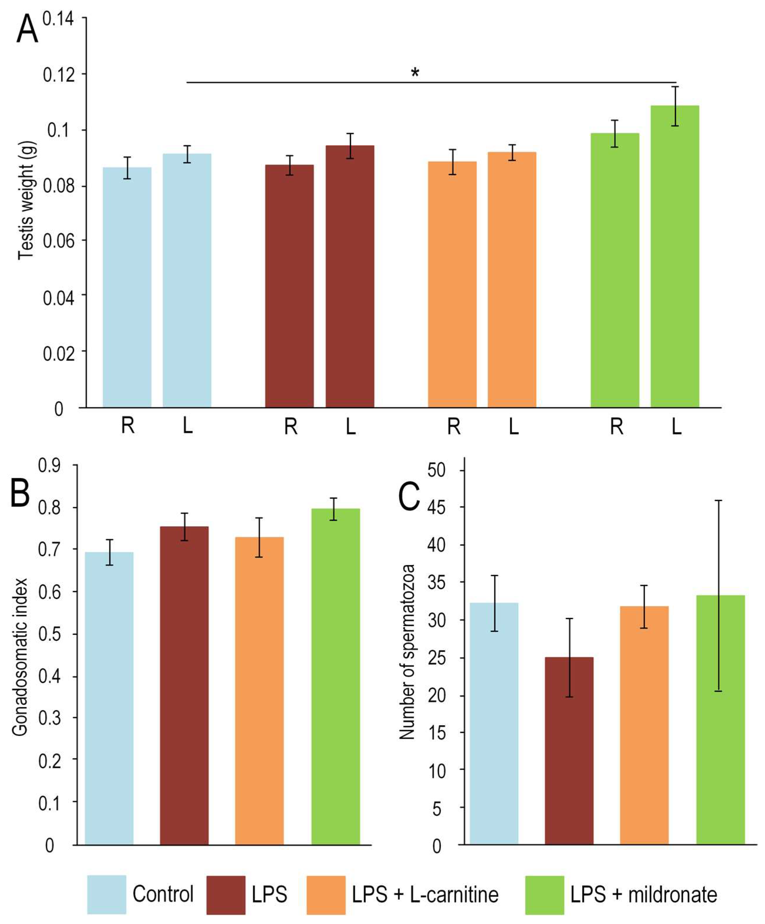
2.2. Effect of LPS Injection and Modulators of L-Carnitine Metabolism on Testis Weight and Sperm Quality

We did not find any differences in the weight of the left testis between the groups, but there were differences in the weight of the right testis. Mice in the LPS + mildronate group had a 19% higher weight in the right testis compared to the control group (p < 0.05) (Figure 3A). However, there was no significant difference in the gonadosomatic index (GSI) between the control group and the LPS + mildronate group (Figure 3B). The mean number of spermatozoa per five big squares of Goryaeva’s camera in the control group was 32 ± 3.7, and in the mildronate-treated group, it was 33 ± 12.7, indicating no significant differences (Figure 3C).

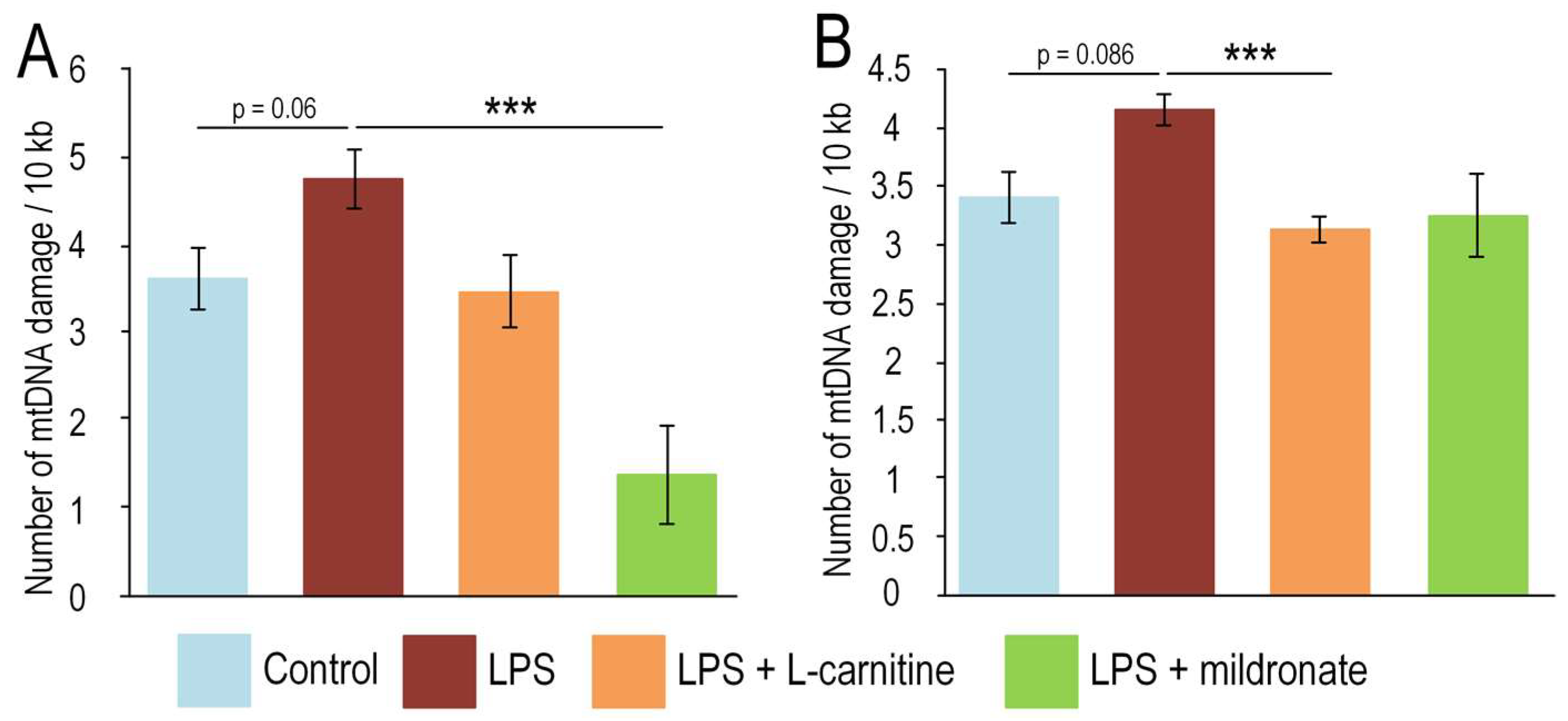
2.3. Impact of the LPS, L-Carnitine and Mildronate on the mtDNA Integrity in the Gut and Testis

LPS injections led to a 31% increase in the amount of mtDNA damage in the testes of mice, but the differences were not statistically significant (p = 0.062). However, in mice consuming mildronate, the amount of damage was 3.5 times lower than in mice receiving only LPS injections (p < 0.001). Mice receiving L-carnitine had similar levels of damage compared to the control group, and the differences with the LPS-injected group were not statistically significant (Figure 4A).
Interestingly, in the gut, L-carnitine provided greater protection against mtDNA damage induced by LPS injections. LPS increased the amount of damage by 22% compared to the control group, but the differences were not statistically significant (p = 0.086). In mice receiving LPS injections along with L-carnitine, the amount of damage was 25% lower than in mice receiving only LPS injections (p < 0.001). Mildronate did not contribute to a statistically significant reduction in mtDNA damage in the gut following LPS injections (Figure 4B).

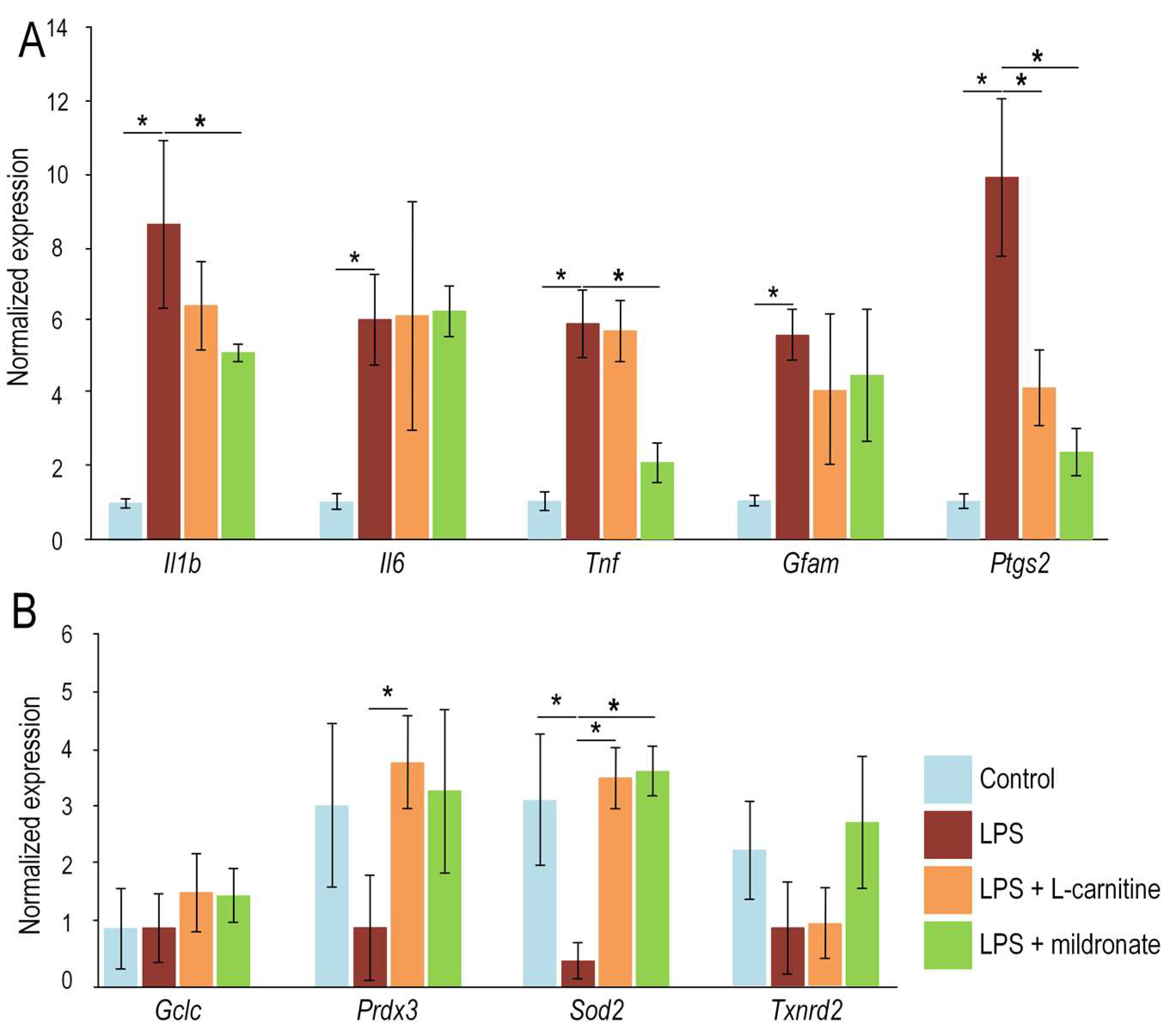
2.4. Effect of LPS, L-Carnitine and Mildronate on the Expression of Inflammatory Markers in the Gut

We found that in the gut, mice receiving LPS injections had significantly increased expression of all the investigated inflammation markers. The expression of Il1b and Il6 was increased by 8-fold and 6-fold, respectively (both p < 0.05). The expression of another pro-inflammatory cytokine, Tnf, was also increased by 6-fold (p < 0.05). LPS injections led to a 6-fold increase in the expression of inflammation markers Gfap and Ptgs (both p < 0.05). However, in mice receiving LPS injections along with mildronate, the expression of Il1b, Tnf, and Ptgs were reduced (all p < 0.05). L-carnitine prevented an increase in Ptgs expression, at only (p < 0.05) (Figure 5A).
We analyzed the expression of genes encoding antioxidant proteins. We found that injections of LPS resulted in a decrease in the expression of the superoxide dismutase 2 (Sod2) gene (p < 0.05). However, in mice receiving mildronate and L-carnitine, the expression of the Sod2 gene was not reduced. Additionally, mice receiving L-carnitine showed an increased expression of the peroxiredoxin 3 (Prdx3) gene compared to mice receiving only LPS injections (p < 0.05). We did not observe any influence of the studied compounds on the expression of the Gclc and Txnrd2 genes (Figure 5B).

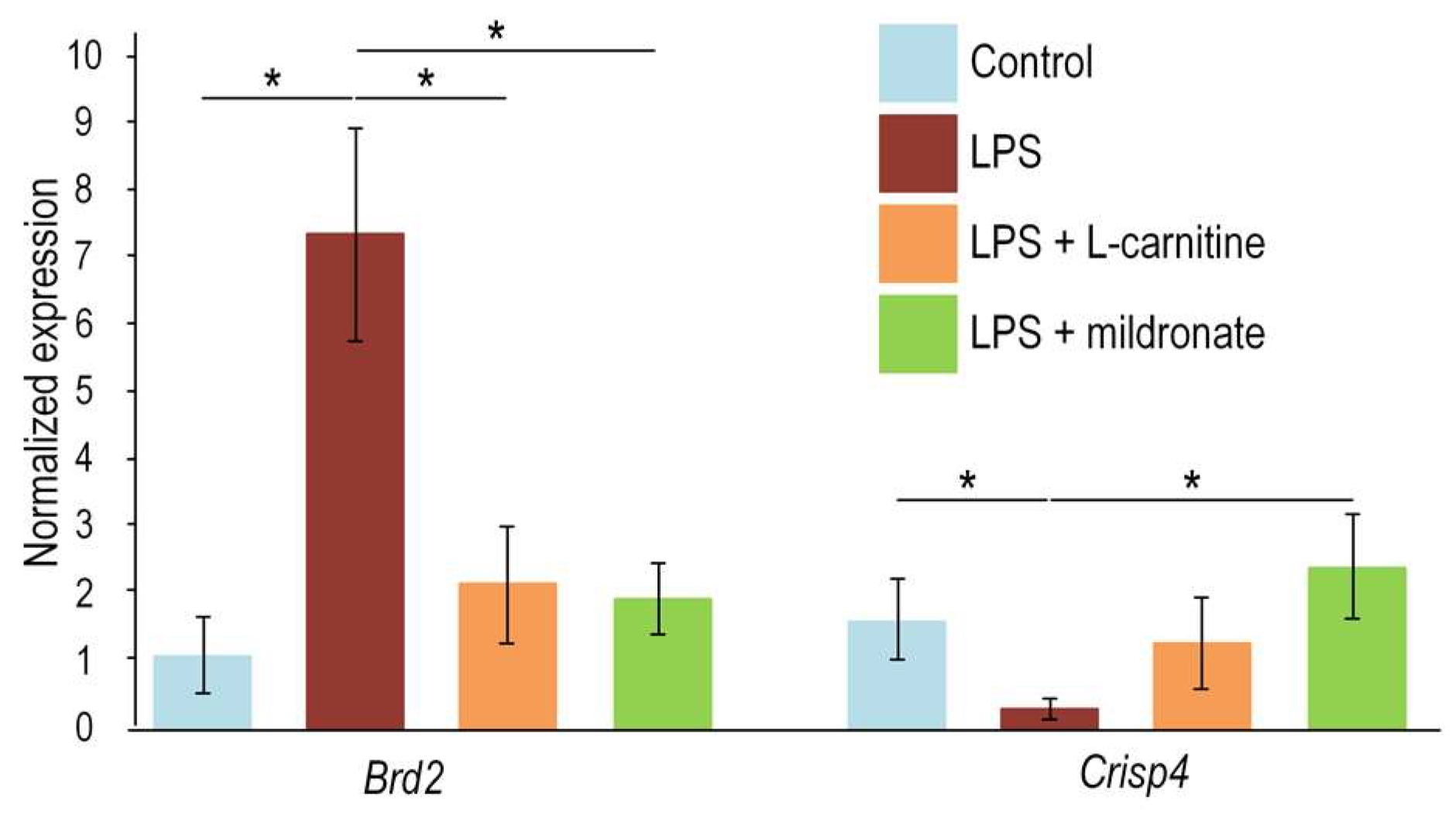
2.5. Effect of LPS Injection and Modulators of L-Carnitine Metabolism on the Gene Expression in the Testis

LPS injections induced overexpression of Brd2 in the mice testis. Both L-carnitine and mildronate treatment normalized of Brd2 expression via its reducing more that tree times (both p < 0.05). LPS injection decreased expression of Crisp4 for four times (p < 0.05). L-carnitine treatment increased expression of Crisp4 for three times compare with LPS-treated mice (p < 0.05). Mildronate treatment increased expression of Crisp4 for five times compare with LPS-treated mice (p < 0.05) (Figure 6).

3. Discussion

The mechanism by which drugs induce a decrease in male fertility can vary, including direct cytotoxic effects, compromised membrane and DNA integrity in spermatozoa, metabolic dysfunctions, and impairment of hypothalamic-pituitary-gonadal functions [23,24]. There is reason to expect that mildronate-inducing changes in metabolism may reduce male fertility. L-carnitine plays a critical role in sperm functions [7] and mildronate led to sharp decrease both in plasma and testis [17]. Our studies did not reveal direct features of deterioration in sperm quality (Figure 3), which were identified by Zhitkova and Khramtsova, 2018 [16]. Although we found that the left testicle of mice receiving mildronate was larger than that of control mice. However, this may be associated with the complex effect of LPS + mildronate drugs. Furthermore, we did not find any difference in the value of the gonadosomatic index. Our findings align more closely with the results reported by Dambrova et al. in 2008, where no decrease in sexual motivation, spermatozoa densities, or motility were observed in rats [16].
In contrast, we have shown that mildronate can protect the testes from LPS-induced inflammation. In the testis, mildronate treatment led to reduce the amount of LPS-induced mtDNA damage (Figure 4A), which may be a marker of oxidative stress [25]. Also, LPS injections induced decrease in Crisp4 expression (Figure 6). Epididymal Cysteine Rich Secretory Proteins 1 and 4 (Crisp1 and Crisp4) are associated with sperm during maturation and play different roles in fertilization. Simultaneous lack of the epididymal proteins results in clear fertility defects [26]. Both mildronate and L-carnitine treatment increases Crisp4 expression (Figure 6), which may be considered as recovery of male reproductive function. On the contrary, LPS injection increased expression of Brd2 in testis (Figure 5B). Brd2 physically associate with the promoters of inflammatory cytokine genes in macrophages and take and involved in LPS-induced inflammation [27]. Both mildronate and L-carnitine treatment decreases LPS-induced Brd2 expression (Figure 6), which may be associated with suppression of inflammation. This assumption is supported by a decrease in the level of mtDNA damage both in the testis and the gut (Figure 4).
The antioxidant and anti-inflammatory effect of mildronate was also observed in the intestine. We have demonstrated that both mildronate and L-carnitine prevent the LPS-induced decrease in Sod2 gene expression (Figure 5B). This protein plays a critical role as a scavenger for superoxide radicals, particularly within the mitochondrial matrix [28]. It is worth noting that neither mildronate nor LPS had an impact on the expression of other mitochondria-targeted antioxidant enzymes, such as Gclc, involved in glutathione synthesis [29], as well as Prdx3 and Txnrd2, crucial for removing H2O2 from mitochondria [30,31]. We found that mildronate reduced the expression of certain pro-inflammatory cytokines and Ptgs2. PTGS2 plays a key role in the synthesis of prostaglandins, which are mediators of inflammation. Inflammatory processes are often accompanied by increased activation of PTGS2 and, accordingly, increased expression of this gene. Increased expression of this gene may be a consequence of the activation of inflammatory signaling pathways, such as NF-κB [32]. Another indicator of inflammatory processes is the Firmicutes/Bacteroidetes ratio. Studies have shown that in inflammatory diseases such as inflammatory bowel diseases (IBD), the Firmicutes/Bacteroidetes ratio may shift towards an increase in Bacteroidetes and a decrease in Firmicutes. This change is associated with increased intestinal permeability and inflammatory processes in the gut [33]. Similar changes were observed in our study, where we induced inflammation using LPS injections. In mice receiving mildronate along with LPS injections, the Firmicutes/Bacteroidetes ratio was closest to the control group (Figure 2).
Note the changes in the abundance of Alloprevotella (Figure 2D). Early it had been showed that Alloprevotella were enriched in patients with low cardiovascular diseases risk [34]. There is evidence that Alloprevotella are associated with the metabolism of short-chain fatty acids (SCFA). An increase in the level of Alloprevotella positively correlates with the production of butyrate but not lactate and acetate [35]. Butyrate can also decrease excessive inflammation through modulation of immune cells such as increasing functionalities of M2 macrophages and regulatory T cells and inhibiting infiltration by neutrophils [36]. LPS injections led to a mild decrease Alloprevotella level. In mice which get LPS injection and L-carnitine supplementation Alloprevotella level was significantly decreased (Figure 2D). These observations suggest that mildronate has less effect on the gut microbiome than L-carnitine. Indeed, it has been repeatedly shown that the addition of L-carnitine to the diet induced changes in the gut microbiome that are associated with the production of TMAO [37,38]. Usually, the TMAO production is associated with the development of cardiovascular diseases [39], but TMAO induces inflammation [19] which harmful to sperm production, associated with oxidative stress and the latter is well known to impair sperm function [1]. Thus, these observations indicate that long-term L-carnitine treatment may have a negative effect due to changes in the composition of the bacterial microbiome, which can lead both to cardiovascular disease and inflammation-induced testis dysfunction. At the same time, it cannot be argued that L-carnitine can only cause negative changes in the microbiome. L-carnitine has vital roles in the endogenous metabolism of short chain fatty acids, which can control various biological processes such as nutrient absorption, lipid and glucose homeostasis, and systemic inflammation [40]. All aspects of L-carnitine metabolism and its effect on the gut microbiome need to be explored more deeply.
Mildronate-induced depletion of L-carnitine did not result in adverse alterations in the bacterial composition of the gut microbiome. This finding is unexpected, as previous studies have demonstrated that long-term mildronate treatment leads to an increase in the abundance of Proteobacteria, which may be indicative of dysbiosis [21]. However, a more in-depth analysis using next-generation sequencing revealed no noticeable changes in the levels of Proteobacteria associated with mildronate administration (Figure 2).

4. Materials and Methods

4.1. Animals

Male of the C57BL/6 strain aged 2 months were used in the experiment. Mice were obtained from Stolbovaya nursery (Moscow region, Russia). Mice were kept under standard conditions of t = 25 °C, 12 h light/dark cycle and relative humidity of 40%. Standard laboratory diet (Ssniff Spezialdiaten GmbH, Soest, Germany) and water were available ad libitum. Mice were sacrificed by a mixture (1 mL/kg) of xylazine (10 mg/kg) and ketamine (90 mg/kg) intraperitoneal. Testis was used to estimate sperm quantity, gene expression, number of mtDNA damage. All experimental procedures with animals and sacrifice were performed by the rules set by Voronezh State University Ethical Committee on Biomedical Research (Section of Animal Care and Use, protocol 42-03 dated 2 April 2019), which correspond to the European Communities Council Directive of 24 November 1986 (86/609/EEC).

4.2. Experiment Design

Second experiment. The protective effect of mildronate and L-carnitine under lipopolysaccharide (LPS)-induced inflammation was studied. Upon the start of the experiment, mice were randomly divided into four groups. The first group (n = 6) received pure drinking water ad libitum for two weeks. During the third week, mice were injected (i.p.) with saline daily. The second group (n = 5) received pure drinking water ad libitum for two weeks and during the third week, mice were injected (i.p.) with LPS (Medgamal, Moscow, Russia) (375 mg/kg/day) daily. The third group (n = 7) received L-carnitine (Sigma-aldrich, St. Louis, MO, USA) (100 mg/kg/day) for two weeks and during the third week, mice were injected (i.p.) with LPS daily. The fourth group (n = 9) received mildronate (Grindex, Riga, Latvia) (100 mg/kg/day) for two weeks and during the third week, mice were injected (i.p.) with LPS daily. After 3 weeks of the experiment, mice were sacrificed. DNA and RNA from testes and gut were isolated for analysis (Figure 7).

4.3. Testis Analysis

The gonadosomatic index (GSI) was calculated as a proportion of the testis mass to the total body mass.
GSI = [testis weight/total mouse weight] × 100
For spermatozoa analysis, a sample of epididymal fluid was collected, diluted with isotonic saline per 20×, and immediately examined on a Goryaev’s camera (MimiMed, Suponevo, Russia) by light microscopy MikMed (Lomo, St. Petersburg, Russia). The number of spermatozoa was determined in five big squares of Goryaev’s camera (0.2 × 0.2 mm) by light microscopy with a magnification of 10 × 40.

4.4. Measurement of Number of mtDNA Damage

Testis were excised and homogenized by a glass–glass homogenizer. DNA isolation was performed using the PROBA-GS (DNA-Technologies, Moscow, Russia). Quantitative PCR (QPCR) analysis was performed using the Bio-Rad CFX96TM Real-TimeSystem (Bio-Rad, Hercules, CA, USA) and Encyclo-polymerase kit (Evrogen, Moscow, Russia) for long-range PCR. The reaction condition steps were as follows: initial denaturation step at 94 °C for 5 min, followed by 35 cycles: denaturation at 94 °C for 10 s, annealing at 59 °C for 20 s, and extension at 72 °C for 4:30 min.
The number of lesions was measured for six mtDNA fragments. The 1st fragment corresponded to rRNAs, the 2nd fragment corresponded to 16S rRNA and ND1, the 3rd fragment corresponded to ND1 and ND2, the 4th fragment corresponded to ND5, the 5th fragment corresponded to ND6 and CytB, and the 6th fragment corresponded to D-loop. The short fragment was amplifying in parallel for normalization of mtDNA damage number to mtDNA copy number. The number of damages was calculated by comparison by ΔCq between the control and experimental mtDNA for the long fragment versus the corresponding ΔCq for the short fragment. Primer sequences for qPCR described in our earlier work [24].

4.5. Gene Expression Measurement

RNA extraction from tissue was performed using the ExtractRNA kit (Evrogen, Moscow, Russia) according to the attached protocol. We used 500 μg of isolated RNA to obtain cDNA using the MMLV reverse transcriptase (Evrogen, Moscow, Russia). qPCR analysis was performed on a CFX96™ Real-Time system thermal cycler (Bio-RAD, Hercules, CA, USA) using the qPCRmix-HS SYBR kit (Evrogen, Moscow, Russia). The normalized expression level was calculated by formula 2(ΔΔCq). The Gapdh gene was used as a reference.
Primer sequences were as follows: Glyceraldehyde 3-phosphate dehydrogenase (Gapdh) forward: 5′-GGCTCCCTAGGCCCCTCCTG-3′; reverse: 5′-TCCCAACTCGGCCCCCAACA-3′; Bromodomain-containing protein 2 (Brd2) forward: 5′-CTTCTGTACCAGCTTTACAAC-3′; reverse: 5′-TTTGTGATAATCCGGCAAAC-3′; Cysteine-Rich Secretory Protein 4 (Crisp4) forward: 5′-TCATGAGGGAAATCATCAAG-3′; reverse: 5′-TCCATCTTCGCAGTTATTTG-3′. Prostaglandin-endoperoxide synthase 2 (Ptgs2) forward: 5′-AGTCCGGGTACAGTCACACTT-3′; reverse: 5′-TTCCAATCCATGTCAAAACCGT-3′; Interleukin 6 (IL-6) forward: 5′-CGGAGAGGAGACTTCACAGAG-3′; reverse: 5′-CATTTCCACGATTTCCCAGA-3′; Interleukin 1 beta (IL-1b) forward: 5′-TTGACGGACCCCAAAAGATG-3′; reverse: 5′-AGAAGGTGCTCATGTCCTCA-3′; Tumor necrosis factor (TNF-α) forward: 5′-TATGGCTCAGGGTCCAACTC-3′; reverse: 5′-GGAAAGCCCATTTGAGTCCT-3′; Glial fibrillary acidic protein (Gfap) forward: 5′-CCACGTTAAGCTAGCCCTGGACAT-3′; reverse: 5′-CTCACCATCCCGCATCTCCACAGT-3′; Glutamate-cysteine ligase catalytic subunit (Gclc) forward: 5′-GGGGTGACGAGGTGGAGTA-3′; reverse: 5′-GTTGGGGTTTGTCCTCTCCC-3′; Peroxiredoxin 3 (Prdx3) forward: 5′-GGTTGCTCGTCATGCAAGTG-3′; reverse: 5′-CCACAGTATGTCTGTCAAACAGG-3′; Superoxide dismutase 2 (Sod2) forward: 5′-CAGACCTGCCTTACGACTATGG-3′; reverse: 5′-CTCGGTGGCCTTGAGATTGTT-3′; Thioredoxin reductase 2 (Txnr2) forward: 5′-GATCCGGTGGCCTAGCTTG-3′; reverse: 5′-AGGGGGAGAAGGTTCCACAT-3′.

4.6. Analysis of Bacterial Composition of Gut Microbiome

The study of the gut microbiome of mice was carried out on the Ion Torrent PGM platform. To assess bacterial diversity, the hypervariable region V3 of the 16S rRNA gene was selected and amplified using universal bacterial primers 337F and 518R [41]. PCR was performed using a 5X ScreenMix-HS Master Mix (Evrogen, Moscow, Russia) in the following mode: 94 °C for 4 min followed by 37 cycles of 94 °C for 30 s, 62 °C for 30 s, and 72 °C for 30 s with the final elongation at 72 °C for 5 min.
Sequencing libraries were prepared using the NEBNext Fast DNA Library Prep kit (New England Biolabs, Ipswich, MA, USA) by the manufacturer’s protocol. The quality of the sequencing libraries was evaluated using the Library Quantification Kit Ion Torrent Platforms (Kapa Biosystems, Wilmington, DE, USA). qPCR was performed by the following mode: 95 °C for 5 min followed by 35 cycles of 95 °C for 30 s, 60 °C for 45 s, and melt curve analysis at 65–95 °C. All library dilutions had a length of about 200 bp and an optimal concentration of 0.087 to 0.87 pM for emulsion PCR.
After that, the libraries were mixed in equimolar volumes for emulsion PCR using the OneTouch 2 system (Thermo Fisher Scientific, Waltham, MA, USA). The degree of enrichment after emulsion PCR was 21%, which is considered optimal. Sequencing was performed on the IonTorrent PGM System using the Ion PGM Hi-Q View Sequencing Kit and Ion 318™ Chip v2 BC (Thermo Fisher Scientific, Waltham, MA, USA).

4.7. Statistical Analysis

Statistical analysis was performed using Statistica 12 (StatSoft Inc., Tulsa, OK, USA). The results were expressed as means ± SEM. The data were analyzed by the Kruskal–Wallis test (H-test). For the calculation of normalized expression, standard Bio-Rad CFX Manager v.3.1 (Bio-Rad, Hercules, USA) was used. For quantification of mtDNA damage used DNADamageCalculator (Voronezh, Russia). After sequencing, about 200,000 reads were obtained for each sample. They were all generated in BAM files for each sample. After that, the files were converted to the FastQ format and analyzed using the R programming language in the RStudio environment v.4.2.1 (Posit, Boston, MA, USA). Raw reads were filtered by length and were quality controlled, using the VSEARCH v.2.8.2 software functionality. After that, the processed reads were aligned with the reference reads in the SILVA database v.123 (https://www.arb-silva.de, (accessed on 13 October 2021)) [41], clustered in the OTU, and taxonomically profiled with the construction of phylogenetic trees and OTU tables. We searched for the OTUs using the UNOISE2 algorithm, which reduces the noise through error correction.

5. Conclusions

Thus, both L-carnitine and mildronate, despite opposite metabolic effects, may have a protective effect against LPS-induced inflammation in the testis and the gut. L-carnitine may directly decrease inflammation and normalized expression of genes, which play a critical role for testis functioning. Mildronate restructures metabolism, which leads to a shift in bacterial composition of gut microbiome. Also, mildronate decreased the extent of mtDNA damage, which was induced by LPS injections. This research confirmed safety of mildronate on male fertility and showed the possibilities of using this metabolic modulator for the treatment of sexual dysfunctions in inflammation.

Author Contributions

Conceptualization, A.P.G. and E.A.S.; methodology, A.P.G.; software, A.D.T., M.V.G.; validation, A.P.G.; formal analysis, E.A.S.; investigation, P.I.B., V.V.N.; resources, A.P.G.; data curation, A.P.G.; writing—original draft preparation, A.P.G.; writing—review and editing, A.P.G.; visualization, A.D.T.; supervision, E.A.S. and M.V.G.; project administration, A.P.G.; funding acquisition, A.P.G. All authors have read and agreed to the published version of the manuscript.

Funding

This work was supported by the Ministry of Science and Higher Education of the Russian Federation under the State assignment for universities in the field of scientific activity (project FZGU-2023-0009) and and scholarships of the President of the Russian Federation for young scientists and graduate students (SP-2802.2021.4).

Institutional Review Board Statement

The study was conducted in accordance with the Declaration of Helsinki, and approved by the Ethics Committee of Voronezh State University (Section of Animal Care and Use, protocol on Biomedical Research 42-03 dated 8 October 2020).

Informed Consent Statement

Not applicable.

Data Availability Statement

The data presented in this study are available on request from the corresponding author. The data are not publicly available since the authors did not use any repositories to collect the data. The raw data files are available in the authors' archive.

Conflicts of Interest

The authors declare no conflict of interest.

References

  1. Azenabor, A.; Ekun, A.O.; Akinloye, O. Impact of Inflammation on Male Reproductive Tract. J. Reprod. Infertil. 2015, 16, 123–129. [Google Scholar] [PubMed]
  2. D’Argenio, V.; Salvatore, F. The role of the gut microbiome in the healthy adult status. Clin. Chim. Acta 2015, 451, 97–102. [Google Scholar] [CrossRef] [PubMed]
  3. Page, M.J.; Kell, D.B.; Pretorius, E. The Role of Lipopolysaccharide-Induced Cell Signalling in Chronic Inflammation. Chronic Stress 2022, 6, 24705470221076390. [Google Scholar] [CrossRef] [PubMed]
  4. Claudino Dos Santos, J.C.; Oliveira, L.F.; Noleto, F.M.; Gusmão, C.T.P.; Brito, G.A.C.; Viana, G.S.B. Gut-microbiome-brain axis: The crosstalk between the vagus nerve, alpha-synuclein and the brain in Parkinson’s disease. Neural Regen. Res. 2023, 18, 2611–2614. [Google Scholar] [CrossRef] [PubMed]
  5. Wang, Y.; Xie, Z. Exploring the role of gut microbiome in male reproduction. Andrology 2022, 10, 441–450. [Google Scholar] [CrossRef]
  6. Mardanshahi, T.; Rezaei, N.; Zare, Z.; Malekzadeh Shafaroudi, M.; Mohammadi, H. Effects of L-Carnitine on the sperm parameters disorders, apoptosis of spermatogenic cells and testis histopathology in diabetic Rats. Int. J. Reprod. Biomed. 2018, 17, 325–336. [Google Scholar] [CrossRef]
  7. Aliabadi, E.; Soleimani Mehranjani, M.; Borzoei, Z.; Talaei-Khozani, T.; Mirkhani, H.; Tabesh, H. Effects of L-carnitine and L-acetyl-carnitine on testicular sperm motility and chromatin quality. Iran J. Reprod. Med. 2012, 10, 77–82. [Google Scholar]
  8. Abd-Allah, A.R.; Helal, G.K.; Al-Yahya, A.A.; Aleisa, A.M.; Al-Rejaie, S.S.; Al-Bakheet, S.A. Pro-inflammatory and oxidative stress pathways which compromise sperm motility and survival may be altered by L-carnitine. Oxid. Med. Cell. Longev. 2009, 2, 73–81. [Google Scholar] [CrossRef]
  9. Sjakste, N.; Gutcaits, A.; Kalvinsh, I. Mildronate: An antiischemic drug for neurological indications. CNS Drug Rev. 2005, 11, 151–168. [Google Scholar] [CrossRef]
  10. Heggermont, W.A.; Papageorgiou, A.P.; Heymans, S.; van Bilsen, M. Metabolic support for the heart: Complementary therapy for heart failure? Eur. J. Heart Fail. 2016, 18, 1420–1429. [Google Scholar] [CrossRef]
  11. Isajevs, S.; Isajeva, D.; Beitnere, U.; Jansone, B.; Kalvinsh, I.; Klusa, V. Mildronate as a regulator of protein expression in a rat model of Parkinson’s disease. Medicina 2011, 47, 79. [Google Scholar] [CrossRef] [PubMed]
  12. Beitnere, U.; van Groen, T.; Kumar, A.; Jansone, B.; Klusa, V.; Kadish, I. Mildronate improves cognition and reduces amyloid-β pathology in transgenic Alzheimer’s disease mice. J. Neurosci. Res. 2014, 92, 338–346. [Google Scholar] [CrossRef] [PubMed]
  13. Bezuglov, E.; Talibov, O.; Butovskiy, M.; Khaitin, V.; Achkasov, E.; Waśkiewicz, Z.; Lazarev, A. The Inclusion in WADA Prohibited List Is Not Always Supported by Scientific Evidence: A Narrative Review. Asian J. Sports Med. 2021, 12, e110753. [Google Scholar] [CrossRef]
  14. Liepinsh, E.; Konrade, I.; Skapare, E.; Pugovics, O.; Grinberga, S.; Kuka, J.; Kalvinsh, I.; Dambrova, M. Mildronate treatment alters γ-butyrobetaine and l-carnitine concentrations in healthy volunteers. J. Pharm. Pharmacol. 2011, 63, 1195–1201. [Google Scholar] [CrossRef] [PubMed]
  15. Wang, S.; Xu, J.; Zheng, J.; Zhang, X.; Shao, J.; Zhao, L.; Hao, J. Anti-Inflammatory and Antioxidant Effects of Acetyl-L-Carnitine on Atherosclerotic Rats. Med. Sci. Monit. 2020, 26, e920250-1. [Google Scholar] [CrossRef] [PubMed]
  16. Zhitkova, V.S.; Khramtsova, Y.S. The influence of excessive physical activity on spermatogenesis in rats against the background of the introduction of mildronate Youth and science in the North. In Proceedings of the III All-Russian (XVIII) Youth Scientific Conference, Rep. Komi, Syktyvkar, Russia, 12–16 March 2018. [Google Scholar]
  17. Dambrova, M.; Cirule, H.; Svalbe, B.; Zvejniece, L.; Pugovichs, O.; Zorenko, T.; Kalvinsh, I.; Liepinsh, E.; Belozertseva, I. Effect of inhibiting carnitine biosynthesis on male rat sexual performance. Physiol. Behav. 2008, 95, 341–347. [Google Scholar] [CrossRef] [PubMed]
  18. Dambrova, M.; Skapare-Makarova, E.; Konrade, I.; Pugovics, O.; Grinberga, S.; Tirzite, D.; Petrovska, R.; Kalvins, I.; Liepins, E. Meldonium decreases the diet-increased plasma levels of trimethylamine N-oxide, a metabolite associated with atherosclerosis. J. Clin. Pharmacol. 2013, 53, 1095–1098. [Google Scholar] [CrossRef]
  19. Berlato, D.G.; Bairros, A.V. Meldonium: Pharmacological, toxicological, and analytical aspects. Toxicol. Res. Appl. 2020, 4, 2397847320915143. [Google Scholar] [CrossRef]
  20. Duttaroy, A.K. Role of Gut Microbiota and Their Metabolites on Atherosclerosis, Hypertension and Human Blood Platelet Function: A Review. Nutrients 2021, 13, 144. [Google Scholar] [CrossRef]
  21. Koeth, R.A.; Wang, Z.; Levison, B.S.; Buffa, J.A.; Org, E.; Sheehy, B.T.; Britt, E.B.; Fu, X.; Wu, Y.; Li, L.; et al. Intestinal microbiota metabolism of L-carnitine, a nutrient in red meat, promotes atherosclerosis. Nat. Med. 2013, 19, 576–585. [Google Scholar] [CrossRef]
  22. Gureev, A.P.; Shaforostova, E.A.; Vitkalova, I.Y.; Sadovnikova, I.S.; Kalinina, Y.I.; Cherednichenko, V.R.; Reznikova, K.A.; Valuyskikh, V.V.; Popov, V.N. Long-term mildronate treatment increased Proteobacteria level in gut microbiome, and caused behavioral deviations and transcriptome change in liver, heart and brain of healthy mice. Toxicol. Appl. Pharmacol. 2020, 398, 115031. [Google Scholar] [CrossRef] [PubMed]
  23. Semet, M.; Paci, M.; Saïas-Magnan, J.; Metzler-Guillemain, C.; Boissier, R.; Lejeune, H.; Perrin, J. The impact of drugs on male fertility: A review. Andrology 2017, 5, 640–663. [Google Scholar] [CrossRef] [PubMed]
  24. Ajayi, A.; Akhigbe, R.E. The physiology of male reproduction: Impact of drugs and their abuse on male fertility. Andrologia 2020, 52, e13672. [Google Scholar] [CrossRef] [PubMed]
  25. Gureev, A.P.; Shaforostova, E.A.; Starkov, A.A.; Popov, V.N. Simplified qPCR method for detecting excessive mtDNA damage induced by exogenous factors. Toxicology 2017, 382, 67–74. [Google Scholar] [CrossRef] [PubMed]
  26. Carvajal, G.; Brukman, N.G.; Weigel Muñoz, M.; Battistone, M.A.; Guazzone, V.A.; Ikawa, M.; Haruhiko, M.; Lustig, L.; Breton, S.; Cuasnicu, P.S. Impaired male fertility and abnormal epididymal epithelium differentiation in mice lacking CRISP1 and CRISP4. Sci. Rep. 2018, 8, 17531. [Google Scholar] [CrossRef] [PubMed]
  27. Belkina, A.C.; Nikolajczyk, B.S.; Denis, G.V. BET protein function is required for inflammation: Brd2 genetic disruption and BET inhibitor JQ1 impair mouse macrophage inflammatory responses. J. Immunol. 2013, 190, 3670–3678. [Google Scholar] [CrossRef] [PubMed]
  28. Mruk, D.D.; Silvestrini, B.; Mo, M.Y.; Cheng, C.Y. Antioxidant superoxide dismutase—A review: Its function, regulation in the testis, and role in male fertility. Contraception 2002, 65, 305–311. [Google Scholar] [CrossRef]
  29. Lu, S.C. Glutathione synthesis. Biochim. Biophys. Acta 2013, 1830, 3143–3153. [Google Scholar] [CrossRef]
  30. Holzerova, E.; Danhauser, K.; Haack, T.B.; Kremer, L.S.; Melcher, M.; Ingold, I.; Kobayashi, S.; Terrile, C.; Wolf, P.; Schaper, J.; et al. Human thioredoxin 2 deficiency impairs mitochondrial redox homeostasis and causes early-onset neurodegeneration. Brain 2016, 139, 346–354. [Google Scholar] [CrossRef]
  31. Chen, L.; Na, R.; Gu, M.; Salmon, A.B.; Liu, Y.; Liang, H.; Qi, W.; Van Remmen, H.; Richardson, A.; Ran, Q. Reduction of mitochondrial H2O2 by overexpressing peroxiredoxin 3 improves glucose tolerance in mice. Aging Cell 2008, 7, 866–878. [Google Scholar] [CrossRef]
  32. Li, R.; Xie, J.; Xu, W.; Zhang, L.; Lin, H.; Huang, W. LPS-induced PTGS2 manipulates the inflammatory response through trophoblast invasion in preeclampsia via NF-κB pathway. Reprod. Biol. 2022, 22, 100696. [Google Scholar] [CrossRef] [PubMed]
  33. Morgan, X.C.; Tickle, T.L.; Sokol, H.; Gevers, D.; Devaney, K.L.; Ward, D.V.; Reyes, J.A.; Shah, S.A.; LeLeiko, N.; Snapper, S.B.; et al. Dysfunction of the intestinal microbiome in inflammatory bowel disease and treatment. Genome Biol. 2012, 13, R79. [Google Scholar] [CrossRef]
  34. Kelly, T.N.; Bazzano, L.A.; Ajami, N.J.; He, H.; Zhao, J.; Petrosino, J.F.; Correa, A.; He, J. Gut Microbiome Associates with Lifetime Cardiovascular Disease Risk Profile Among Bogalusa Heart Study Participants. Circ. Res. 2016, 119, 956–964. [Google Scholar] [CrossRef]
  35. Oh, J.K.; Vasquez, R.; Kim, S.H.; Hwang, I.C.; Song, J.H.; Park, J.H.; Kim, I.H.; Kang, D.K. Multispecies probiotics alter fecal short-chain fatty acids and lactate levels in weaned pigs by modulating gut microbiota. J. Anim. Sci. Technol. 2021, 63, 1142–1158. [Google Scholar] [CrossRef] [PubMed]
  36. Chen, J.; Vitetta, L. The Role of Butyrate in Attenuating Pathobiont-Induced Hyperinflammation. Immune Netw. 2020, 20, e15. [Google Scholar] [CrossRef] [PubMed]
  37. Cretoiu, D.; Ionescu, R.F.; Enache, R.M.; Cretoiu, S.M.; Voinea, S.C. Gut Microbiome, Functional Food, Atherosclerosis, and Vascular Calcifications-Is There a Missing Link? Microorganisms 2021, 9, 1913. [Google Scholar] [CrossRef]
  38. Coutinho-Wolino, K.S.; de F Cardozo, L.F.M.; de Oliveira Leal, V.; Mafra, D.; Stockler, M.B. Can diet modulate trimethylamine N-oxide (TMAO) production? What do we know so far? Eur. J. Nutr. 2021, 60, 3567–3584. [Google Scholar] [CrossRef]
  39. Roncal, C.; Martínez-Aguilar, E.; Orbe, J.; Ravassa, S.; Fernandez-Montero, A.; Saenz-Pipaon, G.; Ugarte, A.; Estella-Hermoso de Mendoza, A.; Rodriguez, J.A.; Fernández-Alonso, S.; et al. Trimethylamine-N-Oxide (TMAO) Predicts Cardiovascular Mortality in Peripheral Artery Disease. Sci. Rep. 2019, 9, 15580. [Google Scholar] [CrossRef]
  40. Ghonimy, A.; Zhang, D.M.; Farouk, M.H.; Wang, Q. The Impact of Carnitine on Dietary Fiber and Gut Bacteria Metabolism and Their Mutual Interaction in Monogastrics. Int. J. Mol. Sci. 2018, 19, 1008. [Google Scholar] [CrossRef]
  41. Sadovnikova, I.S.; Gureev, A.P.; Ignatyeva, D.A.; Gryaznova, M.V.; Chernyshova, E.V.; Krutskikh, E.P.; Novikova, A.G.; Popov, V.N. Nrf2/ARE Activators Improve Memory in Aged Mice via Maintaining of Mitochondrial Quality Control of Brain and the Modulation of Gut Microbiome. Pharmaceuticals 2021, 14, 607. [Google Scholar] [CrossRef]
Figure 1. Principal component analysis (PCA) for genus-level microbiota in fecal samples of mice. The first two principal components, PC1 and PC2, were plotted. Control (n = 4); LPS (n = 4); LPS + L-carnitine (n = 7); LPS + mildronate (n = 4).
Gastrointestdisord 05 00044 g001
Figure 2. Relative abundace of Firmicutes (A) and Bacteroidetes (B). Firmicutes/Bacteroidetes ratio (C). Relative abundance of Alloprevotella (D). Control (n = 4); LPS (n = 4); LPS + L-carnitine (n = 7); LPS + mildronate (n = 4). Differences between group statistically significant: * p < 0.05; ** p < 0.01.
Gastrointestdisord 05 00044 g002
Figure 3. Weight of right (R) and left (L) testes (A). The value of gonadosomatic index (B). The mean number of spermatozoa per five big squares of Goryaeva’s camera (C). Control (n = 6); LPS (n = 5); LPS + L-carnitine (n = 7); LPS + mildronate (n = 9). Differences between group statistically significant: * p < 0.05.
Gastrointestdisord 05 00044 g003
Figure 4. Impact of the LPS, L-carnitine and mildronate on the mtDNA integrity in the testis (A) and the gut (B). Control (n = 6); LPS (n = 5); LPS + L-carnitine (n = 7); LPS + mildronate (n = 9). Differences between group statistically significant: *** p < 0.001.
Gastrointestdisord 05 00044 g004
Figure 5. Impact of the LPS, L-carnitine and mildronate on the proinflammatory (A) and antioxidant genes expression in the gut (B). Control (n = 6); LPS (n = 5); LPS + L-carnitine (n = 7); LPS + mildronate (n = 9). Differences between group statistically significant: * p < 0.05.
Gastrointestdisord 05 00044 g005
Figure 6. Impact of the LPS, L-carnitine and mildronate on the genes expression in the testis. Control (n = 6); LPS (n = 5); LPS + L-carnitine (n = 7); LPS + mildronate (n = 9). Differences between group statistically significant: * p < 0.05.
Gastrointestdisord 05 00044 g006
Figure 7. Scheme of experiments for the study of protective effect of mildronate and L-carnitine under lipopolysaccharide (LPS)-induced inflammation.
Gastrointestdisord 05 00044 g007
Disclaimer/Publisher’s Note: The statements, opinions and data contained in all publications are solely those of the individual author(s) and contributor(s) and not of MDPI and/or the editor(s). MDPI and/or the editor(s) disclaim responsibility for any injury to people or property resulting from any ideas, methods, instructions or products referred to in the content.

