Article

A Multiparametric Method Improves the Serological Characterization of Inflammatory Bowel Diseases: Preliminary Results from a Multicenter Eastern Europe Study

1
Center for Digestive Endoscopy, University Clinic “Dr Dragisa Misovic”, 11000 Belgrade, Serbia
2
School of Medicine, University of Belgrade, 11000 Belgrade, Serbia
3
Gastroenterology Unit, University Hospital of Udine, 33100 Udine, Italy
4
Department of Gastroenterology, Clinical Hospital Center Rijeka, 51000 Rijeka, Croatia
5
Faculty of Health Studies, 51000 Rijeka, Croatia
6
Euroimmun—Medizinische Labordiagnostika AG, 23560 Lübeck, Germany
7
Institute of Clinical Pathology, University Hospital of Udine, 33100 Udine, Italy
8
Department of Medicine, University of Udine, 33100 Udine, Italy
9
General Surgery and Transplantation Unit, University Hospital of Udine, 33100 Udine, Italy
10
Digestive Endoscopy Department, Isola Tiberina Hospital—Gemelli Isola, 00186 Rome, Italy
11
Department of Gastroenterology, Royal Cornwall Hospitals NHS Trust, Truro TR1 3LJ, UK
*
Author to whom correspondence should be addressed.
Gastrointest. Disord. 2024, 6(1), 152-163; https://doi.org/10.3390/gidisord6010011
Submission received: 9 October 2023 / Revised: 20 January 2024 / Accepted: 24 January 2024 / Published: 29 January 2024

Abstract

:
The serological support for early diagnosis and differential diagnosis of inflammatory bowel diseases (IBDs) is actually very limited. In this study, we evaluated the performance of a promising multiparametric method including either well-established and newly developed biomarkers. We conducted a multicenter cross-sectional study at the Gastroenterology Units of Udine (Italy), Rijeka (Croatia) and Belgrade (Serbia). Sera was collected from IBD patients, and autoantibody profiles were determined using a mosaic cell and tissue-based indirect immunofluorescence (IIF) method simultaneously investigating anti-saccharomyces cerevisiae antibodies (ASCAs), anti-atypical perinuclear neutrophilic antibodies (P-ANCAs), anti-pancreatic antigens antibodies (PABs) and anti-goblet cells antibodies (GAB). The study finally enrolled 156 patients with IBD: 100 affected by Crohn’s disease (CD) and 56 by ulcerative colitis (UC). Twenty age-sex matched blood donors (BDs) were included as controls. PAB (anti-CUZD1 and/or anti-GP2 antibodies) were present in 24 CD patients versus none of the UC patients or BDs (24% sensitivity, 100% specificity). As regards CD patients, combined positivity of PAB and ASCA (sensitivity 84%, specificity 71.4%) performed better than ASCA alone. Colon involvement (87.5% vs. 60.5%; p = 0.014), deep mucosal lesions (58.3% vs. 25.0%; p = 0.002) and need for biologic therapies (79.2% vs. 46.1%; p = 0.005) were significantly more prevalent in PAB-positive than in PAB-negative CD patients. Multivariate analysis identified PAB positivity (OR = 3.67; 95%CI = 1.29–10.46) and anti-CUZD1 in particular (OR = 3.54; 95%CI = 1.08–11.63) as significant risk factors for deep mucosal lesion development in CD. A multiparametric diagnostic approach appears very useful to better characterize IBD patients. PABs, whether isolated or combined with other autoantibodies, may support differential diagnosis but above all facilitate the selection of CD patients at risk for more severe disease.

1. Introduction

Inflammatory bowel disease (IBD) represents a chronic inflammatory condition of the gastrointestinal tract which includes ulcerative colitis (UC) and Crohn’s disease (CD). Both conditions display heterogeneity in inflammatory and symptomatic burden, both between patients and within the same individual over time [1].
The differential diagnosis is supported by distinct clinical presentation, involvement of different sections of the gastrointestinal tract and, mostly, histological features observed on biopsy specimen. On the other hand, the contribution made by laboratory markers is still very limited. Recently, new tests with promising performance have been made available on the market, but the real impact on clinical practice is unclear. Anti-Saccharomyces cerevisiae antibodies (ASCAs) represent an established biomarker of CD, while goblet cell antibodies (GABs) and anti-neutrophil cytoplasmic antibodies with atypical perinuclear IIF pattern (P-ANCA) have been significantly associated with UC [2,3]. Among the newly identified biomarkers, increasing evidence suggests a potential role of pancreatic autoantibodies (PABs) in CD. The first work describing the presence of PABs by IIF in the serum of patients with CD dates back to 1984. Subsequently, several other studies addressed this issue reporting the prevalence of PABs in CD to be from 15% to a maximum of 40% [2,4,5,6,7,8,9,10,11,12,13,14,15,16,17,18,19]. Furthermore, while some authors reported a significant correlation between PABs and features of severe disease such as penetrating behavior or perianal localization [13], others did not confirm this association [9,11]. On the other hand, the correlation between PAB positivity and extra-intestinal CD manifestations, such as idiopathic chronic pancreatitis, was reported more consistently [11,13,20,21,22]. PABs were also associated with small bowel involvement and need for surgery [9] as well as early age disease onset [11]. The discrepancies between the studies might be attributed to the lack of standardized methods to test PABs by IIF. More recently, the proper identification of the pancreatic auto-antigenic targets glycoprotein 2 (GP2) [18,23] and CUB/zona pellucida-like domain-containing protein (CUZD1) [24] allowed a more effective and reproducible identification of PABs, offering also a possible explanation of the pathogenic role of PABs in CD. GP2 in particular, appears to have a solid link with the intestinal disease involvement in CD patients. Hase et al. [25,26,27] identified GP2 as a specific receptor on M cells in the intestinal Peyer’s patches. Since then, several researchers provided evidence that GP2 may play an important role in keeping the balance of the intestinal immune system by helping to differentiate between pathogenic and commensal microbiota [28]. The loss of tolerance to pancreatic and/or intestinal GP2 could modulate the pathophysiology of CD [29]. Increased expression of GP2 mRNA and protein was found in intestinal biopsy samples of some patients with CD [23,29]. Several studies reported GP2 as well as CUZD1 antibodies to be associated with distinct clinical phenotypes [30,31,32,33]. Although initial data were promising, PABs have not yet been included among routine laboratory tests conducted in IBD patients. It is still not clear what role they may play within the diagnostic process and above all, easy-to-use and low-cost tests were, up to now, unavailable. We conducted a study in order to evaluate the performance of a novel commercial multiparametric method for diagnosing and profiling IBD patients.

2. Methods

2.1. Patients

The multicenter cross-sectional study was conducted on 156 consecutive patients with IBD enrolled between January 2017 and December 2018 at three participating units: the Gastroenterology of the Academic Hospital of Udine in Italy, the Gastroenterology of the Hospital of Rijeka in Croatia and the Gastroenterology of the University Clinic Dr Dragisa Misovic-Dedinje of Belgrade in Serbia. The study complied with all the relevant national regulations and institutional policies and was conducted in accordance with the Helsinki Declaration. Inclusion criteria implied endoscopically and histologically confirmed CD or UC for at least two years, age above 18 and collaborative capability. Patients with an uncertain diagnosis or with features suggestive of another coexisting intestinal disease were excluded. The following data were collected: demographics, lifestyle habits, age at diagnosis, medical as well as surgical treatments and disease activity (evaluated according to Montreal classification [34]). Patients were sampled during checkups or infusions of biological drugs, without subjecting them to additional visits or withdrawals. Blood donor serum samples were collected at the Laboratory of the Academic Hospital in Udine to be used as a control group.

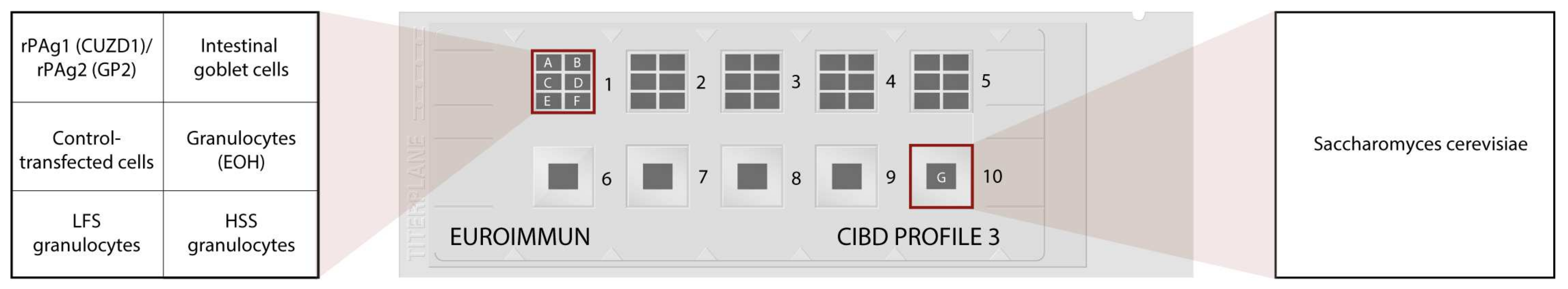
2.2. Laboratory Analysis

Blood samples were collected in tubes without anti-coagulant and then centrifuged at 3500 rpm for 10 min to obtain the serum. Each serum was then aliquoted in 1 mL polypropylene tubes identified by a unique numerical code and frozen at −20 °C until shipping. Subsequently, frozen sera were sent to the laboratory of EUROIMMUN Medizinische Labordiagnostika AG in Lübeck, Germany, where they were tested blindly.
Autoantibody profiles were determined in serum samples by indirect immunofluorescence (IIF) using the mosaic CIBD Profile 3 (EUROIMMUN Medizinische Labordiagnostika AG, Lübeck, Germany) according to the manufacturer’s instructions. Both IgG and IgA auto-antibodies were tested. The BIOCHIP Mosaic comprises different IIF substrates (cell substrates, fungal smears or transfected cells) based on which specific autoantibodies directed against intestinal goblet cells, DNA-bound lactoferrin (that is the major target for anti-neutrophil perinuclear cytoplasmic antibodies in UC [35]), exocrine pancreas antigens (CUZD1 and GP2) and saccharomyces cerevisiae were detected simultaneously (Figure 1). The samples were evaluated by experienced investigators using an Axio Scope A1 microscope from Zeiss (Jena, Germany).

2.3. Statistical Analysis

Categorical variables were compared with a χ2 test or Fisher’s exact test. Results are presented as odds ratios with a 95% confidence interval (CI). The clinical relevance of antibodies was assessed with multivariate logistic regression and exact logistic regression models. p-values were considered significant if p < 0.05. Sensitivity, specificity and positive and negative predictive values were calculated and compared using the McNemar test. Statistical tests were performed by using the SPSS 16.1 statistical software package (SPSS Inc., Chicago, IL, USA).

3. Results

3.1. Patients’ Features

The study finally enrolled 100 patients with CD (57 in Udine, 23 in Rijeka, 20 in Belgrade) and 56 patients with UC (33 in Belgrade, 23 in Udine). The demographic and clinical features of study patients divided according to the recruiting center are illustrated in Table 1. CD patients disclosed several important differences among centers: biologic therapy, young age (<40 years) at diagnosis and perianal disease were features much more present in Udine, while extraintestinal manifestations were apparently absent in Belgrade, where smoking was instead highly frequent. Finally, a stricturing/penetrating disease was more frequent in Rijeka. Such differences were also noticed among the UC patients. These differences reflect not only the epidemiological features but also the access to care modalities of each participating center.

3.2. Results of the Serological Analyses

The overall distribution of the laboratory data obtained from the analysis of the mosaic CIBD profile 3 in patients (CD and UC) and in controls (BD) is shown in Table 2. PAB positivity in general (CUZD1 and/or GP2 IgG/IgA) was present in 24/100 (24%) patients with CD and none of the patients with UC or BD (100% specificity). Among PAB-positive CD patients, 17 were positive for CUZD1 (only IgG: 7%, only IgA: 2%, both: 8%), 11 were positive for GP2 (only IgG: 7%, only IgA: 2%, both: 8%) and 4 patients were positive both for CUZD1 and GP2 antibodies. As expected, ASCA (IgG and/or IgA) were significantly more frequent in CD patients than in UC (p < 0.001), while GAB (IgG and/or IgA) and anti-DNA-bound lactoferrin (LFS) p-ANCA (IgG and/or IgA) were significantly more present in patients with UC than in CD (p < 0.001 both).

3.3. Comparative Analysis of Specificity and Sensitivity

As illustrated in Table 3, PABs were less sensitive in discriminating CD vs. UC in comparison to ASCA (24% vs. 78%, p < 0.001). However, PABs in general, as well as CUZD1 and GP2 individually, showed both 100% specificity and PPV in comparison to 71.4% and 83% of ASCAs, respectively. On the other hand, GABs and anti-LFS granulocytes displayed sensitivity of 50% and 64.3% and specificity of 92% and 75%, respectively, in discriminating UC vs. CD (p = 0.12). Of note, the combined testing of ASCAs and PABs performed better in differentiating CD vs. UC (sensitivity = 84%, specificity = 71.4%) than ASCAs alone (p = 0.014). Supplementary Tables S1–S3 illustrate the results of the serological analyses of each recruiting center. Of note, CD patients recruited in Belgrade had significantly higher prevalence of GABs in comparison with Rijeka and Udine (30% vs. 8.7% vs. 0.0%; p < 0.001). On the other hand, LFS granulocytes IgG antibodies were significantly more present among CD patients recruited in Udine in comparison with Rijeka and Belgrade (24.6% vs. 17.4% vs. 0.0%; p = 0.048).

3.4. Clinical Relevance of PABs in Patients with CD

Table 4 illustrates demographics, lifestyle and clinical and laboratory data in PAB-positive and PAB-negative patients with CD. The patients did not differ for age, sex and smoking. Among the several clinical features investigated, colon involvement (87.5% vs. 60.5%; p = 0.014), development of deep mucosal lesions (58.3% vs. 25.0%; p = 0.002) and need for therapy with biological agents (79.2% vs. 46.1%; p = 0.005) were significantly more prevalent in PAB-positive (CUZD1 IgG/IgA and/or GP2 IgG/IgA) than in PAB-negative patients. Demographics, lifestyle and clinical/laboratory data of CD patients positive for CUZD1 (IgG and/or IgA) and GP2 (IgG and/or IgA) separately are reported in Supplementary Tables S4 and S5. When tested separately, the presence of colon involvement (p = 0.041 for CUZD1 and p = 0.022 for GP2, respectively), deep mucosal lesions (p = 0.013 for CUZD1) and biological therapies (p = 0.01 for CUZD1 and p = 0.05 for GP2, respectively) were more elevated in CUZD1-positive and GP2-positive patients.
The adjusted multivariate analysis (Table 5) confirmed a significant association between PAB positivity in general (CUZD1 and/or GP2) and the presence of deep mucosal lesions in CD patients (OR = 3.67; 95%CI = 1.29–10.46), while association with colon involvement was borderline significant (OR = 3.83; 95%CI = 0.98–14.92). As regards CUZD1 and GP2 individually, only anti-CUZD1 appeared to be a significant risk factor for deep mucosal lesions development in patients with CD (OR = 3.54; 95%CI = 1.08–11.63).

4. Discussion

In this multicenter study we tested the performance of the combined analysis of multiple antibody markers in characterizing IBD patients using an IIF mosaic. Our study confirmed that the presence of PABs is significantly more frequent in patients with CD than with UC and is associated with the development of severe forms of CD, characterized by deep mucosal lesions, colon involvement and need for biologic therapy. We report PAB positivity in general to be 100% specific with high PPV in differentiating CD versus UC, making it a potentially useful tool in situations when other approaches harbor inconclusive results.
Although the literature agrees in reporting a relatively low (maximum 40%) prevalence of PABs in CD patients [2,5,6,7,8,9,10,11,12,13,14,15,16,17,18,19,30,31,32,36,37,38,39,40], there is a great heterogeneity in regard to their correlation with clinical features. PABs have been most frequently reported to be associated with strictures [2,32,36,41,42]. However, some authors have also reported an association with penetrating CD [13,40] and even a negative association with stricturing behavior [8]. In regard to localization of the disease, ileum [30,31,40] and ileocolon [32,36,39] were most frequently associated with PAB positivity, followed by perianal disease [13,32,36]. Interestingly, an association between the presence of PABs and pouchitis has also been proposed [43], offering a possible explanation of the role of PABs in the pathogenic process of IBD. Another feature of CD frequently associated with PABs in the literature is disease onset at early age [11,31,32,42]. In relation to therapeutic intervention, PABs have been associated with need for surgery [9,37,38,41] and therapy with immune suppressors [36]. On the other hand, data on a possible association with biologic therapies are poor, except for one paper reporting a negative association [39]. We showed PABs to be associated with deep mucosal lesions and colonic localization as well as a possible association with early introduction of biologic therapies. To our knowledge, deep mucosal lesions have not yet been associated with PABs. However, frequently reported associations with stricturing or penetrating behavior are in line with our findings, as deep mucosal lesions may precede stricture and penetrating disease. Of note, this is the first study reporting a strong correlation between PAB positivity and colonic localization in CD. As the majority of patients with colonic involvement in our study were also affected by terminal ileal disease, observed correlation cannot be attributed to pathological processes typical for the colon. Moreover, previously conducted studies suggested terminal ileum as a potential localization of disease activity where PABs, in particular GP2, can play a role in the pathogenic process of CD [30]. Although our report is the first to identify the presence of PABs as a possible risk factor for the need for the early introduction of biologic agents, several features of severe CD were already recognized as risk factors for biologic therapies, which is in line with our findings.
The ideal diagnostic test has been defined as one never being positive in a control group and never negative in patients affected by a certain disease [44]. Power et al. [45] suggested that, for a test to be considered useful, the sum of sensitivity and specificity must exceed 1.5, ideally reaching 2.0. We assessed the diagnostic accuracy of the mosaic assay comprising multiple biomarkers in differentiating CD and UC. Among all the markers tested, only ASCAs reached a sum of sensitivity and specificity of 1.5. This was expected as ASCAs have been previously well recognized as a valuable diagnostic tool in CD [46]. However, when addressing the possible role of PABs in differentiating CD versus UC, apart from low sensitivity (24%), we identified 100% specificity and high PPV. We should point out that these data refer to the combined use of CUZD1 and GP2, as our analysis found them to perform better than each one alone. Two recently published meta-analyses summarized data on diagnostic performances of GP2 in CD. Deng et al. [47] reported that the combination of IgA and/or IgG GP2 antibodies have an overall sensitivity of 24% and a specificity of 96%. Gkiouras et al. [48] reported pooled sensitivity of 20% and a median specificity of 97%. Diagnostic tests with low sensitivity and high specificity represent ideal screening tools. Therefore, PABs can be a suitable biomarker for CD in patients with suspected IBD or IBD related symptoms, especially in cases where endoscopy and histopathology remain with inconclusive results. Finally, our study also demonstrated an important advantage when testing PABs and ASCAs together, as combined testing performed better than ASCAs alone. Nevertheless, high quality double-blind cohort studies including consecutive patients with suspected CD are needed to provide more evidence on these findings. According to the European Crohn and Colitis Organization, serological testing is currently not recommended for diagnosis of CD or UC [49], but they are routinely investigated in the current practice, since IBD-like symptoms are very frequent in the general population and serological tests together with fecal calprotectin are very effective at excluding a diagnosis of IBD. In contrast, they may not be sufficient to differentiate the hybrid forms involving the colon, and in these cases, PABs appear to have a very promising role. The opportunity to test these new biomarkers with commercial methods easily performed in the Laboratory is an essential step to improve the serological diagnostics of IBD.
As regards the other antibodies which are included in the mosaic herein tested, our findings on sensitivity and specificity of GABs and anti-LFS granulocytes in UC patients are in line with previously reported data [50]. Of note, GAB-positive UC were more prevalent among patients enrolled in Belgrade, which are those less treated with biological agents. One possible explanation for this difference may be that biological agents would have reduced GAB expression in the other series. But this hypothesis must be confirmed in larger studies focused on UC.
The possibility to test simultaneously all these antibodies appears of great interest especially in the early phase of the diagnostic process, as it may offer the most complete analysis of all the available biomarkers for IBD at the moment.
Our study has some limitations. We performed a multicenter study in order to obtain in a limited time a sufficient number of cases, but any lack of significant association could also be explained by insufficient statistical power. The features of patients recruited in the three centers were different, but these differences eventually led to the creation of a large group of patients with a more balanced representation of the different phenotypic expressions of the two diseases, something that the collection of patients in one center during the same period of time would not have allowed. Since in this study we recruited not only newly diagnosed patients but also those seen during the follow-up and already under treatment with biologic agents, we may have reduced the sensitivity of PABs and GABs and their potential impact on the early diagnosis of CD and UC. However, the percentage of PAB-positive patients was in line with previous studies. Only a study focused on early diagnosis will clarify if PABs and GABs are more present in this setting. On the other hand, the additional value of our findings is represented by the collaboration among international gastroenterologists that has contributed to the dissemination of the knowledge of these new IBD markers and of the current possibility to test them all together with an available laboratory method.
In conclusion, PABs, either isolated, but more in combination with other new and established biomarkers, may support the differential diagnosis of CD, especially in cases when endoscopy and histopathology remain inconclusive. But they can be of greater help as a prognostic marker, since PAB-positive CD patients disclosed more frequently a severe phenotype with a trend of increased need for biological therapies. However, the clinical relevance of our findings needs to be confirmed in larger series and hopefully prospective studies, which will be facilitated by the use of this new commercially available method.

Supplementary Materials

The following supporting information can be downloaded at: https://www.mdpi.com/article/10.3390/gidisord6010011/s1, Table S1: Results of serological analysis among patients with CD (n = 57), UC (n = 23) and blood donors (BD) (n = 20) recruited in Udine; Table S2: Results of serological analysis among patients with CD (n = 20) and UC (n = 33) recruited in Belgrade; Table S3: Results of serological analysis among patients with CD (n = 23) recruited in Rijeka; Table S4: Distribution of demographics, lifestyle habits, clinical characteristic as well as results of other serological analysis in CD patients (n = 100) in relation to presence of anti-CUZD1 antibodies; Table S5: Distribution of demographics, lifestyle habits, clinical characteristic as well as results of other serological analysis in CD patients (n = 100) in relation to presence of anti-GP2 antibodies.

Author Contributions

Conceptualization, M.M. and F.M.; Methodology, S.J., A.C., C.P. and M.F.; Validation, M.B., G.K. and I.R.; Formal analysis, N.P. and M.F.; Investigation, I.K.-Z., G.K. and M.F.; Resources, M.M., G.H., F.C., F.M., E.C., S.F.V.d.P., D.T., I.K.-Z. and I.R.; Data curation, N.P., M.B. and G.K.; Writing—original draft, N.P.; Writing—review & editing, S.J. and M.F.; Supervision, M.F. and G.T.; Funding acquisition, G.T. All authors have read and agreed to the published version of the manuscript.

Funding

Laboratory analyses were partly funded by EUROIMMUN Medizinische Labordiagnostika AG, Luebeck, Germany, in accordance with Good Publication Practice (GPP3) guidelines (http://www.ismpp.org/gpp3 accessed on 19 August 2020).

Institutional Review Board Statement

The research complied with all relevant national regulations and institutional policies and is in accordance with the Helsinki Declaration (as revised in 2013). The study was approved by the respective Institutional Review Board (IRB) or equivalent committee: for Udine IRB 97/2023; for Belgrade n. 01-5331/9; for Rijeka n. 003-05/18-1/105—2170-29-02/1-18-2 approved 23 October 2018.

Informed Consent Statement

Informed consent was obtained from all patients included in this study.

Data Availability Statement

The original contributions presented in the study are included in the article/Supplementary Materials, further inquiries can be directed to the corresponding author.

Acknowledgments

We thank all the nurses of the gastroenterology units and all the technicians of the Laboratory of Immunopathology.

Conflicts of Interest

Authors state no conflicts of interest, and none of them benefits from any potential or actual financial or non-financial gain as a result of this work. Silvia Jacobsen is an employee of EUROIMMUN Medizinische Labordiagnostika AG.

References

  1. Lamb, C.A.; Kennedy, N.A.; Raine, T.; Hendy, P.A.; Smith, P.J.; Limdi, J.K.; Hayee, H.; Lomer, M.C.E.; Parkes, G.C.; Selinger, C.; et al. British Society of Gastroenterology consensus guidelines on the management of inflammatory bowel disease in adults. Gut 2019, 68 (Suppl. S3), s1–s106. [Google Scholar] [CrossRef]
  2. Kovacs, M.; Lakatos, P.L.; Papp, M.; Jacobsen, S.; Nemes, E.; Polgar, M.; Solyom, E.; Bodi, P.; Horvath, A.; Muller, K.E.; et al. Pancreatic autoantibodies and autoantibodies against goblet cells in pediatric patients with inflammatory bowel disease. J. Pediatr. Gastroenterol. Nutr. 2012, 55, 429–435. [Google Scholar] [CrossRef] [PubMed]
  3. Papp, M.; Altorjay, I.; Lakos, G.; Tumpek, J.; Sipka, S.; Dinya, T.; Palatka, K.; Veres, G.; Udvardy, M.; Lakatos, P.L. Evaluation of the combined application of ethanol-fixed and formaldehyde-fixed neutrophil substrates for identifying atypical perinuclear antineutrophil cytoplasmic antibodies in inflammatory bowel disease. Clin. Vaccine Immunol. CVI 2009, 16, 464–470. [Google Scholar] [CrossRef] [PubMed]
  4. Stöcker, W.; Otte, M.; Ulrich, S.; Normann, D.; Stöcker, K.; Jantschek, G. Autoantibodies against the exocrine pancreas and against intestinal goblet cells in the diagnosis of Crohn’s disease and ulcerative colitis. Dtsch. Med. Wochenschr. 1984, 109, 1963–1969. [Google Scholar] [CrossRef] [PubMed]
  5. Stöcker, W.; Otte, M.; Ulrich, S.; Normann, D.; Finkbeiner, H.; Stöcker, K.; Jantschek, G.; Scriba, P.C. Autoimmunity to pancreatic juice in Crohn’s disease. Results of an autoantibody screening in patients with chronic inflammatory bowel disease. Scand. J. Gastroenterol. Suppl. 1987, 139, 41–52. [Google Scholar] [CrossRef]
  6. Conrad, K.; Schmechta, H.; Klafki, A.; Lobeck, G.; Uhlig, H.H.; Gerdi, S.; Henker, J. Serological differentiation of inflammatory bowel diseases. Eur. J. Gastroenterol. Hepatol. 2002, 14, 129–135. [Google Scholar] [CrossRef] [PubMed]
  7. Koutroubakis, I.E.; Drygiannakis, D.; Karmiris, K.; Drygiannakis, I.; Makreas, S.; Kouroumalis, E.A. Pancreatic autoantibodies in Greek patients with inflammatory bowel disease. Dig. Dis. Sci. 2005, 50, 2330–2334. [Google Scholar] [CrossRef] [PubMed]
  8. Joossens, S.; Vermeire, S.; Van Steen, K.; Godefridis, G.; Claessens, G.; Pierik, M.; Vlietinck, R.; Aerts, R.; Rutgeerts, P.; Bossuyt, X. Pancreatic autoantibodies in inflammatory bowel disease. Inflamm. Bowel Dis. 2004, 10, 771–777. [Google Scholar] [CrossRef] [PubMed]
  9. Seibold, F.; Weber, P.; Jenss, H.; Wiedmann, K.H. Antibodies to a trypsin sensitive pancreatic antigen in chronic inflammatory bowel disease: Specific markers for a subgroup of patients with Crohn’s disease. Gut 1991, 32, 1192–1197. [Google Scholar] [CrossRef]
  10. Folwaczny, C.; Noehl, N.; Endres, S.P.; Loeschke, K.; Fricke, H. Antineutrophil and pancreatic autoantibodies in first-degree relatives of patients with inflammatory bowel disease. Scand. J. Gastroenterol. 1998, 33, 523–528. [Google Scholar]
  11. Desplat-Jégo, S.; Johanet, C.; Escande, A.; Goetz, J.; Fabien, N.; Olsson, N.; Ballot, E.; Sarles, J.; Baudon, J.J.; Grimaud, J.C.; et al. Update on Anti-Saccharomyces cerevisiae antibodies, anti-nuclear associated anti-neutrophil antibodies and antibodies to exocrine pancreas detected by indirect immunofluorescence as biomarkers in chronic inflammatory bowel diseases: Results of a multicenter study. World J. Gastroenterol. 2007, 13, 2312–2318. [Google Scholar]
  12. Demirsoy, H.; Ozdil, K.; Ersoy, O.; Kesici, B.; Karaca, C.; Alkim, C.; Akbayir, N.; Erdem, L.K.; Onuk, M.D.; Beyzadeoglu, H.T. Anti-pancreatic antibody in Turkish patients with inflammatory bowel disease and first-degree relatives. World J. Gastroenterol. 2010, 16, 5732–5738. [Google Scholar] [CrossRef] [PubMed]
  13. Lakatos, P.L.; Altorjay, I.; Szamosi, T.; Palatka, K.; Vitalis, Z.; Tumpek, J.; Sipka, S.; Udvardy, M.; Dinya, T.; Lakatos, L.; et al. Pancreatic autoantibodies are associated with reactivity to microbial antibodies, penetrating disease behavior, perianal disease, and extraintestinal manifestations, but not with NOD2/CARD15 or TLR4 genotype in a Hungarian IBD cohort. Inflamm. Bowel Dis. 2009, 15, 365–374. [Google Scholar] [CrossRef] [PubMed]
  14. Klebl, F.H.; Bataille, F.; Huy, C.; Hofstädter, F.; Schölmerich, J.; Rogler, G. Association of antibodies to exocrine pancreas with subtypes of Crohn’s disease. Eur. J. Gastroenterol. Hepatol. 2005, 17, 73–77. [Google Scholar] [CrossRef] [PubMed]
  15. Homsak, E.; Micetić-Turk, D.; Bozic, B. Autoantibodies pANCA, GAB and PAB in inflammatory bowel disease: Prevalence, characteristics and diagnostic value. Wiener klinische Wochenschrift. 2010, 122 (Suppl. S2), 19–25. [Google Scholar] [CrossRef] [PubMed]
  16. Lawrance, I.C.; Hall, A.; Leong, R.; Pearce, C.; Murray, K. A comparative study of goblet cell and pancreatic exocine autoantibodies combined with ASCA and pANCA in Chinese and Caucasian patients with IBD. Inflamm. Bowel Dis. 2005, 11, 890–897. [Google Scholar] [CrossRef]
  17. Roggenbuck, D.; Reinhold, D.; Wex, T.; Goihl, A.; von Arnim, U.; Malfertheiner, P.; Büttner, T.; Porstmann, T.; Porstmann, S.; Liedvogel, B.; et al. Autoantibodies to GP2, the major zymogen granule membrane glycoprotein, are new markers in Crohn’s disease. Clin. Chim. Acta Int. J. Clin. Chem. 2011, 412, 718–724. [Google Scholar] [CrossRef]
  18. Komorowski, L.; Teegen, B.; Probst, C.; Aulinger-Stöcker, K.; Sina, C.; Fellermann, K.; Stöcker, W. Autoantibodies against exocrine pancreas in Crohn’s disease are directed against two antigens: The glycoproteins CUZD1 and GP2. J. Crohn’s Colitis 2013, 7, 780–790. [Google Scholar] [CrossRef]
  19. Schoepfer, A.M.; Schaffer, T.; Mueller, S.; Flogerzi, B.; Vassella, E.; Seibold-Schmid, B.; Seibold, F. Phenotypic associations of Crohn’s disease with antibodies to flagellins A4-Fla2 and Fla-X, ASCA, p-ANCA, PAB, and NOD2 mutations in a Swiss Cohort. Inflamm. Bowel Dis. 2009, 15, 1358–1367. [Google Scholar] [CrossRef]
  20. Goischke, E.M.; Zilly, W. Clinical importance of organ-specific antibodies in ulcerative colitis and Crohn disease. Z. Gastroenterol. 1992, 30, 319–324. [Google Scholar] [PubMed]
  21. Barthet, M.; Hastier, P.; Bernard, J.P.; Bordes, G.; Frederick, J.; Allio, S.; Mambrini, P.; Saint-Paul, M.C.; Delmont, J.P.; Salducci, J.; et al. Chronic pancreatitis and inflammatory bowel disease: True or coincidental association? Am. J. Gastroenterol. 1999, 94, 2141–2148. [Google Scholar] [CrossRef] [PubMed]
  22. Spiess, S.E.; Braun, M.; Vogelzang, R.L.; Craig, R.M. Crohn’s disease of the duodenum complicated by pancreatitis and common bile duct obstruction. Am. J. Gastroenterol. 1992, 87, 1033–1036. [Google Scholar] [PubMed]
  23. Roggenbuck, D.; Hausdorf, G.; Martinez-Gamboa, L.; Reinhold, D.; Büttner, T.; Jungblut, P.R.; Porstmann, T.; Laass, M.W.; Henker, J.; Büning, C.; et al. Identification of GP2, the major zymogen granule membrane glycoprotein, as the autoantigen of pancreatic antibodies in Crohn’s disease. Gut 2009, 58, 1620–1628. [Google Scholar] [CrossRef] [PubMed]
  24. Liaskos, C.; Rigopoulou, E.I.; Orfanidou, T.; Bogdanos, D.P.; Papandreou, C.N. CUZD1 and anti-CUZD1 antibodies as markers of cancer and inflammatory bowel diseases. Clin. Dev. Immunol. 2013, 2013, 968041. [Google Scholar] [CrossRef] [PubMed]
  25. Hase, K.; Kawano, K.; Nochi, T.; Pontes, G.S.; Fukuda, S.; Ebisawa, M.; Kadokura, K.; Tobe, T.; Fujimura, Y.; Kawano, S.; et al. Uptake through glycoprotein 2 of FimH(+) bacteria by M cells initiates mucosal immune response. Nature 2009, 462, 226–230. [Google Scholar] [CrossRef] [PubMed]
  26. Ohno, H.; Hase, K. Glycoprotein 2 (GP2): Grabbing the FimH bacteria into M cells for mucosal immunity. Gut Microbes. 2010, 1, 407–410. [Google Scholar] [CrossRef]
  27. Terahara, K.; Yoshida, M.; Igarashi, O.; Nochi, T.; Pontes, G.S.; Hase, K.; Ohno, H.; Kurokawa, S.; Mejima, M.; Takayama, N.; et al. Comprehensive gene expression profiling of Peyer’s patch M cells, villous M-like cells, and intestinal epithelial cells. J. Immunol. 2008, 180, 7840–7846. [Google Scholar] [CrossRef]
  28. Roggenbuck, D.; Reinhold, D.; Schierack, P.; Bogdanos, D.P.; Conrad, K.; Laass, M.W. Crohn’s disease specific pancreatic antibodies: Clinical and pathophysiological challenges. Clin. Chem. Lab. Med. 2014, 52, 483–494. [Google Scholar] [CrossRef]
  29. Roggenbuck, D.; Reinhold, D.; Werner, L.; Schierack, P.; Bogdanos, D.P.; Conrad, K. Glycoprotein 2 antibodies in Crohn’s disease. Adv. Clin. Chem. 2013, 60, 187–208. [Google Scholar]
  30. Pavlidis, P.; Romanidou, O.; Roggenbuck, D.; Mytilinaiou, M.G.; Al-Sulttan, F.; Liaskos, C.; Smyk, D.S.; Koutsoumpas, A.L.; Rigopoulou, E.I.; Conrad, K.; et al. Ileal inflammation may trigger the development of GP2-specific pancreatic autoantibodies in patients with Crohn’s disease. Clin. Dev. Immunol. 2012, 2012, 640835. [Google Scholar] [CrossRef]
  31. Pavlidis, P.; Shums, Z.; Koutsoumpas, A.L.; Milo, J.; Papp, M.; Umemura, T.; Lakatos, P.L.; Smyk, D.S.; Bogdanos, D.P.; Forbes, A.; et al. Diagnostic clinical significance of Crohn’s disease-specific anti-MZGP2 pancreatic antibodies by a novel ELISA. Clin. Chim. Acta Int. J. Clin. Chem. 2015, 441, 176–181. [Google Scholar] [CrossRef]
  32. Bogdanos, D.P.; Roggenbuck, D.; Reinhold, D.; Wex, T.; Pavlidis, P.; von Arnim, U.; Malfertheiner, P.; Forbes, A.; Conrad, K.; Laass, M.W. Pancreatic-specific autoantibodies to glycoprotein 2 mirror disease location and behaviour in younger patients with Crohn’s disease. BMC Gastroenterol. 2012, 12, 102. [Google Scholar] [CrossRef]
  33. Op De Beéck, K.; Vermeire, S.; Rutgeerts, P.; Bossuyt, X. Antibodies to GP2, the major zymogen granule membrane glycoprotein, in inflammatory bowel diseases. Gut 2012, 61, 162–164. [Google Scholar] [CrossRef]
  34. Silverberg, M.S.; Satsangi, J.; Ahmad, T.; Arnott, I.D.R.; Bernstein, C.N.; Brant, S.R.; Caprilli, R.; Colombel, J.-F.; Gasche, C.; Geboes, K.; et al. Toward an Integrated Clinical, Molecular and Serological Classification of Inflammatory Bowel Disease: Report of a Working Party of the 2005 Montreal World Congress of Gastroenterology. Can J. Gastroenterol. 2005, 19 (Suppl. A), 5A–36A. [Google Scholar] [CrossRef]
  35. Teegen, B.; Niemann, S.; Probst, C.; Schlumberger, W.; Stöcker, W.; Komorowski, L. DNA-bound lactoferrin is the major target for antineutrophil perinuclear cytoplasmic antibodies in ulcerative colitis. Ann. N. Y. Acad. Sci. 2009, 1173, 161–165. [Google Scholar] [CrossRef]
  36. Michaels, M.A.; Jendrek, S.T.; Korf, T.; Nitzsche, T.; Teegen, B.; Komorowski, L.; Derer, S.; Schröder, T.; Baer, F.; Lehnert, H.; et al. Pancreatic Autoantibodies Against CUZD1 and GP2 Are Associated with Distinct Clinical Phenotypes of Crohn’s Disease. Inflamm. Bowel Dis. 2015, 21, 2864–2872. [Google Scholar] [CrossRef]
  37. Papp, M.; Sipeki, N.; Tornai, T.; Altorjay, I.; Norman, G.L.; Shums, Z.; Roggenbuck, D.; Fechner, K.; Stöcker, W.; Antal-Szalmas, P.; et al. Rediscovery of the Anti-Pancreatic Antibodies and Evaluation of their Prognostic Value in a Prospective Clinical Cohort of Crohn’s Patients: The Importance of Specific Target Antigens [GP2 and CUZD1]. J. Crohn’s Colitis 2015, 9, 659–668. [Google Scholar] [CrossRef]
  38. Röber, N.; Noß, L.; Goihl, A.; Reinhold, D.; Jahn, J.; de Laffolie, J.; Johannes, W.; Flemming, G.M.; Roggenbuck, D.; Conrad, K.; et al. Autoantibodies Against Glycoprotein 2 Isoforms in Pediatric Patients with Inflammatory Bowel Disease. Inflamm. Bowel Dis. 2017, 23, 1624–1636. [Google Scholar] [CrossRef] [PubMed]
  39. Zhang, S.; Wu, Z.; Luo, J.; Ding, X.; Hu, C.; Li, P.; Deng, C.; Zhang, F.; Qian, J.; Li, Y. Diagnostic Potential of Zymogen Granule Glycoprotein 2 Antibodies as Serologic Biomarkers in Chinese Patients With Crohn Disease. Medicine 2015, 94, e1654. [Google Scholar] [CrossRef] [PubMed]
  40. Zhang, S.; Luo, J.; Wu, Z.; Roggenbuck, D.; Schierack, P.; Reinhold, D.; Li, J.; Zeng, X.; Zhang, F.; Qian, J.; et al. Antibodies against glycoprotein 2 display diagnostic advantages over ASCA in distinguishing CD from intestinal tuberculosis and intestinal Behçet’s disease. Clin. Transl. Gastroenterol. 2018, 9, e133. [Google Scholar] [CrossRef] [PubMed]
  41. Degenhardt, F.; Dirmeier, A.; Lopez, R.; Lang, S.; Kunst, C.; Roggenbuck, D.; Reinhold, D.; Szymczak, S.; Rogler, G.; Klebl, F.; et al. Serologic Anti-GP2 Antibodies Are Associated with Genetic Polymorphisms, Fibrostenosis, and Need for Surgical Resection in Crohn’s Disease. Inflamm. Bowel Dis. 2016, 22, 2648–2657. [Google Scholar] [CrossRef]
  42. Pavlidis, P.; Komorowski, L.; Teegen, B.; Liaskos, C.; Koutsoumpas, A.L.; Smyk, D.S.; Perricone, C.; Mytilinaiou, M.G.; Stocker, W.; Forbes, A.; et al. Diagnostic and clinical significance of Crohn’s disease-specific pancreatic anti-GP2 and anti-CUZD1 antibodies. Clin. Chem. Lab. Med. 2016, 54, 249–256. [Google Scholar] [CrossRef] [PubMed]
  43. Cummings, D.; Cruise, M.; Lopez, R.; Roggenbuck, D.; Jairath, V.; Wang, Y.; Shen, B.; Rieder, F. Loss of tolerance to glycoprotein 2 isoforms 1 and 4 is associated with Crohn’s disease of the pouch. Aliment. Pharmacol. Ther. 2018, 48, 1251–1259. [Google Scholar] [CrossRef] [PubMed]
  44. Lalkhen, A.G.; McCluskey, A. Clinical tests: Sensitivity and specificity. Contin. Educ. Anaesth. Crit. Care Pain. 2008, 8, 221–223. [Google Scholar] [CrossRef]
  45. Power, M.; Fell, G.; Wright, M. Principles for high-quality, high-value testing. Evid. Based Med. 2013, 18, 5–10. [Google Scholar] [CrossRef] [PubMed]
  46. Zhang, Z.; Li, C.; Zhao, X.; Lv, C.; He, Q.; Lei, S.; Guo, Y.; Zhi, F. Anti-Saccharomyces cerevisiae antibodies associate with phenotypes and higher risk for surgery in Crohn’s disease: A meta-analysis. Dig. Dis. Sci. 2012, 57, 2944–2954. [Google Scholar] [CrossRef] [PubMed]
  47. Deng, C.; Li, W.; Li, J.; Zhang, S.; Li, Y. Diagnostic value of the antiglycoprotein-2 antibody for Crohn’s disease: A PRISMA-compliant systematic review and meta-analysis. BMJ Open 2017, 7, e014843. [Google Scholar] [CrossRef]
  48. Gkiouras, K.; Grammatikopoulou, M.G.; Theodoridis, X.; Pagkalidou, E.; Chatzikyriakou, E.; Apostolidou, A.G.; Rigopoulou, E.; Sakkas, L.; Bogdanos, D.P. Diagnostic and clinical significance of antigen-specific pancreatic antibodies in inflammatory bowel diseases: A meta-analysis. World J. Gastroenterol. 2020, 26, 246–265. [Google Scholar] [CrossRef]
  49. Maaser, C.; Sturm, A.; Vavricka, S.R.; Kucharzik, T.; Fiorino, G.; Annese, V.; Calabrese, E.; Baumgart, D.C.; Bettenworth, D.; Nunes, P.B.; et al. ECCO-ESGAR Guideline for Diagnostic Assessment in IBD Part 1: Initial diagnosis, monitoring of known IBD, detection of complications. J. Crohn’s Colitis 2019, 13, 144–164. [Google Scholar] [CrossRef]
  50. Kovacs, G.; Sipeki, N.; Suga, B.; Tornai, T.; Fechner, K.; Norman, G.L.; Shums, Z.; Antal-Szalmas, P.; Papp, M. Significance of serological markers in the disease course of ulcerative colitis in a prospective clinical cohort of patients. PLoS ONE 2018, 13, e0194166. [Google Scholar] [CrossRef]
Figure 1. BIOCHIP Mosaic for chronic inflammatory bowel disease (CIBD). A schematic representation of the IIF CIBD Profile 3 BIOCHIP Mosaic (EUROIMMUN). The slide contains ten incubation wells. The biochip in the wells of the top row of the slide contains six microwells with different substrates: human endothelial cells transfected to overexpress rPAg1 (CUZD1) and rPAg2 (GP2) pancreatic antigens (BIOCHIP A), intestinal goblet cells (BIOCHIP B), control untransfected cells (BIOCHIP C), granulocytes fixed with ethanol (EOH granulocytes—BIOCHIP D), DNA-bound lactoferrin expressing granulocytes (LFS granulocytes—BIOCHIP E) and control granulocytes fixed in formalin (HSS granulocytes—BIOCHIP F). In the lower row, each well contains a fungal smear of Saccharomyces cerevisiae (BIOCHIP G).
Gastrointestdisord 06 00011 g001
Table 1. Demographic and clinical features of patients with Crohn’s disease (CD) and ulcerative colitis (UC), subdivided based on center of enrollment (UD: Udine, RI: Rijeka, BE: Belgrade) and blood donors (BD).
CD—UD (57)CD—RI (23)CD—BE (20)UC—UD (23)UC—BE (33)BD (20)
Sex (% female)25 (43.8%)7 (30.4%)10 (50%)10 (43.5%)17 (51.5%)7 (35%)
Mean age (years)45.5 ± 14.143 ± 14.547.7 ± 15.451 ± 15.655 ± 12,342.7 ± 13.5
Biologic therapy41 (71.9%)10 (43.4%)3 (15%)10 (43.4%)0 (0%)
Extra-intestinal manifestations21 (36.8%)12 (52.2%)0 (0%)10 (43.4%)0 (0%)
Previous surgery26 (45.6%)12 (52.2%)8 (40%)
Age at dx <40 years43 (75.4%)7 (30.4%)7 (35%)
Smoking22 (38.6%)9 (39.1%)14 (70%)16 (69.6%)2 (6.1%)
Colonic disease41 (71.9%)16 (69.6%)11 (55%)23 (100%)33 (100%)
Perianal disease40 (70.2%)5 (21.7%)1 (5%)
Deep ulcers17 (29.8%)10 (43.4%)7 (35%)3 (13%)9 (27.3%)
Stricturing/penetrating disease22 (38.6%)17 (73.9%)9 (45%)
Table 2. Results of serological analysis among patients with CD (N is the number of patients positive for the indicated antibodies; the correspondent % is indicated in the adjacent column).
CD (n = 100)UC (n = 56)pBD (n = 20)
N N CD vs. UCN
PAB (IgG and/or IgA)2424.0%00.0%0.00000.0%
Anti-CUDZ1 (IgG and/or IgA)1717.0%00.0%0.00100.0%
Anti-CUDZ1 IgG1515.0%00.0%0.00200.0%
Anti-CUDZ1 IgA1010.0%00.0%0.01400.0%
Anti-GP2 (IgG and/or IgA)1111.0%00.0%0.01000.0%
Anti-GP2 IgG99.0%00.0%0.02100.0%
Anti-GP2 IgA66.0%00.0%0.06200.0%
ASCA (IgG and/or IgA)7878.0%1628.6%0.000420.0%
ASCA IgG7070.0%1119.6%0.00000.0%
ASCA IgA6868.0%712.5%0.000420.0%
GAB (IgG and/or IgA)88.0%2850.0%0.00000.0%
GAB IgG88.0%2850.0%0.00000.0%
GAB IgA11.0%610.7%0.00500.0%
Anti-LFS (IgG and/or IgA)2525.0%3664.3%0.00000.0%
Anti-LFS IgG1818.0%3257.1%0.00000.0%
Anti-LFS IgA1313.0%1526.8%0.03100.0%
CD = Crohn’s disease; UC = ulcerative colitis; BD = blood donors PAB = antipancreatic autoantibodies; anti-CUZD1 = anti-CUB/zona pellucida-like domain-containing protein antibodies; anti-GP2 = anti-glycoprotein 2 antibodies; ASCA = anti-Saccharomyces cerevisiae antibodies; GAB = antibodies to goblet cells; anti-LFS = anti-DNA-bound-lactoferrin antibodies.
Table 3. Assay performance parameters for IBD-related antibodies.
Sensitivity (%)Specificity (%)PPV (%)NPV (%)LR+LR−
CD vs. UC
PAB (IgG and/or IgA)2410010042.4-0.76
Anti-CUDZ1 (IgG and/or IgA)1710010040.3-0.83
Anti-CUDZ1 IgG1510010039.7-0.85
Anti-CUDZ1 IgA1010010038.4-0.90
Anti-GP2 (IgG and/or IgA)1110010038.6-0.89
Anti-GP2 IgG910010038.1-0.91
Anti-GP2 IgA610010037.3-0.94
ASCA (IgG and/or IgA)7871.48364.52.730.31
ASCA IgG7080.486.4603.560.37
ASCA IgA6887.590.760.52.690.37
UC vs. CD
GAB (IgG and/or IgA)509277.876.76.250.54
GAB IgG509277.876.76.250.54
GAB IgA10.79985.766.410.70.91
Anti-LFS (IgG and/or IgA)64.3755978.92.570.48
Anti-LFS IgG57.1826477.43.170.52
Anti-LFS IgA26.88753.6682.060.84
CD = Crohn’s disease; UC = ulcerative colitis; PPV = positive predictive value; NPV = negative predictive value; LR+ = likelihood ratio positive; LR− = likelihood ratio negative; PAB = antipancreatic antibodies; anti-CUZD1 = anti-CUB/zona pellucida-like domain-containing protein antibodies; anti-GP2 = anti-glycoprotein 2 antibodies; ASCA = anti-Saccharomyces cerevisiae antibodies; GAB = antibodies to goblet cells; anti-LFS = anti-DNA-bound-lactoferrin antibodies.
Table 4. Prevalence of demographics, lifestyle habits and clinical and laboratory data in PAB-positive (n = 24) and PAB-negative (n = 76) patients with CD.
PAB-PositivePAB-Negative
N N p
Gender
Female833.3%3444.7%0.35
Smoking1041.7%3343.4%0.88
Age at diagnosis
>40312.5%1621.1%0.20
16–401979.2%4559.2%
<1628.3%1519.8%
Clinical characteristics
Perianal disease833.3%2127.6%0.59
Deep mucosal lesions1458.3%1925.0%0.002
Colon involvement2187.5%4660.5%0.014
Disease behavior
None1250.0%4052.6%0.56
Stricturing625.0%2431.6%
Penetrating625.0%1215.8%
Extensive involvement1041.7%1823.7%0.09
Previous surgery1145.8%3546.1%0.99
Biologics1979.2%3546.1%0.005
Extra intestinal manifestations1354.2%3343.4%0.36
Other autoantibodies
ASCA (positive)1875.0%6079.0%0.68
GAB (positive)14.2%79.2%0.43
Anti-LFS granulocytes (positive)937.5%1621.1%0.11
Severe disease
Table 5. Clinical relevance of PAB in patients with CD according to multivariate analysis.
PAB (Multivariate)CUZD1 (Multivariate)GP2 (Multivariate)
OR95%CIOR95%CIOR95%CI
Deep mucosal lesions3.671.29–10.463.541.08–11.633.070.74–12.63
Colon involvement3.830.98–14.923.190.64–15.875.190.60–45.43
Therapy with biologics2.900.80–10.502.920.64–13.333.320.52–21.18
Disclaimer/Publisher’s Note: The statements, opinions and data contained in all publications are solely those of the individual author(s) and contributor(s) and not of MDPI and/or the editor(s). MDPI and/or the editor(s) disclaim responsibility for any injury to people or property resulting from any ideas, methods, instructions or products referred to in the content.

