
Bowel Cleansing Preparations Are Associated with Gastroduodenal Lesions

Division of Gastroenterology, Department of Internal Medicine, University of South Alabama Frederick P. Whiddon College of Medicine, Mobile, AL 36688, USA
*
Author to whom correspondence should be addressed.
Gastrointest. Disord. 2024, 6(1), 359-367; https://doi.org/10.3390/gidisord6010024
Submission received: 29 January 2024
/
Revised: 8 March 2024
/
Accepted: 11 March 2024
/
Published: 15 March 2024
Abstract
:Background: During esophagogastroduodenoscopy performed with colonoscopy, gastric and duodenal erythema, erosions, and ulcerations are often observed. This investigation was designed to review the prevalence of gastroduodenal lesions in patients who have undergone wireless capsule endoscopy using standard bowel cleansing preparations, but no endoscopy or sedation. Methods: A retrospective analysis was conducted on patients referred for capsule endoscopy. Records and capsule reports were reviewed for the patient demographics, preparation prescribed, procedural indications, and gastroduodenal findings. The preparations studied included polyethylene glycol lavage (PEG), PEG plus bisacodyl (PEG + bis), bisacodyl (bis), oral sulfate solution (OSS), and no prep. Results: Among the 1236 records, 498 (40.3%) were men and 738 (59.7%) were women. The mean age was 56 years +/− 18 years SD. The percentage of patients with lesions after any bowel preparation was 52.7% for gastric lesions and 23.6% for duodenal lesions. The percentage of patients with gastroduodenal lesions was 58.3% with prep, compared to 38.2% without prep. These findings were statistically significant, with an RR of 1.53 [1.19–1.94] (p-value = 0.00004). This difference was more pronounced in the OSS group RR of 1.65 [1.29–2.1] and bisacodyl group RR of 1.64 [1.25–2.15] compared to the PEG group RR of 0.95 [0.7–1.3]. Conclusions: This study showed that patients undergoing wireless capsule endoscopy who received bowel preparations had a significant increase in gastric and duodenal lesions. Of the preparations studied, OSS was associated with a greater number of gastroduodenal lesions, while PEG was the least associated with lesions, with an occurrence similar to the non-prep group. The clinical significance of these lesions remains undetermined. Endoscopists should be aware that preparations are associated with gastroduodenal lesions to avoid the misinterpretation and misdiagnosis of these lesions.
1. Introduction
While bowel preparations used for colonoscopy are generally safe, they do not come without some side effects. The impact of bowel preparation on the gastrointestinal (GI) tract is well established in terms of the way it affects the lower GI mucosa. Many existing research has discussed bowel preparation’s adverse events on the lower GI tract such as aphthoid lesions, erosions, and ulcers in the colon and rectum [1,2,3,4,5,6,7,8]. On the other hand, there remains a noticeable gap in understanding how bowel prep affects the upper GI tract, with a paucity of data available on this subject. Two studies have described gastric lesions following sodium phosphate prep (NaP), which is no longer commonly used in the United States due to renal toxicity [2,3]. One notable observational prospective study conducted in France by Hagège et al. [9] described the prevalence of gastric lesions in patients who underwent EGD at the time of colonoscopy following NaP prep. Among the 360 patients who underwent both procedures after NaP prep, 201 of them (consisting of 55.8%) had gastric lesions, primarily in the antral region, indicating a possible irritating effect on the upper GI mucosa. A safety committee reviewed those lesions and determined that the prep was responsible for the lesions in about 10.3% of those patients. Another study performed in Korea by Nam et al. [10] showed an association between NaP bowel prep and hemorrhagic gastropathy, highlighting an increased risk (OR 1.92, 95% CI 1.34–2.74) associated with this particular preparation. Similar studies have not been found on other bowel preps such as oral sulfate solution (OSS)- or polyethylene glycol (PEG)-based preparations.
Gastric and duodenal lesions have recently been recognized by us during EGD performed at the same time as colonoscopy after various bowel preparations. Although these lesions did not result in major complications or require therapeutic interventions, recognizing these lesions as a consequence of bowel preparation is pivotal to avoid the misinterpretation and misdiagnosis of these lesions. The significance of identifying these confounding lesions has been acknowledged in the lower GI mucosa with the suggestion to avoid using sodium phosphate-based bowel preparations in patients with suspicion of Crohn’s or IBD to avoid misinterpreting the findings [4]. Yet, this has not been recognized in the upper GI mucosa so far in the existing literature.
This study aimed to bridge this gap by examining the association between different bowel preparations and the occurrence of gastric and duodenal lesions. Moreover, we aimed to estimate the frequency of these lesions relative to the preparation used, to offer some insights into the safety profile of different bowel preps in relation to the upper GI tract.
2. Methods
Study design and settings: This retrospective cohort study was conducted at the University of South Alabama Division of Gastroenterology, in Mobile, Alabama. Medical records were reviewed for patients who underwent capsule endoscopic evaluation over a span of 20 years, between April 2002 and November 2021. IRB approval was exempted for this study as the analysis involved de-identified data. Patients’ identities and personal information were fully protected.
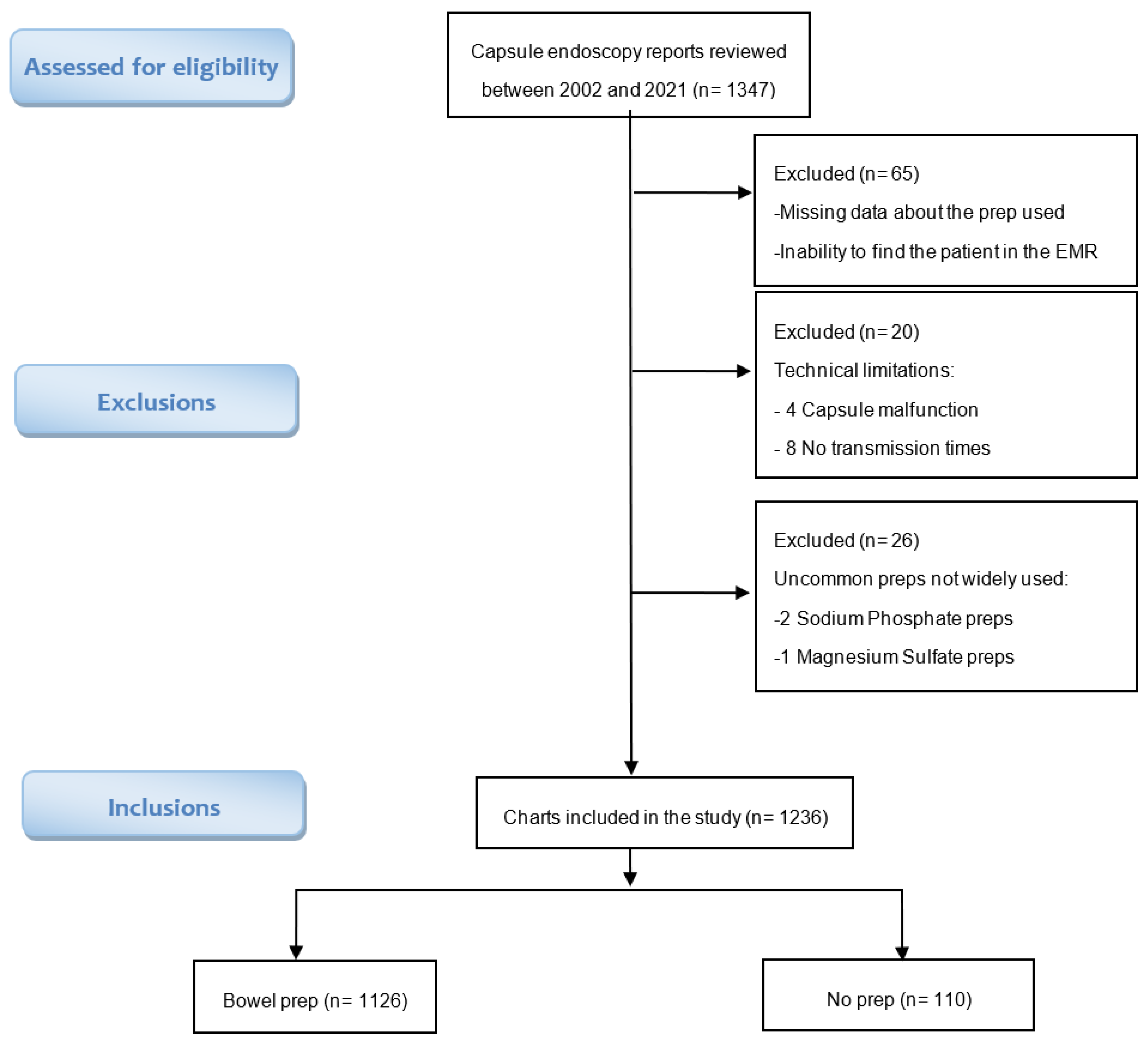
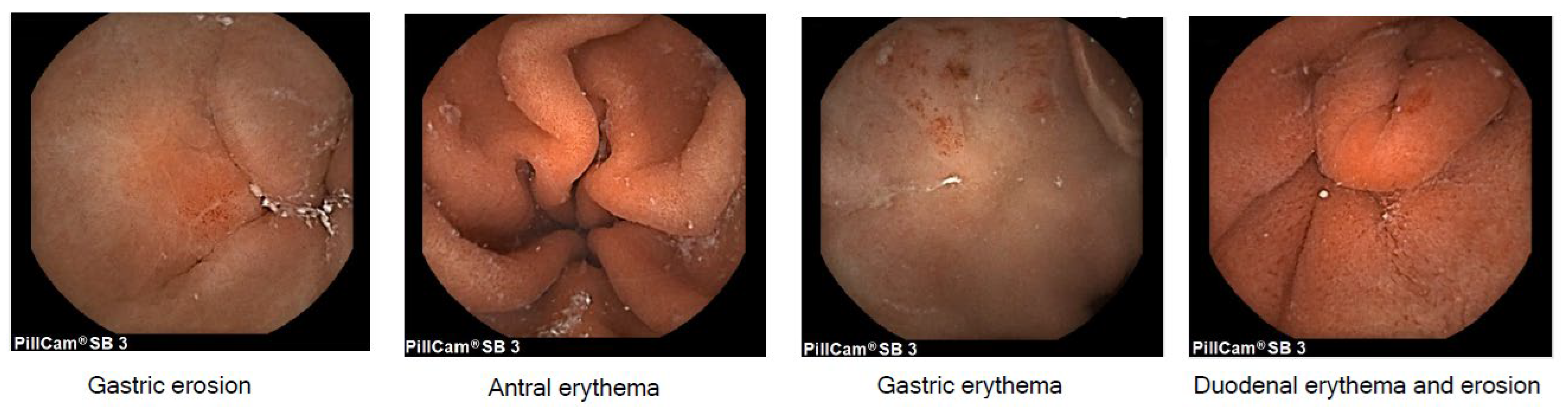
Data collection: Medical records were identified on PillCam SB3 software v9 database and reviewed for patient demographics, indication of the procedure, type of preparation prescribed, timing of preparation administration relative to the procedure, and gastroduodenal findings. Exclusion criteria included missing data regarding the preparation used, incomplete prep protocols, technical malfunctions, or the utilization of uncommon preparations that are not within the scope of this study (Figure 1). Gastroduodenal lesions were defined as erythematous lesions, ulcers, erosions, or inflammation found in the stomach or duodenum (Figure 2). Other lesions that are unlikely to be related to the prep such as masses, polyps, arteriovenous malformations (AVMs), angiodysplasias, angioectasia, and lymphangiectasia, were not counted as gastroduodenal lesions. The presence or absence of gastroduodenal lesions, as recorded on the capsule endoscopy procedure reports, was documented as a binary outcome (Yes/No).
Indications and preparations: The major indications for the capsule endoscopy were GI bleed (52%), IBD or suspicion of IBD (26%), iron deficiency anemia (19%), abnormal imaging or follow up on other conditions (3%).
Preparations studied included polyethylene glycol lavage (PEG), PEG plus bisacodyl (PEG + bis), bisacodyl (bis), and oral sulfate solution (OSS). Preparations were prescribed according to manufacturer’s instructions. OSS was prescribed as split dose, administered both on the evening before and on the day of the procedure. PEG was prescribed as 4L dosage administered on the evening preceding the procedure. The bisacodyl regimen was prescribed as a 20 mg dosage administered on the evening before the procedure. The PEG + bis combination was prescribed as bisacodyl 20 mg administered at noon and 2 L of PEG administered on the evening before the procedure. Patients were asked to not take non-steroidal anti-inflammatory drugs (NSAIDs) for seven days before undergoing capsule endoscopy. Most subjects were given pre-procedure metoclopramide 10 mg and simethicone 80 mg.
Statistical analysis: To analyze the data, categorical variables were reported in frequencies and percentages and compared using the Pearson’s chi-squared test (χ2). Continuous variables were reported as means and standard deviations or as medians and interquartile ranges and compared using analysis of variance (ANOVA). The statistical analysis was performed using the JMP statistical package. A p-value < 0.05 was considered statistically significant.
3. Results
Study cohort: A total of 1347 medical records were identified on the PillCam SB3 software database. A total of 111 records were excluded from the study due to missing data regarding the preparation used, incomplete prep protocols, technical malfunctions, or the utilization of uncommon preparations that are not within the scope of this study (Figure 1). Consequently, the final study group consisted of 1236 adult subjects.
Descriptive analysis: Among the 1236 patients included in the study, 1126 of them (91%) received a bowel preparation prior to the capsule endoscopy, while 110 patients (9%) did not receive any prep prior to the procedure. Among the 1126 patients who received bowel prep prior to the capsule endoscopy, 773 (68.6%) received oral sulphate solution (OSS), 178 (15.8%) received polyethylene glycol (PEG), 137 (12.2%) received bisacodyl only (bis), and 38 (3.4%) received polyethylene glycol and bisacodyl (PEG + bis). There were 498 (40.3%) men and 738 (59.7%) women in the study. The mean age of the study population was 56 years, with a standard deviation of 18 years. The mean age was similar across all the subgroups (ANOVA p = 0.8). Gender differed between the prep group and the no prep group with a male predominance (58%) in the no prep group and a female predominance (61%) in the prep subgroups (Table 1).
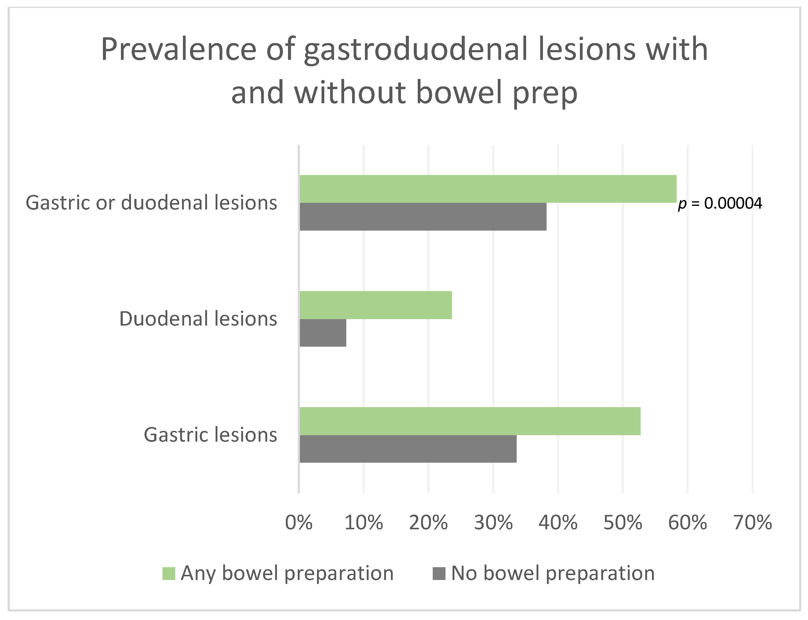
Primary outcomes: There was a significant difference between the rate of gastroduodenal lesions with prep and without prep. The patients who received bowel prep had gastric or duodenal findings in 58.3% of the cases, compared to 38.2% of the patients who did not receive any prep, indicating a relative risk (RR) of 1.53 (95% CI 1.19–1.94) and a p value of 0.00004 (Table 2, Figure 3).
The percentage of gastric lesions alone after any bowel preparation was 52.7% compared to 33.6% without any bowel prep with a statistically significant relative risk (RR) of 1.56 [95% CI 1.2–2] and a p value of 0.0001. Similarly, the relative risk (RR) of duodenal lesions alone was also statistically significant with 23.6% of the patients having duodenal lesions after any bowel prep compared to 7.3% without any bowel prep, yielding a relative risk (RR) of 3.2 [95% CI 1.6–6.4] and a p value < 0.0001. (Table 2, Figure 3). Although the increase in duodenal lesions after prep, RR 3.2 [95% CI 1.6–6.3], was higher than the increase in gastric lesions, RR 1.56 [95% CI 1.2–2], this difference did not reach statistical significance (CI overlap).
Subgroup analysis by bowel preparation type: Among the preps studied, the OSS and bis groups were significantly associated with gastroduodenal lesions. The patients who received OSS had gastric or duodenal lesions in 63.1% of the cases, compared to 38.2% of the patients who did not receive any prep yielding a relative risk (RR) of 1.65 [95% CI 1.3–2.1] and a p value < 0.0001. Bis alone was also highly associated with gastroduodenal lesions, with 62.7% of the patients having gastroduodenal lesions compared to 38.2% of the patients who did not receive prep yielding a relative risk (RR) of 1.64 (95% CI 1.25–2.15) and a p value of 0.0001.
This association was not found in the PEG or PEG + bis preparations. The prevalence of gastroduodenal lesions in the PEG group was 36.5% compared to 38.2% without prep, RR 0.96 (95% 0.7–1.3). Similarly, the prevalence of gastroduodenal lesions in the PEG + bis group was also not statistically significant, with a prevalence of 47.4% compared to 38.2% without prep, RR 1.24 (95% CI 0.82–1.87) (Figure 4).
Stratified Analysis for Suspected Inflammatory Bowel Disease (IBD): In light of the potential confounding effect of Crohn’s disease on upper gastrointestinal lesions, we conducted a stratified analysis focusing on patients who underwent capsule endoscopy due to suspected inflammatory bowel disease (IBD). This subset comprised 321 patients, accounting for approximately 25% of the total cohort and 25% of the gastroduodenal lesions detected. Consistent with the overall findings, a higher prevalence of lesions was observed among patients who underwent bowel preparation, particularly with (OSS) and (bis), compared to those who did not undergo bowel preparation.
While the percentages of lesions were very similar to the overall data (Table 3), statistical significance was somewhat attenuated due to the smaller sample size. Nevertheless, the statistical significance persisted for gastroduodenal lesions in both (OSS) and (bis) groups, as well as for duodenal lesions in the (OSS) group, and gastric lesions in the (bis) group. The patients with no prep had gastroduodenal findings in 35% of the cases, compared to 58% in the (OSS) group (RR 1.68, p = 0.03) and 78% in the (bis) group (RR 2.25, p = 0.003).
4. Discussion
Although it has been reported that some bowel preps may be associated with colonic lesions [1,2,3,4,5,6,7,8,9,10,11], this study is unique as it takes a different route and assesses the relation between different bowel preps and gastric and duodenal lesions. The prevalence of gastroduodenal lesions in the general population varies among studies and has been reported to be as high as 31% in patients without any bowel preparation [12]. In the present study, we found gastroduodenal lesions in 38.2% of the patients who did not receive a prep, compared to 58.3% who did receive bowel preparation, a statistically significant increase compared to the no prep group (RR 1.5; 95% CI 1.19–1.94). These lesions included erythema, ulcers, erosions, or inflammation. It is important to interpret these results with caution, as our study did not take into account the lesions that might be caused by known risk factors (NSAIDs, H. Pylori, inflammatory bowel disease, etc.). This study was not randomized, which may have introduced selection bias. The usage trends of different preparations changed over time, with no prep or PEG being more frequently used earlier in the study, with a median procedure date of March 2009. On the other hand, the OSS was used more frequently later in the study with a median procedure date of March 2018. The definition of gastroduodenal lesions and the manner it was reported was not changed over the study period. Assuming that other conditions are equally distributed between both groups, this study yields an attributable risk of 20.1% that could be the result of bowel preparation. PEG regimens appeared to be less correlated with these lesions and could be considered to mitigate lesion misinterpretation in certain conditions.
As prep-associated gastroduodenal lesions are not well described in the literature, we lack sufficient data regarding their exact mechanism. There are numerous factors that play a role in the integrity of the gastroduodenal mucosa and its defense mechanisms. These include the loss of prostaglandin defense observed in NSAID usage, immune and inflammatory responses triggered by antigens or autoimmune conditions like H. Pylori and IBD, oxidative stress and the release of oxygen radicals induced by factors such as ethanol and smoking, as well as reduced gastric blood flow and mucosal ischemia seen in stress ulcers [13,14,15]. It is crucial to recognize that there is a considerable overlap among these mechanisms, and that an injury cannot be attributed to a single factor alone. We suspect that prep-associated lesions, similar to chemical gastropathy, can be the result of a direct irritant effect exerted by the preparation used owing to the irritant properties of its chemicals, high osmolarity, PH alterations, and direct chemical contact with the mucosa. A possible mechanism is a breach in the gastric mucosal barrier or modulation of luminal substances (prostaglandins, gaseous mediators, and neuropeptides), rendering the mucosa more susceptible to acid-induced injury. Further studies are needed to investigate these mechanisms further.
The strength of this study is that the cohort is relatively large and that the upper gastrointestinal findings were not influenced by endoscopy manipulation or sedation. The limitations of this study include that this is a retrospective study, so patients were not randomized to take into consideration other risk factors for gastroduodenal lesions. There was a small gender imbalance between the prep and the no prep groups, and the 1 week restriction of NSAID use might not have been long enough to rule out the presence of NSAID injury. Since the examinations were by capsule endoscopy, histology was not available to evaluate for distinct features that could distinguish prep-related lesions from other lesions, which will be interesting to investigate in future studies. Most examinations were performed for iron deficiency or suspected or known inflammatory bowel disease. The group with no prep was small (9%).
The clinical implications of these observations remain uncertain, given that this is one of the first and largest studies describing these lesions. These lesions can explain the nausea, vomiting, and abdominal discomfort experienced by patients following prep ingestion. To our knowledge, these lesions have not resulted in major complications or necessitated therapeutic interventions. We suspect that these lesions could be clinically similar to those induced by other irritants such as chemicals, smoking, or alcohol. We hypothesize that these lesions resolve spontaneously as the mucosa undergoes repair and the offending agent is discontinued.
Understanding the etiology of gastroduodenal lesions is essential for accurate diagnosis and appropriate management. If the indication for an esophagogastroduodenoscopy at the time of colonoscopy is abdominal discomfort or bleeding, these gastroduodenal findings could be misinterpreted as the etiology of the indicated symptoms rather than the consequence of the preparation. This report describes findings unrelated to colonoscopy and upper gastrointestinal lesions proposed to be related to preparation. Endoscopists should be aware that preparations are associated with gastroduodenal lesions and should further pursue the etiology of upper gastrointestinal symptoms independent of a planned combined colonoscopy. Our recommendation is that the EGD and colonoscopy be separated when investigating upper GI symptoms. In addition, if the indication of capsule endoscopy is to evaluate for IBD, we recommend choosing certain preparations that might have a lower association with gastroduodenal findings to avoid confounding lesions and misdiagnosis.
The use of capsule endoscopy for the evaluation of the upper GI tract is expected to increase, as part of a panendoscopy or even with a magnetic capsule fostered by the development of AI technologies. If so, these study findings from a large population in USA should be considered in future clinical trials assessing the prevalence of gastroduodenal lesions in capsule endoscopy and its implications.
5. Conclusions
This study showed that patients who received bowel preparations displayed a significant increase in gastric and duodenal lesions. The clinical significance of these lesions remains undetermined. The increase was more pronounced in the duodenum. The frequency and prevalence of the findings varied across different preparations. Of the preparations studied, OSS was associated with a greater number of gastroduodenal lesions, while PEG was the least associated with lesions with an occurrence similar to the non-prep group. Endoscopists should be aware that bowel preparations are associated with gastroduodenal lesions, and try to avoid using certain preparations when evaluating upper GI symptoms or upper GI lesions in Crohn’s disease, to avoid the misinterpretation and misdiagnosis of these lesions.
Author Contributions
A.K. and C.G.M. examined and collected data, analyzed and contributed to the manuscript. J.A.D.P. conceptualized the project, collected data, analyzed results, and contributed to the writing of the manuscript. All authors have read and agreed to the published version of the manuscript.
Funding
This research received no external funding.
Institutional Review Board Statement
The study did not require ethical approval. This study did not meet the criteria of “clinical trial” as defined by the ICMJE.
Informed Consent Statement
Patient consent was waived for this retrospective study. All the patients’ data have been pooled and cannot be identified.
Data Availability Statement
The data is available in USA Health system databases upon request.
Acknowledgments
An earlier version of this manuscript has been presented as a poster in DDW 2022 in San Diego, CA, USA. https://www.giejournal.org/article/S0016-5107(22)00514-4/fulltext (accessed on 1 June 2022).
Conflicts of Interest
Di Palma is a consultant medical director of Braintree Laboratories a part of Sebela Pharmaceuticals. The other authors have no conflicts of interest.
References
- Anastassopoulos, K.; Farraye, F.A.; Knight, T.; Colman, S.; Cleveland, M.V.; Pelham, R.W. A Comparative Study of Treatment-Emergent Adverse Events Following Use of Common Bowel Preparations Among a Colonoscopy Screening Population: Results from a Post-Marketing Observational Study. Dig. Dis. Sci. 2016, 61, 2993–3006. [Google Scholar] [CrossRef] [PubMed]
- Belsey, J.; Epstein, O.; Heresbach, D. Systematic review: Adverse event reports for oral sodium phosphate and polyethylene glycol. Aliment. Pharmacol. Ther. 2009, 29, 15–28. [Google Scholar] [CrossRef] [PubMed]
- Chan, A.; Depew, W.; Vanner, S. Use of oral sodium phosphate colonic lavage solution by Canadian colonoscopists: Pitfalls and complications. Can. J. Gastroenterol. Hepatol. 1997, 11, 797486. [Google Scholar] [CrossRef] [PubMed]
- Zwas, F.R.; Cirillo, N.W.; Ei-Serag, H.B.; Eisen, R.N. Colonic mucosal abnormalities associated with oral sodium phosphate solution. Gastrointest. Endosc. 1996, 43, 463–466. [Google Scholar] [CrossRef] [PubMed]
- Hsu, M.H.; Chang, I.W.; Tai, C.M. White Spots in the Rectum. Gastroenterology 2016, 151, e15–e16. [Google Scholar] [CrossRef] [PubMed]
- Driman, D.K.; Preiksaitis, H.G. Colorectal inflammation and increased cell proliferation associated with oral sodium phosphate bowel preparation solution. Hum. Pathol. 1998, 29, 972–978. [Google Scholar] [CrossRef] [PubMed]
- Watts, D.A.; Lessells, A.M.; Penman, I.D.; Ghosh, S. Endoscopic and histologic features of sodium phosphate bowel preparation-induced colonic ulceration: Case report and review. Gastrointest. Endosc. 2002, 55, 584–587. [Google Scholar] [CrossRef] [PubMed]
- Rejchrt, S.; Bureš, J.; Široký, M.; Kopáčová, M.; Slezák, L.; Langr, F. A prospective, observational study of colonic mucosal abnormalities associated with orally administered sodium phosphate for colon cleansing before colonoscopy. Gastrointest. Endosc. 2004, 59, 651–654. [Google Scholar] [CrossRef] [PubMed]
- Hagège, H.; Laugier, R.; Nahon, S.; Coulom, P.; Isnard-Bagnis, C.; Albert-Marty, A. Real-life conditions of use of sodium phosphate tablets for colon cleansing before colonoscopy. Endosc. Int. Open 2015, 3, e346–e353. [Google Scholar] [CrossRef] [PubMed]
- Nam, S.; Choi, I.; Park, K.; Ryu, K.; Kim, B.; Sohn, D.; Nam, B.; Kim, C. Risk of hemorrhagic gastropathy associated with colonoscopy bowel preparation using oral sodium phosphate solution. Endoscopy 2010, 42, 109–113. [Google Scholar] [CrossRef] [PubMed]
- Wexner, S.D.; Beck, D.E.; Baron, T.H.; Fanelli, R.D.; Hyman, N.; Shen, B.; Wasco, K.E.; American Society of Colon and Rectal Surgeons; American Society for Gastrointestinal Endoscopy; Society of American Gastrointestinal and Endoscopic Surgeons. A consensus document on bowel preparation before colonoscopy: Prepared by a Task Force from The American Society of Colon and Rectal Surgeons (ASCRS), the American Society for Gastrointestinal Endoscopy (ASGE), and the Society of American Gastrointestinal and Endoscopic Surgeons (SAGES). Gastrointest. Endosc. 2006, 63, 894–909. [Google Scholar] [CrossRef] [PubMed]
- Fernández, J.F.J.; Sainz, I.F.-U.; Ollo, B.Z.; Dueñas, C.S.; Guimera, M.M.; González, A.E.; Costas, J.J.V. Gastroduodenal lesions detected during small bowel capsule endoscopy: Incidence, diagnostic and therapeutic impact. Rev. Esp. Enferm. Dig. 2018, 110, 102–108. [Google Scholar] [CrossRef]
- Wallace, J.L. Prostaglandins, NSAIDs, and Gastric Mucosal Protection: Why Doesn’t the Stomach Digest Itself? Physiol. Rev. 2008, 88, 1547–1565. [Google Scholar] [CrossRef] [PubMed]
- Bhattacharyya, A.; Chattopadhyay, R.; Mitra, S.; Crowe, S.E. Oxidative Stress: An Essential Factor in the Pathogenesis of Gastrointestinal Mucosal Diseases. Physiol. Rev. 2014, 94, 329–354. [Google Scholar] [CrossRef] [PubMed]
- Plummer, M.P.; Blaser, A.R.; Deane, A.M. Stress ulceration: Prevalence, pathology and association with adverse outcomes. Crit. Care 2014, 18, 213. [Google Scholar] [CrossRef] [PubMed]
Figure 1.
Flow chart of patients included and excluded in the retrospective study.

Figure 2.
Examples of gastroduodenal lesions as seen on Pillcam.

Figure 3.
Prevalence of gastroduodenal lesions of erythema, ulcers, erosions, or inflammation with and without bowel prep.
Figure 3.
Prevalence of gastroduodenal lesions of erythema, ulcers, erosions, or inflammation with and without bowel prep.

Figure 4.
Comparison of the gastroduodenal lesion prevalence between OSS, PEG, and no prep groups.


Table 1.
Demographic characteristics of the study population across the groups.
| Parameter | Total | OSS | PEG | Bis Only | PEG + bis | No Prep |
|---|---|---|---|---|---|---|
| N (%) | 1236 (100%) | 773 (62.5%) | 178 (14.4%) | 137 (11.1%) | 38 (3.1%) | 110 (8.9%) |
| Age, Mean (SD) | 56 (18) | 57 (17) | 56 (18) | 57 (18) | 57 (18) | 57 (16) |
| Sex, n (%) | ||||||
| Male | 498 (40.3%) | 305 (39%) | 71 (40%) | 15 (39%) | 43 (31%) | 64 (58%) |
| Female | 738 (59.7%) | 468 (61%) | 107 (60%) | 23 (61%) | 94 (69%) | 46 (42%) |
Table 2.
Patients with gastric or duodenal lesions by type of colon cleansing preparation.
| All Patients (N = 1236) | Gastric Findings | Duodenal Findings | Gastric or Duodenal Findings |
|---|---|---|---|
| Any prep (N = 1126) | 594 (52.7%) * | 266 (23.6%) * | 657 (58.3%) * |
| PEG (N = 178) | 57 (32%) | 16 (9%) | 65 (36.5%) |
| PEG + bis (N = 38) | 15 (39.5%) | 4 (10.5%) | 18 (47.4%) |
| Bis only (N = 137) | 81 (59.1%) * | 23 (16.8%) * | 86 (62.7%) * |
| OSS (N = 773) | 441 (57%) * | 223 (28.8%) * | 488 (63.1%) * |
| No prep (N = 110) | 37 (33.6%) | 8 (7.3%) | 42 (38.2%) |
* means statistically significant compared to no prep (p < 0.05).
Table 3.
Gastric or duodenal lesions by type of colon cleansing preparation stratified to “IBD suspicion” indication.
Table 3.
Gastric or duodenal lesions by type of colon cleansing preparation stratified to “IBD suspicion” indication.
| IBD Suspicion (N = 321) | Gastric Lesions | Duodenal Lesions | Gastric or Duodenal Findings |
|---|---|---|---|
| Any prep (N = 298) | 150 (50%) | 56 (19%) | 164 (55%) |
| PEG (N = 55) | 16 (29%) | 2 (4%) | 18 (33%) |
| PEG + bis (N = 11) | 4 (36%) | 3 (27%) | 6 (55%) |
| Bis only (N = 23) | 18 (78%) * | 3 (13%) | 18 (78%) * |
| OSS (N = 209) | 112 (54%) | 48 (23%) * | 122 (58%) * |
| No prep (N = 23) | 8 (35%) | 1 (4%) | 8 (35%) |
* means statistically significant compared to no prep (p < 0.05).
Disclaimer/Publisher’s Note: The statements, opinions and data contained in all publications are solely those of the individual author(s) and contributor(s) and not of MDPI and/or the editor(s). MDPI and/or the editor(s) disclaim responsibility for any injury to people or property resulting from any ideas, methods, instructions or products referred to in the content. |
© 2024 by the authors. Licensee MDPI, Basel, Switzerland. This article is an open access article distributed under the terms and conditions of the Creative Commons Attribution (CC BY) license (https://creativecommons.org/licenses/by/4.0/).
Share and Cite
MDPI and ACS Style
Khouri, A.; Moreno, C.G.; Di Palma, J.A. Bowel Cleansing Preparations Are Associated with Gastroduodenal Lesions. Gastrointest. Disord. 2024, 6, 359-367. https://doi.org/10.3390/gidisord6010024
AMA Style
Khouri A, Moreno CG, Di Palma JA. Bowel Cleansing Preparations Are Associated with Gastroduodenal Lesions. Gastrointestinal Disorders. 2024; 6(1):359-367. https://doi.org/10.3390/gidisord6010024
Chicago/Turabian StyleKhouri, Anas, Cesar G. Moreno, and Jack A. Di Palma. 2024. "Bowel Cleansing Preparations Are Associated with Gastroduodenal Lesions" Gastrointestinal Disorders 6, no. 1: 359-367. https://doi.org/10.3390/gidisord6010024