Article

Morphological and Optical Modification of Melanosomes in Fish Integuments upon Oxidation

1
School of Physics, University of Exeter, Stocker Road, Exeter EX4 4QL, UK
2
Department of Physics, Namur Institute of Structured Matter (NISM), Institute of Life, Earth and Environment (ILEE), University of Namur, Rue de Bruxelles 61, 5000 Namur, Belgium
3
Queensland Brain Institute, The University of Queensland, Brisbane, QLD 4072, Australia
4
School of the Environment, The University of Queensland, Brisbane, QLD 4072, Australia
5
Center for Photonics, Institute of Physics, University of Belgrade, Pregrevica 118, 11080 Belgrade, Serbia
6
Micro- and Nanophotonic Materials Group, University of Mons, Place du Parc 20, 7000 Mons, Belgium
*
Authors to whom correspondence should be addressed.
These authors contributed equally to this work.
Optics 2023, 4(4), 563-572; https://doi.org/10.3390/opt4040041
Submission received: 30 June 2023 / Revised: 25 October 2023 / Accepted: 30 October 2023 / Published: 1 November 2023

Abstract

:
Reactive oxygen species (ROS) such as superoxide radicals O2−, hydroxyl radicals OH−, and hydrogen peroxide H2O2 may have detrimental effects on marine organisms, including their integuments and visual appearances. Although some studies have described the impact of ROS on marine ecosystems and species ecology, the influence on the optical response of the integuments of marine species and on their visual appearances remains unknown. In this article, we used histology and optical characterisation to show, for the first time, that skin melanophores (melanin-containing chromophores) of the coral reef fish, Stegastes apicalis, change their shapes and fluorescent proprieties upon oxidation with H2O2 radicals. Our observations also suggest that pheomelanosomes may occur in fish integuments, where, previously, it was thought that fish melanosomes only contain eumelanin. This investigation relied on light and electron microscopy and steady-state fluorimetry, as well as time-resolved streak imaging systems. We suggest that the changes in the morphological and spectral characteristics of melanophores can be used as a marker of physiological stress induced by environmental factors such as ROS. Moreover, S. apicalis may be used as a potential model for studying the interaction between the surrounding environment and natural organisms in biologically diverse ecosystems, such as the Great Barrier Reef in Australia.

1. Introduction

Colors in nature play an essential role in various biological phenomena such as visual communication, camouflage, and aposematism [1,2,3]. Their origin may be either pigmentary or structural. In fishes, colors originate from different cells in the skin, chromatophores (i.e., containing pigments), leucophores (i.e., comprising light scatterers), and iridophores (containing photonic structures), and from the structure of the scales covering a fish’s body [1,3,4,5,6,7]. In addition to pigments, fish integuments can also emit light at different wavelengths by fluorescence decay [8,9,10]. Fishes use colors and patterns, amongst others, for social communication, mimicry, or camouflage and background adaptation [11,12,13,14].
Reactive oxygen species (ROS) such as superoxide radicals (O2), hydroxyl radicals (OH), and non-radical hydrogen peroxide (H2O2) significantly impact sea organisms [15,16,17,18], including their integuments. ROS can be produced in seawater via various abiotic and biotic processes, leading to the ionisation of organic or inorganic compounds, including the metabolisms of phytoplankton [19]; photolysis of water molecules; or radiolysis arising, e.g., from cosmic rays or 40K decay [15,16]. In addition, climate changes and anthropogenic marine pollution appear to increase the concentration of ROS in seawater, especially in surface layers [18,20,21].
Despite the observed effect of ROS on marine organisms [15,16,17,18], how ROS affect the visual appearance and the optical proprieties of animal skin had not yet been described. In this study, we investigated the influence of ROS on the skin melanophores occurring in a marine fish, namely Stegastes apicalis (De Vis, 1885), through histological and optical characterization. This damselfish species of the family Pomacentridae, commonly known as the Australian gregory or the yellowtip gregory, lives on the Great Barrier Reef off the Australian East coast, in depths of 1–5 m. S. apicalis has a dark brown body coloration, with yellow, orange, or red margins on the dorsal fin and the tip of the upper lobe of the caudal fin (Figure 1a) [22]. Melanophores are cells that appear black or dark brown, as they contain vesicles of melanin called melanosomes within the cytoplasm [23,24]. This research allowed us to get insights into the naturally occurring interactions between the surrounding environment and the visual appearances of marine animals, by tracking the physical and optical properties of the fish melanophores upon oxidation with H2O2 [25], akin to oxidation with ROS within the marine environment. The goal of our study was to highlight the potential effects of oxidation on the visual appearance and optical properties of the integuments of a marine species. Since this study is a new topic of research, we aim at setting a baseline upon which future research using more naturalistic scenarios can build. In addition, microscope-based observations of the integuments unveiled the likely presence of pheomelanosomes, contradicting the common assumption that fish melanophores purely consist of eumelansosomes [23]. Finally, the changes that we found in this study can serve as an indicator of environmentally induced physiological stress. Hence, S. apicalis emerges as a potential model to study the effects of ROS and similar stressors on the reef.

2. Materials and Methods

2.1. Sample Preparation and Treatment

Specimens ( n = 4 ) of S. apicalis were collected (GBRMPA permit number G17/381601; fishery permit number 180731) off the coast of Lizard Island, Queensland, Australia (S 14°41 16.5 , E 145°27 09.6 ), at depth of 1–4 m in August 2018. All four dorsal fins were fixed overnight in a 0.01 M phosphate buffer solution (PBS) containing 4% paraformaldehyde (PFA) and 2.5% glutaraldehyde (GA) before being transferred to a PBS solution with a pH ranging from 7.2 to 7.4. Such fixation protocols are widespread in the scientific literature for histological observations of melanosomes in biological organisms, including fish [26,27,28,29,30]. Ideally, our study should have been carried out with fresh or living tissues. However, in this pioneering study, practical feasibility left us with no choice but to fix the samples. Small areas of the dorsal fins were sampled using a scalpel and tweezers that are typically used for atomic force and electron microscopy following a standard procedure for the integuments of other biological organisms [31,32]. The tools were cleaned with 99.9999% isopropyl alcohol (IPA) prior to dissection. To assess the effect of the oxidative agent on the fluorescence emission from the integuments, some samples were incubated for 5 h in a 10% H2O2 weak-acid solution (pH between 4.5 and 5.0).

2.2. Light Microscopy

The instrument is based on a modified JENAVAL microscopic frame (manufactured by Carl Zeiss, Munich, Germany). The sample was placed on the existing mechanical stage, which was powered by a stepper motor, moving the sample vertically, with a 0.3-µm resolution. The removable prism deflected the beam and enabled the capture of bright field images on a Canon EOS 50D (Tokyo, Japan) digital camera. The images were recorded in reflection mode with a 40 × 0.65 objective.

2.3. Scanning Electron Microscopy

The histological observations were performed using a Tescan MIRA3 (Brno, Czech Republic) high-resolution field emission scanning electron microscope (FEG-SEM) with an accelerating voltage lower than 29 kV. Prior to any observation, the surface of samples was coated with a 2–3-nm-thin gold layer using a Quorum Technologies SC7620 Mini (Lewes, United Kingdom) sputter coater to prevent charging effects.

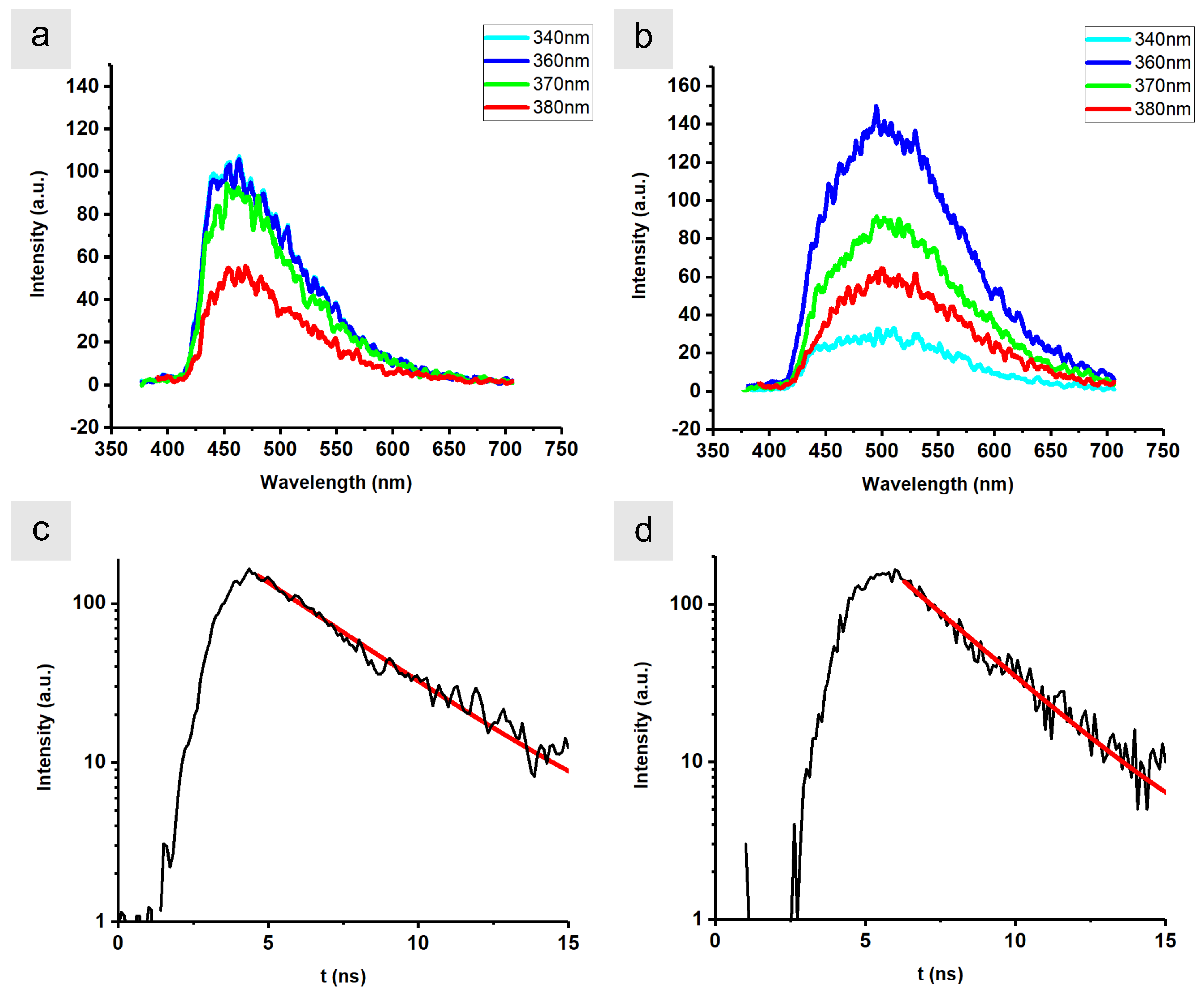
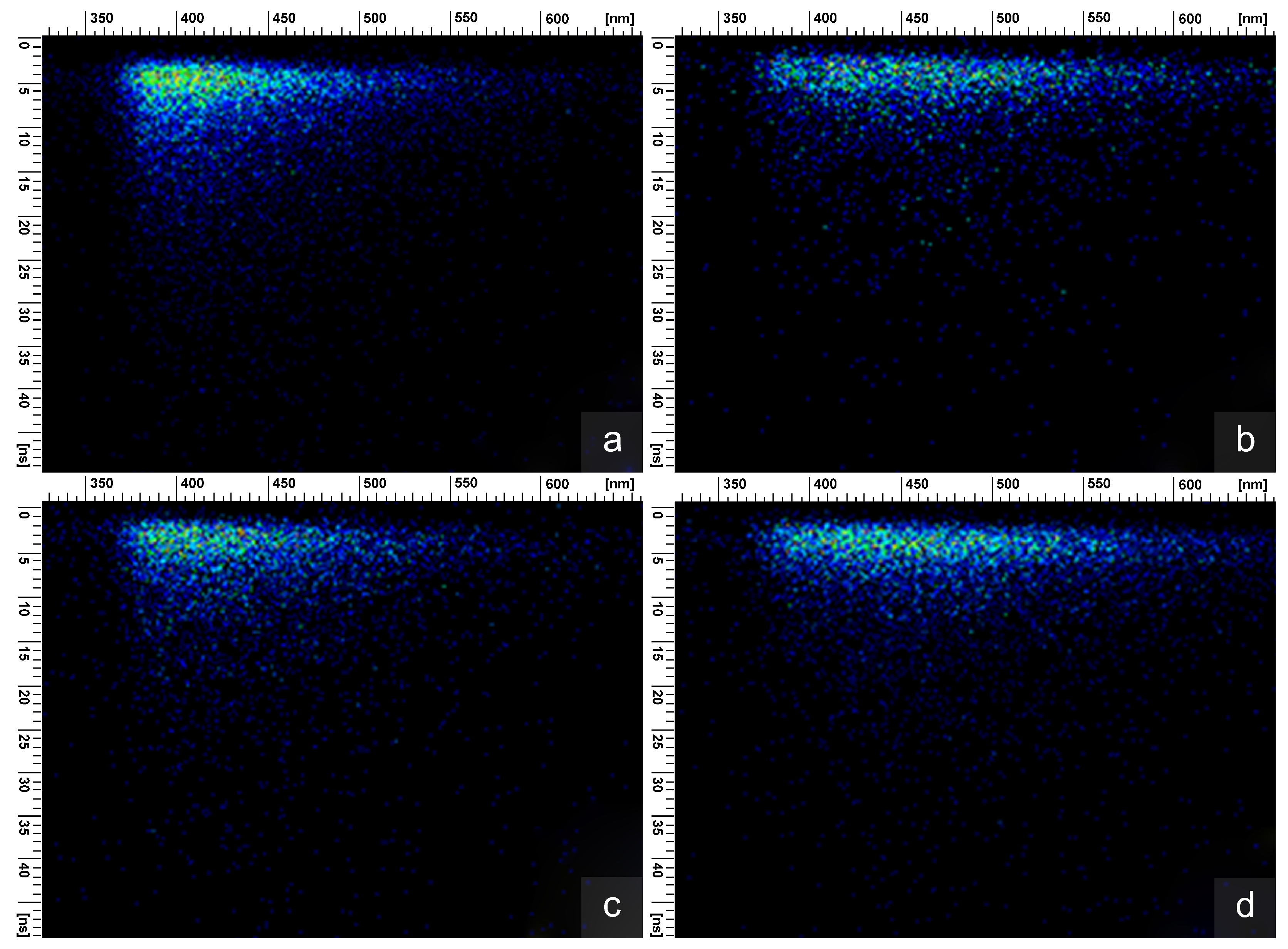
2.4. Time-Resolved and Steady-State Fluorimetry

The fluorimetry analysis was conducted using a pulsed light beam produced by a tunable Nd:YAG laser system (duration of 5 ns and repetition rate of 10 Hz) and by an FLSP920 UV–Vis–NIR spectrometer device (manufactured by Edinburgh Instruments, Livingston, UK). In the latter case, time-resolved measurements were recorded using a 60-W xenon lamp operating at a frequency of 100 Hz, and a Hamamatsu R928P (Hamamatsu City, Japan) photomultiplier tube was used to detect the emission signal in the visible region. In the former case, an optical parametric oscillator (OPO) was pumped by the fourth harmonic of the laser at 266 nm. The output of the OPO could be continuously tuned over a spectral range from 320 nm to 475 nm, and served as excitation beam. A time-resolved streak imaging system acquired the fluorescence steady-state emission spectra from the samples as a function of time. Full widths at half maximum (FWHM) were estimated using Origin 8.5.0 software, assuming a Gaussian function. The time-resolved streak imaging system used a Princeton Instruments SpectraPro 2300i (Trenton, NJ, USA) spectrograph, a Hamamatsu C4334-01 streak camera system, and the High-Performance Digital Temporal Analyzer Streak Camera Control software. The fundamental advantage of the streak camera is its two-dimensional nature that enables the acquisition of the temporal evolution of laser-induced phenomena. The camera was equipped with an image intensifier so that single photons could be detected, allowing the measurement of very-low-fluorescence intensities from the analyzed sample. The excitation and detection optical axes were aligned using a beam splitter to make it possible to adjust the angle of the excitation beam with respect to the sample surface, allowing a high sensitivity of detection using the single-mode fibers. The streak images were acquired in photon-counting mode with 1300 expositions. The time-resolved emission dynamics were observed at the maximum of the emission peak and fitted to single exponential functions, enabling the assessment of the related decay times using Origin software. Such fits by single exponential functions indicate that the excited molecules do not give rise to any non-radiative decay [33].

3. Results and Discussions

The integuments of the brown parts of the S. apicalis dorsal fin exhibit dark spots when observed by optical microscopy (Figure 1b). These spots are due to melanophores that contain melanin vesicles, called melanosomes, that abundantly cover the surface of the scales (Figure 2a) [34,35,36,37,38]. In nature, there are two types of melanosomes: eumelanosomes and pheomelanosomes. The former are rod-shaped and contain eumelanin, giving rise to black, dark brown, gray, and blonde appearances. The latter are spherical and contain pheomelanin, leading to red and brown colorations. In the case of the S. apicalis dorsal fin, the observed organelles were spherical with a diameter of 450–500 nm, likely corresponding to pheomelanosomes (Figure 2a). The consensus in the scientific community is that fish melanosomes only contain eumelanin [39]. However, it has previously been suggested that phenomelanin occurs in the integuments of a strain of the common carp (Cyprinus carpio) [40]. It is likely that pheomelanin in fishes is more common than assumed. Future studies accounting for phylogeny and species richness should be conducted to investigate its prevalence in fishes.
In addition to light absorption, melanin, as a polymer, and regardless of the type, is also believed to strengthen biological materials, thanks to covalent cross-linking processes [41,42,43,44,45,46,47,48]. For instance, melanin in birds has been shown to contribute to the mechanical properties of feathers [42,45,46,47].
Melanins usually do not fluoresce in their natural state under excitation by visible or UV light [49,50]. However, under excitation by UV light with a wavelength within the range of 340–380 nm, the S. apicalis integuments surprisingly emitted light (Figure 3a,c). The main advantage of using the streak camera as a detection system in fluorescence experiments lies in the fact that it allows the simultaneous recording of the time-resolved fluorescence spectra responses (Figure 4). The emission peaks of the S. apicalis integuments were found to be located in the range of 474–478 nm with a FWHM ranging from 86 nm to 101 nm. This light emission corresponds to fluorescence, with a decay time of about 2.2–2.7 ns (Table 1). We believe that this is mostly due to the autofluorescence of molecules such as collagen, flavins, and reduced nicotinamide adenine dinucleotide (NADH) [51,52]. In particular, the NADH fluorescence spectrum matches our measurements [52].
Upon oxidation by H2O2, both the structure and the optical properties of the melanosomes were modified. Histologically, they agglomerated into more substantial and connected structures (Figure 2b). In addition, the fluorescence emission spectra of the fish integuments were modified in terms of shape, peak position, and FWHM (Figure 3b). When excited with UV light (from 340 to 380 nm), the emission peaks were longer shifted in the range of 523–531 nm, with a larger FWHM of 122–130 nm. The decay time (Figure 3d and Table 1) was also significantly shorter (from 1.66 to 2.02 ns), as also seen on the streak camera (Figure 4). The different values of peak location, FWHM, and decay time qualitatively indicate that, when treated with H2O2, very different states were excited, resulting in significantly different relaxation kinetics. Most likely, the difference in the fluorescent signals was caused by different chemical entities.
On one hand, the oxidation of melanin is known to give rise to fluorescence [50,53,54]. The spectra measured in this study are very close to the spectra reported for oxidized melanin [50,54]. Organic fluorophores and pigments like melanin are usually cyclic or polycyclic compounds with OH and COOH functional groups [33,54]. These groups are possible sites for reduction–oxidation reactions [25,54,55] caused by reactive peroxide and oxygen anions arising from H2O2 decomposition. Oxidation by H2O2 affects the electronic configuration of the molecules, leading to fluorescence emission. On the other hand, the oxidation process quenches the autofluorescence from some of the fluorescent materials of the fish integuments. ROS, therefore, have a direct influence on the integumental structure and the chromophores of S. apicalis. The fluorescence emission measured in this study are likely to be negligible for the signaling and behavior of S. apicalis. This is because the quantum yields of natural fluorophores are usually low [56,57,58,59,60], UV light propagates only over short distances in seawater [61], and the proportion of UV light in sunlight and moonlight is limited (i.e., there are ca. 6% of UV and 50% of visible light in the sunlight at sea level) [62]. However, oxidation affects the fluorescence emission of the fish integuments and most likely their color, which could affect the ecology and behavior of fishes, including in intra- and interspecific communication, temperature control, reproduction, and survival. Moreover, older fishes may be more impacted by such ROS oxidation, due to the longer exposure time. This might explain the degradation of their visual appearance and the loss of color during aging. Further physiological and behavioral investigations are needed to test these hypotheses with living animals in their natural marine environment. If the fluorescence emission may only be a likely by-product of another biological function, it could be exploited to detect, identify, and quantify the effects of ROS on fish integuments.

4. Conclusions

This study contributes to the growing body of scientific literature describing the effects of oxidation on biological organisms under different environmental stressors. We first observed the presence of spherical melanosomes in the integuments of S. apicalis, which most likely correspond to pheomelanosomes. As it appears that pheomelanin in fishes could be more common than assumed, we call for future investigations accounting for phylogeny and species richness to establish its prevalence in fishes. In addition, and most importantly, we provide some of the first data on how ROS might impact the visual appearance of biological organisms in the marine environment. Following the oxidation of the integuments, the observed pheomelanosomes agglomerated and the fluorescence response was considerably modified, most likely due to the quenching of the integuments’ autofluorescence and the activation of fluorescence in the oxidized melanin. Hence, these changes can be tracked as markers for physiological stress caused by the surrounding environment, such as ROS. S. apicalis is here identified as a potential model for highlighting the consequences of such stress. The baseline data on how ROS affect the color of fishes that we have provided here create the possibility for more detailed investigations into the effects of ROS under natural conditions (e.g., clean versus polluted seawater). They also provide avenues to study, in detail, the ROS-induced agglomeration of melanosomes and the effects that changes in pH (e.g., due to ocean acidification) might have on these processes, as well as understanding the contributions from biotic and abiotic processes, including metabolic paths in marine environments.

Author Contributions

Conceptualization, S.R.M. and B.K.; methodology, S.R.M., V.L. and B.K.; validation, S.R.M., F.C., V.L., P.V., N.J.M. and B.K.; formal analysis, S.R.M., B.B., V.L. and B.K.; investigation, B.B. and V.L.; resources, S.R.M., F.C., N.J.M. and B.K.; data curation, B.B. and V.L.; writing—original draft preparation, S.R.M., V.L. and B.K.; writing—review and editing, F.C., P.V. and N.J.M.; visualization, B.B. and V.L.; supervision, B.K.; project administration, S.R.M. and B.K.; funding acquisition, S.R.M., F.C., N.J.M. and B.K. All authors have read and agreed to the published version of the manuscript.

Funding

S.R.M. was supported by the Belgian National Fund for Scientific Research (FRS-FNRS) (91400/1.B.309.18F) and a BEWARE Fellowship (Convention n°2110034) of the Walloon Region (COFUND Marie Skłodowska-Curie Actions of the European Union #847587) as a Postdoctoral Researcher. S.R.M. acknowledges a grant of the Agathon De Potter Fund of the Royal Academy of Science, Letters and Fine Arts of Belgium, a travel grant of FRS-FNRS, and a grant from the Namur Institute of Structured Matter (NISM). F.C. and N.J.M. were supported by the Australian Research Council (DE200100620 and FL140100197, respectively). BB and BK acknowledge the support of the of the EU: the EIC Pathfinder Challenges 2022 call through the Research Grant 101115149 (project ARTEMIS), the Office of Naval Research Global through the Research Grant N62902-22-1-2024 and the Ministry of Science, Technological Development and Innovation of the Republic of Serbia. B.K. also acknowledges financial support from FRS-FNRS; Inter-university Attraction Pole: Photonics@be (P7-35, Belgian Science Policy Office). The supporting bodies played no role in the design of the study, the collection of the samples, the analysis and interpretation of the data, and the writing of the manuscript.

Institutional Review Board Statement

The animal study protocol was approved by the Animal Ethics Committees of the Queensland Brain Institute of The University of Queensland (QBI/304/16-2018) and of the University of Belgrade (2019). The samples were collected in the framework of the Great Barrier Reef Marine Park Authority (GBRMPA) permit number G17/38160.1 and Fisheries Queensland permit number 180731. This research complies with European Community (EC) health rules as regards animal by-products and derived products not intended for human consumption (Regulation (EC) n°1069/2009—University of Namur, n°BE 705/0089).

Data Availability Statement

The data that support the findings of this study are available from the authors, upon reasonable request.

Acknowledgments

We acknowledge the Dingaal, Ngurrumungu, and Thanhil peoples as traditional owners of the lands and waters of the Lizard Island region from where the specimens were collected. We would like to thank the staff at Lizard Island Research Station (LIRS), Anne Hoggett, and Lyle Vail for support during field work. All authors acknowledge the help of colleagues from the Institute of Physics in Serbia and the KULeuven in Belgium in the preliminary phase of the presented study.

Conflicts of Interest

The authors declare no conflict of interest.

References

  1. Kinoshita, S. Structural Colors in the Realm of Nature; World Scientific: Singapore, 2008. [Google Scholar]
  2. Mouchet, S.R.; Vukusic, P. Structural colours in lepidopteran scales. In Advances in Insect Physiology; Elsevier: London, UK, 2018; Volume 54, pp. 1–53. [Google Scholar] [CrossRef]
  3. Mouchet, S.R.; Deparis, O. Natural Photonics and Bioinspiration; Artech House: Norwood, MA, USA, 2021; Available online: https://us.artechhouse.com/Natural-Photonics-and-Bioinspiration-P2221.aspx (accessed on 28 June 2023).
  4. Denton, E. Reflectors in Fishes. Sci. Am. 1971, 224, 64–72. [Google Scholar] [CrossRef]
  5. Brown, F., Jr. Chromatophores and color change. In Comparative Animal Physiology; WB Saunders: Philadelphia, PA, USA, 1973; pp. 915–950. [Google Scholar]
  6. Hawkes, J.W. The structure of fish skin. Cell Tissue Res. 1974, 149, 147–158. [Google Scholar] [CrossRef] [PubMed]
  7. Kelsh, R.N.; Schmid, B.; Eisen, J.S. Genetic analysis of melanophore development in zebrafish embryos. Dev. Biol. 2000, 225, 277–293. [Google Scholar] [CrossRef]
  8. Michiels, N.; Anthes, N.; Hart, N.; Herler, J.; Meixner, A.J.; Schleifenbaum, F.; Schulte, G.; Siebeck, U.E.; Sprenger, D.; Wucherer, M.F. Red fluorescence in reef fish: A novel signalling mechanism? BMC Ecol. 2008, 8, 16. [Google Scholar] [CrossRef] [PubMed]
  9. Lagorio, M.G.; Cordon, G.B.; Iriel, A. Reviewing the relevance of fluorescence in biological systems. Photochem. Photobiol. Sci. 2015, 14, 1538–1559. [Google Scholar] [CrossRef]
  10. Marshall, J.; Johnsen, S. Fluorescence as a means of colour signal enhancement. Philos. Trans. R. Soc. B Biol. Sci. 2017, 372, 20160335. [Google Scholar] [CrossRef]
  11. Bond, C. Biology of Fishes; WB Sounders Company: Philadelphia, PA, USA, 1979. [Google Scholar]
  12. Stepien, C.A. Color pattern and habitat differences between male, female and juvenile giant kelpfish (Blennioidei: Clinidae). Bull. Mar. Sci. 1987, 41, 45–58. [Google Scholar]
  13. Hakkinen, J.; Korhonen, H.; Oikari, A.; Karjalainen, J. Melanin concentrations in vendace (Coregonus albula) Whitefish (Coregonus lavaretus) Larvae Five Boreal lakes with different optical properties. Boreal Environ. Res. 2003, 8, 193–202. [Google Scholar]
  14. Stieb, S.M.; Cortesi, F.; Sueess, L.; Carleton, K.L.; Salzburger, W.; Marshall, N.J. Why UV vision and red vision are important for damselfish (Pomacentridae): Structural and expression variation in opsin genes. Mol. Ecol. 2017, 26, 1323–1342. [Google Scholar] [CrossRef]
  15. Blough, N.V.; Zepp, R.G. Reactive oxygen species in natural waters. In Active Oxygen in Chemistry; Springer: Dordrecht, the Netherlands, 1995; pp. 280–333. [Google Scholar]
  16. Manjun, Z. Determination of photochemically-generated reactive oxygen species in natural water. J. Environ. Sci. 2009, 21, 303–306. [Google Scholar]
  17. Kieber, D.J.; Peake, B.; Scully, N.M. Reactive oxygen species in aquatic ecosystems. In UV Effects in Aquatic Organisms and Ecosystems; Helbling, E.W., Ed.; Royal Society of Chemistry: London, UK, 2003; pp. 251–276. [Google Scholar]
  18. Morris, J.J.; Rose, A.L.; Lu, Z. Reactive oxygen species in the world ocean and their impacts on marine ecosystems. Redox Biol. 2022, 52, 102285. [Google Scholar] [CrossRef] [PubMed]
  19. Fan, S.M. Photochemical and biochemical controls on reactive oxygen and iron speciation in the pelagic surface ocean. Mar. Chem. 2008, 109, 152–164. [Google Scholar] [CrossRef]
  20. Müller, R.; Desel, C.; Steinhoff, F.S.; Wiencke, C.; Bischof, K. UV-radiation and elevated temperatures induce formation of reactive oxygen species in gametophytes of cold-temperate/Arctic kelps (Laminariales, Phaeophyceae). Phycol. Res. 2012, 60, 27–36. [Google Scholar] [CrossRef]
  21. Celis-Plá, P.S.; Moenne, F.; Rodríguez-Rojas, F.; Pardo, D.; Lavergne, C.; Moenne, A.; Brown, M.T.; Huovinen, P.; Gómez, I.; Navarro, N.; et al. Antarctic intertidal macroalgae under predicted increased temperatures mediated by global climate change: Would they cope? Sci. Total Environ. 2020, 740, 140379. [Google Scholar] [CrossRef]
  22. Allen, G.R.; Emery, A.R. A review of the pomacentrid fishes of the genus Stegastes from the Indo-Pacific, with descriptions of two new species. In Indo-Pacific Fishes; Bishop Museum: Honolulu, HI, USA, 1985; Volume 3, pp. 1–31. [Google Scholar]
  23. Sköld, H.N.; Aspengren, S.; Cheney, K.L.; Wallin, M. Fish Chromatophores—From Molecular Motors to Animal Behavior. Int. Rev. Cell Mol. Biol. 2016, 321, 171–219. [Google Scholar] [CrossRef]
  24. Salis, P.; Lorin, T.; Laudet, V.; Frédérich, B. Magic Traits in Magic Fish: Understanding Color Pattern Evolution Using Reef Fish. Trends Genet. 2019, 35, 265–278. [Google Scholar] [CrossRef]
  25. Smith, R.; Garrett, B.; Naqvi, K.; Fülöp, A.; Godfrey, S.; Marsh, J.; Chechik, V. Mechanistic insights into the bleaching of melanin by alkaline hydrogen peroxide. Free Radic. Biol. Med. 2017, 108, 110–117. [Google Scholar] [CrossRef]
  26. Goda, M.; Fujii, R. Coloration and Chromatophores of the Domino Damsel, Dascyllus trimaculatus. Zool. Sci. 2001, 18, 165–174. [Google Scholar] [CrossRef]
  27. Futter, C.E.; Ramalho, J.S.; Jaissle, G.B.; Seeliger, M.W.; Seabra, M.C. The Role of Rab27a in the Regulation of Melanosome Distribution within Retinal Pigment Epithelial Cells. Mol. Biol. Cell 2004, 15, 2264–2275. [Google Scholar] [CrossRef]
  28. Franzen, A.J.; Cunha, M.M.; Miranda, K.; Hentschel, J.; Plattner, H.; da Silva, M.B.; Salgado, C.G.; de Souza, W.; Rozental, S. Ultrastructural characterization of melanosomes of the human pathogenic fungus Fonsecaea pedrosoi. J. Struct. Biol. 2008, 162, 75–84. [Google Scholar] [CrossRef]
  29. Amiri, M.; Shaheen, H.M. Chromatophores and color revelation in the blue variant of the Siamese fighting fish (Betta splendens). Micron 2012, 43, 159–169. [Google Scholar] [CrossRef] [PubMed]
  30. Ali, S.A.; Naaz, I. Comparative light and electron microscopic studies of dorsal skin melanophores of Indian toad, Bufo melanostictus. J. Microsc. Ultrastruct. 2014, 2, 230–235. [Google Scholar] [CrossRef]
  31. Mouchet, S.R.; Van Hooijdonk, E.; Welch, V.L.; Louette, P.; Colomer, J.F.; Su, B.L.; Deparis, O. Liquid-induced colour change in a beetle: The concept of a photonic cell. Sci. Rep. 2016, 6, 19322. [Google Scholar] [CrossRef]
  32. Dhungel, B.; Ohno, Y.; Matayoshi, R.; Iwasaki, M.; Taira, W.; Adhikari, K.; Gurung, R.; Otaki, J.M. Distal-less induces elemental color patterns in Junonia butterfly wings. Zool. Lett. 2016, 2, 4. [Google Scholar] [CrossRef]
  33. Lakowicz, J.R. Introduction to fluorescence. In Principles of Fluorescence Spectroscopy; Springer: New York, NY, USA, 1999; pp. 1–23. [Google Scholar]
  34. Lindgren, J.; Moyer, A.; Schweitzer, M.H.; Sjövall, P.; Uvdal, P.; Nilsson, D.E.; Heimdal, J.; Engdahl, A.; Gren, J.A.; Schultz, B.P.; et al. Interpreting melanin-based coloration through deep time: A critical review. Proc. R. Soc. B Biol. Sci. 2015, 282, 20150614. [Google Scholar] [CrossRef] [PubMed]
  35. Donatti, L.; Fanta, E. Morphology of the retina in the freshwater fish Metynnis Roosevelti Eiggenmann (Characidae, Serrasalminae) and the effects of monochromatic red light. Rev. Bras. Zool. 1999, 16, 151–173. [Google Scholar] [CrossRef]
  36. Kawauchi, H. Functions of Melanin-Concentrating Hormone in Fish. J. Exp. Zool. 2006, 305A, 751–760. [Google Scholar] [CrossRef]
  37. Wolnicka-Glubis, A.; Pecio, A.; Podkowa, D.; Kolodziejczyk, L.M.; Plonka, P.M. Pheomelanin in the skin of Hymenochirus Boettgeri (Amphibia: Anura: Pipidae). Exp. Dermatol. 2012, 21, 535–561. [Google Scholar] [CrossRef]
  38. Colleary, C.; Dolocan, A.; Gardner, J.; Singh, S.; Wuttke, M.; Rabenstein, R.; Habersetzer, J.; Schaal, S.; Feseha, M.; Clemens, M.; et al. Chemical, experimental, and morphological evidence for diagenetically altered melanin in exceptionally preserved fossils. Proc. Natl. Acad. Sci. USA 2015, 112, 12592–12597. [Google Scholar] [CrossRef]
  39. Kottler, V.A.; Künstner, A.; Schartl, M. Pheomelanin in fish? Pigment Cell Melanoma Res. 2015, 28, 355–356. [Google Scholar] [CrossRef]
  40. Xu, P.; Zhang, X.; Wang, X.; Li, J.; Liu, G.; Kuang, Y.; Xu, J.; Zheng, X.; Ren, L.; Wang, G.; et al. Genome sequence and genetic diversity of the common carp, Cyprinus Carpio. Nat. Genet. 2014, 46, 1212–1219. [Google Scholar] [CrossRef]
  41. Riley, P. Materia Melanica: Further Dark Thoughts. Pigment Cell Res. 1992, 5, 101–106. [Google Scholar] [CrossRef] [PubMed]
  42. Bonser, R.H.C. Melanin and the Abrasion Resistance of Feathers. Condor 1995, 97, 590–591. [Google Scholar]
  43. Liu, Y.; Hong, L.; Wakamatsu, K.; Ito, S.; Adhyaru, B.; Cheng, C.Y.; Bowers, C.R.; Simon, J.D. Comparison of Structural and Chemical Properties of Black and Red Human Hair Melanosomes. Photochem. Photobiol. 2005, 81, 135–144. [Google Scholar] [CrossRef] [PubMed]
  44. Moses, D.N.; Harreld, J.H.; Stucky, G.D.; Waite, J.H. Melanin and Glycera Jaws. J. Biol. Chem. 2006, 281, 34826–34832. [Google Scholar] [CrossRef] [PubMed]
  45. Schreiber, R.W.; Schreiber, E.A.; Peele, A.M.; Burtt, E.H. Pattern of Damage to Albino Great Frigatebird Flight Feathers Supports Hypothesis of Abrasion by Airborne Particles. Condor 2006, 108, 736–741. [Google Scholar] [CrossRef]
  46. Pannkuk, E.L.; Siefferman, L.M.; Butts, J.A. Colour phases of the eastern screech owl: A comparison of biomechanical variables of body contour feathers. Funct. Ecol. 2010, 24, 347–353. [Google Scholar] [CrossRef]
  47. Galván, I.; Solano, F. Bird Integumentary Melanins: Biosynthesis, Forms, Function and Evolution. Int. J. Mol. Sci. 2016, 17. [Google Scholar] [CrossRef]
  48. Wang, Y.; Li, T.; Wang, X.; Ma, P.; Bai, H.; Dong, W.; Xie, Y.; Chen, M. Superior Performance of Polyurethane Based on Natural Melanin Nanoparticles. Biomacromolecules 2016, 17, 3782–3789. [Google Scholar] [CrossRef]
  49. Huang, Z.; Zeng, H.; Hamzavi, I.; Alajlan, A.; Tan, E.; McLean, D.I.; Lui, H. Cutaneous melanin exhibiting fluorescence emission under near-infrared light excitation. J. Biomed. Opt. 2006, 11, 034010. [Google Scholar] [CrossRef]
  50. Fernandes, B.; Matamá, T.; Guimarães, D.; Gomes, A.; Cavaco-Paulo, A. Fluorescent quantification of melanin. Pigment Cell Melanoma Res. 2016, 29, 707–712. [Google Scholar] [CrossRef] [PubMed]
  51. Zellweger, M.; Goujon, D.; Conde, R.; Forrer, M.; van den Bergh, H.; Wagnières, G. Absolute autofluorescence spectra of human healthy, metaplastic, and early cancerous bronchial tissue in vivo. Appl. Opt. 2001, 40, 3784–3791. [Google Scholar] [CrossRef] [PubMed]
  52. Pu, Y.; Wang, W.; Yang, Y.; Alfano, R.R. Native fluorescence spectra of human cancerous and normal breast tissues analyzed with non-negative constraint methods. Appl. Opt. 2013, 52, 1293–1301. [Google Scholar] [CrossRef]
  53. Sachs, H.W. Über die autogenen Pigmente, besonders das Lipofuscin und seine Abrenzung von Melanin. Beitr. Pathol. Anat. Allg. Pathol. 1943, 108, 268–314. [Google Scholar]
  54. Kayatz, P.; Thumann, G.; Luther, T.T.; Jordan, J.F.; Bartz–Schmidt, K.U.; Esser, P.J.; Schraermeyer, U. Oxidation Causes Melanin Fluorescence. Investig. Ophthalmol. Vis. Sci. 2001, 42, 241–246. [Google Scholar]
  55. Sarathy, S.R.; Mohseni, M. The impact of UV/H2O2 advanced oxidation on molecular size distribution of chromophoric natural organic matter. Environ. Sci. Technol. 2007, 41, 8315–8320. [Google Scholar] [CrossRef]
  56. Iriel, A.; Lagorio, M.G. Is the flower fluorescence relevant in biocommunication? Naturwissenschaften 2010, 97, 915–924. [Google Scholar] [CrossRef]
  57. Iriel, A.; Lagorio, M.G. Implications of reflectance and fluorescence of Rhododendr. Indicum Flowers Biosignaling. Photochem. Photobiol. Sci. 2010, 9, 342–348. [Google Scholar] [CrossRef]
  58. Taboada, C.; Brunetti, A.E.; Pedron, F.N.; Carnevale Neto, F.; Estrin, D.A.; Bari, S.E.; Chemes, L.B.; Peporine Lopes, N.; Lagorio, M.G.; Faivovich, J. Naturally occurring fluorescence in frogs. Proc. Natl. Acad. Sci. USA 2017, 114, 3672–3677. [Google Scholar] [CrossRef]
  59. Ladouce, M.; Barakat, T.; Su, B.L.; Deparis, O.; Mouchet, S.R. Scattering of ultraviolet light by avian eggshells. Faraday Discuss. 2020, 223, 63–80. [Google Scholar] [CrossRef]
  60. Mouchet, S.R.; Verstraete, C.; Bokic, B.; Mara, D.; Dellieu, L.; Orr, A.G.; Deparis, O.; Van Deun, R.; Verbiest, T.; Vukusic, P.; et al. Revealing natural fluorescence in transparent insect wings by linear and nonlinear optical techniques. J. Lumin. 2023, 254, 119490. [Google Scholar] [CrossRef]
  61. Hulburt, E.O. The Penetration of Ultraviolet Light into Pure Water and Sea Water. J. Opt. Soc. Am. 1928, 17, 15–22. [Google Scholar] [CrossRef]
  62. Moan, J. Visible light and UV radiation. In Radiation at Home, Outdoors and in the Workplace; Brune, A., Hellborg, R., Persson, B., Pääkkönen, R., Eds.; Scandinavian Science Publisher: Oslo, Norway, 2001; pp. 69–85. [Google Scholar]
Figure 1. (a) Stegastes apicalis displays a brown color in daylight with yellow, orange, or red margins on its dorsal fin and the tip of the upper lobe of its caudal fin. (b) The brown color is due to melanophores occurring in the integuments of the fish; (a) was reproduced from Dr Anne Hoggett, https://commons.wikimedia.org/wiki/File:StegastApicalAnneHoggett.jpg, accessed on 26 June 2023.
Optics 04 00041 g001
Figure 2. Representative micrographs of Stegastes apicalis dorsal fin skin sections. (a) Melanin granules, called melanosomes, are found in the integuments of the S. apicalis dorsal fin, as observed here by scanning electron microscopy, prior to oxidation by H2O2. (b) Upon oxidation by H2O2, these melanosomes agglomerate. Please note that (a,b) correspond to different magnifications.
Optics 04 00041 g002
Figure 3. Upon excitation by UV light (the wavelength of which ranged from 340 nm to 380 nm), the integuments of the Stegastes apicalis dorsal fin emitted light by fluorescence. (a) The emission peak was located around 475 nm, with a FWHM of ca. 90-95 nm. (b) Upon oxidation by H2O2, the emission peak changed shape and shifted to ca. 530 nm, with a FWHM larger than 120 nm. The time-resolved measurements of the emitted intensities with an excitation wavelength of 360 nm were fitted with single exponential functions (the quality of the fits is given by coefficients of determination R 2 = 0.980 (c) and R 2 = 0.983 (d), respectively), allowing the assessment of the decay times of the integuments (c) before and (d) after oxidation.
Optics 04 00041 g003
Figure 4. Upon oxidation with H2O2, the time-resolved fluorescence response of the Stegastes apicalis dorsal fin integuments considerably changes. Fluorescence emission was recorded with a streak camera with various excitation wavelengths, before (a,c) and after (b,d) oxidation. Horizontal profiles represent emission spectra and vertical profiles represent time-resolved measurements at selected excitation and emission wavelengths. (a) λ ex = 340 nm before oxidation, (b) λ ex = 340 nm after oxidation, (c) λ ex = 360 nm before oxidation, (d) λ ex = 360 nm after oxidation.
Optics 04 00041 g004
Table 1. Upon oxidation, the fluorescence response of the Stegastes apicalis dorsal fin integuments changed in terms of shape, peak wavelength λ max , decay time τ , and FWHM.
Wavelength (nm)
340360370380
λ max , no oxidation (nm)474476474478
λ max , oxidation (nm)523524528531
τ no oxidation (ns)2.702.252.692.61
τ oxidation (ns)1.661.751.922.02
FWHM no oxidation (nm)939486101
FWHM oxidation (nm)122130126126
Disclaimer/Publisher’s Note: The statements, opinions and data contained in all publications are solely those of the individual author(s) and contributor(s) and not of MDPI and/or the editor(s). MDPI and/or the editor(s) disclaim responsibility for any injury to people or property resulting from any ideas, methods, instructions or products referred to in the content.

