Systematic Review

Systematic Review: JAK-STAT Regulation and Its Impact on Inflammation Response in ARDS from COVID-19

by
Irasema Rodriguez
* and
Kate J. F. Carnevale
*
Dr. Kiran C. Patel College of Allopathic Medicine, Nova Southeastern University, Fort Lauderdale, FL 33328, USA
*
Authors to whom correspondence should be addressed.
Immuno 2024, 4(2), 147-158; https://doi.org/10.3390/immuno4020010
Submission received: 26 February 2024 / Revised: 25 April 2024 / Accepted: 8 May 2024 / Published: 14 May 2024

Abstract

:
Severe acute respiratory syndrome coronavirus 2 (SARS-CoV-2) infection has had a global impact and resulted in millions of deaths worldwide. The course of the Janus kinase signaling transducers and activators (JAK-STAT) pathway is an important molecular pathway that is involved in the cellular response to various cytokines and growth factors promoting an inflammatory response. The overactivation of the JAK-STAT signaling pathway in coronavirus disease 2019 (COVID-19) and its effect on acute respiratory distress syndrome (ARDS)-induced inflammatory processes was observed in various clinical articles that focused on JAK-STAT regulation regarding angiotensin converting enzyme 2 (ACE2) expression and cytokine storm release. Down-regulation of the JAK-STAT signaling pathway through inhibitors decreases the inflammatory response by decreasing cytokine storm release. However, the increased regulation of JAK-STAT in severe COVID-19 patients caused cytokines such as interferon alpha (IFN-α) to promote the phosphorylation of STATs. This response indicated an imbalance with JAK-STAT regulation and its inability to induce the transcription of interferon stimulated response elements. Furthermore, an increase in ACE2 regulation was noted to also increase JAK-STAT signaling, yet the down-regulation of JAK-STAT signaling can result in the overexpression of ACE2 by binding to SARS-CoV-2 and increasing STAT1 expression. Data suggest that inflammatory cytokines enhance the activation of ACE2 in endothelial cells via JAK-STAT pathway. Increasing the regulation of the JAK-STAT signaling pathway enhances the release of cytokines such as tumor necrosis factor alpha (TNF-α) and interferon gamma (IFN-γ), further expressing ACE2. The expression of ACE2 regulates STAT1 and STAT2 expression, leading to the up-regulation of the inflammasomal complexes in hyper-inflammatory responses from the JAK-STAT pathway. Through the review of various clinical reports, the effect of the JAK-STAT signaling pathway on ARDS-induced inflammatory response was observed and correlated with the expression of ACE2 and cytokine storm release in severe COVID-19 cases.

1. Introduction

In late December of 2019, Wuhan, Hubei Province, China reported the first cases of severe acute respiratory syndrome coronavirus 2 (SARS-CoV-2) viral infection, preceding the global outbreak of 2019 coronavirus disease (COVID-19) [1]. The worldwide pandemic has resulted in the deaths of more than 6 million individuals [2]. In the last three years, extensive research has been undertaken to further understand SARS-CoV-2 and its mechanism of disease; however, various countries continue to experience outbreaks of COVID-19 due to the emergence of mutant variants of the virus. SAR-CoV-2 is a single-stranded positive-sense RNA virus surrounded by a phospholipid membrane covered with a series of box spike glycoproteins [2]. This virus is a novel betacoronavirus belonging to the same subgenus as the severe acute respiratory syndrome coronavirus (SARS-CoV) and the middle east reparatory syndrome coronavirus (MERS-CoV), which are linked to respiratory, enteric, hepatic, and neurological diseases [2]. In severe COVID-19, viral pneumonia from SARS-CoV-2 infection progresses to acute respiratory distress syndrome (ARDS) [2,3].
ARDS is considered an expeditious progressive non-cardiogenic pulmonary edema, which can present with dyspnea, tachypnea, and hypoxia, quickly leading to respiratory failure [3]. ARDS occurs after a pulmonary or extra-pulmonary insult initiates the release of inflammatory mediators, thus stimulating inflammatory cells that build up in the alveoli and microcirculation of the lungs [3]. This inflammatory response can lead to severe damage within the endothelium and alveolar epithelium of the lungs, which can result in complications such as pulmonary edema, hyaline membrane formation, decreased lung compliance, and decreased gas exchange, which can quickly decline into respiratory failure. Pulmonary inflammation in the ARDS molecular pathways involves impaired immune response and uncontrollable inflammatory innate response [3]. When the pandemic began, the understanding of COVID-19 disease progression was limited and therefore the therapeutic management of this viral disease was inadequate to meet the growing needs. Currently, a variety of therapeutic options are available that include antiviral drugs, anti-SARS-CoV-2 monoclonal antibodies, anti-inflammatory drugs, and immunomodulator agents, which are available under the FDA-Issued Emergency Use Authorization or are being evaluated for the management of COVID-19 [2,4,5,6]. The application of these treatments must be specified based on the gravity of the illness or various risk factors [2].
There are two phases in the clinical course of SARS-CoV-2 infection. In the early phase, the viral replication is greatest before (or soon after) the onset of symptoms. During this phase, antiviral medication and antibody-based treatment are more likely to halt viral processes to clear the infection [2]. The later phase of the viral infection is marked by a hyperinflammatory state induced by cytokines’ release. Treatment options for this phase include anti-inflammatory drugs such as corticosteroids, immunomodulating therapies, or a combination thereof [5]. This later inflammatory phase of the infection has the highest rate of mortality [5].
Various molecular pathways are involved in the pathogenesis of SARS-CoV-2 [3,7]. The Janus kinase signaling transducers and activator of transcription (JAK-STAT) pathway is an important molecular pathway that is implicated in the pathogenesis of inflammatory and autoimmune diseases [8]. The cellular response to dozens of cytokines and growth factors is mediated by the JAK-STAT signaling pathway [8,9]. The responses that activate this pathway include proliferation, differentiation, migration, apoptosis and cell survival, depending on the signal, tissue, and cellular context [8]. Pathway activation occurs via cytokine receptor-ligand binding, which causes the phosphorylation of JAK [10]. Trans-phosphorylated JAKs begin to phosphorylate downstream substrates such as receptors and STATs [10]. Once the STATs are activated, they enter the nucleus and bind as dimers or a complex oligomer to further target genes, regulating their transcription and transmitting cytokine signals [11]. Observations suggested that a cytokine storm associated with SARS-CoV-2 infection overactivates JAK-STAT signaling in target cells to further increase inflammatory response and up-regulate tissue factors [10]. An increase in the serum level of pro-inflammatory cytokines, such as interleukin 6 (IL-6), has been shown to act as a pivotal component in cytokine storm release by further recruiting natural killer cells (NKCs), T-lymphocytes, macrophages, neutrophils, and chemokines to aid with the inflammatory response [11]. Overactivation of JAK-STAT signaling amplifies the pathologic effect of SARS-CoV-2 infection, and a significant correlation between angiotensin-converting enzyme 2 (ACE2) and the JAK-STAT signaling pathway has been observed [9,12]. In standard conditions, it has been indicated that the JAK-STAT signaling pathway is involved in the downstream action of the overactivation of ACE2 [12]. ACE2 is a close homologue of angiotensin-converting enzyme (ACE) and functions as a negative regulator of the angiotensin system [13]. Specifically, in COVID-19, ACE2 is a significant membrane entry receptor for SARS-CoV-2 cellular infection [13]. In the lungs, ACE2 is used to protect against acute lung injury; however, SARS-CoV-2 progression causes the downregulation of ACE2 [13]. The downregulation of ACE2 impacts the function of angiotensin II (Ang II) and the renin–angiotensin–aldosterone system (RAAS) in a variety of tissues, and contributes to lung injury [13].
The role of the JAK-STAT signaling pathway in COVID-19 and its involvement in the inflammatory response that is induced by SAR-CoV-2 infection, leading to the development of ARDS, comprise an extensive and intricate process. While many excellent review articles have been published addressing COVID-19’s pathogenesis [14,15,16], ARDS [3,17,18], and the JAK-STAT signaling pathway [19,20,21], this manuscript focuses on a systematic review of clinical and observational trials conducted at the intersection of these topics to gauge the cellular effects. This review aims to elucidate the current understanding of the intricate interplay between JAK-STAT overactivation and ARDS progression in severe COVID-19, along with what is currently understood about the role of ACE2. The various clinical reports on the effects of regulation of the JAK-STAT signaling pathway on ARDS-induced inflammatory response, including the role of ACE2 and cytokine storm release related to severe COVID-19, are discussed here.

2. Methods

A systematic literature review was conducted using the search engines PubMed, Google Scholar, and ScienceDirect. The filters used were free full text, clinical trials, and randomized controlled trials. Publications from the years 2019–2024 were selected. Articles were included if they contained the key words (a) COVID-19 and/or SARS-CoV-2 infection and (b) JAK-STAT signaling pathway or inflammation, in combination with (c) ACE inhibitors and/or (d) ARDS. Clinical trials published in English were the main articles that were reviewed. Additionally, articles that did not relate to JAK-STAT or inflammatory pathways were excluded. Figure 1 below shows a schematic representation of the systematic literature review, which followed PRISMA guidelines (Figure 1). Summary data and citations for each article analyzed is available in Table 1 below.

3. Results

3.1. JAK-STAT Role in COVID-19 Pathology

Cytokine storm and inflammation are contributors to the systemic symptoms experienced by COVID-19 patients. Researchers have found that cytokine signals induce the JAK signaling cascade through receptor activation by the phosphorylation of the intracellular tails of the receptor, as summarized in Table 1 and Figure 2 [7]. Receptor phosphorylation allows the attachment of members of the STAT family of transcription factors, thus causing dissociation and translocation to the nucleus [7]. Inflammation is elevated in COVID-19 with an increase in IL-6, IL-2, IL-15, and IL-10. IL-6 binds to IL-6 receptor glycoprotein 130, activating JAK-STAT signaling in numerous cells, promoting the release of chemokines and monocytes and neutrophil recruitment [20]. Other cytokines like IL-2 bind to natural killer cells, triggering JAK signaling and increasing NK cytotoxicity, yet IL-15 regulates NK function through JAK1 activation [27]. Furthermore, Ravid and co-workers found that JAK-STAT signaling is activated in epithelial cells and amplifies the production of von Willebrand factor (vWF) and angiopoietin-2, increasing both the vascular and pulmonary endothelium [7]. The activation of endothelial cells then releases tissue factor (TF). However, in SARS-CoV-2-infected cells, the overactivation of JAK-regulated mechanisms causes TF elevation and increases coagulopathy [7]. The increase in TF is related to the suppressed levels of ACE2 in SARS-CoV-2-infected cells. While ACE2 expression is suppressed, angiotensin-2 (ANG-II) is upregulated, increasing TF expression and microvascular thrombosis in COVID-19 patients [28]. The current understanding is that JAK-STAT signaling in COVID-19 patients causes an increase in inflammatory cytokine activation as well as the upregulation of various coagulation cascade factors, such as ANG-II, vWF, and TF.

3.2. JAK-STAT Effects on Cytokines

While studies have been conducted, there has been no direct evidence definitively linking the involvement of specific pro-inflammatory cytokines or chemokines in the pathology of COVID-19 [22]. A correlation has, however, been demonstrated between the severity of the disease and the increase in concentration of cytokines and chemokines in the serum. Pro-inflammatory cytokines such as interleukins (IL)-2, -4, -6, -7, and -10, tumor necrosis factor alpha (TNF-α), and interferon gamma (IFN-γ) were identified in patients with COVID-19 who were hospitalized [22]. While other inflammatory pathway mechanisms can contribute, these and other cytokines and chemokines are released by the distinct intracellular signaling pathways mediated by the JAK signaling pathway [3,19]. Downregulation of the JAK-STAT singling pathway through JAK-STAT inhibitors was found to reduce the pro-inflammatory process induced by SARS-CoV-2 infection [22]. An observational longitudinal trial was conducted using the immunomodulatory medication baricitinib, where 88 patients (44 female and 44 male) who were hospitalized in the Unit of Internal Medicine at the University Hospital of Vernon and Pederzoli Hospital of Peschiera were treated with 4 mg of baricitinib twice a day for 2 days, followed by 4 mg once per day for 7 days [22]. The analysis of the downregulation on molecular targets of baricitinib activity was observed with patients that acquired COVID-19-related pneumonia and displayed an increase in phosphorylated STAT3 (p-STAT3). The application of baricitinib expressed a significant inhibition of p-STAT3 four days after administration in various cells, such as T-lymphocytes, NKCs, monocytes, and neutrophils [22]. The levels of p-STAT3 and change in immune phenotype were compared with serum-derived cytokine levels and antibodies against severe acute respiratory response, resulting in a significant reduction in IL-1B, IL-6, and TNF-α plasma concentrations for patients taking baricitinib [22], while control groups displayed no reduction, indicating that baricitinib treatment inhibits cytokine storm release by regulating the JAK-STAT pathway.
Datasets of healthy volunteers and patients with moderate to severe COVID-19 infections were analyzed to determine the pro-inflammatory cytokines circulating during infection [26]. This dataset included treatment of bone marrow-derived macrophages (BMDMs) with cytokines, such as IL-6, IL-18, IL-15, IL-2, IL-1α, IL-1β, TNF-α and IFN-γ, in various combinations known as cocktails. It was noted that out of 28 combinations tested, combinations with TNF-α and IFN-γ induced an inflammatory response leading to cell death and cytokine storm. Yet, combinations that did not have TNF-α and IFN-γ failed to cause cell death, confirming their importance in inflammation-induced cell death [26]. The production of STAT1 in patients with severe COVID-19 and BMDMs treated with TNF-α and IFN-γ was observed and cells inhibiting STAT1 production were protected from inflammatory cell death [21,26]. Evidently, their findings suggested that STAT1 contributed to inflammatory cell death in severe SARS-CoV-2 patients. Mice that lacked TNF-α receptors displayed reduced lung inflammation and reduced mortality rate following infection [26]. To understand the implications of TNF-α- and IFN-γ-blocking treatments, neutralizing antibodies against TNF-α and IFN-γ were tested. Mice injected with the antibodies displayed neutralized TNF-α and IFN-γ and were protected from inflammatory cell death [29]. To clearly examine the efficacy during a SARS-CoV-2 infection, the murine SARS-CoV-2 infection model was used, and by day 7 post-infection, almost all control-treated mice were infected [29]. Antibody usage against TNF-α and IFN-γ decreased STAT1 production in the JAK-STAT signaling pathway, reducing cell death [26].
In another study by Sataker and co-workers, the up-regulation of the STAT1 in the JAK-STAT signaling pathway was observed in patients with severe COVID-19 and highly impaired IFN type I, reducing IFN-α production and activity, which are considered proinflammatory cytokines [30]. IFN-α is considered to represent a broad class of cytokines, elicited upon challenge to the host defense, and is essential for mobilizing immune defense [31]. JAK phosphorylation, meditated by cytokine receptor activation, leads to the phosphorylation of STATs that translocate into the nucleus to trigger translation of inflammatory mediators [31]. STAT1 signaling is regulated through phosphorylation and is part of the IFN-mediated viral responses that induces complexes such as interferon-stimulated gene factor three (ISGF3), which is known for the application of IFN-mediated signals [23]. To evaluate the functionality of type I and II IFN signaling, peripheral blood mononuclear cells (PBMCs) were obtained from 20 health individuals and 30 COVID-19 patients, then a cytokine storm was stimulated with low doses of either IFN-α or IFN-γ for 48 h [23]. The stimulation induced by IFN-α led to the transcriptional upregulation of STAT1 in CD3+ T-cells and CD19+ B-cells for healthy controls and CD19+ B-cells in mild COVID-19 cases [23]. After stimulation with a low-dose IFN-α or IFN-γ for 48 h, phosphorylation of STAT1 was unchanged for the healthy controls and mildly affected COVID-19 patients. However, severely affected COVID-19 patients showed increased levels of p-STAT1 [23]. Cytokines, including IFNs, use the JAK-STAT signaling pathway with a highly common system of heterogenous molecules that conduct specific signaling through a defined receptor complex that may recruit different STATs and lead to distinct downstream transcription. Therefore, the increased phosphorylation of STAT1 in patients with severe COVID-19 was indicative of the imbalance in the JAK-STAT signaling pathway, inducing transcription of interferon-stimulated response elements (IRSE) to promote interferon signaling for cytokine release [23]. Additionally, 85 confirmed COVID-19 cases were obtained to analyze the immune dysregulation of SARS-CoV-2. This study included 12 critically ill, 20 ward, 1 discharged, and 52 ambulatory patients. Acute samples were gathered during the patient’s hospital stays or within 2 weeks post symptom onset for outpatients [26]. Convalescent samples were obtained after hospital discharge or 26 days post symptom onset for outpatients [26]. Cytokines/chemokines in moderate and severe acute and convalescent COVID-19 plasma samples were observed. It was noted that IL-6 is an important cytokine that drives inflammation responses in SAR-CoV-2 infection. Therefore, the level of IL-6 was measured in moderate and severe and convalescent SARS-CoV-2 samples, where IL-6, IL-8, and IL-10 were significantly upregulated in acute samples [26]. IL-6 and IFN-γ levels were also linked to acute samples from various patients, compared to healthy controls, expressing a rapid innate immune response towards infection [26]. Similar to the results found in the study by Rincon-Arevalo et al., the levels of IFN-γ and IL-6 increased cytokine storm release and the regulation of the JAK-STAT pathway in COVID-19 for immune hyperactivation [22].

3.3. Involvement of ACE2 on JAK-STAT Signaling Pathway

SARS-CoV-2 spike protein shares similar nucleotide sequences and common receptors with ACE2. The gene expression of ACE2 was investigated and correlated with the JAK-STAT signaling pathway [24]. A microarray of datasets from the Gene Expression Omnibus (GEO) database, included 174 human airway epithelial (HAE) samples, with 89 samples from COVID-19 patients, were analyzed by Luo and coworkers [24]. These were further divided into nine groups based on different time points after initial infection. The correlation of ACE2 and specific genes related to the JAK-STAT pathway in airway epithelial cells was analyzed in cells with or without SARS-CoV-2 infection [24]. The association of high ACE2 expression with the presence of members of the JAK-STAT pathway (such as JAK2, STAT1, STAT2, STAT4, and STAT5A) indicated that the activation of ACE2 in SARS-CoV-2 infection is related to type I and II IFNs and the STAT family [24]. It was noted that the increased expression of ACE2 can induce further overactivation of the JAK-STAT signaling pathway and continue to produce an immune response after SARS-CoV-2 infection has been cleared, potentially contributing to the reported “long COVID-19” symptoms [24]. Downregulation of the JAK-STAT pathway can lead to the decreased distribution of interferon-stimulated genes and an increase in STAT1 expression. This results in the overexpression of the ACE2 receptor, which binds SARS-CoV-2 for viral entry into the host cell in severe COVID-19 patients [30].
TNF-α/IFN-γ amplification has been related to associated inflammation in SARS-CoV-2 regarding ACE2 receptors and their correlation with the JAK-STAT pathway. SARS-CoV-2 uptake receptors, such as ACE2 and DPP4, were compared in senescent and non-senescent human umbilical vein endothelial cells (HUVECs) [25]. Data showed that inflammatory cytokines, TNF-α, and IFN-γ have synergistically enhanced activation of ACE receptors in endothelial cells via the JAK-STAT pathway [25]. HUVECs were cultured and stimulated with cytokines for 5–7 days to detect whether viral entry via receptors was present in senescent HUVECs and non-senescent cells. Expression patterns of ACE2 were detected and correlated with the inflammation of the JAK-STAT pathway. It was noted that endothelial cell injury coupled with excessive inflammation is a key driver of COVID-19 severity. TNF-α, IFN-γ, and IL-6 are important markers of cellular senescence and COVID-19 receptor expression [32]. The increased SARS-CoV-2 viral uptake via receptors such as ACE2 is enhanced by TNF-α and IFN-γ in endothelial cells via the JAK-STAT pathway [25]. Senescent cells were shown to increase the burden with aging patients, and data suggested that inflammation and infection are lower in younger children compared to adults. This could be related to the low expression of ACE2 that regulates ANGII signaling and ANGII receptors type 1 and type 2, which play a major role in controlling inflammation [8,16]. These receptors were higher in senescent cells than non-senescent cells, and it was observed that they could contribute to senescent cell burden in patients with severe infection [24]. The HUVECs were exposed to TNF-α or IFN-γ alone or in combination for 3 days and ACE2 expression was analyzed via Western blot. The analysis observed a higher ACE2 expression in cells exposed to TNF-α or IFN-γ alone or in combination for 3 days than in controls [33]. Furthermore, the JAK-STAT pathway was then tested to explain the role of cytokine-induced ACE2 receptor expression in correlation with inflammation. STAT3 phosphorylation increases in response to IFN-γ, which was higher under the upstream regulator, while phosphorylation of STAT1 was increased with a combination of TNF-α and IFN-γ [8,16]. Additionally, the combination of TNF-α and IFN-γ triggered upregulation for inflammasomal complexes in hyper-inflammatory responses of the JAK-STAT pathway and the excessive activation of amplified, prolonged, and sustained STAT1 inflammation [25].

4. Discussion

While the JAK-STAT signaling pathway acts in part as a natural defense to trigger the immune system’s containment of pathogenic attack, as summarized in Table 1 and Figure 2, the inflammation response in COVID-19 can be linked to the involvement and overactivation of the JAK-STAT pathway in various molecular factors, leading to cytokine storm and negative outcomes for patients. It has been observed that autoimmune destruction of immune factors can lead to adverse outcomes for patients experiencing viral infection [34,35,36]. However, overactivation is also harmful, as seen when cytokines such as TNF-α and IFN-γ were observed at a high concentration in the serum of patients that were diagnosed with severe COVID-19 [22]. It was noted that cytokines and chemokines such as IL-2, IL-4, IL-6, IL-7, IL-10, TNF-α, and IFN-γ are released by intracellular signaling pathways that are controlled by the JAK-STAT pathway [22]. Baricitinib, a JAK ½ inhibitor, has been observed to downregulate JAK-STAT and its effects on the inflammatory response, including autoimmune disfunction and viral activation of cytokine storm [9]. It was observed that the downregulation imposed by baricitinib treatment allowed the molecular inhibition of p-STAT3. In patients with a severe acute respiratory response, it was noted that cytokine levels of IL-1B, IL-6, and TNF-α were reduced significantly in the plasma concentrations of patients taking baricitinib [22]. Limited correlation was observed for in vitro studies which challenged cells with the SARS-CoV-2 spike protein alone [37]. Conversely, a clinical trial using an inhaled pan-JAK inhibitor (1–4), nezulcitinib, did not show statistically significant improvement in hospitalized COVID-19 patients compared to placebo-treated controls, but may still be advantageous for use in certain at risk populations [38]. It was concluded by Bronte et al. that the downregulation of the JAK-STAT pathway via baricitinib inhibits cytokine storm release, effectively decreasing the inflammation response [22].
In contrast, a study by Sataker and co-workers examined the up-regulation of the JAK-STAT signaling pathway in patients with severe COVID-19 and observed IFN type 1 impairment which reduced IFN-α production and activity [30]. It was noted that STAT1 was lower among severely affected COVID-19 patients compared to those who with only mild COVID-19 symptoms. The expression of STAT1 was found to be lower in patients diagnosed with severe COVID-19; however, p-STAT1 levels increased among these patients based on IFN signaling [30]. These implications were said to be the cause of viral factors that can impair the proper function of STAT1 when stimulated with IFN-α or IFN-γ. Furthermore, the induced stimulation of these cytokines leads to an up-regulation of p-STAT1 in severe COVID-19 patients after stimulation with a low dose of IFN-α or IFN-γ. This was especially evident in cultured PBMCS (CD19+ B cells and CD3+ cells), where only a small increase in IFN-α or IFN-γ was needed to increase p-STAT1 [30]. The increase in phosphorylated STAT1 suggested the dysregulation of the JAK-STAT signaling pathway in response to stimulation with a low dose of IFN-α or IFN-γ. While there have been various reports of differences in the observations made in vitro versus in vivo for treatment and inflammatory progression for COVID-19 patients, the results of this study seem to indicate the cytokine release measurable in PBMCS can be correlated with the severity of patient symptoms [37,39]. The limitations of the Sataker study involved a limited understanding of the molecular mechanism regarding the upregulation of p-STAT signaling due to reduced levels of IFN regulation [30]. Understanding this mechanism proficiently in the future will assist in patient therapies. Furthermore, in the study performed by Klein et al., TNF-α and IFN-γ were observed in severe COVID-19 and BMDM-treated cells. Cells that inhibited STAT1 production, thus inhibiting JAK-STAT signaling, protected the cells from cell death via inflammatory response. Antibody treatment in mouse models also supported these findings, suggesting that the role that STAT1 plays in the inflammatory response in SARS-CoV-2 infection is important, and that low production via JAK-STAT leads to low mortality in cells during infection. The pursuit of inhibition of the IL-6 receptor, through antibodies and other means, to stem the cascade of JAK-STAT overactivation is also a viable therapeutic option, and has been reviewed elsewhere [40,41]. Additional targets for disease-causing genetic characteristics are being investigated and may yield viable routes for treatments and severe disease prevention [42].
The involvement of the JAK-STAT signaling pathway in inflammation is not limited to cytokines; implications on ACE2 receptors have also been observed. A correlation between ACE2 and specific gene expression related to the JAK-STAT pathway, such as JAK2, STAT1, STAT2, STAT4, and STAT5, has been documented [24]. Primarily, the activation of ACE2 in COVID-19 infection has been linked to type 1 and II IFNs and STAT upregulation. The increase in ACE2 induces an increase in the STAT signaling pathway, continuing an active immune response even after the SARS-CoV-2 infection has been cleared. This overexpression was noted in human airway epithelial cells in various cells, such as B cells, macrophages, helper T cells 1, and CD8+T cells. The correlation between ACE2 and the JAK-STAT signaling pathway indicated that these pathways are involved in the downstream action of the overactivation of ACE2 by type I/II IFNs. Baricitinib is also believed to inhibit nuclear-factor-kappa-beta activating kinase (NAK), which facilitates viral endocytosis following viral binding to ACE2, further mitigating viral infection [9].
Similarly, TNF-α and IFN-γ have been directly linked to the JAK-STAT pathway and its association with the inflammation process. According to a recent study by Kandyhaya-Pillai, inflammatory cytokines such as TNF-α and IFN-γ are linked to the enhanced activation of ACE receptors, not only in endothelial cells via the JAK-STAT pathway [25]. It was expressed that underlying conditions and advanced age played a factor in relation to the severe complications of COVID-19, including cytokine storm release, which can be connected to immune response dysregulation [25]. The increased replication rate of SARS-CoV-2 after cellular entry via ACE2 receptors was enhanced by TNF-α and IFN-γ in endothelial cells, and this was also observed in senescent HUVECs. STAT3 phosphorylation increased in response to IFN-γ, while STAT1 increased when interacting with both TNF-α and IFN-γ. TNF-α and IFN-γ enhanced the expression of SARS-CoV-2 viral entry receptors, amplified senescence-associated inflammation, and started a positive feedback loop via the hyper-activation of the JAK-STAT pathway in endothelial cells [25]. A key limitation of this study is the narrow age range of patients for which data were collected. Clinical trials are currently being conducted to further analyze the molecular mechanism of JAK and cytokine-driven cellular senescence and the link between inflammation and SARS-CoV-2 viral entry into host cells.
In summary, the JAK-STAT signaling pathway is heavily affected by the expression of ACE2 and cytokines during a SARS-CoV-2 infection (Table 1 and Figure 2). The upregulation of ACE2 can lead to the overactivation of the JAK-STAT signaling pathway, increasing the inflammation response. This was seen when ACE2 receptors were exposed to cytokines such as TNF-α and IFN-γ, which further increased the ACE2 response. Furthermore, an increase in phosphorylated STAT1 and STAT3 related to cytokine dosage was observed to affect the downregulation of JAK-STAT signaling. The downregulation of JAK-STAT promotes the phosphorylation of STAT complexes to drive a required immune response. In the future, the enhancement of human clinical trials to observe JAK-STAT signaling regulation and its effects on ACE2 and cytokine release should be further assessed. For example, a clear molecular model of a cytokine storm should be further explored in the future to obtain a better understanding of the effect the JAK-STAT pathway and its relationship with cytokine storm release. Trials for cytokine inhibition, such as IL-6, have also been discussed and could be used in the future to assist JAK-STAT signaling as well. The regulation of various viral diseases like COVID-19 can be better understood and controlled when understanding how the JAK-STAT signaling pathway is associated with the body’s immune response.

Author Contributions

I.R. conducted the literature review, created the tables and figures, and authored the first drafts of the manuscript. K.J.F.C. consulted on content, search parameters, and focus of the systematic review, provided feedback and manuscript editing, and assisted with the review and revision process. All authors have read and agreed to the published version of the manuscript.

Funding

This research received no external funding. Publication costs were covered by the NSU PFRDG fund.

Data Availability Statement

No new data were created for this article.

Conflicts of Interest

The authors declare no conflicts of interest.

References

  1. Chan, J.F.-W.; Kok, K.-H.; Zhu, Z.; Chu, H.; To, K.K.-W.; Yuan, S.; Yuen, K.-Y. Genomic Characterization of the 2019 Novel Human-Pathogenic Coronavirus Isolated from a Patient with Atypical Pneumonia after Visiting Wuhan. Emerg. Microbes Infect. 2020, 9, 221–236. [Google Scholar] [CrossRef]
  2. Cascella, M.; Rajnik, M.; Cuomo, A.; Dulebohn, S.C.; Di Napoli, R. Features, Evaluation and Treatment Coronavirus (COVID-19); StatPearls—NCBI Bookshelf: Treasure Island, FL, USA, 2020. [Google Scholar]
  3. Matthay, M.A.; Zemans, R.L. The Acute Respiratory Distress Syndrome: Pathogenesis and Treatment. Annu. Rev. Pathol. Mech. Dis. 2011, 6, 147–163. [Google Scholar] [CrossRef] [PubMed]
  4. Lui, G.; Guaraldi, G. Drug Treatment of COVID-19 Infection. Curr. Opin. Pulm. Med. 2023, 29, 174–183. [Google Scholar] [CrossRef] [PubMed]
  5. Stasi, C.; Fallani, S.; Voller, F.; Silvestri, C. Treatment for COVID-19: An Overview. Eur. J. Pharmacol. 2020, 889, 173644. [Google Scholar] [CrossRef]
  6. Bramante, C.T.; Buse, J.B.; Liebovitz, D.M.; Nicklas, J.M.; Puskarich, M.A.; Cohen, K.; Belani, H.K.; Anderson, B.J.; Huling, J.D.; Tignanelli, C.J.; et al. Outpatient Treatment of COVID-19 and Incidence of Post-COVID-19 Condition over 10 Months (COVID-OUT): A Multicentre, Randomised, Quadruple-Blind, Parallel-Group, Phase 3 Trial. Artic. Lancet Infect. Dis. 2023, 23, 1119–1148. [Google Scholar] [CrossRef] [PubMed]
  7. Ravid, J.D.; Leiva, O.; Chitalia, V.C. Janus Kinase Signaling Pathway and Its Role in COVID-19 Inflammatory, Vascular, and Thrombotic Manifestations. Cells 2022, 11, 306. [Google Scholar] [CrossRef]
  8. Arbouzova, N.I.; Zeidler, M.P. JAK/STAT Signalling in Drosophila: Insights into Conserved Regulatory and Cellular Functions. Development 2006, 133, 2605–2616. [Google Scholar] [CrossRef] [PubMed]
  9. Tanaka, Y. A Review of Janus Kinase Inhibitors for the Treatment of Covid-19 Pneumonia. Tanaka Inflamm. Regen. 2023, 43, 3. [Google Scholar] [CrossRef]
  10. Harrison, D.A. The Jak/STAT Pathway. Cold Spring Harb. Perspect. Biol. 2012, 4, a011205. [Google Scholar] [CrossRef]
  11. Darnell, J.E.; Kerr, L.M.; Stark, G.R. Jak-STAT Pathways and Transcriptional Activation in Response to IFNs and Other Extracellular Signaling Proteins. Science 1994, 264, 1415–1421. [Google Scholar] [CrossRef]
  12. Datta, P.K.; Liu, F.; Fischer, T.; Rappaport, J.; Qin, X. SARS-CoV-2 Pandemic and Research Gaps: Understanding SARS-CoV-2 Interaction with the ACE2 Receptor and Implications for Therapy. Theranostics 2020, 10, 7448–7464. [Google Scholar] [CrossRef] [PubMed]
  13. Gheblawi, M.; Wang, K.; Viveiros, A.; Nguyen, Q.; Zhong, J.-C.; Turner, A.J.; Raizada, M.K.; Grant, M.B.; Oudit, G.Y. Angiotensin-Converting Enzyme 2: SARS-CoV-2 Receptor and Regulator of the Renin-Angiotensin System. Circ. Res. 2020, 126, 1456–1474. [Google Scholar] [CrossRef] [PubMed]
  14. Yuki, K.; Fujiogi, M.; Koutsogiannaki, S. COVID-19 Pathophysiology: A Review. Clin. Immunol. 2020, 215, 108427. [Google Scholar] [CrossRef] [PubMed]
  15. Lamers, M.M.; Haagmans, B.L. SARS-CoV-2 Pathogenesis. Nat. Rev. Microbiol. 2022, 20, 270–284. [Google Scholar] [CrossRef]
  16. Tay, M.Z.; Poh, C.M.; Rénia, L.; MacAry, P.A.; Ng, L.F.P. The Trinity of COVID-19: Immunity, Inflammation and Intervention. Nat. Rev. Immunol. 2020, 20, 363–374. [Google Scholar] [CrossRef]
  17. Swenson, K.E.; Swenson, E.R. Pathophysiology of Acute Respiratory Distress Syndrome and COVID-19 Lung Injury. Crit. Care Clin. 2021, 37, 749–776. [Google Scholar] [CrossRef]
  18. Bos, L.D.J.; Ware, L.B. Acute Respiratory Distress Syndrome: Causes, Pathophysiology, and Phenotypes. Lancet 2022, 400, 1145–1156. [Google Scholar] [CrossRef]
  19. Hu, X.; Li, J.; Fu, M.; Zhao, X.; Wang, W. The JAK/STAT Signaling Pathway: From Bench to Clinic. Signal Transduct. Target. Ther. 2021, 6, 402. [Google Scholar] [CrossRef]
  20. Morris, R.; Kershaw, N.J.; Babon, J.J. The Molecular Details of Cytokine Signaling via the JAK/STAT Pathway. Protein Sci. 2018, 27, 1984–2009. [Google Scholar] [CrossRef]
  21. Seif, F.; Khoshmirsafa, M.; Aazami, H.; Mohsenzadegan, M.; Sedighi, G.; Bahar, M. The Role of JAK-STAT Signaling Pathway and Its Regulators in the Fate of T Helper Cells. Cell Commun. Signal. 2017, 15, 23. [Google Scholar] [CrossRef]
  22. Bronte, V.; Ugel, S.; Tinazzi, E.; Vella, A.; De Sanctis, F.; Canè, S.; Batani, V.; Trovato, R.; Fiore, A.; Petrova, V.; et al. Baricitinib Restrains the Immune Dysregulation in Patients with Severe COVID-19. J. Clin. Investig. 2020, 130, 6409–6416. [Google Scholar] [CrossRef] [PubMed]
  23. Rincon-Arevalo, H.; Aue, A.; Ritter, J.; Szelinski, F.; Khadzhynov, D.; Zickler, D.; Stefanski, L.; Lino, A.C.; Körper, S.; Eckardt, K.-U.; et al. Altered Increase in STAT1 Expression and Phosphorylation in Severe COVID-19. Eur. J. Immunol. 2022, 52, 138–148. [Google Scholar] [CrossRef] [PubMed]
  24. Luo, J.; Lu, S.; Yu, M.; Zhu, L.; Zhu, C.; Li, C.; Fang, J.; Zhu, X.; Wang, X. The Potential Involvement of JAK-STAT Signaling Pathway in the COVID-19 Infection Assisted by ACE2. Gene 2021, 768, 145325. [Google Scholar] [CrossRef] [PubMed]
  25. Kandhaya-Pillai, R.; Yang, X.; Tchkonia, T.; Martin, G.M.; Kirkland, J.L.; Oshima, J. TNF-α/IFN-γ Synergy Amplifies Senescence-Associated Inflammation and SARS-CoV-2 Receptor Expression via Hyper-Activated JAK/STAT1. Aging Cell 2022, 21, e13646. [Google Scholar] [CrossRef] [PubMed]
  26. Koutsakos, M.; Rowntree, L.C.; Hensen, L.; Chua, B.Y.; van de Sandt, C.E.; Habel, J.R.; Zhang, W.; Jia, X.; Kedzierski, L.; Ashhurst, T.M.; et al. Integrated Immune Dynamics Define Correlates of COVID-19 Severity and Antibody Responses. Cell Rep. Med. 2021, 2, 100208. [Google Scholar] [CrossRef] [PubMed]
  27. Prinz, D.; Klein, K.; List, J.; Knab, V.M.; Menzl, I.; Leidenfrost, N.; Heller, G.; Polić, B.; Putz, E.M.; Witalisz-Siepracka, A.; et al. Loss of NKG2D in Murine NK Cells Leads to Increased Perforin Production upon Long-Term Stimulation with IL-2. Eur. J. Immunol. 2020, 50, 880–890. [Google Scholar] [CrossRef] [PubMed]
  28. Cañas, C.A.; Cañas, F.; Bautista-Vargas, M.; Bonilla-Abadía, F. Role of Tissue Factor in the Pathogenesis of COVID-19 and the Possible Ways to Inhibit It. Clin. Appl. Thromb. Hemost. 2021, 27, 10760296211003984. [Google Scholar] [CrossRef]
  29. Winkler, E.S.; Bailey, A.L.; Kafai, N.M.; Nair, S.; McCune, B.T.; Yu, J.; Fox, J.M.; Chen, R.E.; Earnest, J.T.; Keeler, S.P.; et al. SARS-CoV-2 Infection of Human ACE2-Transgenic Mice Causes Severe Lung Inflammation and Impaired Function. Nat. Immunol. 2020, 21, 1327–1335. [Google Scholar] [CrossRef]
  30. Satarker, S.; Tom, A.A.; Shaji, R.A.; Alosious, A.; Luvis, M.; Nampoothiri, M. JAK-STAT Pathway Inhibition and Their Implications in COVID-19 Therapy. Postgrad. Med. 2021, 133, 489–507. [Google Scholar] [CrossRef]
  31. Feng, X.; Li, S.; Sun, Q.; Zhu, J.; Chen, B.; Xiong, M.; Cao, G. Immune-Inflammatory Parameters in COVID-19 Cases: A Systematic Review and Meta-Analysis. Front. Med. 2020, 7, 301. [Google Scholar] [CrossRef]
  32. Conde, J.N.; Schutt, W.R.; Gorbunova, E.E.; Mackow, E.R. Recombinant ACE2 Expression Is Required for SARS-CoV-2 To Infect Primary Human Endothelial Cells and Induce Inflammatory and Procoagulative Responses. mBio 2020, 11, e03185-20. [Google Scholar] [CrossRef]
  33. Hubackova, S.; Kucerova, A.; Michlits, G.; Kyjacova, L.; Reinis, M.; Korolov, O.; Bartek, J.; Hodny, Z. IFNγ Induces Oxidative Stress, DNA Damage and Tumor Cell Senescence via TGFβ/SMAD Signaling-Dependent Induction of Nox4 and Suppression of ANT2. Oncogene 2016, 35, 1236–1249. [Google Scholar] [CrossRef]
  34. Bastard, P.; Rosen, L.B.; Zhang, Q.; Michailidis, E.; Hoffmann, H.H.; Zhang, Y.; Dorgham, K.; Philippot, Q.; Rosain, J.; Béziat, V.; et al. Autoantibodies against Type I IFNs in Patients with Life-Threatening COVID-19. Science 2020, 370, eabd4585. [Google Scholar] [CrossRef]
  35. Chen, P.K.; Yeo, K.J.; Chang, S.H.; Liao, T.L.; Chou, C.H.; Lan, J.L.; Chang, C.K.; Chen, D.Y. The Detectable Anti-Interferon-γ Autoantibodies in COVID-19 Patients May Be Associated with Disease Severity. Virol. J. 2023, 20, 33. [Google Scholar] [CrossRef]
  36. van Laarhoven, A.; Kurver, L.; Overheul, G.J.; Kooistra, E.J.; Abdo, W.F.; van Crevel, R.; Duivenvoorden, R.; Kox, M.; ten Oever, J.; Schouten, J.; et al. Interferon Gamma Immunotherapy in Five Critically Ill COVID-19 Patients with Impaired Cellular Immunity: A Case Series. Med 2021, 2, 1163–1170. [Google Scholar] [CrossRef]
  37. Barilli, A.; Visigalli, R.; Ferrari, F.; Luciani, G.R.; Soli, M.; Dall’asta, V.; Rotoli, B.M. The JAK1/2 Inhibitor Baricitinib Mitigates the Spike-Induced Inflammatory Response of Immune and Endothelial Cells In Vitro. Biomedicines 2022, 10, 2324. [Google Scholar] [CrossRef]
  38. Belperio, J.; Nguyen, T.; Lombardi, D.A.; Bogus, M.; Moskalenko, V.; Singh, D.; Haumann, B.; Bourdet, D.L.; Kaufman, E.; Pfeifer, N.D.; et al. Efficacy and Safety of an Inhaled Pan-Janus Kinase Inhibitor, Nezulcitinib, in Hospitalised Patients with COVID-19: Results from a Phase 2 Clinical Trial. BMJ Open Respir. Res. 2023, 10, 1627. [Google Scholar] [CrossRef]
  39. Gasmi, A.; Peana, M.; Noor, S.; Lysiuk, R.; Menzel, A.; Gasmi Benahmed, A.; Bjørklund, G. Chloroquine and Hydroxychloroquine in the Treatment of COVID-19: The Never-Ending Story. Appl. Microbiol. Biotechnol. 2021, 105, 1333–1343. [Google Scholar] [CrossRef]
  40. Coomes, E.; Haghbayan, H. Interleukin-6 in COVID-19: A Systematic Review and Meta-Analysis. Rev. Med. Virol. 2020, 30, 1–9. [Google Scholar] [CrossRef]
  41. Choy, E.H.; De Benedetti, F.; Takeuchi, T.; Hashizume, M.; John, M.R.; Kishimoto, T. Translating IL-6 Biology into Effective Treatments. Nat. Rev. Rheumatol. 2020, 16, 335–345. [Google Scholar] [CrossRef]
  42. Baukmann, H.A.; Cope, J.L.; Bannard, C.; Schwinges, A.R.E.C.; Lamparter, M.R.J.; Groves, S.; Ravarani, C.N.J.; Amulic, B.; Klinger, J.E.; Schmidt, M.F. Exploring Disease-Causing Traits for Drug Repurposing in Critically Ill COVID-19 Patients: A Causal Inference Approach. iScience 2023, 26, 108185. [Google Scholar] [CrossRef] [PubMed]
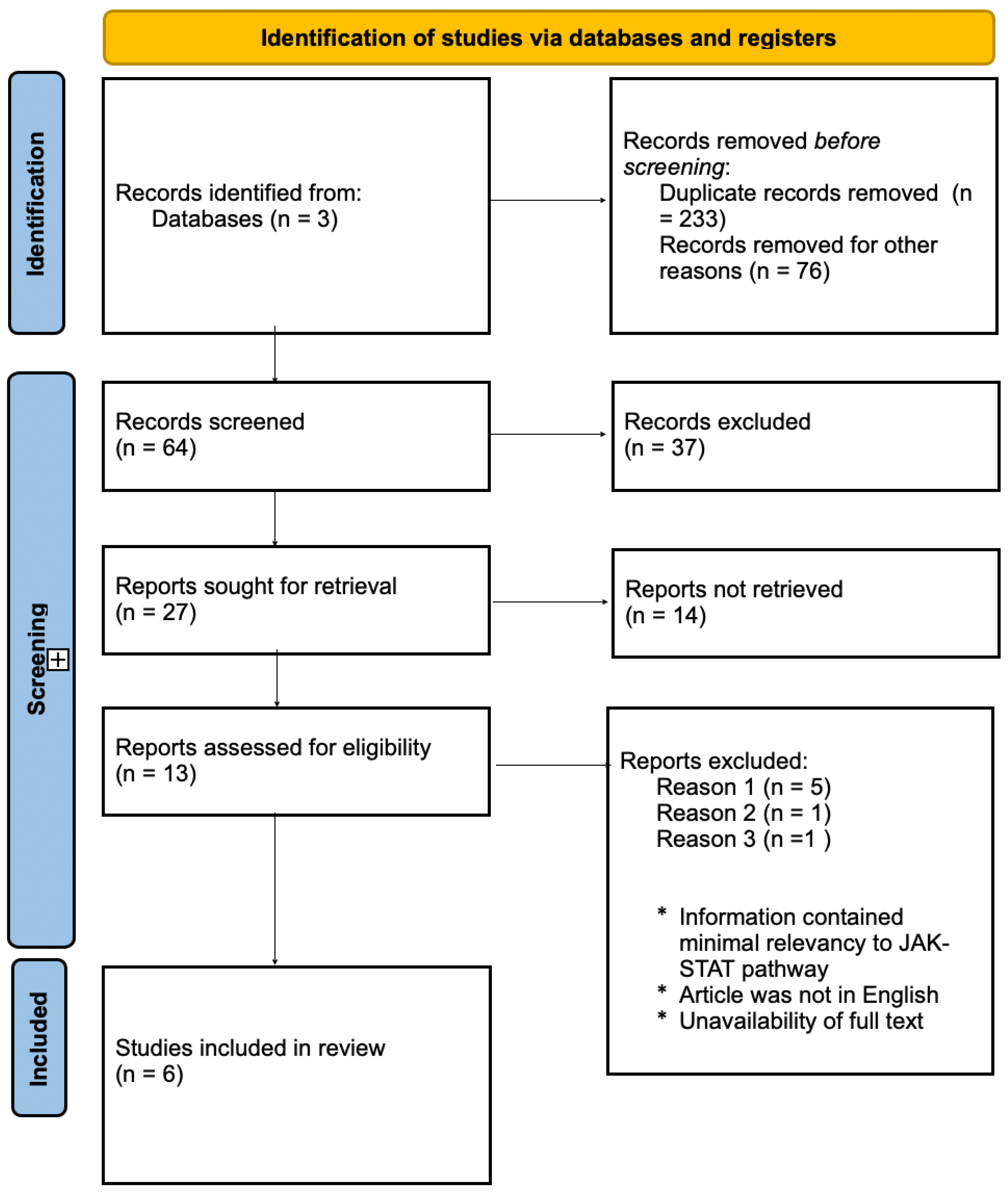
Figure 1. Flow chart of systematic review parameters for the identification of studies from online databases (chart template provided by Prisima®). Articles were excluded if they were not clinical or observational trials published in English, did not pertain to the JAK-STAT pathway, inflammation response, cytokine release storm, COVID-19, SARS-CoV-2, or ACE2, or if the full text was unavailable.
Immuno 04 00010 g001
Figure 2. Cytokines such as TNF-α, IL-6, IFN-γ, and IFN-α bind to their respected receptor in response to SAR-Cov-2 cellular infection. The initiation of JAK phosphorylation is led by the activation of the cytokine receptor, increasing p-STAT production. The phosphorylated STAT complex translocates to the nucleus, initiating transcription and gene expression of inflammatory mediators. In severe COVID-19, low levels of IFN cytokines increase STAT regulation, increasing and prolonging the inflammatory response. Additionally, TNF-α, IL-6, and IFN-γ levels increase ACE2 expression in endothelial cells and interferon signaling for cytokine storm release.
Immuno 04 00010 g002
Table 1. Summary of the literature reviewed, and findings related to COVID-19, ARDS and the JAK-STAT signaling pathway.
ReferenceSample/Subject(s)AnalysesKey Findings Limitations
Bronte
et al.,
2020
[22]
44 females and 44 males with COVID-19 pneumonia treated with baricitinibSerum levels of pSTAT3, cytokines and chemokines were measured pre and post treatment↓ Levels of p-STAT3 and
↓ levels of IL-1B, IL-6, and TNF-α plasma concentrations found on individuals taking baricitinib
Missing immunological parameters for enrolled patients.
Rincon-Arevalo
et al.,
2020
[23]
Peripheral blood mononuclear cells (PBMCs) from 20 healthy patients and 30 COVID-19 patients treated with with low dose of TNF-α and IFN-γCells from healthy patients and mild and severe COVID-19 patients were evaluated for pSTAT and interferion effect↑ p-STAT levels in patients with severe COVID-19, indicating the imblance of JAK-STAT signaling pathway for transcription of interferon-stimulated response elenents (IRSE) and interferon signaling for cytokine releaseThe mechanism of JAK-STAT pathway inhibition at the molecular level remains speculative for further investigation.
Luo et al.,
2021
[24]
GEO database of 174 Human Airway Epithelial (HAE) cell data, 84 of those samples infected with COVID-19ACE2 regulation of STAT with type I and II IFNS and STAT1Downregulation of the JAK-STAT pathway leads to the ↓ distribution of interferon-
stimulated genes and the ↑ of STAT1 expression causing over-expression of ACE2 in HAE cells.
Sample size
limitations and
verification of the relationship
between
COVID-19 and JAK-STAT
signaling pathway in pathogenesis
infection.
Kandhaya-Pillai et al., 2022
[25]
Senescent and non-senescent Human umbilical vein endothelial cells (HUVECs) cultured with cytokinesViral entry was
measured by over-
expression of ACE2,
associated with
regulation of the JAK-STAT pathway and its inflammation response
Combination of TNF-α or IFN-γ expressed upregulation for inflammasome complexes in hyper-inflammatory
responses for the JAK-STAT signaling pathway. ↑ STAT3
phosphorylation under IFN-γ response. ↑ activation of STAT1 amplifies, prolongs, and sustains
inflammation.
Sample age limitation focused on the elderly population. Limited clinical trials for JAK-STAT inhibition pathway for COVID-19
Koutsakos et al.,
2021
[26]
Blood from actue and convalescent COVID-19 individuals (85) using 3 multi-parameter flow cytometric panels and TrackSOMInnate and adaptive immune response in longitudinal acute and convalescent blood samples were studied to analyze IL-6
signaling
Identification of IL-6 and IL-8 receptors correlated to
COVID-19 severity regarding dysregulation of cytokines and immune hyperactivation.
Study only implemented one time series algorithm to tract infection activation
Karki et al.
2021
[26]
Evaluation of pro-inflammatory cytokines such as TNF-α and IFN-γ that are upregulated in SARS-CoV-2Bone marrow-derived macrophages (BMDMs) were treated with different combinations of common high regulated cytokines. Mouse models were used to observe antibody treatment with specific cytokines.Combination of TNF-α and IFN-γ induced inflammatory cell death and targets JAK-STAT signaling pathway. Treatment of antibodies against TNF-α and IFN-γ
decreased mortality rate during SARS-CoV-2 infection and cytokine shock.
Insufficient information for molecular pathway for cytokine storm.
Disclaimer/Publisher’s Note: The statements, opinions and data contained in all publications are solely those of the individual author(s) and contributor(s) and not of MDPI and/or the editor(s). MDPI and/or the editor(s) disclaim responsibility for any injury to people or property resulting from any ideas, methods, instructions or products referred to in the content.