Article

Histopathological and Quantitative Lesions of the Cardiac Conduction System and Its Vascularization Related to Chronic Cocaine Abusers and Sudden Unexpected Death

by
Damián Sánchez-Quintana
1,*,
Sergio Alama
2,
Yolanda Macías
3,
José-Ángel Cabrera
4,
María Santos
5 and
Manuel Salguero
6
1
Departamento de Anatomía y Biología Celular, Facultad de Medicina, Universidad de Extremadura, 06006 Badajoz, Spain
2
Instituto de Medicina Legal y Ciencias Forenses, Avda. Elvas s/n Campus Universitario de Badajoz, 06006 Badajoz, Spain
3
Departamento de Terapéutica Médica y Quirúrgica, Facultad de Veterinaria, Universidad de Extremadura, 10071 Cáceres, Spain
4
Departamento de Cardiología, Hospital Universitario Quirón-Madrid, Universidad Europea de Madrid, 28223 Madrid, Spain
5
Servicio de Patología Forense Instituto de Medicina Legal y Ciencias Forenses de Sevilla, 41015 Sevilla, Spain
6
Instituto Nacional de Toxicología y Ciencias Forenses, 41009 Sevilla, Spain
*
Author to whom correspondence should be addressed.
Forensic Sci. 2022, 2(2), 423-437; https://doi.org/10.3390/forensicsci2020032
Submission received: 7 May 2022 / Revised: 20 May 2022 / Accepted: 25 May 2022 / Published: 8 June 2022
(This article belongs to the Special Issue Feature Papers in Forensic Sciences in 2022)

Abstract

:
Cocaine abuse is associated with multiple cardiovascular events, including myocardial infarction, arrhythmias, and sudden death. A total of 40 hearts were studied. The purpose of this study was to compare the cardiac conduction tissue and its vascularization in 20 young adults without a history of drug use and/or arrhythmias and 20 hearts of young adults with a history of chronic cocaine use who have died of sudden unexpected death, in which toxicological analyzes were performed in blood and urine as a means to establish chronic cocaine use. We have applied serial histological sections, techniques of morphometry, and image analysis to quantify the density and affectation of connective/adipose tissue of the conduction system and intramyocardial vessels. The conduction system after chronic cocaine use showed potentially lethal changes with an increase in connective/adipose tissue at the level of the intranodal or perinodal tissue of the sinus node in 35% (7 hearts) and the atrioventricular node in 75% (15 hearts), with the most affected structure being the left bundle branch and the AV nodal artery (100%, 20 hearts). In conclusion, the histopathologic changes in nodes, the perinodal area, and small vessels may be a morphological substrate that offers an explanation about the mechanism of arrhythmias and sudden death in this population.

1. Introduction

Cocaine is an alkaloid derived from the leaves of the Erythroxylum coca shrub of the Andes mountains. Cocaine administered by inhalation, injection, and smoking has a long history of use and abuse. A significant number of emergency room admissions are due to the cardiovascular effects of cocaine [1], thus there is a need to further characterize the cardiovascular consequences of the drug. Cocaine causes myocyte cell death and fibrosis, resulting in structural remodeling related to its sympathomimetic effects: reduced systolic function and increased heart rate, blood pressure, and myocardial oxygen demand, which result in high incidences of cardiac arrhythmias and sudden death [2,3,4,5].
Cocaine also produces an electrical remodeling by inhibiting several potassium channels, cardiac sodium channels in two phases (rapid and slower phase), and alters the function of calcium-related channels and calcium-handling proteins [6,7,8,9]. Its electrical effects produce a delay in ventricular conduction, with a widening of the QRS complex, an increase in the variability of the QT interval, and the generation of atrial arrhythmias, with sinus tachycardia being the most common atrial arrhythmia and potentially fatal ventricular arrhythmias [10,11,12,13]. In cases of sudden unexpected death in cocaine abusers, especially in young subjects, the magistrate generally has the idea of limiting the forensic investigation to autopsy and toxicological analysis [14,15].
As other authors, we think that a detailed study (although it is more laborious) of the lesions in the cardiac conduction system could explain more clearly the sudden death of these patients [16,17], especially in cases in which cocaine has been detected in blood and urine. For this reason, what we consider absolutely necessary is that when the pathologist is faced with the microscopic study of the autopsy of a person who has died as a result of the consumption or abuse of drugs, especially cocaine, they can clearly identify which are the lesions that this drug has left in the conduction tissue of the heart and its vessels. The purpose of this histopathological study was to compare the cardiac conduction system and its vascularization related to chronic cocaine use and utilize hearts from autopsies of similar-age men/women who died due to different motifs but who did not have a history of drug addiction or arrhythmias, which we use as control hearts in an attempt to find possible morphologic changes explaining the cause of sudden death in the cocaine users.

2. Materials and Methods

2.1. Cases

We examined histologically the cardiac conduction system (sinus node, AV node, bundle of His, and its two branches) as well as its vascularization (arteries of the sinus node and AV node) in 20 hearts: 17 male/3 female cases of chronic cocaine abusers who were victims of a sudden, unexpected death. All hearts belong to young adult subjects, 25.3 ± 5 years of age; the hearts were of normal size. The mean heart weight was 286 ± 30 g and all forensic autopsies were performed at the Institute of Legal Medicine, Faculty of Medicine, Coimbra. Portugal, between the years 2013 and 2019. The epicardial coronary arteries were normal in all the hearts without luminal stenosis due to arteriosclerotic plaques. We compared our findings with those obtained from 20 hearts: 14 male/6 female who died from other causes (suicide, traffic accidents, and various noncardiac pathologies) and who did not have a history of drug addiction or arrhythmias, which we consider to be a control group. All control hearts were studied in the Histopathology Service of National Institute of Toxicology and Forensic Sciences of Seville, between 2010 and 2018. The mean age of subjects in the control group was (27.5 ± 4 years) and their heart weight was 300 ± 25 g. The study was conducted in accordance with the Declaration of Helsinki and approved by the Bioethics and Biosafety Committee of the University of Extremadura (registration code no 9/2015). See Table 1.
The autopsies of chronic cocaine abusers were performed within the first 18–24 h after death. We had no access, however, to any electrocardiographic recordings or history of arrhythmias that might have been made or observed during the life of these victims. The confirmation of cocaine abusers was initially based on antecedent police reports and death scene investigations (death at home and mainly on weekends). In most of our cases cocaine had been consumed by inhalation (by sniffing the powder or by lung inhalation of what is known as base or crack). Regular users of cocaine often consume around 0.50–1 g per intake, and sometimes punctual consumption of 2–3 g is reached. At autopsy, blood from the right internal jugular vein and urine from them bladder were collected and examined in the laboratory. The identification of the products of metabolism of cocaine in the urine and blood from toxicologic analysis established the diagnosis in all cocaine abusers. The cocaine victims of a sudden unexpected death were included when toxicological analyses revealed, in blood and urine as measured by gas chromatography/mass spectrometry, the presence of cocaine and its metabolites with a potentially lethal level according to published data [15]. In the 20 cases,) ≥850 ng/mL of benzoylecgonine (a major metabolite of cocaine) was detected, in 9 of the 20 individuals ecgonine methyl ester (another cocaine metabolite) was also detected at ≥600 ng/mL, in 14 (70%) of the 20 individuals simultaneous consumption of cocaine and ethanol was determined by the presence of cocaethylene at 600 ng/mL. It is noteworthy that 17 cases (85% of cocaine abusers) have antecedents of concomitant cigarette smoking, and finally, in 7 of the 20 individuals, methadone was detected.

2.2. Histological Examination of the Cardiac Conduction System and Its Vascularization

In all hearts of the cocaine victims of a sudden unexpected death, at least six sample blocks were made and fixed in 7% buffered formalin and embedded in paraffin, one at the level of the sinoatrial junctional area which was removed in a block from each heart, and cross-serially sectioned at 10 µm thickness through the sinus node, and the terminal crest included a short portion of the superior caval vein and extended to the entrance of the inferior caval vein, similar to the method described by Sánchez-Quintana et al. [18]. Another at the level of the AV node, the full thickness of the triangle of Koch, along with the adjacent aortic root, was removed as a solitary block. The block was then serially sectioned at 10 µm thickness, cutting at right angles to the hinge of the septal leaflet of the tricuspid valve, as was described in one of our previous studies [19]. The sections were then stained using either Masson’s trichrome or the Picrosirius red technique. From the samples of the sinus node and Triangle of Koch, a total of 25 and 90 sections were stained from each heart, respectively. Finally, the histological examinations of the hearts included at least 5–7 slides made from the working myocardium wall of the left ventricle. Two independent pathologists blind to the sample’s origin (control or cocaine group) performed the microscopic examination of all of them.

2.3. Morphometry and Image Analysis

As described previously [20,21], digital images were taken of the sections stained with Masson’s using a Nikon SMZ 1500 microscope (Nikon, New York, NY, USA). Morphometry was performed at least in 5–8 fields per atrial (sinus node and AV node) or ventricular sample, in the regions of maximum extent of the affected area, using a grid covering an area of 5 mm2 of the atrial or ventricular sample. The areas of myolysis or fibrosis were expressed as a percentage of the entire limit of the grid. Capillary vessels, perivascular interstitial tissue, and intertrabecular spaces between the cardiomyocytes were excluded from this morphometric analysis.
To study the possible stenotic alterations of the vascular lumen in control patients and cocaine abusers, 15 to 20 photographs were taken of each histological section using a stereomicroscope equipped with a digital camera (Nikon SMZ 1500). Using ImageJ software (National Institutes of Health, New York, NY, USA), quantitative morphometric measurements of the lumen of the arteries of the sinus and AV nodes and intramyocardial arterioles/capillaries of the walls of the left ventricle close to the papillary muscles and interventricular septum were taken, calculating the percentage of the total surface that the lumen represents in each vessel. The identification of the areas of myolysis or fibrosis and vascular lumen were performed by direct visualization of the histological preparation by two expert anatomopathologists at proper magnification.

2.4. Data Analysis

Quantitative data were expressed as the mean ± standard deviation. The ordinary two-way.
ANOVA test with Dunnett’s multiple comparison correction was used to evaluate the significance of the changes from control sinus node and AV node to selected cocaine specimens. The ANOVA test was also used to assess the significance of the evolving changes in cocaine lumen vessel morphology (arteries of the sinus node, AV node, and left ventricular wall). The paired t-test was used to assess the significance of the changes in cocaine specimens. A value of p < 0.05 was considered significant, while for p ˂ 0.001 we consider that the test is highly significant. All analyses were performed using SPSS v.22.0 (IBMSPSS, Chicago, IL, USA).

3. Results

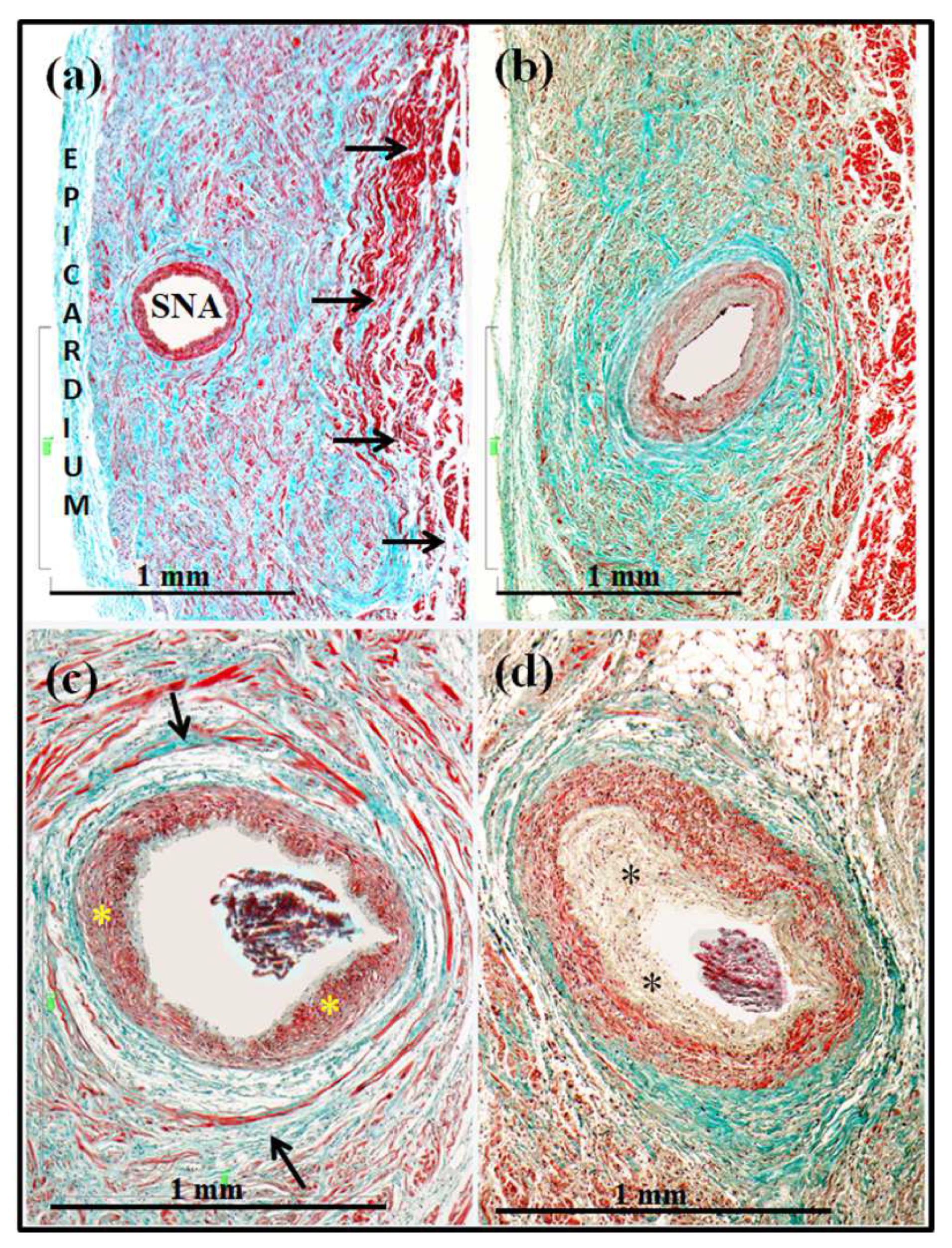
3.1. Sinus Node and Artery

The intrinsic function of the sinus node is to be the source where the rhythm of the heart emanates, being responsible for the wave P of the electrocardiogram, representing atrial activity. In the majority of individuals, the sinus node has a subepicardial location and is located like a wedge at the junction of the superior vena cava with the atrium and the right atrial appendage, running directly below the terminal sulcus and just above the terminal crest (crista terminalis). It usually has its head inferior to the crest of the right atrial appendage, with a tail extending towards the inferior caval vein. In most cases, the node is cigar or arc-shaped and is built around a prominent central sinus node artery. As described previously [18], structurally, the sinus node is made up of small round or ovoid myocytes (less than 7 µm in length) that are paler in staining than those of normal myocardium, and with the appearance of empty cytoplasm, but with a long central round nucleus. These myocytes are found surrounding the artery of the sinus node within a dense interstitial matrix of connective tissue, within which the myocytes of the node are intertwined without a precise order (Figure 1). In the periphery, nodal and transitional myocytes directly contact normal atrial myocytes.
The differences between the two groups (normal hearts and cocaine victims) regarding the histology of the sinus node and its vascularization are characterized because in the cocaine abusers we observed an increase in density and replacement in the sinus node tissue by adipose ⁄fibrous tissue sinus node in 35% of cases (7 hearts). In fact, the morphometric study detected highly significant statistical differences (p < 0.001) between the two groups (Figure 1). In all other cases (13 hearts), we did not find significant alterations in the density of connective or adipose tissue. Only in two cases were focal inflammatory infiltration by lymphocytes found in the group of cocaine abusers (Table 2).
In order to quantify the degree of stenosis in the histological sections at the level of the sinus node artery, we observed that the percentage of lumen of this artery in the control hearts was 73 ± 7%, while in the group of cocaine abusers there were different changes, in some cases (5 hearts) there was a dysplasia with a great proliferation and increased thickness of the intima layer, which greatly stenosed the lumen of the vessel to 15 ± 6% (Figure 1); in other cases (10 hearts) the dysplasia was observed due to smooth muscle proliferation of the middle and thick adventitial layer, although the lumen of the vessel was stenosed less than in the previous case 36 ± 5% (Figure 1), and in other cases (5 hearts), we did not find significant alterations in relation to the control group. Therefore, in the cocaine patient abusers (15 of the 20 individuals), the sinus node artery was highly significantly stenosed in its lumen (p < 0.001) (Table 2).

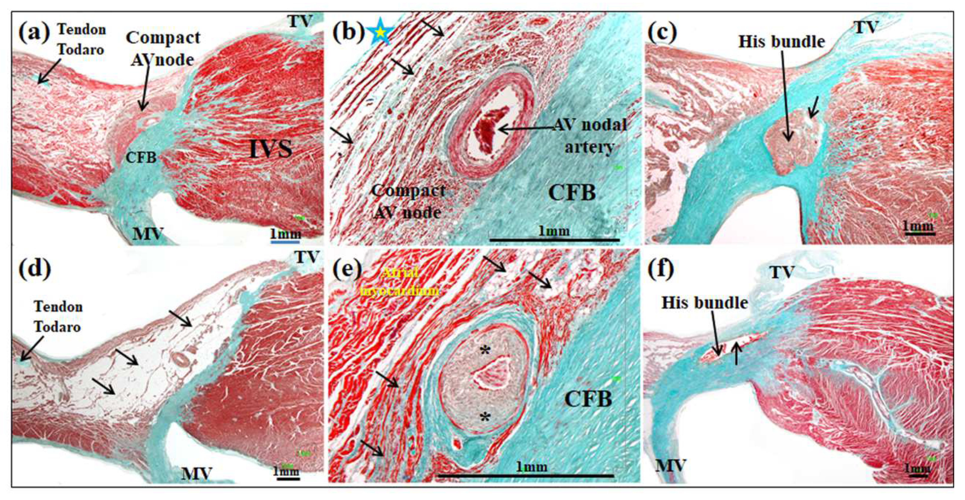
3.2. Atrioventricular Conduction System and Vascularization

As described previously [22], the AV node is located inside Koch’s triangle in its antero-superior area next to the apex, on the right atrial wall over the ventricular septum. The node is a semioval-shaped structure that is supported by the fibroconnective tissue of the right fibrous trigone and consists of a compact node and myocytes transitional zone between the node and the atrial myocardium, which constitute a kind of bridge between the normal myocardium and the AV node (Figure 2). Histologically, the control specimen node is a collection of short spindle-shaped myocardial cell groupings of irregularly whorled interlacing bundles immersed in a loose matrix of connective tissue, in contrast to the ribbon-shaped myocardial cells at the atrial and ventricular chambers (Figure 2). The nodal cells in the compact zone are tightly attached to each other, but the nodal cells in the transitional zone are separated by connective tissue. The atrial myocardium is in close contact with the transitional myocytes and the AV nodal cells and directly below the endocardium of the right atrium (Figure 2).
The bundle of His is the continuity of the AV node just when it is completely surrounded by connective tissue that isolates it completely from the surrounding myocardium and penetrates into the right fibrous trigone (Figure 2) connecting the atria and ventricles [23].
The bundle extends as a cordlike collection of spindle-shaped conduction cells at the junction between the membranous septum and the ventricular septal crest. The bundle runs along the inferior border of the membranous septum at the crest of the ventricular septum. Histologically, in the control specimens, the specialized myocytes of the bundle of His present a more uniform and parallel arrangement than that observed in the myocytes of the AV node, and they are surrounded by a loose interstitial matrix of connective tissue (Figure 2).
The differences between the two groups regarding the histology of the AV node are evident given that in 15 of the 20 hearts (75% of the cases) of the cocaine abusers they presented a substitution of the myocytes that are replaced by adipose and/or fibrous tissue (Figure 2 and Figure 3). This replacement usually affects transitional myocytes, sometimes the nodal myocytes themselves, and sometimes all myocytes (Table 2). When quantifying the density of connective/adipose tissue in the AV node of the cocaine victims, we detected 72 ± 5% and 32 ± 5% in the control group, that is, highly significant statistical differences (p < 0.001). In the remaining five cases (25%), the same tissue was within normal limits. In the histological sections at the level of the AV nodal artery, we found that in the controls the percentage of lumen artery was 72 ± 3.5%, while in 7 cases of cocaine abusers (35%) they had fibromuscular dysplasia due to smooth muscle proliferation of the middle layer; although in most cases, 13 cases (65%), moderate or great thickening of the intima was observed (Figure 3), thus the lumen artery was very narrowed, with the percentage of lumen being 12 ± 3.5% (p < 0.001) (Table 2). In one case, the narrowing was even complete (Figure 3). Moreover, the focal or diffuse infiltration of lymphocytes was found in the surrounding area of the AV node artery in five cases (25%) affecting the body of the AV node. (Figure 3). This infiltration was not observed in any control specimens.
We have observed less histological affectation in the bundle of His than in the AV node in cocaine-abusing patients; the specialized myocytes of the main bundle were replaced by a small-to-moderate amount of fatty and/or fibrous tissue in seven cases (35%). Sometimes the involvement was scattered throughout the bundle and other times it was concentrated in the distal part of the bundle (Figure 2). Quantitatively, the density of connective/adipose tissue in the bundle of His in the group control was 17 ± 5%, in contrast, in the group of cocaine abusers, it was 55 ± 4%, which is highly significant (p < 0.001) (Table 2).
As previously described [23,24], as the bundle of His heads for the ventricles and penetrates into the right trigone fibrosus, the axis runs a nonbranching course of variable length on the crest of the muscular ventricular septum, located below the membranous septum and above the apex of the muscular portion of the interventricular septum, dividing later into its right and left branches. The left branch of His has a subendocardial path on the left side of the septum and was separated from the ventricular myocardium by a thin layer of connective tissue (Figure 4); its thickness varies from 0.4–1 mm, and histologically in the control group among specialized myocytes there are fine tracts of connective tissue and some adipocytes (Figure 4). In the group of cocaine abusers, the left branch is the most affected structure of the conducting tissue. In 12 hearts (60%), there was a loss of an important part of myocyte conduction observed in the origin of the left branch, and an important fatty infiltration could be seen in the proximal and distal parts of the left bundle branch, so that the specialized myocytes faded away, leaving a “ghost” structure. In eight hearts (40%), the myocytes were replaced by a moderate amount of fatty and/or fibrous tissue. The density in the control group of connective/adipose tissue in the left branch was 19 ± 5%, in contrast, in the group of cocaine abusers, it was 95 ± 4%, which is highly significant (p < 0.001) (Table 2).
The right branch of the bundle of His is intramyocardial in origin, and in the upper or middle third of the right side of the interventricular septum it becomes subendocardial (Figure 4). Histologically, in the control hearts, it is a very fine structure with a thickness of 0.1–0.3 mm, and between its myocytes, fine tracts of connective tissue and some adipocytes were observed. In the group of cocaine abusers, the right branch was affected in 40% of cases (eight hearts). Myocytes were being replaced by adipose/connective tissue that follows the course of the branch. In the rest of the hearts (12 hearts), we did not find significant alterations in the density of connective or adipose tissue. The density of connective/adipose tissue in the right branch of the control group was 17 ± 5%, in contrast, in the group of cocaine abusers, it was 58 ± 4%, which is highly significant (p < 0.001) (Table 2). It is important to point out that in cocaine abusers, six hearts (30%) had both branches seriously affected.

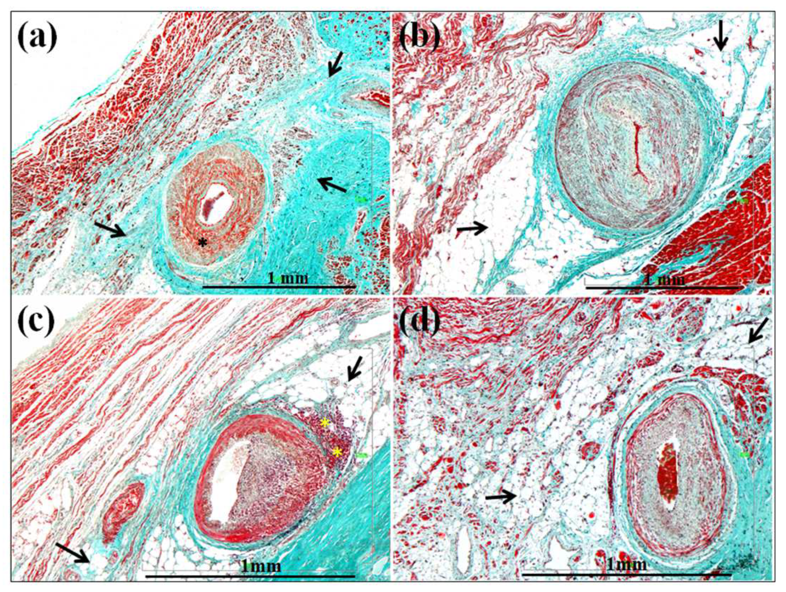
3.3. Affectation of the Walls and Lumen of the Small Intramyocardial Vessels

We have qualitatively observed in cocaine-abusing patients a serious affectation of the small intramyocardial arterioles of the left ventricular wall and interventricular septum with a fibromuscular hypertrophy or hyperplasia of the middle layer that over time from cocaine produces a necrosis of the smooth muscle cells that are replaced by connective tissue and that sometimes produce a lysis of the middle layer, the latter indicating disease progression from chronic cocaine use (Figure 5). In these vessels, there is also significant periadventitial fibrosis. In the majority of cocaine abusers (12 cases, 60%), the measurements made in the arterioles indicate that the percentage that the lumen of the vessel represents with respect to the total of its wall is 5.0 ± 3.5%, and in the control group it is 35.5 ± 2% (Table 2). Therefore, we found highly significant differences (p < 0.001). The histopathological consequence of the stenosis or obstruction of the small intramyocardial vessels was the necrosis of the working ventricular myocytes, which were replaced by connective tissue with visible microscopic scars, fundamentally in the subendocardium of the papillary muscles of the left ventricle (Figure 5). Only three cases (15%) with focal inflammatory infiltration in papillary muscles were found in the group of cocaine victims.

4. Discussion

Previous studies have reported that cocaine addicts are associated with arrhythmias and cardiac arrest and that these can cause sudden death. Cardiac arrythymias in cocaine users have been studied by a number of authors, both in cases of acute intoxication and in chronic cocaine addicts [25,26,27,28]. In our study, in all cases (20, cases, 100%) benzoylecgonine (main metabolite of cocaine), and in 9 of the 20 (45% of cases) ecgonine methyl ester, (another metabolite of cocaine) were detected in urine and blood, with values that are considerably higher than reported by other authors [29,30]. This finding corroborates the chronic nature of the abuse and suggests increased tolerance. However, the metabolites–cocaine concentrations should be interpreted cautiously because of some patients becoming sick with relatively low blood concentrations, whereas others tolerate large quantities without consequences [31,32]. Thus, according to Karch and Stephens [33], cocaine-associated sudden death is not dose-related, and the relationship between cocaine blood concentrations and toxicity has not been established.
Different mechanisms have been proposed to explain the phenomenon after cocaine administration. A direct effect of cocaine is that it can produce cardiac electrical remodeling by altering gating properties of various ions such as potassium, sodium, and calcium channels [6,7,8,9]. Other explanations are based on the fact that cocaine produces a structural remodeling of the myocardium [2,3,4,5]. It has also been suggested that the lesions observed in the atrioventricular conduction system in six cases of chronic cocaine abusers, aged between 19–35 years, may be considered as a possible cause of death [17]. Alcohol potentiates actions of cocaine by forming cocaethylene, a metabolite of alcohol and cocaine that inhibits inward-rectifying potassium channels [34], which has been associated with a 25-fold increase in sudden cardiac death [28,35,36]. In our study, cocaethylene was detected in 70% of cocaine-abuse-related sudden deaths, indicating a simultaneous cocaine and ethanol abuse in the majority of cases. Moreover, in our study, we detected that methadone was present in 7 of the 20 cases (35%), and various authors have suggested whether administering this substance could have caused possible arrhythmia and perhaps death itself by prolonging the QT interval following intravenous administration of methadone [37,38,39]. However, no histological studies have been performed to date that correlate methadone with cardiac conduction tissue or myocardium of the left ventricle.
Our results show that in the 20 presented cases of young adult subjects who were chronic cocaine abusers of 25.3 ± 5 years of age, different lesions of the cardiac conduction system were identified; the lesions are characterized because they are not uniform in all specialized myocardial structures. To the best of our knowledge, there is no evidence to show postmortem studies that histologically analyze and quantify the amount of connective/adipose tissue in the sinus node and main sinus artery lesions in young adult chronic cocaine abusers who suffered sudden death to distinguish them from controls. Loss of myocardial sinus node cells, replaced by various amounts of adipose ⁄fibrous tissue was found in 35% of the cocaine addict group, as well as different degrees of stenosis with or without lumen thrombosis of the sinus artery due to the proliferation of the intima layer or the middle and adventitia layer of the artery. Sinus tachycardia is the most common atrial arrhythmia that results from cocaine toxicity, but supraventricular tachycardia and atrial fibrillation can also occur [25]. Few studies have documented sinus bradycardia (rate of ≤50 beats/min) secondary to chronic cocaine use because of the representation of the antithesis of its adrenergic effects [10]. Concordant with these findings, Franklin et al. [40] reported that habitual cocaine use was associated with a sevenfold increase in the risk of sinus bradycardia and suggested a potential mechanism for sinus bradycardia may be related to cocaine-induced desensitization of the beta-adrenergic receptor secondary to continuous exposure. We must emphasize that our study is not an electrophysiological essay, since the tools and techniques that we used are purely anatomical and we do not clinically know the arrhythmic antecedents of the specimens. In our opinion, the most striking histological characteristic is the fibrofatty degeneration of sinus node tissue that can create an ideal substrate for reentrant circuits that may decrease impulse conduction; in addition, the premature atherosclerosis of the sinus artery can be related to the development of sinus node dysfunction and may constitute a mechanism of sudden death in this population.
Previous studies on the myocardium of chronic cocaine users indicate that their abuse results in pathologic lesions in the atrioventricular conduction system [17,41,42]. Our findings confirm greater qualitative and quantitative alterations in the atrioventricular conduction system in comparison with the normal group; the more interesting findings especially affecting the AV node (15 of the 20 hearts, 75% of the cases) and the left bundle branch of His (100% of the cases) with intense loss of specialized myocytes that gives rise to its replacement by large amounts of fatty and/or fibrous tissue, which affects nodal or transitional myocytes, or both with highly significant statistical differences with respect to the control group (p <0.001). Even in 12 hearts (60% of the cases), the myocytes of the left bundle branch seem to fade away, being replaced by adipose tissue, leaving it as a “ghost” structure. In some cases (six hearts, 30%), the lesion affects both branches of the bundle of His, left and right; the consequences of these alterations is a complete anatomical block between the atria and ventricles. Previous studies have reported that the effect of cocaine on arterioles is greater than that of heroin [43], and that the AV nodal artery is one of the most affected [17]. In our study, the AV nodal artery with or without lumen thrombosis had affected walls and narrowing of its lumen in 100% of the cases. The artery of the AV node vascularizes the AV node and the left branch of the bundle of His, and both are very affected in cocaine abusers, which probably could be the origin of different types of ventricular lethal arrhythmias. However, the number of histological reports regarding the documentation of ventricular arrhythmias in cocaine users are very scarce. Moreover, the cocaine-induced Brugada pattern has been described [44,45]. Alcohol potentiates actions of cocaine, producing a significantly altered PR interval, QRS, and QTc compared with those with addictions who did not use alcohol [46]. Our morphological and quantitative data are consistent that this type of degeneration and fibroadipose replacement suffered by the AV tissue (especially the left branch of His) and the structural changes of the AV node artery may be related to the development of a dysfunction or blockage of the AV system and may indicate a proarrhythmic state as the mechanism of unexpected sudden death in this population.
Inflammatory infiltration by lymphocytes was found in some samples from the cocaine group (two cases affecting sinus node, five cases (25%) affecting the body of the AV node, and three cases affecting the subendocardium of the papillary muscles of the left ventricle). These could probably be attributed to many causes: admixtures of cocaine, nonsterile injection techniques of intravenous drug users, and immunosuppression caused by chronic abuse, among other causes. It has been suggested [47,48] that the inflammatory infiltration, probably from toxins because of the inflammatory process, affects the conduction system and its surrounding area, and this favors the development of myolysis, interstitial edema, and fibrosis, which may result in the dysfunction of these structures and may facilitate the onset and perpetuation of different types of arrhythmias.
It has been suggested that coronary vasospasm is probably related to endothelial dysfunction or damage, which causes a loss of inhibition of platelet aggregation and favors the development of a premature and accelerated form of atherosclerosis, which promotes thrombosis and can contribute to myocardial infarction [2,33]. According to other studies, our observations show a high prevalence of small vessel disease in the left papillary muscles and left ventricular wall, consisting of media hypertrophy, intimal proliferation, and periadventitial fibrosis [28,49]. In addition, the progression is towards a lysis of the middle layer and areas of fibrosis that give rise to sclerosis of the arterial wall, accounting for narrowed lumen in 60% of the cases (12 hearts), demonstrating highly significant differences with the control specimens (p < 0.001). These data demonstrate that cocaine has the potential to injure vessel walls, and by this action may induce the early onset of vascular disease. In our study, chronic cocaine use produced myocardial ischemia that is revealed by the presence of multifocal myocyte necrosis and interstitial fibrosis around the small vessels, as well as, in the endocardium of the papillary muscles of the left ventricle with progressive structural changes in the myocardium. According to Rajab et al. [50], myocardial fibrosis is multifocal in 21% of cocaine users, and magnetic resonance studies in asymptomatic cocaine addicts have observed fibrosis in 73% of cases, which may or may not follow ischemic patterns [51]. The density of the fibrosis and/or the framework of the intramyocardial connective tissue are morphological substrates for reentry arrhythmias [52], and therefore, these individuals could be at higher risk of sudden unexpected death.

Limitations of the Study

Our study is based on structurally pathological hearts (cocaine abusers) and normal hearts, however, we had no access in either group to any electrocardiographic recordings or history of arrhythmias that might have been made or observed during the life of these victims. We recognize that our study has obvious limitations in that we have no functional data with which to compare our findings. On the other hand, we always tried to use the same methodology for the identification of the areas of myolysis or fibrosis and vascular lumen in both the cocaine abuser group and the control group sampled, despite the inconvenience of traveling from Badajoz to Seville and vice versa.

5. Conclusions

Cocaine use causes structural and electrical remodeling of the atria and ventricles with subsequent electrocardiogram changes and ensuing atrial and ventricular arrhythmias such as sinus bradycardia, atrial fibrillation, torsade de pointes, ventricular tachycardia, and cardiac arrest. The question that we asked ourselves in our study is whether the qualitative and quantitative histological lesions that appear in the cardiac conduction tissue and its vascularization are linked to cocaine use and are a sufficient cause for the sudden death of these young patients. We clearly demonstrated that the lesions we observed at the level of the sinus node, and especially the AV node and branches of the His bundle and its vascularization, may constitute a morphological substrate for sudden death, and they may well have been the result of chronic cocaine use. Our study demonstrated significant differences in the conduction tissue between the two groups. These differences surely should be acknowledged when seeking to correlate the findings in an experimental situation with clinical experience.

Author Contributions

Conceptualization, D.S.-Q., S.A. and M.S. (Manuel Salguero); methodology, S.A., Y.M. and M.S. (María Santos); formal analysis and investigation, D.S.-Q., M.S. (Manuel Salguero), S.A. and M.S. (María Santos); writing, D.S.-Q., S.A., Y.M. and J.-Á.C.; writing—review and editing, D.S.-Q. and M.S. (Manuel Salguero); supervision, S.A., Y.M., J.-Á.C. and M.S. (María Santos). All authors have read and agreed to the published version of the manuscript.

Funding

The authors declare that they have not received any external or specific funding for this research.

Institutional Review Board Statement

This study was conducted in accordance with the Declaration of Helsinki and approved by the Ethics Committee of University of Extremadura, protocol code no. 9/2015. Date: September 2015.

Informed Consent Statement

Informed consent was obtained from all subjects involved in the study.

Acknowledgments

The authors would like to thank Maria Cristina de Mendonça, Institute of Legal Medicine, Faculdade de Medicina, Coimbra, Portugal for donations in materials used for histological study, and Agustin G. Nogales, Department of Statistics, University of Extremadura, Badajoz, Spain for assistance with the statistical analysis of the article.

Conflicts of Interest

The authors declare that there are no conflict of interest to report.

References

  1. UNODC. World Drug Report. 2018. Available online: https://www.unodc.org/wdr2018/prelaunch/WDR18_Booklet_1_EXSUM.pdf (accessed on 9 October 2019).
  2. Karch, S.B. Cocaine and the heart: Clinical and pathological correlations. Adv. Biosci. 1991, 80, 211–218. [Google Scholar]
  3. Qureshi, A.I.; Suri, M.F.; Guterman, L.R.; Hopkins, L.N. Cocaine use and the likelihood of nonfatal myocardial infarction and stroke: Data from the third national health and nutrition examinations survey. Circulation 2001, 103, 502–506. [Google Scholar] [CrossRef] [Green Version]
  4. Egred, M.; Davis, G.K. Cocaine and the heart. Postgrad. Med. J. 2005, 81, 568–571. [Google Scholar] [CrossRef]
  5. Liaudet, L.; Calderari, B.; Pacher, P. Pathophysiological mechanisms of catecholamine and cocaine-mediated cardiotoxicity. Heart Fail. Rev. 2014, 19, 815–824. [Google Scholar] [CrossRef] [PubMed] [Green Version]
  6. Wu, S.N.; Chang, H.D.; Sung, R.J. Cocaine-induced inhibition of ATP-sensitive K+ channels in rat ventricular myocytes and in heart-derived H9c2 cells. Basic Clin. Pharmacol. Toxicol. 2006, 98, 510–517. [Google Scholar] [CrossRef] [PubMed]
  7. Reyes, S.; Kane, G.C.; Zingman, L.V.; Yamada, S.; Terzic, A. Targeted disruption of K(ATP) channels aggravates cardiac toxicity in cocaine abuse. Clin. Transl. Sci. 2009, 2, 361–365. [Google Scholar] [CrossRef] [PubMed]
  8. Crumb, W.J., Jr.; Clarkson, C.W. Characterization of cocaine-induced block of cardiac sodium channels. Biophys. J. 1990, 57, 589–599. [Google Scholar] [CrossRef] [Green Version]
  9. Renard, D.C.; Delaville, F.J.; Thomas, A.P. Inhibitory effects of cocaine on Ca2+ transients and contraction in single cardiomyocytes. Am. J. Physiol. 1994, 266 Pt 2, H555–H567. [Google Scholar] [CrossRef] [PubMed]
  10. Sharma, J.; Rathnayaka, N.; Green, C.; Moeller, F.G.; Schmitz, J.M.; Shoham, D.; Dougherty, A.H. Bradycardia as a marker of chronic cocaine use: A novel cardiovascular finding. Behav. Med. 2016, 42, 1–8. [Google Scholar] [CrossRef] [PubMed] [Green Version]
  11. Jackman, W.M.; Friday, K.J.; Anderson, J.L.; Aliot, E.M.; Clark, M.; Lazzara, R. The long QT syndromes: A critical review, new clinical observations and a unifying hypothesis. Prog. Cardiovasc. Dis. 1988, 31, 115–172. [Google Scholar] [CrossRef]
  12. Gupta, A.; Lawrence, A.T.; Krishnan, K.; Kavinsky, C.J.; Trohman, R.G. Current concepts in the mechanisms and management of drug-induced QT prolongation and torsade de pointes. Am. Heart J. 2007, 153, 891–899. [Google Scholar] [CrossRef]
  13. Atkinson, T.S.; Sanders, N.; Mancino, M.; Oliveto, A. Effects of disulfiram on QTc interval in non-opioid-dependent and methadone-treated cocainedependent patients. J. Addict. Med. 2013, 7, 243–248. [Google Scholar] [CrossRef]
  14. Virmani, R.; Robinowitz, M.; Smialek, J.E.; Smyth, D.F. Cardiovascular effects of cocaine: An autopsy study of 40 patients. Am. Heart J. 1988, 115, 1068–1076. [Google Scholar] [CrossRef]
  15. Winek, C.L.; Wahba, W.W.; Winek, C.L., Jr.; Balzer, T.W. Drug and chemical blood-level data 2001. Forensic Sci. Int. 2001, 122, 107–123. [Google Scholar] [CrossRef]
  16. Bauman, J.L.; Grawe, J.J.; Winecoff, A.P.; Hariman, R.J. Cocaine-related sudden cardiac death: A hypothesis correlating basic science and clinical observations. J. Clin. Pharmacol. 1994, 34, 902–911. [Google Scholar] [CrossRef]
  17. Michaud, K.; Augsburger, M.; Sporkert, F.; Bollmann, M.; Krompecher, T.; Mangin, P. Interpretation of lesions of the cardiac conduction system in cocaine-related fatalities. J. Forensic Leg. Med. 2007, 14, 416–422. [Google Scholar] [CrossRef]
  18. Sánchez-Quintana, D.; Cabrera, J.A.; Farré, J.; Climent, V.; Anderson, R.H.; Ho, S.Y. Sinus node revisited in the era of electroanatomical mapping and catheter ablation. Heart 2005, 91, 189–194. [Google Scholar] [CrossRef] [Green Version]
  19. Anderson, R.H.; Sánchez-Quintana, D.; Mori, S.; Cabrera, J.A.; Sternick, B.E. Re-evaluation of the structure of the atrioventricular node and its connections with the atrium. Europace 2020, 22, 821–830. [Google Scholar] [CrossRef]
  20. Climent, V.; Hurlé, A.; Ho, S.Y.; Sáenz-Santamaría, J.; Nogales, A.G.; Sánchez-Quintana, D. Early morphologic changes following microwave endocardial ablation for treatment of chronic atrial fibrillation during mitral valve surgery. J. Cardiovasc. Electrophysiol. 2004, 15, 1277–1283. [Google Scholar] [CrossRef]
  21. Amorós-Figueras, G.; Roselló-Diez, E.; Sánchez-Quintana, D.; Casabella-Ramón, S.; Jorge, E.; Nevado-Medina, J.; Arzamendi, D.; Millán, X.; Alonso-Martin, C.; Guerra, J.M.; et al. Changes in local atrial electrograms and surface ECG induced by acute atrial myocardial infarction. Front. Physiol. 2020, 11, 264–273. [Google Scholar] [CrossRef]
  22. Sánchez-Quintana, D.; Ho, S.Y.; Cabrera, J.A.; Farré, J.; Anderson, R.H. Topographic anatomy of the inferior pyramidal space: Relevance to radiofrequency catheter ablation. J. Cardiovasc. Electrophysiol. 2001, 12, 210–217. [Google Scholar] [CrossRef]
  23. Sánchez-Quintana, D.; Anderson, R.H.; Tretter, J.T.; Cabrera, J.A.; Sternick, E.B.; Farré, J. Anatomy of the conduction tissues 100 years on: What have we learned? Heart 2021, 2021, 320304. [Google Scholar] [CrossRef]
  24. Cabrera, J.A.; Anderson, R.H.; Porta-Sánchez, A.; Macías, Y.; Cano, O.; Spicer, D.E.; Sánchez-Quintana, D. The Atrioventricular Conduction Axis and its Implications for Permanent Pacing. Arrhythm. Electrophysiol. Rev. 2021, 10, 181–189. [Google Scholar] [CrossRef]
  25. Dominic, P.; Ahmad, J.; Awwab, H.; Bhuiyan, M.S.; Kevil, C.G.; Goeders, N.E.; Murnane, K.S.; Patterson, J.C.; Sandau, K.E.; Gopinathannair, R.; et al. Stimulant drugs of abuse and cardiac arrhythmias. Circ. Arrhythm. Electrophysiol. 2022, 15, 71–83. [Google Scholar] [CrossRef]
  26. Schwartz, B.G.; Rezkalla, S.; Kloner, R.A. Cardiovascular effects of cocaine. Circulation 2010, 122, 2558–2569. [Google Scholar] [CrossRef] [Green Version]
  27. Zhou, W.; Lin, P.H.; Bush, R.L.; Nguyen, L.; Lumsdem, A.B. Acute arterial thrombosis associated with cocaine abuse. J. Vasc. Surg. 2004, 40, 291–295. [Google Scholar] [CrossRef] [Green Version]
  28. Lucena, J.; Blanco, M.; Jurado, C.; Rico, A.; Salguero, M.; Vazquez, R.; Thiene, G.; Basso, C. Cocaine-related sudden death: A prospective investigation in south-west Spain. Eur. Heart J. 2010, 31, 318–329. [Google Scholar] [CrossRef] [Green Version]
  29. Jenkins, A.J.; Levine, B.; Titus, J.; Smialek, J.E. The interpretation of cocaine and benzoilecgonine concentrations in postmortem cases. Forensic Sci. Int. 1999, 101, 17–25. [Google Scholar] [CrossRef]
  30. Blaho, K.; Logan, B.; Winbery, S.; Park, L.; Schwilke, E. Blood cocaine and metabolite concentrations, clinical findings, and outcome of patients presenting to an ED. Am. J. Emerg. Med. 2000, 18, 593–598. [Google Scholar] [CrossRef]
  31. Karch, S.B. Interpretation of blood cocaine and metabolite concentrations. Am. J. Emerg. Med. 2000, 18, 635–636. [Google Scholar] [CrossRef]
  32. Bertol, E.; Trignano, C.; Di Milia, M.G.; Di Padua, M.; Mari, F. Cocaine-related deaths: An enigma still under investigation. Forensic Sci. Int. 2008, 176, 121–123. [Google Scholar] [CrossRef] [PubMed]
  33. Karch, S.B.; Stephens, B.S. When is cocaine the cause of death? Am. J. Forensic Med. Pathol. 1991, 12, 1–2. [Google Scholar] [CrossRef] [PubMed]
  34. O’Leary, M.E. Inhibition of HERG potassium channels by cocaethylene: A metabolite of cocaine and ethanol. Cardiovasc. Res. 2002, 53, 59–67. [Google Scholar] [CrossRef] [Green Version]
  35. Harris, D.; Everhart, E.T.; Mendelson, J.; Jones, R.T. The pharmacology of cocaethylene in humans following cocaine and ethanol administration. Drug Alcohol Depend. 2003, 72, 169–182. [Google Scholar] [CrossRef]
  36. Wilson, L.D.; Henning, R.J.; Suttheiner, C.; Lavins, E.; Balraj, E.; Earl, S. Cocaethylene causes dose-dependent reductions in cardiac functions in anesthetized dogs. J. Cardiovasc. Pharmacol. 1995, 26, 965–973. [Google Scholar] [CrossRef]
  37. Clark, J.C.; Milroy, C.M.; Forrest, A.R.W. Deaths from methadone use. J. Clin. Forensic Med. 1995, 2, 143–144. [Google Scholar] [CrossRef]
  38. Krantz, M.J.; Lowery, C.M.; Martell, B.A.; Gourevitch, M.N.; Arnsten, J.H. Effects of methadone on QT-interval dispersion. Pharmacotherapy 2005, 25, 1523–1529. [Google Scholar] [CrossRef]
  39. Argo, A.; Spatola, G.F.; Zerbo, S.; Sortino, C.; Lanzarone, A.; Uzzo, M.L.; Pitruzzella, A.; Farè, F.; Roda, G.; Gambaro, V.; et al. A possible biomarker for methadone related deaths. J. Forensic Leg. Med. 2017, 49, 8–14. [Google Scholar] [CrossRef]
  40. Franklin, S.M.; Thihalolipavan, S.; Fontaine, J.M. Sinus bradycardia in habitual cocaine users. Am. J. Cardiol. 2017, 119, 1611–1615. [Google Scholar] [CrossRef]
  41. Iacobellis, G.; Kemp, W. Cardiomyocyte apoptosis in cocaine-induced myocarditis with involvement of bundle of His and left bundle branch. Int. J. Cardiol. 2006, 112, 116–118. [Google Scholar] [CrossRef]
  42. Gilloteaux, J.; Ekwedike, N.N. Cocaine causes atrial Purkinje fiber damage. Ultrastruct. Pathol. 2010, 34, 90–98. [Google Scholar] [CrossRef]
  43. Darke, S.; Kaye, S.; Duflou, J. Comparative cardiac pathology among deaths due to cocaine toxicity, opioid toxicity and non-drug-related causes. Addiction 2006, 101, 1771–1777. [Google Scholar] [CrossRef]
  44. Bebarta, V.S.; Summers, S. Brugada electrocardiographic pattern induced by cocaine toxicity. Ann. Emerg. Med. 2007, 49, 827–829. [Google Scholar] [CrossRef]
  45. Ortega-Carnicer, J.; Bertos-Polo, J.; Gutierrez-Tirado, C. Aborted sudden death, transient Brugada pattern, and wide QRS dysrrhythmias after massive cocaine ingestion. J. Electrocardiol. 2001, 34, 345–349. [Google Scholar] [CrossRef]
  46. Mahoney, J.J., 3rd; Haile, C.N.; De La Garza, R., 2nd; Thakkar, H.; Newton, T.F. Electrocardiographic characteristics in individuals with cocaine use disorder. Am. J. Addict. 2017, 26, 221–227. [Google Scholar] [CrossRef]
  47. Nerantzis, C.E.; Koulouris, S.N.; Marianou, S.K.; Pastromas, S.C.; Koutsaftis, P.N.; Agapitos, E.B. Histologic findings of the sinus node and the perinodal area in street heroin addicts, victims of sudden unexpected death. J. Forensic. Sci. 2011, 56, 645–648. [Google Scholar] [CrossRef]
  48. Nerantzis, C.E.; Couvaris, C.M.; Pastromas, S.C.; Marianou, S.K.; Boghiokas, I.D.; Koutsaftis, P.N. Histological findings of the atrioventricular conductive system in street heroin addicts, victims of sudden unexpected death. J. Forensic. Sci. 2013, 58 (Suppl. 1), S99–S104. [Google Scholar] [CrossRef]
  49. Mosseri, M.; Yarom, R.; Gostman, M.S.; Hasin, Y. Histologic evidence for small-vessel coronary artery disease in patients with angina pectoris and patent large coronary arteries. Circulation 1986, 74, 964–972. [Google Scholar] [CrossRef] [Green Version]
  50. Rajab, R.; Stearns, E.; Baithun, S. Autopsy pathology of cocaine users from the Eastern district of London: A retrospective cohort study. J. Clin. Pathol. 2008, 61, 848–850. [Google Scholar] [CrossRef]
  51. Aquaro, G.D.; Gabutti, A.; Meini, M.; Prontera, C.; Pasanisi, E.; Passino, C.; Emdin, M.; Lombardi, M. Silent myocardial damage in cocaine addicts. Heart 2011, 97, 2056–2062. [Google Scholar] [CrossRef]
  52. De Jong, S.; van Veen, T.A.; van Rijen, H.V.; de Bakker, J.M. Fibrosis and cardiac arrhythmias. J. Cardiovasc. Pharmacol. 2011, 57, 630–638. [Google Scholar] [CrossRef]
Figure 1. This is a figure showing sinus node tissue with a sinus node artery (SNA) in the middle of it. (a) In this image, note the normal density of connective tissue (green) and nodal myocytes and the contact of the node with the epicardium and atrial myocytes (black arrows). (b) This image shows the destruction of the sinus node tissue with marked amount of connective tissue which replaced the nodal myocytes in a cocaine abuser. (c) This panel shows a sinus node artery with moderate middle-layer hypertrophy (asterisk) associated with periadventitial fibrosis (arrows) and slight lumen thrombosis in a cocaine addict. (d) This image shows a sinus node with marked intimal proliferation (asterisk) (atherosclerotic type) and lumen thrombosis.
Forensicsci 02 00032 g001
Figure 2. Figure showing part of the atrioventricular conduction system (AV node and His bundle). (a) The image shows the normal arrangement of the AV node in the histological sections and how the compact node is formed at the apex of the inferior pyramidal space. (b) The compact node is in close contact with the transitional myocytes (arrows) and the atrial myocardium is directly below the endocardium of the right atrium (star). Note the normal density of connective/adipose tissue at the level of intranodal or perinodal tissue. (c) The penetrating bundle or His bundle is when the conduction axis is insulated by the fibrous tissue of the right fibrous trigone derived from the area of mitral-to-tricuspid fibrous continuity. Note in normal hearts the density of connective/adipose tissue at the level of the His bundle (arrow). (d) This image shows destruction of the compact AV node tissue with marked amount of adipose tissue (arrows) which replaced the nodal myocytes in a cocaine abuser. (e) This panel shows destruction of the compact tissue with moderate amount of connective/adipose tissue (arrows) which replaced the nodal myocytes and with a marked intimal proliferation (asterisks) at the AV nodal artery in a cocaine addict. (f) This image shows destruction of the His myocytes with marked replaced amount of adipose tissue (arrow) in a cocaine abuser. CFB = central fibrous Body, IVS = interventricular septum, MV = mitral valve, and TV = tricuspid valve.
Forensicsci 02 00032 g002
Figure 3. The figure show the pathological features of the AV nodal artery and compact AV node in cocaine abusers. (a) This panel shows an AV nodal artery with moderate medial hypertrophy (asterisk) associated with periadventitial fibrosis and marked amount of connective tissue (arrows) which replaced the compact nodal myocytes (b) Note the AV nodal artery with great thickening of the media, thus the lumen artery was narrowed completely, and there is marked amount of adipose tissue (arrows) which replaced the compact nodal myocytes. (c) This image shows the AV node artery with marked intimal proliferation and a markedly narrowed lumen. The asterisks point to foci of inflammation around the artery cells and the compact AV node tissue with marked amount of adipose tissue (arrows). (d) This panel shows destruction of the compact tissue with marked amount of adipose tissue (arrows) and a marked intimal layer proliferation at the AV nodal artery. Note the presence of diffuse inflammatory cells constituted of lymphocytes in the AV node area.
Forensicsci 02 00032 g003
Figure 4. The figure shows the left and right branches of the His bundle. (a) The panel shows the branching of the His bundle on the apex of the muscular ventricular septum and below the membranous septum (MS) in a normal heart. (b) This image corresponds to the case of a cocaine abuser: both branches are seriously affected with an important adipose infiltration. (c) The specialized myocytes of the left bundle branch fade away, leaving a “ghost” structure in a cocaine addict. (d) This image shows destruction of the left bundle branch tissue with marked amount of adipose tissue in a cocaine abuser. (e) This panel shows destruction of the left bundle branch tissue with marked amount of connective tissue (asterisks) which replaced the myocytes. (f) This image shows destruction of the right bundle branch myocytes being replaced by adipose tissue. IVS = Interventricular Septum, MV = Mitral Valve, TV = Tricuspid Valve.
Forensicsci 02 00032 g004
Figure 5. The figure shows the pathological features of the small intramyocardial vessels at the level of the left ventricular wall and papillary muscles in cocaine abusers. (a) Note the medial hypertrophy associated with periadventitial fibrosis and intimal thickening with narrowed lumen. (b) The repeated vasoconstrictor stimulus produces necrosis of the smooth muscle cells of the middle layer (mediolysis) of some vessels and sclerosis of the lumen, as well as areas of fibrosis around the vessels. (c) The obstruction of the lumen of the small vessels produced necrosis of the working ventricular myocytes, which were replaced by connective tissue with visible microscopic scars. (d) This image is one of the cases in which focal areas of interstitial fibrosis were seen in the subendocardium of the antero-superior papillary muscle.
Forensicsci 02 00032 g005
Table 1. Characteristics of the postmortem cases.
Number of CasesGenderAge/YearsHeart WeightArrhythmic History
20 specimens chronic cocaine abusers 17 male/3 female25.3 ± 5286 ± 30 gUnknown
20 specimens that did not have a history of drug addiction14 male/6 female27.5 ± 4300 ± 25 gUnknown
Table 2. Histological findings of the conductive tissue in the two groups.
Cocaine Abusers Group Control Group
SamplesFindingsHeart Cases%Heart Cases%p
Sinus node
structure
Increase adipose/
fibrous tissue
7 351 5p < 0.001
Sinus node
artery
Lumen stenosis15 75525p < 0.001
AV node
structure
Increase adipose/
fibrous tissue
15756.532p < 0.001
AV node
artery
Lumen stenosis201006.532p < 0.001
His bundle
structure
Increase adipose/
fibrous tissue
11553.517p < 0.001
Left-branch structureIncrease adipose/
fibrous tissue
1995419p < 0.001
Right-branch structureIncrease adipose/
fibrous tissue
11.6583.517p < 0.001
Small vessels
atherosclerotic
lesion
Lumen stenosis1260735p < 0.001
Inflammatory
infiltration
Surrounding area84000p < 0.001
Publisher’s Note: MDPI stays neutral with regard to jurisdictional claims in published maps and institutional affiliations.

