Brief Report

Sex-Specific Differences in Cytokine Production Capacity in Patients with Gout Compared to Controls

1
Department of Medical Genetics, Iuliu Hațieganu University of Medicine and Pharmacy, 400349 Cluj-Napoca, Romania
2
Department of Internal Medicine, Radboud University Medical Centre, 6525 GA Nijmegen, The Netherlands
3
Department of Rheumatology, Iuliu Hațieganu University of Medicine and Pharmacy, 400006 Cluj-Napoca, Romania
4
Department of Genetics, Clinical Emergency Hospital for Children, 400370 Cluj-Napoca, Romania
*
Author to whom correspondence should be addressed.
These authors contributed equally to this work.
Membership of the HINT Consortium are listed in Appendix A.
§
These authors shared senior authorship.
Gout Urate Cryst. Depos. Dis. 2024, 2(2), 133-143; https://doi.org/10.3390/gucdd2020012
Submission received: 28 February 2024 / Revised: 10 April 2024 / Accepted: 15 April 2024 / Published: 22 April 2024

Abstract

:
Gout, an inflammatory disease orchestrated by interleukin-1β activation and release, is more prevalent in men. The clinical profiles of patients with gout report differences by sex. This study aims to investigate sex-specific cytokine profiles in circulation and in stimulated peripheral blood mononuclear cells (PBMCs) of patients with gout and controls. Participants included in the gout group met the criteria of the American College of Rheumatology/European League Against Rheumatism (ACR/EULAR). The control group included individuals with varying levels of serum urate and absence of gout. PBMCs were stimulated in vitro for 24 h with various TLR ligands. Cytokines were determined in culture supernatants and plasma. Plasma IL-1Ra and high-sensitivity C-reactive protein (hsCRP) were higher in men with gout compared to men without gout whereas no significant differences in circulating cytokines were observed in women. PBMCs of patients with gout showed higher cytokine production of IL-1β, IL-1Ra, and TNF-α following 24 h stimulation, predominantly observed in women. We identified sex-specific cytokine production in gout in response to in vitro stimulation. While men with gout had higher levels of circulating cytokines, stimulated PBMCs of women with gout show an enhanced capacity for cytokine production. These data may suggest potentially different regulatory mechanisms of inflammation in men and women with gout.

1. Introduction

Gout is an inflammatory disease driven by innate immune mechanisms around interleukin-1β activation and release [1,2]. Since gout is more prevalent in men compared to women, it has mostly been considered a condition that impacts men [3]. Still, clinical profiles of patients with gout are reported to differ by sex: older age, higher prevalence of comorbidities, use of diuretics have been reported more in women [4], and high consumption of alcohol, especially beer, in men [5]. Women report greater disease severity, which affects their daily activities, despite their better adherence to dietary modifications and receiving the same treatment as men [5,6]. These differences could result in bias in clinical care since a study shows that women receive medical advice for disease management less often than men [6].
Men are more frequently and severely affected by infectious diseases [7], while autoimmune diseases predominantly affect women [8]. Initial explanations for sexual dimorphism in immunity have first centered around the differences in the levels of sex hormones or the different compositions of sex chromosomes. The role of sex hormones in regulating immune responses has been extensively studied [9], and it shows that their levels correlate with immune circulating mediators [10]. Furthermore, a study showing differences in LPS-induced cytokine responses in the immune cells of pre-pubescent males and females suggests that sex hormones alone cannot completely account for the distinct immune responses observed in females and males [11]. In SARS-CoV-2 infection, female sex hormones interact with the oestrogen receptors to inhibit immune responses, indicating a potential protective role for oestrogens; moreover, women over the age of 55 present an increased mortality rate [12]. Some studies propose that sex chromosomes also contribute to the observed sexual dimorphism, as many genes on the X chromosome are linked to the regulation of immune system functions [13]. A possible regulatory mechanism for sex-specific immune responses might involve microRNA since the X chromosome is known to contain 10% of the microRNA (miRNA) in the human genome as compared to 2% miRNA on the Y chromosome [14]. When stimulated with TLR-specific ligands, monocytes of men show higher cytokine production compared to women or individuals with Klinefelter syndrome XXY, suggesting that regulation of sex-specific immune responses may be dependent on the X-linked genes [15].
Additional evidence from in vitro studies reveals that stimulation of peripheral blood mononuclear cells (PBMCs) of healthy individuals results in increased monocyte-derived cytokine responses in men [16]. In individuals with metabolic syndrome, men exhibit higher levels of various circulating pro-inflammatory markers such as IL-6 and leptin, while women present lower levels of anti-inflammatory adiponectin [17]. In vitro stimulationof PBMCs from men with metabolic syndrome shows an enhanced capacity for cytokine production [17]. While these sex-specific immune differences cannot be explained by hormonal differences alone, they are greatly impacted by age [16,17]. Overall, men present an elevated immune response as indicated by a comprehensive meta-analysis that looked at cytokine production upon LPS stimulation in 15 study populations. Interestingly, these changes are not age-dependent nor disease-dependent [18]. Under stress-related conditions, such as exposure to cortisol stimulation, men exhibit higher levels of proinflammatory cytokines, whereas women tend to have a shift towards higher levels of anti-inflammatory mediators [19].
The objective of this study is to gain insight into the immune responses of patients with gout and controls by examining sex-specific cytokine profiles in both circulation and stimulated peripheral blood mononuclear cells (PBMCs).

2. Materials and Methods

2.1. Participants

Participants for this study were recruited from the Rheumatology Department of the Iuliu Haţieganu University of Medicine and Pharmacy, Cluj-Napoca, Romania. The enrolled patients are part of a larger study, the HINT Project (Hyperuricemia-induced Inflammation: Targeting the central role of uric acid in rheumatic and cardiovascular diseases, ID P 37 762; MySMIS 103587) implemented in Cluj-Napoca Romania at the Iuliu Haţieganu University of Medicine and Pharmacy. All participants gave written informed consent before being included in this study. The study participants consisted of patients with diagnosed gout and individuals without gout. Peripheral blood was drawn from the cubital vein on EDTA tubes under sterile conditions. Approval for the current study was granted by the Ethical Committee of the “Iuliu Hațieganu” University of Medicine and Pharmacy, Cluj-Napoca (approval no. 425/2016). Individuals were included in the gout group if they had experienced at least, one clinically diagnosed gout flare and had either confirmed the presence of MSU crystals in their synovial fluid or a score of at least 8 according to the American College of Rheumatology/European League Against Rheumatism (ACR/EULAR).
Throughout the sections of this report, we employ the terms ‘women’ and ‘men’ to refer to participants of female and male biological sex, respectively. This distinction is deliberate and aimed at clearly distinguishing the context in which we are discussing sex and gender.

2.2. Cell Preparation and Culture Conditions

PBMCs were obtained through density gradient centrifugation using Ficoll–Paque (GE Healthcare, Chicago, IL, USA) and subsequently washed 3 times with phosphate-buffered saline (PBS, Sigma, St. Louis, MO, USA) at 4 °C. PBMCs were counted by the Coulter Counter method (Multisizer 4e Beckman Coulter, Brea, CA, USA). Cells were plated in a round-bottom 96-well plate (Greiner Bio-One, Kremsmünster, Austria) at 5 × 10⁵ PBMCs in a volume of 50 µL per well with Dutch Modified RPMI 1640 medium (Sigma) supplemented with 50 µg/mL gentamycin (Sigma), 2 mM GlutaMAX (Gibco, Waltham, MA USA), and 1 mM pyruvate (Gibco).
PBMCs isolated from patients and controls were treated for 24 h with the following stimuli in two separate experiments using round-bottom plates: first experiment—C16 (200 µM palmitate conjugated with human albumin), C16 + MSU (monosodium urate) crystals (300 µg/mL) and RPMI + albumin for negative control; second experiment—LPS (lipopolysaccharide) high dose (0.1 µg/mL), LPS low dose (0.001 µg/mL), LPS low dose + MSU crystals (300 µg/mL), PHA (10 µg/mL; Phytohaemagglutinin InvivoGen, San Diego, CA, USA), Poly: IC (10 µg/mL; InvivoGen), CpG (1 µg/mL; InvivoGen), heat-killed Candida albicans (106 col/mL), Staphylococcus aureus (106 col/mL), Escherichia coli (106 col/mL), Borellia burgdorferi (106 col/mL), Mycobacterium tuberculosis lysate (5 µg/mL) and RPMI for negative control for IL-1β and basal level measurements for IL-1Ra. All stimuli were prepared prior to conducting the experiments, aliquoted, and stored at −20 °C. Replicate wells were used for each experimental condition at the stated concentrations in a total volume of 200 µL. The supernatants were pooled before being stored at −20 °C until the ELISA measurements were performed. All cells were incubated in a Galay 170 R incubator (Eppendorf, Hamburg, Germany) at 37 °C with 5% CO2.
LPS (E. coli serotype 055:b5; Sigma), suitable for cell culture, was ultra-purified before cell culture experiments. MSU crystals were prepared as previously described [20] and sonicated before being added to the cell culture.

2.3. Cytokine Measurements

Supernatants collected at 24 h were used for cytokine measurement using enzyme-linked immunosorbent assay with commercial ELISA kits for IL-1β, IL-1Ra, and TNF according to the manufacturer’s instructions (R&D Systems, Minneapolis, MN, USA). The absorbance at 450 nm was measured with Synergy HTX Multi-mode reader from Bio-Teck. The lowest range of detection was 39 pg/mL for IL-1β, 390 pg/mL for IL-1Ra and 78 pg/mL for TNF. Samples were diluted before assay 10-fold for IL-1β and IL-1Ra and 5-fold for TNF. EDTA Plasma IL-1Ra and hsCRP were diluted prior assay and measured following the manufacturer’s instructions (Quantikine ELISA Kit, R&D Systems, Minneapolis, MN, USA). The lowest range of detection was 0.3105 µg/mL for hsCRP and 31.2 pg/mL for IL-1Ra. For the circulating markers and cytokines produced following PBMC stimulation, any measurement falling below or exceeding the detection limit was adjusted to the corresponding detection limit. In the analysis of in vitro cytokine production, samples measuring IL-1β levels exceeding twice the value of the lowest standard, equivalent to 78 pg/mL, were excluded from the dataset, to account for potential contamination. This exclusion criterion was applied across all cytokines for the corresponding sample.

2.4. Statistical Analysis

The statistical analysis was performed using R (R Foundation for Statistical Computing, Vienna, Austria) software (4.2.2 and 4.2.3 versions) with R Studio IDE (as an integrated development environment). Considering data distribution, statistical evaluation was performed using Student’s t-test or Mann–Whitney and values of p < 0.05 were considered statistically significant. Pearson’s Chi-squared test was applied to categorical data.
For the in vitro cytokine production analysis, a linear model was created using the ‘lm’ function in R. The model was run 3 times: for all data with sex and age as covariates, and separately for women and men, disregarding the correction factor for “sex”.
Model1 = log(cytokine_production)~Group + Age + Sex for ALL in the case of gout vs. non-gout
Model2 = log(cytokine_production)~Group + Age in the case of female/male

3. Results

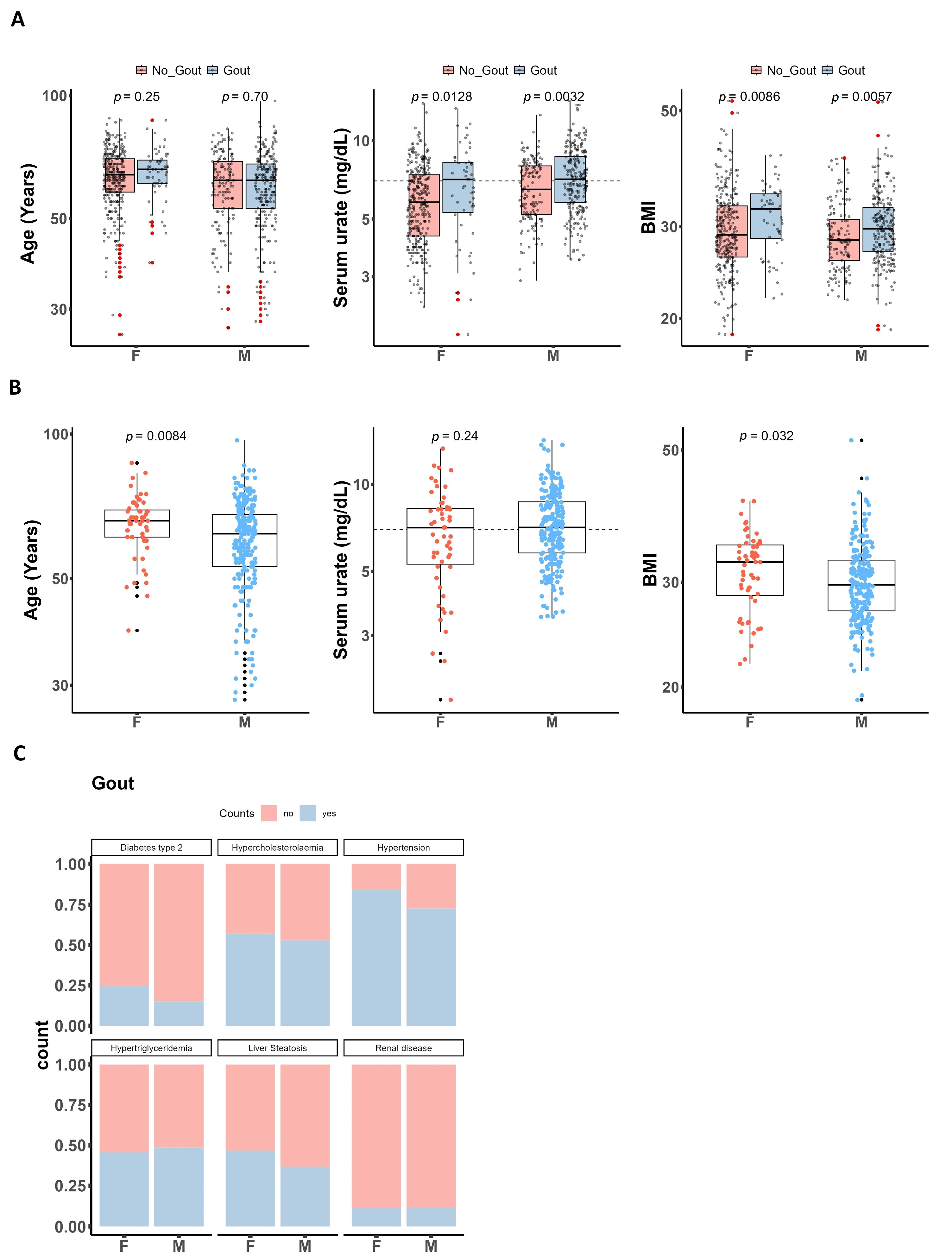
3.1. Characteristics of Patients with Gout and Healthy Controls

Table 1 illustrates the total number of patients recruited within the HINT study. The prevalence of gout was 19% in women and 81% in men, while a larger percentage (68%) were women in the group without gout. Pearson’s Chi-squared test finds evidence of an association between group and sex that is statistically significant (p < 0.001). In addition, our results indicate that gout is more prevalent in men compared to women by approximately 3–4:1 and this is in accordance with what has been reported in the literature [21]. Both groups were similar in terms of age (IQR = 63, p = 0.3). Serum urate levels were significantly higher in the gout group compared to patients without gout (p < 0.001). Body mass index (BMI) was higher in patients with gout (p = 0.03).
When analyzing the data dividedby sex, we observed that patients with gout, whether they were women or men, did not significantly differ from their respective control groups in terms of age. In patients affected by gout, it was notable that women tended to be older than men (Figure 1B). Furthermore, both women and men with gout displayed elevated serum urate levels compared to their control counterparts (Figure 1A), although interestingly, within the gout group, there were no substantial differences in urate levels between men and women (Figure 1B). Moreover, among patients with gout, women exhibited a higher body mass index (BMI) than men (Figure 1B), and a higher frequency for comorbidities type 2 diabetes, hypertension, or liver steatosis (Figure 1C). However, when analyzing the data by sex, both women and men with gout had higher BMIs compared to the corresponding controls without gout (Figure 1A).

3.2. Sex-Specific Cytokine Production upon 24 h In Vitro Stimulation of PBMCs

After filtering out the samples that measured IL-1β in RPMI above 78 mg/dL, had missing information on age, sex, and serum urate, the dataset for in vitro cytokine production analysis included data from two experiments: the first experiment included a total number of n = 134 patients with gout (28 women and 106 men) and n = 227 patients without gout (154 women and 73 men); the second included a total number of n = 131 patients with gout (25 women and 106 men) and n = 224 patients without gout (150 women and 74 men). The bar plot illustrates the distribution percentages of women and men within the groups (Figure 2A). Summary statistics for the baseline characteristics of this participant subset are available in the Supplementary Materials and reflect the characteristics of the larger dataset.
To gain a deeper understanding of the inflammatory aspects of gout, we conducted an analysis examining the association between in vitro cytokine production (IL-1β, IL-1Ra, TNF) in response to several stimuli based on a linear regression model. Age and sex were included as co-variates since it is well documented in the literature that they can impact the immune system. When the analysis was stratified by sex, age was the only covariate added. The overall findings indicate a significant association between gout and increased cytokine production, even after adjusting for age and sex (Figure 2C,D). Despite not reaching statistical significance after adjusting for age and sex, there is an observable trend of increased production of IL-1β and IL-1Ra in response to stimulation with MSU crystals and TLR agonists such as LPS (Figure 2G,H, Supplementary Figures S3 and S4). Box plots showing sample distribution for all stimuli and samples have been included in the Supplementary Material. We observed significant positive associations predominantly in women with gout for all the cytokines after adjusting for age, irrespective of the stimuli used (Figure 2C,D).

3.3. Circulating Markers Are Elevated in Men with Gout

When looking at high-sensitive CRP in circulation, patients with gout show elevated levels, with a significant increase observed only in men with gout. Among women with gout, the levels of high-sensitive CRP were comparable to those in the control group. In the case of plasma IL-1Ra, a trend toward higher levels was observed in patients with gout, which became significant in men with gout, after stratifying by sex (Figure 3A,B).

4. Discussion

The impact of sex is often overlooked in research studies as it is commonly seen as a confounder that can skew results. Studies on aging are starting to explore the potential biological differences between men and women [22,23], which could result in distinct outcomes in terms of disease states or infections [24]. Gout, an inflammatory disease associated with IL-1β activation and higher prevalence in men compared to women, displays distinct clinical profiles based on sex. This study explores sex-specific cytokine production in response to TLR in vitro stimulation in patients with gout. PBMCs from patients with gout show increased IL-1β, IL-1Ra, and TNF production upon 24 h stimulation, particularly in women. On the other hand, men with gout had elevated levels of plasma IL-1Ra and high-sensitive CRP, but no significant differences in circulating cytokines were observed in women. In the gout group, women were older and had a slightly higher BMI compared to men and presented with comorbidities more often.
Gout can develop even at a younger age in men [25], while it is relatively rare in premenopausal women [26]. In our study, we observed that women with gout tend to be of older age than men with gout (Figure 1), findings that are supported by the literature [5]. Overall, patients with gout showed markedly higher serum urate concentrations when compared with control subjects, patients without gout but with either normal or elevated serum urate (referred to as normo- or hyperuricemic individuals). Most often, men with gout present higher levels of serum urate compared to women [27]. Notably, there was no difference in serum urate between men and women with gout in our group. The BMI measurements in women with gout were significantly higher compared to men with gout (Figure 1), in line with the association between excess adiposity and an increased risk of gout in women [28]. In a study following 534 healthy individuals exposed to various stimuli, the analysis reveals that factors like age and sex significantly influence cytokine production but remain unaffected by smoking and body mass index (BMI) [16].
Men and women may have different baseline levels of certain immune cells which could result in differences in their immune system responses. A study on immune changes among in individuals aged 50 and older observed over the course of a year that men and women had different cell populations, with women having more CD4+ T cells and men more CD8+ T cells [29]. Furthermore, compared to men, PBMCs of women may exhibit a lower percentage of natural killer (NK) cells, and a higher percentage of plasma cells [30]. Neutrophils are essential in clearing microorganisms but switch to a proinflammatory phenotype in several autoimmune diseases. Type 1 interferon pathway is up-regulated in neutrophils from healthy women, through a mechanism that involves estradiol-dependent maturation. Neutrophils isolated from women and primed in vitro with IFN-α show increased proinflammation on restimulation with viral-like ligands, suggesting possible hyperresponsiveness to danger signals [31]. The interferon signature was also detected in monocytes of post-menopausal women presenting low-grade inflammation, defined as hsCRP above 3 μg/mL [32]. In our study, we found increased innate cytokine production predominantly in stimulated PBMCs of women with gout (Figure 2), suggesting women may mount more robust inflammatory responses to infections. While this could be beneficial to resolution of an infection, it may also contribute to exacerbation of symptoms. Within the dataset of the UK Biobank cohort, it was observed that women diagnosed with gout presented increased susceptibility to COVID-19-related mortality [33]. Healthy men showed increased cytokine capacity in response to in vitro stimulation in both PBMCs and whole blood, particularly of immune innate cytokines [16]. In addition, PBMCs from men with metabolic syndrome display an enhanced capacity for cytokine production as well. Circulating markers reveal that men with metabolic syndrome had elevated levels of several pro-inflammatory markers such as IL-6 and leptin, while women with metabolic syndrome showed lower levels of anti-inflammatory adiponectin [17]. In our study, men with gout show increased levels of pro-inflammatory hsCRP and anti-inflammatory IL-1Ra (Figure 3). These findings may suggest an interplay of inflammatory factors in male gout patients, where heightened inflammation as indicated by hsCRP is potentially counteracted by an increase in the anti-inflammatory IL-1Ra.
We recognize the need for caution when drawing robust conclusions solely based on the group with a smaller sample size. To enhance the credibility of our findings, further research is warranted to replicate and validate these observations, ideally with more balanced sex distributions among the groups. Additionally, we acknowledge that only sex and age have been considered in the analysis. However, it is important to recognize that other factors like lifestyle, medication use, hormones, or underlying health conditions in patients with gout may also impact cytokine production. Given the substantial amount of missing data in various clinical variables, our model incorporated only age and sex as covariates. In this sense, it is important that these results are further investigated in relation to other datasets as well. Moreover, regarding in vitro production of IL-1β, some samples were measured at the upper limit of the standard curve, which could potentially mask significant differences between groups.
The elevated cytokine production in women with gout following stimulation suggests a potentially more robust immune response in this subgroup. On the other hand, the elevated levels of IL-1Ra and hsCRP in men with gout highlight the presence of systemic inflammation. These results possibly underline distinct immune responses between men and women with gout. Further functional studies will help elucidate some of the mechanistic links between cytokine dysregulation and gout pathogenesis. Omics techniques, like RNA sequencing, could advance our understanding of the pathways involved and complex interplay between cytokine production and sex in gout pathogenesis, with the ultimate goal of improving patient care and establishing a base for future therapeutic strategies.

Supplementary Materials

The following supporting information can be downloaded at: https://www.mdpi.com/article/10.3390/gucdd2020012/s1, Figure S1: Box plots showing distribution of cytokine levels in PBMCs from patients with and without gout (A) and split by sex (B) that were stimulated in vitro for 24 h with C16 and C16 + MSU; Figure S2: Box plots showing distribution of TNF-α levels in PBMCs from patients with and without gout (A) and split by sex (B) that were stimulated in vitro for 24 h; Figure S3: Box plots showing distribution of IL-1β levels in PBMCs from patients with and without gout (A) and split by sex (B) that were stimulated in vitro for 24 h; Figure S4: Box plots showing distribution of IL-1Ra levels in PBMCs from patients with and without gout (A) and split by sex (B) that were stimulated in vitro for 24 h; Table S1: Baseline characteristics of the gout and control cohorts in all patients included for experiment 1; Table S2: Baseline characteristics only for the gout patients included for experiment 1; Table S3: Baseline characteristics of the gout and control cohorts in all patients included for experiment 2; Table S4: Baseline characteristics only for the gout patients included for experiment 2.

Author Contributions

Conceptualization, M.B., O.I.G., T.O.C. and L.A.B.J.; Methodology, M.B., O.I.G., T.O.C. and L.A.B.J.; Investigation, M.B., O.I.G., I.H., V.N., A.M.M., D.M., C.P. and S.R.; Supervision, T.O.C. and L.A.B.J.; Project administration, R.A.P.; Data curation, M.B. and O.I.G.; Formal analysis, M.B.; Visualization, M.B.; Writing—original draft, M.B. and O.I.G.; Writing—review & editing, M.B., O.I.G., T.O.C. and L.A.B.J. All authors contributed to data interpretation, revised the manuscript, and approved the final version. All authors have read and agreed to the published version of the manuscript.

Funding

This study was funded by a Competitiveness Operational Programme grant of the Romanian Ministry of European Funds (P_37_762, MySMIS 103587). LABJ is supported by a grant of the Romanian Ministry of European Investments and Projects, PNRR-III-C9-2022-I8, CF 85/15.11.2022. T.O.C. was supported by a grant of the Ministry of Research, Innovation and Digitization, CNCS/CCCDI—UEFISCDI, project number PD-2019-0802, within PNCDI III (Romania). MB was supported by “Iuliu Hațieganu” University of Medicine and Pharmacy, 400012 Cluj-Napoca, Romania, grant number 1529/6 PCD project. OIG was supported by a grant for doctoral research projects (2462/22) of the University of Medicine and Pharmacy “Iuliu Hațieganu”, Cluj-Napoca, Romania.

Institutional Review Board Statement

In vitro experiments were approved by the Ethical Committee of the “Iuliu Hațieganu” University of Medicine and Pharmacy, Cluj-Napoca (approval no. 425/24.11.2016).

Informed Consent Statement

All participants provided written informed consent before inclusion. All experiments were conducted according to the principles of the Declaration of Helsinki.

Data Availability Statement

The data underlying this article will be shared on reasonable request to the corresponding author.

Conflicts of Interest

The authors declare no conflicts of interest.

Appendix A

HINT consortium: Leo A. B. Joosten, Ioan V. Pop, Radu A. Popp, Simona Rednic, Cristina Pamfil, Tania O. Crişan, Marius Farcaş, Dragoş H. Mărginean, Orsolya I. Gaal, Medeea O. Badii, Ioana Hotea, Loredana Peca, Andreea-Manuela Mirea, Georgiana Cabău, Valentin Nica, Doina Colcear, Mariana S. Pop, Ancuta Rus.

References

  1. Martinon, F.; Pétrilli, V.; Mayor, A.; Tardivel, A.; Tschopp, J. Gout-associated uric acid crystals activate the NALP3 inflammasome. Nature 2006, 440, 237–241. [Google Scholar] [CrossRef]
  2. So, A.; Dumusc, A.; Nasi, S. The role of IL-1 in gout: From bench to bedside. Rheumatology 2018, 57, i12–i19. [Google Scholar] [CrossRef] [PubMed]
  3. Zhu, Y.; Pandya, B.J.; Choi, H.K. Prevalence of gout and hyperuricemia in the US general population: The National Health and Nutrition Examination Survey 2007–2008. Arthritis Rheum. 2011, 63, 3136–3141. [Google Scholar] [CrossRef]
  4. te Kampe, R.; Janssen, M.; van Durme, C.; Jansen, T.L.; Boonen, A. Sex differences in the clinical profile among patients with gout: Cross-sectional analyses of an observational study. J. Rheumatol. 2021, 48, 286–292. [Google Scholar] [CrossRef]
  5. Harrold, L.R.; Etzel, C.J.; Gibofsky, A.; Kremer, J.M.; Pillinger, M.H.; Saag, K.G.; Schlesinger, N.; Terkeltaub, R.; Cox, V.; Greenberg, J.D. Sex differences in gout characteristics: Tailoring care for women and men. BMC Musculoskelet. Disord. 2017, 18, 108. [Google Scholar] [CrossRef]
  6. Bergsten, U.; Dehlin, M.; Klingberg, E.; Landgren, A.J.; Jacobsson, L.T.H. Gender differences in illness perceptions and disease management in patients with gout, results from a questionnaire study in Western Sweden. BMC Musculoskelet. Disord. 2023, 24, 300. [Google Scholar] [CrossRef] [PubMed]
  7. Van Lunzen, J.; Altfeld, M. Sex differences in infectious diseases-common but neglected. J. Infect. Dis. 2014, 209, 79–80. [Google Scholar] [CrossRef] [PubMed]
  8. Angum, F.; Khan, T.; Kaler, J.; Siddiqui, L.; Hussain, A. The Prevalence of Autoimmune Disorders in Women: A Narrative Review. Cureus 2020, 12, e8094. [Google Scholar] [CrossRef]
  9. Klein, S.L.; Flanagan, K.L. Sex differences in immune responses. Nat. Rev. Immunol. 2016, 16, 626–638. [Google Scholar] [CrossRef]
  10. Verthelyi, D.; Klinman, D.M. Sex hormone levels correlate with the activity of cytokine-secreting cells in vivo. Immunology 2000, 100, 384–390. [Google Scholar] [CrossRef]
  11. Casimir, G.J.; Heldenbergh, F.; Hanssens, L.; Mulier, S.; Heinrichs, C.; Lefevre, N.; Désir, J.; Corazza, F.; Duchateau, J. Gender differences and inflammation: An in vitro model of blood cells stimulation in prepubescent children. J. Inflamm. 2010, 7, 28. [Google Scholar] [CrossRef]
  12. Li, F.; Boon, A.C.M.; Michelson, A.P.; Foraker, R.E.; Zhan, M.; Payne, P.R.O. Estrogen hormone is an essential sex factor inhibiting inflammation and immune response in COVID-19. Sci. Rep. 2022, 12, 9462. [Google Scholar] [CrossRef] [PubMed]
  13. Schurz, H.; Salie, M.; Tromp, G.; Hoal, E.G.; Kinnear, C.J.; Möller, M. The X chromosome and sex-specific effects in infectious disease susceptibility. Hum. Genom. 2019, 13, 2. [Google Scholar] [CrossRef] [PubMed]
  14. Pinheiro, I.; Dejager, L.; Libert, C. X-chromosome-located microRNAs in immunity: Might they explain male/female differences?: The X chromosome-genomic context may affect X-located miRNAs and downstream signaling, thereby contributing to the enhanced immune response of females. BioEssays 2011, 33, 791–802. [Google Scholar] [CrossRef] [PubMed]
  15. Lefèvre, N.; Corazza, F.; Valsamis, J.; Delbaere, A.; De Maertelaer, V.; Duchateau, J.; Casimir, G. The number of X chromosomes influences inflammatory cytokine production following Toll-like receptor stimulation. Front. Immunol. 2019, 10, 1052. [Google Scholar] [CrossRef] [PubMed]
  16. ter Horst, R.; Jaeger, M.; Smeekens, S.P.; Oosting, M.; Swertz, M.A.; Li, Y.; Kumar, V.; Diavatopoulos, D.A.; Jansen, A.F.M.; Lemmers, H.; et al. Host and Environmental Factors Influencing Individual Human Cytokine Responses. Cell 2016, 167, 1111–1124.e13. [Google Scholar] [CrossRef] [PubMed]
  17. ter Horst, R.; van den Munckhof, I.C.L.; Schraa, K.; Aguirre-Gamboa, R.; Jaeger, M.; Smeekens, S.P.; Brand, T.; Lemmers, H.; Dijkstra, H.; Galesloot, T.E.; et al. Sex-specific regulation of inflammation and metabolic syndrome in obesity. Arterioscler. Thromb. Vasc. Biol. 2020, 40, 1787–1800. [Google Scholar] [CrossRef] [PubMed]
  18. Beenakker, K.G.M.; Westendorp, R.G.J.; De Craen, A.J.M.; Chen, S.; Raz, Y.; Ballieux, B.E.P.B.; Nelissen, R.G.H.H.; Later, A.F.L.; Huizinga, T.W.; Slagboom, P.E.; et al. Men Have a Stronger Monocyte-Derived Cytokine Production Response upon Stimulation with the Gram-Negative Stimulus Lipopolysaccharide than Women: A Pooled Analysis Including 15 Study Populations. J. Innate Immun. 2020, 12, 142–153. [Google Scholar] [CrossRef]
  19. Da Pozzo, E.; Giacomelli, C.; Cavallini, C.; Martini, C. Cytokine secretion responsiveness of lymphomonocytes following cortisol cell exposure: Sex differences. PLoS ONE 2018, 13, e0200924. [Google Scholar] [CrossRef]
  20. Joosten, L.A.B.; Netea, M.G.; Mylona, E.; Koenders, M.I.; Malireddi, R.K.S.; Oosting, M.; Stienstra, R.; Van De Veerdonk, F.L.; Stalenhoef, A.F.; Giamarellos-Bourboulis, E.J.; et al. Engagement of fatty acids with toll-like receptor 2 drives interleukin-1β production via the ASC/caspase 1 pathway in monosodium urate monohydrate crystal-induced gouty arthritis. Arthritis Rheum. 2010, 62, 3237–3248. [Google Scholar] [CrossRef]
  21. Singh, J.A.; Gaffo, A. Gout epidemiology and comorbidities. Semin. Arthritis Rheum. 2020, 50, S11–S16. [Google Scholar] [CrossRef] [PubMed]
  22. Hägg, S.; Jylhävä, J. Sex differences in biological aging with a focus on human studies. eLife 2021, 10, e63425. [Google Scholar] [CrossRef] [PubMed]
  23. Márquez, E.J.; Chung, C.H.; Marches, R.; Rossi, R.J.; Nehar-Belaid, D.; Eroglu, A.; Mellert, D.J.; Kuchel, G.A.; Banchereau, J.; Ucar, D. Sexual-dimorphism in human immune system aging. Nat. Commun. 2020, 11, 751. [Google Scholar] [CrossRef] [PubMed]
  24. Bongen, E.; Lucian, H.; Khatri, A.; Fragiadakis, G.K.; Bjornson, Z.B.; Nolan, G.P.; Utz, P.J.; Khatri, P. Sex Differences in the Blood Transcriptome Identify Robust Changes in Immune Cell Proportions with Aging and Influenza Infection. Cell Rep. 2019, 29, 1961–1973.e4. [Google Scholar] [CrossRef] [PubMed]
  25. Evans, P.L.; Prior, J.A.; Belcher, J.; Hay, C.A.; Mallen, C.D.; Roddy, E. Gender-specific risk factors for gout: A systematic review of cohort studies. Adv. Rheumatol. 2019, 59, 24. [Google Scholar] [CrossRef] [PubMed]
  26. Patel, A.V.; Gaffo, A.L. Managing Gout in Women: Current Perspectives. J. Inflamm. Res. 2022, 15, 1591–1598. [Google Scholar] [CrossRef] [PubMed]
  27. Tin, A.; Marten, J.; Halperin Kuhns, V.L.; Li, Y.; Wuttke, M.; Kirsten, H.; Sieber, K.B.; Qiu, C.; Gorski, M.; Yu, Z.; et al. Target genes, variants, tissues and transcriptional pathways influencing human serum urate levels. Nat. Genet. 2019, 51, 1459–1474. [Google Scholar] [CrossRef] [PubMed]
  28. McCormick, N.; Yokose, C.; Lu, N.; Joshi, A.D.; Curhan, G.C.; Choi, H.K. Impact of adiposity on risk of female gout among those genetically predisposed: Sex-specific prospective cohort study findings over >32 years. Ann. Rheum. Dis. 2022, 81, 556–563. [Google Scholar] [CrossRef]
  29. Lakshmikanth, T.; Muhammad, S.A.; Olin, A.; Chen, Y.; Mikes, J.; Fagerberg, L.; Gummesson, A.; Bergström, G.; Uhlen, M.; Brodin, P. Human Immune System Variation during 1 Year. Cell Rep. 2020, 32, 107923. [Google Scholar] [CrossRef]
  30. Huang, Z.; Chen, B.; Liu, X.; Li, H.; Xie, L.; Gao, Y.; Duan, R.; Li, Z.; Zhang, J.; Zheng, Y.; et al. Effects of sex and aging on the immune cell landscape as assessed by single-cell transcriptomic analysis. Proc. Natl. Acad. Sci. USA 2021, 118, e2023216118. [Google Scholar] [CrossRef]
  31. Gupta, S.; Nakabo, S.; Blanco, L.P.; O’Neil, L.J.; Wigerblad, G.; Goel, R.R.; Mistry, P.; Jiang, K.; Carmona-Rivera, C.; Chan, D.W.; et al. Sex differences in neutrophil biology modulate response to type I interferons and immunometabolism. Proc. Natl. Acad. Sci. USA 2020, 117, 16481–16491. [Google Scholar] [CrossRef] [PubMed]
  32. So, J.; Tai, A.K.; Lichtenstein, A.H.; Wu, D.; Lamon-Fava, S. Sexual dimorphism of monocyte transcriptome in individuals with chronic low-grade inflammation. Biol. Sex Differ. 2021, 12, 43. [Google Scholar] [CrossRef] [PubMed]
  33. Topless, R.K.; Gaffo, A.; Stamp, L.K.; Robinson, P.C.; Dalbeth, N.; Merriman, T.R. Gout and the risk of COVID-19 diagnosis and death in the UK Biobank: A population-based study. Lancet Rheumatol. 2022, 4, e274–e281. [Google Scholar] [CrossRef] [PubMed]
Figure 1. Baseline characteristics. (A) Box plots split into women (F) and men (M) with and without gout. (B) Box plots of only patients with gout split into women (F) and men (M). Unpaired two-sample Wilcoxon test (also known as Wilcoxon rank sum test or Mann–Whitney test); outlier samples are indicated by the red (A) and black (B) dots. (C) Comorbidities distribution in patients with gout split by sex.
Gucdd 02 00012 g001
Figure 2. Association of cytokine production in PBMCs and sex in patients with and without gout. (A) Percentage of women (F) and men (M) in both groups. (B) Experimental design. (C,D) p-values of association between gout and cytokine levels in all patients or split by sex. In all patients, age and sex have been added as co-variates, rather when split by sex, only age was added as a covariate. All p-values were calculated using linear regression and cytokine data were log transformed. Grey color belongs to non-significant p-values (>0.05). (E,F) Sample distribution example of basal IL-1Ra levels (RPMI) and in stimulation with C16 and MSU in all patients or split by sex. (G,H) Sample distribution of IL-1β and IL-1Ra levels after stimulation with LPS low dose and MSU in all patients or split by sex; On the y-axis, values are presented without commas for numbers exceeding four digits.
Gucdd 02 00012 g002
Figure 3. Circulating markers. (A) Box plots for high-sensitive CRP and plasma IL-1Ra for patients with and without gout. (B) Box plots split into women (F) and men (M). Unpaired two-sample Wilcoxon test (also known as Wilcoxon rank sum test or Mann–Whitney test); Black (A) and red (B) dots mark outlier samples.
Gucdd 02 00012 g003
Table 1. Baseline characteristics of the gout and control cohorts in all patients included.
CharacteristicnGout, n = 299 1No_Gout, n = 480 1p-Value 2
Sex777 <0.001
F (female) 56 (19%)324 (68%)
M (male) 243 (81%)154 (32%)
Unknown 02
Age (years)76363 (55, 69)63 (57, 70)0.3
Unknown 511
Urate (mg/dL)7627.10 (5.70, 8.69)6.10 (4.70, 7.60)<0.001
Unknown 710
BMI74330.0 (26.8, 33.2)28.6 (26.2, 32.3)0.003
Unknown 828
1 n (%); Median (IQR); 2 Pearson’s Chi-squared test or Wilcoxon rank sum test; Unkown accounts for missing information.
Disclaimer/Publisher’s Note: The statements, opinions and data contained in all publications are solely those of the individual author(s) and contributor(s) and not of MDPI and/or the editor(s). MDPI and/or the editor(s) disclaim responsibility for any injury to people or property resulting from any ideas, methods, instructions or products referred to in the content.

