Endometritis and Fibrosis: An Evolving Story
Share This Topical Collection
Editors
 Prof. Dr. Jordi Miró Roig
 Prof. Dr. Jordi Miró Roig
E-Mail
Website
Guest Editor
Equine Reproduction Service, Department of Animal Medicine and Surgery, Faculty of Veterinary Sciences, Autonomous University of Barcelona, E-08193 Bellaterra, Spain
Interests: assisted reproductive technology; semen evaluation; reproductive biology; semen analysis; cryopreservation; endometritis; endometriosis; horse, donkey; reproduction biology
 Dr. Graça Ferreira-Dias
 Dr. Graça Ferreira-Dias
E-Mail
Website
Guest Editor
Department of Morphology and Function, CIISA- Centre for Interdisciplinary Research in Animal Health, Faculty of Veterinary Medicine, University of Lisbon, 1649-004 Lisbon, Portugal
Interests: reproduction; mare; corpus luteum; oviduct; endometrium; endometriosis; endocrinology; cytokines; fibrosis; epigenetics
Topical Collection Information
Dear Colleagues,
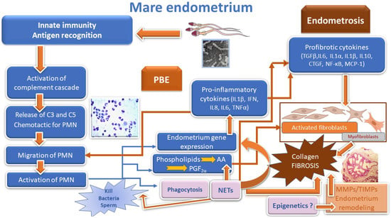
Natural breeding and artificial insemination induce a physiological endometrial response, resulting in inflammatory cell migration into the uterus. In many species, innate and adaptive immune mechanisms are essential for the physiological regulation of uterine function. This interplay among inflammatory cells orchestrates endometrial inflammatory response and remodeling events. Pathogenesis of endometritis is very complex, bridging the action of steroid hormones, eicosanoids, cytokines, growth factors, antioxidants, and others. Physiological endometritis differs among species, depending on semen deposition location, reproductive tract anatomy, and other species specificities. In species with vaginal semen deposition, post-breeding endometritis is reduced, while species with intrauterine semen deposition mount an exuberant physiological endometrial reaction. Seminal plasma induces and controls the inflammatory response. Failure of defense mechanisms in the reproductive tract can induce pathological persistent endometritis or metritis. Persistence of endometrial inflammation leads to pro-fibrotic factor release, collagen deposition, and fibrosis, causing uterine failure and infertility. Each species has its own reproductive strategy. As such, knowledge of similar pathways can improve the assisted reproduction outcome by increasing fertility rates in different species. Thus, this Special Issue aims to gather sound knowledge that will help to provide new treatments for pathological endometritis and avoid endometrial degenerative changes.
Prof. Dr. Jordi Miró Roig
Dr. Graça Ferreira Dias
Guest Editors
Manuscript Submission Information
Manuscripts should be submitted online at www.mdpi.com by registering and logging in to this website. Once you are registered, click here to go to the submission form. Manuscripts can be submitted until the deadline. All submissions that pass pre-check are peer-reviewed. Accepted papers will be published continuously in the journal (as soon as accepted) and will be listed together on the collection website. Research articles, review articles as well as short communications are invited. For planned papers, a title and short abstract (about 100 words) can be sent to the Editorial Office for announcement on this website.
Submitted manuscripts should not have been published previously, nor be under consideration for publication elsewhere (except conference proceedings papers). All manuscripts are thoroughly refereed through a single-blind peer-review process. A guide for authors and other relevant information for submission of manuscripts is available on the Instructions for Authors page. Animals is an international peer-reviewed open access semimonthly journal published by MDPI.
Please visit the Instructions for Authors page before submitting a manuscript.
The Article Processing Charge (APC) for publication in this open access journal is 2400 CHF (Swiss Francs).
Submitted papers should be well formatted and use good English. Authors may use MDPI's
English editing service prior to publication or during author revisions.
Keywords
- physiological endometritis
- pathological endometritis
- endometrosis
- mammals
Published Papers (10 papers)
Open AccessCommunication
Characterization of Bacillus pumilus Strains Isolated from Bovine Uteri
by
Panagiotis Ballas, Christoph Gabler, Karen Wagener, Marc Drillich and Monika Ehling-Schulz
Viewed by 1832
Abstract
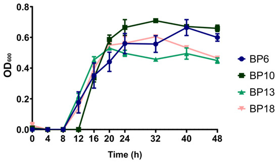
Uterine infections are a major source of economic losses to dairy farmers. The uterine microbiota as well as opportunistic uterine contaminants can contribute to the development of endometritis in dairy cows during the postpartum period. Therefore, it is important to characterize potential pathogens
[...] Read more.
Uterine infections are a major source of economic losses to dairy farmers. The uterine microbiota as well as opportunistic uterine contaminants can contribute to the development of endometritis in dairy cows during the postpartum period. Therefore, it is important to characterize potential pathogens and to further elucidate their role in the disease. In this study, we aimed to characterize
Bacillus pumilus field isolates to obtain more details regarding their effect on uterine cells by using an in vitro endometrial epithelial primary cells model. We found that
B. pumilus isolates possessed the keratinase genes
ker1 and
ker2 and therefore may produce keratinases. When primary endometrial epithelial cells were infected with 4 different
B. pumilus strains, an effect on cellular viability was observed over the course of 72 h. The effect was dose-dependent and time-dependent. Nevertheless, significant differences between the strains were not observed. All tested strains reduced the viability of the primary cells after 72 h of incubation, indicating that
B. pumilus potentially has a pathogenic effect on endometrial epithelial cells.
Full article
►▼
Show Figures
Open AccessArticle
Characterization of Myeloperoxidase in the Healthy Equine Endometrium
by
Sonia Parrilla Hernández, Thierry Franck, Carine Munaut, Émilie Feyereisen, Joëlle Piret, Frédéric Farnir, Fabrice Reigner, Philippe Barrière and Stéfan Deleuze
Cited by 3 | Viewed by 2124
Abstract
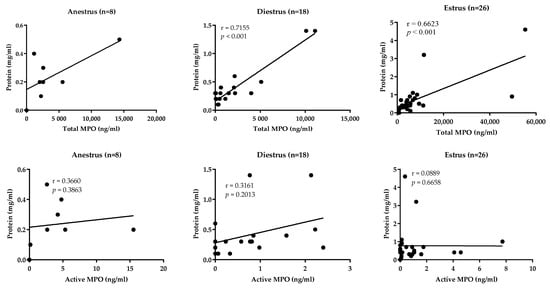
Myeloperoxidase (MPO), as a marker of neutrophil activation, has been associated with equine endometritis. However, in absence of inflammation, MPO is constantly detected in the uterine lumen of estrous mares. The aim of this study was to characterize MPO in the uterus of
[...] Read more.
Myeloperoxidase (MPO), as a marker of neutrophil activation, has been associated with equine endometritis. However, in absence of inflammation, MPO is constantly detected in the uterine lumen of estrous mares. The aim of this study was to characterize MPO in the uterus of mares under physiological conditions as a first step to better understand the role of this enzyme in equine reproduction. Total and active MPO concentrations were determined, by ELISA and SIEFED assay, respectively, in low-volume lavages from mares in estrus (
n = 26), diestrus (
n = 18) and anestrus (
n = 8) in absence of endometritis. Immunohistochemical analysis was performed on 21 endometrial biopsies randomly selected: estrus (
n = 11), diestrus (
n = 6) and anestrus (
n = 4). MPO, although mostly enzymatically inactive, was present in highly variable concentrations in uterine lavages in all studied phases, with elevated concentrations in estrus and anestrus, while in diestrus, concentrations were much lower. Intracytoplasmic immunoexpression of MPO was detected in the endometrial epithelial cells, neutrophils and glandular secretions. Maximal expression was observed during estrus in mid and basal glands with a predominant intracytoplasmic apical reinforcement. In diestrus, immunopositive glands were sporadic. In anestrus, only the luminal epithelium showed residual MPO immunostaining. These results confirm a constant presence of MPO in the uterine lumen of mares in absence of inflammation, probably as part of the uterine mucosal immune system, and suggest that endometrial cells are a source of uterine MPO under physiological cyclic conditions.
Full article
►▼
Show Figures
Open AccessArticle
Dynamics and Diversity of Intrauterine Anaerobic Microbiota in Dairy Cows with Clinical and Subclinical Endometritis
by
Panagiotis Ballas, Harald Pothmann, Isabella Pothmann, Marc Drillich, Monika Ehling-Schulz and Karen Wagener
Cited by 5 | Viewed by 2373
Abstract
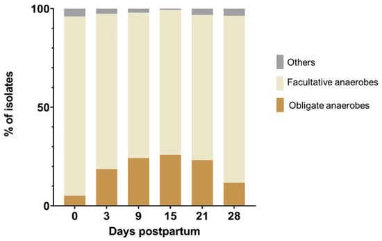
The aim of the study was to characterize the dynamics of anaerobic cultivable postpartum microbiota in the uterus of dairy cows. In total, 122 dairy cows were enrolled and sampled on day 0 (day of calving) and on days 3, 9, 15, 21,
[...] Read more.
The aim of the study was to characterize the dynamics of anaerobic cultivable postpartum microbiota in the uterus of dairy cows. In total, 122 dairy cows were enrolled and sampled on day 0 (day of calving) and on days 3, 9, 15, 21, and 28 postpartum (pp). Samples were cultivated anaerobically and analyzed by MALDI-TOF MS. In total, 1858 isolates were recovered. The most prevalent facultative anaerobic genera were
Trueperella (27.8%),
Streptococcus (25.4%), and
Escherichia (13.1%). The most prevalent obligate anaerobes were
Peptoniphilus (9.3%),
Bacteroides (3.3%), and
Clostridium (2.4%). The microbial communities were highly dynamic and diverse. On the animal level,
Trueperella pyogenes on day 21 and 28 pp was associated with clinical endometritis, and
E. coli on day 21 pp was associated with subclinical endometritis. The occurrence of
Streptococcus pluranimalium on day 28 was related to uterine health. The presence of
T. pyogenes,
Streptococcus, and
Peptoniphilus was significantly associated with an increased risk for purulent vaginal discharge. Primiparous cows showed a higher prevalence of
T. pyogenes,
Fusobacterium necrophorum,
Porphyromonas levii, and
Peptoniphilus spp. than multiparous cows but were not more susceptible to uterine diseases. This study might provide a suitable basis for future co-cultivation studies to elucidate potential synergistic interactions between microbiota.
Full article
►▼
Show Figures
Open AccessArticle
Oxidative Stress Induces Bovine Endometrial Epithelial Cell Damage through Mitochondria-Dependent Pathways
by
Pengjie Song, Chen Liu, Mingkun Sun, Jianguo Liu, Pengfei Lin, Aihua Wang and Yaping Jin
Cited by 12 | Viewed by 3016
Abstract
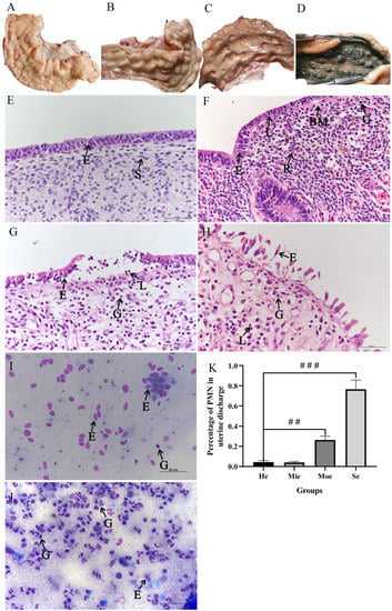
Bovine endometritis is a mucosal inflammation that is characterized by sustained polymorphonuclear neutrophil (PMN) infiltration. Elevated PMN counts in the uterine discharge of dairy cows affected by endometritis suggest that oxidative stress may be among the causes of impaired fertility due to the
[...] Read more.
Bovine endometritis is a mucosal inflammation that is characterized by sustained polymorphonuclear neutrophil (PMN) infiltration. Elevated PMN counts in the uterine discharge of dairy cows affected by endometritis suggest that oxidative stress may be among the causes of impaired fertility due to the condition. Nevertheless, the effects of oxidative stress-mediated endometritis in dairy cows largely remain uninvestigated. Therefore, fresh uterine tissue and uterine discharge samples were collected to diagnose the severity of endometritis according to the numbers of inflammatory cells in the samples. Twenty-six fresh uteri were classified into healthy, mild, moderate, and severe endometritis groups based on hematoxylin and eosin stain characteristics and the percentage of PMNs in discharge. BEECs were treated with graded concentrations of H
2O
2 from 50 μM to 200 μM in vitro as a model to explore the mechanism of oxidative stress during bovine graded endometritis. The expressions of antioxidant stress kinases were detected by quantitative fluorescence PCR to verify the oxidative stress level in uteri with endometritis. Reactive oxygen species were detected by fluorescence microscope, and inflammation-related mRNA expression increased significantly after H
2O
2 stimulation. Moreover, mRNA expression levels of antioxidant oxidative stress-related enzymes (glutathione peroxidase, superoxide dismutase, and catalase) and mitochondrial membrane potential both decreased. Further investigation revealed that expression of the apoptosis regulator Bcl-2/Bax decreased, whereas expression of the mitochondrial apoptosis-related proteins cytochrome c and caspase-3 increased in response to oxidative stress. Our results indicate that an imbalance exists between oxidation and antioxidation during bovine endometritis. Moreover, apoptosis induced in vitro by oxidative stress was characterized by mitochondrial damage in BEECs.
Full article
►▼
Show Figures
Open AccessReview
Evolution of the Concepts of Endometrosis, Post Breeding Endometritis, and Susceptibility of Mares
by
Terttu Katila and Graça Ferreira-Dias
Cited by 20 | Viewed by 4734
Abstract
In this paper, the evolution of our understanding about post breeding endometritis (PBE), the susceptibility of mares, and events leading to endometrosis are reviewed. When sperm arrive in the uterus, pro-inflammatory cytokines and chemokines are released. They attract neutrophils and induce modulatory cytokines
[...] Read more.
In this paper, the evolution of our understanding about post breeding endometritis (PBE), the susceptibility of mares, and events leading to endometrosis are reviewed. When sperm arrive in the uterus, pro-inflammatory cytokines and chemokines are released. They attract neutrophils and induce modulatory cytokines which control inflammation. In susceptible mares, this physiological defense can be prolonged since the pattern of cytokine release differs from that of resistant mares being delayed and weaker for anti-inflammatory cytokines. Delayed uterine clearance due to conformational defects, deficient myometrial contractions, and failure of the cervix to relax is detected by intrauterine fluid accumulation and is an important reason for susceptibility to endometritis. Multiparous aged mares are more likely to be susceptible. Untreated prolonged PBE can lead to bacterial or fungal endometritis called persistent or chronic endometritis. Exuberant or prolonged neutrophilia and cytokine release can have deleterious and permanent effects in inducing endometrosis. Interactions of neutrophils, cytokines, and prostaglandins in the formation of collagen and extracellular matrix in the pathogenesis of fibrosis are discussed. Endometritis and endometrosis are interconnected, influencing each other. It is suggested that they represent epigenetic changes induced by age and hostile uterine environment.
Full article
►▼
Show Figures
Open AccessArticle
Inflammatory Markers in Uterine Lavage Fluids of Pregnant, Non-Pregnant, and Intrauterine Device Implanted Mares on Days 10 and 15 Post Ovulation
by
Maria Montserrat Rivera del Alamo, Tiina Reilas, Karolina Lukasik, Antonio M. Galvão, Marc Yeste and Terttu Katila
Cited by 3 | Viewed by 2872
Abstract
Intrauterine devices (IUDs) are used in mares to suppress oestrous behaviour, but the underlying mechanism is yet to be elucidated. The presence of an embryo or an IUD prevents cyclooxygenase-2 (COX-2) and, subsequently, prostaglandin (PG) release and luteolysis. However, inflammation may also be
[...] Read more.
Intrauterine devices (IUDs) are used in mares to suppress oestrous behaviour, but the underlying mechanism is yet to be elucidated. The presence of an embryo or an IUD prevents cyclooxygenase-2 (COX-2) and, subsequently, prostaglandin (PG) release and luteolysis. However, inflammation may also be involved. Endometrial inflammatory markers in uterine lavage fluid were measured on Day 10 (EXP 1,
n = 25) and Day 15 (EXP 2,
n = 27) after ovulation in inseminated mares, non-pregnant or pregnant, and in mares in which a small plastic sphere had been inserted into the uterus 4 (EXP 1) or 3 days (EXP 2) after ovulation. Uterine lavage fluid samples were analysed for nitric oxide (NO), prostaglandin E
2 (PGE
2) (only EXP 1), prostaglandin F
2α (PGF
2α), inhibin A and cytokines, and blood samples for progesterone and oestradiol. On Day 10, the concentration of PGF
2α was lower (
p < 0.05) in the IUD group than in pregnant mares. The concentration of the modulatory cytokine IL-10 was significantly higher in the IUD group in comparison to non-pregnant mares, and inhibin A was significantly higher in IUD mares than in the pregnant counterparts on Day 15. The results suggest that the presence of IUD causes endometrial inflammation which is at a resolution stage on Day 15.
Full article
Open AccessArticle
Characterization of an Ex Vivo Equine Endometrial Tissue Culture Model Using Next-Generation RNA-Sequencing Technology
by
Maithê R. Monteiro de Barros, Mina C. G. Davies-Morel, Luis A. J. Mur, Christopher J. Creevey, Roger H. Alison and Deborah M. Nash
Viewed by 4497
Abstract
Persistent mating-induced endometritis is a major cause of poor fertility rates in the mare. Endometritis can be investigated using an ex vivo equine endometrial explant system which measures uterine inflammation using prostaglandin F
2α as a biomarker. However, this model has yet to
[...] Read more.
Persistent mating-induced endometritis is a major cause of poor fertility rates in the mare. Endometritis can be investigated using an ex vivo equine endometrial explant system which measures uterine inflammation using prostaglandin F
2α as a biomarker. However, this model has yet to undergo a wide-ranging assessment through transcriptomics. In this study, we assessed the transcriptomes of cultured endometrial explants and the optimal temporal window for their use. Endometrium harvested immediately post-mortem from native pony mares (
n = 8) were sampled (0 h) and tissue explants were cultured for 24, 48 and 72 h. Tissues were stored in RNALater, total RNA was extracted and sequenced. Differentially expressed genes (DEGs) were defined using DESeq2 (R/Bioconductor). Principal component analysis indicated that the greatest changes in expression occurred in the first 24 h of culture when compared to autologous biopsies at 0 h. Fewer DEGs were seen between 24 and 48 h of culture suggesting the system was more stable than during the first 24 h. No genes were differentially expressed between 48 and 72 h but the low number of background gene expression suggested that explant viability was compromised after 48 h.
ESR1,
MMP9,
PTGS2,
PMAIP1,
TNF,
GADD45B and
SELE genes were used as biomarkers of endometrial function, cell death and inflammation across tissue culture timepoints. STRING assessments of gene ontology suggested that DEGs between 24 and 48 h were linked to inflammation, immune system, cellular processes, environmental information processing and signal transduction, with an upregulation of most biomarker genes at 24 h. Taken together our observations indicated that 24–48 h is the optimal temporal window when the explant model can be used, as explants restore microcirculation, perform wound healing and tackle inflammation during this period. This key observation will facilitate the appropriate use of this as a model for further research into the equine endometrium and potentially the progression of mating-induced endometritis to persistent inflammation between 24 and 48 h.
Full article
►▼
Show Figures
Open AccessArticle
Specific Seminal Plasma Fractions Are Responsible for the Modulation of Sperm–PMN Binding in the Donkey
by
Jordi Miró, Jaime Catalán, Henar Marín, Iván Yánez-Ortiz and Marc Yeste
Cited by 7 | Viewed by 2528
Abstract
While artificial insemination (AI) with frozen-thawed sperm results in low fertility rates in donkeys, the addition of seminal plasma, removed during cryopreservation, partially counteracts that reduction. Related to this, an apparent inflammatory reaction in jennies is induced following AI with frozen-thawed sperm, as
[...] Read more.
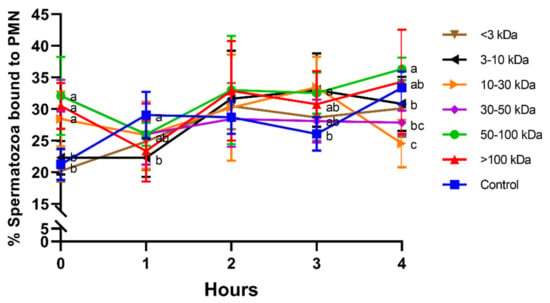
While artificial insemination (AI) with frozen-thawed sperm results in low fertility rates in donkeys, the addition of seminal plasma, removed during cryopreservation, partially counteracts that reduction. Related to this, an apparent inflammatory reaction in jennies is induced following AI with frozen-thawed sperm, as a high amount of polymorphonuclear neutrophils (PMN) are observed within the donkey uterus six hours after AI. While PMN appear to select the sperm that ultimately reach the oviduct, two mechanisms, phagocytosis and NETosis, have been purported to be involved in that clearance. Remarkably, sperm interacts with PMN, but the presence of seminal plasma reduces that binding. As seminal plasma is a complex fluid made up of different molecules, including proteins, this study aimed to evaluate how different seminal plasma fractions, separated by molecular weight (<3, 3–10, 10–30, 30–50, 50–100, and >100 kDa), affect sperm–PMN binding. Sperm motility, viability, and sperm–PMN binding were evaluated after 0 h, 1 h, 2 h, 3 h, and 4 h of co-incubation at 38 °C. Two seminal plasma fractions, including 30–50 kDa or 50–100 kDa proteins, showed the highest sperm motility and viability. As viability of sperm not bound to PMN after 3 h of incubation was the highest in the presence of 30–50 and 50–100 kDa proteins, we suggest that both fractions are involved in the control of the jenny’s post-breeding inflammatory response. In conclusion, this study has shown for the first time that specific fractions rather than the entire seminal plasma modulate sperm–PMN binding within the donkey uterus. As several proteins suggested to be involved in the control of post-AI endometritis have a molecular weight between 30 and 100 kDa, further studies aimed at determining the identity of these molecules and evaluating their potential effect in vivo are much warranted.
Full article
►▼
Show Figures
Open AccessArticle
Effects of Intrauterine Infusion of a Chitosan Solution on Recovery and Subsequent Reproductive Performance of Early Postpartum Dairy Cows with Endometritis: A Pilot Field Trial
by
Hiroaki Okawa, Missaka M.P. Wijayagunawardane, Peter L.A.M. Vos, Osamu Yamato, Masayasu Taniguchi and Mitsuhiro Takagi
Cited by 4 | Viewed by 2857
Abstract
This study investigated the efficacy of intrauterine infusion of a chitosan solution (CHT) on uterine recovery in early postpartum dairy cows with or without endometritis, and their subsequent reproductive performance. In Experiment 1, cows with endometritis at 3 weeks postpartum were administered CHT
[...] Read more.
This study investigated the efficacy of intrauterine infusion of a chitosan solution (CHT) on uterine recovery in early postpartum dairy cows with or without endometritis, and their subsequent reproductive performance. In Experiment 1, cows with endometritis at 3 weeks postpartum were administered CHT (n = 5) and prostaglandin F
2α (PGF
2α) (n = 4). Untreated cows (n = 7) served as the control group. In Experiment 2, 18 cows with a normally recovered uterus at the fresh cow check (mean, 35 days postpartum) were assigned to the CHT (n = 10) and control (n = 8) groups, and intrauterine infusion was conducted in the CHT group. Overall, in Experiment 1, the percentage of polymorphonuclear leukocytes significantly declined in the CHT group (32.3 ± 10.2 to 5.5 ± 2.4,
p < 0.05) from week 3 to week 5, but no decline occurred in the PGF
2α and control groups. In Experiment 2, the CHT and control groups showed no significant differences in reproductive parameters, suggesting the absence of adverse effects of CHT on fertility. These results suggest that intrauterine infusion of CHT in the early postpartum period effectively accelerates uterine recovery from endometritis and might be a suitable replacement for PGF
2α administration.
Full article
Open AccessArticle
Endometrial Status in Queens Evaluated by Histopathology Findings and Two Cytological Techniques: Low-Volume Uterine Lavage and Uterine Swabbing
by
Alba Martí, Anna Serrano, Josep Pastor, Teresa Rigau, Ugné Petkevičiuté, Maria Àngels Calvo, Esteban Leonardo Arosemena, Aida Yuste, David Prandi, Adrià Aguilar and Maria Montserrat Rivera del Alamo
Cited by 4 | Viewed by 3025
Abstract
Endometritis is associated with fertility problems in many species, with endometrial biopsy being the main diagnostic tool. In feline queens, the reduced size of the uterus may make it difficult to obtain representative diagnostic samples. Endometrial cytology may represent a valuable diagnostic tool
[...] Read more.
Endometritis is associated with fertility problems in many species, with endometrial biopsy being the main diagnostic tool. In feline queens, the reduced size of the uterus may make it difficult to obtain representative diagnostic samples. Endometrial cytology may represent a valuable diagnostic tool for evaluating the health status of the endometrium in queens. Fifty domestic shorthair queens were included and divided into two cytological diagnostic technique groups, the uterine lavage (UL; n = 28) and uterine swabbing (US; n = 22) groups. Cytological results were compared with histopathological and bacteriological information. Changes in the histopathological patterns were also evaluated and compared with progesterone levels to confirm previous published data. Furthermore, the results from both cytological sampling methods were compared to evaluate the utility of each method. Endometritis was ruled out in all queens by means of histology and microbiology. Leukocyte counts and red blood cell/endometrial cell ratios were significantly higher in US than UL samples. Additionally, UL sampling is less affected by blood contamination and cells are better preserved. The combination of endometrial cytology and uterine culture might be useful for evaluating the endometrial characteristics in queens. The UL evaluation method is more representative of the actual endometrial status than the US technique.
Full article
►▼
Show Figures


