Ion Channels in Cancer Therapies

A topical collection in Cancers (ISSN 2072-6694). This collection belongs to the section "Cancer Therapy".

Viewed by 54577

Editor


E-Mail Website
Collection Editor
AG Oncophysiology, Max Planck Institute for Multidisciplinary Sciences, Hermann-Rein-Str. 3, 37075 Göttingen, Germany
Interests: ion channels; voltage-gated potassium channels; membrane potential; cell biology; neuroscience; targeted therapies
Topical Collection Information

Dear Colleagues,

The idea that ion channels play crucial roles in the onset and development of all types of cancer has gained overwhelming support in the last several decades. Besides the purely scientific interest that this concept has for cancer biologists, the available extensive knowledge of the physiology and pharmacology of ion channels opens a window for therapeutic interventions that can correct or modify the behavior of tumor cells and their microenvironment and result in a benefit for the patients.

There are many aspects related to ion channels in the context of tumor therapy. First, not only channels in the tumor cells but also those in cells of the microenvironment can be targeted. Moreover, ion channels play relevant roles in some of the undesired effects of more classical treatment regimes for cancer patients, and can thus be addressed to diminish toxicity. The strategies to target channels are also very diverse. In some cases, the function of ion channels is directly tackled by pharmacological modulators, which can be either repurposed drugs in current or past clinical use for other indications or newly identified natural or synthetic compounds with improved selectivity profile and potency. Other strategies take advantage of the aberrant expression of particular ion channels to direct other agents to the tumor cells or aim to correct the pathways altered by pathological channel expression.

In summary, this Topical Collection aims to offer an overview of the present and potential importance of ion channels in cancer therapy.

Prof. Dr. Luis A. Pardo
Collection Editor

Manuscript Submission Information

Manuscripts should be submitted online at www.mdpi.com by registering and logging in to this website. Once you are registered, click here to go to the submission form. Manuscripts can be submitted until the deadline. All submissions that pass pre-check are peer-reviewed. Accepted papers will be published continuously in the journal (as soon as accepted) and will be listed together on the collection website. Research articles, review articles as well as communications are invited. For planned papers, a title and short abstract (about 100 words) can be sent to the Editorial Office for announcement on this website.

Submitted manuscripts should not have been published previously, nor be under consideration for publication elsewhere (except conference proceedings papers). All manuscripts are thoroughly refereed through a single-blind peer-review process. A guide for authors and other relevant information for submission of manuscripts is available on the Instructions for Authors page. Cancers is an international peer-reviewed open access semimonthly journal published by MDPI.

Please visit the Instructions for Authors page before submitting a manuscript. The Article Processing Charge (APC) for publication in this open access journal is 2900 CHF (Swiss Francs). Submitted papers should be well formatted and use good English. Authors may use MDPI's English editing service prior to publication or during author revisions.

Keywords

  • cancer treatment
  • ion channels
  • ion channel pharmacology
  • toxins
  • immunotherapy
  • drug resistance
  • targeted therapies

Published Papers (20 papers)

2024

Jump to: 2023, 2022, 2021

15 pages, 2038 KiB  
Article
Increased PIEZO1 Expression Is Associated with Worse Clinical Outcomes in Hormone-Receptor-Negative Breast Cancer Patients
by Rylee Ann Poole, Qingfei Wang, Alo Ray, Kazuaki Takabe, Mateusz Opyrchal and Eriko Katsuta
Cancers 2024, 16(4), 683; https://doi.org/10.3390/cancers16040683 - 6 Feb 2024
Viewed by 2293
Abstract
PIEZO1 plays a crucial role in the human body as a mechanosensory ion channel. It has been demonstrated that PIEZO1 is important in tissue development and regulating many essential physiological processes. Studies have suggested that the PIEZO1 ion channel plays a role in [...] Read more.
PIEZO1 plays a crucial role in the human body as a mechanosensory ion channel. It has been demonstrated that PIEZO1 is important in tissue development and regulating many essential physiological processes. Studies have suggested that the PIEZO1 ion channel plays a role in invasion and progression in cancer; elevated levels of PIEZO1 have been correlated with increased migration in breast cancer cells, chemo-resistance and invasion in gastric cancer cells, and increased invasion of osteosarcoma cells. In addition, high PIEZO1 expression levels were correlated with a worse prognosis in glioma patients. On the other hand, studies in lung cancer have attributed high PIEZO1 levels to better patient outcomes. However, the clinical impact of PIEZO1 in breast cancer is not well characterized. Therefore, our goal was to determine the clinical relevance of PIEZO1 in breast cancer. An analysis of breast cancer data from The Cancer Genome Atlas (TCGA) was conducted to investigate PIEZO1 expression levels and correlation to survival, followed by validation in an independent dataset, GSE3494. We also performed gene set enrichment analysis (GSEA) and pathway enrichment analysis. We also analyzed the immune cell composition in breast tumors from TCGA through a CIBERSORT algorithm. Our results demonstrated that the PIEZO1 expression levels are higher in hormone-receptor (HR)-negative than in HR-positive cohorts. High PIEZO1 expression is correlated with a significant decrease in survival in HR-negative cohorts, especially in triple-negative breast cancer (TNBC), suggesting that PIEZO1 could be utilized as a prognostic biomarker in HR-negative breast cancer. GSEA showed that various signaling pathways associated with more invasive phenotypes and resistance to treatments, including epithelial–mesenchymal transition (EMT), hypoxia, and multiple signaling pathways, are enriched in high-PIEZO1 HR-negative tumors. Our results also demonstrated a decrease in CD8+ and CD4+ T cell infiltration in high-PIEZO1 HR-negative tumors. Further investigations are necessary to elucidate the mechanistic roles of PIEZO1 in HR-negative breast cancer. Full article
Show Figures

Figure 1

2023

Jump to: 2024, 2022, 2021

32 pages, 8341 KiB  
Article
Voltage-Gated Sodium Channel NaV1.7 Inhibitors with Potent Anticancer Activities in Medullary Thyroid Cancer Cells
by Piyasuda Pukkanasut, Jason Whitt, Rachael Guenter, Shannon E. Lynch, Carlos Gallegos, Margarita Jacaranda Rosendo-Pineda, Juan Carlos Gomora, Herbert Chen, Diana Lin, Anna Sorace, Renata Jaskula-Sztul and Sadanandan E. Velu
Cancers 2023, 15(10), 2806; https://doi.org/10.3390/cancers15102806 - 17 May 2023
Cited by 3 | Viewed by 3101
Abstract
Our results from quantitative RT-PCR, Western blotting, immunohistochemistry, and the tissue microarray of medullary thyroid cancer (MTC) cell lines and patient specimens confirm that VGSC subtype NaV1.7 is uniquely expressed in aggressive MTC and not expressed in normal thyroid cells and [...] Read more.
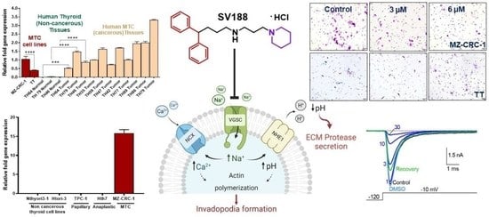
Our results from quantitative RT-PCR, Western blotting, immunohistochemistry, and the tissue microarray of medullary thyroid cancer (MTC) cell lines and patient specimens confirm that VGSC subtype NaV1.7 is uniquely expressed in aggressive MTC and not expressed in normal thyroid cells and tissues. We establish the druggability of NaV1.7 in MTC by identifying a novel inhibitor (SV188) and investigate its mode of binding and ability to inhibit INa current in NaV1.7. The whole-cell patch-clamp studies of the SV188 in the NaV1.7 channels expressed in HEK-293 cells show that SV188 inhibited the INa current in NaV1.7 with an IC50 value of 3.6 µM by a voltage- and use-dependent blockade mechanism, and the maximum inhibitory effect is observed when the channel is open. SV188 inhibited the viability of MTC cell lines, MZ-CRC-1 and TT, with IC50 values of 8.47 μM and 9.32 μM, respectively, and significantly inhibited the invasion of MZ-CRC-1 cells by 35% and 52% at 3 μM and 6 μM, respectively. In contrast, SV188 had no effect on the invasion of TT cells derived from primary tumor, which have lower basal expression of NaV1.7. In addition, SV188 at 3 μM significantly inhibited the migration of MZ-CRC-1 and TT cells by 27% and 57%, respectively. Full article
Show Figures

Graphical abstract

19 pages, 3660 KiB  
Article
Combination Therapy with a Bispecific Antibody Targeting the hERG1/β1 Integrin Complex and Gemcitabine in Pancreatic Ductal Adenocarcinoma
by Tiziano Lottini, Claudia Duranti, Jessica Iorio, Michele Martinelli, Rossella Colasurdo, Franco Nicolás D’Alessandro, Matteo Buonamici, Stefano Coppola, Valentina Devescovi, Vincenzo La Vaccara, Alessandro Coppola, Roberto Coppola, Elena Lastraioli and Annarosa Arcangeli
Cancers 2023, 15(7), 2013; https://doi.org/10.3390/cancers15072013 - 28 Mar 2023
Cited by 10 | Viewed by 2403
Abstract
Pancreatic ductal adenocarcinoma (PDAC) represents an unmet medical need. Difficult/late diagnosis as well as the poor efficacy and high toxicity of chemotherapeutic drugs result in dismal prognosis. With the aim of improving the treatment outcome of PDAC, we tested the effect of combining [...] Read more.
Pancreatic ductal adenocarcinoma (PDAC) represents an unmet medical need. Difficult/late diagnosis as well as the poor efficacy and high toxicity of chemotherapeutic drugs result in dismal prognosis. With the aim of improving the treatment outcome of PDAC, we tested the effect of combining Gemcitabine with a novel single chain bispecific antibody (scDb) targeting the cancer-specific hERG1/β1 integrin complex. First, using the scDb (scDb-hERG1-β1) in immunohistochemistry (IHC), Western blot (WB) analysis and immunofluorescence (IF), we confirmed the presence of the hERG1/β1 integrin complex in primary PDAC samples and PDAC cell lines. Combining Gemcitabine with scDb-hERG1-β1 improved its cytotoxicity on all PDAC cells tested in vitro. We also tested the combination treatment in vivo, using an orthotopic xenograft mouse model involving ultrasound-guided injection of PDAC cells. We first demonstrated good penetration of the scDb-hERG1-β1 conjugated with indocyanine green (ICG) into tumour masses by photoacoustic (PA) imaging. Next, we tested the effects of the combination at either therapeutic or sub-optimal doses of Gemcitabine (25 or 5 mg/kg, respectively). The combination of scDb-hERG1-β1 and sub-optimal doses of Gemcitabine reduced the tumour masses to the same extent as the therapeutic doses of Gemcitabine administrated alone; yielded increased survival; and was accompanied by minimised side effects (toxicity). These data pave the way for a novel therapeutic approach to PDAC, based on the combination of low doses of a chemotherapeutic drug (to minimize adverse side effects and the onset of resistance) and the novel scDb-hERG1-β1 targeting the hERG1/β1 integrin complex as neoantigen. Full article
Show Figures

Figure 1

19 pages, 3454 KiB  
Article
TRPV6 Calcium Channel Targeting by Antibodies Raised against Extracellular Epitopes Induces Prostate Cancer Cell Apoptosis
by Aurélien Haustrate, George Shapovalov, Corentin Spriet, Clément Cordier, Artem Kondratskyi, Lucile Noyer, François Foulquier, Natalia Prevarskaya and V’yacheslav Lehen’kyi
Cancers 2023, 15(6), 1825; https://doi.org/10.3390/cancers15061825 - 17 Mar 2023
Cited by 3 | Viewed by 2184
Abstract
The TRPV6 calcium channel is known to be up-regulated in various tumors. The efforts to target the TRPV6 channel in vivo are still ongoing to propose an effective therapy against cancer. Here, we report the generation of two antibodies raised against extracellular epitopes [...] Read more.
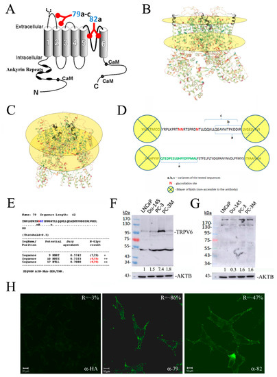
The TRPV6 calcium channel is known to be up-regulated in various tumors. The efforts to target the TRPV6 channel in vivo are still ongoing to propose an effective therapy against cancer. Here, we report the generation of two antibodies raised against extracellular epitopes corresponding to the extracellular loop between S1 and S2 (rb79) and the pore region (rb82). These antibodies generated a complex biphasic response with the transient activation of the TRPV6 channel. Store-operated calcium entry was consequently potentiated in the prostate cancer cell line LNCaP upon the treatment. Both rb79 and rb82 antibodies significantly decreased cell survival rate in a dose-dependent manner as compared to the control antibodies of the same isotype. This decrease was due to the enhanced cell death via apoptosis revealed using a sub-G1 peak in a cell cycle assay, TUNEL assay, and a Hoechst staining, having no effects in the PC3Mtrpv6−/− cell line. Moreover, all TUNEL-positive cells had TRPV6 membrane staining as compared to the control antibody treatment where TRPV6-positive cells were all TUNEL negative. These data clearly demonstrate that TRPV6 channel targeting using rb79 and rb82 antibodies is fatal and may be successfully used in the anticancer therapies. Full article
Show Figures

Figure 1

27 pages, 4124 KiB  
Article
Transcriptional Basis of Ca2+ Remodeling Reversal Induced by Polyamine Synthesis Inhibition in Colorectal Cancer Cells
by Enrique Pérez-Riesgo, Elena Hernando-Pérez, Verónica Feijóo, Sendoa Tajada, Lucía Núñez and Carlos Villalobos
Cancers 2023, 15(5), 1600; https://doi.org/10.3390/cancers15051600 - 4 Mar 2023
Cited by 7 | Viewed by 2070
Abstract
Colorectal cancer (CRC) is associated with mutations in APC/Wnt leading to c-myc activation and the overexpression of ODC1, the limiting step in polyamine synthesis. CRC cells also display a remodeling of intracellular Ca2+ homeostasis that contributes to cancer hallmarks. As polyamines may [...] Read more.
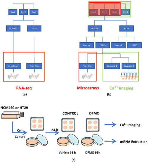
Colorectal cancer (CRC) is associated with mutations in APC/Wnt leading to c-myc activation and the overexpression of ODC1, the limiting step in polyamine synthesis. CRC cells also display a remodeling of intracellular Ca2+ homeostasis that contributes to cancer hallmarks. As polyamines may modulate Ca2+ homeostasis during epithelial tissue repair, we investigated whether polyamine synthesis inhibition may reverse Ca2+ remodeling in CRC cells and, if so, the molecular basis for this reversal. To this end, we used calcium imaging and transcriptomic analysis in normal and CRC cells treated with DFMO, an ODC1 suicide inhibitor. We found that polyamine synthesis inhibition partially reversed changes in Ca2+ homeostasis associated with CRC, including a decrease in resting Ca2+ and SOCE along with an increased Ca2+ store content. We also found that polyamine synthesis inhibition reversed transcriptomic changes in CRC cells without affecting normal cells. Specifically, DFMO treatment enhanced the transcription of SOCE modulators CRACR2A; ORMDL3; and SEPTINS 6, 7, 8, 9, and 11, whereas it decreased SPCA2, involved in store-independent Orai1 activation. Therefore, DFMO treatment probably decreased store-independent Ca2+ entry and enhanced SOCE control. Conversely, DFMO treatment decreased the transcription of the TRP channels TRPC1 and 5, TRPV6, and TRPP1 while increasing TRPP2, thus probably decreasing Ca2+ entry through TRP channels. Finally, DFMO treatment enhanced the transcription of the PMCA4 Ca2+ pump and mitochondrial channels MCU and VDAC3 for enhanced Ca2+ extrusion through the plasma membrane and mitochondria. Collectively, these findings suggested the critical role of polyamines in Ca2+ remodeling in colorectal cancer. Full article
Show Figures

Figure 1

2022

Jump to: 2024, 2023, 2021

27 pages, 4086 KiB  
Article
Voltage-Gated Sodium Channel NaV1.5 Controls NHE−1−Dependent Invasive Properties in Colon Cancer Cells
by Osbaldo Lopez-Charcas, Lucile Poisson, Oumnia Benouna, Roxane Lemoine, Stéphanie Chadet, Adrien Pétereau, Widad Lahlou, Serge Guyétant, Mehdi Ouaissi, Piyasuda Pukkanasut, Shilpa Dutta, Sadanandan E. Velu, Pierre Besson, Driffa Moussata and Sébastien Roger
Cancers 2023, 15(1), 46; https://doi.org/10.3390/cancers15010046 - 22 Dec 2022
Cited by 10 | Viewed by 2903
Abstract
Colorectal cancer (CRC) is the second leading cause of death worldwide, with 0.9 million deaths per year. The metastatic stage of the disease is identified in about 20% of cases at the first diagnosis and is associated with low patient-survival rates. Voltage-gated sodium [...] Read more.
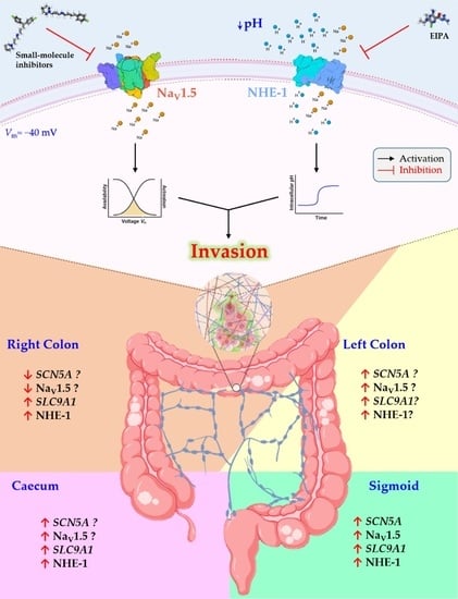
Colorectal cancer (CRC) is the second leading cause of death worldwide, with 0.9 million deaths per year. The metastatic stage of the disease is identified in about 20% of cases at the first diagnosis and is associated with low patient-survival rates. Voltage-gated sodium channels (NaV) are abnormally overexpressed in several carcinomas including CRC and are strongly associated with the metastatic behavior of cancer cells. Acidification of the extracellular space by Na+/H+ exchangers (NHE) contributes to extracellular matrix degradation and cell invasiveness. In this study, we assessed the expression levels of pore-forming α-subunits of NaV channels and NHE exchangers in tumor and adjacent non-malignant tissues from colorectal cancer patients, CRC cell lines and primary tumor cells. In all cases, SCN5A (gene encoding for NaV1.5) was overexpressed and positively correlated with cancer stage and poor survival prognosis for patients. In addition, we identified an anatomical differential expression of SCN5A and SLC9A1 (gene encoding for NHE-1) being particularly relevant for tumors that originated on the sigmoid colon epithelium. The functional activity of NaV1.5 channels was characterized in CRC cell lines and the primary cells of colon tumors obtained using tumor explant methodologies. Furthermore, we assessed the performance of two new small-molecule NaV1.5 inhibitors on the reduction of sodium currents, as well as showed that silencing SCN5A and SLC9A1 substantially reduced the 2D invasive capabilities of cancer cells. Thus, our findings show that both NaV1.5 and NHE-1 represent two promising targetable membrane proteins against the metastatic progression of CRC. Full article
Show Figures

Graphical abstract

19 pages, 2972 KiB  
Article
Tumoricidal, Temozolomide- and Radiation-Sensitizing Effects of KCa3.1 K+ Channel Targeting In Vitro Are Dependent on Glioma Cell Line and Stem Cell Fraction
by Nicolai Stransky, Katrin Ganser, Ulrike Naumann, Stephan M. Huber and Peter Ruth
Cancers 2022, 14(24), 6199; https://doi.org/10.3390/cancers14246199 - 15 Dec 2022
Cited by 2 | Viewed by 1622
Abstract
Reportedly, the intermediate-conductance Ca2+-activated potassium channel KCa3.1 contributes to the invasion of glioma cells into healthy brain tissue and resistance to temozolomide and ionizing radiation. Therefore, KCa3.1 has been proposed as a potential target in glioma therapy. [...] Read more.
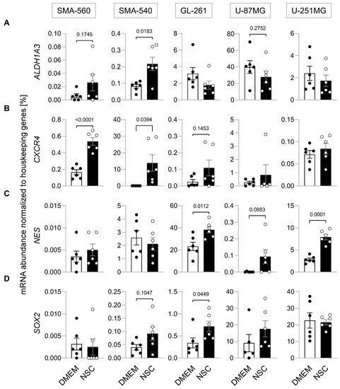
Reportedly, the intermediate-conductance Ca2+-activated potassium channel KCa3.1 contributes to the invasion of glioma cells into healthy brain tissue and resistance to temozolomide and ionizing radiation. Therefore, KCa3.1 has been proposed as a potential target in glioma therapy. The aim of the present study was to assess the variability of the temozolomide- and radiation-sensitizing effects conferred by the KCa3.1 blocking agent TRAM-34 between five different glioma cell lines grown as differentiated bulk tumor cells or under glioma stem cell-enriching conditions. As a result, cultures grown under stem cell-enriching conditions exhibited indeed higher abundances of mRNAs encoding for stem cell markers compared to differentiated bulk tumor cultures. In addition, stem cell enrichment was paralleled by an increased resistance to ionizing radiation in three out of the five glioma cell lines tested. Finally, TRAM-34 led to inconsistent results regarding its tumoricidal but also temozolomide- and radiation-sensitizing effects, which were dependent on both cell line and culture condition. In conclusion, these findings underscore the importance of testing new drug interventions in multiple cell lines and different culture conditions to partially mimic the in vivo inter- and intra-tumor heterogeneity. Full article
Show Figures

Figure 1

16 pages, 2000 KiB  
Review
Chloride Intracellular Channel Proteins (CLICs) and Malignant Tumor Progression: A Focus on the Preventive Role of CLIC2 in Invasion and Metastasis
by Saya Ozaki, Kanta Mikami, Takeharu Kunieda and Junya Tanaka
Cancers 2022, 14(19), 4890; https://doi.org/10.3390/cancers14194890 - 6 Oct 2022
Cited by 6 | Viewed by 2406
Abstract
CLICs are the dimorphic protein present in both soluble and membrane fractions. As an integral membrane protein, CLICs potentially possess ion channel activity. However, it is not fully clarified what kinds of roles CLICs play in physiological and pathological conditions. In vertebrates, CLICs [...] Read more.
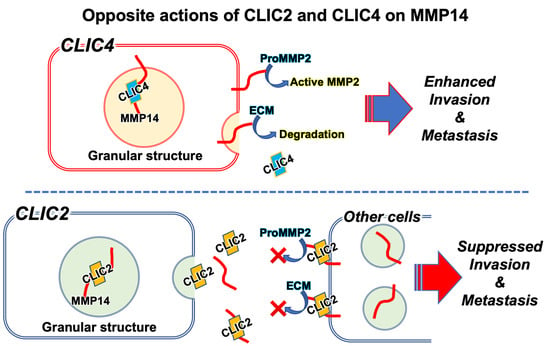
CLICs are the dimorphic protein present in both soluble and membrane fractions. As an integral membrane protein, CLICs potentially possess ion channel activity. However, it is not fully clarified what kinds of roles CLICs play in physiological and pathological conditions. In vertebrates, CLICs are classified into six classes: CLIC1, 2, 3, 4, 5, and 6. Recently, in silico analyses have revealed that the expression level of CLICs may have prognostic significance in cancer. In this review, we focus on CLIC2, which has received less attention than other CLICs, and discuss its role in the metastasis and invasion of malignant tumor cells. CLIC2 is expressed at higher levels in benign tumors than in malignant ones, most likely preventing tumor cell invasion into surrounding tissues. CLIC2 is also expressed in the vascular endothelial cells of normal tissues and maintains their intercellular adhesive junctions, presumably suppressing the hematogenous metastasis of malignant tumor cells. Surprisingly, CLIC2 is localized in secretory granules and secreted into the extracellular milieu. Secreted CLIC2 binds to MMP14 and inhibits its activity, leading to suppressed MMP2 activity. CLIC4, on the other hand, promotes MMP14 activity. These findings challenge the assumption that CLICs are ion channels, implying that they could be potential new targets for the treatment of malignant tumors. Full article
Show Figures

Graphical abstract

23 pages, 4253 KiB  
Article
Relevance of Abnormal KCNN1 Expression and Osmotic Hypersensitivity in Ewing Sarcoma
by Sebastian Fuest, Christoph Post, Sebastian T. Balbach, Susanne Jabar, Ilka Neumann, Sandra Schimmelpfennig, Sarah Sargin, Elke Nass, Thomas Budde, Sareetha Kailayangiri, Bianca Altvater, Andreas Ranft, Wolfgang Hartmann, Uta Dirksen, Claudia Rössig, Albrecht Schwab and Zoltán Pethő
Cancers 2022, 14(19), 4819; https://doi.org/10.3390/cancers14194819 - 1 Oct 2022
Cited by 4 | Viewed by 2354
Abstract
Ewing sarcoma (EwS) is a rare and highly malignant bone tumor occurring mainly in childhood and adolescence. Physiologically, the bone is a central hub for Ca2+ homeostasis, which is severely disturbed by osteolytic processes in EwS. Therefore, we aimed to investigate how [...] Read more.
Ewing sarcoma (EwS) is a rare and highly malignant bone tumor occurring mainly in childhood and adolescence. Physiologically, the bone is a central hub for Ca2+ homeostasis, which is severely disturbed by osteolytic processes in EwS. Therefore, we aimed to investigate how ion transport proteins involved in Ca2+ homeostasis affect EwS pathophysiology. We characterized the expression of 22 candidate genes of Ca2+-permeable or Ca2+-regulated ion channels in three EwS cell lines and found the Ca2+-activated K+ channel KCa2.1 (KCNN1) to be exceptionally highly expressed. We revealed that KCNN1 expression is directly regulated by the disease-driving oncoprotein EWSR1-FL1. Due to its consistent overexpression in EwS, KCNN1 mRNA could be a prognostic marker in EwS. In a large cohort of EwS patients, however, KCNN1 mRNA quantity does not correlate with clinical parameters. Several functional studies including patch clamp electrophysiology revealed no evidence for KCa2.1 function in EwS cells. Thus, elevated KCNN1 expression is not translated to KCa2.1 channel activity in EwS cells. However, we found that the low K+ conductance of EwS cells renders them susceptible to hypoosmotic solutions. The absence of a relevant K+ conductance in EwS thereby provides an opportunity for hypoosmotic therapy that can be exploited during tumor surgery. Full article
Show Figures

Figure 1

14 pages, 2665 KiB  
Article
AKT Regulation of ORAI1-Mediated Calcium Influx in Breast Cancer Cells
by Alice Hui Li Bong, Trinh Hua, Choon Leng So, Amelia A. Peters, Mélanie Robitaille, Yin Yi Tan, Sarah J. Roberts-Thomson and Gregory R. Monteith
Cancers 2022, 14(19), 4794; https://doi.org/10.3390/cancers14194794 - 30 Sep 2022
Cited by 5 | Viewed by 2430
Abstract
Although breast cancer cells often exhibit both abnormal AKT signaling and calcium signaling, the association between these two pathways is unclear. Using a combination of pharmacological tools, siRNA and CRISPR/Cas9 gene silencing techniques, we investigated the association between PTEN, AKT phosphorylation and calcium [...] Read more.
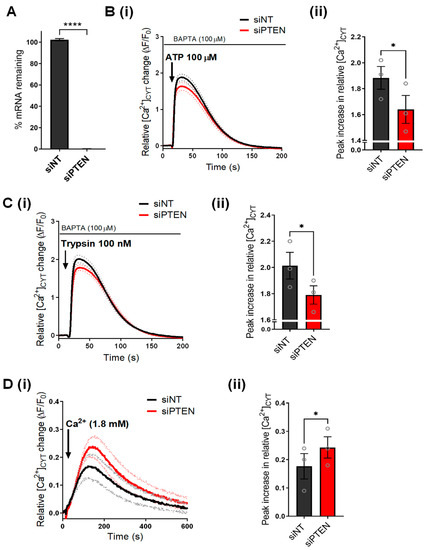
Although breast cancer cells often exhibit both abnormal AKT signaling and calcium signaling, the association between these two pathways is unclear. Using a combination of pharmacological tools, siRNA and CRISPR/Cas9 gene silencing techniques, we investigated the association between PTEN, AKT phosphorylation and calcium signaling in a basal breast cancer cell line. We found that siRNA-mediated PTEN silencing promotes AKT phosphorylation and calcium influx in MDA-MB-231 cells. This increase in AKT phosphorylation and calcium influx was phenocopied by the pharmacological AKT activator, SC79. The increased calcium influx associated with SC79 is inhibited by silencing AKT2, but not AKT1. This increase in calcium influx is suppressed when the store-operated calcium channel, ORAI1 is silenced. The results from this study open a novel avenue for therapeutic targeting of cancer cells with increased AKT activation. Given the association between ORAI1 and breast cancer, ORAI1 is a possible therapeutic target in cancers with abnormal AKT signaling. Full article
Show Figures

Figure 1

14 pages, 1572 KiB  
Review
Cancer as a Channelopathy—Appreciation of Complimentary Pathways Provides a Different Perspective for Developing Treatments
by Harry J. Gould III and Dennis Paul
Cancers 2022, 14(19), 4627; https://doi.org/10.3390/cancers14194627 - 23 Sep 2022
Cited by 3 | Viewed by 1827
Abstract
Life depends upon the ability of cells to evaluate and adapt to a constantly changing environment and to maintain internal stability to allow essential biochemical reactions to occur. Ions and ion channels play a crucial role in this process and are essential for [...] Read more.
Life depends upon the ability of cells to evaluate and adapt to a constantly changing environment and to maintain internal stability to allow essential biochemical reactions to occur. Ions and ion channels play a crucial role in this process and are essential for survival. Alterations in the expression of the transmembrane proteins responsible for maintaining ion balance that occur as a result of mutations in the genetic code or in response to iatrogenically induced changes in the extracellular environment is a characteristic feature of oncogenesis and identifies cancer as one of a constellation of diseases known as channelopathies. The classification of cancer as a channelopathy provides a different perspective for viewing the disease. Potentially, it may expand opportunities for developing novel ways to affect or reverse the deleterious changes that underlie establishing and sustaining disease and developing tolerance to therapeutic attempts at treatment. The role of ions and ion channels and their interactions in the cell’s ability to maintain ionic balance, homeostasis, and survival are reviewed and possible approaches that mitigate gain or loss of ion channel function to contribute to new or enhance existing cancer therapies are discussed. Full article
Show Figures

Figure 1

16 pages, 1139 KiB  
Review
How the Potassium Channel Response of T Lymphocytes to the Tumor Microenvironment Shapes Antitumor Immunity
by Martina Chirra, Hannah S. Newton, Vaibhavkumar S. Gawali, Trisha M. Wise-Draper, Ameet A. Chimote and Laura Conforti
Cancers 2022, 14(15), 3564; https://doi.org/10.3390/cancers14153564 - 22 Jul 2022
Cited by 12 | Viewed by 3102
Abstract
Competent antitumor immune cells are fundamental for tumor surveillance and combating active cancers. Once established, tumors generate a tumor microenvironment (TME) consisting of complex cellular and metabolic elements that serve to suppress the function of antitumor immune cells. T lymphocytes are key cellular [...] Read more.
Competent antitumor immune cells are fundamental for tumor surveillance and combating active cancers. Once established, tumors generate a tumor microenvironment (TME) consisting of complex cellular and metabolic elements that serve to suppress the function of antitumor immune cells. T lymphocytes are key cellular elements of the TME. In this review, we explore the role of ion channels, particularly K+ channels, in mediating the suppressive effects of the TME on T cells. First, we will review the complex network of ion channels that mediate Ca2+ influx and control effector functions in T cells. Then, we will discuss how multiple features of the TME influence the antitumor capabilities of T cells via ion channels. We will focus on hypoxia, adenosine, and ionic imbalances in the TME, as well as overexpression of programmed cell death ligand 1 by cancer cells that either suppress K+ channels in T cells and/or benefit from regulating these channels’ activity, ultimately shaping the immune response. Finally, we will review some of the cancer treatment implications related to ion channels. A better understanding of the effects of the TME on ion channels in T lymphocytes could promote the development of more effective immunotherapies, especially for resistant solid malignancies. Full article
Show Figures

Figure 1

23 pages, 14586 KiB  
Article
Inhibition of a Mitochondrial Potassium Channel in Combination with Gemcitabine and Abraxane Drastically Reduces Pancreatic Ductal Adenocarcinoma in an Immunocompetent Orthotopic Murine Model
by Weiwei Li, Gregory C. Wilson, Magdalena Bachmann, Jiang Wang, Andrea Mattarei, Cristina Paradisi, Michael J. Edwards, Ildiko Szabo, Erich Gulbins, Syed A. Ahmad and Sameer H. Patel
Cancers 2022, 14(11), 2618; https://doi.org/10.3390/cancers14112618 - 25 May 2022
Cited by 11 | Viewed by 2770
Abstract
Pancreas ductal adenocarcinoma (PDAC) is one the most aggressive cancers and associated with very high mortality, requiring the development of novel treatments. The mitochondrial voltage-gated potassium channel, Kv1.3 is emerging as an attractive oncologic target but its function in PDAC is unknown. Here, [...] Read more.
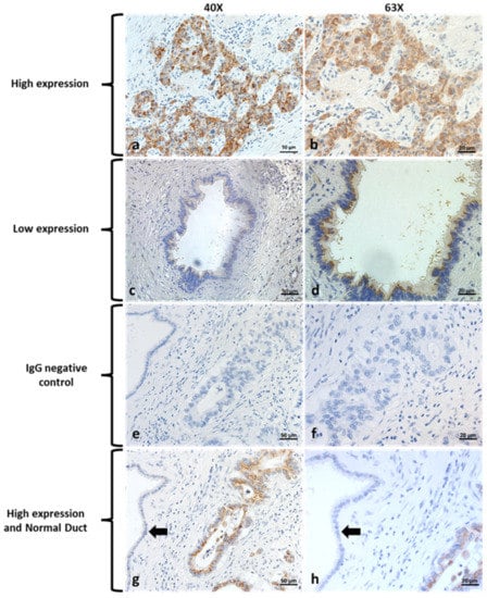
Pancreas ductal adenocarcinoma (PDAC) is one the most aggressive cancers and associated with very high mortality, requiring the development of novel treatments. The mitochondrial voltage-gated potassium channel, Kv1.3 is emerging as an attractive oncologic target but its function in PDAC is unknown. Here, we evaluated the tissue expression of Kv1.3 in resected PDAC from 55 patients using immunohistochemistry (IHC) and show that all tumors expressed Kv1.3 with 60% of tumor specimens having high Kv1.3 expression. In Pan02 cells, the recently developed mitochondria-targeted Kv1.3 inhibitors PCARBTP and PAPTP strongly reduced cell survival in vitro. In an orthotopic pancreas tumor model (Pan02 cells injected into C57BL/6 mice) in immune-competent mice, injection of PAPTP or PCARBTP resulted in tumor reductions of 87% and 70%, respectively. When combined with clinically used Gemcitabine plus Abraxane (albumin-bound paclitaxel), reduction reached 95% and 80% without resultant organ toxicity. Drug-mediated tumor cell death occurred through the p38-MAPK pathway, loss of mitochondrial membrane potential, and oxidative stress. Resistant Pan02 clones to PCARBTP escaped cell death through upregulation of the antioxidant system. In contrast, tumor cells did not develop resistance to PAPTP. Our data show that Kv1.3 is highly expressed in resected human PDAC and the use of novel mitochondrial Kv1.3 inhibitors combined with cytotoxic chemotherapies might be a novel, effective treatment for PDAC. Full article
Show Figures

Figure 1

20 pages, 10310 KiB  
Article
Design of New Potent and Selective Thiophene-Based KV1.3 Inhibitors and Their Potential for Anticancer Activity
by Špela Gubič, Louise Antonia Hendrickx, Xiaoyi Shi, Žan Toplak, Štefan Možina, Kenny M. Van Theemsche, Ernesto Lopes Pinheiro-Junior, Steve Peigneur, Alain J. Labro, Luis A. Pardo, Jan Tytgat, Tihomir Tomašič and Lucija Peterlin Mašič
Cancers 2022, 14(11), 2595; https://doi.org/10.3390/cancers14112595 - 24 May 2022
Cited by 5 | Viewed by 2316 | Correction
Abstract
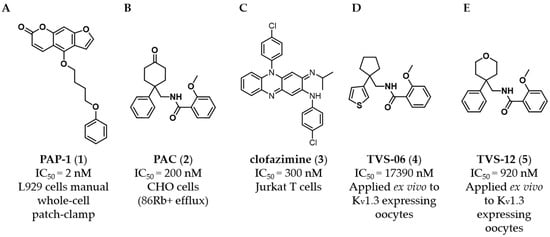
The voltage-gated potassium channel KV1.3 has been recognized as a tumor marker and represents a promising new target for the discovery of new anticancer drugs. We designed a novel structural class of KV1.3 inhibitors through structural optimization of benzamide-based [...] Read more.
The voltage-gated potassium channel KV1.3 has been recognized as a tumor marker and represents a promising new target for the discovery of new anticancer drugs. We designed a novel structural class of KV1.3 inhibitors through structural optimization of benzamide-based hit compounds and structure-activity relationship studies. The potency and selectivity of the new KV1.3 inhibitors were investigated using whole-cell patch- and voltage-clamp experiments. 2D and 3D cell models were used to determine antiproliferative activity. Structural optimization resulted in the most potent and selective KV1.3 inhibitor 44 in the series with an IC50 value of 470 nM in oocytes and 950 nM in Ltk cells. KV1.3 inhibitor 4 induced significant apoptosis in Colo-357 spheroids, while 14, 37, 43, and 44 significantly inhibited Panc-1 proliferation. Full article
Show Figures

Figure 1

10 pages, 837 KiB  
Article
Calcium Channel Blocker Use and the Risk for Breast Cancer: A Population-Based Nested Case-Control Study
by Victoria Rotshild, Bruria Hirsh Raccah, Muna Gazawe and Ilan Matok
Cancers 2022, 14(9), 2344; https://doi.org/10.3390/cancers14092344 - 9 May 2022
Cited by 8 | Viewed by 2575
Abstract
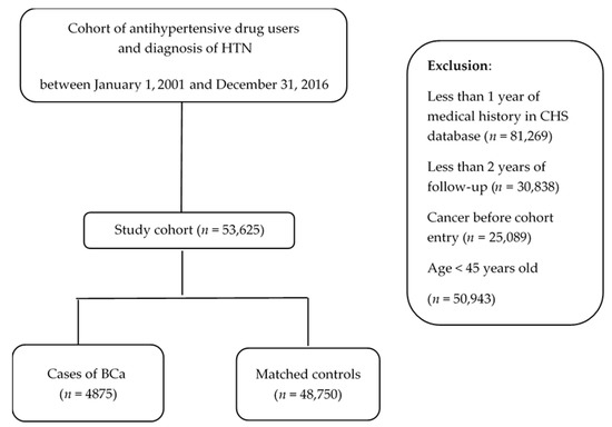
We investigated whether long-term exposure to calcium channel blockers (CCBs) is associated with an increased risk of breast cancer (BCa). We designed a nested case–control study based on data from the Clalit electronic database, the largest Israeli Health Services organization. All newly diagnosed [...] Read more.
We investigated whether long-term exposure to calcium channel blockers (CCBs) is associated with an increased risk of breast cancer (BCa). We designed a nested case–control study based on data from the Clalit electronic database, the largest Israeli Health Services organization. All newly diagnosed breast cancer (BCa) cases were selected from a cohort of patients with hypertension. Ten controls were matched for each BCa case. The odds ratios (ORs) of BCa among CCBs users were calculated using multivariate conditional logistic regression analyses. A total of 4875 patients with newly diagnosed BCa were identified from the cohort with a median follow-up of 5.15 years. The exposure to CCBs was not associated with an increased risk of BCa (OR = 0.98; 95% CI, 0.92–1.04). Additionally, there was no association between long-term exposure to CCBs (above eight years) and increased BCa risk (OR = 0.91; 95% CI, 0.67–1.21). Higher cumulative doses of CCBs were not associated with an elevated risk of BCa (OR = 0.997; 95% CI, 0.962–1.034, calculated per 1000 DDD). Based on this large population-based study, long-term exposure to CCBs was not associated with an increased risk of BCa. Considering that CCBs are widely used medications, our results provide important safety information on a population level, especially for patients with an increased risk of BCa. Full article
Show Figures

Figure 1

16 pages, 2787 KiB  
Article
Role of Amino Acid Transporter SNAT1/SLC38A1 in Human Melanoma
by Ines Böhme-Schäfer, Sandra Lörentz and Anja Katrin Bosserhoff
Cancers 2022, 14(9), 2151; https://doi.org/10.3390/cancers14092151 - 26 Apr 2022
Cited by 17 | Viewed by 2862
Abstract
The tumor metabolism is an important driver of cancer cell survival and growth, as rapidly dividing tumor cells exhibit a high demand for energetic sources and must adapt to microenvironmental changes. Therefore, metabolic reprogramming of cancer cells and the associated deregulation of nutrient [...] Read more.
The tumor metabolism is an important driver of cancer cell survival and growth, as rapidly dividing tumor cells exhibit a high demand for energetic sources and must adapt to microenvironmental changes. Therefore, metabolic reprogramming of cancer cells and the associated deregulation of nutrient transporters are a hallmark of cancer cells. Amino acids are essential for cancer cells to synthesize the necessary amount of protein, DNA, and RNA. Although cancer cells can synthesize glutamine de novo, most cancer cells show an increased uptake of glutamine from the tumor microenvironment. Especially SNAT1/SLC38A1, a member of the sodium neutral amino acid transporter (SNAT) family, plays an essential role during major net import of glutamine. In this study, we revealed a significant upregulation of SNAT1 expression in human melanoma tissue in comparison to healthy epidermis and an increased SNAT1 expression level in human melanoma cell lines when compared to normal human melanocytes (NHEMs). We demonstrated that functional inhibition of SNAT1 with α-(methylamino) isobutyric acid (MeAIB), as well as siRNA-mediated downregulation reduces cancer cell growth, cellular migration, invasion, and leads to induction of senescence in melanoma cells. Consequently, these results demonstrate that the amino acid transporter SNAT1 is essential for cancer growth, and indicates a potential target for cancer chemotherapy. Full article
Show Figures

Graphical abstract

2021

Jump to: 2024, 2023, 2022

16 pages, 3667 KiB  
Article
NS-11021 Modulates Cancer-Associated Processes Independently of BK Channels in Melanoma and Pancreatic Duct Adenocarcinoma Cell Lines
by Alessia Remigante, Paolo Zuccolini, Raffaella Barbieri, Loretta Ferrera, Rossana Morabito, Paola Gavazzo, Michael Pusch and Cristiana Picco
Cancers 2021, 13(23), 6144; https://doi.org/10.3390/cancers13236144 - 6 Dec 2021
Cited by 14 | Viewed by 2375
Abstract
Potassium channels have emerged as regulators of carcinogenesis, thus introducing possible new therapeutic strategies in the fight against cancer. In particular, the large-conductance Ca2+-activated K+ channel, often referred to as BK channel, is involved in several cancer-associated processes. Here, we [...] Read more.
Potassium channels have emerged as regulators of carcinogenesis, thus introducing possible new therapeutic strategies in the fight against cancer. In particular, the large-conductance Ca2+-activated K+ channel, often referred to as BK channel, is involved in several cancer-associated processes. Here, we investigated the effects of different BK activators, NS-11021, NS-19504, and BMS-191011, in IGR39 (primary melanoma cell line) and Panc-1 (primary pancreatic duct carcinoma cell line), highly expressing the channel, and in IGR37 (metastatic melanoma cell line) that barely express BK. Our data showed that NS-11021 and NS-19504 potently activated BK channels in IGR39 and Panc-1 cells, while no effect on channel activation was detected in IGR37 cells. On the contrary, BK channel activator BMS-191011 was less effective. However, only NS-11021 showed significant effects in cancer-associated processes, such as cell survival, migration, and proliferation in these cancer cell lines. Moreover, NS-11021 led to an increase of intracellular Ca2+ concentration, independent of BK channel activation, thus complicating any interpretation of its role in the regulation of cancer-associated mechanisms. Overall, we conclude that the activation of the BK channel by itself is not sufficient to produce beneficial anti-cancer effects in the melanoma and PDAC cell lines examined. Importantly, our results raise an alarm flag regarding the use of presumably specific BK channel openers as anti-cancer agents. Full article
Show Figures

Figure 1

20 pages, 4667 KiB  
Article
Molecular Activation of the Kv11.1 Channel Reprograms EMT in Colon Cancer by Inhibiting TGFβ Signaling via Activation of Calcineurin
by Najmeh Eskandari, Vitalyi Senyuk, Jennifer Moore, Zane Kalik, Qiyue Luan, Ian Papautsky, Arfa Moshiri, Maurizio Bocchetta, Seyed Alireza Salami, Shahrbanoo Oryan and Saverio Gentile
Cancers 2021, 13(23), 6025; https://doi.org/10.3390/cancers13236025 - 30 Nov 2021
Cited by 8 | Viewed by 2964
Abstract
Control of ionic gradients is critical to maintain cellular homeostasis in both physiological and pathological conditions, but the role of ion channels in cancer cells has not been studied thoroughly. In this work we demonstrated that activity of the Kv11.1 potassium channel plays [...] Read more.
Control of ionic gradients is critical to maintain cellular homeostasis in both physiological and pathological conditions, but the role of ion channels in cancer cells has not been studied thoroughly. In this work we demonstrated that activity of the Kv11.1 potassium channel plays a vital role in controlling the migration of colon cancer cells by reversing the epithelial-to-mesenchymal transition (EMT) into the mesenchymal-to-epithelial transition (MET). We discovered that pharmacological stimulation of the Kv11.1 channel with the activator molecule NS1643 produces a strong inhibition of colon cancer cell motility. In agreement with the reversal of EMT, NS1643 treatment leads to a depletion of mesenchymal markers such as SNAIL1, SLUG, TWIST, ZEB, N-cadherin, and c-Myc, while the epithelial marker E-cadherin was strongly upregulated. Investigating the mechanism linking Kv11.1 activity to reversal of EMT into MET revealed that stimulation of Kv11.1 produced a strong and fast inhibition of the TGFβ signaling. Application of NS1643 resulted in de-phosphorylation of the TGFβ downstream effectors R-SMADs by activation of the serine/threonine phosphatase PP2B (calcineurin). Consistent with the role of TGFβ in controlling cancer stemness, NS1643 also produced a strong inhibition of NANOG, SOX2, and OCT4 while arresting the cell cycle in G0/G1. Our data demonstrate that activation of the Kv11.1 channel reprograms EMT into MET by inhibiting TGFβ signaling, which results in inhibition of motility in colon cancer cells. Full article
Show Figures

Graphical abstract

13 pages, 2450 KiB  
Article
Investigation of Novel Small Molecular TRPM4 Inhibitors in Colorectal Cancer Cells
by Paulina Stokłosa, Anna Borgström, Barbara Hauert, Roland Baur and Christine Peinelt
Cancers 2021, 13(21), 5400; https://doi.org/10.3390/cancers13215400 - 28 Oct 2021
Cited by 9 | Viewed by 2940
Abstract
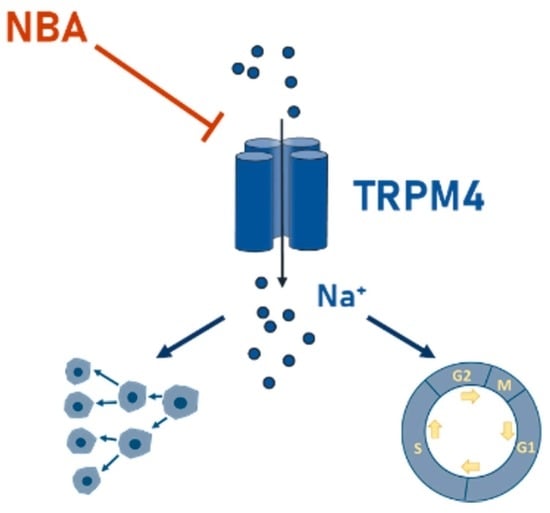
(1) Background: Transient receptor potential melastatin (TRPM4) ion channel aberrant expression or malfunction contributes to different types of cancer, including colorectal cancer (CRC). However, TRPM4 still needs to be validated as a potential target in anti-cancer therapy. Currently, the lack of potent and [...] Read more.
(1) Background: Transient receptor potential melastatin (TRPM4) ion channel aberrant expression or malfunction contributes to different types of cancer, including colorectal cancer (CRC). However, TRPM4 still needs to be validated as a potential target in anti-cancer therapy. Currently, the lack of potent and selective TRPM4 inhibitors limits further studies on TRPM4 in cancer disease models. In this study, we validated novel TRPM4 inhibitors, CBA, NBA, and LBA, in CRC cells. (2) Methods: The potency to inhibit TRPM4 conductivity in CRC cells was assessed with the whole-cell patch clamp technique. Furthermore, the impact of TRPM4 inhibitors on cellular functions, such as viability, proliferation, and cell cycle, were assessed in cellular assays. (3) Results: We show that in CRC cells, novel TRPM4 inhibitors irreversibly block TRPM4 currents in a low micromolar range. NBA decreases proliferation and alters the cell cycle in HCT116 cells. Furthermore, NBA reduces the viability of the Colo205 cell line, which highly expresses TRPM4. (4) Conclusions: NBA is a promising new TRPM4 inhibitor candidate, which could be used to study the role of TRPM4 in cancer disease models and other diseases. Full article
Show Figures

Graphical abstract

17 pages, 2453 KiB  
Article
Cell Systems Bioelectricity: How Different Intercellular Gap Junctions Could Regionalize a Multicellular Aggregate
by Alejandro Riol, Javier Cervera, Michael Levin and Salvador Mafe
Cancers 2021, 13(21), 5300; https://doi.org/10.3390/cancers13215300 - 22 Oct 2021
Cited by 16 | Viewed by 4841
Abstract
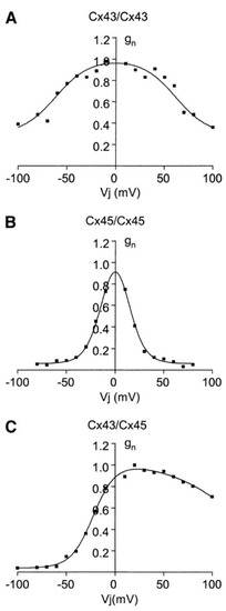
Electric potential distributions can act as instructive pre-patterns for development, regeneration, and tumorigenesis in cell systems. The biophysical states influence transcription, proliferation, cell shape, migration, and differentiation through biochemical and biomechanical downstream transduction processes. A major knowledge gap is the origin of spatial [...] Read more.
Electric potential distributions can act as instructive pre-patterns for development, regeneration, and tumorigenesis in cell systems. The biophysical states influence transcription, proliferation, cell shape, migration, and differentiation through biochemical and biomechanical downstream transduction processes. A major knowledge gap is the origin of spatial patterns in vivo, and their relationship to the ion channels and the electrical synapses known as gap junctions. Understanding this is critical for basic evolutionary developmental biology as well as for regenerative medicine. We computationally show that cells may express connexin proteins with different voltage-gated gap junction conductances as a way to maintain multicellular regions at distinct membrane potentials. We show that increasing the multicellular connectivity via enhanced junction function does not always contribute to the bioelectrical normalization of abnormally depolarized multicellular patches. From a purely electrical junction view, this result suggests that the reduction rather than the increase of specific connexin levels can also be a suitable bioelectrical approach in some cases and time stages. We offer a minimum model that incorporates effective conductances ultimately related to specific ion channel and junction proteins that are amenable to external regulation. We suggest that the bioelectrical patterns and their encoded instructive information can be externally modulated by acting on the mean fields of cell systems, a complementary approach to that of acting on the molecular characteristics of individual cells. We believe that despite the limitations of a biophysically focused model, our approach can offer useful qualitative insights into the collective dynamics of cell system bioelectricity. Full article
Show Figures

Figure 1

Planned Papers

The below list represents only planned manuscripts. Some of these manuscripts have not been received by the Editorial Office yet. Papers submitted to MDPI journals are subject to peer-review.

Title: Polyamine depletion reverses transcriptomic and calcium remodeling in colon cancer cells
Authors: Enrique Pérez-Riesgo1,2, Elena Hernando-Pérez1,2, Lucía Núñez1,2 and Carlos Villalobos1,*
Affiliation: Universidad de Valladolid
Abstract: In 2020, colorectal cancer was the third most common cancer worldwide and the second most deadly form of cancer. Alterations in the Wnt/β-catenin pathway are the most frequent cause of colon cancer development. Because of these alterations, the oncogene c-Myc is overexpressed which in turn promotes overexpression of ornithine decarboxylase (ODC) and excess polyamine synthesis, a process that is limited by alpha-Difluoromethylornithine (DFMO), a suicide ODC inhibitor considered a potential treatment against colorectal cancer. Furthermore, it has been suggested that intracellular calcium transport mechanisms are deeply altered in colorectal cancer. Here we asked whether polyamine depletion induced by DFMO may impinge on transcriptional and intracellular calcium remodeling in colorectal cancer. For this end we used calcium imaging, transcriptomic and statistical analysis in HT29 and NCM460 cells used here as models of human colorectal cancer and normal colonic cells, respectively. We found that colon cancer cells display dramatic changes in intracellular Ca2+ homeostasis (calcium remodeling) relative to normal cells. Specifically, cancer cells show enhanced store-operated calcium entry (SOCE) and resting cytosolic basal calcium levels, but decreased calcium store content relative to normal cells. On the other hand, cancer cells displayed also differential expression of more than 6500 genes relative to normal cells, 56 among them related to intracellular calcium transport. Polyamine depletion using DFMO reverses largely calcium remodeling in cancer cells. In addition, polyamine depletion changed the expression of 17 genes related to calcium homeostasis in HT29, including 11 of them that were reversed to mimic expression levels in normal cells. Specifically, polyamine depletion increased expression of modulators of store-operated Ca2+ entry and the calcium pump PMCA4 along with decreased expression of TRPC1, TRPC5 and TRPV6 channels and the secretory pathway SPCA2 pump. These effects were observed only in cancer cells. We conclude that polyamine depletion reverses calcium remodeling specifically in colon cancer cells acting on a few Ca2+ transport systems and modulators.

Back to TopTop