



Topical Advisory Panel applications are now closed. Please contact the Editorial Office with any queries.
-
Post and Core for Telescopic Crown-Retained Dentures—A Comparison of Different Materials Using Chewing Simulation -
Mapping Dental Anxiety Across Oral and Systemic Health Pathways -
A Clinical Framework for Interpreting Endodontic–Periodontal Lesions -
Cyclic Fatigue of Rotary Versus Reciprocating Endodontic Files: An In Vitro Study of Engine-Driven Endodontic Files
Journal Description
Dentistry Journal
Dentistry Journal
is an international, peer-reviewed, open access journal on dentistry, published monthly online by MDPI.
- Open Access— free for readers, with article processing charges (APC) paid by authors or their institutions.
- High Visibility: indexed within Scopus, ESCI (Web of Science), PubMed, PMC, and other databases.
- Journal Rank: JCR - Q1 (Dentistry, Oral Surgery and Medicine) / CiteScore - Q1 (General Dentistry)
- Rapid Publication: manuscripts are peer-reviewed and a first decision is provided to authors approximately 25.7 days after submission; acceptance to publication is undertaken in 5.9 days (median values for papers published in this journal in the first half of 2026).
- Recognition of Reviewers: reviewers who provide timely, thorough peer-review reports receive vouchers entitling them to a discount on the APC of their next publication in any MDPI journal, in appreciation of the work done.
Impact Factor:
3.4 (2025);
5-Year Impact Factor:
3.5 (2025)
Latest Articles
Single-Retained Lithium Disilicate “Maryland” with Pontic-Derived Stamp Technique for Anterior Symmetry: A Case Report
Dent. J. 2026, 14(7), 427; https://doi.org/10.3390/dj14070427 - 10 Jul 2026
Abstract
Background/Objectives: Single-retainer Maryland adhesive restorations provide a minimally invasive fixed option for replacing missing anterior teeth in young patients. When congenital absence of a maxillary lateral incisor is associated with a contralateral conoid lateral incisor, the clinical problem extends beyond tooth replacement to
[...] Read more.
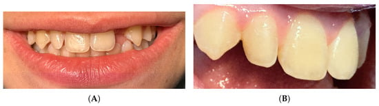
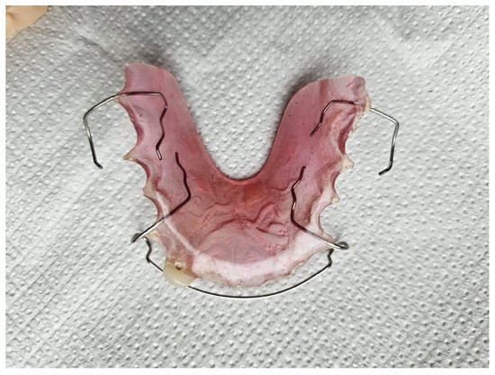
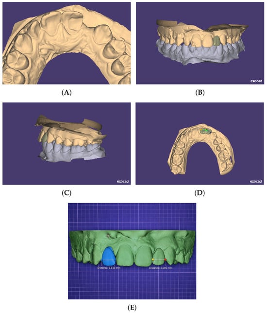
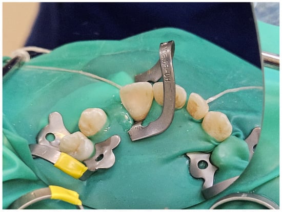
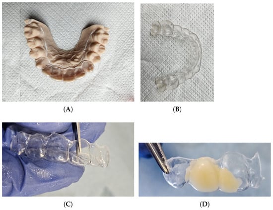
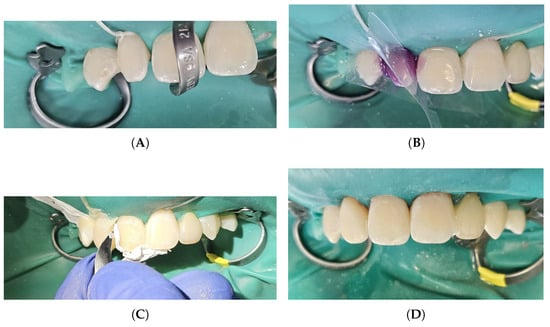
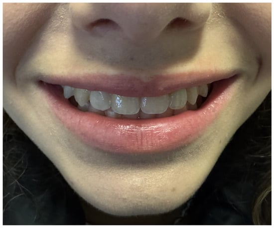
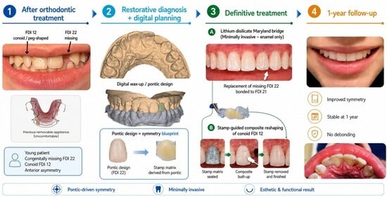
Background/Objectives: Single-retainer Maryland adhesive restorations provide a minimally invasive fixed option for replacing missing anterior teeth in young patients. When congenital absence of a maxillary lateral incisor is associated with a contralateral conoid lateral incisor, the clinical problem extends beyond tooth replacement to bilateral symmetry management. This case report describes a digital-restorative workflow combining a single-retainer lithium disilicate Maryland adhesive restoration with a pontic-derived stamp technique for contralateral symmetry correction. Methods: A medically healthy 13-year-old patient presented with congenital absence of the maxillary left lateral incisor (FDI 22; Universal 10) after orthodontic treatment and had been wearing a removable appliance to replace the single missing tooth. The contralateral maxillary right lateral incisor (FDI 12; Universal 7) presented with conoid morphology, further compromising anterior symmetry. Clinical assessment confirmed ideal mesiodistal space at FDI 22, a vital and unrestored FDI 21 with intact enamel and healthy periodontal support, and the absence of abnormal overjet, overbite, or excursive contact pattern. Based on the findings, a single-retainer Maryland adhesive restoration was fabricated from lithium disilicate (IPS e.max CAD) and bonded to the maxillary left central incisor (FDI 21; Universal 9) using an enamel-only preparation and adhesive cementation protocol. The fixed restoration was combined with a pontic-derived stamp workflow to guide direct composite reshaping of the contralateral conoid lateral incisor. Results: At 1-year follow-up, no debonding, sensitivity, marginal discoloration, or soft-tissue inflammation was observed. The contralateral composite reshaping remained clinically stable, and the patient and guardian reported improved comfort and esthetic satisfaction compared with the previous removable appliance. Conclusions: This case suggests the short-term clinical feasibility of combining a single-retainer lithium disilicate Maryland adhesive restoration with a pontic-derived stamp workflow to achieve minimally invasive tooth replacement and contralateral symmetry correction. Longer follow-up and broader case series are required to confirm the long-term predictability and reproducibility of this approach.
Full article
(This article belongs to the Topic Digital Patient Care: Bridging Technology and Clinical Practice)
►
Show Figures
Open AccessArticle
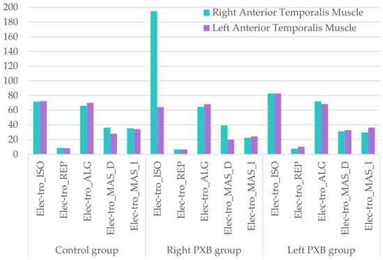
Masticatory Muscle Function in Growing Patients with Unilateral Posterior Crossbite: A Case–Control Study Combining Surface Electromyography and Myotonometry
by
Lara Díaz-González, Carlos López-de-Celis, Albert Pérez-Bellmunt and Francisco Guinot
Dent. J. 2026, 14(7), 426; https://doi.org/10.3390/dj14070426 - 10 Jul 2026
Abstract
Background: Unilateral posterior crossbite (UPXB) is a common malocclusion in growing patients, often associated with functional mandibular deviation and asymmetric masticatory function. However, its relationship with muscle activity remains unclear. This study aimed to evaluate masticatory muscle activity and viscoelastic properties in
[...] Read more.
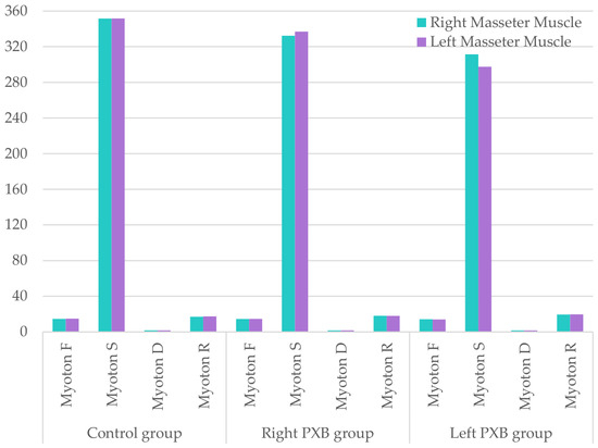
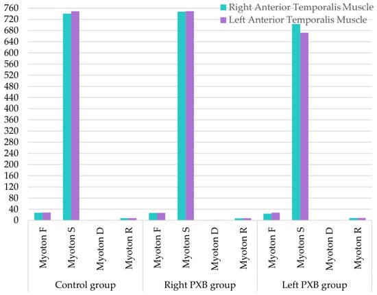
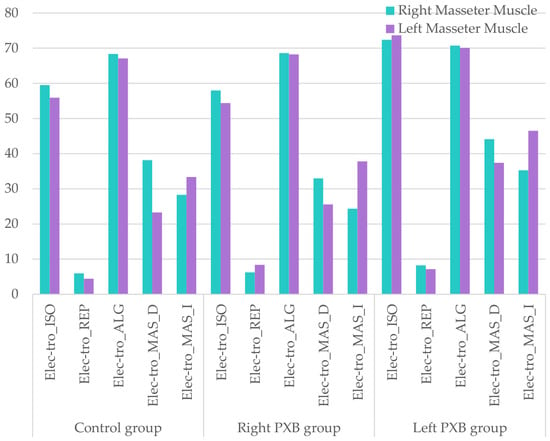
Background: Unilateral posterior crossbite (UPXB) is a common malocclusion in growing patients, often associated with functional mandibular deviation and asymmetric masticatory function. However, its relationship with muscle activity remains unclear. This study aimed to evaluate masticatory muscle activity and viscoelastic properties in growing patients with UPXB compared to subjects with normal occlusion. Methods: This case–control study included 140 growing patients (60 controls and 80 with UPXB: 36 right-side and 44 left-side). Surface electromyography (sEMG) was used to assess the activity of the superficial masseter and anterior temporalis muscles during standardized static and dynamic tasks. Muscle viscoelastic properties were evaluated using myotonometry. To minimize potential bias related to craniofacial morphology, only mesofacial subjects (Ricketts’ VERT −0.5 to +0.5) were included. Statistical significance was set at p < 0.05. Results: No differences were found in sex distribution, whereas the control group was slightly older than the UPXB group. Myotonometric analysis showed limited between-group differences, with isolated findings including higher stiffness of the non-crossbite anterior temporalis in left UPXB patients (p = 0.014), and minor differences in stiffness and relaxation of the left masseter between groups (p = 0.049; p = 0.045). Electromyographic results showed no significant differences during maximum voluntary contraction in intercuspation. In controls, higher activity was found in the left anterior temporalis during contraction on cotton rolls (p = 0.039) and in the right masseter at rest (p = 0.033). In left UPXB patients, the non-crossbite temporalis showed higher activity (p = 0.023). Increased activity of the crossbite-side left masseter was found in patients with UPXB during contraction and mastication (p = 0.012; p = 0.002). During mastication, both groups exhibited higher activity on the chewing side. Conclusions: UPXB is associated with specific, condition-dependent alterations in masticatory muscle activity and viscoelastic properties rather than generalized neuromuscular asymmetry. These findings suggest the presence of functional adaptations in growing patients, particularly under certain loading conditions, and highlight the importance of early diagnosis to better understand its potential impact on craniofacial development.
Full article
(This article belongs to the Special Issue Oral Health in the Maternal, Infant and Adolescent Populations)
►▼
Show Figures

Figure 1
Open AccessReview
Biocompatibility of Pit and Fissure Sealants: Scoping Review of In Vitro and In Vivo Evidence
by
Marija Badrov, Karmela Džaja, Barbara Badrov, Ana Glavina and Antonija Tadin
Dent. J. 2026, 14(7), 425; https://doi.org/10.3390/dj14070425 - 10 Jul 2026
Abstract
Objectives: This scoping review summarized the evidence on the biocompatibility of pit and fissure sealants, focusing on cytotoxicity, genotoxicity, and overall biological safety of commercial and experimental materials evaluated in vitro and in vivo. Methods: Following the PRISMA-ScR guidelines, eligibility was defined using
[...] Read more.
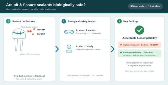
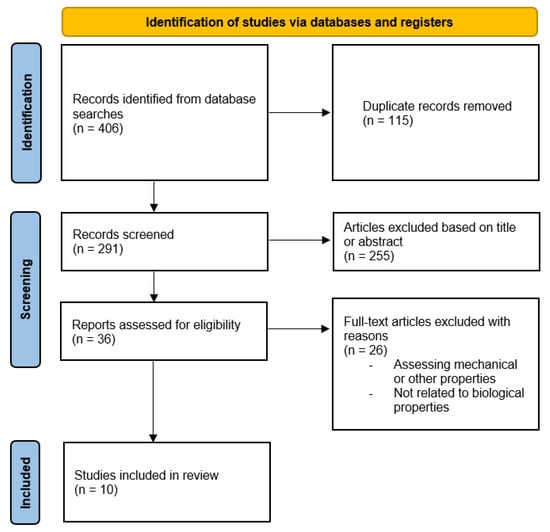
Objectives: This scoping review summarized the evidence on the biocompatibility of pit and fissure sealants, focusing on cytotoxicity, genotoxicity, and overall biological safety of commercial and experimental materials evaluated in vitro and in vivo. Methods: Following the PRISMA-ScR guidelines, eligibility was defined using the Population, Concept, and Context (PCC) framework: the population comprised cell cultures, animal models, or human participants exposed to sealants; the concept was biocompatibility, including cytotoxicity, genotoxicity, and inflammatory or tissue response; and the context encompassed commercial and experimental pit and fissure sealants used in preventive dentistry, particularly in pediatric populations. PubMed and Scopus platforms were searched without restrictions on publication year or language. Studies assessing biocompatibility (cytotoxicity, genotoxicity, inflammatory or tissue response) in cell cultures, animal models, or humans were eligible; those evaluating only clinical efficacy were excluded. Two reviewers independently performed study selection and data extraction. Results: Of 406 records (291 after deduplication), 10 studies were included—nine in vitro and one in vivo. Resin-based sealants predominated, mainly assessing residual monomers (TEGDMA, Bis-GMA) and their effects on fibroblasts, keratinocytes, periodontal ligament cells, and buccal epithelial cells. TEGDMA was released most frequently, whereas Bis-GMA showed the highest cytotoxicity. Experimental sealants containing nano-calcium fluoride, calcium phosphate, bioactive glass, or antibacterial monomers generally showed favorable biocompatibility, although high additive concentrations reduced cell viability. The single in vivo study reported good biocompatibility without significant genotoxicity. Conclusions: Pit and fissure sealants generally show acceptable biocompatibility and remain safe for caries prevention, although the biological response depends on composition, degree of polymerization, and residual monomer release. Further standardized long-term in vivo research is needed, particularly in pediatric populations.
Full article
(This article belongs to the Section Dental Materials)
►▼
Show Figures

Graphical abstract
Open AccessArticle
Conversational Artificial Intelligence as a Source of Oral Health Information: A Cross-Sectional Study in a Romanian Population
by
Marina Antoneta Pop, Abel Emanuel Moca, Mihai Porumb and Anca Porumb
Dent. J. 2026, 14(7), 424; https://doi.org/10.3390/dj14070424 - 10 Jul 2026
Abstract
Background/Objectives: Large language models have created new pathways for patients to access health information, yet little is known about how the general population uses these conversational artificial intelligence (AI) tools for oral health concerns. This study investigated patterns of AI use as a
[...] Read more.
Background/Objectives: Large language models have created new pathways for patients to access health information, yet little is known about how the general population uses these conversational artificial intelligence (AI) tools for oral health concerns. This study investigated patterns of AI use as a source of oral health information, the nature of data shared with these systems, users’ perceptions, and the impact on dental care-seeking behavior. Methods: A cross-sectional study was conducted among adults from Bihor County, Romania, using a structured 16-item online questionnaire distributed via social media, with eligibility restricted to individuals who had previously used conversational AI for oral health information. The final sample comprised 393 valid responses from this self-selected group of users. Fisher’s exact test and Z-tests with Bonferroni correction were applied (α = 0.05). Results: Most participants were female (68.2%), university-educated (52.9%), and lived in an urban setting (88.3%). Significant differences in patterns of AI use for oral health information were identified according to age, sex, and living environment (p < 0.001). Younger participants used AI more frequently, while older individuals perceived the information as less clear. The vast majority used AI for informational or preliminary guidance purposes, with very few treating it as a substitute for professional opinion. A relevant subset shared visual data (intraoral photographs or radiographs) with AI systems, raising data privacy concerns. Rural participants more frequently delayed dental visits and less often discussed AI-derived information with their dentist compared to urban participants. Conclusions: When used by the public as a source of oral health information, AI is increasingly adopted, with adoption shaped by age, sex, and socioeconomic context. These findings concern only this informational use and do not extend to other applications of AI in dentistry, such as diagnostic support, image analysis, or clinical decision-making. Dental professionals should proactively engage patients about their use of AI for oral health information to ensure that digitally obtained content is appropriately contextualized.
Full article
(This article belongs to the Special Issue AI in Dentistry and Oral Medicine: Transforming Diagnosis, Treatment, and Practice)
Open AccessArticle
Digital Dental Triage and Access to Care: A Qualitative Study of Experiences in a Teaching Hospital
by
Alaa Husni Qari, Ola Abdullah Sheiko, Mohammed Saed Althagafi, Wael Yaghmoor and Rayan Sharka
Dent. J. 2026, 14(7), 423; https://doi.org/10.3390/dj14070423 - 9 Jul 2026
Abstract
Background/Objectives: Digital triage systems are increasingly used in dentistry to support patient access and improve service efficiency, particularly in academic and public healthcare settings. However, limited evidence exists on how these systems function in practice from the perspectives of both patients and
[...] Read more.
Background/Objectives: Digital triage systems are increasingly used in dentistry to support patient access and improve service efficiency, particularly in academic and public healthcare settings. However, limited evidence exists on how these systems function in practice from the perspectives of both patients and providers. This study aimed to explore patients’ and staff members’ experiences, perceptions, and expectations of a digital dental triage system at a teaching dental hospital. Methods: A qualitative descriptive study was conducted at the Dental Teaching Hospital of Umm Al-Qura University (DTH-UQU), Saudi Arabia. Semi-structured interviews were carried out with 25 participants, including dental professionals, administrative staff, and patients who had recently used the digital triage system. Interviews were audio-recorded, transcribed verbatim, and analyzed using Braun and Clarke’s thematic analysis approach. Results: Analysis identified six interconnected themes. Patients generally found the system easy to use and consistent with their digital habits, yet this usability did not translate into satisfaction due to limited communication, post-registration silence, and unclear care pathways. Staff emphasized clinical accuracy, educational requirements, and capacity constraints as key drivers of triage decisions that were often invisible to patients. Communication breakdowns, misclassification, duplicate registrations, and unmet expectations emerged as central challenges, contributing to workflow inefficiencies. Participants suggested that improvements focused on enhanced communication, guided data entry, system integration, and expanded teledentistry functionality. Conclusions: The effectiveness of digital dental triage systems depends not only on usability but also on transparent communication, alignment with institutional capacity, and integration within broader care pathways.
Full article
(This article belongs to the Special Issue Dental Public Health Landscape: Challenges, Technological Innovation and Opportunities in the 21st Century)
Open AccessArticle
Development of a Quantitative Detection System for Porphyromonas gingivalis Based on Immunochromatography
by
Kaoru Kobayashi, Ryota Yamasaki, Michihiko Usui, Masahiro Morita, Maki Inoue, Yoshie Nagai-Yoshioka, Masanori Iwasaki, Keisuke Nakashima, Tatsuji Nisihara and Wataru Ariyoshi
Dent. J. 2026, 14(7), 422; https://doi.org/10.3390/dj14070422 - 9 Jul 2026
Abstract
►▼
Show Figures
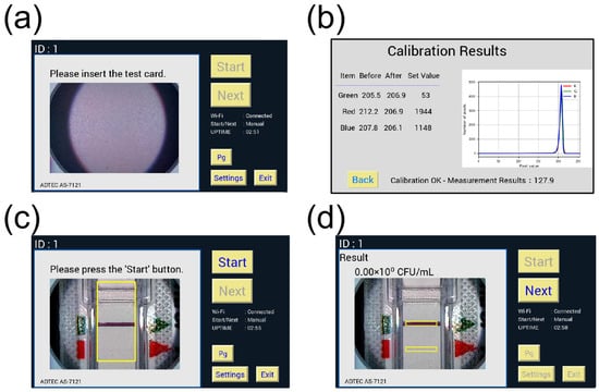
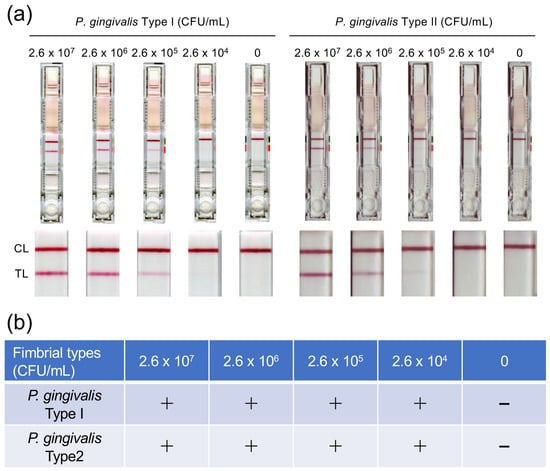
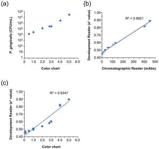
Background: Porphyromonas gingivalis is the principal bacterial species implicated in the pathogenesis of periodontitis. Despite its central role, direct detection of periodontopathic bacteria is not routinely performed in dental clinics. Although test kits using monoclonal antibodies against P. gingivalis have been reported,
[...] Read more.
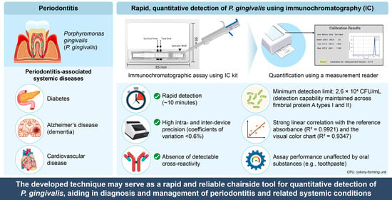
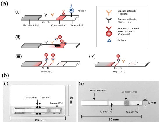
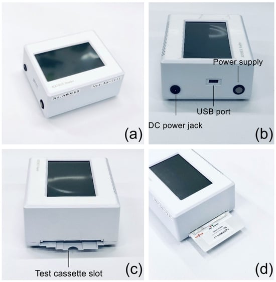
Background: Porphyromonas gingivalis is the principal bacterial species implicated in the pathogenesis of periodontitis. Despite its central role, direct detection of periodontopathic bacteria is not routinely performed in dental clinics. Although test kits using monoclonal antibodies against P. gingivalis have been reported, no practical chairside method has yet been established. Objectives: In this study, we developed an immunochromatography system that enables rapid and quantitative detection of P. gingivalis within 10 min. Methods: We evaluated the performance of the developed immunochromatography system and its dedicated measurement reader. Results: The kit demonstrated a minimum detection limit of 2.6 × 10⁴ CFU/mL and maintained detection capability across fimbrial types I and II. The dedicated reader showed strong correlations with a reference reader (R2 = 0.9921) and visual color chart evaluation (R2 = 0.9347). High intra- and inter-device precision was observed, with coefficients of variation less than 0.6%. Although the presence of 2% whole blood interfered with visual interpretation, other oral substances, including toothpastes and mouthwashes, did not significantly affect the assay performance. Conclusions: Collectively, these findings indicate that the developed system may serve as a rapid and reliable chairside tool for quantitative detection of P. gingivalis, with potential utility in the diagnosis and management of periodontitis and related systemic conditions.
Full article

Graphical abstract
Open AccessReview
Human Enamel Formation: A Scoping Review for Oral Health Professionals
by
Patrick Unterbrink, Bernhard Ganss, Hardy Limeback, Birte Hollmann, Pascal Fandrich, Ingo Winschel, Erik Schulze zur Wiesche, Bennett Tochukwu Amaechi, Malgorzata Pawinska, Elzbieta Paszynska and Joachim Enax
Dent. J. 2026, 14(7), 421; https://doi.org/10.3390/dj14070421 - 9 Jul 2026
Abstract
►▼
Show Figures
Background: Tooth enamel is the hardest and most highly mineralized tissue in the human body. It serves as a protective barrier against chemical, mechanical, and microbial challenges. Despite its durability, enamel remains vulnerable to developmental and posteruptive defects such as fluorosis, hypomineralization,
[...] Read more.
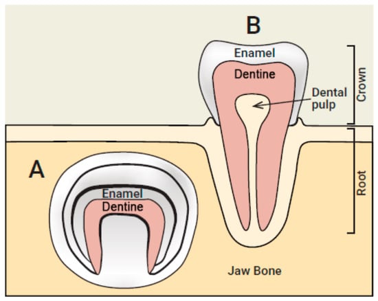
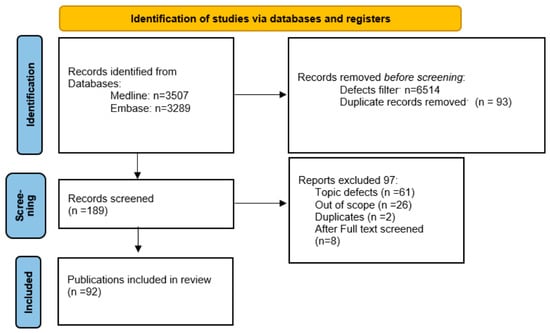
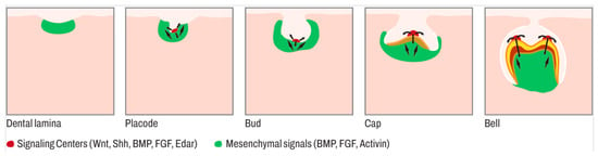
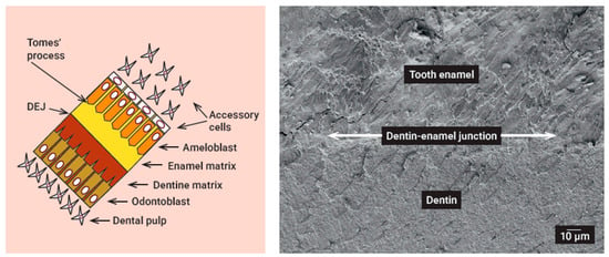
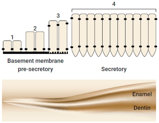
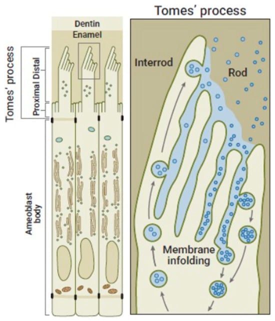
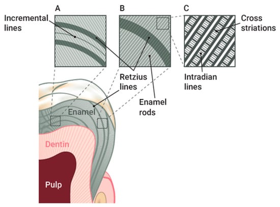
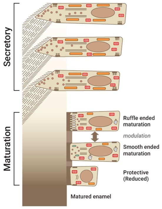
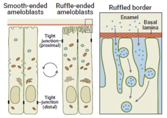
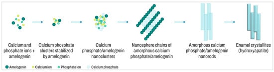
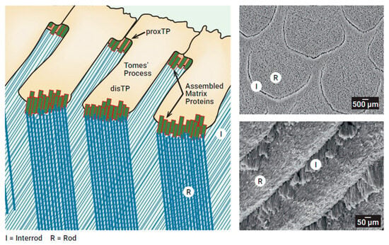
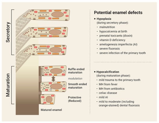
Background: Tooth enamel is the hardest and most highly mineralized tissue in the human body. It serves as a protective barrier against chemical, mechanical, and microbial challenges. Despite its durability, enamel remains vulnerable to developmental and posteruptive defects such as fluorosis, hypomineralization, and amelogenesis imperfecta (AI). For oral health professionals, a clear understanding of the biological and molecular mechanisms underlying enamel formation is essential for advancing preventive and therapeutic strategies in clinical practice. This review synthesizes current knowledge on enamel formation, with emphasis on its cellular, molecular, and structural determinants, and discusses clinically relevant disruptions as well as emerging biomimetic approaches. Methods: This scoping review was conducted according to the PRISMA-ScR guidelines. A systematic literature search of the mechanisms of enamel formation was performed via Embase and Medline. Titles and abstracts were screened independently by three authors. Studies that primarily addressed enamel defects were excluded from the systematic synthesis; however, these studies were retained for narrative discussion. Following the screening process, 92 publications met the inclusion criteria and were incorporated into the thematic synthesis. Results: Enamel formation is a complex, multistage process involving epithelial–mesenchymal interactions and the sequential activity of ameloblasts during presecretory, secretory, transition, and maturation stages. Key mechanisms include the secretion of enamel matrix proteins (e.g., amelogenin, ameloblastin, and enamelin), proteolytic processing by enzymes such as MMP20 and KLK4, and controlled ion transport, leading to hydroxyapatite crystal growth and organization into rod and interrod structures. The structural arrangement endows enamel with exceptional mechanical resistance. Narrative sections address “What can go wrong?”, summarizing genetic, epigenetic, and environmental causes of fluorosis, hypomineralization, and amelogenesis imperfecta, and other developmental defects, whereas “What can we learn from nature?” highlights biomimetic strategies. Conclusions: Human enamel formation is a highly coordinated biomineralization process regulated at the cellular, structural, and molecular levels. Disruptions in these processes underlie major enamel pathologies. Integrating mechanistic insights from natural enamel development with emerging biomimetic technologies offers promising avenues for prevention, diagnosis, and treatment in dentistry. This review provides oral health professionals with a biologically grounded framework to guide evidence-based management of enamel-related conditions.
Full article

Figure 1
Open AccessArticle
Estradiol Enhances Alveolar Bone Resorption by Promoting Osteoclast Differentiation in Experimental Periodontitis
by
Keisuke Yasuda, Shinji Matsuda, Takumi Memida, Tetsuya Yoshimoto, Fuminori Nakashima, Yurika Ninomiya, Tomoya Ueda, Shogo Shimada, Shizu Hirata-Tsuchiya, Mikihito Kajiya, Kazuhisa Ouhara and Noriyoshi Mizuno
Dent. J. 2026, 14(7), 420; https://doi.org/10.3390/dj14070420 - 9 Jul 2026
Abstract
Background/Objectives: Estrogen is a key female hormone; however, its role in periodontitis remains poorly understood. This study investigated the effects of 17β-estradiol (E2) on experimental periodontitis using an ovariectomy (OVX) model with E2 administration. Methods: Female mice aged 8–10 weeks underwent
[...] Read more.
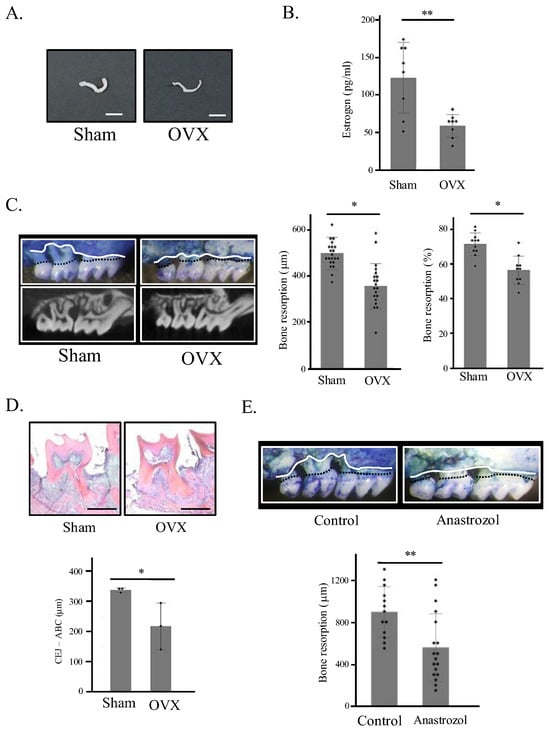
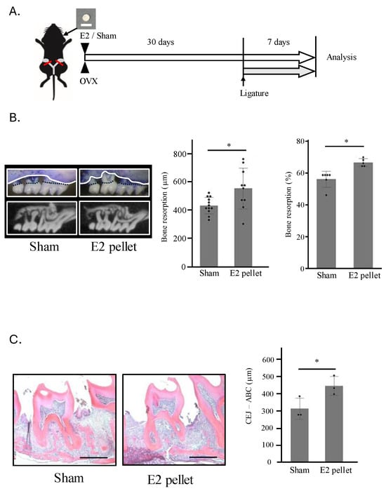
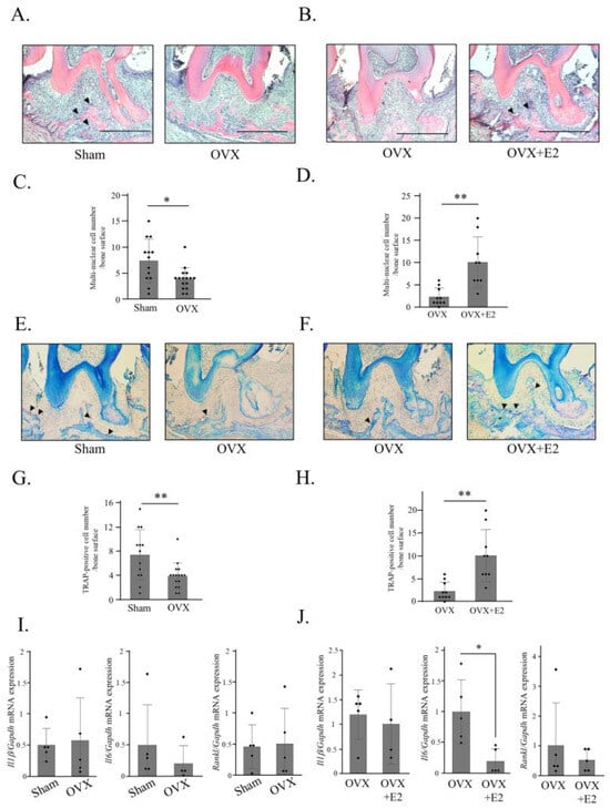
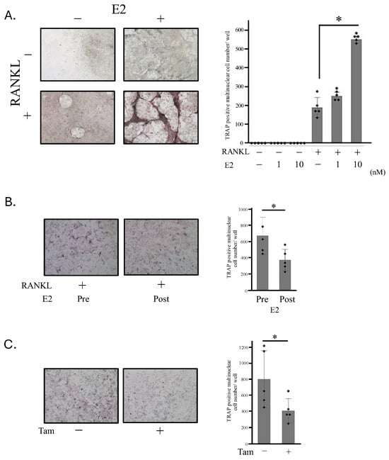
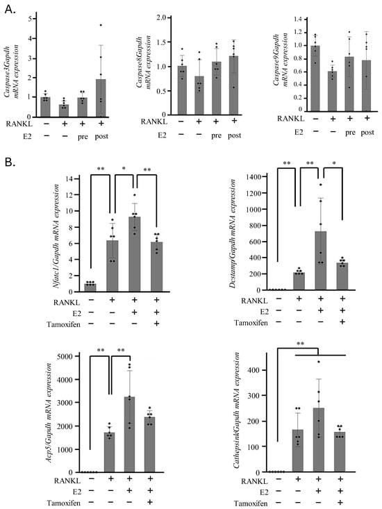
Background/Objectives: Estrogen is a key female hormone; however, its role in periodontitis remains poorly understood. This study investigated the effects of 17β-estradiol (E2) on experimental periodontitis using an ovariectomy (OVX) model with E2 administration. Methods: Female mice aged 8–10 weeks underwent OVX, followed by induction of ligature-induced periodontitis, and subsequent quantification of alveolar bone resorption. Additional groups received an aromatase inhibitor or E2 supplementation after OVX, with subsequent induction of periodontitis and evaluation of bone resorption. Histological analysis assessed multinucleated giant cells and tartrate-resistant acid phosphatase-positive osteoclasts on the bone surface. Gingival tissue was analyzed for gene expression related to osteoclastogenesis. The effect of E2 on osteoclast differentiation from bone marrow cells was also examined. Results: OVX significantly reduced serum E2 levels and decreased alveolar bone resorption. Aromatase inhibitor administration similarly reduced bone loss. Histological evaluation revealed a reduced number of resorbing osteoclasts in OVX mice, whereas E2 supplementation increased osteoclast numbers. No significant changes in inflammatory cytokine or receptor activator of nuclear factor-kappa B ligand (RANKL) expression were observed. E2 promoted osteoclast differentiation in vitro, and treatment with E2 prior to RANKL stimulation further increased the number of osteoclasts. This effect was suppressed by an estrogen receptor antagonist. Moreover, E2 enhanced the expression of osteoclast differentiation–associated genes in the presence of RANKL, an effect abolished by tamoxifen. Conclusions: E2 increased alveolar bone resorption in experimental periodontitis, likely by promoting osteoclast differentiation, independent of inflammatory cytokine or RANKL gene expression.
Full article
(This article belongs to the Section Oral Hygiene, Periodontology and Peri-implant Diseases)
►▼
Show Figures

Figure 1
Open AccessArticle
Selective Caries Removal as a Restorative Strategy for Caries Management: Residual Dentin Quality After Chemomechanical and Mechanical Excavation
by
Gustavo Gerardo Ramirez-Martinez, Christian Andrea Lopez-Ayuso, Rogelio Jose Scougall-Vilchis, Silvia Rojas-Rueda, Carlos A. Jurado, Abdulrahman Alshabib, Hamid Nurrohman and Rene Garcia-Contreras
Dent. J. 2026, 14(7), 419; https://doi.org/10.3390/dj14070419 - 8 Jul 2026
Abstract
Background/Objectives: Advances in restorative caries management have shifted operative dentistry from complete caries excavation toward selective, tissue-preserving strategies that maintain remineralizable dentin and support adhesive restoration. Chemomechanical systems and self-limiting burs may reduce unnecessary dentin removal, but their effects on residual dentin quality
[...] Read more.
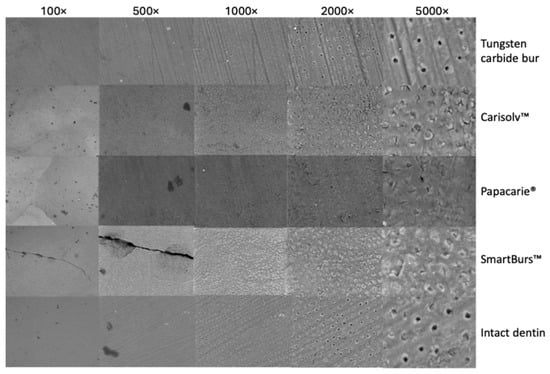
Background/Objectives: Advances in restorative caries management have shifted operative dentistry from complete caries excavation toward selective, tissue-preserving strategies that maintain remineralizable dentin and support adhesive restoration. Chemomechanical systems and self-limiting burs may reduce unnecessary dentin removal, but their effects on residual dentin quality remain relevant to restorative decision-making. This in vitro study compared Carisolv™, Papacarie®, Smart Burs™, and conventional tungsten carbide burs with respect to caries removal time, residual bacterial load, laser fluorescence, Vickers microhardness, and scanning electron microscopy (SEM) features of residual dentin. Methods: Forty-eight extracted permanent molars were selected. Standardized class I cavities were prepared and subjected to an artificial caries protocol using a demineralizing solution for 240 h. A cariogenic biofilm model was then established using Streptococcus mutans. Specimens were allocated to four excavation groups: Carisolv™, Papacarie®, Smart Burs™, and conventional carbide burs. Caries removal was performed according to each system, and the operative time was recorded. Residual bacterial contamination was estimated through colony-forming unit counts. Residual dentin was assessed using DIAGNOdent™ laser fluorescence, Vickers microhardness testing, and SEM at multiple magnifications. Results: Mean removal times were 2.82 ± 1.64 min for Carisolv™, 2.50 ± 1.15 min for Papacarie®, 2.05 ± 0.70 min for Smart Burs™, and 0.61 ± 0.44 min for carbide burs. Residual bacterial growth was detected in all groups. Vickers microhardness values were 79.2 ± 24.66 VHN for Carisolv™, 79.6 ± 22.30 VHN for Papacarie®, 83.4 ± 28.73 VHN for Smart Burs™, and 92.9 ± 24.66 VHN for intact dentin control. SEM revealed dentin morphology closer to the control after carbide bur excavation, partially obliterated tubules after Carisolv™, and more evident tubular obliteration after Papacarie® and Smart Burs™. Conclusions: Chemomechanical excavation, particularly Carisolv™, generated a residual dentin substrate with microhardness values compatible with a conservative restorative approach, although with longer operative time than conventional burs. The findings support the role of selective caries removal in minimally invasive restorative protocols and highlight the need to connect residual dentin characteristics with adhesive performance, bioactive materials, and long-term restoration outcomes.
Full article
(This article belongs to the Special Issue Advancements in Restorative Techniques for Caries Management)
►▼
Show Figures

Figure 1
Open AccessArticle
The Spread of Non-Evidence-Based Health Claims on Social Media: The Case of #Mouthtape on Instagram, a Cross-Sectional Study
by
Maria Eleonora Bizzoca, Christian Durante, Francisco José Vázquez Santos, Gennaro Musella, Lorenzo Lo Muzio, Eleonora Trecca, Alessandra Lucchese and Vito Carlo Alberto Caponio
Dent. J. 2026, 14(7), 418; https://doi.org/10.3390/dj14070418 - 8 Jul 2026
Abstract
Background: Social media platforms like Instagram have become major sources of health information, often amplifying unverified claims. Nocturnal mouth-taping is one such trend promoted as beneficial for sleep and overall health despite limited scientific evidence. Methods: In April 2025, 530 Instagram
[...] Read more.
Background: Social media platforms like Instagram have become major sources of health information, often amplifying unverified claims. Nocturnal mouth-taping is one such trend promoted as beneficial for sleep and overall health despite limited scientific evidence. Methods: In April 2025, 530 Instagram reels tagged #mouthtape were retrieved via the official API. After exclusions, 390 reels were analyzed. Two independent reviewers assessed each reel for relevance, stance on mouth-taping, presenter characteristics, engagement metrics, and content type. Spearman’s correlation, Mann–Whitney, Kruskal–Wallis, and Chi-Square tests were applied. Results: Overall, 94.4% of reels endorsed mouth-taping. Most content was produced by companies (55.4%), while only 11.6% was posted by medical professionals. Supportive posts had significantly higher shares than critical ones (mean 923 vs. 193; p = 0.015). Male presenters’ reels achieved more likes and shares, though female presenters had more followers. English-language posts generated greater engagement. Commercial and medical claims predominated, with limited representation of evidence-based information. Conclusions: Instagram content overwhelmingly promotes mouth-taping, largely driven by commercial actors. High engagement with supportive posts, regardless of scientific validity, underscores the role of algorithms in spreading non-evidence-based health claims.
Full article
Open AccessArticle
Deliberate Error-Based Learning in Dental Radiography: An Educational Study
by
Andy Wai Kan Yeung, Ray Tanaka, Kuo Feng Hung, Varut Vardhanabhuti and Andrew Nalley
Dent. J. 2026, 14(7), 417; https://doi.org/10.3390/dj14070417 - 8 Jul 2026
Abstract
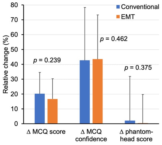
Objectives: We aimed to explore whether incorporating a deliberate error-based learning activity, adapted from error management training (EMT), could enhance undergraduate dental students’ understanding of common intra-oral radiographic faults and their phantom-head imaging performance. Methods: This randomized two-arm educational study involved
[...] Read more.
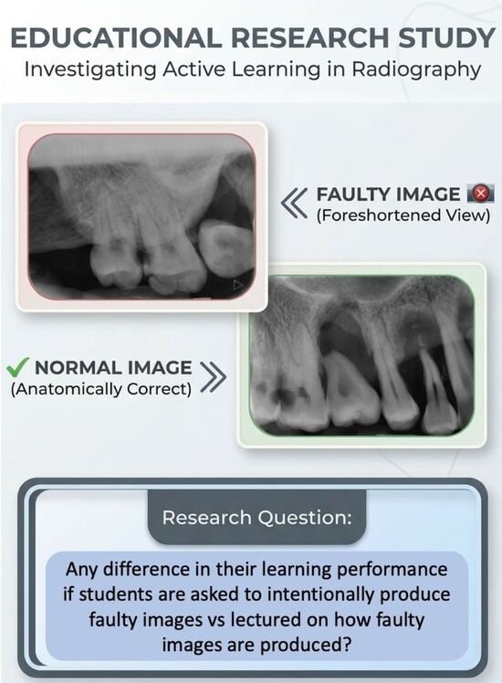
Objectives: We aimed to explore whether incorporating a deliberate error-based learning activity, adapted from error management training (EMT), could enhance undergraduate dental students’ understanding of common intra-oral radiographic faults and their phantom-head imaging performance. Methods: This randomized two-arm educational study involved Year 2 dental undergraduates who completed pre- and post-intervention phantom-head imaging and a short multiple-choice test on radiographic errors. Students were allocated either to a brief, slide-based teaching session on imaging faults (conventional group) or to a hands-on activity in which they intentionally produced and corrected faulty images under supervision (EMT group). Main outcomes included MCQ scores, confidence ratings, overall imaging performance (percentage of maximum possible score), and distribution of diagnostic image quality categories. Two-sample t tests were used to check for inter-group differences in the change in the post–pre numerical scores. The Cochran–Mantel–Haenszel (CMH) linear-by-linear test was used to check for inter-group differences in the change in image distribution by diagnostic quality before and after the intervention. Results: Eighty-seven students contributed a total of 735 radiographs. Both groups showed improvements in MCQ scores and confidence within groups. Diagnostic acceptability of student radiographs was already high at baseline and remained so afterwards, with no significant differences between groups in MCQ gains, confidence changes, imaging scores, or shifts in diagnostic image quality distributions. Conclusions: Although the single, short EMT-style activity did not outperform conventional teaching on immediate outcomes, the study demonstrates that deliberate error-based learning is feasible in a pre-clinical radiography setting and well-received by students. The findings also help to clarify the circumstances under which EMT is most likely to yield benefits, suggesting that longer or more structured EMT sessions, particularly those involving metacognitive scaffolding or more challenging imaging scenarios, may be needed before performance differences can emerge.
Full article
(This article belongs to the Special Issue Dental Education: Innovation and Challenge)
►▼
Show Figures

Graphical abstract
Open AccessSystematic Review
Diagnostic Performance of Artificial Intelligence Models for Periodontitis Disease Detection Using Panoramic Radiographs: A Systematic Review
by
Khalid Almutairi, Tariq Almanseer, Enrique España Guerrero, Antonio José España and Gerardo Moreu
Dent. J. 2026, 14(7), 416; https://doi.org/10.3390/dj14070416 - 7 Jul 2026
Abstract
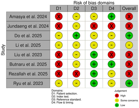
Background/Objectives: Periodontitis is a highly prevalent inflammatory disease and a major cause of tooth loss worldwide. Accurate diagnosis requires integration of clinical and radiographic findings, but interpretation of panoramic radiographs is subject to variability. Artificial intelligence (AI) has emerged as a promising
[...] Read more.
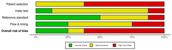
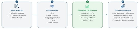
Background/Objectives: Periodontitis is a highly prevalent inflammatory disease and a major cause of tooth loss worldwide. Accurate diagnosis requires integration of clinical and radiographic findings, but interpretation of panoramic radiographs is subject to variability. Artificial intelligence (AI) has emerged as a promising adjunct for radiographic assessment. This systematic review evaluated the diagnostic performance of AI-based models for detecting periodontitis using panoramic radiographic images. Methods: A systematic search of PubMed, Scopus, and Web of Science identified studies published between 1 January 2015 and 1 March 2026. Eligible studies assessed AI models for periodontitis detection on panoramic radiographs and used either clinically confirmed periodontal diagnosis or expert radiographic annotation as the reference standard. Data extraction and quality assessment were performed independently by two reviewers using the QUADAS-2 tool. Owing to heterogeneity in AI architectures, datasets, and outcome measures, a narrative synthesis was conducted. Results: Nine studies met the inclusion criteria, comprising more than 20,000 radiographs. AI models included convolutional neural networks (CNNs), segmentation-based systems, and hybrid architectures. Sensitivity ranged from 0.795 to 1.00, specificity from 0.784 to 0.99, and AUC values from 0.843 to 0.967. Studies using clinical periodontal diagnosis as the reference standard generally reported lower performance than those relying solely on expert annotation. Only four studies performed external validation, and dataset sizes varied widely. One study combining panoramic and periapical radiographs showed moderate diagnostic performance. Conclusions: AI-based diagnostic models demonstrate promising performance for detecting periodontitis on panoramic radiographs, with several studies reporting high sensitivity and AUC values. However, heterogeneity in reference standards, limited external validation, and inconsistent dataset quality restrict generalizability. AI should be considered an adjunct to, rather than a replacement for, comprehensive clinical periodontal examination. Standardized datasets and robust external validation are needed to support clinical implementation.
Full article
(This article belongs to the Special Issue Artificial Intelligence Technology in Periodontology and Implantology: Development and Prospect)
►▼
Show Figures

Graphical abstract
Open AccessCase Report
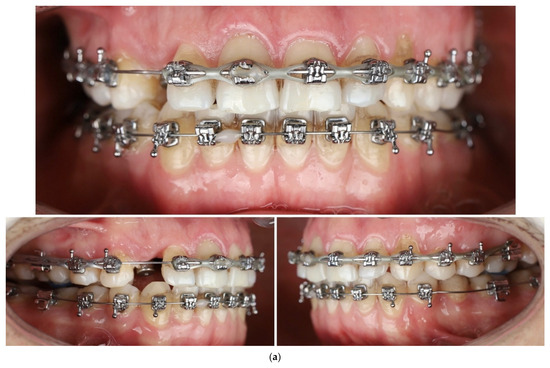
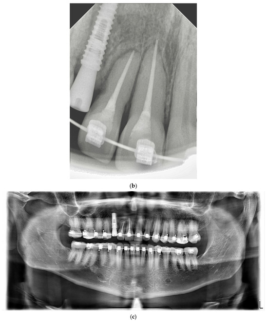
Traumatic Extrusive and Lateral Luxation of Maxillary Incisors During Orthodontic Treatment: A Multidisciplinary Management Approach
by
Marina Mirzabekian, Narine Arutiunian, Nina Karpoeva, Angelina Petiuleva, Tigran Minasyan, Ekaterina Zernitckaia and Sofiia Khadzhieva
Dent. J. 2026, 14(7), 415; https://doi.org/10.3390/dj14070415 - 7 Jul 2026
Abstract
►▼
Show Figures
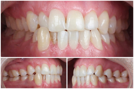
Background/Objectives: Traumatic dental injuries involving the maxillary anterior teeth are common; however, combined extrusive and lateral dislocation during active orthodontic treatment is uncommon and presents unique management challenges. This case report describes the multidisciplinary management of traumatic extrusive and lateral dislocation of
[...] Read more.
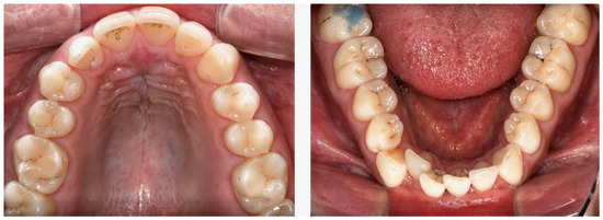
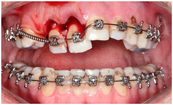
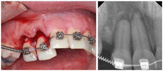
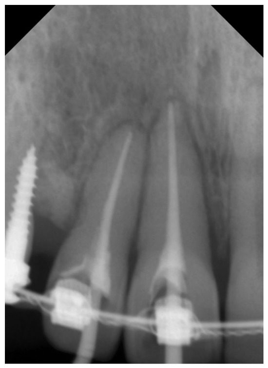
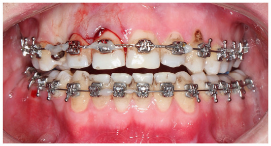
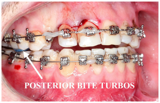
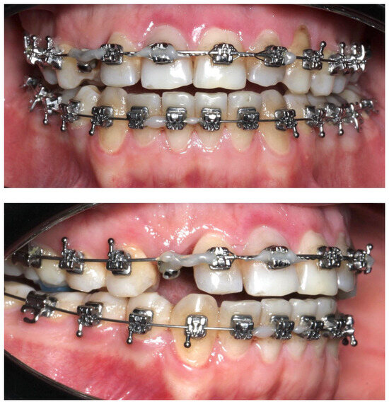
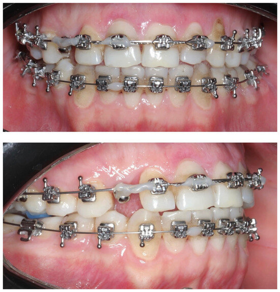
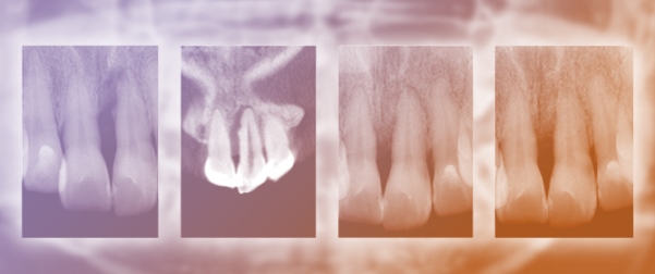
Background/Objectives: Traumatic dental injuries involving the maxillary anterior teeth are common; however, combined extrusive and lateral dislocation during active orthodontic treatment is uncommon and presents unique management challenges. This case report describes the multidisciplinary management of traumatic extrusive and lateral dislocation of the maxillary right lateral incisor (#7) and maxillary right central incisor (#8) in a 27-year-old patient undergoing fixed orthodontic treatment following sports-related trauma. Methods: Clinical examination revealed marked coronal and lateral displacement, pathologic mobility, gingival laceration, active bleeding, and deformation of the orthodontic archwire. Both affected teeth had complete root formation. Emergency management included immediate repositioning of the displaced teeth and urgent endodontic treatment. Semi-rigid stabilization was achieved using metallic ligatures and a rectangular stainless-steel orthodontic archwire, supplemented by temporary skeletal anchorage with a mini-screw and posterior bite turbos to reduce occlusal loading. The patient was monitored clinically and radiographically throughout healing and subsequent orthodontic treatment. Results: Follow-up examinations demonstrated favorable periodontal healing, stable retention of the traumatized teeth, absence of pathologic mobility, and successful continuation of orthodontic treatment. Radiographic evaluation showed no evidence of inflammatory root resorption or other significant complications. At the 16-month follow-up, the affected teeth remained functional and stable, and implant placement was successfully completed at the congenitally missing maxillary canine site. Conclusions: This case highlights the importance of immediate interdisciplinary management of traumatic dental dislocation during orthodontic treatment. Fixed orthodontic appliances, when appropriately incorporated into emergency stabilization protocols, may contribute to successful tooth retention and favorable long-term outcomes.
Full article

Figure 1
Open AccessArticle
Lateral Wall Failure During Implant Osteotomy Preparation in a Standardized Narrow-Ridge Model: Osseodensification Versus Conventional Drilling—An Ex Vivo Controlled Study
by
Radomir Nikolic, Jelena Vulovic, Milan Bojovic, Gavrilo Ilic, Andrej Kravanja, Dragana Gabric and Zoran Tatic
Dent. J. 2026, 14(7), 414; https://doi.org/10.3390/dj14070414 - 7 Jul 2026
Abstract
Background: The aim of this ex vivo study was to compare the resistance to lateral wall failure during sequential implant site preparation using osseodensification (OD) burs and conventional rotary drills in a standardized narrow ridge model. The primary outcome was the drill
[...] Read more.
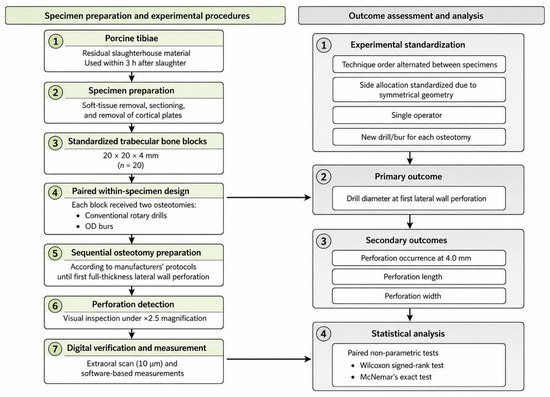
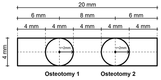
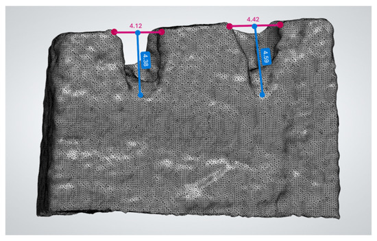
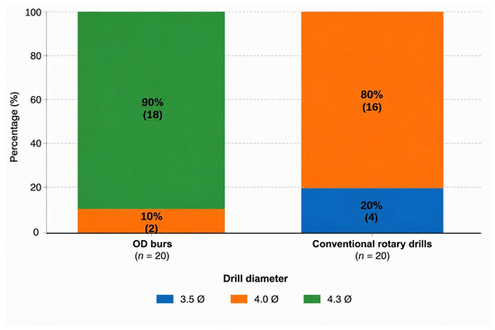
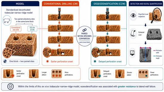
Background: The aim of this ex vivo study was to compare the resistance to lateral wall failure during sequential implant site preparation using osseodensification (OD) burs and conventional rotary drills in a standardized narrow ridge model. The primary outcome was the drill diameter at first lateral wall perforation. Secondary outcomes included paired perforation occurrence at a clinically relevant diameter (4.0 mm) and perforation morphology (length and width). Methods: Twenty standardized trabecular bone blocks were prepared in a paired within-specimen design, with each block receiving two osteotomies: one prepared using conventional rotary drills and the other using OD burs. Osteotomy diameter was sequentially increased according to the manufacturers’ protocols until lateral wall perforation occurred. The drill diameter at first perforation was recorded. Perforation morphology (length and width) was quantified using high-resolution extraoral digital scanning and software-based measurements. Statistical analysis was performed using paired non-parametric tests (α = 0.05). Results: OD burs significantly delayed lateral wall perforation compared with conventional drills (median 4.30 vs. 4.00 mm; p = 0.0000171). At the 4.0 mm drill step, perforation occurred in 16/20 (80%) conventional versus 2/20 (10%) OD preparations (p = 0.0013). Perforation length was significantly greater with conventional drilling (p = 0.000681), while no significant difference was observed for perforation width (p = 0.073). Conclusions: Within the limits of this decorticated trabecular ex vivo model, OD increased resistance to lateral wall failure, delayed perforation onset, and reduced perforation length compared with conventional drilling.
Full article
(This article belongs to the Section Dental Implantology)
►▼
Show Figures

Graphical abstract
Open AccessReview
Crown Lengthening Procedures: Biological Foundations, Clinical Outcomes, and Contemporary Approaches in Flapless and Flap Techniques—A Narrative Review
by
Blagovesta Yaneva, Aleksandra Pecheva and Meri Hristamyan
Dent. J. 2026, 14(7), 413; https://doi.org/10.3390/dj14070413 - 7 Jul 2026
Abstract
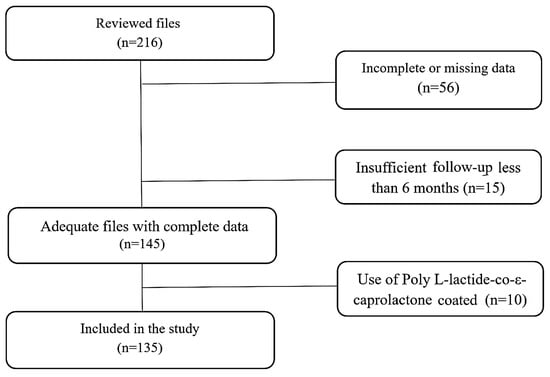
Background: Crown lengthening procedures are widely used in restorative and periodontal therapy to expose sufficient tooth structure for functional and esthetic rehabilitation. Traditionally performed using an open-flap approach, these procedures have evolved with the introduction of minimally invasive flapless techniques. This narrative review
[...] Read more.
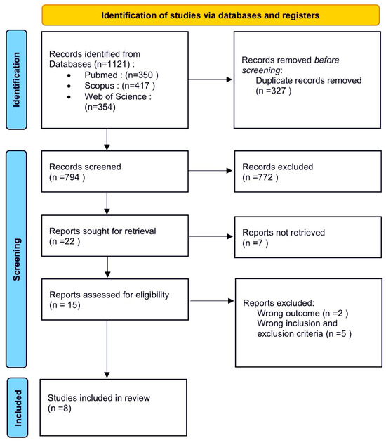
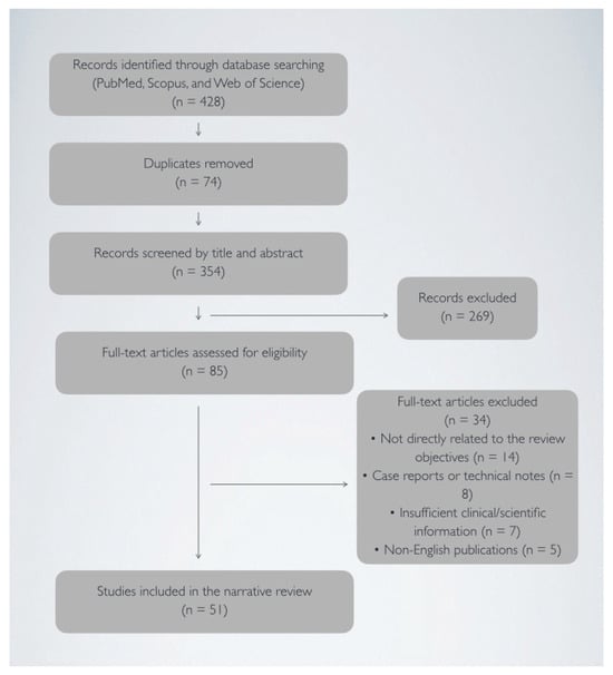
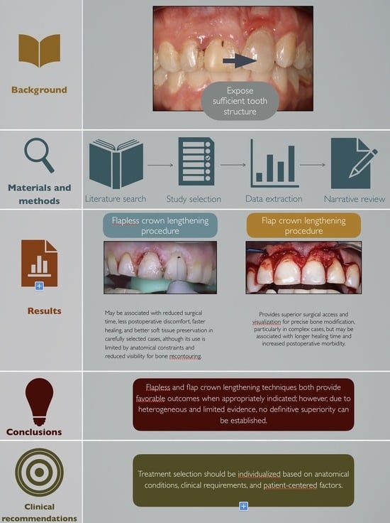
Background: Crown lengthening procedures are widely used in restorative and periodontal therapy to expose sufficient tooth structure for functional and esthetic rehabilitation. Traditionally performed using an open-flap approach, these procedures have evolved with the introduction of minimally invasive flapless techniques. This narrative review aims to summarize the biological foundations, clinical outcomes, and contemporary approaches associated with crown lengthening procedures, with particular emphasis on flapless and flap techniques, healing processes, technological advancements, and clinical decision-making considerations. Methods: A narrative literature review was conducted using electronic databases, including PubMed, Scopus, and Web of Science, to identify relevant publications on crown lengthening procedures. The search focused on studies addressing flapless and flap techniques, biological principles, healing dynamics, clinical outcomes, postoperative considerations, and contemporary approaches such as laser-assisted and digitally guided procedures. Titles and abstracts were screened for relevance, followed by full-text assessment of eligible articles. The study selection process was documented using a simplified PRISMA-style flow diagram to enhance transparency. Results: The reviewed literature suggests that flapless crown lengthening may be associated with reduced surgical time, less postoperative discomfort, faster healing, and preservation of soft tissue architecture in appropriately selected cases. However, its applicability appears to be influenced by anatomical factors, including bone morphology and the need for adequate visualization during osseous recontouring. Conventional flap techniques continue to be widely used and may offer improved surgical access and visibility, particularly in more complex situations requiring extensive bone modification. Although these procedures have been associated with longer healing periods and greater postoperative morbidity, the heterogeneity of the available evidence and the narrative nature of this review limit definitive comparisons between the two approaches. Conclusions: In conclusion, both flapless and flap crown lengthening procedures demonstrate high clinical efficacy when appropriately indicated. The choice of technique should be guided by case-specific factors, clinician expertise, and patient expectations. Further randomized controlled trials are recommended to strengthen the evidence base and establish standardized clinical guidelines.
Full article
(This article belongs to the Special Issue Advances in Periodontal and Peri-Implant Tissues Health Management: 2nd Edition)
►▼
Show Figures

Graphical abstract
Open AccessTutorial
Modified Reverse Scan Technique for Abutment Teeth
by
Pavel Hyspler, Jiri Borovec and Tatjana Dostalova
Dent. J. 2026, 14(7), 412; https://doi.org/10.3390/dj14070412 (registering DOI) - 6 Jul 2026
Abstract
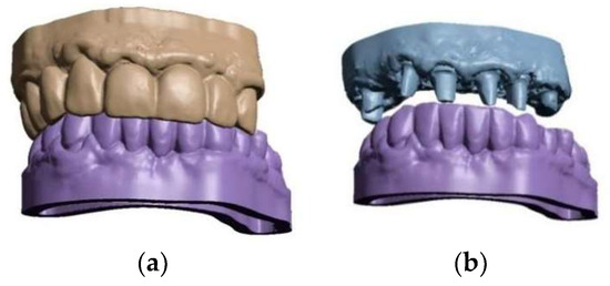
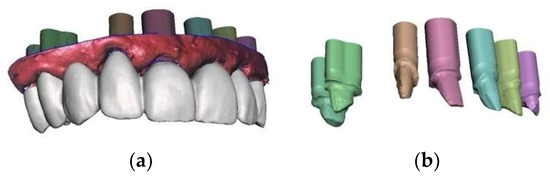
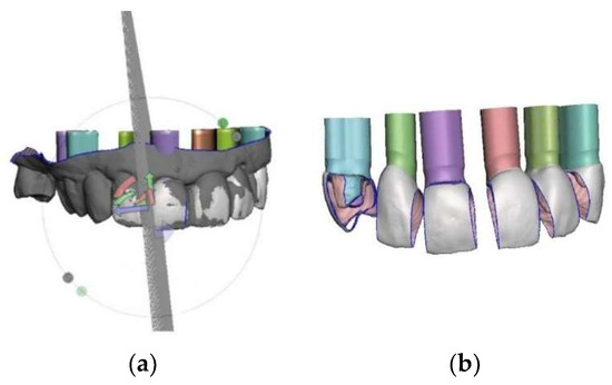
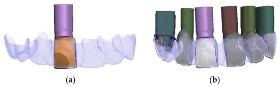
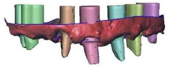
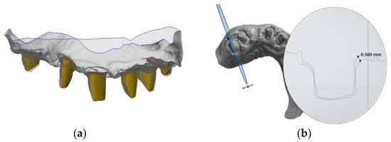
This article introduces a modified reverse scan technique for abutment teeth that incorporates laboratory scanners into a fully digital workflow to enhance accuracy and reduces the impact of stitching and merging errors associated with long-span intraoral scans when fabricating extensive tooth-supported prostheses. This
[...] Read more.
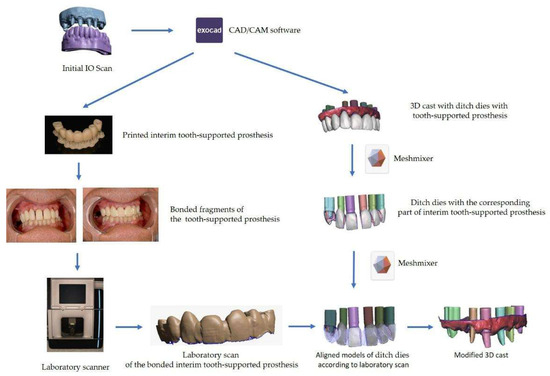
This article introduces a modified reverse scan technique for abutment teeth that incorporates laboratory scanners into a fully digital workflow to enhance accuracy and reduces the impact of stitching and merging errors associated with long-span intraoral scans when fabricating extensive tooth-supported prostheses. This cost-effective, time-saving, and versatile procedure uses a 3D-printed interim tooth-supported prosthesis that is sectioned, bonded intraorally, and scanned with a laboratory scanner to improve the virtual definitive cast. The modification is performed using free software.
Full article
(This article belongs to the Special Issue Feature Papers in Digital Dentistry)
►▼
Show Figures

Figure 1
Open AccessArticle
The Impact of Limited Access to Dental Care on Emergency Room Service Utilization: A Study of Primary Healthcare in a Rural Inland Region of Portugal
by
Alexandra Prada, Ana Galvão, Matilde Monteiro-Soares and Cláudia Camila Dias
Dent. J. 2026, 14(7), 411; https://doi.org/10.3390/dj14070411 (registering DOI) - 6 Jul 2026
Abstract
Background/Objectives: This cross-sectional observational study investigated factors associated with emergency room (ER) utilization for dental pain in a rural inland region of Portugal. The main objective was to examine the relationship between access to dental care, sociodemographic characteristics, oral health behaviors, and clinical
[...] Read more.
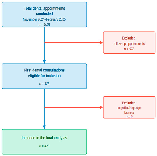
Background/Objectives: This cross-sectional observational study investigated factors associated with emergency room (ER) utilization for dental pain in a rural inland region of Portugal. The main objective was to examine the relationship between access to dental care, sociodemographic characteristics, oral health behaviors, and clinical outcomes with the use of emergency room services for dental problems. Methods: The study sample comprised 423 participants from the districts of Bragança and Vinhais, in Trás-os-Montes, aged 4 to 90 years, who attended their first dental medicine consultation. Participants completed a structured questionnaire addressing sociodemographic characteristics, general health, oral health behaviors, and dental prosthetic use, and underwent oral examination for assessment of the Decayed, Missing, and Filled Teeth (DMFT) index. Associations with reported ER utilization due to toothache were analyzed using Fisher’s exact test and the Mann–Whitney U test. Results: Overall, 28.4% of participants reported having visited the ER due to dental pain, and most cases were managed with medication followed by discharge. ER utilization was significantly associated with behavioral risk factors such as smoking, as well as poorer oral hygiene practices, including less frequent tooth brushing. In addition, participants who sought ER care presented higher DMFT scores, indicating a greater burden of untreated dental decay and tooth loss. Conclusions: These findings suggest that limited preventive dental care and unfavorable oral health behaviors are associated with to avoidable ER visits for dental pain in rural settings. This study reinforces the need to strengthen access to preventive oral health services and to advance the integration of dental care into Portugal’s National Health Service (SNS), particularly in underserved inland regions.
Full article
(This article belongs to the Special Issue The Ethical and Professional Nature of Dentistry)
►▼
Show Figures

Figure 1
Open AccessArticle
Oral Health Values, Oral Hygiene Habits, and Preventive Dental Attendance Among Health-Related and Non-Health-Related University Students: A Cross-Sectional Study
by
Klara Dulić, Marija Čandrlić, Ivan Miškulin, Kristina Kralik, Davor Jurlina, Katarina Major Poljak, Ingrid Kovačević, Dora Dragičević Tomičić, Emanuela Ham and Slavko Čandrlić
Dent. J. 2026, 14(7), 410; https://doi.org/10.3390/dj14070410 (registering DOI) - 6 Jul 2026
Abstract
Background/Objectives: This study aimed to assess oral health values, oral hygiene habits, preventive dental attendance, and lifestyle-related risk factors among students at the University of Osijek and to compare findings between students enrolled in health-related and non-health-related study programs. Methods: A cross-sectional survey
[...] Read more.
Background/Objectives: This study aimed to assess oral health values, oral hygiene habits, preventive dental attendance, and lifestyle-related risk factors among students at the University of Osijek and to compare findings between students enrolled in health-related and non-health-related study programs. Methods: A cross-sectional survey was conducted among 331 students (186 health-related and 145 non-health-related). Participants were recruited using a convenience sampling approach. Data were collected using an anonymous questionnaire comprising demographic information, oral health–related behaviors, and the Croatian version of the Oral Health Values Scale (OHVS-CRO). Group differences were analyzed using nonparametric statistical tests. Results: Students enrolled in health-related study programs reported significantly more favorable oral hygiene behaviors, including more frequent toothbrushing, greater use of dental floss, interdental brushes, and mouthwash, as well as more regular preventive dental attendance (all p < 0.05). They also achieved significantly higher OHVS-CRO scores across all domains and on the total scale (median total score: 42 vs. 40; p < 0.001). No significant differences were observed regarding smoking, alcohol consumption, or refined sugar intake. Conclusions: Students enrolled in health-related study programs demonstrated higher oral health values and more favorable oral health-related behaviors than students enrolled in non-health-related study programs. These findings suggest an association between educational orientation and oral health values and behaviors and may inform future oral health promotion initiatives targeting university students.
Full article
(This article belongs to the Section Preventive Dentistry)
Open AccessArticle
Implant Failure Rate in Transcrestal and Lateral Window Sinus Augmentations: A Descriptive Retrospective Cohort Study
by
Eran Gabay, Yuval Shafran, Tarek Mtanis, Eli E. Machtei, Ofir Ginesin, Hadar Zigdon-Giladi and Yaniv Mayer
Dent. J. 2026, 14(7), 409; https://doi.org/10.3390/dj14070409 (registering DOI) - 6 Jul 2026
Abstract
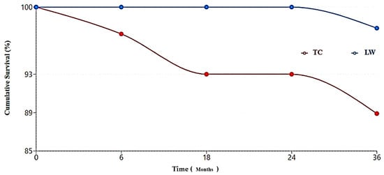
Objectives: This study aimed to describe implant survival rates and associated risk factors in patients treated with two sinus augmentation techniques—Lateral Window (LW) and Transcrestal (TC)—at a single center. Methods: A retrospective cohort analysis was performed using medical records of patients who underwent
[...] Read more.
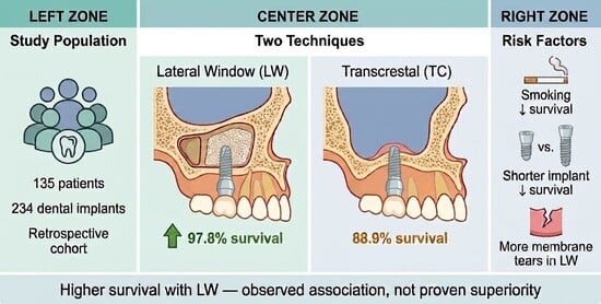
Objectives: This study aimed to describe implant survival rates and associated risk factors in patients treated with two sinus augmentation techniques—Lateral Window (LW) and Transcrestal (TC)—at a single center. Methods: A retrospective cohort analysis was performed using medical records of patients who underwent maxillary sinus augmentation and subsequent dental implant placement between 2015 and 2019 at the Rambam Healthcare Center. Data on demographic variables, surgical approach, graft materials, implant characteristics, and clinical outcomes were collected from electronic medical records. Results: A total of 135 patients met the inclusion criteria, contributing 144 TC procedures (with 144 implants) and 46 LW procedures (90 implants) to the final analysis. The overall implant failure rate was 7.7%, with a significantly higher failure rate for TC (11.11%) compared to LW (2.22%). Procedure type was a significant predictor of implant survival (OR 1.62); however, as residual bone height (RBH) data were unavailable, confounding by indication cannot be excluded and this finding should be interpreted with caution. Implant length demonstrated a significant influence on outcomes (OR 1.28 per mm increase); shorter implants (8 mm) exhibited lower survival rates (81.3%) compared to 10 mm implants (92.3%). Smoking status was identified as a negative predictor of implant survival (OR 0.23). Schneiderian membrane ruptures occurred in nine cases, seven of which were LW procedures. Smoking was the only factor affecting graft survival (OR 0.29). Conclusions: In this retrospective cohort, implant survival rates were higher in the LW group than in the TC group; however, given that individual residual bone height data were unavailable and case assignment, although governed by standardized departmental criteria, was not randomized, the possibility of confounding by indication cannot be excluded. Smoking and shorter implants were significant risk factors for implant failure. Smoking was also associated with graft failure.
Full article
(This article belongs to the Special Issue Implant Dentistry—the Surgical Prosthetic Interplay)
►▼
Show Figures

Graphical abstract
Open AccessSystematic Review
Protective and Healing Effects of Zinc L-Carnosine on the Oral Mucosa: A Systematic Review and Meta-Analysis
by
Pierpaolo De Francesco, Paolo Vescovi, Giuseppe Pedrazzi and Ilaria Giovannacci
Dent. J. 2026, 14(7), 408; https://doi.org/10.3390/dj14070408 - 5 Jul 2026
Abstract
Background/Objectives: Oral mucosal injury is a frequent complication in oncologic and surgical settings, significantly affecting patient quality of life. Zinc L-carnosine (ZnC) is a cytoprotective compound with anti-inflammatory, antioxidant, and epithelial reparative properties. This systematic review evaluated its protective and healing effects
[...] Read more.
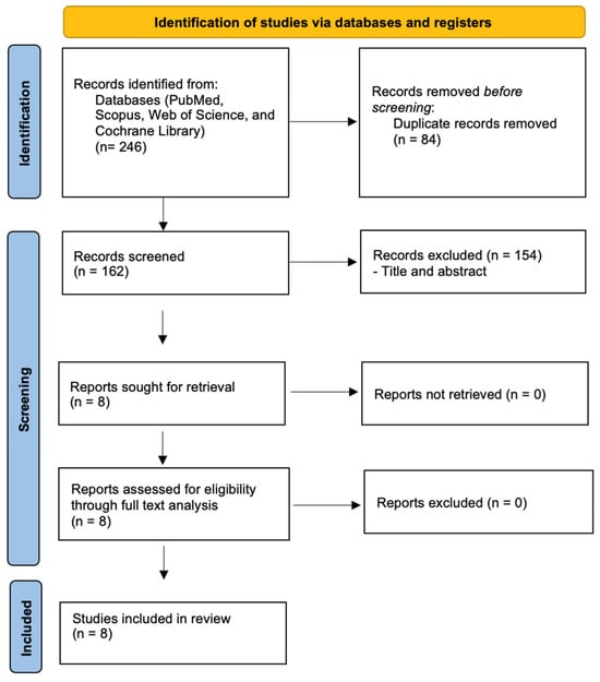
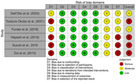
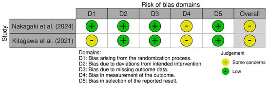
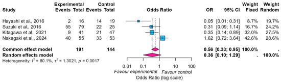
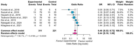
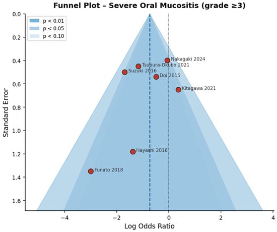
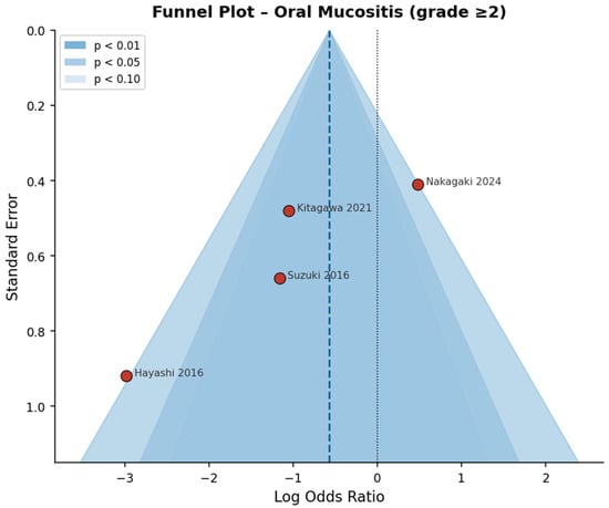
Background/Objectives: Oral mucosal injury is a frequent complication in oncologic and surgical settings, significantly affecting patient quality of life. Zinc L-carnosine (ZnC) is a cytoprotective compound with anti-inflammatory, antioxidant, and epithelial reparative properties. This systematic review evaluated its protective and healing effects on oral mucosa. Methods: A systematic search followed PRISMA guidelines was conducted across PubMed, Scopus, Web of Science, and Cochrane Library (2015–2026). Randomized and non-randomized controlled studies assessing ZnC in patients with or at risk of oral mucosal injury were included. Risk of bias was evaluated using RoB 2 and ROBINS-I tools. Meta-analyses were conducted under both common- and random-effects models. The certainty of evidence was evaluated according to the GRADE guidelines. Results: Eight studies (n = 544) were included. Six non-randomized studies showed moderate or serious risk of bias, while randomized trials presented some concerns. ZnC was administered in different formulations, including mouthwashes, lozenges, and mucoadhesive suspensions based on sodium alginate, polyacrylic acid, and carboxyvinyl polymer, and across different clinical settings. Meta-analysis showed a reduced incidence of severe oral mucositis (grade ≥ 3) under the Common Effect model (OR 0.48; 95% CI 0.32–0.72), although statistical significance was not maintained under random-effects models (OR 0.44; 95% CI 0.18–1.06). Similar results were observed for grade ≥ 2 mucositis. According to the GRADE assessment, the certainty of evidence was low for oral mucositis outcomes and very low for oral mucosal healing. Only one study suggested improved surgical wound healing. No serious adverse events were reported. Conclusions: ZnC may support oral mucosal protection and healing, particularly in preventing oral mucositis. However, substantial heterogeneity and limited high-quality randomized evidence restrict the strength of conclusions. Further well-designed randomized trials are needed.
Full article
(This article belongs to the Special Issue Feature Review Papers in Dentistry: 2nd Edition)
►▼
Show Figures

Figure 1
Journal Menu
► ▼ Journal Menu-
- Dentistry Journal Home
- Aims & Scope
- Editorial Board
- Reviewer Board
- Topical Advisory Panel
- Instructions for Authors
- Special Issues
- Topics
- Sections & Collections
- Article Processing Charge
- Indexing & Archiving
- Editor’s Choice Articles
- Most Cited & Viewed
- Journal Statistics
- Journal History
- Journal Awards
- Conferences
- Editorial Office
Journal Browser
► ▼ Journal BrowserHighly Accessed Articles
Latest Books
E-Mail Alert
News
Topics
Topic in
Dentistry Journal, IJMS, JCM, Medicina, Applied Sciences
Oral Health Management and Disease Treatment
Topic Editors: Christos Rahiotis, Felice Lorusso, Sergio Rexhep TariDeadline: 31 July 2026
Topic in
Applied Sciences, Dentistry Journal, Polymers, Applied Biosciences, Bioengineering, Materials
Advances in Biomaterials—2nd Edition
Topic Editors: Satoshi Komasa, Yoshiro Tahara, Tohru Sekino, Hideaki Sato, Yoshiya Hashimoto, Tetsuya AdachiDeadline: 31 December 2026
Conferences
Special Issues
Special Issue in
Dentistry Journal
Caries Risk Assessment and Preventive Care Protocols
Guest Editor: Svante TwetmanDeadline: 15 July 2026
Special Issue in
Dentistry Journal
Recent Progress in Digital Technology in Prosthodontics and Implantology
Guest Editor: Stefanos KourtisDeadline: 20 July 2026
Special Issue in
Dentistry Journal
Antimicrobial Resistance and Stewardship in Dentistry: Challenges, Evidence, and Clinical Strategies
Guest Editors: Noha Seoudi, Melanie WilsonDeadline: 26 July 2026
Special Issue in
Dentistry Journal
Contemporary Dentistry: Classical and Modern Approaches
Guest Editor: Horia Mihail BarbuDeadline: 31 July 2026
Topical Collections
Topical Collection in
Dentistry Journal
Light and Laser Dentistry
Collection Editors: Samir Nammour, Aldo Brugnera Junior
Topical Collection in
Dentistry Journal
Novel Ceramic Materials in Dentistry
Collection Editors: Roberto Sorrentino, Gianrico Spagnuolo
Topical Collection in
Dentistry Journal
Bio-Logic Approaches to Implant Dentistry
Collection Editors: Luigi Canullo, Donato Antonacci, Piero Papi, Francesco Gianfreda, Bianca Di Murro, Carlo Raffone
Topical Collection in
Dentistry Journal
Dental Traumatology and Sport Dentistry
Collection Editor: Enrico Spinas