Journal Description
Receptors
Receptors
is an international, peer-reviewed, open access journal on all aspects of receptors published quarterly online by MDPI.
- Open Access— free for readers, with article processing charges (APC) paid by authors or their institutions.
- High Visibility: indexed within ESCI (Web of Science), Scopus and other databases.
- Rapid Publication: manuscripts are peer-reviewed and a first decision is provided to authors approximately 43.1 days after submission; acceptance to publication is undertaken in 7.2 days (median values for papers published in this journal in the first half of 2026).
- Recognition of Reviewers: APC discount vouchers, optional signed peer review and reviewer names are published annually in the journal.
- Receptors is a companion journal of Biomolecules.
Latest Articles
The Prorenin Receptor: Multitasking Its Way Through Cardiovascular, Metabolic and Renal Diseases
Receptors 2026, 5(3), 26; https://doi.org/10.3390/receptors5030026 - 11 Aug 2026
Abstract
►
Show Figures
The renin–angiotensin–aldosterone system (RAAS) is a fundamental regulator of blood pressure, electrolyte balance, fluid homeostasis, and tissue remodeling. The discovery of the prorenin receptor (PRR), the protein encoded by the ATP6AP2 gene, has substantially expanded the classical RAAS paradigm by demonstrating that prorenin
[...] Read more.
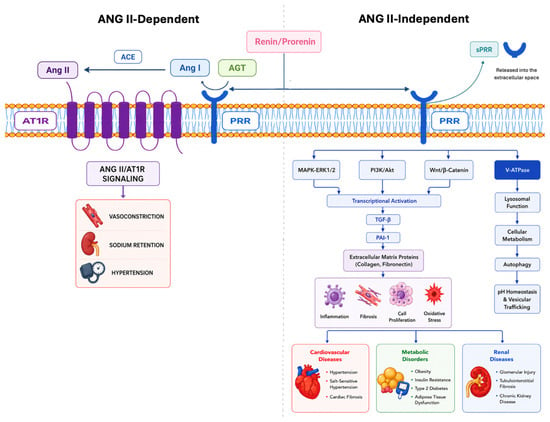
The renin–angiotensin–aldosterone system (RAAS) is a fundamental regulator of blood pressure, electrolyte balance, fluid homeostasis, and tissue remodeling. The discovery of the prorenin receptor (PRR), the protein encoded by the ATP6AP2 gene, has substantially expanded the classical RAAS paradigm by demonstrating that prorenin possesses biological activity beyond its proteolytic conversion to renin. Binding of renin or prorenin to PRR enhances local angiotensin II (Ang II) generation while simultaneously initiating Ang II-independent intracellular signaling pathways, including ERK1/2, mitogen-activated protein kinases, PI3K/Akt, transforming growth factor-β, and nuclear factor-κB, thereby promoting inflammation, oxidative stress, fibrosis, cellular proliferation, and extracellular matrix accumulation. Beyond its receptor function, PRR serves as an essential accessory component of the vacuolar H+-ATPase (V-ATPase) complex, regulating vesicular acidification, lysosomal function, autophagy, protein trafficking, cellular metabolism, and Wnt/β-catenin signaling. These diverse functions explain its indispensable role in embryonic development, cell differentiation, and tissue homeostasis, as evidenced by the embryonic lethality associated with ATP6AP2 gene deficiency. PRR is predominantly localized to intracellular organelles, including the endoplasmic reticulum, Golgi apparatus, endosomes, lysosomes, and autophagic vesicles, although membrane-bound and soluble forms also contribute to physiological and pathological processes. Increasing evidence implies dysregulated PRR signaling in the development and progression of hypertension, cardiovascular disease, chronic kidney disease, diabetes, obesity, and other metabolic disorders. This review summarizes current advances in PRR and soluble PRR biology, discusses unresolved mechanistic and translational questions, and evaluates the potential of PRR as a biomarker and therapeutic target for cardiovascular, renal, and metabolic diseases.
Full article
Open AccessReview
The Role of Toll-like Receptors (TLRs) in Fish Immunity
by
Surajit Ghosh, Ananya Paul, Auroshree Sadhu, Triparna Roy, Nimai Chandra Saha, Aabid Hussain and Shubhajit Saha
Receptors 2026, 5(3), 25; https://doi.org/10.3390/receptors5030025 - 7 Aug 2026
Abstract
►▼
Show Figures
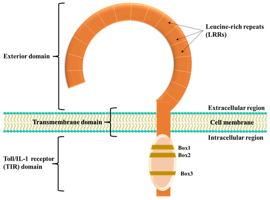
The innate system’s recognition of non-self and danger signals is mediated by a limited number of germline-encoded pattern recognition receptors (PRRs) that recognize pathogen-associated molecular patterns (PAMPs). Toll-like receptors (TLRs) are single, non-catalytic, membrane-spanning PRRs present in invertebrates and vertebrates. They act by
[...] Read more.
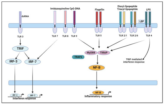
The innate system’s recognition of non-self and danger signals is mediated by a limited number of germline-encoded pattern recognition receptors (PRRs) that recognize pathogen-associated molecular patterns (PAMPs). Toll-like receptors (TLRs) are single, non-catalytic, membrane-spanning PRRs present in invertebrates and vertebrates. They act by specifically recognizing PAMPs of a variety of microbes and activate signalling cascades to induce innate immunity. A large number of TLRs has been identified in various teleosts and basal vertebrates. The varied evolutionary lineages of aquatic and warm-blooded higher animals result in some unique characteristics in their TLRs. In innate immunity and pathogen recognition, the majority of them share conserved signalling pathways. Comparing the immunology of teleosts, chondrichthyans, agnathans, and warm-blooded vertebrates requires a functional analysis of novel TLRs in teleosts and basal vertebrates. Furthermore, TLRs play important roles in adaptive immunity in addition to innate immunity. This review describes the discovery of TLRs and their role in fish immunity along their signalling pathway and ligand specificity.
Full article

Figure 1
Open AccessReview
Neurotrophic Glucocorticoid Signalling
by
Freddy Jeanneteau
Receptors 2026, 5(3), 24; https://doi.org/10.3390/receptors5030024 - 27 Jul 2026
Abstract
Glucocorticoids are stress hormones released into circulation that play central roles in the immediate and slower adaptive responses of the brain and body to environmental and internal stimuli, whether perceived or imagined. Receptors for glucocorticoids translocate from the cytosol to the nucleus and
[...] Read more.
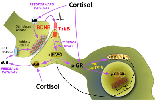
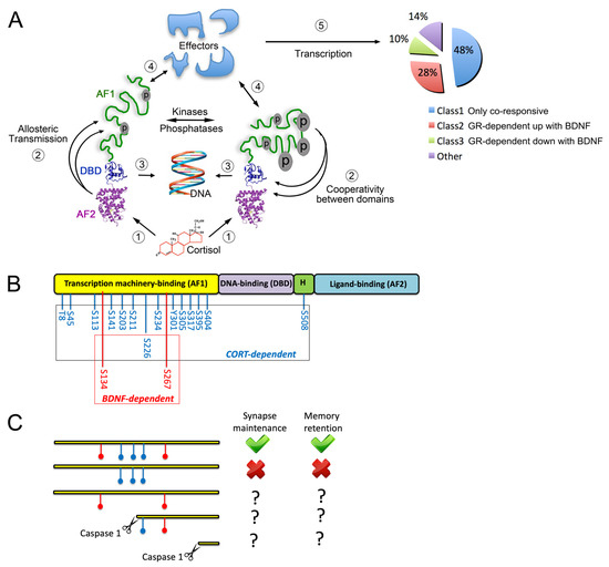
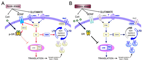
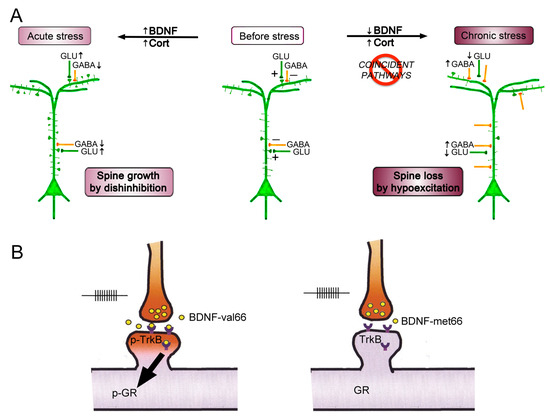
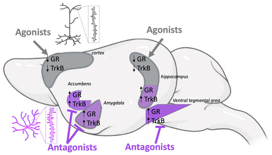
Glucocorticoids are stress hormones released into circulation that play central roles in the immediate and slower adaptive responses of the brain and body to environmental and internal stimuli, whether perceived or imagined. Receptors for glucocorticoids translocate from the cytosol to the nucleus and mitochondria to regulate the expression of numerous genes involved in neuroplasticity, metabolism, inflammation, immunity, and the cytoskeleton. Rapid effects of glucocorticoids are mediated by distinct receptor-associated mechanisms to facilitate or suppress neurotransmitter release. Feedforward and feedback mechanisms in neurotransmission thus depend on glucocorticoid receptor localization and functional states across synapses. Post-translational modifications of glucocorticoid receptors generate specific docking sites for signalling effectors which vary between active and inactive neurons. Diversity of glucocorticoid reactivity produces heterogeneous cellular responses that integrate prior experience. The activity-dependent trans-synaptic neurotrophic messenger BDNF emerges as a key modulator of glucocorticoid receptor signalling diversity. These mechanisms have been investigated with deletion mutants in a variety of preclinical models, pointing not only at possible causes of human diseases but also at potential strategies for mitigating them. This review specifically focuses on neurotrophic glucocorticoid signalling as a coincidence detection mechanism between BDNF and glucocorticoids, extending beyond classical genomic and non-genomic frameworks.
Full article
(This article belongs to the Collection Receptors: Exceptional Scientists and Their Expert Opinions)
►▼
Show Figures

Figure 1
Open AccessReview
Protective and Detrimental Roles of NLRP6 in Infection and Cancer
by
Takayoshi Yamauchi, Vaibhav Jain and Simon G. Gregory
Receptors 2026, 5(3), 23; https://doi.org/10.3390/receptors5030023 - 8 Jul 2026
Abstract
►▼
Show Figures
NLRP6 is a member of the NOD-like receptor family that was initially characterized as an inflammasome-forming sensor in the intestine. However, accumulating evidence over the past decade has revealed that the functions of NLRP6 extend far beyond this canonical role. NLRP6 operates in
[...] Read more.
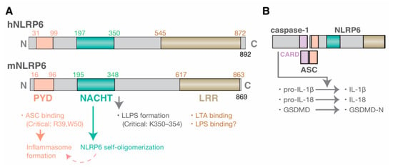
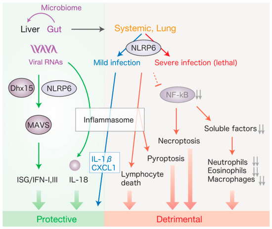
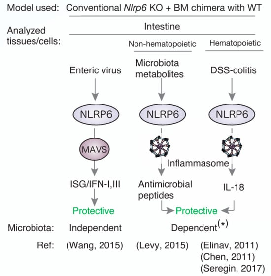
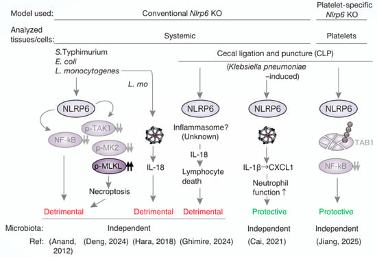
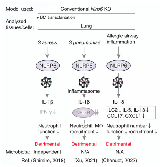
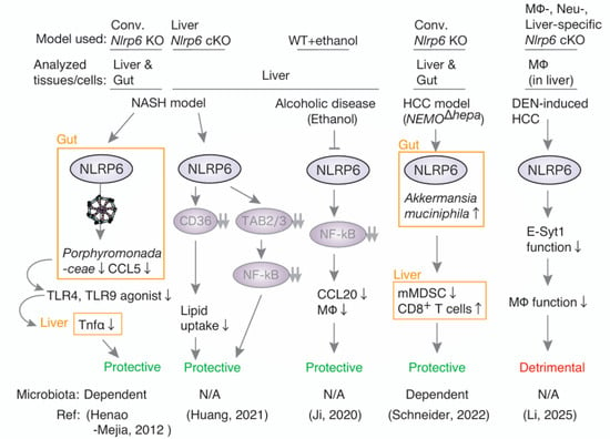
NLRP6 is a member of the NOD-like receptor family that was initially characterized as an inflammasome-forming sensor in the intestine. However, accumulating evidence over the past decade has revealed that the functions of NLRP6 extend far beyond this canonical role. NLRP6 operates in a wide range of tissues, including the intestine, liver, lung, and immune system, where it exerts context-dependent effects that can be either protective or detrimental. In the intestine, NLRP6 is most consistently associated with host protection, contributing to antiviral defense, epithelial barrier integrity, and the maintenance of microbial and metabolic homeostasis through both inflammasome-dependent and -independent mechanisms. In contrast, in systemic infection models and in certain inflammatory settings, NLRP6 can also promote pathology by suppressing NF-κB signaling; inducing IL-18–mediated lymphocyte death, or enhancing inflammatory cell death pathways. Moreover, studies using both conventional and tissue-specific knockout models have highlighted the importance of the gut–organ axis; particularly the gut–liver axis, in shaping NLRP6-dependent disease outcomes. Here, we summarize recent advances in understanding the upstream regulation, downstream signaling, and tissue-specific functions of NLRP6.
Full article

Figure 1
Open AccessReview
Transsynaptic Bridges: Molecular Architects of Synaptic Identity, Plasticity and Disease
by
Swetha K. Godavarthi
Receptors 2026, 5(3), 22; https://doi.org/10.3390/receptors5030022 - 7 Jul 2026
Abstract
►▼
Show Figures
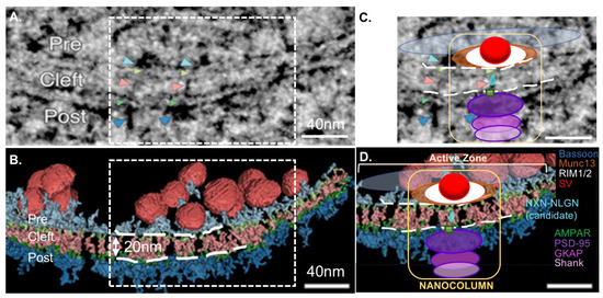
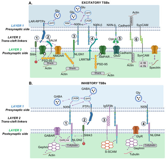
Transsynaptic bridges are molecular complexes spanning the synaptic cleft that physically couple presynaptic release machinery to postsynaptic receptor fields. That this architecture is the direct target of autoimmune attack in myasthenia gravis and a primary locus of genetic risk in autism spectrum disorder
[...] Read more.
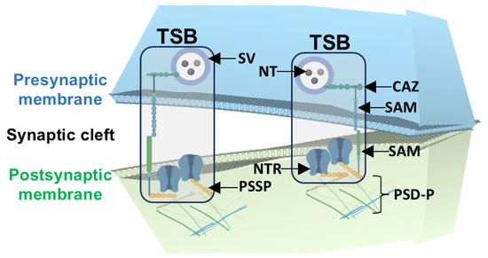
Transsynaptic bridges are molecular complexes spanning the synaptic cleft that physically couple presynaptic release machinery to postsynaptic receptor fields. That this architecture is the direct target of autoimmune attack in myasthenia gravis and a primary locus of genetic risk in autism spectrum disorder and schizophrenia underscores that transsynaptic bridges are not only important during synapse development but are continuously required organizers of neural function. This review traces the evolution of structural, molecular, and functional evidence that shaped our understanding of the synapse as a single integrated trans-cellular unit. Several defining properties of a synapse emerge as a consequence of transsynaptic bridges—for example, the nanoscale alignment they impose between vesicle fusion sites and receptor nanodomains determines transmission efficacy, the bidirectional communication they coordinate across the cleft drives signaling specificity, and they undergo activity-dependent remodeling, thereby physically encoding synaptic history.
Full article

Figure 1
Open AccessReview
The Dopamine D3 Receptor as an Emerging Therapeutic Target in Parkinson’s Disease: Structural Advances, Signaling Bias and Neuroprotective Perspectives
by
Felipe Patricio, Eliud Morales Dávila, Aleidy Patricio-Martínez, Abel Villa-Mancera, Jose Manuel Pérez-Aguilar and Ilhuicamina Daniel Limón
Receptors 2026, 5(2), 21; https://doi.org/10.3390/receptors5020021 - 18 Jun 2026
Abstract
►▼
Show Figures
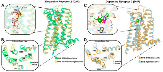
The dopamine D3 receptor (D3R) has long been considered a secondary target in the treatment of Parkinson’s disease (PD), with therapeutic strategies primarily focused on D2 receptor–mediated motor control. However, accumulating evidence now supports D3R as a
[...] Read more.
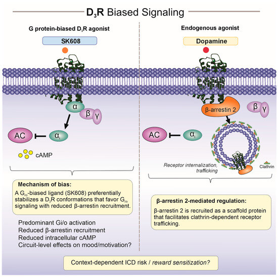
The dopamine D3 receptor (D3R) has long been considered a secondary target in the treatment of Parkinson’s disease (PD), with therapeutic strategies primarily focused on D2 receptor–mediated motor control. However, accumulating evidence now supports D3R as a functionally distinct dopaminergic receptor subtype with specific relevance to non-motor symptom domains and dopaminergic signaling under hypodopaminergic conditions. Recent advances in high-resolution structural biology have elucidated the molecular basis of D3R/D2R discrimination, revealing how subtle residue-level and microstructural differences within a conserved G protein–coupled receptor framework shape ligand recognition and receptor activation. In parallel, the emergence of ligand-dependent biased signaling has refined current understanding of D3R pharmacology. Selected ligands can preferentially engage Gαi/o-mediated pathways while limiting β-arrestin recruitment and associated regulatory processes, providing a mechanistic rationale for more stable modulation of mesolimbic dopaminergic circuits involved in affective and motivational regulation. Beyond symptomatic modulation, preclinical studies suggest that D3R signaling may influence neuronal resilience, synaptic plasticity, and adaptive responses to dopaminergic injury; however, such effects remain experimental and have not been demonstrated in clinical PD. This review integrates recent structural, signaling, and functional insights into D3R biology, with particular emphasis on biased agonism and emerging therapeutic concepts. Although D3R-targeted strategies do not currently represent disease-modifying interventions, they offer a rational framework for the development of next-generation dopaminergic therapies aimed at improving precision, tolerability, and long-term signaling stability in Parkinson’s disease.
Full article

Figure 1
Open AccessReview
Ectopic Olfactory Receptors: Expression and Functions Outside of the Nasal Cavity
by
Mary Beth Genter
Receptors 2026, 5(2), 20; https://doi.org/10.3390/receptors5020020 - 8 Jun 2026
Abstract
►▼
Show Figures
Olfactory (or odorant) receptors (ORs) were initially characterized in 1991 by Drs. Richard Axel and Linda Buck, and subsequent additional efforts have contributed to our understanding of their canonical function in odorant identification in the nasal cavity, including ligands for many of the
[...] Read more.
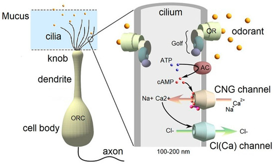
Olfactory (or odorant) receptors (ORs) were initially characterized in 1991 by Drs. Richard Axel and Linda Buck, and subsequent additional efforts have contributed to our understanding of their canonical function in odorant identification in the nasal cavity, including ligands for many of the ORs and the signaling pathways involved. More recently, OR transcripts and proteins have been identified in cells and organs outside of the nasal cavity, ranging from skin to sperm to tumors, suggesting that they have biological roles in ectopic locations other than their canonical function of odorant molecule detection in the nose. This mini narrative review discusses ectopic human ORs and their potential ligand-activated functions in the skin, lung, and sperm, as well as in diseases such as nonalcoholic steatohepatitis (NASH), melanoma and prostate cancer.
Full article

Figure 1
Open AccessReview
PIEZO Channels in Breast Cancer: Emerging Roles and Therapeutic Potential
by
Elizabeth Adams, Madison Reddock, Bryn Gillen, Xiyu Wang, Qingfei Wang, Mateusz Opyrchal and Tao Yu
Receptors 2026, 5(2), 19; https://doi.org/10.3390/receptors5020019 - 5 Jun 2026
Abstract
►▼
Show Figures
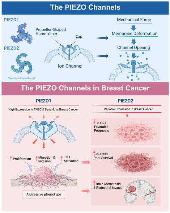
The mechanosensitive PIEZO family channels, PIEZO1 and PIEZO2, are essential for mechanotransduction and play roles in many cellular processes, including cell volume regulation, tissue development, touch sensation, and proprioception. Emerging evidence suggests roles for PIEZO channels in cancer biology; however, direct mechanistic evidence
[...] Read more.
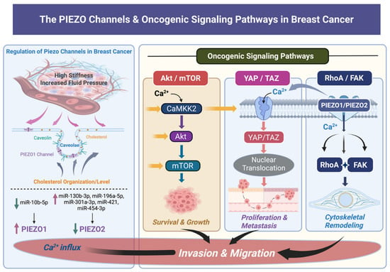
The mechanosensitive PIEZO family channels, PIEZO1 and PIEZO2, are essential for mechanotransduction and play roles in many cellular processes, including cell volume regulation, tissue development, touch sensation, and proprioception. Emerging evidence suggests roles for PIEZO channels in cancer biology; however, direct mechanistic evidence in breast cancer remains limited. They have been shown to promote proliferation, epithelial-to-mesenchymal transition (EMT), and migration; however, these roles are varied and context-dependent. In breast cancer specifically, the two PIEZO channels may play opposing and complex roles in tumor progression, the tumor microenvironment (TME), and the tumor immune microenvironment (TIME), potentially impacting therapeutic response and prognosis. Where breast cancer-specific mechanistic data are lacking, we integrate findings from other tumor types to generate testable hypotheses relevant to breast cancer. In this review, we will explore the importance of PIEZO channels in breast cancer development, progression, and therapeutic response, and explore therapeutics and potential strategies to improve patient outcomes.
Full article

Figure 1
Open AccessReview
The Central Role of Immune Checkpoint Receptors in Genitourinary Tumor Immunotherapy: Mechanisms, Biomarkers, and Therapeutic Landscape
by
Alcides Chaux
Receptors 2026, 5(2), 18; https://doi.org/10.3390/receptors5020018 - 29 May 2026
Abstract
►▼
Show Figures
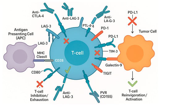
Immune checkpoint receptors (ICRs) play a pivotal role in modulating antitumor immunity and have become central targets in the immunotherapy of genitourinary (GU) malignancies. This review provides a comprehensive overview of the fundamental mechanisms of ICR signaling, the expression and pathophysiological roles of
[...] Read more.
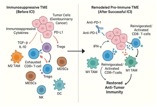
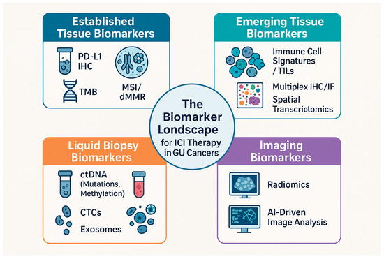
Immune checkpoint receptors (ICRs) play a pivotal role in modulating antitumor immunity and have become central targets in the immunotherapy of genitourinary (GU) malignancies. This review provides a comprehensive overview of the fundamental mechanisms of ICR signaling, the expression and pathophysiological roles of these receptors in GU cancers (kidney, bladder, prostate, testicular, and penile), and the evolving therapeutic landscape. Key ICRs, including PD-1, CTLA-4, LAG-3, TIM-3, and TIGIT, orchestrate complex signaling cascades that can lead to T-cell exhaustion and tumor immune evasion. Their expression varies significantly across GU cancer types, histological subtypes, and tumor stages, influencing prognosis and therapeutic response. Immune checkpoint inhibitors (ICIs) reinvigorate antitumor immunity by disrupting these inhibitory pathways and remodeling the tumor microenvironment (TME); however, resistance mechanisms (primary, adaptive, and acquired) and immune-related adverse events (irAEs) pose significant clinical challenges. Established biomarkers such as PD-L1 expression, tumor mutational burden (TMB), and microsatellite instability (MSI)/deficient mismatch repair (dMMR) status guide ICI use, but their predictive power has limitations. Consequently, emerging tissue-based (e.g., immune cell signatures, multiplex IHC/IF, spatial transcriptomics), liquid biopsy-based (e.g., ctDNA, CTCs, exosomes), and imaging-based (radiomics, AI-driven analysis) biomarkers are under active investigation to refine patient selection and monitor treatment efficacy. The therapeutic armamentarium is rapidly expanding with novel ICIs targeting new receptors, bispecific antibodies, and innovative combination strategies involving ICIs with chemotherapy, targeted therapies, radiotherapy, and other immunotherapies. Furthermore, ICIs are increasingly explored in neoadjuvant, adjuvant, and maintenance settings. This review highlights the dynamic progress in understanding ICR biology and its clinical translation, emphasizing the ongoing efforts to develop more personalized and effective immunotherapeutic strategies for patients with genitourinary tumors.
Full article

Figure 1
Open AccessArticle
Proxalutamide as Potential Inhibitor of ACE2 and TMPRSS2 Receptors?
by
Helyson Lucas Bezerra Braz, Aline Diogo Marinho, Antônio Marcelo Alves Lima, Raul Victor Magalhães Souza, João Alison de Moraes Silveira, Danilo Galvão Rocha, Mirna Marques Bezerra, Marcos Serrou do Amaral, Danilo da Silva Olivier, Geanne Matos de Andrade and Roberta Jeane Bezerra Jorge
Receptors 2026, 5(2), 17; https://doi.org/10.3390/receptors5020017 - 26 May 2026
Abstract
►▼
Show Figures
Background: The World Health Organization (WHO) declared a pandemic due to Severe Acute Respiratory Syndrome Coronavirus 2 (SARS-CoV-2), the etiologic agent responsible for Coronavirus Disease 2019 (COVID-19). Although case numbers declined after the initial outbreak, Brazil experienced slight increases in COVID-19 cases
[...] Read more.
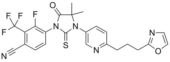
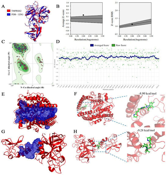
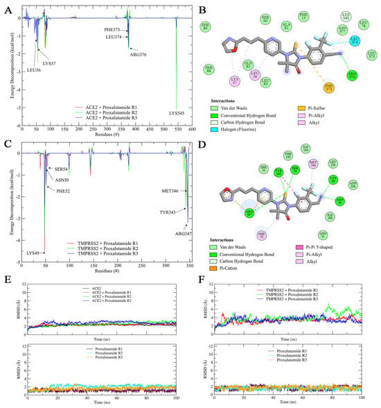
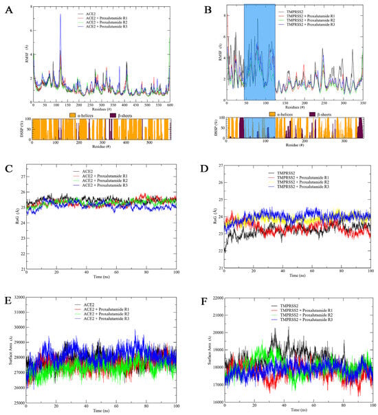
Background: The World Health Organization (WHO) declared a pandemic due to Severe Acute Respiratory Syndrome Coronavirus 2 (SARS-CoV-2), the etiologic agent responsible for Coronavirus Disease 2019 (COVID-19). Although case numbers declined after the initial outbreak, Brazil experienced slight increases in COVID-19 cases in 2024 and 2025, underscoring the persistent need for effective therapeutic interventions. Recently, proxalutamide—an androgen receptor antagonist—has been proposed as a potential therapeutic agent against COVID-19, as supported by several clinical studies. Methods: In the present work, we aimed to elucidate the molecular interactions between proxalutamide and key proteins involved in the viral fusion and replication processes of SARS-CoV-2. Computational techniques, including molecular docking and molecular dynamics simulations, were employed. Results: Our analyses indicated a high success rate, with stable conformations and favorable binding affinity values for ACE2 (−8.90 kcal/mol) and TMPRSS2 (−9.28 kcal/mol), resulting in strong docking scores. Moreover, molecular dynamics simulations confirmed the stability of these complexes, as evidenced by consistent mean square deviation values, low structural flexibility, a stable radius of gyration, and maintained surface rigidity over a 100 ns simulation period. Conclusions: These combined docking and dynamics results suggest that proxalutamide interacts firmly with the active sites, indicating high binding affinity that may interfere with SARS-CoV-2 entry. Nevertheless, experimental validation and rigorous safety assessments are warranted to confirm this potential.
Full article

Figure 1
Open AccessReview
Interleukin-17A (IL-17A): Molecular Mechanisms and Its Roles in Immune and Neuroimmune Systems
by
Sae Sanaka, Asumi Kubo, Sara Kamiya, Kenyu Nakamura and Tetsuya Sasaki
Receptors 2026, 5(2), 16; https://doi.org/10.3390/receptors5020016 - 21 May 2026
Abstract
►▼
Show Figures
Interleukin-17A (IL-17A) is a proinflammatory cytokine that plays a pivotal role in immune responses and tissue homeostasis. Its expression is strictly regulated by transcription factors including RORγt, and it is mainly produced by Th17 cells, γδ T cells, and innate lymphoid cells. IL-17A
[...] Read more.
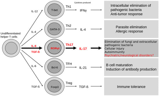
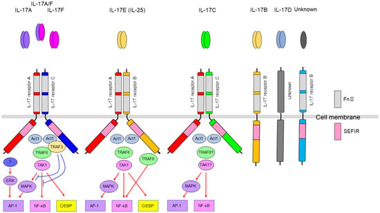
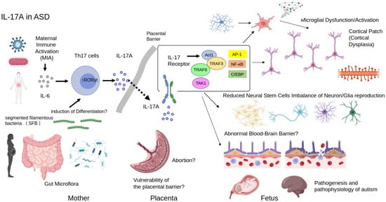
Interleukin-17A (IL-17A) is a proinflammatory cytokine that plays a pivotal role in immune responses and tissue homeostasis. Its expression is strictly regulated by transcription factors including RORγt, and it is mainly produced by Th17 cells, γδ T cells, and innate lymphoid cells. IL-17A signals through a heterodimeric receptor complex consisting of IL-17RA and IL-17RC, activating NF-κB, MAPK, and C/EBP pathways via the adaptor protein Act1. IL-17 signaling is counterbalanced by negative regulators including A20 and Regnase-1. Beyond its classical roles in antimicrobial defense and autoimmune inflammation, recent studies have highlighted its functions in the central nervous system, with associations to multiple sclerosis, autism spectrum disorder, and Alzheimer’s disease. The development of IL-17A inhibitors, including the dual IL-17A/F antagonist bimekizumab, has advanced markedly, with demonstrated efficacy in immune-mediated diseases such as psoriasis and psoriatic arthritis. This review provides a comprehensive overview of current knowledge of IL-17A, from its molecular characteristics to clinical applications.
Full article

Figure 1
Open AccessArticle
Analyzing the Molecular Effects of Endomorphin-2 Degradation on Stabilizing Interactions at the μ-Opioid Receptor
by
Celvic Coomber, Jakob J. Kresse, Surahit Chewle, Marcus Weber, Christof Schütte and Vikram Sunkara
Receptors 2026, 5(2), 15; https://doi.org/10.3390/receptors5020015 - 28 Apr 2026
Abstract
Background: Endogenous opioids, such as endomorphin-2, are key regulators of the body’s pain pathways and mediate analgesia by engaging the -opioid receptor. This class of opioids are distinguished by their transient activation of the -opioid receptor, which is attributed to
[...] Read more.
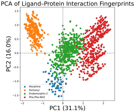
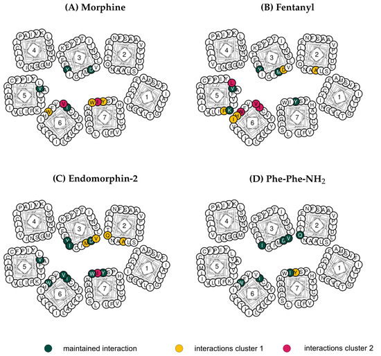
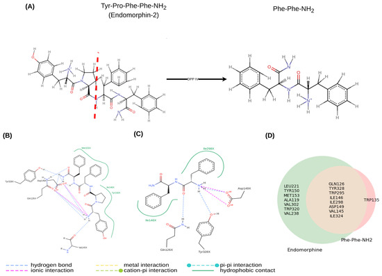
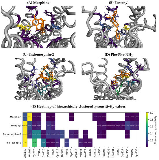
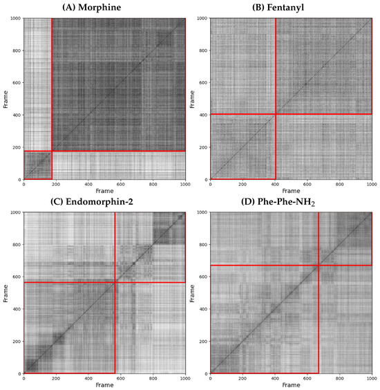
Background: Endogenous opioids, such as endomorphin-2, are key regulators of the body’s pain pathways and mediate analgesia by engaging the -opioid receptor. This class of opioids are distinguished by their transient activation of the -opioid receptor, which is attributed to rapid enzymatic degradation. Methods: To understand how degradation of endomorphin-2 by the enzyme DPP IV affects its interaction with the -opioid receptor, we analyzed the ligand–receptor conformational dynamics and interaction patterns of molecular dynamics simulations data of morphine, fentanyl and endomorphin-2 and one degradation product Phe-Phe-NH2, using molecular fingerprints and the mathematical framework ISOKANN. Results: Our analyses revealed that both the clinically relevant opioids, morphine and fentanyl, as well as the endogenous opioid endomorphin-2, adopt a set of recurring binding conformations within the -opioid receptor binding pocket, maintaining overlapping interaction motifs throughout the simulations. In contrast, Phe-Phe-NH2 failed to maintain a persistent binding mode over the simulated timescale. This instability arises from the dipeptidyl peptidase IV mediated cleavage of endomorphin-2, which generates Phe-Phe-NH2 and removes critical proline and tyrosine residues, thereby leading to the loss of stabilizing hydrophobic contacts with receptor residues Tyr1503,33, Val2385,43 and Val3026,55. Conclusion: By mapping structural interaction motifs essential for stable -opioid receptor binding, this study provides mechanistic insights into how endogenous degradation reshapes ligand–receptor interactions.
Full article
(This article belongs to the Collection Receptors: Exceptional Scientists and Their Expert Opinions)
►▼
Show Figures

Figure 1
Open AccessReview
Classical Immune Pattern Recognition Receptors Involved in Inflammatory Trigger of Sickle Cell Anemia
by
Hershiley Oliveira Jácome, Jonatas Alencar Castro Campelo and Alexander Leonardo Silva-Junior
Receptors 2026, 5(2), 14; https://doi.org/10.3390/receptors5020014 - 21 Apr 2026
Abstract
►▼
Show Figures
Sickle cell anemia (SCA) is a genetic disorder characterized by chronic hemolysis, primarily driven by red blood cell lysis. Its pathophysiology is centered, though not exclusively, on the increased release of intracellular components, such as hemoglobin degradation products, which are known to stimulate
[...] Read more.
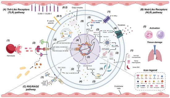
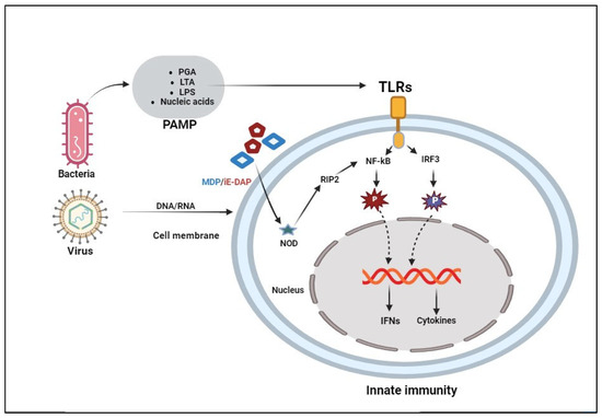
Sickle cell anemia (SCA) is a genetic disorder characterized by chronic hemolysis, primarily driven by red blood cell lysis. Its pathophysiology is centered, though not exclusively, on the increased release of intracellular components, such as hemoglobin degradation products, which are known to stimulate innate immune responses and promote prothrombotic states. Current therapies alleviate symptoms, yet patients remain exposed to a chronic inflammatory milieu punctuated by episodes of acute pain. The recurrence of these crises can be life-threatening due to ischemia–reperfusion injury, hypercoagulability, and respiratory complications. Central mechanisms are marked by elevated hemolysis, heightened inflammatory signaling, and increased procoagulant activity, largely driven by soluble molecules released into the plasma, such as hemoglobin, nuclear molecules and other products. These compounds are recognized from sensors on immune and endothelial cells, named Pattern Recognition Receptors (PRRs), and constitute canonical pathways for intracellular activation. Four main types have been extensively studied in the literature over recent years in both infectious and sterile inflammatory contexts; still, only a few have elucidated the mechanisms underlying acute and chronic inflammation in patients with SCA. Although Toll receptors were shown to be major in triggering immunity, other receptors were found to be important regarding this function, which suggested a multifactorial mechanism for this triggering. Therefore, here, we propose a comprehensive review of previously published findings regarding the expression, activation, and dynamics of Toll-like, NOD-like, and RIG-I–like receptors in the progression of SCA and its associated inflammatory features.
Full article

Figure 1

Open AccessOpinion
Does Biochemical Life Exist for a Receptor Agonist Outside Its Receptor? The Case of Melatonin
by
Jean A. Boutin and Jérôme Leprince
Receptors 2026, 5(2), 13; https://doi.org/10.3390/receptors5020013 - 15 Apr 2026
Abstract
Melatonin is reported to exert two types of actions: those based on its interaction with cognate receptors (characterized by very high affinities—1 nM and below), and those mediated by unknown targets (characterized by high concentrations—100 µM and above). Whereas receptor-mediated activities are known
[...] Read more.
Melatonin is reported to exert two types of actions: those based on its interaction with cognate receptors (characterized by very high affinities—1 nM and below), and those mediated by unknown targets (characterized by high concentrations—100 µM and above). Whereas receptor-mediated activities are known to regulate circadian rhythm, the high-dose effects are reported to be independent of these receptors and to produce literally dozens of beneficial effects in almost all human diseases, including cancer, neurodegenerative disorders, viral infections, obesity and many others. In the present opinion paper, we discuss this extensive set of claims and place them in perspective with a sum of evidence that collectively challenges the validity of these alleged beneficial effects.
Full article
Open AccessReview
From the Plate to the Nucleus: Dietary Control of Nuclear Receptors in the Development and Prevention of Metabolic Diseases
by
Ivan Torre-Villalvazo, Claudia Tovar-Palacio, Andrea Díaz-Villaseñor and Berenice Palacios-González
Receptors 2026, 5(2), 12; https://doi.org/10.3390/receptors5020012 - 9 Apr 2026
Cited by 1
Abstract
►▼
Show Figures
Nutrient-sensing nuclear receptors (NSNRs), including PPARs, FXR, LXRs, RAR/RXR, VDR, and related orphan receptors, integrate a molecular interface that allows diet to communicate directly with the genome. By binding fatty acids, bile acids, sterols, vitamins, polyphenols, and other food-derived metabolites, NSNRs translate qualitative
[...] Read more.
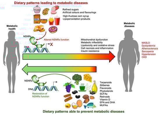
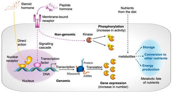
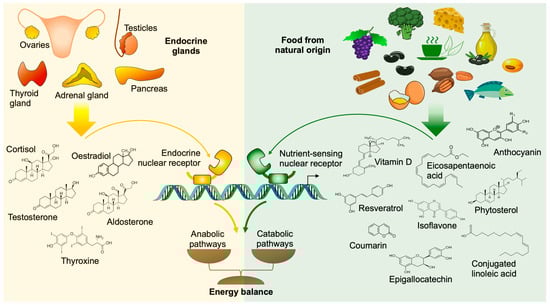
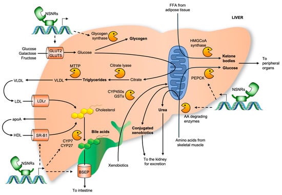
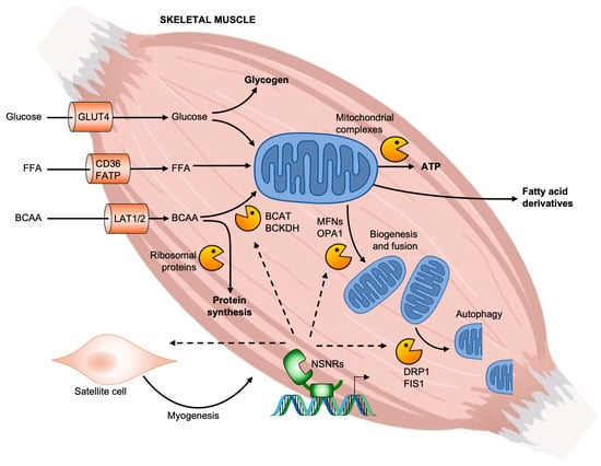
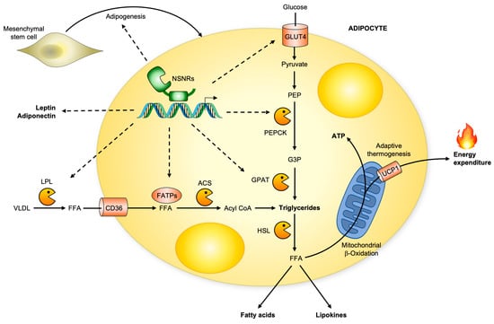
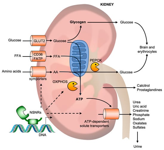
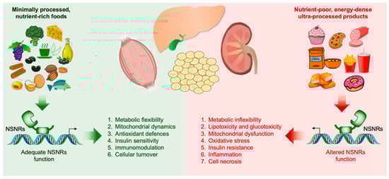
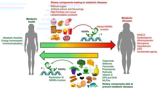
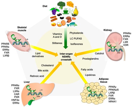
Nutrient-sensing nuclear receptors (NSNRs), including PPARs, FXR, LXRs, RAR/RXR, VDR, and related orphan receptors, integrate a molecular interface that allows diet to communicate directly with the genome. By binding fatty acids, bile acids, sterols, vitamins, polyphenols, and other food-derived metabolites, NSNRs translate qualitative and quantitative features of the diet into coordinated transcriptional programmes across metabolically active organs. This ligand-dependent signalling network integrates dietary information to orchestrate inter-organ lipid and glucose metabolism, mitochondrial function, thermogenesis, and immune response, thereby enabling the organism to adapt dynamically to fasting–feeding cycles. In this review, we synthesise current evidence on the integrated roles of major NSNRs in the liver, skeletal muscle, white and brown adipose tissue, and kidney, emphasising how receptor networks within and between metabolic organs collectively govern energy expenditure, substrate partitioning, and systemic metabolic flexibility. We propose a conceptual framework in which diet functions as an “external endocrine organ”, acting as the primary source of chemically diverse NSNR ligands, while metabolic tissues serve as secondary signal amplifiers and integrators. Through circulating lipid species, bile acids, oxysterols, and other metabolites, these organs engage in continuous bidirectional communication that reprograms NSNR activity across tissues. We then examine how the global shift from minimally processed, nutrient-rich foods to nutrient-poor, energy-dense ultra-processed diets leads to a reduction in NSNR ligand diversity, promoting hepatic steatosis, muscle metabolic inflexibility, adipose tissue dysfunction, renal lipotoxicity, and chronic low-grade inflammation, ultimately causing obesity, type 2 diabetes, and cardiometabolic disease. Finally, we explore strategies to restore NSNR function, including Mediterranean and plant-based dietary patterns, as well as diets enriched with ω-3 polyunsaturated fatty acids, monounsaturated fats, and polyphenols. By integrating molecular, physiological, and clinical evidence, this review aims to clarify how NSNR networks translate dietary cues into coordinated inter-organ metabolism and how nutrient-poor diets lead to metabolic diseases trough a loss of metabolic information, rather than merely by energy excess. This framework supports a paradigm shift from calorie-centred nutrition to diet quality as the main therapeutic target for preventing metabolic diseases and promoting health.
Full article

Graphical abstract
Open AccessReview
FAM3 Cytokine-like Proteins, Their Putative Receptors and Signaling Pathways in Metabolic Diseases and Cancers
by
José E. Belizário, Izabela D. S. Caldeira, Bruna Moreira, João Marcelo Occhiucci, Brant Burkhardt and Humberto M. Garay-Malpartida
Receptors 2026, 5(2), 11; https://doi.org/10.3390/receptors5020011 - 30 Mar 2026
Abstract
►▼
Show Figures
FAM3A, FAM3B, FAM3C and FAM3D are members of the “family with sequence similarity 3” (FAM3) gene family, an emerging class of cytokine-like proteins with a unique structural globular β-β-α fold and distinct biological functions. With widespread expression in tissue, organs and in many
[...] Read more.
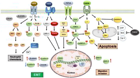
FAM3A, FAM3B, FAM3C and FAM3D are members of the “family with sequence similarity 3” (FAM3) gene family, an emerging class of cytokine-like proteins with a unique structural globular β-β-α fold and distinct biological functions. With widespread expression in tissue, organs and in many cell types, their specific roles in human diseases have been the focus of much research. FAM3A acts as a positive regulator of metabolic health, typically activating canonical pro-survival and metabolic pathways. FAM3B, also called PANDER (PANcreatic DERived Factor), exerts critical physiological functions in the regulation of glycemic levels via promotion of hepatic glucose production and pancreatic β-cell insulin secretion. FAM3C, also named ILEI (Interleukin-like EMT inducer), is involved as an inducer of epithelial–mesenchymal transition (EMT) and cancer metastasis, as well as osteoblast differentiation and bone mineralization. FAM3D is a gut-secreted protein and potential regulator of gastrointestinal homeostasis and microbiota-induced inflammation. Here we provide an overview of previous studies supporting that FAM3 proteins act through putative membrane receptors and co-partners, including fibroblast growth factor receptor (FGFR), leukemia inhibitory factor receptor (LIFR), formyl peptide receptor (FPR1/2), to activate diverse downstream signaling pathways on different cellular contexts. Basic and clinical studies suggest that the FAM3 family influences both obesity, diabetes, and other metabolic disorders; thus, its expression may have diagnostic potential. The differential and often cancer-specific expression patterns make members of the FAM3 family promising candidates for biomarkers and therapeutic targets of some types of neoplasia.
Full article

Figure 1

Open AccessReview
Ion Channels as Targets of the Vitamin D Receptor: A Long Journey with a Promising Future
by
Verna Cázares-Ordoñez, Ramiro José González-Duarte, Michiyasu Ishizawa, Luis A. Pardo and Makoto Makishima
Receptors 2026, 5(2), 10; https://doi.org/10.3390/receptors5020010 - 26 Mar 2026
Cited by 1
Abstract
►▼
Show Figures
The vitamin D receptor (VDR) acts as both a nuclear transcription factor and a non-genomic mediator that regulates a broad spectrum of physiological processes beyond calcium and phosphate homeostasis. VDR plays an important role in the modulation of ion channels across multiple tissues,
[...] Read more.
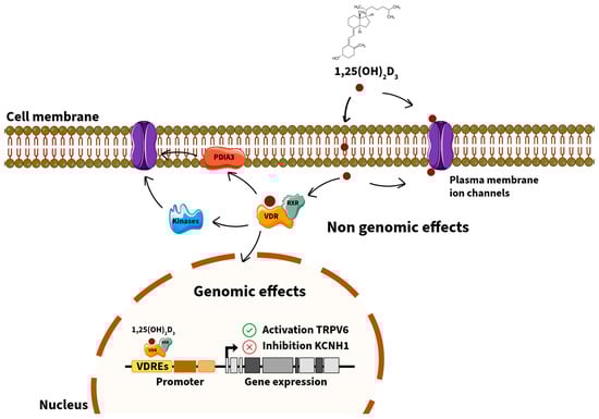
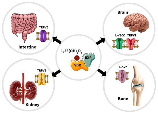
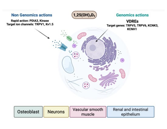
The vitamin D receptor (VDR) acts as both a nuclear transcription factor and a non-genomic mediator that regulates a broad spectrum of physiological processes beyond calcium and phosphate homeostasis. VDR plays an important role in the modulation of ion channels across multiple tissues, including osteoblasts, renal and intestinal epithelial cells, neurons, and vascular smooth muscle. These regulatory mechanisms encompass genomic actions through vitamin D response elements in target genes—such as TRPV5, TRPV6, KCNK3, and KCNH1—as well as rapid, non-genomic actions at the plasma membrane involving protein disulfide isomerase A3 and associated signaling cascades. VDR-mediated transcriptional control of calcium, potassium, and chloride channels contributes to the fine-tuning of cellular excitability, calcium transport, and mitochondrial function. Evidence also implicates VDR–ion channel crosstalk in various pathological contexts, including renal cell carcinoma, breast and cervical cancers, pulmonary arterial hypertension, and osteoporosis. Understanding the molecular interplay between VDR and ion channels provides new perspectives on the pleiotropic effects of vitamin D and offers promising therapeutic opportunities in oncology, cardiovascular disease, and skeletal disorders. This review synthesizes previous and current evidence on the genomic and non-genomic mechanisms underlying VDR–ion channel regulation and highlights novel frontiers in vitamin D signaling relevant to human health and disease.
Full article

Graphical abstract
Open AccessReview
Function and Modulation of Sphingosine-1-Phosphate Receptors in the Central Nervous System
by
Elizabeth Gulliksen, Sriya Darsi, Ladan Haidarbaigi, Lucas J. Codispoti, Devam Purohit, Ashley Jung, Aishwarya Chilamula and Jason Newton
Receptors 2026, 5(1), 9; https://doi.org/10.3390/receptors5010009 - 17 Mar 2026
Cited by 1
Abstract
►▼
Show Figures
Sphingolipids, first discovered in 1874 by Johann Thudicum, are among the eight recognized classes of lipids and are present in essentially all plants, animals, and fungi, as well as some viruses and prokaryotes. In mammals, sphingolipids are enriched in the central nervous system
[...] Read more.
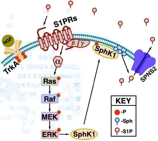
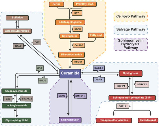
Sphingolipids, first discovered in 1874 by Johann Thudicum, are among the eight recognized classes of lipids and are present in essentially all plants, animals, and fungi, as well as some viruses and prokaryotes. In mammals, sphingolipids are enriched in the central nervous system (CNS), where they play vital roles in tissue development; membrane structure; cell adhesion and recognition; and, importantly, signaling. A subset of sphingolipids including ceramide, glucosylceramide, and sphingosine has been shown to have bioactive properties, but two sphingolipids in particular (ceramide-1-phosphate and sphingosine-1-phosphate) have been shown to exert their effects at least in part due to the activation of cell surface-expressed G protein-coupled receptors. In the CNS, sphingosine-1-phosphate signaling has specifically emerged as a productive therapeutic target for the treatment of neurodegenerative disease, with the first small molecule targeting sphingosine-1-phosphate receptors approved roughly 15 years ago for the treatment of multiple sclerosis. As more specific activators and inhibitors of these receptors have been developed and entered the clinical trial pipeline, now is an appropriate time to examine the current state of our knowledge of the role that these receptors play in the CNS and highlight the current landscape of available modulators targeting these pathways.
Full article

Figure 1
Open AccessReview
The Role of Progesterone in the Reproductive Physiology of Females of Viviparous Squamata
by
Norma Berenice Cruz-Cano, Uriel Ángel Sánchez-Rivera, Carmen Álvarez-Rodríguez, Hibraim Adán Pérez-Mendoza and Martín Martínez-Torres
Receptors 2026, 5(1), 8; https://doi.org/10.3390/receptors5010008 - 27 Feb 2026
Abstract
►▼
Show Figures
Progesterone (P4) regulates diverse reproductive processes across vertebrates through nuclear receptors; however, its mechanisms in squamate reptiles—particularly in viviparous species—remain poorly understood. In Squamata, P4 primarily acts through progesterone receptor (PR) isoforms A and B, although relatively few reptilian PR
[...] Read more.
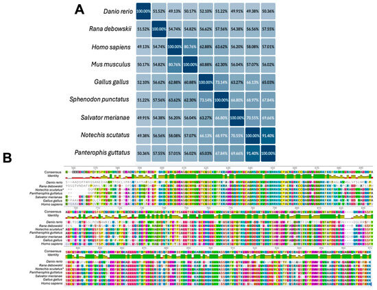
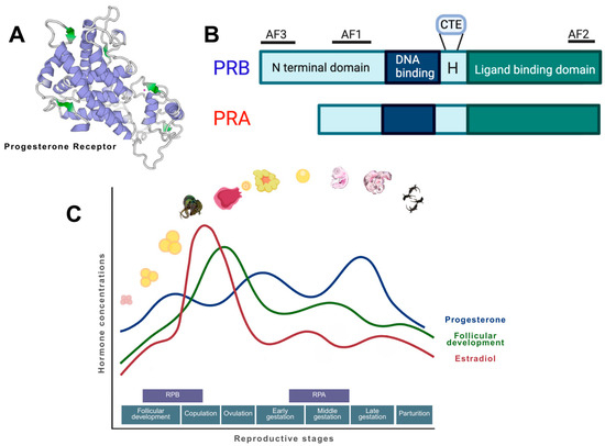
Progesterone (P4) regulates diverse reproductive processes across vertebrates through nuclear receptors; however, its mechanisms in squamate reptiles—particularly in viviparous species—remain poorly understood. In Squamata, P4 primarily acts through progesterone receptor (PR) isoforms A and B, although relatively few reptilian PR sequences have been characterized to date. Squamate PR exhibits ~50% overall sequence divergence from mammalian homologs yet retains striking conservation in both the ligand and DNA-binding domain across vertebrates. Despite the broadly conserved physiological roles of P4 (folliculogenesis, ovulation, courtship behavior, pregnancy maintenance, and parturition/oviposition), P4 dynamics in viviparous squamates remain unresolved due to heterogeneous circulating hormone concentrations and limited PR phylogeny and structure studies. While mammalian models dominate P4 research due to their biomedical relevance, squamates offer unique evolutionary insights: as the only reptile order exhibiting both oviparity and viviparity within the same clade, squamates represent an ideal model for investigating transitions in parity mode. Elucidating P4 mechanisms in squamates will help bridge this critical evolutionary gap, with important implications for reproductive biology and conservation.
Full article

Figure 1

Open AccessReview
Coevolution Between Three-Finger Toxins and Target Receptors
by
Jéssica Lopes de Oliveira and Henrique Roman-Ramos
Receptors 2026, 5(1), 7; https://doi.org/10.3390/receptors5010007 - 14 Feb 2026
Abstract
►▼
Show Figures
Background: Three-finger toxins (3FTxs) are a major axis of functional diversification in advanced snake venoms, with canonical paralytic activity mediated through muscle-type nicotinic acetylcholine receptors (nAChRs) and a broader set of non-nicotinic targets. This review integrates evidence bearing on coevolution between 3FTxs
[...] Read more.
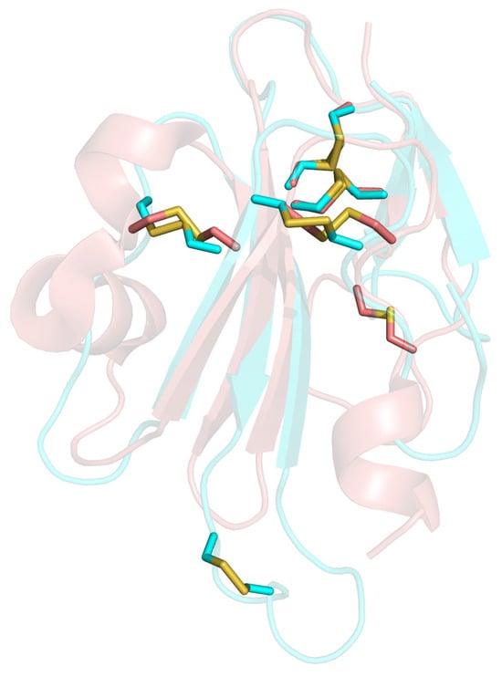
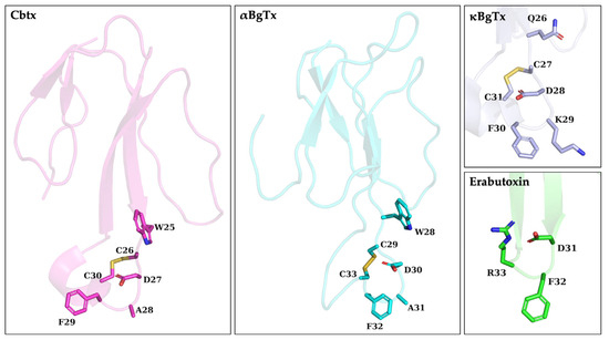
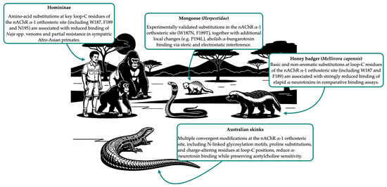
Background: Three-finger toxins (3FTxs) are a major axis of functional diversification in advanced snake venoms, with canonical paralytic activity mediated through muscle-type nicotinic acetylcholine receptors (nAChRs) and a broader set of non-nicotinic targets. This review integrates evidence bearing on coevolution between 3FTxs and target receptors, spanning toxin origin, diversification, receptor evolution, and ecological context. Methods: The synthesis draws on comparative genomic and transcriptomic studies of 3FTx gene-family evolution, codon-model analyses of selection, structural characterisation of toxin–receptor interfaces, and functional assays (including receptor-mimicking peptide binding) that link sequence variation to binding and toxicity. Results: Across lineages, 3FTx diversification is repeatedly structured by strong constraint on the disulphide-rich scaffold with accelerated change concentrated in solvent-exposed loops, alongside birth–death dynamics and exon/segment-level innovation that expand binding specificity. On the receptor side, resistance-associated variation is most intensively characterised for the nAChR α1 orthosteric site and includes convergent, mechanistically distinct solutions such as electrostatic repulsion and glycosylation-mediated steric interference. Within the predominantly elapid systems currently examined, integrative datasets indicate that prey-selective binding and geographically variable susceptibility can arise from modest substitutions at toxin–receptor interfaces, but they also reveal substantial taxonomic and target-specific biases. Conclusions: Current evidence supports adaptive diversification in both toxins and receptors, while broader evolutionary interpretations are limited by uneven sampling and the frequent lack of matched toxin and receptor variants analysed within a common evolutionary framework. Development of predictive models will require joint pipelines linking genomics, structure-informed evolutionary inference, scalable functional assays, and explicit ecological network context.
Full article

Graphical abstract
Highly Accessed Articles
Latest Books
E-Mail Alert
News
Topics
Conferences
Special Issues
Topical Collections
Topical Collection in
Receptors
Receptors: Exceptional Scientists and Their Expert Opinions
Collection Editor: Stephen H. Safe