Selected Papers from the XIX International Meeting on Crystal Chemistry, X-ray Diffraction and Spectroscopy of Minerals

A special issue of Minerals (ISSN 2075-163X). This special issue belongs to the section "Crystallography and Physical Chemistry of Minerals & Nanominerals".

Deadline for manuscript submissions: closed (15 September 2019) | Viewed by 39417

Special Issue Editor


E-Mail
Guest Editor
1. Kola Science Center, Russian Academy of Sciences, Fersmana str. 14, 184209 Apatity, Russia
2. Department of Crystallography, Institute of Earth Sciences, St. Petersburg State University, University Emb. 7/9, 199034 St. Petersburg, Russia
Interests: mineralogy; crystallography; structural complexity; uranium
Special Issues, Collections and Topics in MDPI journals

Special Issue Information

Dear Colleagues,

Technological innovations have brought forth tremendous advances in traditional experimental techniques used in the investigation of composition, structure, and properties of minerals: X-ray diffraction, spectroscopy, and mineral physics. On the other hand, the development of novel theoretical concepts has led to the true Renaissance of theoretical mineralogy and crystallography. The XIX International Meeting on Crystal Chemistry, X-ray Diffraction and Spectroscopy of Minerals will be held on 2–5 July 2019 in Apatity, Kola peninsula, Russia, a famous mineral locality, where the largest number of new mineral species in the history of mineralogy have been discovered. The Meeting is devoted to the memory of great Russian crystallographer, mineralogist, petrologist, and mining engineer Academician E.S. Fedorov (10(22).12.1853–21.05.1919), one of the founders of modern crystallography and discoverer of 230 space groups. It will cover a broad range of topics, including general problems of inorganic crystal chemistry and structural mineralogy, X-ray crystallography, mineral physics and spectroscopy, high-pressure and high-temperature crystal chemistry, theoretical crystal chemistry, and mineralogy.

This Special Issue will include high-quality research papers presented at the conference. We cordially invite you to participate in the Meeting and to contribute your manuscript to this Special Issue.

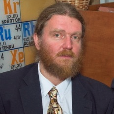
Prof. Dr. Sergey V. Krivovichev
Guest Editor

Manuscript Submission Information

Manuscripts should be submitted online at www.mdpi.com by registering and logging in to this website. Once you are registered, click here to go to the submission form. Manuscripts can be submitted until the deadline. All submissions that pass pre-check are peer-reviewed. Accepted papers will be published continuously in the journal (as soon as accepted) and will be listed together on the special issue website. Research articles, review articles as well as short communications are invited. For planned papers, a title and short abstract (about 100 words) can be sent to the Editorial Office for announcement on this website.

Submitted manuscripts should not have been published previously, nor be under consideration for publication elsewhere (except conference proceedings papers). All manuscripts are thoroughly refereed through a single-blind peer-review process. A guide for authors and other relevant information for submission of manuscripts is available on the Instructions for Authors page. Minerals is an international peer-reviewed open access monthly journal published by MDPI.

Please visit the Instructions for Authors page before submitting a manuscript. The Article Processing Charge (APC) for publication in this open access journal is 2400 CHF (Swiss Francs). Submitted papers should be well formatted and use good English. Authors may use MDPI's English editing service prior to publication or during author revisions.

Published Papers (9 papers)

Order results
Result details
Select all
Export citation of selected articles as:

Research

Jump to: Review

10 pages, 4453 KiB  
Article
(K,Na)2[AsB6O12]2[B3O3(OH)3], a New Microporous Material, and Its Comparison to Teruggite
by Yulia A. Pankova and Sergey V. Krivovichev
Minerals 2019, 9(12), 781; https://doi.org/10.3390/min9120781 - 13 Dec 2019
Viewed by 2125
Abstract
Single crystals of the novel boroarsenate (K,Na)2[As2B12O24][B3O3(OH)3] (I) were obtained using the borax flux method. The crystal structure of I was found to be triclinic, P-1, [...] Read more.
Single crystals of the novel boroarsenate (K,Na)2[As2B12O24][B3O3(OH)3] (I) were obtained using the borax flux method. The crystal structure of I was found to be triclinic, P-1, a = 8.414(5), b = 10.173(6), c = 15.90(1) Å, α = 79.56(1), β = 78.68(1), γ = 70.91(1), V = 1251(1) Å3, Z = 2. The crystal structure of I is based upon the novel [AsB6O12] microporous boroarsenate framework formed by B and As coordination polyhedra. This framework can be subdivided into borate units that are interlinked by AsO4 tetrahedra. In the case of I, the borate substructure is a chain consisting of triborate rings, ☐2Δ, formed by two (BO3) triangles and one (BO4) tetrahedron connected through shared common oxygen atoms. The chains are extended along [0 1 ¯ 1] and are interlinked by (AsO4) tetrahedra in the [011] direction. As a result, the framework has large channels parallel to [100], having an effective diameter of 4.2 × 5.6 Å2. The channels contain occluded electroneutral ring triborate complexes, [B3O3(OH)3]0, formed by three (BO2(OH)) triangles sharing common O atoms, as well as K+ and Na+ cations. The triborate [B3O3(OH)3]0 units correspond to similar clusters found in the crystal structure of the α-form of metaboric acid, HBO2. According to information-based complexity calculations, the crystal structure of I should be described as complex, with IG = 5.781 bits/atom and IG,total = 625.950 bits/cell. Teruggite, Ca4Mg[B6As(OH)6O11]2(H2O)14, the only known boroarsenate of natural origin, has almost twice as much information per unit cell, with IG,total = 1201.992 bits/cell. The observed difference in structural complexity between I and teruggite is the consequence of their chemistry (hydration state) and different formation conditions. Full article
Show Figures

Figure 1

13 pages, 3130 KiB  
Article
Calcium Oxalates in Lichens on Surface of Apatite-Nepheline Ore (Kola Peninsula, Russia)
by Olga V. Frank-Kamenetskaya, Gregory Yu. Ivanyuk, Marina S. Zelenskaya, Alina R. Izatulina, Andrey O. Kalashnikov, Dmitry Yu. Vlasov and Evgeniya I. Polyanskaya
Minerals 2019, 9(11), 656; https://doi.org/10.3390/min9110656 - 25 Oct 2019
Cited by 23 | Viewed by 4121
Abstract
The present work contributes to the essential questions on calcium oxalate formation under the influence of lithobiont community organisms. We have discovered calcium oxalates in lichen thalli on surfaces of apatite-nepheline rocks of southeastern and southwestern titanite-apatite ore fields of the Khibiny peralkaline [...] Read more.
The present work contributes to the essential questions on calcium oxalate formation under the influence of lithobiont community organisms. We have discovered calcium oxalates in lichen thalli on surfaces of apatite-nepheline rocks of southeastern and southwestern titanite-apatite ore fields of the Khibiny peralkaline massif (Kola Peninsula, NW Russia) for the first time; investigated biofilm calcium oxalates with different methods (X-ray powder diffraction, scanning electron microscopy, and EDX analysis) and discussed morphogenetic patterns of its formation using results of model experiments. The influence of inorganic and organic components of the crystallization medium on the phase composition and morphology of oxalates has been analyzed. It was shown that, among the complex of factors controlling the patterns of biogenic oxalate formation, one of the main roles belongs to the metabolic activity of the lithobiont community organisms, which differs significantly from the activity of its individuals. Full article
Show Figures

Graphical abstract

12 pages, 270 KiB  
Article
On the Chemical Identification and Classification of Minerals
by Ferdinando Bosi, Cristian Biagioni and Roberta Oberti
Minerals 2019, 9(10), 591; https://doi.org/10.3390/min9100591 - 28 Sep 2019
Cited by 33 | Viewed by 5749
Abstract
To univocally identify mineral species on the basis of their formula, the IMA-CNMNC recommends the use of the dominant-valency rule and/or the site-total-charge approach, which can be considered two procedures complementary to each other for mineral identification. In this regard, several worked examples [...] Read more.
To univocally identify mineral species on the basis of their formula, the IMA-CNMNC recommends the use of the dominant-valency rule and/or the site-total-charge approach, which can be considered two procedures complementary to each other for mineral identification. In this regard, several worked examples are provided in this study along with some simple suggestions for a more consistent terminology and a straightforward use of mineral formulae. IMA-CNMNC guidelines subordinate the mineral structure to the mineral chemistry in the hierarchical scheme adopted for classification. Indeed, a contradiction appears when we first classify mineral species to form classes (based on their chemistry) and subsequently we group together them to form supergroups (based on their structure topology): To date, more than half of recognized mineral supergroups include species with different anions or anionic complexes. This observation is in contrast to the current use of chemical composition as the distinguishing factor at the highest level of mineral classification. Full article
12 pages, 9719 KiB  
Article
Dmisteinbergite, CaAl2Si2O8, a Metastable Polymorph of Anorthite: Crystal-Structure and Raman Spectroscopic Study of the Holotype Specimen
by Andrey A. Zolotarev, Sergey V. Krivovichev, Taras L. Panikorovskii, Vladislav V. Gurzhiy, Vladimir N. Bocharov and Mikhail A. Rassomakhin
Minerals 2019, 9(10), 570; https://doi.org/10.3390/min9100570 - 20 Sep 2019
Cited by 24 | Viewed by 4979
Abstract
The crystal structure of dmisteinbergite has been determined using crystals from the type locality in Kopeisk city, Chelyabinsk area, Southern Urals, Russia. The mineral is trigonal, with the following structure: P312, a = 5.1123(2), c = 14.7420(7) Å, V = 333.67(3) Å [...] Read more.
The crystal structure of dmisteinbergite has been determined using crystals from the type locality in Kopeisk city, Chelyabinsk area, Southern Urals, Russia. The mineral is trigonal, with the following structure: P312, a = 5.1123(2), c = 14.7420(7) Å, V = 333.67(3) Å3, R1 = 0.045, for 762 unique observed reflections. The most intense bands of the Raman spectra at 327s, 439s, 892s, and 912s cm −1 correspond to different types of tetrahedral stretching vibrations: Si–O, Al–O, O–Si–O, and O–Al–O. The weak bands at 487w, 503w, and 801w cm−1 can be attributed to the valence and deformation modes of Si–O and Al–O bond vibrations in tetrahedra. The weak bands in the range of 70–200 cm−1 can be attributed to Ca–O bond vibrations or lattice modes. The crystal structure of dmisteinbergite is based upon double layers of six-membered rings of corner-sharing AlO4 and SiO4 tetrahedra. The obtained model shows an ordering of Al and Si over four distinct crystallographic sites with tetrahedral coordination, which is evident from the average <T–O> bond lengths (T = Al, Si), equal to 1.666, 1.713, 1.611, and 1.748 Å for T1, T2, T3, and T4, respectively. One of the oxygen sites (O4) is split, suggesting the existence of two possible conformations of the [Al2Si2O8]2 layers, with different systems of ditrigonal distortions in the adjacent single layers. The observed disorder has a direct influence upon the geometry of the interlayer space and the coordination of the Ca2 site. Whereas the coordination of the Ca1 site is not influenced by the disorder and is trigonal antiprismatic (distorted octahedral), the coordination environment of the Ca2 site includes disordered O atoms and is either trigonal prismatic or trigonal antiprismatic. The observed structural features suggest the possible existence of different varieties of dmisteinbergite that may differ in: (i) degree of disorder of the Al/Si tetrahedral sites, with completely disordered structure having the P63/mcm symmetry; (ii) degree of disorder of the O sites, which may have a direct influence on the coordination features of the Ca2+ cations; (iii) polytypic variations (different stacking sequences and layer shifts). The formation of dmisteinbergite is usually associated with metastable crystallization in both natural and synthetic systems, indicating the kinetic nature of this phase. Information-based complexity calculations indicate that the crystal structures of metastable CaAl2Si2O8 polymorphs dmisteinbergite and svyatoslavite are structurally and topologically simpler than that of their stable counterpart, anorthite, which is in good agreement with Goldsmith’s simplexity principle and similar previous observations. Full article
Show Figures

Figure 1

15 pages, 1869 KiB  
Article
Dritsite, Li2Al4(OH)12Cl2·3H2O, a New Gibbsite-Based Hydrotalcite Supergroup Mineral
by Elena S. Zhitova, Igor V. Pekov, Ilya I. Chaikovskiy, Elena P. Chirkova, Vasiliy O. Yapaskurt, Yana V. Bychkova, Dmitry I. Belakovskiy, Nikita V. Chukanov, Natalia V. Zubkova, Sergey V. Krivovichev and Vladimir N. Bocharov
Minerals 2019, 9(8), 492; https://doi.org/10.3390/min9080492 - 17 Aug 2019
Cited by 19 | Viewed by 6334
Abstract
Dritsite, ideally Li2Al4(OH)12Cl2·3H2O, is a new hydrotalcite supergroup mineral formed as a result of diagenesis in the halite−carnallite rock of the Verkhnekamskoe salt deposit, Perm Krai, Russia. Dritsite forms single lamellar or tabular [...] Read more.
Dritsite, ideally Li2Al4(OH)12Cl2·3H2O, is a new hydrotalcite supergroup mineral formed as a result of diagenesis in the halite−carnallite rock of the Verkhnekamskoe salt deposit, Perm Krai, Russia. Dritsite forms single lamellar or tabular hexagonal crystals up to 0.25 mm across. The mineral is transparent and colourless, with perfect cleavage on {001}. The chemical composition of dritsite (wt. %; by combination of electron microprobe and ICP−MS; H2O calculated by structure refinement) is: Li2O 6.6, Al2O3 45.42, SiO2 0.11, Cl 14.33, SO3 0.21, H2Ocalc. 34.86, O = Cl − 3.24, total 98.29. The empirical formula based on Li + Al + Si = 6 apfu (atom per formula unit) is Li1.99Al4.00Si0.01[(OH)12.19Cl1.82(SO4)0.01]Σ14.02·2.60(H2O). The Raman spectroscopic data indicate the presence of O–H bonding in the mineral, whereas CO32– groups are absent. The crystal structure has been refined in the space group P63/mcm, a = 5.0960(3), c = 15.3578(13) Å, and V = 345.4(5) Å3, to R1 = 0.088 using single-crystal data. The strongest lines of the powder X-ray diffraction pattern (d, Å (I, %) (hkl)) are: 7.68 (100) (002), 4.422 (61) (010), 3.832 (99) (004, 012), 2.561 (30) (006), 2.283 (25) (113), and 1.445 (26) (032). Dritsite was found as 2H polytype, which is isotypic with synthetic material and shows strong similarity to chlormagalumite-2H. The mineral is named in honour of the Russian crystallographer and mineralogist Prof. Victor Anatol`evich Drits. Full article
Show Figures

Figure 1

16 pages, 3525 KiB  
Article
Crystal Chemistry and High-Temperature Behaviour of Ammonium Phases NH4MgCl3·6H2O and (NH4)2Fe3+Cl5·H2O from the Burned Dumps of the Chelyabinsk Coal Basin
by Andrey A. Zolotarev, Jr., Elena S. Zhitova, Maria G. Krzhizhanovskaya, Mikhail A. Rassomakhin, Vladimir V. Shilovskikh and Sergey V. Krivovichev
Minerals 2019, 9(8), 486; https://doi.org/10.3390/min9080486 - 14 Aug 2019
Cited by 8 | Viewed by 3724
Abstract
The technogenic mineral phases NH4MgCl3·6H2O and (NH4)2Fe3+Cl5·H2O from the burned dumps of the Chelyabinsk coal basin have been investigated by single-crystal X-ray diffraction, scanning electron microscopy and [...] Read more.
The technogenic mineral phases NH4MgCl3·6H2O and (NH4)2Fe3+Cl5·H2O from the burned dumps of the Chelyabinsk coal basin have been investigated by single-crystal X-ray diffraction, scanning electron microscopy and high-temperature powder X-ray diffraction. The NH4MgCl3·6H2O phase is monoclinic, space group C2/c, unit cell parameters a = 9.3091(9), b = 9.5353(7), c = 13.2941(12) Å, β = 90.089(8)° and V = 1180.05(18) Å3. The crystal structure of NH4MgCl3·6H2O was refined to R1 = 0.078 (wR2 = 0.185) on the basis of 1678 unique reflections. The (NH4)2Fe3+Cl5·H2O phase is orthorhombic, space group Pnma, unit cell parameters a = 13.725(2), b = 9.9365(16), c = 7.0370(11) Å and V = 959.7(3) Å3. The crystal structure of (NH4)2Fe3+Cl5·H2O was refined to R1 = 0.023 (wR2 = 0.066) on the basis of 2256 unique reflections. NH4MgCl3·6H2O is stable up to 90 °C and then transforms to the less hydrated phase isotypic to β-Rb(MnCl3)(H2O)2 (i.e., NH4MgCl3·2H2O), the latter phase being stable up to 150 °C. (NH4)2Fe3+Cl5·H2O is stable up to 120 °C and then transforms to an X-ray amorphous phase. Hydrogen bonds provide an important linkage between the main structural units and play the key role in determining structural stability and physical properties of the studied phases. The mineral phases NH4MgCl3·6H2O and (NH4)2Fe3+Cl5·H2O are isostructural with natural minerals novograblenovite and kremersite, respectively. Full article
Show Figures

Figure 1

13 pages, 4691 KiB  
Article
Thermal Behavior and Phase Transition of Uric Acid and Its Dihydrate Form, the Common Biominerals Uricite and Tinnunculite
by Alina R. Izatulina, Vladislav V. Gurzhiy, Maria G. Krzhizhanovskaya, Nikita V. Chukanov and Taras L. Panikorovskii
Minerals 2019, 9(6), 373; https://doi.org/10.3390/min9060373 - 22 Jun 2019
Cited by 13 | Viewed by 4437
Abstract
Single crystals and powder samples of uric acid and uric acid dihydrate, known as uricite and tinnunculite biominerals, were extracted from renal stones and studied using single-crystal and powder X-ray diffraction (SC and PXRD) at various temperatures, as well as IR spectroscopy. The [...] Read more.
Single crystals and powder samples of uric acid and uric acid dihydrate, known as uricite and tinnunculite biominerals, were extracted from renal stones and studied using single-crystal and powder X-ray diffraction (SC and PXRD) at various temperatures, as well as IR spectroscopy. The results of high-temperature PXRD experiments revealed that the structure of uricite is stable up to 380 °C, and then it loses crystallinity. The crystal structure of tinnunculite is relatively stable up to 40 °C, whereas above this temperature, rapid release of H2O molecules occurs followed by the direct transition to uricite phase without intermediate hydration states. SCXRD studies and IR spectroscopy data confirmed the similarity of uricite and tinnunculite crystal structures. SCXRD at low temperatures allowed us to determine the dynamics of the unit cells induced by temperature variations. The thermal behavior of uricite and tinnunculite is essentially anisotropic; the structures not only expand, but also contract with temperature increase. The maximal expansion occurs along the unit cell parameter of 7 Å (b in uricite and a in tinnunculite) as a result of the shifts of chains of H-bonded uric acid molecules and relaxation of the π-stacking forces, the weakest intermolecular interactions in these structures. The strongest contraction in the structure of uricite occurs perpendicular to the (101) plane, which is due to the orthogonalization of the monoclinic angle. The structure of tinnunculite also contracts along the [010] direction, which is mostly due to the stretching mechanism of the uric acid chains. These phase transitions that occur within the range of physiological temperatures emphasize the particular importance of the structural studies within the urate system, due to their importance in terms of human health. The removal of supersaturation in uric acid in urine at the initial stages of stone formation can occur due to the formation of metastable uric acid dihydrate in accordance with the Ostwald rule, which would serve as a nucleus for the subsequent growth of the stone at further formation stages; afterward, it irreversibly dehydrates into anhydrous uric acid. Full article
Show Figures

Figure 1

11 pages, 4933 KiB  
Article
Jahn-Teller Distortion and Cation Ordering: The Crystal Structure of Paratooite-(La), a Superstructure of Carbocernaite
by Sergey V. Krivovichev, Taras L. Panikorovskii, Andrey A. Zolotarev, Vladimir N. Bocharov, Anatoly V. Kasatkin and Radek Škoda
Minerals 2019, 9(6), 370; https://doi.org/10.3390/min9060370 - 20 Jun 2019
Cited by 3 | Viewed by 3169
Abstract
The crystal structure of paratooite-(La) has been solved using crystals from the type locality, Paratoo copper mine, near Yunta, Olary Province, South Australia, Australia. The mineral is orthorhombic, Pbam, a = 7.2250(3) Å, b = 12.7626(5) Å, c = 10.0559(4) Å, V [...] Read more.
The crystal structure of paratooite-(La) has been solved using crystals from the type locality, Paratoo copper mine, near Yunta, Olary Province, South Australia, Australia. The mineral is orthorhombic, Pbam, a = 7.2250(3) Å, b = 12.7626(5) Å, c = 10.0559(4) Å, V = 927.25(6) Å3, and R1 = 0.063 for 1299 unique observed reflections. The crystal structure contains eight symmetrically independent cation sites. The La site, which accommodates rare earth elements (REEs), but also contains Sr and Ca, has a tenfold coordination by seven carbonate groups. The Ca, Na1, and Na2 sites are coordinated by eight, eight, and six O atoms, respectively, forming distorted CaO8 and Na1O8 cubes, and Na2O6 octahedra. The Cu site is occupied solely by copper and possess a distorted octahedral coordination with four short (1.941 Å) and two longer (2.676 Å) apical Cu–O bonds. There are three symmetrically independent carbonate groups (CO3)2− with the average <C–O> bond lengths equal to 1.279, 1.280, and 1.279 Å for the C1, C2, and C3 sites, respectively. The crystal structure of paratooite-(La) can be described as a strongly distorted body-centered lattice formed by metal cations with (CO3)2− groups filling its interstices. According to the chemical and crystal-structure data, the crystal-chemical formula of paratooite-(La) can be described as (La0.74Ca0.11Sr0.07)4CuCa(Na0.75Ca0.15)(Na0.63)(CO3)8 or REE2.96Ca1.59Na1.38CuSr0.28(CO3)8. The idealized formula can be written as (La,Sr,Ca)4CuCa(Na,Ca)2(CO3)8. The structure of paratooite is a 1 × 2 × 2 superstructure of carbocernaite, CaSr(CO3)2. The superstructure arises due to the ordering of the chemically different Cu2+ cations, on one hand, and Na+ and Ca2+ cations, on the other hand. The formation of a superstructure due to the cation ordering in paratooite-(La) compared to carbocernaite results in the multiple increase of structural complexity per unit cell. Therefore, paratooite-(La) versus carbocernaite represents a good example of structural complexity increasing due to the increasing chemical complexity controlled by different electronic properties of mineral-forming chemical elements (transitional versus alkali and alkaline earth metals). Full article
Show Figures

Figure 1

Review

Jump to: Research

20 pages, 21331 KiB  
Review
E. S. Fedorov Promoting the Russian-German Scientific Interrelationship
by Peter Paufler and Stanislav K. Filatov
Minerals 2020, 10(2), 181; https://doi.org/10.3390/min10020181 - 18 Feb 2020
Cited by 4 | Viewed by 3998
Abstract
At the dawn of crystal structure analysis, the close personal contact between researchers in Russia and Germany, well documented in the “Zeitschrift für Krystallographie und Mineralogie”, contributed significantly to the evolution of our present knowledge of the crystalline state. The impact of the [...] Read more.
At the dawn of crystal structure analysis, the close personal contact between researchers in Russia and Germany, well documented in the “Zeitschrift für Krystallographie und Mineralogie”, contributed significantly to the evolution of our present knowledge of the crystalline state. The impact of the Russian crystallographer E. S. Fedorov upon German scientists such as A. Schoenflies and P. Groth and the effect of these contacts for Fedorov are highlighted hundred years after the death of the latter. A creative exchange of ideas paved the way for the analysis of crystal structures with the aid of X-ray diffraction. Full article
Show Figures

Graphical abstract

Back to TopTop