


Cells 2024, 13(9), 786; https://doi.org/10.3390/cells13090786 (registering DOI) - 04 May 2024
Abstract
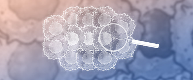
Ovarian cancer is a highly lethal form of gynecological cancer. This disease often goes undetected until advanced stages, resulting in high morbidity and mortality rates. Unfortunately, many patients experience relapse and succumb to the disease due to the emergence of drug resistance that
[...] Read more.
Ovarian cancer is a highly lethal form of gynecological cancer. This disease often goes undetected until advanced stages, resulting in high morbidity and mortality rates. Unfortunately, many patients experience relapse and succumb to the disease due to the emergence of drug resistance that significantly limits the effectiveness of currently available oncological treatments. Here, we discuss the molecular mechanisms responsible for resistance to carboplatin, paclitaxel, polyadenosine diphosphate ribose polymerase inhibitors, and bevacizumab in ovarian cancer. We present a detailed analysis of the most extensively investigated resistance mechanisms, including drug inactivation, drug target alterations, enhanced drug efflux pumps, increased DNA damage repair capacity, and reduced drug absorption/accumulation. The in-depth understanding of the molecular mechanisms associated with drug resistance is crucial to unveil new biomarkers capable of predicting and monitoring the kinetics during disease progression and discovering new therapeutic targets.
Full article
(This article belongs to the Special Issue Unlocking the Secrets behind Drug Resistance at the Cellular Level)


